Embed Size (px)
Citation preview
Placental Pharmacology – Implications for Therapy in Pregnancy
by
Priya Bapat
A thesis submitted in conformity with the requirements for the degree of Doctor of Philosophy
Department of Pharmacology and Toxicology University of Toronto
© Copyright by Priya Bapat (2016)
ii
Placental Pharmacology – Implications for Therapy in Pregnancy
Priya Bapat
Doctor of Philosophy
Department of Pharmacology and Toxicology University of Toronto
2016
Abstract
Medication use during pregnancy requires a balance between treating a condition in the mother,
and minimizing potential risks in the fetus. In recent years, it has been estimated that between 50
and 70% of pregnant women in North America will take at least one prescription medication
during their pregnancy. The decision to begin or continue treatment during pregnancy relies
heavily on evaluating the risk-benefit ratio, and an important determinant in this risk assessment
is estimating fetal drug exposure. The use of medications in pregnant women can be especially
challenging, as there is very limited safety data in pregnancy. Recently, novel oral anticoagulants
have been developed and approved for clinical use. However, the information regarding their
fetal safety and placental transfer in humans is unknown. We used the ex vivo placenta perfusion
model to investigate anticoagulant (dabigatran, rivaroxaban, apixaban) transfer across the human
placenta. Rivaroxaban and apixaban rapidly crossed the term human placenta, while dabigatran
crossed the placenta to a lesser extent. The placenta perfusion results were adjusted to account
for protein binding and pH differences between the mother and fetus. We also developed a
pharmacokinetic model that adequately described the transplacental transfer of anticoagulants by
using data from our experiments. While the placenta perfusion model can be technically
challenging, its results strongly correlate with in vivo placental transfer data. By evaluating the
success rate of the perfusion model in our laboratory, we determined that establishing the fetal
iii
circulation is an important stage of the protocol. This information can be used to create a focused
training program to help increase the overall success rate of this model and productivity of the
lab. Drug use in pregnancy is multifactorial, and placental transfer data is important in assessing
which drugs can be used to treat the mother while protecting the unborn.
iv
Acknowledgments
I would like to thank many individuals for their guidance, support and contributions during the
course of my training.
First and foremost, I would like to express my profound respect and gratitude to Dr. Shinya Ito
for his exceptional mentorship during my graduate studies. I thank him for his support and
encouragement in pursuing my ambitions and career goals. I would like to acknowledge Dr.
Gideon Koren for his continuous guidance and for providing me with countless opportunities.
I would like to thank Dr. Howard Berger for being a wonderful mentor and for the opportunity to
collaborate together on many projects. I thank him for taking time to proofread my work, and for
being a dedicated member of my graduate advisory committee. I would like to thank Dr.
Micheline Piquette-Miller for her wise words and invaluable guidance during committee
meetings. I wish to acknowledge Drs. Bhushan Kapur and Prateek Lala for their mentorship and
enthusiasm in working with graduate students.
A very special thank you to Angelika Lubetsky for sharing her expertise of the placenta
perfusion model and working tirelessly for successful experiments. I would like to thank
Leonardo Pinto for all his contributions to these studies. I would especially like to acknowledge
the staff in the Labour and Delivery Ward at St. Michael’s Hospital. Thank you to the
obstetricians, residents, and nurses for all their help in facilitating tissue collection. I would like
to acknowledge Dr. Katarina Aleksa, Ariane Mandel, Paula Walasek, Michael Leadley, and
Hayley Craig-Barnes for their assistance with analytical methods in the laboratory. I want to
thank Dr. Masanobu Takeuchi for introducing me to the exciting field of pharmacokinetic
modeling, and taking the time to answer all my questions.
I would like to thank my fellow graduate students who have made this experience more than I
could have hoped for. This talented group of people played an important role in making graduate
school fun and I will always cherish the great conversations and meals we shared. I consider
myself lucky for having shared the lab with you all and I couldn’t have imagined a better group
of brilliant people. I am grateful for the friendships that will extend far beyond our time in the
lab.

v
Last, but certainly not least, a very special thank you to my family and friends for their
unwavering support, encouragement, and patience over the last few years. Thank you to my
parents for being phenomenal role models and showing me what hard work and determination
look like. They have been my cheerleaders every step of the way and I cannot thank them
enough.
vi
Table of Contents
Acknowledgments .......................................................................................................................... iv
Table of Contents ........................................................................................................................... vi
List of Tables ................................................................................................................................. ix
List of Figures ..................................................................................................................................x
List of Abbreviations .................................................................................................................... xii
List of Appendices ....................................................................................................................... xiv
Chapter 1 General Introduction .......................................................................................................1
Drug Use in Pregnancy ...............................................................................................................1 1
1.1 Transplacental Drug Transfer ..............................................................................................1
1.1.1 Pregnancy-Related Physiological and Pharmacokinetic Changes ...........................2
1.1.2 Human Placenta: Structure and Function ................................................................4
1.1.3 Methods for Studying Transplacental Drug Transfer ............................................12
1.2 Anticoagulation Therapy in Pregnancy .............................................................................17
1.2.1 Overview of the Coagulation Cascade ...................................................................18
1.2.2 Maternal and Fetal Coagulation Systems ..............................................................20
1.2.3 Current Therapies ...................................................................................................22
1.3 Novel Oral Anticoagulants ................................................................................................24
1.3.1 Dabigatran ..............................................................................................................26
1.3.2 Rivaroxaban ...........................................................................................................27
1.3.3 Apixaban ................................................................................................................29
1.4 Overall Rationale and Primary Objectives ........................................................................30
Chapter 2 Transfer of Dabigatran and Dabigatran Etexilate Mesylate across the Dually Perfused Human Placenta. ........................................................................................................32
2.1 Abstract ..............................................................................................................................33
2.2 Introduction ........................................................................................................................34
vii
2.3 Materials and Methods .......................................................................................................35
2.4 Results ................................................................................................................................38
2.5 Discussion ..........................................................................................................................42
Chapter 3 Rivaroxaban Transfer across the Dually Perfused Isolated Human Placental Cotyledon. .................................................................................................................................44
3.1 Abstract ..............................................................................................................................45
3.2 Introduction ........................................................................................................................46
3.3 Materials and Methods .......................................................................................................47
3.3.1 Ex vivo perfusion of human placental cotyledon ...................................................47
3.3.2 Experimental period ...............................................................................................47
3.3.3 Sample analysis ......................................................................................................47
3.3.4 Statistical analysis ..................................................................................................48
3.4 Results ................................................................................................................................48
3.5 Discussion ..........................................................................................................................54
Chapter 4 Examining the Transplacental Passage of Apixaban Using the Dually Perfused Human Placenta. .......................................................................................................................57
4.1 Abstract ..............................................................................................................................58
4.2 Introduction ........................................................................................................................59
4.3 Methods..............................................................................................................................60
4.3.1 Dual perfusion of human placental cotyledon ex vivo ...........................................60
4.3.2 Sample analysis ......................................................................................................60
4.3.3 Prediction of in vivo fetal-to-maternal (F:M) ratio of plasma drug concentration ..........................................................................................................61
4.3.4 Statistical analysis ..................................................................................................63
4.4 Results ................................................................................................................................63
4.5 Discussion ..........................................................................................................................65
Chapter 5 Prediction of human placental pharmacokinetics of novel anticoagulants using the ex vivo placenta perfusion and physiologically based models. .................................................69
viii
5.1 Abstract ..............................................................................................................................70
5.2 Introduction ........................................................................................................................71
5.3 Methods..............................................................................................................................72
5.3.1 Ex vivo placenta perfusion .....................................................................................72
5.3.2 Prediction of in vivo fetal-to-maternal (F:M) ratio of plasma drug concentration ..........................................................................................................72
5.3.3 Statistical analysis ..................................................................................................73
5.3.4 Model construction ................................................................................................73
5.4 Results ................................................................................................................................75
5.4.1 Ex vivo placenta perfusion .....................................................................................75
5.4.2 Model analysis .......................................................................................................78
5.5 Discussion ..........................................................................................................................80
Chapter 6 Evaluating the success rate of the ex vivo dual perfusion of the human placenta. ........83
6.1 Abstract ..............................................................................................................................84
6.2 Introduction ........................................................................................................................85
6.3 Methods..............................................................................................................................86
6.4 Results ................................................................................................................................88
6.5 Discussion ..........................................................................................................................91
Chapter 7 General Discussion ........................................................................................................94
7.1 Summary of Research Findings and Future Directions .....................................................94
7.2 Overall Significance .........................................................................................................104
Chapter 8 References ...................................................................................................................105
Appendices ...................................................................................................................................129
Copyright Acknowledgements .....................................................................................................137
ix
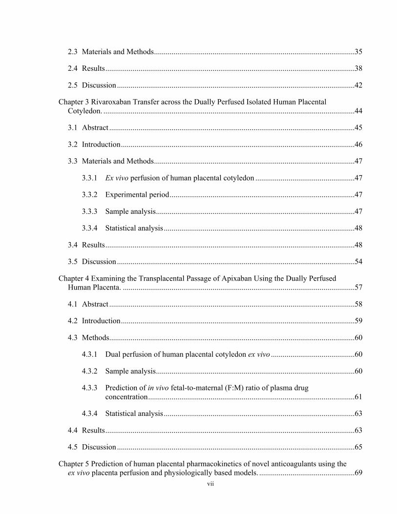
List of Tables
Table 1.1. Advantages and disadvantages of various methods used to study placental drug
transfer. ......................................................................................................................................... 13
Table 1.2. Physicochemical and pharmacokinetic properties of novel anticoagulants ................. 26
Table 2.1. Measurements of placental viability and metabolic capacity during the perfusion
experiments (n=6). ........................................................................................................................ 38
Table 3.1. Measurements of placental viability and integrity during the pre-experimental and
experimental periods of perfusion (n=9). ..................................................................................... 49
Table 4.1. Markers of placental viability during the pre-experimental and experimental periods of
perfusion (n=5). ............................................................................................................................ 64
Table 5.1. Pharmacokinetic parameters of transplacental transfer of rivaroxaban, apixaban and
dabigatran. ..................................................................................................................................... 78
x
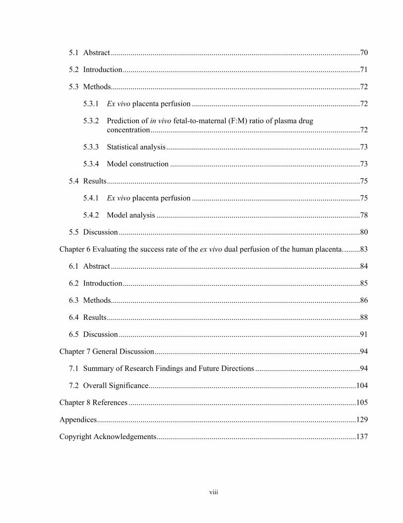
List of Figures
Figure 1.1. Trophoblast differentiation. .......................................................................................... 6
Figure 1.2. Anatomy of the term human placenta .......................................................................... 8
Figure 1.3. ATP-binding cassette drug transport proteins expressed in the human placenta ....... 11
Figure 1.4. Schematic representation of the placental perfusion model ....................................... 14
Figure 1.5. The coagulation cascade. ............................................................................................ 20
Figure 1.6. Chemical structures of novel anticoagulants .............................................................. 25
Figure 2.1. Dabigatran and dabigatran etexilate mesylate concentrations in maternal and fetal
reservoirs during the 3-h experimental phase of the placenta perfusions ..................................... 40
Figure 2.2. Fetal-to-maternal ratios for antipyrine, dabigatran, and dabigatran etexilate mesylate
during the 3-h experimental phase of the perfusions .................................................................... 41
Figure 3.1. Rivaroxaban concentrations in maternal and fetal circulations during placenta
perfusions after rivaroxaban was added to the maternal circulation only .................................... 50
Figure 3.2. Fetal-to-maternal ratios for antipyrine and rivaroxaban during the 3-h experimental
phase of the placenta perfusion ..................................................................................................... 51
Figure 3.3. Rivaroxaban concentrations in maternal and fetal circulations during the placenta
perfusions after rivaroxaban was added to the fetal circulation only ........................................... 52
Figure 3.4. Rivaroxaban concentrations in maternal and fetal circulations during the placenta
perfusions after rivaroxaban was added to both the maternal and fetal circulation ...................... 53
Figure 4.1. Apixaban concentrations in maternal and fetal circulations during the dual perfusion
of an isolated placental cotyledon ................................................................................................. 65
Figure 5.1. Three-compartment pharmacokinetic model of rivaroxaban, apixaban, and dabigatran
transfer across the placenta ........................................................................................................... 75
xi
Figure 5.2. Fetal-to-maternal (F:M) drug concentration ratios for apixaban, rivaroxaban, and
dabigatran during placenta perfusions .......................................................................................... 76
Figure 5.3. Placenta perfusion and predicted fetal-to-maternal (F:M) drug concentration ratios for
dabigatran, rivaroxaban, and apixaban after 180-min perfusion .................................................. 77
Figure 5.4. Concentration-time profiles for dabigatran, rivaroxaban and apixaban in maternal and
fetal circulations ............................................................................................................................ 79
Figure 6.1. Placenta perfusion protocol divided into various steps .............................................. 87
Figure 6.2. Flow diagram showing the number of placentae at each stage of the perfusion
protocol. ........................................................................................................................................ 89
Figure 6.3. Success rate (%) at each checkpoint of the placenta perfusion, calculated in relation
to the total number of placentae collected .................................................................................... 91
xii
List of Abbreviations
AAG α-1 acid glycoprotein
ABC ATP-binding cassette
APS antiphospholipid antibody syndrome
ATP adenosine triphosphate
AUC area under the curve
B/U bound/unbound ratio
BCRP breast cancer resistance protein
CL clearance
Cmax maximal plasma concentration
CYP cytochrome P450
DVT deep vein thrombosis
ELISA enzyme-linked immunosorbent assay
EPI enhanced product ion
F:M fetal-to-maternal
GFR glomerular filtration rate
hCG human chorionic gonadotropin
HCl hydrochloric acid
HIT heparin-induced thrombocytopenia (HIT)
IQR interquartile range
K12 influx rate constant from maternal to placental compartment
K21 efflux rate constant from placental to maternal compartment
K23 efflux rate constant from placental to fetal compartment
K32 influx rate constant from fetal to placental compartment
LMWH low molecular weight heparin
xiii
MRP multi-drug resistance associated protein
NOAC novel oral anticoagulant
NSAID nonsteroidal anti-inflammatory drug
OAT organic anion transporter
OCT organic cation transporter
P-gp P-glycoprotein
PAI plasminogen activator inhibitor
PE pulmonary embolism
pKa log dissociation constant
t1/2 elimination half-life
TAFI thrombin-activatable fibrinolysis inhibitor
TF tissue factor
TFPI tissue factor pathway inhibitor
tPA tissue-type plasminogen activator
UFH unfractionated heparin
UGT uridine 5’-diphosphate glucuronosyl-transferase
UHPLC ultrahigh-performance liquid chromatography
uPA urokinase-type plasminogen activator
Vd volume of distribution
Vf volume of distribution in fetal compartment
Vm volume of distribution in maternal compartment
Vp volume of distribution in placental compartment
VTE venous thromboembolism
ZPI protein Z-dependent protease inhibitor

xiv
List of Appendices
Appendix A. Letter of Information and Consent Form ........................................................ 130
Appendix B. Composition of M199 Culture Medium in Perfusate .................................... 136
1
Chapter 1
General Introduction
Drug Use in Pregnancy 1Medication use during pregnancy requires a fine balance between treating a condition in the
intended patient, the pregnant woman, and minimizing potential adverse risks in the unintended
patient, the fetus. Although a number of prenatal drug exposures are known to cause major
malformations, there is limited information on the risks and safety for the vast majority of
medications. With newer medications, there is typically no information about safety or efficacy
in pregnancy, as clinical trials often specifically exclude pregnant women and ensure that women
of childbearing potential do not become pregnant during the period of drug exposure. This is in
contrast to recent trends indicating that the antepartum use of both over-the-counter and
prescription medications have increased in the last 30 years. Retrospective database studies have
estimated that between 50 and 70% of women in North America take at least one prescription
medication during pregnancy (Kulaga et al. 2009; Mitchell et al. 2011). The decision to begin or
continue treatment during pregnancy relies heavily on weighing the benefits of the drug to the
mother against the potential risk to the fetus. An important determinant in this risk assessment is
the estimation of fetal drug exposure, based on quantifying the amount of drug that crosses the
placenta.
1.1 Transplacental Drug Transfer
During pregnancy, there are several physiological and anatomical changes that can alter the
maternal and fetal pharmacokinetics of medications. These changes can impact the absorption,
distribution, metabolism, and elimination of medications, and may affect their pharmacodynamic
properties during pregnancy. Transplacental pharmacokinetics must also be considered in the
context of fetal safety, as the placenta plays an integral role in fetal drug disposition.
2
1.1.1 Pregnancy-Related Physiological and Pharmacokinetic Changes
Absorption
Nausea and vomiting in the first trimester of pregnancy can decrease the amount of drug that is
absorbed following oral administration. Therefore, oral medications should ideally be taken
when nausea and vomiting are minimal. During pregnancy, gastric acid secretions are decreased
and mucus secretion is increased, leading to an elevation in gastric pH (Gryboski & Spiro 1956;
Loebstein et al. 1997). A change in gastric pH can increase ionization of weak acids and reduce
their absorption. Weak bases will be primarily un-ionized, and absorbed more readily. In
addition, slower intestinal motility can alter drug absorption and oral bioavailability. In late
pregnancy, the gastrointestinal transit time is increased by 30–50% (Parry et al. 1970; Chiloiro et
al. 2001). As well, increases in cardiac output and intestinal blood flow may increase drug
absorption (Lees et al. 1967). After accounting for changes in gastric pH, intestinal motility, and
cardiac output, it appears that pregnancy-related physiological changes may alter the rate of drug
absorption.
Distribution
There are numerous physiological changes during pregnancy that can affect the distribution of
drugs. Throughout pregnancy, there are marked increases in total body water, blood volume, and
plasma volume. Blood volume begins to increase at 6–8 weeks gestation, and continues to
increase until 32–34 weeks (Lund & Donovan 1967). The significant increases in extracellular
fluid space and total body water will increase the volume of distribution (Vd) for hydrophilic
drugs. This can lead to a decrease in the maximal concentration (Cmax) for many drugs, if the
dose is not adjusted. Maternal body fat increases by 3–6 kg, thereby increasing the Vd for
lipophilic drugs (Lederman et al. 1997). These increases in total body water and fat content can
affect the Vd of many drugs (Loebstein et al. 1997). The placenta and fetus can act as additional
compartments for drug distribution, further increasing the apparent Vd for certain drugs.
Changes in plasma protein binding can affect the unbound fraction of a drug, and potentially
alter its pharmacodynamics, as only the unbound form of the drug elicits a pharmacological
effect. Throughout pregnancy, albumin concentrations decrease to approximately 70–80% of
non-pregnant values in the third trimester (Krauer et al. 1984). While this decrease in albumin
3
has been attributed to dilution due to an increased plasma volume, another explanation is that this
decrease is caused by a reduction in the rate of albumin synthesis or an increase in the rate of its
catabolism (Frederiksen 2001). This is supported by the fact that levels of α-1 acid glycoprotein
(AAG) remain relatively stable during pregnancy (Piafsky & Woolner 1982; Krauer et al. 1984).
Protein binding is important in the context of placental drug transfer, and this will be discussed
in section 1.1.2.2.
Metabolism
Drug metabolism is altered in pregnancy, and these changes become more pronounced as the
pregnancy progresses. The activities of many phase I cytochrome P450 (CYP) enzymes are
increased in pregnancy, leading to increased clearance overall. The hepatic activities of CYP
3A4, CYP 2A6, CYP 2D6 and CYP 2C9 are all increased in pregnancy (Tracy et al. 2005;
Hebert et al. 2008; Ryu et al. 2016). Due to increased clearance, many drugs will have sub-
therapeutic concentrations, and may require an increased dose to maintain a therapeutic effect. It
should be noted that some phase I enzymes, namely CYP 1A2 and CYP 2C19, have decreased
activity in pregnancy (Brazier et al. 1983; Carter et al. 1986; Villani et al. 2006). Drugs that are
metabolized by CYP 1A2 and CYP 2C19 may require smaller doses to minimize potential
toxicity. Phase II enzymes are also altered in pregnancy, with the activity of uridine 5’-
diphosphate glucuronosyl-transferases (UGTs) increasing by up to 300% in the third trimester
(Pennell et al. 2004). The effect of pregnancy on enzyme activity varies with genotype, ethnicity,
age, and certain disease states unrelated to pregnancy. In general, changes in drug metabolism
and clearance may require dose adjustments for certain medications used in pregnancy.
Following delivery, these doses may need to be re-adjusted, as enzyme activity returns to pre-
pregnancy levels.
Elimination
Several renal changes occur during pregnancy, which can alter the elimination of renally-cleared
drugs. Anatomically, the size of the kidneys increases during pregnancy, accompanied by a 60–
80% increase in renal blood flow. As a result, the glomerular filtration rate (GFR) increases by
50% in the first trimester, and continues to increase throughout pregnancy. In some individuals,
GFR begins to decrease in the last few weeks of pregnancy (Davison & Hytten 1974; Davison &
Dunlop 1980; Odutayo & Hladunewich 2012). Studies using probe drugs have measured the
4
apparent activity of renal drug transporters, and found that the activity of P-glycoprotein (P-gp),
organic cation transporter 2 (OCT2), and organic anion transporter 1 (OAT1) are all increased
during pregnancy (Andrew et al. 2007; Hebert et al. 2008; Eyal et al. 2010). The overall increase
in renal clearance can significantly increase the elimination rates of renally-cleared drugs, and
drug dosages may need to be increased by 20–65% to maintain therapeutic levels (Anderson
2005).
1.1.2 Human Placenta: Structure and Function
1.1.2.1 Placental Development
Development of the placenta and fetus is a highly regulated process, which begins at the time of
fertilization. After fertilization, the first three days of development occur in the fallopian tube,
and on the fourth day, the morula enters the uterus. On the 5th day, the morula becomes a
blastocyst, as a result of fluid accumulation and polarization of the cells. The blastocyst has an
outer layer of cells, called trophoblasts, which will form the placenta and fetal membranes, and
an inner cell mass, which will eventually form the embryo (Boyd & Hamilton 1970; Gude et al.
2004). The inner cell mass and outer layer of cells rapidly proliferate, and the zona pellucida
surrounding the blastocyst is shed. Uterine secretions temporarily provide oxygen and metabolic
substrates for the blastocyst until approximately day 6, when the blastocyst implants into the
uterine lining (Aplin 2000). The uterine lining provides access to substrates, such as glycogen-
filled stromal cells, which are necessary for fetal growth.
Implantation involves the movement of the blastocyst to usually the mid-to-upper anterior or
posterior wall of the uterus, followed by adhesion and invasion. Trophoblasts rapidly proliferate
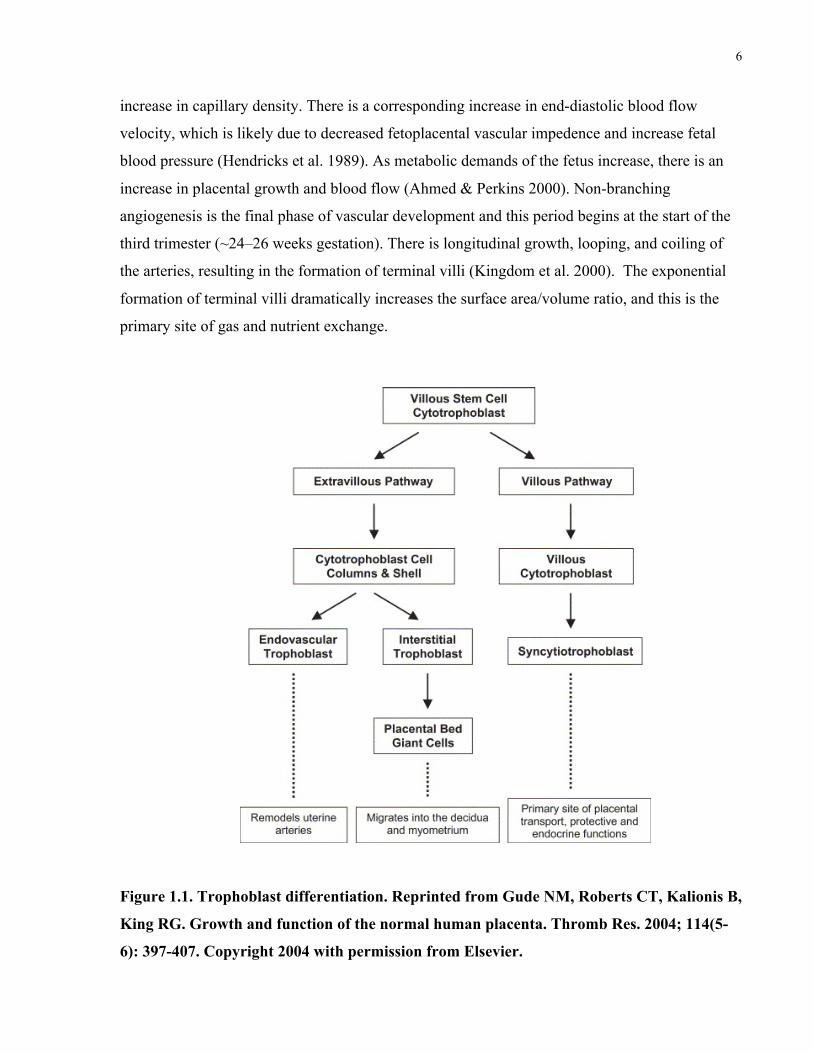
and differentiate along two pathways to form either villous or extravillous cytotrophoblasts
(Figure 1.1). Villous cytotrophoblasts ultimately form the outer cellular layer of
syncytiotrophoblast, which is responsible for the transport of gases, nutrients and waste products,
and the synthesis of peptide and steroid hormones. Extravillous trophoblasts (EVTs) eventually
form structural components of the placenta, including columns, cell islands, and septa (Boyd &
Hamilton, 1970). EVTs have been shown to express high levels of human placental lactogen
(hPL), and very little human chorionic gonadotropin (hCG) in vitro (Tarrade et al. 2001).
5
The syncytiotrophoblast extends into the endometrial epithelium and invades the connective
tissue. As the trophoblast moves deeper into the endometrial surface (called decidua), vacuoles
or empty spaces start to form. These vacuoles become confluent to form lacunar networks, and
the lacunar space eventually becomes filled with maternal blood to form the intervillous space,
thus establishing uteroplacental circulation (Aplin 2000). By the second week of placental
development, a syncytiotrophoblast layer with a core of cytotrophoblast cells evaginates into the
lacunar space to form primary mesenchymal villi. After further development, they become
secondary villi by acquiring an inner core of embryonic mesoderm. By day 21, the embryonic
mesoderm differentiates into blood vessels, which subsequently connect to vessels in the
umbilical cord and embryo, thus forming tertiary villi (Boyd & Hamilton, 1970). Some villi are
anchored to the maternal decidua, and others float freely in the lacunae.
At 4–5 weeks gestation, EVTs form columns as part of the cytotrophoblastic shell, which is at
the feto-maternal interface. Proliferative trophoblast cells are found at the base of the columns,
and invasive trophoblasts are at the distal portion of the columns. Invasive EVT are further
categorized into interstitial EVT, which invade the decidua, and endovascular EVT, which
remodel maternal blood vessels in the uterine decidua. Interstitial EVT promote the
circumferential expansion of the placenta and recruitment of maternal arterioles, which
ultimately results in growth the villous region of the placenta (6). Interstitial trophoblasts become
multinucleated and more rounded to form placenta bed giant cells (Boyd & Hamilton, 1970;
Gude et al. 2004).
During the first trimester of pregnancy, the placenta differentiates and grows in a low oxygen
environment. Endovascular EVT plug uterine spiral arterioles, and thus restrict uterine blood
flow to the fetus (Rodesch et al. 1992). At 10–14 weeks gestation, the trophoblastic plugs are
displaced and blood flows into the intervillous space to facilitate gas and nutrient exchange. The
proportion of the placenta occupied by blood vessels increases throughout gestation to facilitate
nutrient transport (Gude et al. 2004).
The three stages of human placental vascular development are: (1) vasculogenesis, (2) branching
angiogenesis, and (3) non-branching angiogenesis. Approximately 21 days after conception,
placental villi undergo vasculogenesis and blood vessels are formed (Risau 1997). Branching
angiogenesis involves the formation of new branches from pre-existing vessels, resulting in an
6
increase in capillary density. There is a corresponding increase in end-diastolic blood flow
velocity, which is likely due to decreased fetoplacental vascular impedence and increase fetal
blood pressure (Hendricks et al. 1989). As metabolic demands of the fetus increase, there is an
increase in placental growth and blood flow (Ahmed & Perkins 2000). Non-branching
angiogenesis is the final phase of vascular development and this period begins at the start of the
third trimester (~24–26 weeks gestation). There is longitudinal growth, looping, and coiling of
the arteries, resulting in the formation of terminal villi (Kingdom et al. 2000). The exponential
formation of terminal villi dramatically increases the surface area/volume ratio, and this is the
primary site of gas and nutrient exchange.
Figure 1.1. Trophoblast differentiation. Reprinted from Gude NM, Roberts CT, Kalionis B,
King RG. Growth and function of the normal human placenta. Thromb Res. 2004; 114(5-
6): 397-407. Copyright 2004 with permission from Elsevier.
7
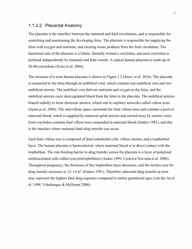
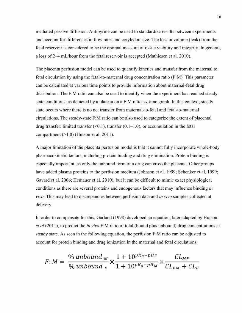
1.1.2.2 Placental Anatomy
The placenta is the interface between the maternal and fetal circulations, and is responsible for
nourishing and maintaining the developing fetus. The placenta is responsible for supplying the
fetus with oxygen and nutrients, and clearing waste products from the fetal circulation. The
functional unit of the placenta is a lobule, formally termed a cotyledon, and each cotyledon is
perfused independently by maternal and fetal vessels. A typical human placenta is made up of
20-40 cotyledons (Syme et al. 2004).
The structure of a term human placenta is shown in Figure 1.2 (Serov et al. 2016). The placenta
is connected to the fetus through an umbilical cord, which contains one umbilical vein and two
umbilical arteries. The umbilical vein delivers nutrients and oxygen to the fetus, and the
umbilical arteries carry deoxygenated blood from the fetus to the placenta. The umbilical arteries
branch radially to form chorionic arteries, which end in capillary networks called villous trees
(Syme et al. 2004). The intervillous space surrounds the fetal villous trees and contains a pool of
maternal blood, which is supplied by maternal spiral arteries and carried away by uterine veins.
Each cotyledon contains fetal villous trees suspended in maternal blood (Enders 1981), and this
is the interface where maternal-fetal drug transfer can occur.
Each fetal villous tree is composed of fetal endothelial cells, villous stroma, and a trophoblast
layer. The human placenta is haemochorial, where maternal blood is in direct contact with the
trophoblast. The rate-limiting barrier to drug transfer across the placenta is a layer of polarized,
multinucleated cells called syncytiotrophoblasts (Audus 1999; Ceckova-Novotna et al. 2006).
Throughout pregnancy, the thickness of this trophoblast layer decreases, and the surface area for
drug transfer increases to 12–14 m2 (Enders 1981). Therefore, placental drug transfer at term
may represent the highest fetal drug exposure compared to earlier gestational ages (van der Aa et
al. 1998; Vahakangas & Myllynen 2006).
8
Figure 1.2. Anatomy of the term human placenta. Reprinted and modified from Serov AS,
Salafia C, Grebenkov DS, Filoche M. The role of morphology in mathematical models of
placental gas exchange. J Appl Physiol (1985). 2016 Jan 1; 120(1): 17-28. Copyright 2016
the American Physiological Society (permission not required).
1.1.2.3 Factors Affecting Placental Drug Transfer
The rate and extent of drug transfer across the placenta depends on the physicochemical
characteristics of the drug, including lipid solubility, molecular size, and degree of ionization.
Protein binding in maternal and fetal plasma can restrict placental drug transfer, as only the
unbound form of a drug can cross the placenta (Hill & Abramson 1988). Drug transfer is also
related to the gestational age of the placenta, as the thickness of the trophoblast layer decreases
Cotyledon
Cotyledon
9
as the pregnancy progresses (Enders 1981). In addition, the expression of placental drug
transporters and metabolic enzymes changes throughout pregnancy, and this can impact the
amount of drug available to cross the placenta and reach the fetal circulation (Hutson et al. 2010;
Iqbal et al. 2012).
Endogenous and xenobiotic compounds that are lipophilic and un-ionized, with a molecular
weight less than 600 Da, can readily cross the placenta via passive diffusion (Syme et al. 2004).
As a result, most small-molecule drugs tend to cross the placenta via diffusion, which is driven
by the concentration gradient between maternal and fetal circulations (Audus 1999; Ala-Kokko
et al. 2000). In general, molecules that are relatively lipophilic can dissolve in lipid membranes,
and diffuse across the syncytiotrophoblast layer more readily than hydrophilic molecules.
Because of this, molecular size does not heavily influence the diffusion of lipophilic drugs across
the placenta, and their transfer is likely dependent on utero-placental blood flow (Giroux et al.
1997; Syme et al. 2004). Blood flow to the placenta increases during pregnancy from ~50
mL/min at 10 weeks to 600 mL/min at term. Molecular size can influence the placental transfer
of hydrophilic drugs, as the rate of diffusion tends to decrease with increasing molecular size
(Syme et al. 2004).
The extent of drug ionization in the maternal plasma is an important factor in the context of
placental drug transfer, as only the un-ionized form of a drug can cross the placenta via diffusion
(Syme et al. 2004). The log dissociation constant (pKa) of a drug can provide information about
the degree of ionization in maternal and fetal plasma. The fetal circulation is slightly more acidic
than the maternal circulation (7.35 vs 7.4, respectively), and this can affect the placental transfer
for weak acid or basic drugs whose pKa is close to physiological pH (Reynolds & Knott 1989).
For example, weak bases can become more ionized in the fetal plasma, resulting in ion trapping.
Protein Binding
Another important consideration in the disposition of drugs across the placenta is protein
binding, as only the unbound form of the drug can equilibrate across the placenta. Two important
binding proteins that bind a wide variety of drugs are albumin and α-1 acid glycoprotein (AAG).
In general, albumin binds acidic, lipophilic drugs (Kragh-Hansen 1981), while AAG binds basic,
lipophilic drugs (Piafsky & Woolner 1982; Paxton 1983). However, it should be noted that a
specific drug may partially bind to albumin, AAG or other plasma proteins. Several factors can
10
influence the protein binding of a drug in the maternal and fetal plasma. These factors include
the concentrations of plasma proteins in maternal and fetal plasma, the presence of competing
endogenous or xenobiotic ligands, and saturation of drug-protein binding (Hill & Abramson
1988).
The concentrations of albumin and AAG differ in fetal and maternal plasma, and this can affect
the transplacental transfer of drugs that bind these proteins. Throughout pregnancy, maternal
albumin levels gradually decrease and fetal albumin levels tend to increase, with the fetal-to-
maternal (F:M) albumin ratio increasing from 0.28 in the first trimester to 1.20 closer to term
(Reboud et al. 1963; Krauer et al. 1984). As well, there is a 3-fold increase in the concentration
of free fatty acids in the maternal circulation that can displace drugs from bound maternal
albumin (Ridd et al. 1983; Nau et al. 1984). This can lead to increased binding of a drug to fetal
albumin, which can, in turn, lead to increased placental drug transfer and possibly fetal drug
accumulation. By comparison, AAG levels remain relatively constant in maternal plasma, and
fetal levels gradually increase throughout pregnancy, with the AAG F:M ratio increasing from
0.09 in the first trimester to 0.37 at term (Krauer et al. 1984).
Active Transport
The placenta expresses numerous drug transport proteins that play an important role in limiting
fetal drug exposure. Unlike diffusion, active transport requires energy, usually in the form of
adenosine triphosphate (ATP) or via an electrical gradient generated by [H+], [Na+] or [Cl–] ions.
Active transport in the placenta is mediated by transport proteins located on the brush-border
apical and basolateral membranes of syncytiotrophoblast cells (Syme et al. 2004). Since active
transport typically occurs against a concentration gradient, these transporters can concentrate
nutrients in the fetal circulation (via influx) or prevent transfer of certain maternal drugs (via
efflux).
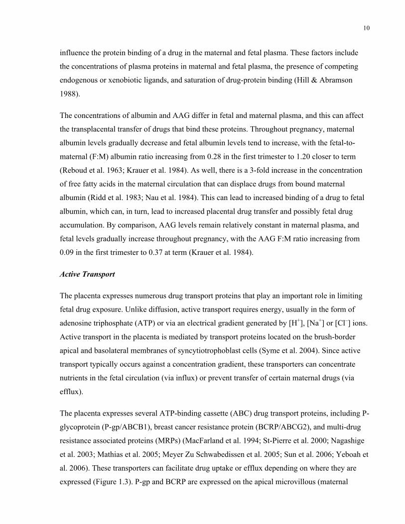
The placenta expresses several ATP-binding cassette (ABC) drug transport proteins, including P-
glycoprotein (P-gp/ABCB1), breast cancer resistance protein (BCRP/ABCG2), and multi-drug
resistance associated proteins (MRPs) (MacFarland et al. 1994; St-Pierre et al. 2000; Nagashige
et al. 2003; Mathias et al. 2005; Meyer Zu Schwabedissen et al. 2005; Sun et al. 2006; Yeboah et
al. 2006). These transporters can facilitate drug uptake or efflux depending on where they are
expressed (Figure 1.3). P-gp and BCRP are expressed on the apical microvillous (maternal
11
blood-facing) membrane of the syncytiotrophoblast (Ceckova-Novotna et al. 2006; Hahnova-
Cygalova et al. 2011), where they limit drug transfer across the placenta to the fetal circulation
and play a crucial role in fetal protection against maternal toxins. In addition to xenobiotics,
placental transporters have endogenous substrates such as amino acids, hormones, and vitamins
(Ganapathy et al. 2000).
Figure 1.3. ATP-binding cassette (ABC) drug transport proteins expressed in the human
placenta. Reprinted from Hutson JR, Koren G, Matthews SG. Placental P-glycoprotein
and breast cancer resistance protein: influence of polymorphisms on fetal drug exposure
and physiology. Placenta. 2010 May; 31(5): 351-7. Copyright 2010 with permission from
Elsevier.
12
Pregnancy is a dynamic state in which the expression and activity of drug transporters changes as
the pregnancy progresses. ABCB1 mRNA and P-gp protein are highly expressed in
syncytiotrophoblasts of first trimester human placentas, but expression levels gradually decrease
as the pregnancy progresses to term (Gil et al. 2005; Mathias et al. 2005; Sun et al. 2006). By
comparison, studies have reported inconsistent findings on age-dependent changes in expression
of human placental ABCG2 mRNA and BCRP protein. The levels of ABCG2 mRNA remain
relatively stable in the human placenta from the first to third trimester (Mathias et al. 2005;
Yeboah et al. 2006), and one study showed BCRP was more strongly expressed in term
placentae than in the first trimester (Yeboah et al. 2006). Other studies have reported that mRNA
levels for ABCG2 (BCRP) were much higher than those of ABCB1 (P-gp) in isolated primary
term trophoblast cells (Serrano et al. 2007; Manceau et al. 2012).
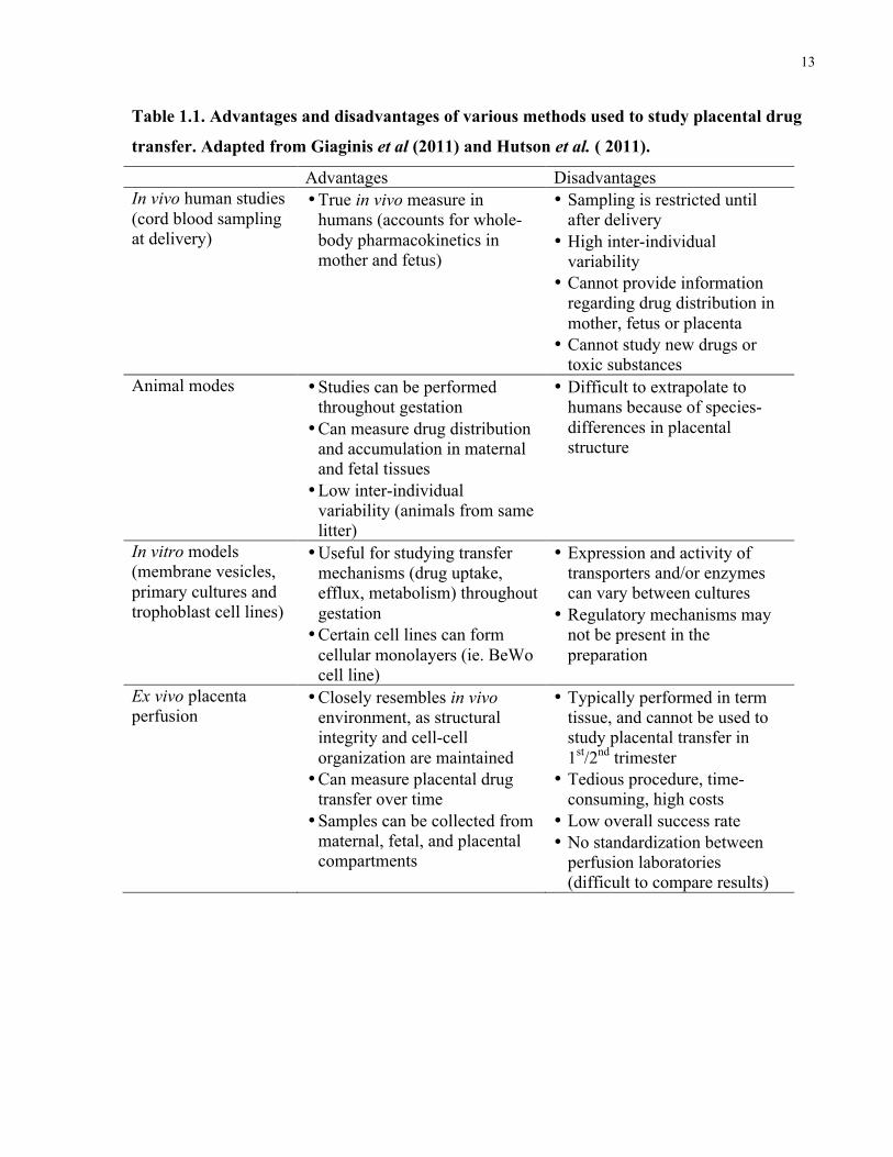
1.1.3 Methods for Studying Transplacental Drug Transfer
The placenta plays a major role in determining fetal drug exposure, and the extent of exposure
can be estimated by quantifying the amount of drug that crosses the placenta. Transplacental
pharmacokinetic data can be useful in assessing fetal safety, but this data is usually very limited
for new drugs. Due to ethical constraints, it is not possible to conduct controlled trials for new
medications in pregnant women. For some medications, drug concentrations can be measured in
cord blood from neonates exposed during pregnancy, but this requires mothers to receive the
drug prior to delivery. Cord blood levels provide information for a single time-point, and are
subject to high inter-individual variability. As well, placenta transfer in animal studies cannot
always be extrapolated to humans because of species differences in the anatomical and
biochemical structure of the placenta (Ala-Kokko et al. 2000). In vitro cell cultures and
membrane vesicles are useful for studying mechanisms of placental transfer (passive diffusion
vs. active transport), but this may not reflect the true in vivo environment as these models lack
anatomic integrity and blood flow. The ex vivo placenta perfusion model resolves many of the
issues associated with these other models. Table 1.1 outlines some of the key advantages and
disadvantages of different methods used to study placental drug transfer.
13
Table 1.1. Advantages and disadvantages of various methods used to study placental drug
transfer. Adapted from Giaginis et al (2011) and Hutson et al. ( 2011).
Advantages Disadvantages In vivo human studies (cord blood sampling at delivery)
• True in vivo measure in humans (accounts for whole-body pharmacokinetics in mother and fetus)
• Sampling is restricted until after delivery
• High inter-individual variability
• Cannot provide information regarding drug distribution in mother, fetus or placenta
• Cannot study new drugs or toxic substances
Animal modes • Studies can be performed throughout gestation • Can measure drug distribution
and accumulation in maternal and fetal tissues • Low inter-individual
variability (animals from same litter)
• Difficult to extrapolate to humans because of species-differences in placental structure
In vitro models (membrane vesicles, primary cultures and trophoblast cell lines)
• Useful for studying transfer mechanisms (drug uptake, efflux, metabolism) throughout gestation • Certain cell lines can form
cellular monolayers (ie. BeWo cell line)
• Expression and activity of transporters and/or enzymes can vary between cultures
• Regulatory mechanisms may not be present in the preparation
Ex vivo placenta perfusion
• Closely resembles in vivo environment, as structural integrity and cell-cell organization are maintained • Can measure placental drug
transfer over time • Samples can be collected from
maternal, fetal, and placental compartments
• Typically performed in term tissue, and cannot be used to study placental transfer in 1st/2nd trimester
• Tedious procedure, time-consuming, high costs
• Low overall success rate • No standardization between
perfusion laboratories (difficult to compare results)
14
Ex vivo placenta perfusion
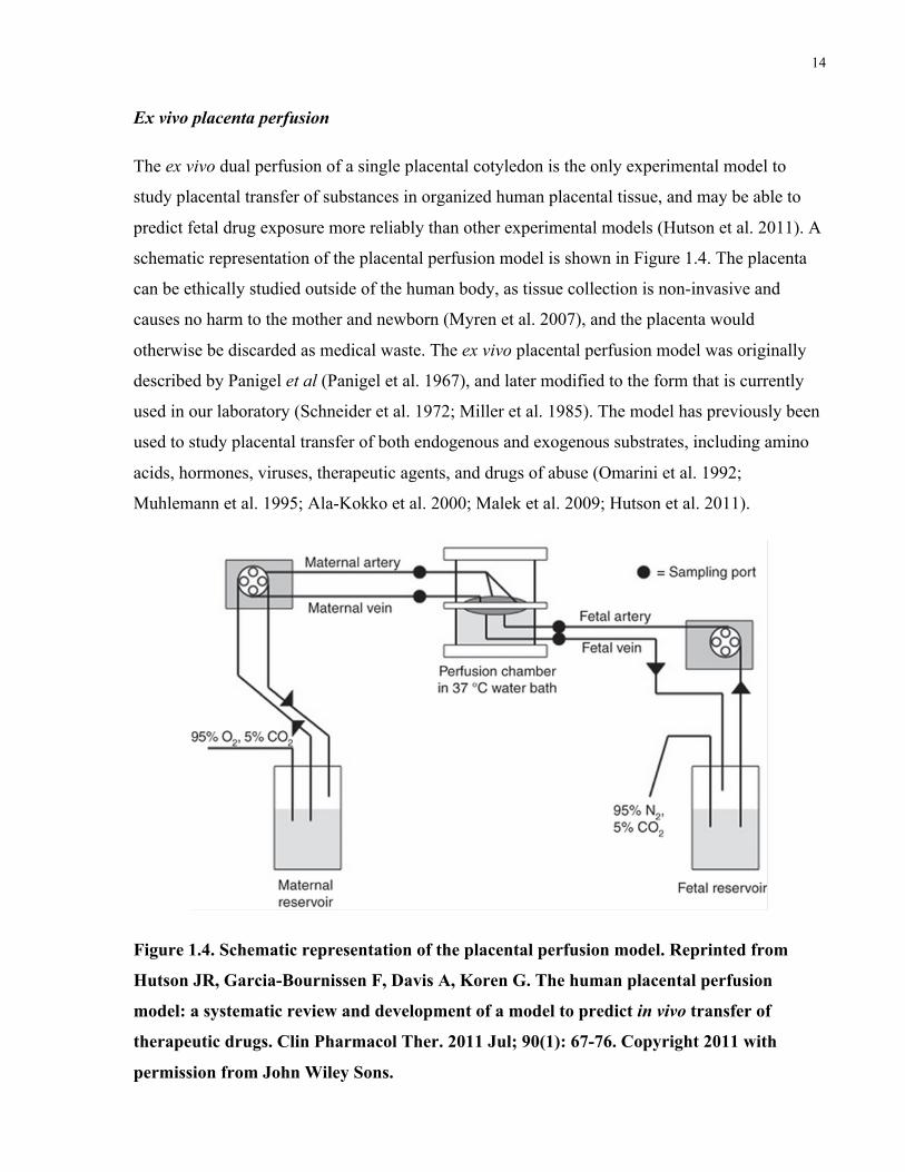
The ex vivo dual perfusion of a single placental cotyledon is the only experimental model to
study placental transfer of substances in organized human placental tissue, and may be able to
predict fetal drug exposure more reliably than other experimental models (Hutson et al. 2011). A
schematic representation of the placental perfusion model is shown in Figure 1.4. The placenta
can be ethically studied outside of the human body, as tissue collection is non-invasive and
causes no harm to the mother and newborn (Myren et al. 2007), and the placenta would
otherwise be discarded as medical waste. The ex vivo placental perfusion model was originally
described by Panigel et al (Panigel et al. 1967), and later modified to the form that is currently
used in our laboratory (Schneider et al. 1972; Miller et al. 1985). The model has previously been
used to study placental transfer of both endogenous and exogenous substrates, including amino
acids, hormones, viruses, therapeutic agents, and drugs of abuse (Omarini et al. 1992;
Muhlemann et al. 1995; Ala-Kokko et al. 2000; Malek et al. 2009; Hutson et al. 2011).
Figure 1.4. Schematic representation of the placental perfusion model. Reprinted from
Hutson JR, Garcia-Bournissen F, Davis A, Koren G. The human placental perfusion
model: a systematic review and development of a model to predict in vivo transfer of
therapeutic drugs. Clin Pharmacol Ther. 2011 Jul; 90(1): 67-76. Copyright 2011 with
permission from John Wiley Sons.
15
Following delivery, term placentae are collected and immediately transported to our on-site
laboratory at St. Michael’s Hospital in Toronto, Ontario. A fetal artery-vein pair supplying a
clearly defined cotyledon is identified and the corresponding maternal surface is checked for any
signs of physical trauma. The fetal-artery vein pair is cannulated and the fetal circulation is
established. The lobule is then clamped into a plexiglass chamber with the fetal side down and
the excess placental tissue is removed. The perfusion chamber is placed in a water bath to
maintain physiological temperature (37°C). The maternal circulation is established through the
insertion of blunt-tipped needles into the intervillous space, roughly 2-3 mm below the decidual
surface. Maternal venous samples are collected from multiple venous openings in the decidual
plate.
The fetal and maternal flow rates are maintained at 2-3 mL/min and 12-15 mL/min, respectively,
through independently-controlled roller pumps. Other perfusion laboratories use different flow
rates (Mathiesen et al. 2010), and this can affect the time to needed to measure transplacental
transfer and reach steady state between the fetal and maternal circulations (Bassily et al. 1995;
He et al. 2001). The circulations are equilibrated with 95%-O2/5%-CO2 (maternal) and 95%-
N2/5%-CO2 (fetal) to mimic physiological conditions. Other perfusion laboratories use room air
instead of 95% O2/5% CO2 to avoid hyperoxic toxicity (Mathiesen et al. 2010).
Once the maternal and fetal circulations are established, there is a control period where the blood
is washed out and baseline parameters of placental viability are measured. After the control
period, the drug of interest is added to maternal or fetal circulations to measure placental transfer
in the maternal-to-fetal or fetal-to-maternal direction. The drug can also be added in equal
concentrations to both circulations in order to measure drug accumulation. Closed (recirculating)
perfusions can be used to evaluate drug transfer and maternal-placental-fetal drug distribution.
Open (single-pass, non-recirculating) perfusions can be used to study drug clearance at steady
state concentrations.
Several quality control parameters are monitored to ensure placental viability and integrity
during the control and experimental phase of the placenta perfusion. These include arterial
inflow pressure in the fetal circulation, pH, oxygen consumption, fetal oxygen transfer, glucose
consumption, lactate production, and secretion of human chorionic gonadotropin (hCG) (Hutson
et al. 2011). Antipyrine is typically added to the maternal circulation as a marker of flow-
16
mediated passive diffusion. Antipyrine can be used to standardize results between experiments
and account for differences in flow rates and cotyledon size. The loss in volume (leak) from the
fetal reservoir is considered to be the optimal measure of tissue viability and integrity. In general,
a loss of 2–4 mL/hour from the fetal reservoir is accepted (Mathiesen et al. 2010).
The placenta perfusion model can be used to quantify kinetics and transfer from the maternal to
fetal circulation by using the fetal-to-maternal drug concentration ratio (F:M). This parameter
can be calculated at various time points to provide information about maternal-fetal drug
distribution. The F:M ratio can also be used to identify when the experiment has reached steady
state conditions, as depicted by a plateau on a F:M ratio-vs-time graph. In this context, steady
state occurs where there is no net transfer from maternal-to-fetal and fetal-to-maternal
circulations. The steady-state F:M ratio can be also used to categorize the extent of placental
drug transfer: limited transfer (<0.1), transfer (0.1–1.0), or accumulation in the fetal
compartment (>1.0) (Hutson et al. 2011).
A major limitation of the placenta perfusion model is that it cannot fully incorporate whole-body
pharmacokinetic factors, including protein binding and drug elimination. Protein binding is
especially important, as only the unbound form of a drug can cross the placenta. Other groups
have added plasma proteins to the perfusion medium (Johnson et al. 1999; Schenker et al. 1999;
Gavard et al. 2006; Hemauer et al. 2010), but it can be difficult to mimic exact physiological
conditions as there are several proteins and endogenous factors that may influence binding in
vivo. This may lead to discrepancies between perfusion data and in vivo samples collected at
delivery.
In order to compensate for this, Garland (1998) developed an equation, later adapted by Hutson
et al (2011), to predict the in vivo F:M ratio of total (bound plus unbound) drug concentrations at
steady state. As seen in the following equation, the perfusion F:M ratio can be adjusted to
account for protein binding and drug ionization in the maternal and fetal circulations,
!!! ! !!!!"#$!"%!!!!!"#$!"%!! !
!! !"!"!!!"!!! !"!"!!!"! !
!"!"!"!" ! !"!
17
where % unboundM and % unboundF are the percentages of free drug in the maternal and fetal
circulations in vivo; pHM and pHF are the pH values of maternal and fetal blood in vivo; pKa is
the log dissociation constant for the drug; and CLMF, CLFM, and CLF are the maternal-to-fetal,
fetal-to-maternal, and fetal clearances of the drug (Garland 1998). CLF is assumed to be
negligible, and the perfusion F:M ratio at steady-state can be used to represent CLMF/CLFM
(Hutson et al. 2011).
A systematic review of the placenta perfusion model evaluated its effectiveness in predicting
placental drug transfer in vivo (Hutson et al. 2011). In that study, fetal-to-maternal concentration
ratios from perfusion experiments were compared with cord-to-maternal blood ratios collected at
the time of delivery for 70 drugs. In general, the results from placenta perfusion experiments
correlated well with the data in vivo. After adjusting the perfusion fetal-to-maternal ratio of 24
drugs for protein binding and pH differences between the fetal and maternal circulation, there
was a stronger correlation between the in vivo results of cord-to-maternal drug concentration
ratio and the perfusion experiments’ derived fetal-to-maternal ratio. The authors concluded that
the perfusion model can be used to accurately predict placental transfer of small molecule drugs
in vivo (Hutson et al. 2011). In instances where information regarding placental transfer may not
be available, the placenta perfusion model is a safe and ethical tool to predict placental transfer
in vivo.
1.2 Anticoagulation Therapy in Pregnancy
Coagulation is the process by which blood forms a clot and is an important part of hemostasis at
a site of vessel injury. When a blood vessel is damaged, a platelet- and fibrin-containing clot is
formed at the site of injury to stop the bleeding and allow for vessel repair to begin. This
important role of blood coagulation necessitates that the response be quick, localized, and
carefully regulated. Blood coagulation is initiated almost immediately after injury to the
endothelial lining of a blood vessel. Proteins, such as tissue factor, are activated to initiate the
aggregation of blood platelets to form a plug at the site of injury. The clotting process is further
propagated by other coagulation factors in the plasma that respond in a complex cascade to form
stable fibrin clots. These additional fibrin clots serve to strengthen the platelet plug. The clotting
process is then terminated by antithrombotic control mechanisms, and as the injured tissue heals,
the blood clot is subsequently removed by fibrinolysis (Furie & Furie 2005). The pathways of
18
fibrin clot formation and plasmin-induced fibrinolysis are linked and carefully regulated by each
other (Lane et al. 2005). Blood coagulation disorders can occur when specific elements of the
coagulation cascade are missing or dysfunctional, and can lead to increased risk of bleeding or
thrombosis.
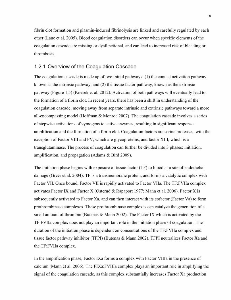
1.2.1 Overview of the Coagulation Cascade
The coagulation cascade is made up of two initial pathways: (1) the contact activation pathway,
known as the intrinsic pathway, and (2) the tissue factor pathway, known as the extrinsic
pathway (Figure 1.5) (Knesek et al. 2012). Activation of both pathways will eventually lead to
the formation of a fibrin clot. In recent years, there has been a shift in understanding of the
coagulation cascade, moving away from separate intrinsic and extrinsic pathways toward a more
all-encompassing model (Hoffman & Monroe 2007). The coagulation cascade involves a series
of stepwise activations of zymogens to active enzymes, resulting in significant response
amplification and the formation of a fibrin clot. Coagulation factors are serine proteases, with the
exception of Factor VIII and FV, which are glycoproteins, and factor XIII, which is a
transglutaminase. The process of coagulation can further be divided into 3 phases: initiation,
amplification, and propagation (Adams & Bird 2009).
The initiation phase begins with exposure of tissue factor (TF) to blood at a site of endothelial
damage (Greer et al. 2004). TF is a transmembrane protein, and forms a catalytic complex with
Factor VII. Once bound, Factor VII is rapidly activated to Factor VIIa. The TF:FVIIa complex
activates Factor IX and Factor X (Osterud & Rapaport 1977; Mann et al. 2006). Factor X is
subsequently activated to Factor Xa, and can then interact with its cofactor (Factor Va) to form
prothrombinase complexes. These prothrombinase complexes can catalyze the generation of a
small amount of thrombin (Butenas & Mann 2002). The Factor IX which is activated by the
TF:FVIIa complex does not play an important role in the initiation phase of coagulation. The
duration of the initiation phase is dependent on concentrations of the TF:FVIIa complex and
tissue factor pathway inhibitor (TFPI) (Butenas & Mann 2002). TFPI neutralizes Factor Xa and
the TF:FVIIa complex.
In the amplification phase, Factor IXa forms a complex with Factor VIIIa in the presence of
calcium (Mann et al. 2006). The FIXa:FVIIIa complex plays an important role in amplifying the
signal of the coagulation cascade, as this complex substantially increases Factor Xa production
19
(at a rate 50–100 times greater than the TF:FVIIa complex) (Butenas & Mann 2002; Mann et al.
2006). The increased levels of Factor Xa drive the accelerated production of thrombin. In the
presence of calcium, the FIXa:FVIIIa and prothrombinase (FXa:FVa) complexes create a
positive feedback loop, resulting in further thrombin generation (Mann et al. 2003). Thrombin
enhances platelet aggregation, and activates FVIIIa, by dissociating FVIII from its complex with
von Willebrand factor (FVIII:vWF). Thrombin also catalyzes the conversion of Factor XI to
Factor XIa (Monroe & Hoffman 2006; Adams & Bird 2009). These additional positive feedback
loops result in increased amounts of thrombin generation in a short amount of time.
During the propagation phase, activated platelets aggregate at the site of injury to ensure
localized thrombin generation. The increased thrombin production results in a ‘thrombin burst’
that leads to the conversion of fibrinogen to fibrin to produce a stable clot. Fibrin monomers
coalesce into a fibrin polymer gel, and factor XIIIa covalently cross-links fibrin strands to form a
stable fibrin network (Greer et al. 2004). The newly formed thrombin also activates thrombin-
activatable fibrinolysis inhibitor (TAFI), which prevents plasmin-mediated fibrinolysis (Bouma
& Mosnier 2003).
Fibrinolysis, or the breakdown of a clot, is an essential component in maintaining hemostatic
equilibrium. Plasmin is an important mediator of fibrinolysis, and it acts by cleaving fibrin at
specific lysine and arginine residues into soluble degradation products (Greer et al. 2004).
Plasmin is produced from its precursor, plasminogen, via tissue-type plasminogen activator
(tPA) and urokinase-type plasminogen activator (uPA) (Adams & Bird 2009). These
plasminogen activators are regulated by plasminogen activator inhibitors (PAIs), which are
present at high levels in the blood. PAIs form inactivating complexes with tPA and uPA to limit
plasmin generation (Sprengers & Kluft 1987).
The coagulation cascade is regulated at each level, and this is important in maintaining
hemostasis. Tissue factor is regulated by tissue factor pathway inhibitor (TFPI), which
neutralizes the activity of Factor Xa and inhibits the TF:FVIIa complex (Broze 1995; Dahlback
2000). Antithrombin is a serine protease inhibitor, and plays an important role in preventing
coagulation by preferentially inhibiting free thrombin and Factor Xa. The anticoagulant actions
of antithrombin can be enhanced by the administration of heparin (Dahlback 2000; Adams &
Bird 2009). Protein C is activated when it binds to thrombomodulin in the presence of thrombin.
20
Activated protein C, along with its co-factor protein S, inactivates Factor Va and Factor VIIIa
(Adams & Bird 2009). Protein Z inhibits Factor Xa via protein Z-dependent protease inhibitor
(ZPI) (Corral et al. 2007). Vitamin K is an essential co-factor for Factors II, VII, IX and X, as
well as Protein S, Protein C and Protein Z.
Figure 1.5. The coagulation cascade. Reprinted from Knesek D, Peterson TC, Markel DC.
Thromboembolic prophylaxis in total joint arthroplasty. Thrombosis. 2012; 2012: 837896.
Copyright © 2012 David Knesek et al (permission not required).
1.2.2 Maternal and Fetal Coagulation Systems
Pregnancy is classically a state of hypercoagulability, characterized by an increased level of
many clotting factors, a decrease in the amount of natural anticoagulants, and a reduction in
fibrinolytic activity in the blood (Dahlman et al. 1985; Bremme 2003; O'Riordan & Higgins
2003; Townsley 2013). These hemostatic changes become more pronounced as the pregnancy
progresses (Bremme 2003) and are likely caused by natural hormonal changes that are associated
with pregnancy (Sattar et al. 1999). Platelet counts decrease in healthy pregnancies, with the
21
maximal decrease occurring in the third trimester (Verdy et al. 1997; Boehlen et al. 2000).
Levels of factors VIII, X, XII, von Willebrand factor, and fibrinogen increase by 20-200%
during pregnancy (Stirling et al. 1984; Esmon 1993; Bremme 2003; Hellgren 2003; O'Riordan &
Higgins 2003). Factor VII substantially increases throughout pregnancy, with levels up to 1000%
at term (Dalaker & Prydz 1984; Bremme 2003). Factor XIII, which stabilizes fibrin, increases in
the first trimester but reduces to 50% of non-pregnant levels at term (Persson et al. 1980). Factor
II (prothrombin) and V levels are slightly changed early in pregnancy, but return to non-pregnant
levels at term (Bremme 2003).
In terms of antithrombotic proteins, antithrombin and protein C levels remain unchanged during
pregnancy, while protein S decreases (Kjellberg et al. 1999; Bremme 2003). Fibrinolytic activity
is reduced as the levels of fibrinolytic inhibitors, thrombin-activatable fibrinolysis inhibitor
(TAFI), plasminogen activator inhibitor-1 (PAI-1), and PAI-2 are significantly increased
throughout pregnancy (Ku et al. 2003). The hypercoagulable state in pregnancy confers a
survival advantage by minimizing blood loss after delivery, but it also predisposes pregnant
women to higher risks for thromboembolism (Hellgren 1996; Thornton & Douglas 2010).
Maternal coagulation factors cannot cross the placenta to the fetal circulation (Cade et al. 1969).
The fetal coagulation system is dynamic throughout gestation, and gradually evolves toward the
neonatal and adult state. The synthesis of coagulation factors, such as fibrinogen, begins as early
as 5.5 weeks, and fetal blood is able to form a clot at 11 weeks gestation (Zilliacus et al. 1966;
Gitlin & Biasucci 1969). One study measured the concentrations of coagulation factors in
healthy fetuses, ranging from 19 to 38 weeks gestation, and found that levels of coagulation
factors typically increase with advancing gestational age. The levels of antithrombin, protein C,
and protein S were also much lower, suggesting that fetal hemostasis maintains equilibrium
between activators and inhibitors of coagulation (Reverdiau-Moalic et al. 1996). Compared to
preterm and term neonates, levels of fetal plasma coagulation factors are generally much lower,
and only factors V and VIII reach adult values at birth (Andrew et al. 1987, 1988, 1990;
Reverdiau-Moalic et al. 1996). The neonatal coagulation system continues to develop through
childhood, and matures to the adult form by late adolescence (Andrew et al. 1992).
22
Thromboembolic Disease
During pregnancy, women are at an increased risk of arterial and venous thromboembolism. The
risk for arterial embolism, which can result in stroke and heart attack, is 3-4-fold higher in
pregnant women, compared to non-pregnant women of childbearing age (James et al. 2005,
2006a). The risk for developing venous thromboembolism (VTE) during pregnancy is increased
by 4–5-fold, and increases by up to 20-fold in the postpartum period (Heit et al. 2005; Pomp et
al. 2008). The increased risk for VTE is likely due to a decrease in venous capacitance and
outflow, mechanical obstruction by the uterus, and possibly by decreased mobility (Macklon et
al. 1997; Danilenko-Dixon et al. 2001; James 2009).
VTE, which includes deep vein thrombosis (DVT) and pulmonary embolism (PE), is one of the
most common causes of maternal mortality in Canada and the United States (Chang et al. 2003;
Lisonkova et al. 2011). A retrospective study found that VTE affected 28 per 100,000
pregnancies antepartum, and 65 per 100,000 postpartum (Simpson et al. 2001); another study
reported that approximately 80% of VTE events in pregnancy are DVT, and 20% are PE (Heit et
al. 2005; James et al. 2006b). Approximately 33% of pregnancy-related DVT events and 50% of
PE events occurred in the postpartum period (Gherman et al. 1999; Ray & Chan 1999; Simpson
et al. 2001; James et al. 2005).
Two important risk factors for the development of VTE include a history of thrombosis, and
inherited or acquired thrombophilias. Additional risk factors include varicose veins,
inflammatory bowel disease, urinary tract infection, diabetes, body mass index ≥30 kg/m2,
increased maternal age (≥35 years), smoking, stillbirth, obstetric hemorrhage, preterm delivery,
and caesarean section (Ginsberg & Hirsh 1998; Abdul Sultan et al. 2013; Sultan et al. 2013).
1.2.3 Current Therapies
As with many drugs prescribed during pregnancy, the use of anticoagulant therapy can be
challenging because of the possibility for both maternal and fetal complications. Anticoagulant
therapy may be required during pregnancy in women at high risk for venous thromboembolism,
and in women with prosthetic or mechanical heart valves, atrial fibrillation, cerebral venous
sinus thrombosis, and left ventricular dysfunction. In some cases, anticoagulants are used in
23
combination with aspirin for the prevention of recurrent pregnancy loss in women with
antiphospholipid antibody syndrome (APS) (Bates et al. 2012).
Low molecular weight heparin (LMWH) and unfractionated heparin (UFH) are the preferred
choices of anticoagulants for the prevention and treatment of thromboembolism in pregnant
women, as they do not cross the placenta and are not associated with teratogenic effects
(Forestier et al. 1984, 1987; Bates 2002; Greer & Nelson-Piercy 2005; Bates et al. 2012).
LMWH and UFH bind antithrombin to induce a conformational change, which significantly
accelerates antithrombin’s inactivation of factor Xa and thrombin, resulting in slower
coagulation overall (Marcum et al. 1984; Klein et al. 2002). LMWHs are generally preferred
over UFHs because LMWH has a higher bioavailability, longer plasma half-life, and more
predictable pharmacokinetic response. In addition, LMWH has a lower risk of maternal heparin-
induced thrombocytopenia (HIT) and heparin-associated osteoporosis compared to UFH (Bates
et al. 2012). UFH is a less expensive alternative to LMWH, and may be preferred in later stages
of pregnancy (closer to delivery) when rapid temporal control of anticoagulation is required
(James 2011).
LWMHs and UFH are administered via subcutaneous injection, up to twice daily, and can result
in pain at the site of injection. As a result of pregnancy-related physiological changes, both
LMWH and UFH have shorter half-lives and lower peak plasma concentrations in pregnant
women, compared to non-pregnant adults (Brancazio et al. 1995; Casele et al. 1999; Barbour et
al. 2004). These changes may require the use of higher doses and more frequent drug
administration.
Danaparoid sodium is a low molecular weight heparinoid, which may be used in pregnant
women who develop HIT and require ongoing anticoagulant therapy (Bates et al. 2012).
Danaparoid sodium is an effective antithrombotic agent that inhibits Factor Xa (de Valk et al.
1995). Animal studies and human case reports have shown that danaparoid sodium does not
cross the placenta (Peeters et al. 1986; Henny et al. 1986; Greinacher et al. 1993; Lindhoff-Last
et al. 2005), and it has a low cross-reactivity with UFH (Magnani 1993). Fetal toxicity has not
been demonstrated with maternal use of danaparoid, but the quality of this evidence is low (Bates
et al. 2012). Due to shortages, danaparoid was withdrawn from the US market in 2002, but it is
still available in Canada, Australia, Japan and European countries.
24
Fondaparinux is a synthetic polysaccharide, which acts by inhibiting Factor Xa. Its structure is
based on the active moiety of heparin. Using a dually perfused human cotyledon, fondaparinux
did not cross the placenta from maternal to fetal circulation (Lagrange et al. 2002).
Approximately 10% of maternal anti-Xa activity was detected in the umbilical cord plasma of
five neonates whose mothers were treated with fondaparinux (Dempfle 2004). While there are
reports of the use of fondaparinux during pregnancy (Parody et al. 2003; Mazzolai et al. 2006;
Wijesiriwardana et al. 2006; Gerhardt et al. 2007), the information still remains very limited. The
American College of Chest Physicians recommends limiting the use of fondaparinux during
pregnancy to women with severe reactions to heparin who cannot receive danaparoid (Bates et
al. 2012). Argatroban is a parenteral direct thrombin inhibitor, which requires continuous
intravenous administration. Agratroban may be used in individuals with severe reactions to
heparins who cannot receive danaparoid or fondaparinux (Bates et al. 2012). To date, only a few
cases have described the use of argatroban in late pregnancy (Taniguchi et al. 2008; Young et al.
2008; Ekbatani et al. 2010).
The classical anticoagulant, warfarin, is contraindicated during pregnancy because of its
associated embryopathy, characterized by nasal and limb hypoplasia, as well as congenital
cardiac anomalies (McLintock 2013). The use of warfarin in pregnancy was also associated with
early miscarriage, but studies could not determine if miscarriages were due to the use of warfarin
itself or the underlying conditions for which warfarin was administered (Schaefer et al. 2006).
Warfarin and other vitamin K antagonists freely cross the placenta, and have the potential to
cause both fetal bleeding and teratogenic effects (Iturbe-Alessio et al. 1986; Ginsberg et al. 1989;
Schaefer et al. 2006). Vitamin K antagonists inhibit the synthesis of vitamin K-dependent
clotting factors, which include Factors II, VII, IX, and X, and the anticoagulant proteins C and S.
These medications can cause fetal bleeding at any stage of pregnancy, but the risk for
teratogenicity is highest with administration between 6 and 12 weeks gestation (Hall et al. 1980;
Howie 1986; Iturbe-Alessio et al. 1986).
1.3 Novel Oral Anticoagulants
Recently, novel oral anticoagulants (NOACs) have been developed and approved for clinical use
in non-pregnant adults. A major advantage is that these new medications can be prescribed at
fixed doses without the need for routine monitoring or dose adjustment. Compared to vitamin K
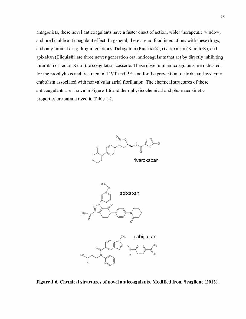
25
antagonists, these novel anticoagulants have a faster onset of action, wider therapeutic window,
and predictable anticoagulant effect. In general, there are no food interactions with these drugs,
and only limited drug-drug interactions. Dabigatran (Pradaxa®), rivaroxaban (Xarelto®), and
apixaban (Eliquis®) are three newer generation oral anticoagulants that act by directly inhibiting
thrombin or factor Xa of the coagulation cascade. These novel oral anticoagulants are indicated
for the prophylaxis and treatment of DVT and PE; and for the prevention of stroke and systemic
embolism associated with nonvalvular atrial fibrillation. The chemical structures of these
anticoagulants are shown in Figure 1.6 and their physicochemical and pharmacokinetic
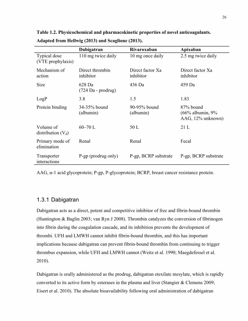
properties are summarized in Table 1.2.
Figure 1.6. Chemical structures of novel anticoagulants. Modified from Scaglione (2013).
apixaban
rivaroxaban
dabigatran
26
Table 1.2. Physicochemical and pharmacokinetic properties of novel anticoagulants.
Adapted from Hellwig (2013) and Scaglione (2013).
Dabigatran Rivaroxaban Apixaban Typical dose (VTE prophylaxis)
110 mg twice daily 10 mg once daily 2.5 mg twice daily
Mechanism of action
Direct thrombin inhibitor
Direct factor Xa inhibitor
Direct factor Xa inhibitor
Size 628 Da (724 Da - prodrug)
436 Da 459 Da
LogP 3.8 1.5 1.83
Protein binding 34-35% bound (albumin)
90-95% bound (albumin)
87% bound (66% albumin, 9% AAG, 12% unknown)
Volume of distribution (Vd)
60–70 L 50 L 21 L
Primary mode of elimination
Renal Renal Fecal
Transporter interactions
P-gp (prodrug only) P-gp, BCRP substrate P-gp, BCRP substrate
AAG, α-1 acid glycoprotein; P-gp, P-glycoprotein; BCRP, breast cancer resistance protein.
1.3.1 Dabigatran
Dabigatran acts as a direct, potent and competitive inhibitor of free and fibrin-bound thrombin
(Huntington & Baglin 2003; van Ryn J 2008). Thrombin catalyzes the conversion of fibrinogen
into fibrin during the coagulation cascade, and its inhibition prevents the development of
thrombi. UFH and LMWH cannot inhibit fibrin-bound thrombin, and this has important
implications because dabigatran can prevent fibrin-bound thrombin from continuing to trigger
thrombus expansion, while UFH and LMWH cannot (Weitz et al. 1990; Maegdefessel et al.
2010).
Dabigatran is orally administered as the prodrug, dabigatran etexilate mesylate, which is rapidly
converted to its active form by esterases in the plasma and liver (Stangier & Clemens 2009;
Eisert et al. 2010). The absolute bioavailability following oral administration of dabigatran
27
etexilate mesylate is low at about 7% (Stangier & Clemens 2009), and maximal plasma
concentrations (Cmax) levels are reached within 0.5–2 hours. Dabigatran is partially bound to
plasma proteins (35%) and its volume of distribution is 60–70 L, indicating moderate tissue
distribution (Blech et al. 2008).
Dabigatran etexilate mesylate is not metabolized by CYP enzymes, and approximately 20% is
conjugated with glucuronic acid and excreted via the biliary system (Ebner et al. 2010). The
active drug, dabigatran, is the major circulating compound, and it is primarily excreted
unchanged through the kidneys (Stangier & Clemens 2009). The average half-life (t1/2) of
dabigatran ranges from 8 to 10 hours after a single dose, and increases to 14–17 hours after
multiple doses (Stangier et al. 2007). In cases of renal impairment, there are elevated plasma
concentrations and a prolonged half-life for dabigatran (Stangier et al. 2010). The prodrug,
dabigatran etexilate mesylate, is a substrate for P-glycoprotein, and therefore, co-administration
with P-gp inhibitors or inducers may affect plasma levels of dabigatran (Eisert et al. 2010).
To date, there are no clinical trials evaluating the safety and efficacy of dabigatran in pregnancy
in humans. Animal studies have investigated the potential teratogenic effects of dabigatran in rats
and rabbits. There was an adverse effect on female fertility as noted by a decrease in
implantation and an increase in pre-implantation loss at a dose of 70 mg/kg, which is 5-fold
higher than the human dose. At higher doses (up to 10-fold higher than the human doses), there
was a decrease in fetal body weight and viability in rats and rabbits. No major malformations
were observed in fetal rabbits (Boehringer Ingelheim Canada 2016).
1.3.2 Rivaroxaban
Rivaroxaban is a direct, reversible, competitive inhibitor of factor Xa, which is an important
component of both the intrinsic and extrinsic pathways in the coagulation cascade. As previously
decribed, factor Xa directly converts prothrombin to thrombin via the prothrombinase complex,
and its inhibition limits thrombin generation and the subsequent formation of a fibrin clot. Due to
signal amplification in the coagulation cascade, one molecule of Factor Xa is able to generate
more than 1,000 molecules of thrombin (Hoffman & Monroe 2007), making Factor Xa an ideal
target for anticoagulation therapy (Spyropoulos 2007). Rivaroxaban can inhibit free Factor Xa,
Factor Xa already bound in the prothrombinase complex, and clot-bound Factor Xa (Roehrig et
al. 2005; Laux et al. 2007). As well, rivaroxaban has a high specificity for Factor Xa, as it does
28
not inhibit other serine proteases including thrombin, Factor VIIa, Factor IXa, Factor XIa, and
activated protein C (Perzborn et al. 2005).
Rivaroxaban has a low water solubility and high membrane permeability, and it is rapidly
absorbed following oral administration. It has a high oral bioavailability (80–100%) and Cmax
levels are achieved 2–4 hours after administration (Kubitza et al. 2005b). There is very limited
presystemic first-pass extraction, and rivaroxaban remains highly bound to plasma proteins (92–
95%), with serum albumin as the predominant binding protein (Weinz et al. 2005). Rivaroxaban
has an apparent Vd of 50 L at steady state (Bayer Canada 2013).
Rivaroxaban is metabolized to inactive metabolites via CYP enzymes (primarily CYP 3A4 and
CYP 2C8) (Lang et al. 2009). Approximately 66% of rivaroxaban is excreted via the kidneys,
with the remaining 34% excreted in feces (Bayer Canada 2013). Rivaroxaban has a terminal
half-life (t1/2) of 5–9 hours in young individuals (Kubitza et al. 2005a, b), and this can increase
up to 11–13 hours in the elderly as a result of age-related declines in renal function (Kubitza et
al. 2008, 2010). Previous studies have shown that rivaroxaban is a substrate of the transporters P-
gp and BCRP (Gnoth et al. 2011; Gong et al. 2013).
To date, there have been few reported cases of rivaroxaban exposure in human pregnancy. A
case report described the use of rivaroxaban up to 19 weeks gestation. A male infant was born at
40 weeks gestation with no complications. The authors reported a small baby by weight, length,
and cranial circumference (13th, 30th, and 7th percentiles, respectively), but attributed this to
maternal smoking (Konigsbrugge et al. 2014). Recently, a case series was published including 37
women who were inadvertently exposed to rivaroxaban in the first and second trimesters. There
was one case of a conotruncal cardiac defect in a woman with several co-medications and a
previous fetus with a cardiac malformation in the absence of rivaroxaban exposure (Hoeltzenbein
et al. 2016). These cases demonstrate the prospect that as prescriptions rates of novel oral
anticoagulants (NOACs) continue to increase (Desai et al. 2014), the number of women with an
unplanned pregnancy who are taking a NOAC will also likely increase.
Animal studies have examined the reproductive and developmental toxicity of rivaroxaban.
Studies in rats and rabbits have shown reproductive toxicity related to the pharmacodynamic
effect of rivaroxaban (ie. hemorrhagic complications). It should be noted that rivaroxaban was
administered up to 38-fold (rat) and up to 89-fold (rabbit) higher doses than the typical
29
therapeutic exposure in humans. Even at the highest tested doses, there were no observed signs
of teratogenicity. As well, rivaroxaban did not show any effect on female fertility at doses up to
200 mg/kg (Bayer Canada 2013).
1.3.3 Apixaban
Similar to rivaroxaban, apixaban is a reversible, direct inhibitor of factor Xa, and it can bind to
free and prothrombinase-bound Factor Xa (Jiang et al. 2009). Apixaban is highly selective for
factor Xa, and does not affect activated protein C, Factors IXa and VIIa, and thrombin (Pinto et
al. 2007). Importantly, when compared to other NOACs (including dabigatran, rivaroxaban, and
edoxaban), apixaban was associated with a relatively low risk of bleeding events while showing
similar efficacy for the treatment of acute VTE (Mantha & Ansell 2015).
Following oral administration, apixaban is rapidly absorbed and Cmax levels are achieved within
3–4 hours (Raghavan et al. 2009). Apixaban has an oral bioavailability of approximately 50% for
doses up to 10 mg. Compared to dabigatran and rivaroxaban, apixaban has a lower volume of
distribution at ~21 L, and remains highly bound to plasma proteins (87%) (Pfizer Canada 2015).
The lower Vd is likely due to limited distribution in extravascular tissue, and not due to extensive
plasma protein binding (He et al. 2011).
Apixaban is metabolized via CYP 3A4/5 with minor contributions from CYP 1A2 and 2J2
(Wang et al. 2010). The major circulating compound is unchanged apixaban and there are no
active metabolites. Apixaban is eliminated via multiple routes, with ~50% excreted unchanged in
the feces and 25% excreted via the kidneys (Raghavan et al. 2009). Apixaban has an elimination
half-life of approximately 8–9 hours (Frost et al. 2014). Using in vitro models, it has been shown
that apixaban is a substrate for both P-gp and BCRP transporters (Zhang et al. 2013); plasma
concentrations and the elimination half-life of apixaban may be altered if it is administered with
inhibitors or inducers of these transporters.
Apixaban is a relatively new drug, and currently there are no clinical trials that have evaluated
the safety and efficacy of apixaban in pregnancy in humans. Studies in mice, rats, and rabbits did
not indicate direct or indirect effects of reproductive toxicity. There was an increased incidence
of maternal bleeding when animals were administered levels of apixaban much higher than
human therapeutic levels. There were no maternal or fetal deaths related to bleeding, and no
30
evidence of an increased risk of fetal malformations following in utero exposure to apixaban
(Pfizer Canada 2015). A study in rats showed limited placental transfer of [14C]-apixaban, with a
peak fetal blood concentration that was approximately 35% of the maternal concentration (Wang
et al. 2011)
1.4 Overall Rationale and Primary Objectives
Recent trends have shown that medication use in pregnancy has steadily increased in the last 30
years, and the decision to begin or continue treatment in pregnancy heavily relies on weighing
the maternal benefits of the drug against the potential fetal risk. The use of newer medications in
pregnancy is especially challenging, as there is typically no information about safety or efficacy
in pregnancy. Clinical trials usually exclude pregnant women and ensure that women of
childbearing potential do not become pregnant during the period of drug exposure. Several novel
oral anticoagulants have been developed and approved for clinical use in recent years, and these
medications have clear advantages over existing therapies. Dabigatran, rivaroxaban, and
apixaban are three newer generation oral anticoagulants that can be used in the prevention and
treatment of venous thromboembolism (VTE). However, information regarding fetal safety and
placental transfer of these drugs is currently very limited. As prescription rates for novel oral
anticoagulants continue to increase, the number of women with an unplanned pregnancy who are
taking novel oral anticoagulants (NOACs) will likely increase.
The main objective of this thesis was to investigate the transplacental kinetics of novel
anticoagulants using the placenta perfusion model. By determining the rate and extent of
placental drug transfer, we will be able to estimate fetal drug exposure. The proposed studies
will focus on 3 anticoagulants (dabigatran, rivaroxaban, and apixaban), and placental drug
transfer will be evaluated separately for each drug. The first objective of this thesis was to
determine the rate and extent of the placental transfer of dabigatran. The second and third
objectives were to examine the placental transfer of rivaroxaban and apixaban, respectively.
Compared to rivaroxaban and apixaban, dabigatran exhibits more hydrogen bonding and has
a higher polar surface area, which are characteristics that may reduce transport across the
placental barrier. The large molecular weight and high lipophilicty of dabigatran may also
limit its transfer. Based on its physicochemical properties, we hypothesize that dabigatran will
have limited placental transfer compared with unbound rivaroxaban and apixaban.
31
While the placenta perfusion model can be highly predictive of placental transfer in vivo, a
limitation of this model is that it cannot account for differences in protein binding between
maternal and fetal plasma. A recent study has showed that placenta perfusion data can be
mathematically adjusted in order to predict the fetal-to-maternal (F:M) ratio of total (bound plus
unbound) drug concentration in plasma in vivo. After adjusting for protein binding differences,
the authors noted a stronger correlation between placental perfusion and in vivo placental transfer
(Hutson et al. 2011). Dabigatran exhibits low protein binding, while rivaroxaban and apixaban
remain highly bound to plasma proteins in vivo.
The fourth objective of this study was to compare the placental transfer of three novel oral
anticoagulants, after adjusting for differences in protein binding. Since rivaroxaban and
apixaban are highly bound to plasma proteins, we hypothesize that the F:M concentration
ratios for these two medications will be altered after the adjustment. We also developed a
pharmacokinetic model to estimate transplacental transfer parameters. We hypothesize that
placenta perfusion data can be fitted to a pharmacokinetic model, and that this model will be
able to predict transfer parameters.
The dual perfusion of a single placental cotyledon ex vivo has been widely used among placental
researchers to study transplacental drug transfer in organized human placental tissue. While this
model can be highly predictive of in vivo placental transfer, a major practical challenge of this
model is the low overall success rate. This can result in high costs and longer time to complete a
study examining placental drug transfer. Different perfusion laboratories report success rates
between 5 and 20%, and in general, 4-6 successful perfusions are ideally needed to establish
transfer parameters. The method is technically challenging, and there are several criteria during
the perfusion procedure that must be met in order to produce a successful experiment.
The fifth objective of this thesis was to evaluate the success rate of the placenta perfusion
model at several logistical stages throughout the experiment. By examining the success rate at
various checkpoints in the experiment, we will be able to identify stages in need of technical
improvement. We hypothesize that establishing the fetal circulation will be an important stage
in the perfusion protocol. We will also determine if the overall success rate of the placenta
perfusion is similar between placentae obtained from vaginal and caesarean deliveries. We
hypothesize that success rates will be similar between the two modes of delivery.
32
Chapter 2
Transfer of Dabigatran and Dabigatran Etexilate Mesylate across the Dually Perfused Human Placenta.
Priya Bapat1,2, Reuven Kedar1, Angelika Lubetsky1, Jeremy N. Matlow1, Katarina Aleksa1,
Howard Berger3, Gideon Koren1,2
1 Division of Clinical Pharmacology and Toxicology, Hospital for Sick Children, 555 University
Avenue, Toronto, Ontario, M5G 1X8, Canada
2 Department of Pharmacology and Toxicology, University of Toronto, 1 King’s College Circle,
Toronto, Ontario, M5S 1A8, Canada
3 Department of Obstetrics and Gynecology, St. Michael’s Hospital, 30 Bond St, Toronto,
Ontario M5B 1W8, Canada.
Contribution of this thesis author (P. Bapat): I performed laboratory analysis of samples to
measure drug concentrations and markers of placental viability with assistance from A. Lubetsky
and K. Aleksa. I analyzed the data with assistance from J.N. Matlow and drafted the manuscript.
All authors provided input and approved the final version of the manuscript.
This work has been published:
Bapat, P., Kedar, R., Lubetsky, A., Matlow, J. N., Aleksa, K., Berger, H., & Koren G. (2014).
Transfer of dabigatran and dabigatran etexilate mesylate across the dually perfused human
placenta. Obstet Gynecol, 123(6), 1256-1261.
33
Transfer of dabigatran and dabigatran etexilate 2mesylate across the dually perfused human placenta.
2.1 Abstract
Objective: To assess the transplacental pharmacokinetics at term of the oral thrombin inhibitor,
dabigatran, and its prodrug, dabigatran etexilate mesylate, to estimate fetal drug exposure.
Methods: Placentae were obtained with informed consent after cesarean delivery of healthy term
pregnancies in Toronto, Ontario, Canada. The transplacental transfer of dabigatran and
dabigatran etexilate mesylate was separately assessed using the ex vivo dual perfusion of an
isolated human placental cotyledon. Dabigatran, at a concentration of 35 ng/mL, was added to
the maternal circulation at the start of the experimental phase. Maternal and fetal samples were
taken throughout the preexperimental (1 hour) and experimental (3 hours) phases for
measurement of dabigatran and markers of placental viability. Separate placenta perfusions with
dabigatran etexilate mesylate were conducted at an initial maternal concentration of 3.5 ng/mL.
Dabigatran and dabigatran etexilate mesylate were measured using liquid chromatography-
tandem mass spectrometry. Results: There was slower transfer of dabigatran compared with
antipyrine from the maternal-to-fetal circulation, and the median fetal-to-maternal concentration
ratio of dabigatran was 0.33 (interquartile range 0.29-0.38) after 3 hours (n=3). The prodrug,
dabigatran etexilate mesylate, had limited placental transfer as characterized by a fetal-to-
maternal ratio of 0.17 (interquartile range 0.15-0.17) after 3 hours (n=3). Placental viability
markers for all perfusions were within normal ranges. Conclusion: This report provides direct
evidence of the transfer of dabigatran and its prodrug across the term human placenta from the
mother to the fetus. From a clinical perspective, these data suggest that, pending further study,
dabigatran should not be used for anticoagulation of pregnant women, because the drug may
have an adverse effect on fetal blood coagulation.
34
2.2 Introduction
Anticoagulant therapy often is prescribed in pregnancy for a variety of clinical indications.
Common indications include: prophylaxis of venous thromboembolism in pregnancies at high or
intermediate risk for thromboembolic disease, treatment of acute venous thromboembolism in
pregnancy, management of pregnancy in women with artificial heart valves, and the prevention
of pregnancy-related complications in women with inherited or acquired thrombophilias and
hypercoagulable antibody syndrome (Royal College of Obstetricians and Gynaecologists 2009;
Bates et al. 2012). Pregnancy itself is a hypercoagulable state (Dahlman et al. 1985; Bremme
2003; O'Riordan & Higgins 2003); thus, effective treatment for these women is critical (Fogerty
& Connors 2009; James 2009; Che Yaakob et al. 2010; Greer 2012). The classic oral
anticoagulant, warfarin, is contraindicated during pregnancy because it is associated with a
severe embryopathy (Chan et al. 2000; Hassouna & Allam 2010; McLintock 2013). For this
reason, unfractionated heparin or low-molecular-weight heparins are currently the drug of choice
for most women requiring anticoagulation therapy during pregnancy (Bates et al. 2012).
However, these are administered subcutaneously, resulting in patient discomfort, financial
burden, and low adherence.
The need for effective and well-tolerated oral antithrombotic agents with more predictable
pharmacokinetic profiles has led to the development of new agents that directly target thrombin
(Huntington & Baglin 2003; Bauer 2011). Recently, the U.S. Food and Drug Administration
approved the direct thrombin inhibitor, dabigatran etexilate mesylate, to reduce the risk of stroke
and systemic embolism in patients with nonvalvular atrial fibrillation (Hankey & Eikelboom
2011). Dabigatran is orally administered as the prodrug, dabigatran etexilate mesylate, which is
rapidly converted by a serum esterase (Stangier & Clemens 2009; Eisert et al. 2010). Unlike
warfarin and heparin, direct thrombin inhibitors are able to inhibit both free and fibrin-bound
thrombin (Huntington & Baglin 2003; van Ryn J 2008), potentially enabling more effective
inhibition of coagulation (Weitz et al. 1990, 1998).
Although dabigatran offers a few of advantages over existing anticoagulants, there are currently
no data regarding its placental transfer (Bates et al. 2012; Cutts et al. 2013). The aim of the
present study was to assess the transplacental pharmacokinetics at term of dabigatran and its
prodrug, dabigatran etexilate mesylate, by using the dual-perfusion human placental model.
35
2.3 Materials and Methods
The dual perfusion of a single placental cotyledon was previously described by Miller et al
(1985) and adapted in our laboratory (Derewlany et al. 1991; Pollex et al. 2008). The present
study was approved by the research ethics board at St Michael's Hospital in Toronto, Ontario,
Canada. Placentae were obtained with informed consent after caesarean or vaginal delivery of
uncomplicated term singleton pregnancies. We excluded placentae from women who were taking
any medication during pregnancy or from women who had any disease during pregnancy
including, but not limited to, hypertension, diabetes, thyroid dysfunction, cancer and infection
(e.g., chorioamnionitis and TORCH infections [toxoplasmosis, other (syphilis, varicella-zoster,
parvovirus B19), rubella, cytomegalovirus, herpes], or positive anti-HIV, anti-HBV, or anti-HCV
serology). Immediately following delivery, the placentae were transported to the on-site
laboratory in ice-cold heparinized phosphate-buffered saline. A fetal artery-vein pair supplying a
well-defined cotyledon was identified, and the corresponding maternal surface of the cotyledon
was examined to have an intact basal plate and no evident physical damage. If there were signs
of physical damage, a different artery-vein pair and cotyledon were selected and examined. The
ideal fetal-artery vein pair had minimal branching, and a cotyledon on the periphery of the
placenta was preferred. The selected fetal artery-vein pair was cannulated, and independent
maternal and fetal circulations were established within 30 minutes of delivery (Derewlany et al.
1991).
Perfusate consisted of 10.9 g/L M199 tissue culture medium containing 40,000 molecular-weight
dextran (maternal 7.5 g/L; fetal 30.0 g/L), glucose (1.0 g/L), heparin (2,000 units/L), and
kanamycin (100 mg/L). Antipyrine (1 mM) is added to the maternal perfusate as a flow-
dependent marker of passive diffusion (Schneider et al. 1972). To mimic physiologic conditions
in maternal and fetal blood, maternal and fetal perfusates were buffered to pH 7.4 and 7.35
(Reynolds & Knott 1989), respectively, by the addition of small volumes of sodium bicarbonate
and hydrochloric acid (HCl).
A single-perfusion experiment consisted of a 1-hour preexperimental control phase followed by a
3-hour experimental phase. During both phases, flow rates were maintained at 2–3 and 13–14
mL/min in the fetal and maternal circuits, respectively. The maternal perfusate was equilibrated
36
with 95% O2 and 5% CO2 and the fetal with 95% N2 and 5% CO2, and the temperature of the
circuits and perfusion chamber was kept at 37°C.
The fetal and maternal circulations were maintained until all residual blood was cleared out of
the vessels. At this point, the maternal and fetal circuits were closed and replaced with 250 mL
and 150 mL of fresh perfusate, respectively. To confirm tissue viability, maternal and fetal
samples were taken every 15 minutes to analyze O2 and CO2 content, pH, glucose concentration,
and lactate production using an on-site blood gas analyzer. Additional samples were taken from
the maternal and fetal reservoirs every 15 minutes for analysis of human chorionic gonadotropin
(hCG) secretion and antipyrine transfer. The integrity of the placenta was analyzed by
monitoring fetal reservoir volume and fetal arterial inflow pressure. The perfusion was
terminated if there was a loss in fetal reservoir volume greater than 4 mL/h or if fetal arterial
inflow pressure deviated from 30 to 60 mmHg for an extended period of time. Before the
experimental period began, the perfusates in the maternal and fetal reservoirs were replaced with
fresh media, and the circulations were closed and recirculated.
In a closed-circuit experiment, dabigatran or dabigatran etexilate mesylate was added to the
maternal circulation at a therapeutic concentration of 35 ng/mL or 3.5 ng/mL, respectively.
Samples were taken from the maternal and fetal reservoirs for analysis of dabigatran transfer, O2
and CO2 content, pH, glucose consumption, and lactate production every 10 minutes for the first
half hour and then every 30 minutes until the end of the 3-hour experimental period. Additional
samples were taken directly from the maternal and fetal reservoirs every 30 minutes for analysis
of hCG secretion and antipyrine transfer.
Perfusate samples were stored at -20°C until analysis. Antipyrine samples were assayed using an
ultraviolet-visible recording spectrophotometer W-160 reading at 350 nm, and hCG samples
were assayed using an enzyme-linked immunosorbent assay (ELISA) kit and a Biotek Synergy
HT microplate reader at 450 nm.
Our method for extraction of dabigatran and dabigatran etexilate mesylate from perfusate was
derived from Blech et al (Blech et al. 2008) and Delavenne et al (Delavenne et al. 2012). Briefly,
50 µL of perfusate sample was added into a 1.5-mL polypropylene tube along with 41.7 µL of
0.6 mg/mL of Dabigatran-d7 internal standard diluted in methanol:0.1 N HCl (90:10). A standard
curve was also prepared at the following level: 0, 1, 5, 10, 25, and 50 ng by adding dabigatran
37
(0.7 mg/mL) and dabigatran etexilate mesylate (1 mg/mL) and 10 µL of the internal standard (0.6
mg/mL) with 900 µL methanol:0.1 N HCl (90:10). All samples were vortexed and centrifuged at
13,400×g for 5 minutes at room temperature. The supernatant was transferred to a 2-mL vial and
10 µL was injected on the ultrahigh-performance liquid chromatography (UHPLC).
The analysis of the samples was performed on an ABSciex QTRAP5500 and Shimadzu Nexera
UHPLC system. Chromatography ran at a flow rate of 300 µL/min on a Kinetex XB-C18 column
50×2.1 mm, 2.6 micron with a gradient starting at 93% A (2 mM ammonium formate in ddH2O
with 0.2% formic acid) and 7% B (2 mM ammonium formate in acetonitrile with 0.2% formic
acid) ramping to 90% B at 0.6 minutes followed by isocratic elution at 90% B for 2.7 minutes.
The total run time was 5 minutes, including reequilibration at the initial conditions. This method
allowed for baseline chromatographic resolution of the dabigatran and dabigatran etexilate
mesylate.
The mass spectrometer was operated in positive electrospray ionization mode with a source
temperature of 500°C and an internal standard voltage setting of 5,000 V. Precursor-to-product
ion mass transitions were established by standard infusions. Data were acquired in multiple
reaction monitoring mode with mass transitions as follows: 472.1à289.2 m/z for dabigatran 1
(transition used for quantitation), 472.1à324.0 m/z for dabigatran 2, 472.1à306.1 m/z for
dabigatran 3, and 478.2à292.0 m/z for dabigatran-d7 1 (retention time 0.47 minutes); and
628.2à289.1 m/z for dabigatran etexilate mesylate 1 (transition used for quantitation) and
628.2à434.2 m/z for dabigatran etexilate mesylate 2 (retention time 0.56 minutes). Data
analysis and peak integration were performed using Analyst 1.6 software from ABSciex. Sample
concentrations were calculated by plotting peak area ratios (analyte/internal standard) against
calibration curves of extracted matrix spiked standards.
All data are presented as median and interquartile range (IQR) unless stated otherwise and
comparisons between preexperimental and experimental phases were analyzed using a Wilcoxon
sign rank test for nonparametric data.
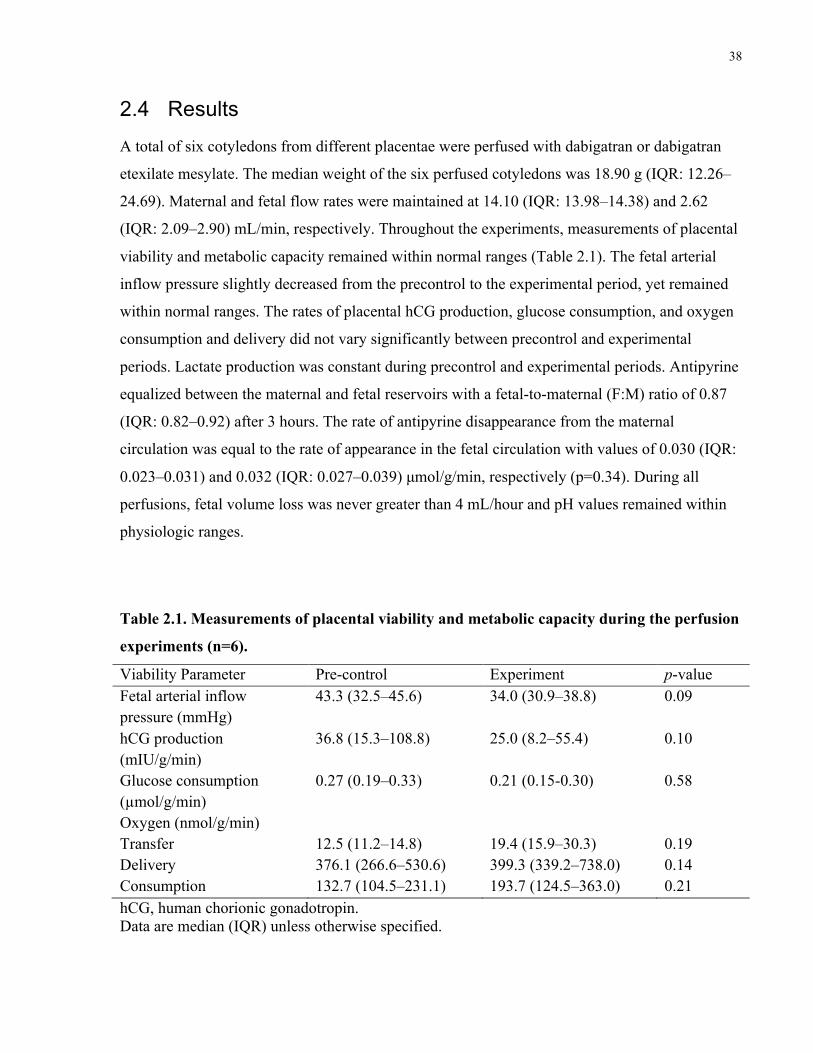
38
2.4 Results
A total of six cotyledons from different placentae were perfused with dabigatran or dabigatran
etexilate mesylate. The median weight of the six perfused cotyledons was 18.90 g (IQR: 12.26–
24.69). Maternal and fetal flow rates were maintained at 14.10 (IQR: 13.98–14.38) and 2.62
(IQR: 2.09–2.90) mL/min, respectively. Throughout the experiments, measurements of placental
viability and metabolic capacity remained within normal ranges (Table 2.1). The fetal arterial
inflow pressure slightly decreased from the precontrol to the experimental period, yet remained
within normal ranges. The rates of placental hCG production, glucose consumption, and oxygen
consumption and delivery did not vary significantly between precontrol and experimental
periods. Lactate production was constant during precontrol and experimental periods. Antipyrine
equalized between the maternal and fetal reservoirs with a fetal-to-maternal (F:M) ratio of 0.87
(IQR: 0.82–0.92) after 3 hours. The rate of antipyrine disappearance from the maternal
circulation was equal to the rate of appearance in the fetal circulation with values of 0.030 (IQR:
0.023–0.031) and 0.032 (IQR: 0.027–0.039) µmol/g/min, respectively (p=0.34). During all
perfusions, fetal volume loss was never greater than 4 mL/hour and pH values remained within
physiologic ranges.
Table 2.1. Measurements of placental viability and metabolic capacity during the perfusion
experiments (n=6).
Viability Parameter Pre-control Experiment p-value Fetal arterial inflow pressure (mmHg)
43.3 (32.5–45.6) 34.0 (30.9–38.8) 0.09
hCG production (mIU/g/min)
36.8 (15.3–108.8) 25.0 (8.2–55.4) 0.10
Glucose consumption (µmol/g/min)
0.27 (0.19–0.33) 0.21 (0.15-0.30) 0.58
Oxygen (nmol/g/min) Transfer Delivery Consumption
12.5 (11.2–14.8) 376.1 (266.6–530.6) 132.7 (104.5–231.1)
19.4 (15.9–30.3) 399.3 (339.2–738.0) 193.7 (124.5–363.0)
0.19 0.14 0.21
hCG, human chorionic gonadotropin. Data are median (IQR) unless otherwise specified.
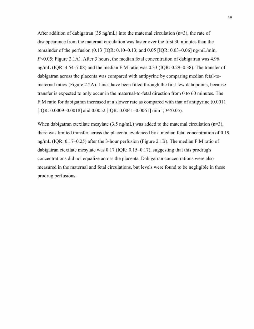
39
After addition of dabigatran (35 ng/mL) into the maternal circulation (n=3), the rate of
disappearance from the maternal circulation was faster over the first 30 minutes than the
remainder of the perfusion (0.13 [IQR: 0.10–0.13; and 0.05 [IQR: 0.03–0.06] ng/mL/min,
P<0.05; Figure 2.1A). After 3 hours, the median fetal concentration of dabigatran was 4.96
ng/mL (IQR: 4.54–7.08) and the median F:M ratio was 0.33 (IQR: 0.29–0.38). The transfer of
dabigatran across the placenta was compared with antipyrine by comparing median fetal-to-
maternal ratios (Figure 2.2A). Lines have been fitted through the first few data points, because
transfer is expected to only occur in the maternal-to-fetal direction from 0 to 60 minutes. The
F:M ratio for dabigatran increased at a slower rate as compared with that of antipyrine (0.0011
[IQR: 0.0009–0.0018] and 0.0052 [IQR: 0.0041–0.0061] min-1; P<0.05).
When dabigatran etexilate mesylate (3.5 ng/mL) was added to the maternal circulation (n=3),
there was limited transfer across the placenta, evidenced by a median fetal concentration of 0.19
ng/mL (IQR: 0.17–0.25) after the 3-hour perfusion (Figure 2.1B). The median F:M ratio of
dabigatran etexilate mesylate was 0.17 (IQR: 0.15–0.17), suggesting that this prodrug's
concentrations did not equalize across the placenta. Dabigatran concentrations were also
measured in the maternal and fetal circulations, but levels were found to be negligible in these
prodrug perfusions.
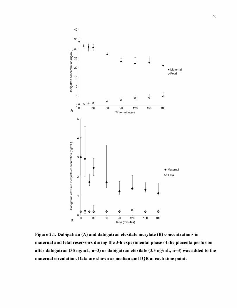
40
Figure 2.1. Dabigatran (A) and dabigatran etexilate mesylate (B) concentrations in
maternal and fetal reservoirs during the 3-h experimental phase of the placenta perfusion
after dabigatran (35 ng/mL, n=3) or dabigatran etexilate (3.5 ng/mL, n=3) was added to the
maternal circulation. Data are shown as median and IQR at each time point.
41
Figure 2.2. Fetal-to-maternal ratios for antipyrine and dabigatran (A; n=3) and dabigatran
etexilate mesylate (B; n=3) during the 3-h experimental phase of the perfusion. Regression
lines have been fitted through the first few points (0-60 min) and extended to 180 min. Data
are shown as median and IQR at each time point.
42
2.5 Discussion
The results of this perfusion study document that dabigatran crosses the term human placenta
relatively slowly, reaching a F:M ratio of 0.33 after 3 hours. The selected dabigatran
concentration of 35 ng/mL was within the therapeutic range but not high enough to theoretically
cause drug saturation of the single placental lobule in our model. The F:M ratio is reported and it
is expected that a similar F:M ratio would be achieved at higher therapeutic concentrations (150
ng/mL). Because dabigatran is administered as a prodrug, additional perfusions were conducted
with the prodrug dabigatran etexilate mesylate. Dabigatran etexilate mesylate was found to have
limited placental transfer, as evidenced by a F:M ratio of 0.16 after 3 hours. The results of the
dabigatran perfusions are of greater clinical relevance, because this is the form that reaches the
placenta through the maternal circulation, because dabigatran etexilate mesylate is rapidly
cleaved to dabigatran through plasma esterase (Eisert et al. 2010).
Dabigatran is highly polar and exhibits hydrogen bonding, both of which are factors that can
reduce transport across the placental barrier (Pacifici & Nottoli 1995). The relatively large
molecular weight of dabigatran (molecular weight 628 Da) likely also limited its transfer,
because molecules above 600 Da cross the placental barrier less readily (Pacifici & Nottoli
1995). In vivo studies have shown that P-gp may be involved in the efflux of dabigatran (Hartter
et al. 2013). Taken together, the physicochemical and pharmacokinetic properties of dabigatran
may be able to explain its limited placental transfer shown in the placenta perfusions.
The placenta expresses several efflux transporters, including P-gp and BCRP, and thus plays a
major role in determining fetal drug exposure. However, the role of placental transporters was
not analyzed in the present study. Future studies with the placental perfusion model or with cell
culture preparations can determine a potential role of placental P-gp in the disposition of
dabigatran across the human placenta.
A limitation of this model is that full-term placentae are used. As a result, it is not possible to
extrapolate our results to earlier gestational ages when placental P-gp expression is higher.
However, pregnancy is classically a state of hypercoagulability, and hemostatic changes become
more pronounced as the pregnancy progresses (Dahlman et al. 1985; Bremme 2003; O'Riordan
& Higgins 2003). There is a fivefold increase in the risk of developing venous thromboembolism
during pregnancy and postpartum as compared with nonpregnant women of childbearing age
43
(Pomp et al. 2008), suggesting that anticoagulant therapy may be required at later stages in
pregnancy and closer to term.
In conclusion, the results of this perfusion study demonstrate that dabigatran crosses the term
human placenta to some extent. Future studies will need to explore the role of placental drug
transporters, especially P-gp, in affecting this process. From a clinical perspective, these data
suggest that, pending further study, dabigatran should not be used for anticoagulation of pregnant
women, because the drug may have an adverse effect on fetal blood coagulation.
44
Chapter 3
Rivaroxaban Transfer across the Dually Perfused Isolated Human Placental Cotyledon.
Priya Bapat1,2, Leonardo SR Pinto1, Angelika Lubetsky1, Howard Berger3, Gideon Koren1,2
1 Division of Clinical Pharmacology and Toxicology, Hospital for Sick Children, 555 University
Avenue, Toronto, Ontario, M5G 1X8, Canada
2 Department of Pharmacology and Toxicology, University of Toronto, 1 King’s College Circle,
Toronto, Ontario, M5S 1A8, Canada
3 Department of Obstetrics and Gynecology, St. Michael’s Hospital, 30 Bond St, Toronto,
Ontario M5B 1W8, Canada.
Contribution of this thesis author (P. Bapat): I was part of the collaborative team to design the
experiment. I conducted the placenta perfusion experiments with technical assistance from
L.S.R. Pinto and A. Lubetsky. I performed laboratory analysis of samples to measure drug
concentrations and markers of placental viability with assistance from A. Lubetsky. I analyzed
the data and drafted the manuscript. All authors provided input and approved the final version of
the manuscript.
This work has been published and reproduced with permissions:
Bapat, P., Pinto, L. S., Lubetsky, A., Berger, H., & Koren, G. (2015). Rivaroxaban transfer
across the dually perfused isolated human placental cotyledon. Am J Obstet Gynecol,
213(5), 710.e1-710.e6.
45
Rivaroxaban transfer across the dually perfused 3isolated human placental cotyledon.
3.1 Abstract
Objective: The purpose of this study was to determine the rate and extent of rivaroxaban transfer
across the term human placenta and determine whether passive diffusion was the primary
mechanism involved in this transfer. Study Design: The transplacental pharmacokinetics of
rivaroxaban was determined with the ex vivo placenta perfusion model. Rivaroxaban was added
to the maternal or fetal circulation only (250 ng/mL). Additional experiments were conducted
under equilibrative conditions with the addition of rivaroxaban to both the maternal and fetal
circulations (250 ng/mL). Rivaroxaban concentrations were measured with the use of liquid
chromatography-tandem mass spectrometry. Results: There was rapid transfer of rivaroxaban
across the human placenta in both the maternal-to-fetal and fetal-to-maternal directions, as
evidenced by transfer ratios of 0.69 (IQR: 0.58-0.73; n = 5) and 0.69 (IQR: 0.67-0.71; n = 2),
respectively, after 3 hours. Under equilibrative conditions (n = 2), rivaroxaban concentrations
remained relatively constant, which suggests that rivaroxaban crosses the placenta down a
concentration gradient. Conclusion: This is the first direct evidence of rivaroxaban transfer
across the term human placenta from both the mother-to-fetus and fetus-to-mother directions.
Our results document that unbound rivaroxaban rapidly crosses the placental barrier via passive
diffusion. However, because rivaroxaban is highly bound to plasma proteins (up to 95%), this
suggests that the amount of unbound drug that may reach the fetus is likely much lower.
Additional studies will need to explore its safety before administering rivaroxaban to a pregnant
woman.
46
3.2 Introduction
Anticoagulant therapy is required in some high-risk pregnancies for the prevention and treatment
of venous thromboembolism (VTE) in women at high or intermediate risk for thromboembolic
disease; for the prevention and treatment of systemic embolism in women with mechanical heart
valves; and, for the prevention of pregnancy-related complications in women with
antiphospholipid antibody syndrome (Royal College of Obstetricians and Gynaecologists 2009;
Bates et al. 2012). A retrospective cohort study reported that VTE affected 28 per 100,000
pregnancies antepartum, and 65 per 100,000 postpartum (Simpson et al. 2001). Although current
guidelines recommend the use of unfractionated or low-molecular weight heparins (LMWH)
(Bates et al. 2012), a recent open-label, randomized trial comparing antepartum prophylactic
dalteparin (LMWH) with no dalteparin found that dalteparin did not reduce the occurrence of
VTE, pregnancy loss, or placenta-mediated pregnancy complications, including severe pre-
eclampsia, small-for-gestational-age infants, and placental abruption, in pregnant women with
thrombophilia who were at high risk of these complications (Rodger et al. 2014). Similar to other
drugs prescribed during pregnancy, the use of anticoagulant therapy can be challenging because
of the possibility for both maternal and fetal complications.
Rivaroxaban (Xarelto®) is a new oral anticoagulant, with low solubility and high membrane
permeability, which is increasingly being prescribed to treat deep vein thrombosis (DVT) and
pulmonary embolism (PE) (Bayer Canada 2013). Following oral administration, rivaroxaban is
rapidly absorbed and remains unchanged in plasma with no major or active circulating
metabolites (Weinz et al. 2009). Rivaroxaban directly inhibits Factor Xa, which is an important
component of both the intrinsic and extrinsic pathways of the coagulation cascade (Hoffman &
Monroe 2007). Since factor Xa catalyzes the conversion of prothrombin (Factor II) to thrombin
(Factor IIa) (Spyropoulos 2007), it is an ideal target for anticoagulation therapy.
The objectives of our study were to determine its transplacental pharmacokinetics at term, in
order to estimate fetal drug exposure, and determine if passive diffusion is the primary
mechanism involved in rivaroxaban transfer across the human placenta.
47
3.3 Materials and Methods
3.3.1 Ex vivo perfusion of human placental cotyledon
The dual perfusion of a single placental cotyledon has previously been described by Miller et al
(Miller et al. 1985) and adapted for use in our laboratory (Derewlany et al. 1991; Pollex et al.
2008). The study was approved by the research ethics board at St Michael’s Hospital in Toronto,
Ontario, Canada, and mothers gave written consent prior to delivery. A single experiment
consisted of a 1-hour control period, followed by a 3-hour experimental period. A full
description of this method is presented in section 2.3.
3.3.2 Experimental period
Rivaroxaban was added to the maternal or fetal reservoir only, or simultaneously to both the
maternal and fetal reservoirs at a therapeutic concentration of 250 ng/mL at the start of the
experimental period. Samples were collected from the maternal and fetal reservoirs for analysis
of rivaroxaban, O2 and CO2 content, pH, glucose consumption, and lactate production at 0, 10,
20, 30, 60, 90, 120, 150 and 180 min. Additional samples were taken directly from the maternal
and fetal reservoirs every 30 minutes for analysis of hCG secretion and antipyrine transfer.
3.3.3 Sample analysis
Perfusate samples were stored at –20°C until analysis. Antipyrine concentrations were analyzed
using a UV-visible recording spectrophotometer W-160 (Shimadzu, Tokyo, Japan) at 350 nm.
HCG levels were determined using an ELISA kit (Alpha Diagnostic International, San Antonio,
TX) and a Biotek Synergy HT microplate reader (Biotek instruments, Winooski, VT) at 450 nm.
Rivaroxaban analysis was performed by the Analytical Facility for Bioactive Molecules of the
Centre for the Study of Complex Childhood Diseases at the Hospital for Sick Children, Toronto,
Canada. A standard curve ranging from 0–500 ng/mL for rivaroxaban (Cedarlane, Burlington,
ON, Canada) was prepared in maternal or fetal perfusate. Mobile phases were (A) 90% water
and 10% acetonitrile and (B) 10% water and 90% acetonitrile, with both consisting of 5 mM
ammonium formate (pH 3.2). All samples and standards were vortexed and centrifuged at 13 000
x g for 5 minutes at 4°C. 50 µL of standards or samples were added to 450 µL of the mobile
phase A containing 10 ng/mL internal standard rivaroxaban-d4 (CacheSyn, Mississauga,
Canada). Sample analysis was performed on an AB Sciex QTrap 5500 and Agilent 1290 UHPLC
48
system. 2 µL of sample or standard were injected at a flow rate of 500 µL/min through a Kinetex
XB-C18 column (50 x 3.0 mm, 2.6 µm)(Phenomenex, Torrance, CA, USA) isocratically in 1.6
min using a mobile phase composition of 60% A and 40% B.
The mass spectrometer was operated in positive electrospray ionization mode with a source
temperature of 600°C and an internal standard voltage setting of 5,500. Precursor-to-product ion
mass transitions were established by standard infusions. Rivaroxaban concentrations were
acquired by multiple reaction monitoring mode using transitions 436.1→145 m/z and
436.1→231.1 m/z for rivaroxaban and 440.2→145 m/z and 440.2→235.25 m/z for rivaroxaban-
d4. The range of rivaroxaban quantification was 0.5–500 ng/mL. Data analysis and peak
integration were performed using Analyst 1.6 software from ABSciex. Sample concentrations
were calculated by plotting peak area ratios (analyte/internal standard) against calibration curves
of extracted matrix spiked standards.
Using the area under the curve (AUC) of rivaroxaban from the placenta perfusions, physiological
rivaroxaban concentrations were simulated with 95% protein binding to estimate maternal and
fetal exposure.
3.3.4 Statistical analysis
All data are presented as median and interquartile range unless stated otherwise and comparisons
between pre-experimental and experimental phases were analyzed using a Wilcoxon sign rank
test for non-normal data.
3.4 Results
A total of 9 cotyledons from different placentae were perfused with rivaroxaban and the physical
parameters for the perfusions are shown in Table 3.1. All successfully perfused placentae were
collected from caesarian deliveries. The (mean ± SD) maternal age was 33.0 ± 6.0 years, with a
gestational age of 39.0 ± 0.4 weeks. The mean weight of the perfused cotyledons was 23.2 ± 7.9
g. Maternal and fetal flow rates were 14.1 ± 0.2 and 2.4 ± 0.3 mL/min, respectively. Measures of
placental viability, integrity, and function remained within normal ranges and were not
significantly different between the pre-experimental and experimental periods (Table 3.1).
Lactate production remained constant for the duration of the perfusion. For all perfusions,
antipyrine equalized between the maternal and fetal reservoirs after 3 hours with a final fetal-to-
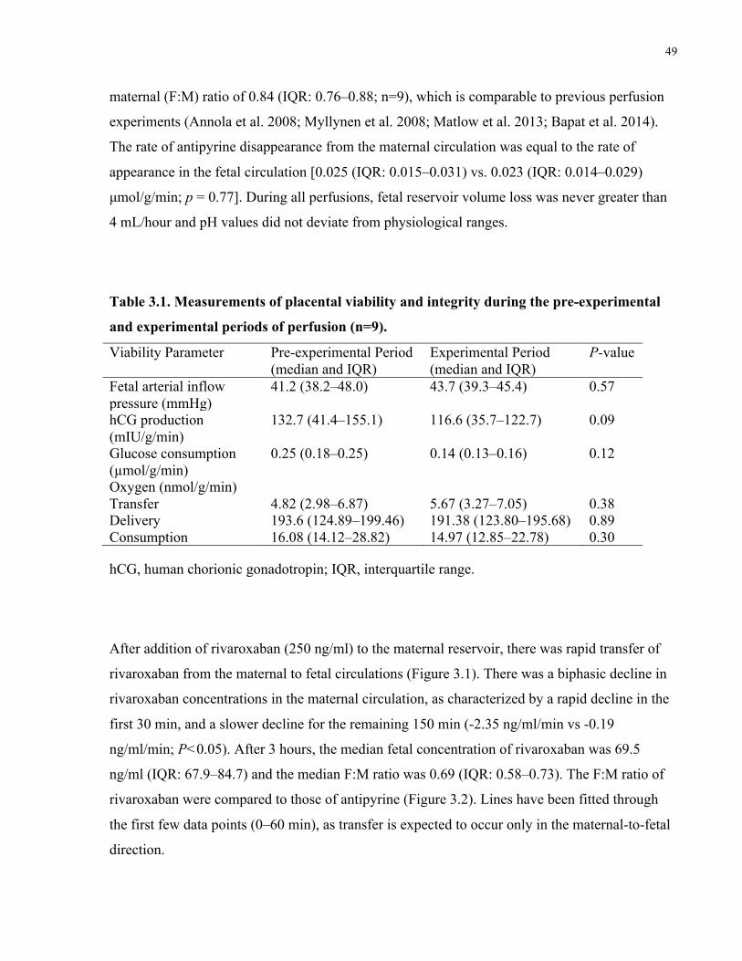
49
maternal (F:M) ratio of 0.84 (IQR: 0.76–0.88; n=9), which is comparable to previous perfusion
experiments (Annola et al. 2008; Myllynen et al. 2008; Matlow et al. 2013; Bapat et al. 2014).
The rate of antipyrine disappearance from the maternal circulation was equal to the rate of
appearance in the fetal circulation [0.025 (IQR: 0.015–0.031) vs. 0.023 (IQR: 0.014–0.029)
µmol/g/min; p = 0.77]. During all perfusions, fetal reservoir volume loss was never greater than
4 mL/hour and pH values did not deviate from physiological ranges.
Table 3.1. Measurements of placental viability and integrity during the pre-experimental
and experimental periods of perfusion (n=9).
Viability Parameter Pre-experimental Period (median and IQR)
Experimental Period (median and IQR)
P-value
Fetal arterial inflow pressure (mmHg)
41.2 (38.2–48.0) 43.7 (39.3–45.4) 0.57
hCG production (mIU/g/min)
132.7 (41.4–155.1) 116.6 (35.7–122.7) 0.09
Glucose consumption (µmol/g/min)
0.25 (0.18–0.25) 0.14 (0.13–0.16) 0.12
Oxygen (nmol/g/min) Transfer Delivery Consumption
4.82 (2.98–6.87) 193.6 (124.89–199.46) 16.08 (14.12–28.82)
5.67 (3.27–7.05) 191.38 (123.80–195.68) 14.97 (12.85–22.78)
0.38 0.89 0.30
hCG, human chorionic gonadotropin; IQR, interquartile range.
After addition of rivaroxaban (250 ng/ml) to the maternal reservoir, there was rapid transfer of
rivaroxaban from the maternal to fetal circulations (Figure 3.1). There was a biphasic decline in
rivaroxaban concentrations in the maternal circulation, as characterized by a rapid decline in the
first 30 min, and a slower decline for the remaining 150 min (-2.35 ng/ml/min vs -0.19
ng/ml/min; P<0.05). After 3 hours, the median fetal concentration of rivaroxaban was 69.5
ng/ml (IQR: 67.9–84.7) and the median F:M ratio was 0.69 (IQR: 0.58–0.73). The F:M ratio of
rivaroxaban were compared to those of antipyrine (Figure 3.2). Lines have been fitted through
the first few data points (0–60 min), as transfer is expected to occur only in the maternal-to-fetal
direction.
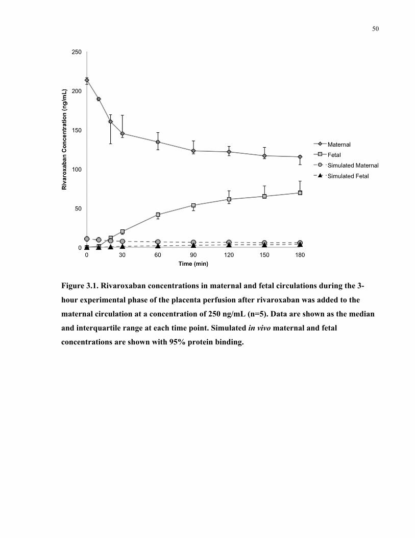
50
Figure 3.1. Rivaroxaban concentrations in maternal and fetal circulations during the 3-
hour experimental phase of the placenta perfusion after rivaroxaban was added to the
maternal circulation at a concentration of 250 ng/mL (n=5). Data are shown as the median
and interquartile range at each time point. Simulated in vivo maternal and fetal
concentrations are shown with 95% protein binding.
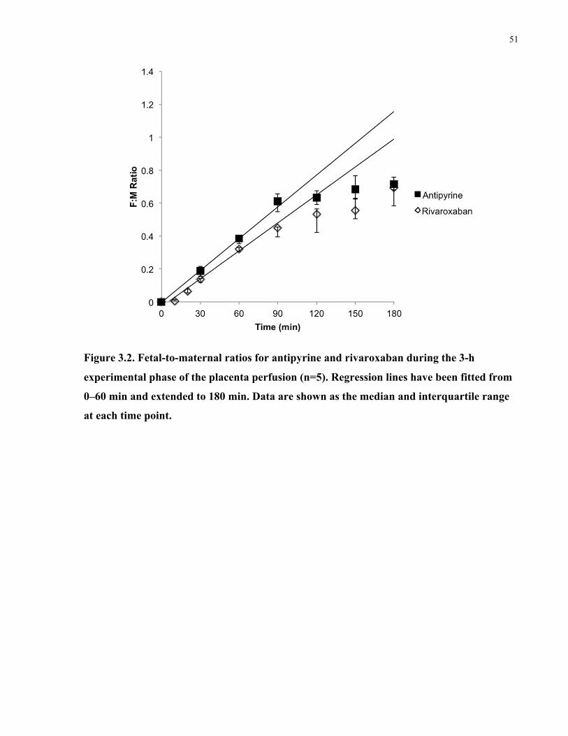
51
Figure 3.2. Fetal-to-maternal ratios for antipyrine and rivaroxaban during the 3-h
experimental phase of the placenta perfusion (n=5). Regression lines have been fitted from
0–60 min and extended to 180 min. Data are shown as the median and interquartile range
at each time point.
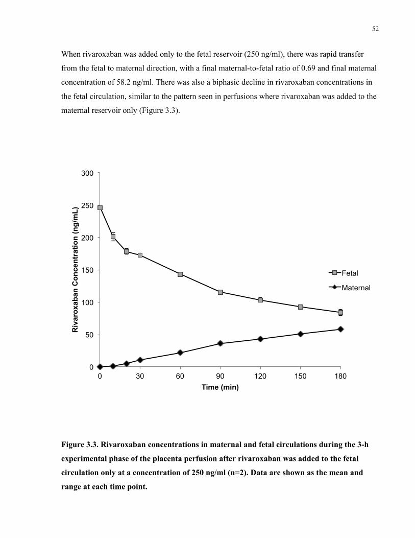
52
When rivaroxaban was added only to the fetal reservoir (250 ng/ml), there was rapid transfer
from the fetal to maternal direction, with a final maternal-to-fetal ratio of 0.69 and final maternal
concentration of 58.2 ng/ml. There was also a biphasic decline in rivaroxaban concentrations in
the fetal circulation, similar to the pattern seen in perfusions where rivaroxaban was added to the
maternal reservoir only (Figure 3.3).
Figure 3.3. Rivaroxaban concentrations in maternal and fetal circulations during the 3-h
experimental phase of the placenta perfusion after rivaroxaban was added to the fetal
circulation only at a concentration of 250 ng/ml (n=2). Data are shown as the mean and
range at each time point.
0
50
100
150
200
250
300
0 30 60 90 120 150 180
Riv
arox
aban
Con
cent
ratio
n (n
g/m
L)
Time (min)
Fetal
Maternal
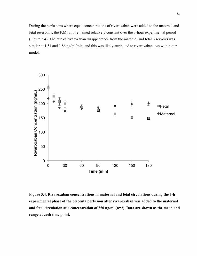
53
During the perfusions where equal concentrations of rivaroxaban were added to the maternal and
fetal reservoirs, the F:M ratio remained relatively constant over the 3-hour experimental period
(Figure 3.4). The rate of rivaroxaban disappearance from the maternal and fetal reservoirs was
similar at 1.51 and 1.86 ng/ml/min, and this was likely attributed to rivaroxaban loss within our
model.
Figure 3.4. Rivaroxaban concentrations in maternal and fetal circulations during the 3-h
experimental phase of the placenta perfusion after rivaroxaban was added to the maternal
and fetal circulation at a concentration of 250 ng/ml (n=2). Data are shown as the mean and
range at each time point.
0
50
100
150
200
250
300
0 30 60 90 120 150 180
Riv
arox
aban
Con
cent
ratio
n (n
g/m
L)
Time (min)
Fetal
Maternal
54
3.5 Discussion
The results of this perfusion study show that rivaroxaban rapidly crosses the term human
placenta, reaching a F:M ratio of 0.69 after 3 hours. The final F:M ratios of rivaroxaban and
antipyrine are likely indicative of steady state, as these ratios remained relatively constant after
120 min (Figure 3.2), suggesting that net maternal-to-fetal transfer was no longer increasing.
The perfusions in which rivaroxaban was added to the fetal reservoir only were important to
elucidate the role of the placenta in clearing the drug from the fetal circulation. The final transfer
ratio in these perfusions was similar to that of the perfusions where rivaroxaban was added to the
maternal circulation only, suggesting that rivaroxaban interacts with the placental barrier in a
similar fashion in both directions across the placenta. Under equilibrative conditions,
rivaroxaban concentrations remained relatively constant, suggesting that rivaroxaban crosses the
placenta down a concentration gradient. The low molecular weight of rivaroxaban (436 Da) also
supports the notion that rivaroxaban likely crosses the placenta via passive diffusion.
A limitation of the placenta perfusion model is that only full-term placentae are used. As a result,
we cannot make generalizations to earlier gestational ages when placental structure is vastly
different. Another important consideration with the placenta perfusion is that this model does not
fully account for non-placental pharmacokinetic factors, including protein binding, clearance
rate, and distribution in the maternal and fetal compartments. Protein binding is especially
important since rivaroxaban is highly bound to plasma proteins (92–95% bound, mainly
albumin) (Bayer Canada 2013). Differences in maternal and fetal plasma protein concentrations
can affect the transplacental transfer of drugs that are highly bound. Throughout pregnancy, fetal
albumin levels tend to increase, with the fetal-to-maternal albumin ratio increasing from 0.28 in
the first trimester to 1.20 closer to term (Krauer et al. 1984). As well, the increased concentration
of free fatty acids in the maternal circulation toward term can displace drugs for the binding spot
on albumin (Ridd et al. 1983; Nau et al. 1984). This leads to increased binding of a drug to
albumin in the fetal circulation compared to the maternal circulation, which can, in turn, lead to
increased placental drug transfer and fetal drug accumulation since only the unbound form of the
drug can equilibrate across the placental barrier. Assuming plasma protein binding of
rivaroxaban at 95%, the fetal exposure to rivaroxaban is predicted to be only a small fraction of
what is shown in the perfusion of the unbound drug (Figure 3.1).
55
Therefore, it is likely that fetal levels in vivo will be substantially lower than the values obtained
in our perfusion experiments, as a majority of the drug will remain bound to serum albumin in
the maternal circulation. The proportion of rivaroxaban that is bound and unbound may be
slightly different in maternal and fetal circulations, as compared to non-pregnant adults due to
differences in albumin levels (Nau et al. 1984). Perfusion experiments that include proteins in the
perfusion medium are technically challenging due to the high viscosity of the perfusate. As well,
when trying to mimic physiological conditions, there are numerous proteins and endogenous
factors that can affect binding in vivo (Johnson et al. 1999; Schenker et al. 1999; Gavard et al.
2006; Nanovskaya et al. 2009; Hutson et al. 2011).
During the experiments, several parameters were measured to monitor placental viability and
integrity. These parameters included arterial inflow pressure in the fetal circulation, pH, oxygen
consumption, net fetal oxygen transfer, glucose consumption, lactate production, and secretion of
human chorionic gonadotropin (hCG). As well, it has been suggested that fetal volume loss is the
optimal measure of tissue viability (Mathiesen et al. 2010; Hutson et al. 2011). A loss up to 4
ml/hr from the fetal reservoir is typically accepted (Mathiesen et al. 2010). Antipyrine, which is a
flow-limited marker that only undergoes passive diffusion, can also be used to measure tissue
integrity. While the model cannot fully incorporate non-placental pharmacokinetic factors such
as protein binding and liver metabolism, the placenta perfusion is the only experimental model
that can be used to study human placental transfer of substances in organized placental tissue and
may be able to more reliably predict fetal exposure compared to other experimental methods
(Hutson et al. 2011). While animal studies can be very useful, they cannot always be easily
extrapolated to humans because the placenta is the most species-specific mammalian organ
(Carter 2007).
Previous studies have shown that rivaroxaban is a substrate for P-glycoprotein (P-gp) in vitro and
in vivo (Lang et al. 2009; Gnoth et al. 2011). P-gp generally has a unidirectional effect on
transcellular transport, and in the placenta, P-gp effluxes substrates from the fetal to the maternal
side. Since the final transfer ratios were similar when rivaroxaban was added to the maternal or
fetal circulation alone, it is unlikely that placental P-gp has a major role on the transplacental
disposition of rivaroxaban at term.
56
A case report was recently published describing the use of rivaroxaban during the first and part
of the second trimester of pregnancy (Konigsbrugge et al. 2014). A male infant was born
spontaneously in the 40th gestational week without complications. The baby birth measurements
were in the 13th, 30th, and 7th percentiles for weight, length, and cranial circumference,
respectively, but the authors likely attributed the low growth percentiles to maternal smoking.
Another study measured thrombin generation and activated partial thromboplastin time in adult
plasma and neonatal cord samples with increasing concentrations of rivaroxaban (Novak et al.
2011). This article was useful in showing that rivaroxaban exerts similar hemostatic effects in
neonatal cord and adult plasma.
In conclusion, this is the first direct evidence of rivaroxaban transfer across the human placenta.
Our results document that unbound rivaroxaban rapidly crosses the term human placenta.
However, since rivaroxaban is highly bound to plasma proteins (up to 95%) with an elimination
half-life of 5–9 hours, the likely amount of unbound drug that may reach the fetus is relatively
small. Future studies will need to explore safety before administering rivaroxaban to a pregnant
woman.
57
Chapter 4
Examining the Transplacental Passage of Apixaban Using the Dually Perfused Human Placenta.
Priya Bapat1,2, Leonardo SR Pinto1, Angelika Lubetsky1, Katarina Aleksa1, Howard Berger3,
Gideon Koren1,2, Shinya Ito1,2
1 Division of Clinical Pharmacology and Toxicology, Hospital for Sick Children, 555 University
Avenue, Toronto, Ontario, M5G 1X8, Canada
2 Department of Pharmacology and Toxicology, University of Toronto, 1 King’s College Circle,
Toronto, Ontario, M5S 1A8, Canada
3 Department of Obstetrics and Gynecology, St. Michael’s Hospital, 30 Bond St, Toronto,
Ontario M5B 1W8, Canada.
Contribution of this thesis author (P. Bapat): I was part of the collaborative team to design the
experiment. I conducted the placenta perfusion experiments with technical assistance from
L.S.R. Pinto and A. Lubetsky. I performed laboratory analysis of samples to measure drug
concentrations and markers of placental viability with assistance from A. Lubetsky and K.
Aleksa. I analyzed the data and drafted the manuscript. All authors provided input and approved
the final version of the manuscript.
This work has been published and reproduced with permissions:
Bapat, P., Pinto, L. S., Lubetsky, A., Aleksa, K., Berger, H., Koren, G., & Ito S. (2016).
Examining the transplacental passage of apixaban using the dually perfused human
placenta. J Thromb Haemost [Epub ahead of print].
58
Examining the transplacental passage of apixaban 4using the dually perfused human placenta.
4.1 Abstract
Background: Apixaban is a novel oral anticoagulant that is increasingly being prescribed to
women of reproductive age. However, information regarding its placental transfer is non-
existent. Objective: To determine the rate and extent of placental transfer of apixaban, using the
human placenta ex vivo. Methods: Placentae collected after caesarean or vaginal delivery of
healthy term infants were perfused in the respective maternal and fetal circulation. At the start of
the experiment, apixaban was added to the maternal circulation at a concentration of 150 ng/ml,
and samples from maternal and fetal reservoirs were collected over 3 hours. Results: There was
a rapid decline of apixaban in the maternal compartment, followed by emergence in the fetal
compartment with a median fetal-to-maternal drug concentration ratio of 0.77 (IQR: 0.76–0.81)
and fetal concentration of 39.0 ng/mL (IQR: 36.8–40.6) after 3 hours (n=5). The perfusion
results were subsequently adjusted to account for differences in the concentration of plasma
proteins in maternal and fetal blood, as apixaban remains highly bound to albumin and alpha-1
acid glycoprotein. After the adjustment, the predicted fetal-to-maternal ratio of total (bound plus
unbound) apixaban concentrations in vivo ranged from 0.35 to 0.90. Conclusions: We conclude
that unbound apixaban rapidly crosses from maternal to fetal circulation. We further predict that
total apixaban concentrations in cord blood in vivo are 35-90% of the corresponding maternal
levels, suggesting that apixaban could have a possible adverse effect on fetal and neonatal
coagulation.
59
4.2 Introduction
Even in pregnancy, anticoagulant therapy is often indicated for women with conditions such as
venous thromboembolic events (VTE), atrial fibrillation, and artificial heart valves (Royal
College of Obstetricians and Gynaecologists 2009; Bates et al. 2012). In particular, VTE,
including deep vein thrombosis (DVT) and pulmonary embolism (PE), are a major cause of
maternal morbidity and mortality during pregnancy or after delivery. The risk for VTE in
pregnant women is 4–5 times higher than the risk among non-pregnant women of the same age
(Conti et al. 2014). Vitamin K antagonists are contraindicated in pregnancy because of their
potential to cause adverse teratogenic effects (Hall et al. 1980; Iturbe-Alessio et al. 1986;
Ginsberg et al. 1989; Schaefer et al. 2006).
Apixaban (Eliquis®) is a novel oral anticoagulant (NOAC) that is increasingly used for the
prevention and treatment of venous thromboembolic events, including DVT and PE. Apixaban is
also prescribed for the prevention of stroke and systemic embolism in patients with atrial
fibrillation. The drug is rapidly absorbed with peak concentrations appearing 3–4 hours after
tablet intake, and unchanged apixaban is the major component in human plasma with no active
circulating metabolites (Pfizer Canada 2015). In human plasma, apixaban is highly bound to
plasma proteins (87–93%) (Pfizer Canada 2015). Apixaban is a reversible, direct and highly
selective inhibitor of free and prothrombinase-bound Factor Xa (Jiang et al. 2009). Factor Xa is
activated via the intrinsic and extrinsic pathways of the coagulation cascade, and its inhibition
prevents thrombin generation and the subsequent formation of a thrombus. Importantly, when
compared to other NOACs (including dabigatran, rivaroxaban, and edoxaban), apixaban is
associated with a relatively low risk of bleeding events while showing similar efficacy for the
treatment of acute VTE (Mantha & Ansell 2015).
To date, no studies have examined the safety of apixaban in pregnant patients and its placental
transfer in humans is currently unknown. As prescription rates for NOACs continue to increase
(Desai et al. 2014), the number of women with an unplanned pregnancy who are taking NOACs
will likely increase. Therefore, the objective of our study was to examine the disposition of
apixaban across the term human placenta ex vivo, in order to estimate fetal drug exposure.
60
4.3 Methods
4.3.1 Dual perfusion of human placental cotyledon ex vivo
The dual perfusion of an isolated placental cotyledon ex vivo is a validated method to examine
placental transfer of drug, as previously described by Miller et al (1985) and adapted for use in
our laboratory (Derewlany et al. 1991; Bapat et al. 2014, 2015). This study was approved by the
research ethics boards at St. Michael’s Hospital and The Hospital for Sick Children in Toronto,
Ontario Canada, and this research was conducted in accordance with the Declaration of Helsinki.
Women provided written consent prior to delivery at St. Michael’s Hospital, and term placentae
were collected immediately following elective cesarean or vaginal delivery of healthy singleton
pregnancies (>38 weeks). We excluded placentae from women who were taking any medication
during pregnancy or from women who had any disease during pregnancy including, but not
limited to, hypertension, diabetes, thyroid dysfunction, cancer and infection (e.g.,
chorioamnionitis and TORCH infections, or positive anti-HIV, anti-HBV, or anti-HCV
serology). A single experiment consisted of a 1-hour control period, followed by a 3-hour
experimental period. A full description of this method is presented in section 2.3.
At the beginning of experimental period, the maternal and fetal reservoirs were replaced with
fresh perfusate, and apixaban was added to the maternal circulation at a concentration of 150
ng/mL, which is in the range of therapeutic plasma concentrations (Frost et al. 2013a, b).
Samples were collected from the maternal and fetal reservoirs at 0, 10, 20, 30, 60, 90, 120, 150
and 180 min for measurement of apixaban levels, O2 and CO2 content, pH, glucose consumption,
lactate production, antipyrine transfer and hCG secretion. Samples were stored at –20°C until
analysis.
4.3.2 Sample analysis
Antipyrine concentrations were determined using a UV-visible recording spectrophotometer W-
160 (Shimadzu, Tokyo, Japan) set at 350 nm. HCG levels were determined using an ELISA kit
(Alpha Diagnostic International, San Antonio, TX) and a Biotek Synergy HT microplate reader
(Biotek instruments, Winooski, VT) at 450 nm.
A standard curve ranging from 0 to 300 ng/mL for apixaban (AK Scientific Inc, Union City, CA,
USA) was prepared in maternal or fetal perfusate. The internal standard, [13C,2H7]-apixaban, was
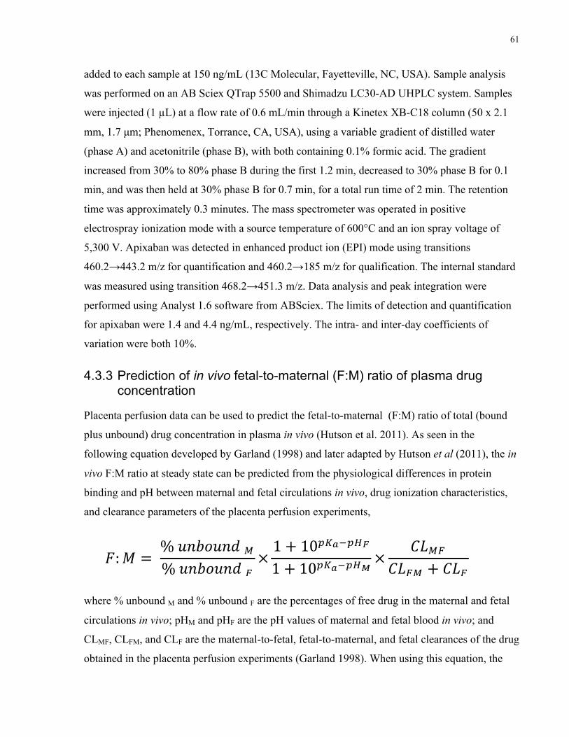
61
added to each sample at 150 ng/mL (13C Molecular, Fayetteville, NC, USA). Sample analysis
was performed on an AB Sciex QTrap 5500 and Shimadzu LC30-AD UHPLC system. Samples
were injected (1 µL) at a flow rate of 0.6 mL/min through a Kinetex XB-C18 column (50 x 2.1
mm, 1.7 µm; Phenomenex, Torrance, CA, USA), using a variable gradient of distilled water
(phase A) and acetonitrile (phase B), with both containing 0.1% formic acid. The gradient
increased from 30% to 80% phase B during the first 1.2 min, decreased to 30% phase B for 0.1
min, and was then held at 30% phase B for 0.7 min, for a total run time of 2 min. The retention
time was approximately 0.3 minutes. The mass spectrometer was operated in positive
electrospray ionization mode with a source temperature of 600°C and an ion spray voltage of
5,300 V. Apixaban was detected in enhanced product ion (EPI) mode using transitions
460.2→443.2 m/z for quantification and 460.2→185 m/z for qualification. The internal standard
was measured using transition 468.2→451.3 m/z. Data analysis and peak integration were
performed using Analyst 1.6 software from ABSciex. The limits of detection and quantification
for apixaban were 1.4 and 4.4 ng/mL, respectively. The intra- and inter-day coefficients of
variation were both 10%.
4.3.3 Prediction of in vivo fetal-to-maternal (F:M) ratio of plasma drug concentration
Placenta perfusion data can be used to predict the fetal-to-maternal (F:M) ratio of total (bound
plus unbound) drug concentration in plasma in vivo (Hutson et al. 2011). As seen in the
following equation developed by Garland (1998) and later adapted by Hutson et al (2011), the in
vivo F:M ratio at steady state can be predicted from the physiological differences in protein
binding and pH between maternal and fetal circulations in vivo, drug ionization characteristics,
and clearance parameters of the placenta perfusion experiments,
where % unbound M and % unbound F are the percentages of free drug in the maternal and fetal
circulations in vivo; pHM and pHF are the pH values of maternal and fetal blood in vivo; and
CLMF, CLFM, and CLF are the maternal-to-fetal, fetal-to-maternal, and fetal clearances of the drug
obtained in the placenta perfusion experiments (Garland 1998). When using this equation, the
!!! ! !!!!"#$!"%!!!!!"#$!"%!! !!! !"!"!!!"!!! !"!"!!!"! !
!"!"!"!" ! !"!
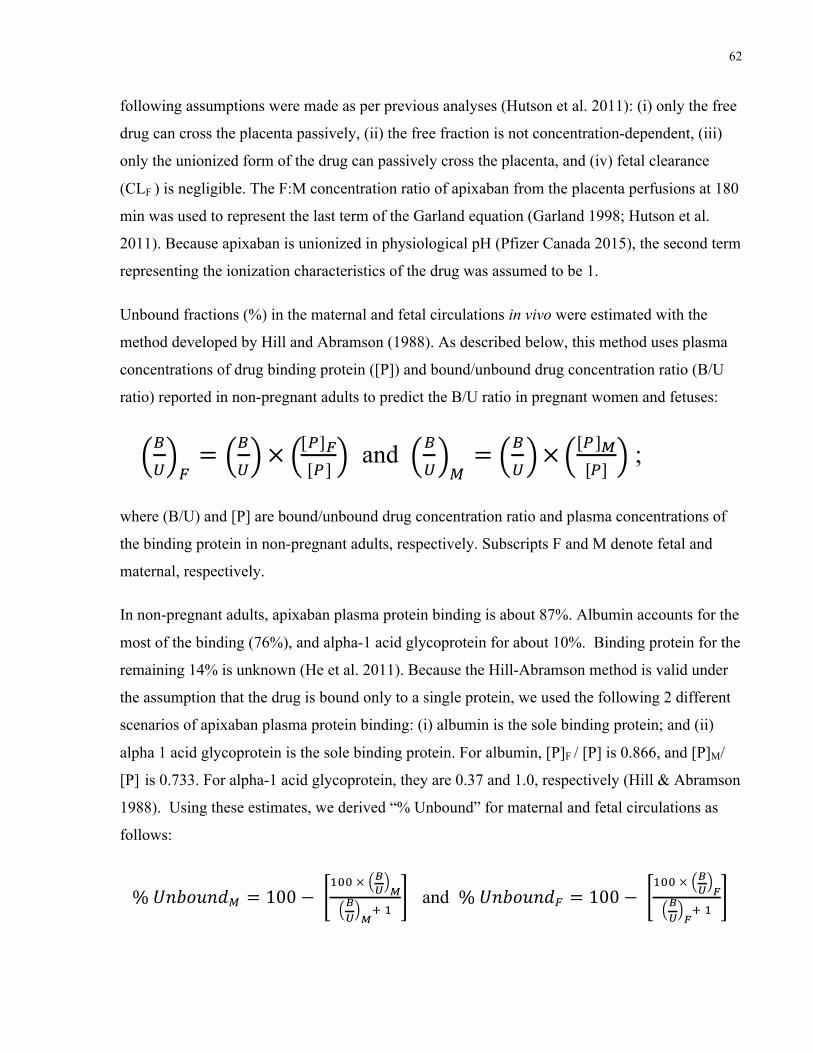
62
following assumptions were made as per previous analyses (Hutson et al. 2011): (i) only the free
drug can cross the placenta passively, (ii) the free fraction is not concentration-dependent, (iii)
only the unionized form of the drug can passively cross the placenta, and (iv) fetal clearance
(CLF ) is negligible. The F:M concentration ratio of apixaban from the placenta perfusions at 180
min was used to represent the last term of the Garland equation (Garland 1998; Hutson et al.
2011). Because apixaban is unionized in physiological pH (Pfizer Canada 2015), the second term
representing the ionization characteristics of the drug was assumed to be 1.
Unbound fractions (%) in the maternal and fetal circulations in vivo were estimated with the
method developed by Hill and Abramson (1988). As described below, this method uses plasma
concentrations of drug binding protein ([P]) and bound/unbound drug concentration ratio (B/U
ratio) reported in non-pregnant adults to predict the B/U ratio in pregnant women and fetuses:
where (B/U) and [P] are bound/unbound drug concentration ratio and plasma concentrations of
the binding protein in non-pregnant adults, respectively. Subscripts F and M denote fetal and
maternal, respectively.
In non-pregnant adults, apixaban plasma protein binding is about 87%. Albumin accounts for the
most of the binding (76%), and alpha-1 acid glycoprotein for about 10%. Binding protein for the
remaining 14% is unknown (He et al. 2011). Because the Hill-Abramson method is valid under
the assumption that the drug is bound only to a single protein, we used the following 2 different
scenarios of apixaban plasma protein binding: (i) albumin is the sole binding protein; and (ii)
alpha 1 acid glycoprotein is the sole binding protein. For albumin, [P]F / [P] is 0.866, and [P]M/
[P] is 0.733. For alpha-1 acid glycoprotein, they are 0.37 and 1.0, respectively (Hill & Abramson
1988). Using these estimates, we derived “% Unbound” for maternal and fetal circulations as
follows:
!! !
! !! ! ! !
! and !! !
! !! ! ! !
! ;
!!!"#$%"&! ! !""! !!""!!! !! !
!! !
!!! and !!!"#$%"&! ! !""! !
!""!!! !! !!! !
!!!
63
4.3.4 Statistical analysis
All data are presented as median and interquartile range (IQR) unless stated otherwise and
comparisons between pre-experimental and experimental phases were analyzed using a
Wilcoxon sign rank test for non-normal data. Any p-values less than 0.05 were considered to be
statistically significant.
4.4 Results
Of the 25 screened and retrieved placentae, five cotyledons from five different placentae were
successfully perfused with apixaban. Four of the perfused placentae were obtained from
caesarian deliveries, and one from a vaginal delivery. The (mean ± SD) maternal age was 33.4 ±
6.5 years, with a gestational age of 39.6 ± 0.9 weeks. The weight of the perfused cotyledons was
24.21 ± 10.32 g. Maternal and fetal flow rates were 12.84 ± 0.40 and 2.63 ± 0.40 mL/min,
respectively. Markers of placenta function and viability remained within normal physiological
ranges and did not significantly change between the pre-experimental and experimental periods
(Table 4.1). Lactate production remained also constant for the duration of the perfusion,
indicating integrity of the perfused placentae. Antipyrine concentrations equilibrated across the
placenta with a F:M concentration ratio of 0.96 (IQR: 0.86–0.99) after 3 hours, further indicating
the integrity of the fetal capillary bed. During all experiments, pH values did not deviate from
physiological ranges (maternal = 7.4; fetal = 7.35) (Reynolds & Knott 1989), and the volume
loss in the fetal reservoir was never greater than 4 mL/hour. These parameters were all within the
accepted ranges of the quality control measures.
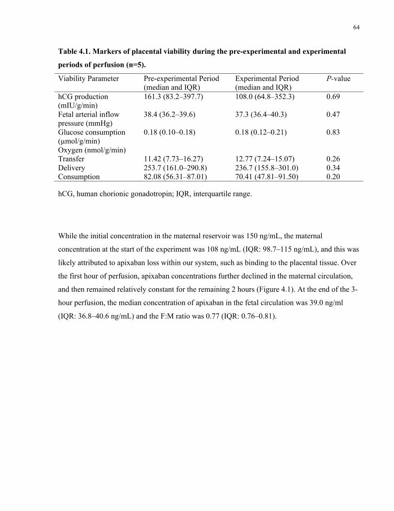
64
Table 4.1. Markers of placental viability during the pre-experimental and experimental
periods of perfusion (n=5).
Viability Parameter Pre-experimental Period (median and IQR)
Experimental Period (median and IQR)
P-value
hCG production (mIU/g/min)
161.3 (83.2–397.7) 108.0 (64.8–352.3) 0.69
Fetal arterial inflow pressure (mmHg)
38.4 (36.2–39.6) 37.3 (36.4–40.3) 0.47
Glucose consumption (µmol/g/min)
0.18 (0.10–0.18) 0.18 (0.12–0.21) 0.83
Oxygen (nmol/g/min) Transfer Delivery Consumption
11.42 (7.73–16.27) 253.7 (161.0–290.8) 82.08 (56.31–87.01)
12.77 (7.24–15.07) 236.7 (155.8–301.0) 70.41 (47.81–91.50)
0.26 0.34 0.20
hCG, human chorionic gonadotropin; IQR, interquartile range.
While the initial concentration in the maternal reservoir was 150 ng/mL, the maternal
concentration at the start of the experiment was 108 ng/mL (IQR: 98.7–115 ng/mL), and this was
likely attributed to apixaban loss within our system, such as binding to the placental tissue. Over
the first hour of perfusion, apixaban concentrations further declined in the maternal circulation,
and then remained relatively constant for the remaining 2 hours (Figure 4.1). At the end of the 3-
hour perfusion, the median concentration of apixaban in the fetal circulation was 39.0 ng/ml
(IQR: 36.8–40.6 ng/mL) and the F:M ratio was 0.77 (IQR: 0.76–0.81).
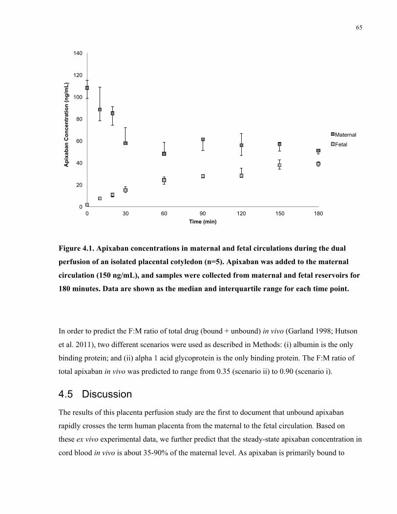
65
Figure 4.1. Apixaban concentrations in maternal and fetal circulations during the dual
perfusion of an isolated placental cotyledon (n=5). Apixaban was added to the maternal
circulation (150 ng/mL), and samples were collected from maternal and fetal reservoirs for
180 minutes. Data are shown as the median and interquartile range for each time point.
In order to predict the F:M ratio of total drug (bound + unbound) in vivo (Garland 1998; Hutson
et al. 2011), two different scenarios were used as described in Methods: (i) albumin is the only
binding protein; and (ii) alpha 1 acid glycoprotein is the only binding protein. The F:M ratio of
total apixaban in vivo was predicted to range from 0.35 (scenario ii) to 0.90 (scenario i).
4.5 Discussion
The results of this placenta perfusion study are the first to document that unbound apixaban
rapidly crosses the term human placenta from the maternal to the fetal circulation. Based on
these ex vivo experimental data, we further predict that the steady-state apixaban concentration in
cord blood in vivo is about 35-90% of the maternal level. As apixaban is primarily bound to
0
20
40
60
80
100
120
140
0 30 60 90 120 150 180
Api
xaba
n C
once
ntra
tion
(ng/
mL)
Time (min)
Maternal
Fetal
66
albumin, we speculate that the in vivo F:M ratio is likely closer to the albumin-based estimate of
0.90.
In instances where information regarding placental transfer may not be available, the placenta
perfusion model is a safe and ethical tool to predict placental transfer in vivo. Apixaban is a
relatively new drug and there are currently no published case reports describing its use in
pregnant patients. A systematic review of the placenta perfusion results evaluated the model in
predicting placental drug transfer in vivo (Hutson et al. 2011). In that study, F:M concentration
ratios from perfusion experiments were compared with cord-to-maternal blood ratios collected at
the time of delivery for 70 drugs. In general, the results from placenta perfusion experiments
correlated well with the data in vivo. After adjusting the perfusion F:M ratios of 24 drugs for
protein binding and pH differences between the fetal and maternal circulation, there was a
stronger correlation between the in vivo results of cord-to-maternal drug concentration ratio and
the perfusion experiments-derived fetal-to-maternal ratio. The authors concluded that the
perfusion model can be used to predict placental transfer of small molecule drugs in vivo (Hutson
et al. 2011).
In our analyses, the predicted ratio of F:M apixaban concentrations in vivo ranged from 0.35 to
0.90, suggesting that fetal apixaban exposure may be similar to the level of maternal therapeutic
exposure. In general, a F:M drug concentration ratio can be used to categorize the placental
transferability of drug: limited transfer (<0.1), transfer (0.1–1.0), or accumulation in the fetal
compartment (>1.0) (Hutson et al. 2011). Although our prediction needs to be verified with in
vivo data, we anticipate that apixaban concentrations in the cord blood will be in the vicinity of
maternal therapeutic levels.
An important consideration with using any anticoagulant in pregnancy relates to the state of the
coagulation system in maternal and fetal plasma. Pregnancy is classically a state of
hypercoagulability, characterized by an increased level of many clotting factors, a decrease in the
amount of natural anticoagulants, and a reduction in fibrinolytic activity in the blood (Dahlman
et al. 1985; Bremme 2003; O'Riordan & Higgins 2003). More specifically, levels of Factor X
increase in the early stages of pregnancy and remain elevated until 1 week after delivery
(Hellgren & Blomback 1981). By comparison, fetal and neonatal levels of Factor X are markedly
lower, ranging from 20–40 percent of adult levels (Andrew et al. 1990; Reverdiau-Moalic et al.
67
1996). This suggests that fetuses and neonates may be more sensitive to the anticoagulant effects
of apixaban. If intrauterine exposures to apixaban occur, coagulation monitoring of the infants
may be necessary.
The therapeutic options are limited for pregnant women who require anticoagulation
management. Unfractionated heparin (UFH) and low-molecular weight heparins (LMWH) do
not cross the placenta and are not associated with an increased risk of teratogenicity or increased
fetal bleeding (Bates 2002; Bates et al. 2012). For this reason, UFH and LMWHs are currently
recommended for most women requiring anticoagulation therapy during pregnancy (Bates et al.
2012). However, parenteral administration poses significant challenges. We recently evaluated
the placental transfer of two other novel oral anticoagulants: dabigatran and rivaroxaban using
the same perfusion model (Bapat et al. 2014, 2015). Dabigatran and its pro-drug, dabigatran
etexilate mesylate, had lower placental transfer as characterized by F:M ratios of 0.33 and 0.17,
respectively (Bapat et al. 2014). Similar to apixaban, rivaroxaban concentrations in the maternal
circulation rapidly declined in the first hour and remained relatively constant over the final 2
hours. The F:M ratio of rivaroxaban was 0.69 after 3 hours, and this transfer was most likely via
to passive diffusion (Bapat et al. 2015). Warfarin is another option, although it is teratogenic
particularly at higher doses (Schaefer et al. 2006).
The placenta expresses numerous efflux transporters, such as P-glycoprotein (P-gp) and breast
cancer resistance protein (BCRP), which can play a protective role in limiting fetal drug
exposure (Iqbal et al. 2012). Using in vitro models, it has been shown that apixaban is a substrate
for both P-gp and BCRP (Zhang et al. 2013), and thus, one could expect lower placental transfer,
as exemplified in drugs such as glyburide and metformin (Kraemer et al. 2006; Pollex et al.
2008; Hemauer et al. 2010). A study in rats showed limited placental transfer of [14C]-apixaban,
with a peak fetal blood concentration that was approximately 35% of the maternal concentration
(Wang et al. 2011). The authors suggested that BCRP might play an important role in limiting
placental transfer of apixaban to the fetus. However, due to its relatively small size and no
ionization at physiological pH (Pfizer Canada 2015), it is likely that apixaban crosses the
placenta extensively via passive diffusion (Pacifici & Nottoli 1995). Future studies are required
to determine if human placental P-gp and BCRP are specifically involved in the distribution of
apixaban in maternal and fetal circulations.
68
A limitation of this placenta perfusion model is that only full-term placentae are used. As such,
we cannot make extrapolations about placental transfer in the first and second trimester.
However, it should be noted that the need for anticoagulant therapy typically becomes more
pronounced as the pregnancy progresses (Bates et al. 2012), suggesting that information
regarding its placental transfer in later stages of pregnancy may be more clinically relevant.
Another consideration with the placenta perfusion model is that it does not account for time-
dependent dynamic changes of drug concentrations in the maternal and fetal compartments.
Protein binding is also important as apixaban is characterized by relatively high plasma protein
binding (87–93%) (Pfizer Canada 2015), and only the unbound form of a drug can cross the
placenta (Pacifici & Nottoli 1995).
In conclusion, unbound apixaban rapidly crosses the placenta from the maternal to the fetal
circulation in the human placenta perfusion model. We further predict that fetal apixaban
concentrations in vivo at steady state are 35-90% of the corresponding maternal levels, raising a
possibility for neonatal complications due to their hypocoagulability status. Future studies will
need to explore safety before clinicians can consider the use of apixaban in pregnant women. As
such, the recommendation still remains that women who become pregnant while taking apixaban
should be switched to another anticoagulant such as LMWH or UFH.
69
Chapter 5
Prediction of human placental pharmacokinetics of novel anticoagulants using the ex vivo placenta perfusion and
physiologically based models.
Priya Bapat1,2, Masanobu Takeuchi2, Howard Berger3, Shinya Ito1,2.
1 Department of Pharmacology & Toxicology, University of Toronto, Toronto, ON, Canada
2 Division of Clinical Pharmacology & Toxicology, The Hospital for Sick Children, Toronto,
ON, Canada.
3 Department of Obstetrics & Gynecology, St. Michael’s Hospital, Toronto, ON, Canada.
Contribution of this thesis author (P. Bapat): I was part of the collaborative team to design the
research study. I performed the in vivo prediction analyses, and developed the pharmacokinetic
model with M. Takeuchi. I analyzed the data and drafted the manuscript. All authors provided
input and approved the final version of the manuscript.
70
Prediction of human placental pharmacokinetics of 5novel anticoagulants using the ex vivo placenta perfusion and physiologically based models.
5.1 Abstract
Background: Our previous studies have provided information regarding the placental transfer of
dabigatran, rivaroxaban, and apixaban using the dually perfused placenta ex vivo. A limitation of
this perfusion model is that it cannot account for differences in protein binding between maternal
and fetal plasma. The purpose of this study was to compare the placental transfer of three novel
oral anticoagulants, after adjusting for differences in protein binding. As well, transplacental
transfer parameters were estimated using a pharmacokinetic model. Methods: To account for the
physiological differences in protein binding and pH between maternal and fetal circulation in
vivo, the equation developed by Garland (1998) and later adapted by Hutson (2011) was used to
predict the fetal-to-maternal (F:M) ratio of total (bound plus unbound) drug concentrations in
plasma in vivo. A three-compartment model, consisting of maternal, placental, and fetal
compartments was used to estimate transplacental pharmacokinetic parameters. Results: The
predicted F:M ratios in vivo of dabigatran and rivaroxaban were 0.39 and 0.81, respectively. For
apixaban, the predicted F:M ratio in vivo ranged from 0.35-0.90. The experimental and predicted
F:M ratios were significantly different between dabigatran and apixaban. The concentration
profiles of rivaroxaban, apixaban and dabigatran in the maternal and fetal reservoir were
adequately described using a three-compartment transplacental pharmacokinetic model.
Conclusions: In conclusion, the in vivo fetal levels of dabigatran, rivaroxaban and apixaban were
predicted to be 39%, 80%, and 90% of corresponding maternal levels at steady state. We noted
that in vivo estimates of the F:M ratio were 15% higher than what was shown in placenta
perfusion experiments, and this was primarily attributed to plasma protein binding.
71
5.2 Introduction
Anticoagulants are often prescribed during pregnancy for the prevention or treatment of
complications in women with venous thromboembolism (VTE), mechanical or artificial heart
valves, and inherited or acquired thrombophilias (Royal College of Obstetricians and
Gynaecologists 2009; Bates et al. 2012). Vitamin K antagonists, such as warfarin, are
contraindicated in pregnancy because of an associated embryopathy characterized by nasal and
limb hypoplasia, as well as congenital heart anomalies (Chan et al. 2000; Hassouna & Allam
2010). As well, vitamin K antagonists cross the placenta and have the potential to cause fetal
bleeding and teratogenicity (Hall et al. 1980; Ginsberg et al. 1989; Schaefer et al. 2006). By
comparison, low molecular weight heparins (LMWH) and unfractionated heparins (UFH) do not
cross the placenta and are currently recommended for the prevention and treatment of VTE
during pregnancy (Bates et al. 2012, 2016). As with any medication during pregnancy, the use of
anticoagulants can be challenging due to the potential for maternal and fetal complications.
More recently, novel oral anticoagulants (dabigatran, rivaroxaban, apixaban) have been approved
for use in non-pregnant adults for the prevention and treatment of VTE, including pulmonary
embolism and deep vein thrombosis. These novel oral anticoagulants are reversible, direct
inhibitors of thrombin or Factor Xa, and can be prescribed at fixed doses without the need for
routine monitoring or dose adjustment. In general, there are no food interactions with these
drugs, and only limited drug-drug interactions (Fox et al. 2012; Bauer 2013; Heidbuchel et al.
2013). To date, there is limited information regarding the use and safety of these drugs in
pregnant women. Moreover, the information about placental drug transfer in vivo and fetal safety
is unknown.
The decision to begin or continue treatment during pregnancy heavily relies on weighing the
maternal benefits of the drug against the potential adverse fetal risks. An important determinant
in this risk assessment is the estimation of fetal drug exposure, based on quantifying the amount
of drug that crosses the placenta. By using the dually perfused human placenta ex vivo, our
previous studies have provided information regarding the placental transfer of dabigatran,
rivaroxaban, and apixaban (Bapat et al. 2014, 2015, 2016). A limitation of this perfusion model
is that it cannot account for differences in protein binding between maternal and fetal plasma.
However, a previous study has showed that placenta perfusion data can be adjusted in order to
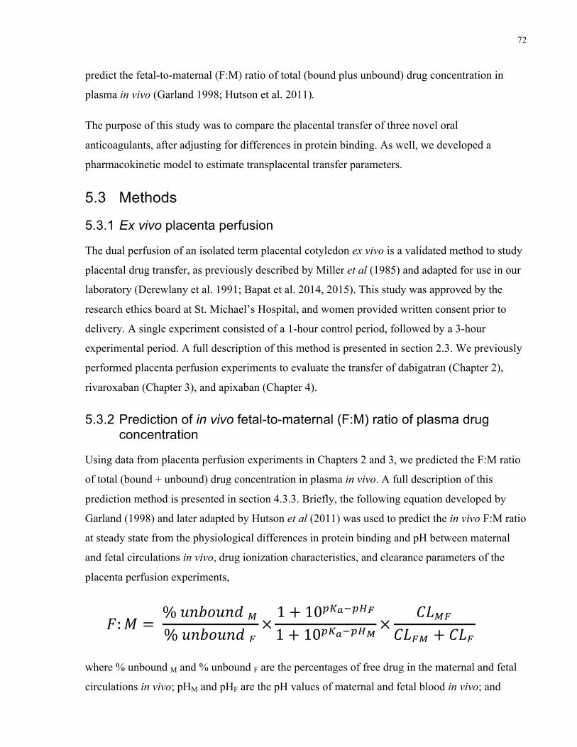
72
predict the fetal-to-maternal (F:M) ratio of total (bound plus unbound) drug concentration in
plasma in vivo (Garland 1998; Hutson et al. 2011).
The purpose of this study was to compare the placental transfer of three novel oral
anticoagulants, after adjusting for differences in protein binding. As well, we developed a
pharmacokinetic model to estimate transplacental transfer parameters.
5.3 Methods
5.3.1 Ex vivo placenta perfusion
The dual perfusion of an isolated term placental cotyledon ex vivo is a validated method to study
placental drug transfer, as previously described by Miller et al (1985) and adapted for use in our
laboratory (Derewlany et al. 1991; Bapat et al. 2014, 2015). This study was approved by the
research ethics board at St. Michael’s Hospital, and women provided written consent prior to
delivery. A single experiment consisted of a 1-hour control period, followed by a 3-hour
experimental period. A full description of this method is presented in section 2.3. We previously
performed placenta perfusion experiments to evaluate the transfer of dabigatran (Chapter 2),
rivaroxaban (Chapter 3), and apixaban (Chapter 4).
5.3.2 Prediction of in vivo fetal-to-maternal (F:M) ratio of plasma drug concentration
Using data from placenta perfusion experiments in Chapters 2 and 3, we predicted the F:M ratio
of total (bound + unbound) drug concentration in plasma in vivo. A full description of this
prediction method is presented in section 4.3.3. Briefly, the following equation developed by
Garland (1998) and later adapted by Hutson et al (2011) was used to predict the in vivo F:M ratio
at steady state from the physiological differences in protein binding and pH between maternal
and fetal circulations in vivo, drug ionization characteristics, and clearance parameters of the
placenta perfusion experiments,
where % unbound M and % unbound F are the percentages of free drug in the maternal and fetal
circulations in vivo; pHM and pHF are the pH values of maternal and fetal blood in vivo; and
!!! ! !!!!"#$!"%!!!!!"#$!"%!! !!! !"!"!!!"!!! !"!"!!!"! !
!"!"!"!" ! !"!
73
CLMF, CLFM, and CLF are the maternal-to-fetal, fetal-to-maternal, and fetal clearances of the drug
obtained in the placenta perfusion experiments (Garland 1998). When using this equation, the
following assumptions were made as per previous analyses (Hutson et al. 2011): (i) only the free
drug can cross the placenta passively, (ii) the free fraction is not concentration-dependent, (iii)
only the unionized form of the drug can passively cross the placenta, and (iv) fetal clearance
(CLF ) is negligible.
In non-pregnant adults, dabigatran and rivaroxaban bind albumin at 35% and 95%, respectively
(Weinz et al. 2005; Blech et al. 2008). Unbound fractions (%) in the maternal and fetal
circulations in vivo were estimated with the method developed by Hill and Abramson (1988) as
described in section 4.3.3. For the second term of the Garland equation, the maternal and fetal
pH values were 7.4 and 7.35, respectively (Reynolds & Knott 1989), and a pKa value of 12.4 was
used for dabigatran (Blech et al. 2008). Since rivaroxaban is unionized at physiological pH
(Bayer Canada 2013), the second term of the equation was assumed to be 1. The F:M
concentration ratios from the placenta perfusions at 180 min were used to represent the last term
of the Garland equation (1998).
5.3.3 Statistical analysis
All data are presented as the median and interquartile range (IQR), unless stated otherwise.
Comparisons between different perfusion groups were analyzed with a Kruskal-Wallis test using
Dunn's (1964) procedure with a Bonferroni correction for multiple comparisons. Any p values
less than 0.05 were considered statistically significant. All statistical analyses were performed in
SPSS for MacIntosh Version 20.0 (IBM Corp., Armonk, NY, USA).
5.3.4 Model construction
As a simple structural representative of the experimental setup, a three-compartment model,
consisting of maternal, placental, and fetal compartments, was used to estimate the following
transplacental pharmacokinetic parameters (Figure 5.1); K12 (influx rate constant from maternal
to placental compartment), K21 (efflux rate constant from placental to maternal compartment),
K23 (efflux rate constant from placental to fetal compartment), and K32 (influx rate constant from
fetal to placental compartment). Vm and Vf, representing the volumes of distribution in the
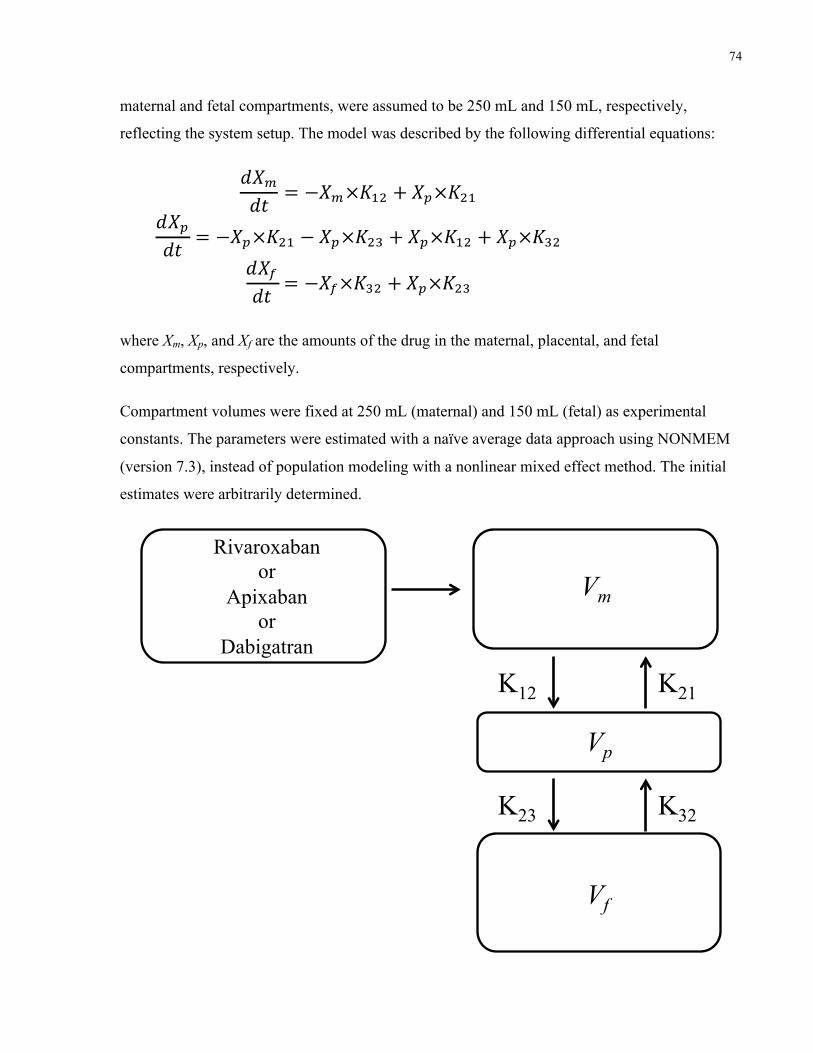
74
maternal and fetal compartments, were assumed to be 250 mL and 150 mL, respectively,
reflecting the system setup. The model was described by the following differential equations:
where Xm, Xp, and Xf are the amounts of the drug in the maternal, placental, and fetal
compartments, respectively.
Compartment volumes were fixed at 250 mL (maternal) and 150 mL (fetal) as experimental
constants. The parameters were estimated with a naïve average data approach using NONMEM
(version 7.3), instead of population modeling with a nonlinear mixed effect method. The initial
estimates were arbitrarily determined.
!!!!" ! !!!!!!" ! !!!!!"!
!!!!" ! !!!!!!" ! !!!!!" ! !!!!!" ! !!!!!"!
!!!!" ! !!!!!!" ! !!!!!"!
Vm
Vf
Vp
K12
Rivaroxaban or
Apixaban or
Dabigatran
K23
K21
K32
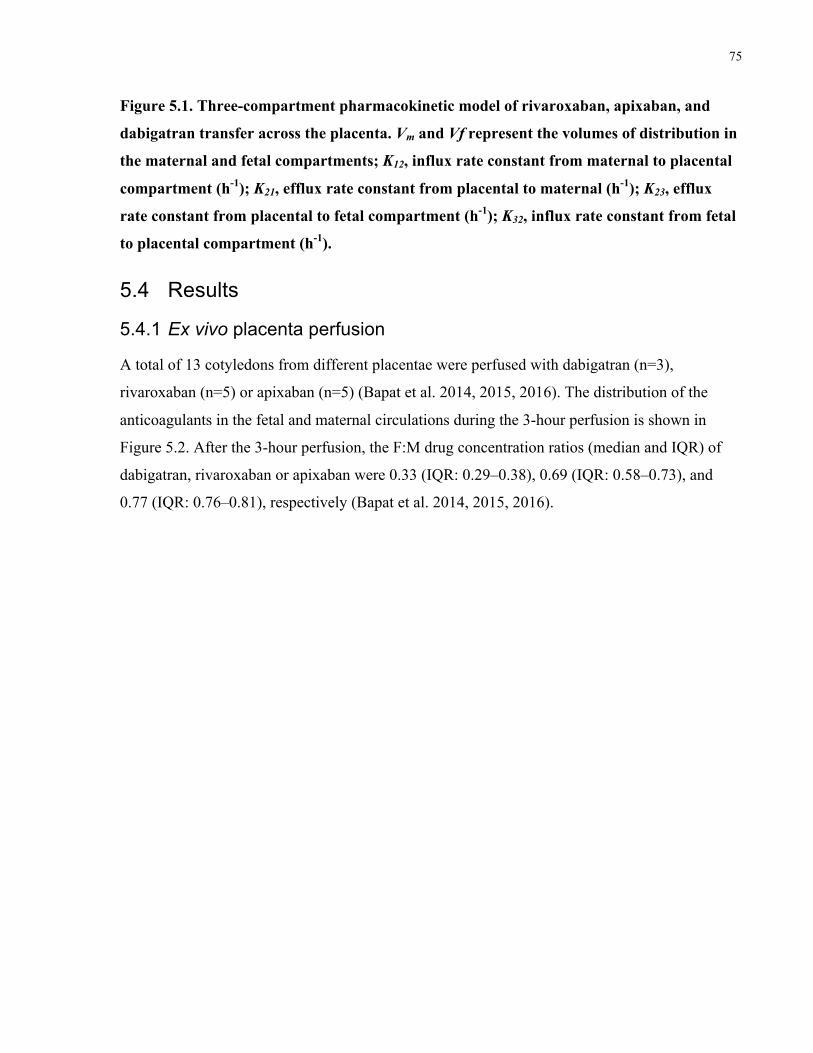
75
Figure 5.1. Three-compartment pharmacokinetic model of rivaroxaban, apixaban, and
dabigatran transfer across the placenta. Vm and Vf represent the volumes of distribution in
the maternal and fetal compartments; K12, influx rate constant from maternal to placental
compartment (h-1); K21, efflux rate constant from placental to maternal (h-1); K23, efflux
rate constant from placental to fetal compartment (h-1); K32, influx rate constant from fetal
to placental compartment (h-1).
5.4 Results
5.4.1 Ex vivo placenta perfusion
A total of 13 cotyledons from different placentae were perfused with dabigatran (n=3),
rivaroxaban (n=5) or apixaban (n=5) (Bapat et al. 2014, 2015, 2016). The distribution of the
anticoagulants in the fetal and maternal circulations during the 3-hour perfusion is shown in
Figure 5.2. After the 3-hour perfusion, the F:M drug concentration ratios (median and IQR) of
dabigatran, rivaroxaban or apixaban were 0.33 (IQR: 0.29–0.38), 0.69 (IQR: 0.58–0.73), and
0.77 (IQR: 0.76–0.81), respectively (Bapat et al. 2014, 2015, 2016).
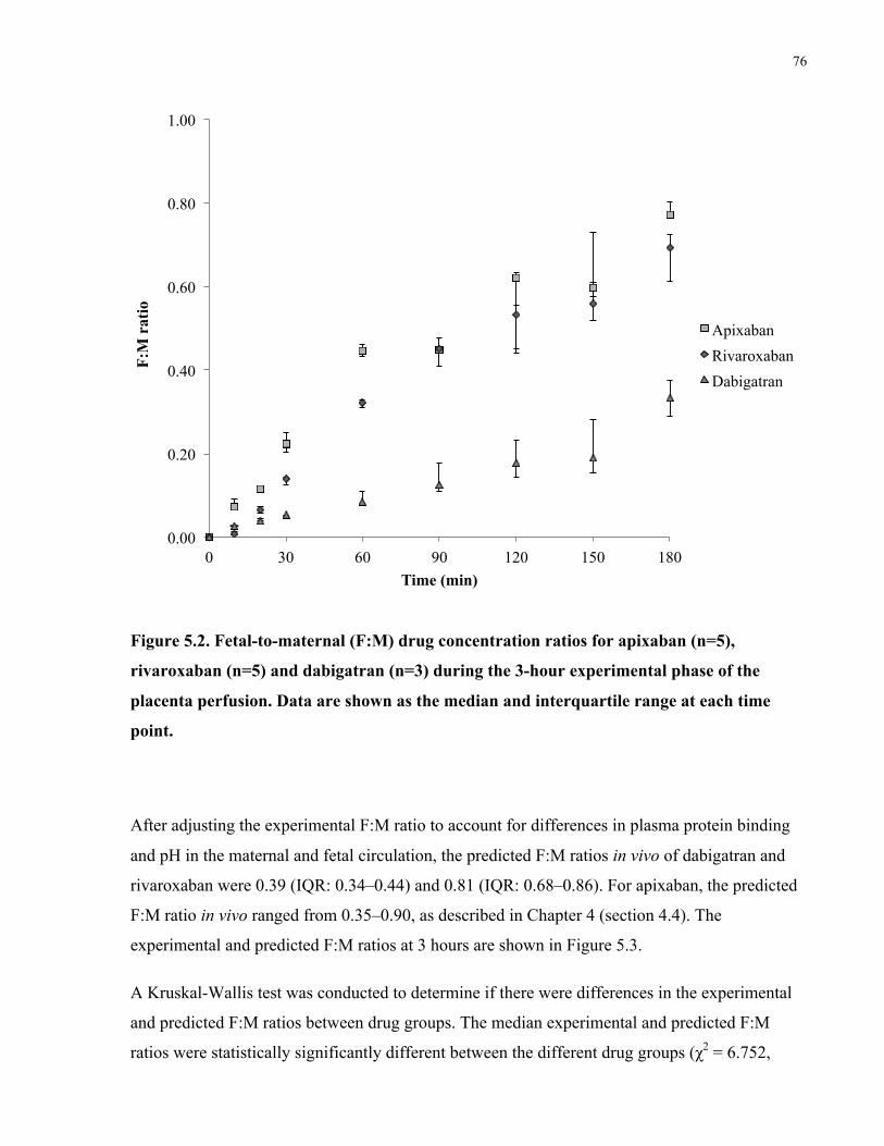
76
Figure 5.2. Fetal-to-maternal (F:M) drug concentration ratios for apixaban (n=5),
rivaroxaban (n=5) and dabigatran (n=3) during the 3-hour experimental phase of the
placenta perfusion. Data are shown as the median and interquartile range at each time
point.
After adjusting the experimental F:M ratio to account for differences in plasma protein binding
and pH in the maternal and fetal circulation, the predicted F:M ratios in vivo of dabigatran and
rivaroxaban were 0.39 (IQR: 0.34–0.44) and 0.81 (IQR: 0.68–0.86). For apixaban, the predicted
F:M ratio in vivo ranged from 0.35–0.90, as described in Chapter 4 (section 4.4). The
experimental and predicted F:M ratios at 3 hours are shown in Figure 5.3.
A Kruskal-Wallis test was conducted to determine if there were differences in the experimental
and predicted F:M ratios between drug groups. The median experimental and predicted F:M
ratios were statistically significantly different between the different drug groups (χ2 = 6.752,
0.00
0.20
0.40
0.60
0.80
1.00
0 30 60 90 120 150 180
F:M
rat
io
Time (min)
Apixaban
Rivaroxaban
Dabigatran
77
p=0.034). Pairwise comparisons were performed using Dunn’s procedure with a Bonferroni
correction for multiple comparisons. This post-hoc analysis revealed statistically significant
differences in median F:M ratios between dabigatran and apixaban (p=0.034), but not between
dabigatran and rivaroxaban or rivaroxaban and apixaban.
Figure 5.3. Placenta perfusion and predicted fetal-to-maternal (F:M) drug concentration
ratios for dabigatran (n=3), rivaroxaban (n=5) and apixaban (n=5) after 180-min
perfusion. Placenta perfusion F:M ratios were obtained directly from experiments, and the
predicted F:M ratios were calculated using the Garland equation (1998) with albumin is
the sole binding protein. Data are shown as the median and interquartile range at each
time point.
0.0
0.2
0.4
0.6
0.8
1.0
Dabigatran Rivaroxaban Apixaban
F:M
rat
io
Placenta Perfusion
Predicted
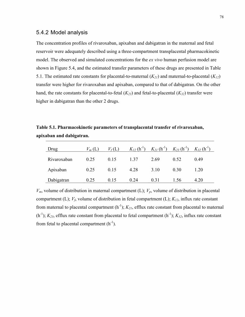
78
5.4.2 Model analysis
The concentration profiles of rivaroxaban, apixaban and dabigatran in the maternal and fetal
reservoir were adequately described using a three-compartment transplacental pharmacokinetic
model. The observed and simulated concentrations for the ex vivo human perfusion model are
shown in Figure 5.4, and the estimated transfer parameters of these drugs are presented in Table
5.1. The estimated rate constants for placental-to-maternal (K21) and maternal-to-placental (K12)
transfer were higher for rivaroxaban and apixaban, compared to that of dabigatran. On the other
hand, the rate constants for placental-to-fetal (K23) and fetal-to-placental (K32) transfer were
higher in dabigatran than the other 2 drugs.
Table 5.1. Pharmacokinetic parameters of transplacental transfer of rivaroxaban,
apixaban and dabigatran.
Drug Vm (L) Vf (L) K12 (h-1) K21 (h-1) K23 (h-1) K32 (h-1)
Rivaroxaban 0.25 0.15 1.37 2.69 0.52 0.49
Apixaban 0.25 0.15 4.28 3.10 0.30 1.20
Dabigatran 0.25 0.15 0.24 0.31 1.56 4.20
Vm, volume of distribution in maternal compartment (L); Vp, volume of distribution in placental
compartment (L); Vf, volume of distribution in fetal compartment (L); K12, influx rate constant
from maternal to placental compartment (h-1); K21, efflux rate constant from placental to maternal
(h-1); K23, efflux rate constant from placental to fetal compartment (h-1); K32, influx rate constant
from fetal to placental compartment (h-1).
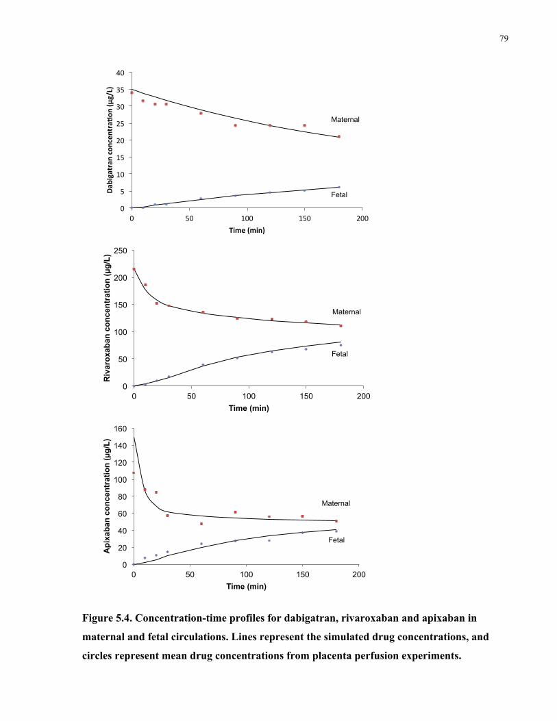
79
Figure 5.4. Concentration-time profiles for dabigatran, rivaroxaban and apixaban in
maternal and fetal circulations. Lines represent the simulated drug concentrations, and
circles represent mean drug concentrations from placenta perfusion experiments.
!"
#"
$!"
$#"
%!"
%#"
&!"
&#"
'!"
!" #!" $!!" $#!" %!!"
!"#
$%&#'(#)
!*+)
*,)'(#-+
)!./&012!
3%4,!.4%)2!
Maternal
Fetal
0
50
100
150
200
250
0 50 100 150 200
R
ivar
oxab
an c
once
ntra
tion
(!g/
L)
Time (min)
Maternal
Fetal
0
20
40
60
80
100
120
140
160
0 50 100 150 200
A
pixa
ban
conc
entr
atio
n (!
g/L)
Time (min)
Maternal
Fetal
80
5.5 Discussion
The results of our study demonstrate the transplacental transfer of dabigatran, rivaroxaban, and
apixaban in full-term human placentae, after adjusting for protein binding. In general, we noticed
that our predicted in vivo F:M ratios were about 15% higher than the experimental F:M ratios,
which highlights the importance of accounting for protein binding in the placenta perfusion.
Other groups have added plasma proteins directly to the perfusate, but it can be difficult to
mimic exact physiological conditions (Johnson et al. 1999; Schenker et al. 1999; Gavard et al.
2006; Nanovskaya et al. 2009; Hutson et al. 2011). The equation developed by Garland (1998) is
an alternate method of accounting for differences in plasma protein concentrations and pH
between mother and fetus, and has previously been used as a means of adjusting perfusion data
to better predict in vivo placental drug transfer (Hutson et al. 2011).
Rivaroxaban and apixaban have similar physicochemical characteristics, in terms of molecular
size (435 and 459 Da) and ionization, and this may explain their similar patterns of placental
transfer shown in Figure 2. Dabigatran showed the least amount of placental transfer, likely due
to its increased lipophilicity and relatively larger molecular size (628 Da).
Another important consideration in the disposition of drugs across the placenta is plasma protein
binding, as the unbound form of the drug can equilibrate across the placenta. The concentrations
of albumin and α-1 acid glycoprotein (AAG) differ in fetal and maternal plasma, and this can
affect total drug concentrations (bound + unbound) in each compartment at equilibrium.
Throughout pregnancy, maternal albumin levels gradually decrease and fetal albumin levels tend
to increase, with the fetal-to-maternal albumin ratio increasing from 0.28 in the first trimester to
1.20 closer to term (Krauer et al. 1984). As well, there is a 3-fold increase in the concentration of
free fatty acids in the maternal circulation that can displace drugs from binding to maternal
albumin (Ridd et al. 1983; Nau et al. 1984). This can lead to increased binding of a drug to fetal
albumin, which can, in turn lead to increased placental drug transfer and possibly fetal drug
accumulation. By comparison, AAG levels remain relatively constant in maternal plasma, and
fetal levels gradually increase throughout pregnancy, with the AAG F:M ratio increasing from
0.09 in the first trimester to 0.37 at term (Krauer et al. 1984). The major binding protein for
dabigatran and rivaroxaban is albumin, and this may explain why the predicted F:M ratios in vivo
were 15% higher than the ratio obtained from the experimental observations in our placenta
81
perfusion model of unbound drug transfer. The predicted in vivo F:M ratio for apixaban was
estimated to be between 0.35 and 0.90, based on binding solely to AAG or albumin. We
speculate that the in vivo F:M ratio is likely much closer to the albumin-based estimate of 0.90,
as apixaban is primarily bound to albumin.
To date, there have been no reported cases of dabigatran and apixaban exposure in human
pregnancy. A case report described the use of rivaroxaban up to 19 weeks gestation. A male
infant was born at 40 weeks gestation with no complications. The authors reported a small baby
for weight, length, and cranial circumference (13th, 30th, and 7th percentiles, respectively), but
likely attributed this to maternal smoking (Konigsbrugge et al. 2014). Recently, there was a case
series of 37 women who were inadvertently exposed to rivaroxaban in the first and second
trimester. There was one case of a conotruncal cardiac defect in a woman with several co-
medications and a previous fetus with a cardiac malformation in the absence of rivaroxaban
exposure (Hoeltzenbein et al. 2016). Placental drug transfer of rivaroxaban was not measured in
these cases. These cases reiterate the notion that as prescriptions rates of NOACs continue to
increase (Desai et al. 2014), the number of women with an unplanned pregnancy who are taking
a NOAC will also likely increase.
The three-compartment pharmacokinetic model adequately described the concentration-time
profiles for NOACs in the placenta perfusion experimental system. This model estimated the
influx and efflux rate constants between maternal, placental and fetal compartments. In
particular, the efflux rate constants from the placenta to maternal compartment (K21) were similar
for apixaban and rivaroxaban at 2.69 and 3.10 h-1, which were higher than that of dabigatran
(0.31 h-1). Previous studies have shown that apixaban and rivaroxaban are substrates of the
transporters, P-gp and BCRP (Gnoth et al. 2011; Gong et al. 2013; Zhang et al. 2013). As these
transporters are expressed in the maternal interface of the placenta, where they move substances
from the placenta to maternal circulation, the differences in the estimated K21 are consistent with
their substrate status for these transporters. Interpretation of the other rate constants awaits
further studies on expression, localization, and substrate specificity of other transporters in the
placenta.
In conclusion, the in vivo fetal levels of dabigatran, rivaroxaban and apixaban were predicted to
be 39%, 80%, and 90% of corresponding maternal levels at steady state. We noted that in vivo
82
estimates of the F:M ratio were 15% higher than what was shown in placenta perfusion
experiments, and this was primarily attributed to plasma protein binding in vivo. Furthermore, we
developed a pharmacokinetic model that was able to describe the concentration-time profiles of
placental transfer of NOACs at term, and estimate transfer parameters. Our modeling approach
provides another example of proof-of-principle demonstration, which will lay a foundation for
physiologically-based pharmacokinetic modeling in pregnancy.
83
Chapter 6
Evaluating the success rate of the ex vivo dual perfusion of the human placenta.
Priya Bapat1,2, Howard Berger3, Shinya Ito1,2
1 Department of Pharmacology and Toxicology, University of Toronto, 1 King’s College Circle,
Toronto, Ontario, M5S 1A8, Canada
2 Division of Clinical Pharmacology and Toxicology, Hospital for Sick Children, 555 University
Avenue, Toronto, Ontario, M5G 1X8, Canada
3 Department of Obstetrics and Gynecology, St. Michael’s Hospital, 30 Bond St, Toronto,
Ontario M5B 1W8, Canada.
Contribution of this thesis author (P. Bapat): I was part of the collaborative team to design the
research study. I collected the data from all placenta perfusion experiments, analyzed the data
and drafted the manuscript. All authors provided input and approved the final version of the
manuscript.
84
Evaluating the success rate of the ex vivo dual 6perfusion of the human placenta.
6.1 Abstract
Background: The dual perfusion of a single placental cotyledon ex vivo has been widely used
among placental researchers to study placental transfer in organized human placental tissue.
While this model can be highly predictive, a major challenge of this model is its low overall
success rate, which can result in high costs and more time to complete a study. The method is
technically challenging, and there are several criteria during the perfusion procedure that must be
met in order to have a successful experiment. The objective of this study was to evaluate the
success rate at several stages throughout the dual perfusion of an isolated placental cotyledon.
Methods: Placentae were collected with informed consent following caesarean or vaginal
delivery of uncomplicated term pregnancies for a 3-hour perfusion of various test substances.
Upon completion of each experiment, placentae were assigned one of six checkpoints
corresponding to important stages of the placenta perfusion protocol. Results: 297 women
provided written consent to participate in our study over a 20-month period, and of these, 251
placentae were collected. 99 placentae did not have an adequate fetal artery-vein pair for
cannulation, and 113 placentae were prematurely terminated due to a fetal reservoir leak. The
maternal circulation was established in the remaining 39 placentae, and of these, 25 placentae
were successfully perfused for 3 hours. This corresponds to an overall success rate of 9.96%.
Conclusion: The results of our study show an overall success rate of 10% for the dual perfusion
of an isolated human placental cotyledon ex vivo. By using the proposed checkpoints, we
identified that setting up the fetal circulation is a critical stage in determining experimental
success, and that this stage may be in need of technical improvement.
.
85
6.2 Introduction
The placenta is a complex organ that is responsible for nourishing and maintaining the fetus via
the umbilical cord. The placenta is made up of vascular tissue in which oxygen and nutrients can
pass from maternal to fetal circulation, while carbon dioxide and waste products can pass in the
opposite direction (Syme et al. 2004). The placental functional unit is a lobule, termed cotyledon,
and each cotyledon is independently perfused by maternal and fetal vessels. A typical human
placenta is made up of 20-40 cotyledons (Syme et al. 2004).
The placenta also plays a major role in determining fetal drug exposure. It is possible to estimate
total fetal drug exposure by measuring the amount of drug that crosses the placenta. Substances
can cross the placenta via passive or active transport, as the placenta highly expresses
transporters such as P-glycoprotein and breast cancer resistance protein (Ceckova-Novotna et al.
2006; Iqbal et al. 2012). Although animal studies are often conducted to study placental transfer
of various compounds, it is difficult to extrapolate these results to humans because of species-
differences in blood flow pattern, as well as biochemical and anatomical structure of the placenta
(Leiser & Kaufmann 1994; Ala-Kokko et al. 2000). Also, animal studies are often performed at
doses that are much higher than the relevant pharmacokinetic human doses (Brent 2004).
The dual perfusion of a single placental cotyledon ex vivo has been widely used among placental
researchers to study placental transfer in organized human placental tissue (Hutson et al. 2011).
The placental perfusion model avoids several issues that are associated with other placental
techniques (e.g. ethical implications of in vivo human studies, physiological discrepancies in cell
cultures) (Hutson et al. 2011). This model has previously been used to investigate the placental
transfer of endogenous and exogenous substrates, such as amino acids, hormones, electrolytes,
viruses, therapeutics, and illicit drugs (Omarini et al. 1992; Muhlemann et al. 1995; Ala-Kokko
et al. 2000; Malek et al. 2009; Bapat et al. 2015). A systematic review of this model found that
fetal-to-maternal drug concentration ratios from perfusion experiments matched well with
umbilical cord-to-maternal blood ratios collected at the time of delivery. The authors concluded
that the placenta perfusion model can reliably be used to predict placental transfer of small
molecule drugs in vivo (Hutson et al. 2011).
A major challenge of this model is its low overall success rate, which can result in high costs and
more time to complete a study. Different perfusion laboratories report success rates between 5
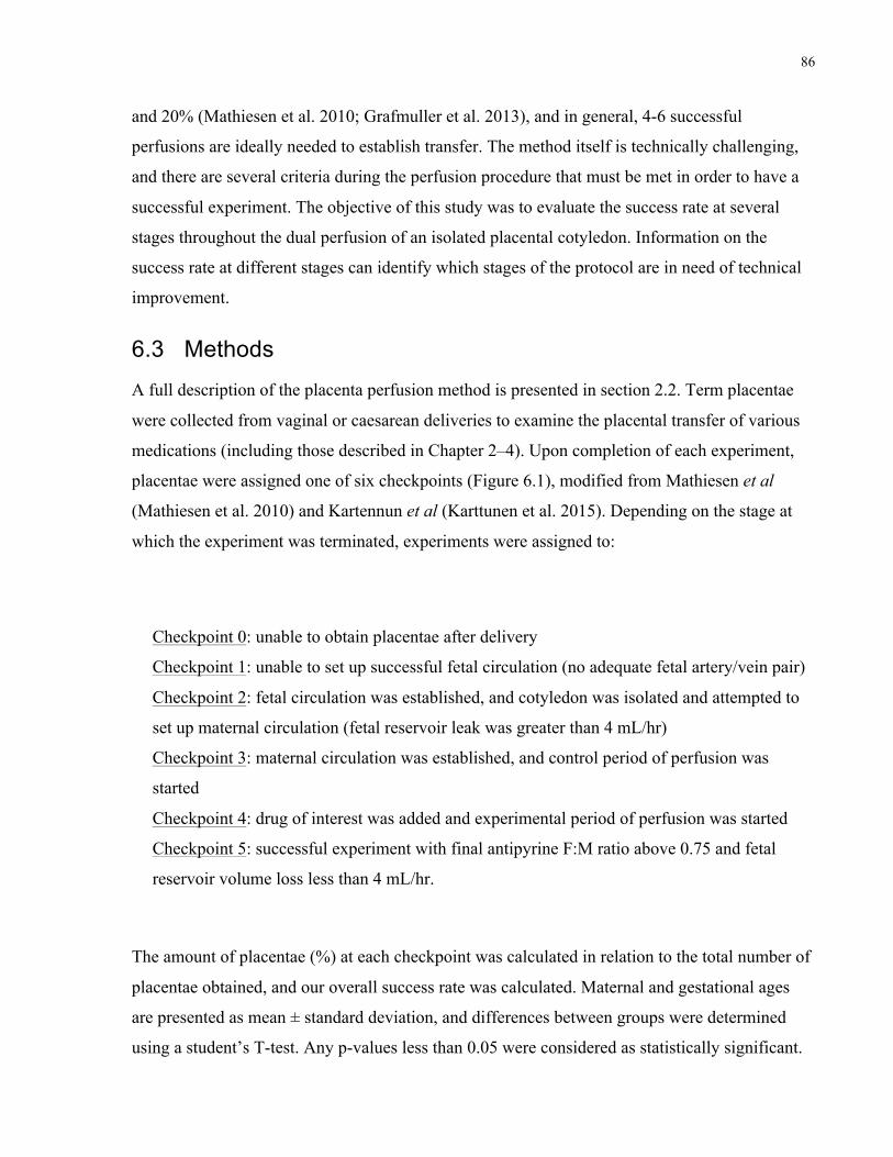
86
and 20% (Mathiesen et al. 2010; Grafmuller et al. 2013), and in general, 4-6 successful
perfusions are ideally needed to establish transfer. The method itself is technically challenging,
and there are several criteria during the perfusion procedure that must be met in order to have a
successful experiment. The objective of this study was to evaluate the success rate at several
stages throughout the dual perfusion of an isolated placental cotyledon. Information on the
success rate at different stages can identify which stages of the protocol are in need of technical
improvement.
6.3 Methods
A full description of the placenta perfusion method is presented in section 2.2. Term placentae
were collected from vaginal or caesarean deliveries to examine the placental transfer of various
medications (including those described in Chapter 2–4). Upon completion of each experiment,
placentae were assigned one of six checkpoints (Figure 6.1), modified from Mathiesen et al
(Mathiesen et al. 2010) and Kartennun et al (Karttunen et al. 2015). Depending on the stage at
which the experiment was terminated, experiments were assigned to:
Checkpoint 0: unable to obtain placentae after delivery
Checkpoint 1: unable to set up successful fetal circulation (no adequate fetal artery/vein pair)
Checkpoint 2: fetal circulation was established, and cotyledon was isolated and attempted to
set up maternal circulation (fetal reservoir leak was greater than 4 mL/hr)
Checkpoint 3: maternal circulation was established, and control period of perfusion was
started
Checkpoint 4: drug of interest was added and experimental period of perfusion was started
Checkpoint 5: successful experiment with final antipyrine F:M ratio above 0.75 and fetal
reservoir volume loss less than 4 mL/hr.
The amount of placentae (%) at each checkpoint was calculated in relation to the total number of
placentae obtained, and our overall success rate was calculated. Maternal and gestational ages
are presented as mean ± standard deviation, and differences between groups were determined
using a student’s T-test. Any p-values less than 0.05 were considered as statistically significant.
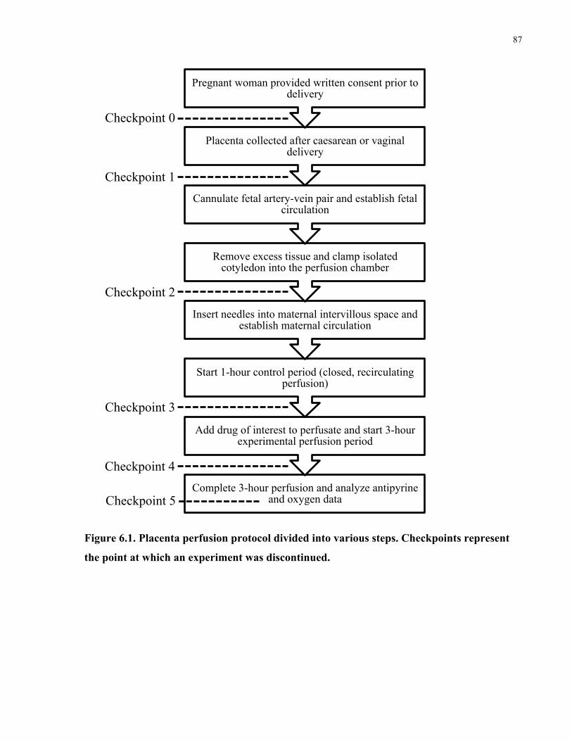
87
Figure 6.1. Placenta perfusion protocol divided into various steps. Checkpoints represent
the point at which an experiment was discontinued.
Complete 3-hour perfusion and analyze antipyrine and oxygen data
Add drug of interest to perfusate and start 3-hour experimental perfusion period
Start 1-hour control period (closed, recirculating perfusion)
Insert needles into maternal intervillous space and establish maternal circulation
Remove excess tissue and clamp isolated cotyledon into the perfusion chamber
Cannulate fetal artery-vein pair and establish fetal circulation
Placenta collected after caesarean or vaginal delivery
Pregnant woman provided written consent prior to delivery
Checkpoint 0
Checkpoint 1
Checkpoint 2
Checkpoint 3
Checkpoint 4
Checkpoint 5
88
6.4 Results
In total, 297 women provided written consent to participate in our studies over a 20-month
period, and of these, 251 placentae were collected from caesarean or vaginal deliveries. At the
time of delivery, the mean maternal age was 34.2 ± 5.0 years and mean gestational age was 39.1
± 0.7 weeks. The majority of placentae collected were obtained from caesarean delivery (85%).
46 placentae were not obtained due to scheduling issues (ie. if the delivery occurred outside of
working hours), or if the participant was part of another research study requiring collection of the
whole placenta (Checkpoint 0). If multiple deliveries occurred within a short time period, we
were only able to collect one placenta as our currently laboratory setup can accommodate one
placenta at a time, and the protocol requires the fetal circulation to be established within 15
minutes of delivery.
Of the 251 collected placentae, 99 did not have a suitable artery/vein pair and/or the maternal
decidual plate was not intact, and the fetal circulation could not be established (Checkpoint 1).
The fetal circulation was set up in a closed, recirculating configuration in the remaining 152
placentae. The experiment was prematurely terminated in 113 placentae primarily due to a leak
in the fetal reservoir greater than 4 mL/hr, or if the fetal arterial inflow pressure deviated from
30–60 mmHg for an extended period of time (Checkpoint 2). The maternal circulation was
successfully established in 39 experiments. At this point, both the maternal and fetal perfusates
were replaced with fresh perfusate and the 1-hour control period was started. 12 placentae
experienced a major leak (>4 mL/hr) during the control period and were immediately terminated
(Checkpoint 3). In 27 experiments, there were no issues experienced in the control period. The
maternal and fetal perfusates were replaced again and the drug of interest was added to start the
experimental period. There was a leak in 2 of these experiments which were prematurely
terminated (Checkpoint 4), and 25 placentae were perfused for the full 3-hour period (Checkpoint
5). This corresponds to an overall success rate of 9.96% (25 successful experiments from 251
collected placentae). A summary of this process is shown in Figure 6.2.
89
Figure 6.2. Flow diagram showing the number of placentae at each stage of the perfusion
protocol.
297 women provided consent
251 placentae collected
Fetal circulation established in 152 placentae
Maternal circulation established, and control period started in 39 placentae
Started experimental period in 27 placentae
Checkpoint 5: Successful perfusion
in 25 placentae
Checkpoint 4: Major leak from fetal
reservoir in 2 placentae
Checkpoint 3: Major leak from fetal
reservoir in 12 placentae
Checkpoint 2: Major leak from fetal
reservoir in 113 placentae
Checkpoint 1: Unable to set up fetal
circulation in 99 placentae
Checkpoint 0: 46 placentae not collected
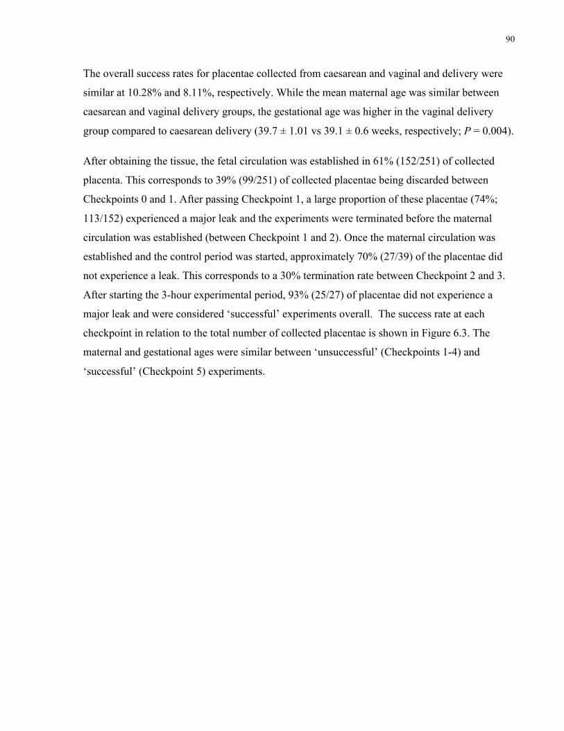
90
The overall success rates for placentae collected from caesarean and vaginal and delivery were
similar at 10.28% and 8.11%, respectively. While the mean maternal age was similar between
caesarean and vaginal delivery groups, the gestational age was higher in the vaginal delivery
group compared to caesarean delivery (39.7 ± 1.01 vs 39.1 ± 0.6 weeks, respectively; P = 0.004).
After obtaining the tissue, the fetal circulation was established in 61% (152/251) of collected
placenta. This corresponds to 39% (99/251) of collected placentae being discarded between
Checkpoints 0 and 1. After passing Checkpoint 1, a large proportion of these placentae (74%;
113/152) experienced a major leak and the experiments were terminated before the maternal
circulation was established (between Checkpoint 1 and 2). Once the maternal circulation was
established and the control period was started, approximately 70% (27/39) of the placentae did
not experience a leak. This corresponds to a 30% termination rate between Checkpoint 2 and 3.
After starting the 3-hour experimental period, 93% (25/27) of placentae did not experience a
major leak and were considered ‘successful’ experiments overall. The success rate at each
checkpoint in relation to the total number of collected placentae is shown in Figure 6.3. The
maternal and gestational ages were similar between ‘unsuccessful’ (Checkpoints 1-4) and
‘successful’ (Checkpoint 5) experiments.
91
Figure 6.3. Success rate (%) at each checkpoint of the placenta perfusion, calculated in
relation to the total number of placentae collected (n=251).
6.5 Discussion
The results of our study show an overall success rate of 10% for the dual perfusion of an isolated
human placental cotyledon ex vivo. While this rate was similar to reported success rates from
other perfusion laboratories, we evaluated the success rate at various checkpoints throughout the
experiment. The highest proportion of experiments was terminated between Checkpoints 0 and 2,
suggesting that this stage is in need of improvement. After obtaining the tissue, the quality of the
placenta was initially accessed by looking for a fetal artery/vein pair and signs of physical
trauma and/or calcifications in the tissue. In many instances, there was an adequate fetal
artery/vein pair, but the corresponding maternal side of the placenta had many tears and
abrasions. Complex branching of the fetal arteries and veins also made it difficult to identify
which artery/vein pair supplied a specific cotyledon.
0
10
20
30
40
50
60
70
80
90
100
0 1 2 3 4 5
Succ
ess
rate
(%)
Checkpoint
92
After the fetal vessels were cannulated, a majority of experiments (75%) were prematurely
terminated due to a major leak in the fetal reservoir (>4 mL/hr). The period between Checkpoint
1 and 2 includes several important steps such as cutting the surrounding areas of the cotyledon to
remove excess tissue, clamping the cotyledon in the perfusion chamber and inserting maternal
cannulae into the intervillous space to establish the maternal circulation. Previous studies have
suggested that cutting the excess tissue and clamping the unperfused area surrounding the
cotyledon can disrupt the closed fetal circulation (Miller et al. 2003; Myllynen et al. 2008;
Mathiesen et al. 2009).
Only 25% of the placentae that were cannulated on the fetal side were cannulated on the
maternal side as well. Given that one of the termination criteria is a fetal reservoir leak greater
than 4 mL/hr, this suggests that cannulating fetal vessels and establishing a closed fetal circuit
are important early steps in the experiment. Other perfusion laboratories accept fetal reservoir
leaks from 2 to 4 mL/hr (Schneider & Huch 1985; Miller et al. 2003; Myllynen et al. 2003;
Mathiesen et al. 2009). Using more stringent criteria for an acceptable leak (2 mL/hr) may help
in identifying unsuccessful perfusions earlier, which can save time and valuable lab reagents.
Interestingly, if an experiment reached the control period without any issues, the likelihood of
having a successful experiment increased, as the proportion of terminated experiments decreased
after Checkpoint 3 (see Figure 3). Once the 3-hour experimental period was started, the
likelihood of a successful experiment dramatically increased to 93%.
In our study, the vast majority of collected placentae were obtained from caesarean deliveries.
One explanation for this could be due to timing of delivery, as the majority of placentae were
collected from scheduled caesarean sections. This allowed for appropriate planning and access to
tissue during regular working hours. The timing of vaginal deliveries was highly variable and
unpredictable in many instances. A previous study showed that placentae delivered vaginally
displayed higher levels oxidative stress, inflammatory cytokines, and angiogenic regulators
compared to placentae delivered by caesarean section (Cindrova-Davies et al. 2007). The authors
suggested that a placenta subjected to labour does not reflect the normal in vivo state at a
molecular level, and that a placenta from a caesarean section is a better representation of the
functional term placenta. However, previous perfusion studies have shown that mode of delivery
does not affect the transfer of antipyrine (Mathiesen et al. 2010; Mose et al. 2012) and the
integrity of the placenta as measured by fetal reservoir volume loss during perfusions (Karttunen
93
et al. 2015). While the number of placenta collected from vaginal and caesarean deliveries
differed, the overall rates of success were similar for both modes of delivery.
There are several challenges with the placenta perfusion model. Due to its low overall success
rate, a large supply of placentae is needed complete a transplacental transfer study, which
typically requires access to a large obstetric ward. Our perfusion laboratory was ideally located
on the Labor and Delivery floor at St. Michael’s Hospital, and this minimized the time needed to
transport the placenta to the laboratory after delivery. The obstetricians and nursing staff also
need to be informed about this study in order to facilitate tissue collection. Additionally,
researchers need to be trained with the technical skills and knowledge in order to generate
reproducible results between experiments, and there can be high initial costs to set up a perfusion
laboratory. Despite these challenges, the placenta perfusion is the only experimental model that
can be used to study placental transfer of substances in organized human placental tissue (Hutson
et al. 2011).
Furthermore, there is no standardized placenta perfusion protocol, making it difficult to directly
compare results between laboratories. Most groups agree that measuring fetal reservoir volume
loss and using a reference compound, such as antipyrine, are important when judging a
successful experiment (Mathiesen et al. 2010; Karttunen et al. 2015). It has also been suggested
that measuring oxygen transfer from maternal to fetal circulation is a good indication of the
overlap between the circulations, and also indicates the oxygen state of the tissue (hypoxic vs
hyperoxic) (Wier & Miller 1985). Differences in cannulation and perfusion techniques, and
perfusate composition can affect the success rate, as well as the transplacental transfer of various
compounds. A study by Mathiesen et al. showed that adding albumin to perfusate increased the
placental transfer of benzo(α)pyrene in the placenta perfusion model (Mathiesen et al. 2009). By
using the proposed checkpoints, we identified that the fetal cannulation is a critical stage in
determining experimental success, and that this stage may be in need of technical improvement.
Our lab currently uses metal cannulae fastened with surgical ligatures, while other groups have
reported using umbilical catheters (Miller et al. 2003). If other perfusion studies report their
success rates using these checkpoints, this can give insight into what changes can be made to
improve the overall success rate of this model.
94
Chapter 7
General Discussion 7
7.1 Summary of Research Findings and Future Directions
Transplacental Transfer of Novel Anticoagulants
The use of newer medications in pregnant women is especially challenging, as there is typically
no information about safety or efficacy in pregnancy. Clinical trials usually exclude pregnant
women and ensure that women of childbearing potential do not become pregnant during the
period of drug exposure. Several novel oral anticoagulants (NOACs) have been developed and
approved for clinical use in recent years, and these medications have clear advantages over
existing therapies. Dabigatran, rivaroxaban, and apixaban are three newer generation oral
anticoagulants that can be used in the prevention and treatment of venous thromboembolism
(VTE). However, the information regarding fetal safety and placental transfer of these drugs is
currently unknown. As prescription rates for novel oral anticoagulants (NOACs) continue to
increase, the number of women with an unplanned pregnancy who are taking NOACs will likely
increase. We investigated the transplacental kinetics of novel anticoagulants using the placenta
perfusion model.
• In the placenta perfusion model, unbound dabigatran crossed the term human placenta
relatively slowly, reaching a fetal-to-maternal (F:M) ratio of 0.33 (IQR: 0.29–0.38) after
3 hours. Because dabigatran is administered as a prodrug, additional perfusions were
conducted with its prodrug dabigatran etexilate mesylate. Dabigatran etexilate mesylate
was found to have limited placental transfer, as evidenced by a F:M ratio of 0.17 (IQR:
0.15–0.17) after 3 hours.
• Dabigatran is highly polar and exhibits significant hydrogen bonding – both of which are
characteristics that can reduce transport across the placental barrier. Therefore, the
physicochemical properties of dabigatran may be able to explain its limited placental
transfer shown in the placenta perfusions.
• In the placenta perfusion model, unbound rivaroxaban rapidly crossed the term human
placenta, reaching a F:M ratio of 0.69 (IQR: 0.58–0.73) after 3 hours. This was likely
95
indicative of steady state, as this ratio remained relatively stable in the final hour of the
experiment.
• Addition placental perfusions were conducted where rivaroxaban was added to the fetal
reservoir only; these were important to understand the role of the placenta in clearing the
drug from the fetal circulation. The final transfer ratio in these perfusions was 0.69 after 3
hours, suggesting that rivaroxaban interacts with the placental barrier in a similar manner
in both directions across the placenta. Under equilibrative conditions, rivaroxaban
concentrations remained relatively constant, suggesting that rivaroxaban crosses the
placenta down a concentration gradient via passive diffusion.
• In the placenta perfusion model, unbound apixaban rapidly crossed the term human
placenta, reaching a F:M ratio of 0.77 (IQR: 0.76–0.81) after 3 hours. The placenta
perfusion reached steady state, as concentrations in the maternal and fetal reservoirs
remained relatively constant in the last hour of perfusion.
• Rivaroxaban and apixaban have very similar physicochemical characteristics, and
therefore, it is likely that apixaban also crosses the term placenta via passive diffusion.
A limitation of this model is that only full-term placentae are used, and as such, we cannot make
extrapolations about placental drug transfer in the first and second trimester of pregnancy.
However, due to anatomical and physiological changes in placental structure, drug transfer at
term may represent the highest level of fetal drug exposure compared to earlier gestational ages
(Vahakangas & Myllynen 2006). Placentae were collected from healthy pregnancies, and women
were excluded if they were taking any medications or if they had any illness during pregnancy.
These exclusion criteria were set to ensure a large supply of placentae would be available for
experiments. As well, if a woman had a major illness during pregnancy, the placenta was usually
sent to the pathology department to be examined after delivery. This is an important limitation as
certain disease states, including inflammation and infection, have an altered expression of drug
transport proteins in the placenta (Wang et al. 2005; Camus et al. 2006; Mason et al. 2011;
Petrovic et al. 2015), suggesting that healthy and disease-state placentae may be very different at
the molecular and cellular level. In future studies, it would be interesting to determine if there are
major differences in placental drug transfer between healthy and disease-state placentae. Given
that certain disease states have an altered expression of placental drug transporters, it is possible
that drugs that are substrates for these transporters will have an altered rate and extent of
96
placental drug transfer. It would be interesting to examine if certain disease states have altered
placental transfer of drugs that cross the placenta via passive diffusion.
Placental metabolism can also affect the extent of fetal drug exposure. Compared to the liver, the
activities of drug metabolizing enzymes in the placenta are relatively low (Hakkola et al. 1996).
Despite this, placental metabolism can result in metabolites with toxic potential entering the fetal
circulation system. The expression of these enzymes is depending on the developmental stage of
the placenta (Myllynen et al. 2007). For phase I enzymes, CYP 1A1 is the predominant isoform
present in the placenta, and it is expressed throughout gestation (Myllynen et al. 2009). CYP 1A2
mRNA has been detected in the first trimester placenta, but not at term. The functional activities
of CYP 2C, CYP 2D6, and CYP 3A have not been reported (Syme et al. 2004; Myllynen et al.
2009). For phase II enzymes, UGTs are expressed throughout gestation, and have an important
role in placental metabolic activity (Syme et al. 2004). Dabigatran is administered as the
prodrug, dabigatran etexilate mesylate, which is rapidly converted to the active drug via plasma
esterases. In the prodrug-only perfusions, the active drug was not detected in perfusate samples,
indicating that the placentae did not metabolize and activate the prodrug. In our perfusion
experiments with rivaroxaban and apixaban, we did not measure their respective metabolites.
However, it should be noted that neither of these drugs have active metabolites.
During placenta perfusion experiments, many parameters were measured as markers of placental
viability and quality control. These parameters included arterial inflow pressure in the fetal
circuit, fetal reservoir volume (leak), and circuit flow rates, which were measured throughout the
experiment. An on-site blood gas analyzer provided real-time measurements of O2 and CO2
content, pH, glucose, and lactate levels in the artery and vein of maternal and fetal circulations.
Samples for antipyrine transfer and hCG secretion were analyzed after the experiment was
complete. Prior to adding our drug of interest to the system, there was a 1-hour control period to
ensure the perfusion system was working optimally, and to ensure that markers of placental
viability were within acceptable ranges.
We noted a large variability between oxygen transfer, delivery, and consumption in the different
sets of perfusions. While the oxygen data is corrected to account for the weight of the cotyledon,
the physical surface area of the perfused cotyledon was not measured. Gas exchange occurs at
the syncytiotrophoblast layer, and it is possible that certain cotyledons had a larger surface area
97
for this layer. While it may not be feasible to measure the surface area of the syncytiotrophoblast
layer in our perfused cotyledon, the correlation between cotyledon weight and surface area of the
syncytiotrophoblast layer may require further evaluation. The physical surface area of the
cotyledon may be a better representation of the surface area of the syncytiotrophoblast layer. As
well, oxygen was delivered via two cannulas, which serve as maternal arteries. There could have
been better oxygen and perfusate distribution if we increased the number of maternal cannulas. It
is possible that rivaroxaban had an impact on the syncytiotrophoblast at the cellular level, and
this may explain why oxygen consumption is much lower in placenta perfusions with
rivaroxaban than those with dabigatran or apixaban. However, intra-experimental variability is
an important concern, and major changes in viability markers between the control period and
experimental period can indicate that the perfusion system may have malfunctioned, or that the
perfused drug may be having an effect on the placental tissue. In our experiments, we did not
find significant differences between the control and experimental periods for oxygen transfer,
delivery, and consumption.
During pregnancy, hCG is critically important in maintaining the pregnancy, regulating blood
flow to the fetoplacental unit, and delivering nutrients to the fetus (Cole 2010). In our placenta
perfusion experiments, the secretion of hCG is an important marker of placenta viability.
Between individual experiments, there was high variability in hCG secretion rates. Similar to
oxygen parameters, hCG secretion is adjusted for cotyledon weight. It is possible that hCG
secretion is not directly proportional to cotyledon weight, and that a different adjustment should
be applied to the results. As well, hCG levels were measured using ELISA kits, and these kits
can be subject to high variability. In the future, it is advisable to measure multiple samples using
the same standards and quality control samples, and to ensure that all ELISA kits were from the
same batch or lot number from the manufacturer. A previous study concluded that formic acid is
placentotoxic as placental hCG secretion decreased between the control and experimental
periods (Hutson et al. 2013). We did not have similar differences in our experiments, and cannot
conclude if any of the studied NOACs are placentotoxic.
While the placenta perfusion model can be highly predictive of placental transfer in vivo, a
limitation of this model is that it cannot account for differences in protein binding between
maternal and fetal plasma. A recent study has showed that placenta perfusion data can be
adjusted in order to predict the F:M ratio of total (bound plus unbound) drug concentration in
98
plasma in vivo. After adjusting for protein binding differences, the authors noted a stronger
correlation between placental perfusion and in vivo placental transfer (Hutson et al. 2011).
Dabigatran exhibits low protein binding, while rivaroxaban and apixaban remain highly bound to
plasma proteins in vivo. We compared the placental transfer of these novel anticoagulants, after
adjusting for differences in protein binding, and developed a transplacental pharmacokinetic
model to estimate transfer parameters.
• After adjusting for differences in protein binding, our in vivo estimates of the F:M ratio
were 15% higher than what was shown in placenta perfusion experiments. The in vivo
fetal levels of dabigatran, rivaroxaban and apixaban were predicted to be 39%, 80%, and
90% of corresponding maternal levels at steady state. Since only the unbound form of the
drug can cross the placenta, it is possible that our predictions are an overestimation of the
fetal levels of rivaroxaban and apixaban, as these drugs remain highly bound to plasma
proteins in maternal blood.
• We developed a pharmacokinetic model that was able to describe the concentration-time
profiles of placental transfer of novel anticoagulants at term, and estimate transfer
parameters. There was a higher efflux rate from the placenta to the maternal compartment
for rivaroxaban and apixaban, and this may be explained by their interactions with
placental drug transporters (P-gp and BCRP).
As with any medication in pregnancy, the use of anticoagulant therapy can be challenging
because of the possibility for both maternal and fetal complications. It is important to weigh the
maternal benefits of the drug against the potential fetal risks, and consider the potentially
deleterious effects of untreated maternal illness. The use of anticoagulants is especially
challenging because of the hypercoagulability status of the mother, compared to the
hypocoagulable state of the fetus.
Pregnancy itself is a risk factor for VTE, and physiological changes in pregnancy suggest that
women may require higher doses of an anticoagulant to achieve therapeutic levels of a drug. In
this case, the fetus could be at increased risk for bleeding complications. The risk-benefit ratio is
important to consider, as untreated VTE may lead to catastrophic outcomes. In fact, VTE, which
consists of deep vein thrombosis and pulmonary embolism, is one of the most common causes of
maternal mortality in Canada and the United States.
99
To date, there have no been reported cases of dabigatran and apixaban exposure in human
pregnancy. Recently, there have been a few published case reports and a case series describing
the use of rivaroxaban in the first and second trimesters (Konigsbrugge et al. 2014; Hoeltzenbein
et al. 2016; Myers et al. 2016). Placental drug transfer of rivaroxaban was not measured in these
cases. As prescription rates for novel anticoagulants continue to rise, the number of women who
become pregnant while taking one of these medications will also likely increase. For this reason,
it is important to study their placental transfer, in order to estimate the level of fetal drug
exposure. To our knowledge, our studies are the first to examine the transplacental
pharmacokinetics of dabigatran, rivaroxaban, and apixaban in term human placentae. We noted
that rivaroxaban and apixaban have comparable patterns of placental transfer, and this is likely
explained by their similar physicochemical characteristics, in terms of molecular size (435 and
459 Da, respectively) and ionization status at physiological pH. Dabigatran showed the lowest
amount of placental transfer, likely due to its increased lipophilicity and relatively larger
molecular size (628 Da).
Low molecular weight heparins (LMWH) still remain the gold standard for use in pregnant
women requiring anticoagulation therapy (Bates et al. 2012). However, a recent open-label,
randomized trial comparing antepartum prophylactic dalteparin (LMWH) versus no dalteparin
found that dalteparin did not reduce the occurrence of VTE, pregnancy loss, or placenta-
mediated pregnancy complications, including severe pre-eclampsia, small-for-gestational-age
infants, and placental abruption, in pregnant women with thrombophilia who were at high risk of
these complications (Rodger et al. 2014). It should be noted that LMWH do not cross the
placenta in the second and third trimester (Forestier et al. 1984, 1987).
Many medications used in pregnancy are prescribed ‘off-label’, as drug manufacturers
commonly exclude pregnant women from clinical trials when seeking to obtain approval from
regulatory agencies. For commonly used medications, there are typically few (if any) controlled
studies in pregnancy, and their use in pregnancy usually falls outside of the terms of the drug
license. Medication use in pregnancy can depend on a variety of factors, such as the indication of
the drug, the dose, route of administration, elimination half-life, duration of treatment, and the
gestational age of the pregnancy. Case reports and case series can provide valuable, if anecdotal,
information regarding pregnancy outcomes. Prospective, observational cohort studies can
provide higher quality evidence to examine the safety and efficacy of a drug in pregnancy, but
100
these cannot achieve the same rigor and high quality of randomized controlled trials. For newer
medications with no published case reports, animal studies can provide potentially useful
information about reproductive and fetal toxicity. These animal studies should be conducted at
doses that are comparable to human levels, but inter-species differences in placental structure
must be considered. Information regarding placental transfer can help estimate fetal drug
exposure, but this simply provides information for one piece of a very complicated puzzle.
The results of our placenta perfusion are considered to be ‘preclinical’, and perfusion data alone
are not enough to make a recommendation about the use of a medication in pregnancy or change
clinical practice guidelines. Future studies are needed to examine the safety and efficacy of these
novel anticoagulants in pregnant women. As well, with more women becoming inadvertently
exposed to novel anticoagulants in pregnancy, we urge clinicians to continue publishing these as
case reports and case series. Data accumulating in large drug registries measuring pregnancy
outcomes over the next few years will provide valuable insights. Another interesting area to
consider is the use of these anticoagulants in lactating women, as the extent of NOAC excretion
in breastmilk is not currently well defined. The risk for VTE is substantially higher in the
postpartum period (Heit et al. 2005; Pomp et al. 2008), and therefore, women may require the
use of anticoagulation therapy while they are breastfeeding.
We developed a pharmacokinetic model to describe the concentration-time profile of the
placental transfer of novel anticoagulants using our perfusion data. While our model still needs
to be validated, it allowed for the estimation of transfer parameters, which may be able to explain
the observed pattern of drug transfer. Pharmacokinetic and pharmacodynamic models have
widely been used to estimate drug disposition and clearance in non-pregnant populations. To
date, only a few studies have used pharmacokinetic modeling to examine placental transfer and
fetal safety. One study developed a pharmacokinetic model that used transfer parameters from
placental perfusions and pharmacodynamic data from animal studies to quantitatively predict the
human fetal toxicity of nonsteroidal anti-inflammatory drugs (NSAIDs) (Shintaku et al. 2012).
This study demonstrates that pharmacokinetic modeling is a powerful tool that can be used to
examine fetal drug safety. In future studies, it may be interesting to combine animal data with
our pharmacokinetic model of placental transfer to describe the whole-body pharmacokinetic
profiles of the mother and fetus, and predict possible adverse effects of novel anticoagulants.
Pharmacokinetic modeling may be another important tool that can be used in conjunction with
101
the placental perfusion model to provide safety data for new medications where no safety data in
pregnancy is available.
Evaluation of the Placenta Perfusion Model
The dual perfusion of a single placental cotyledon ex vivo has been widely used among placental
researchers to study placental drug transfer in organized human placental tissue. This model can
be highly predictive of in vivo placental transfer, yet a major challenge of this model is its low
overall success rate. This can result in high costs and longer time to complete a study examining
placental drug transfer. The method is technically challenging, and there are several criteria
during the perfusion procedure that must be met in order to achieve a successful experiment. We
evaluated the success rate of the placental perfusion model at several stages throughout the
experiment.
• We report an overall success rate of 10% for the dual perfusion of an isolated human
placental cotyledon ex vivo, which is similar to reported success rates from other
perfusion laboratories.
• By using the proposed checkpoints, we identified that cannulation of the fetal artery-vein
pair is a critical stage in determining experimental success, and that this stage may be in
need of technical improvement. Our lab currently uses metal cannulae fastened with
surgical ligatures, while other groups have reported using umbilical catheters (Miller et
al. 2003).
• While the number of placentae collected from vaginal and caesarean deliveries differed,
the overall rates of experimental success were similar using placentae from either mode
of delivery.
There are several challenges with the placenta perfusion model. Due to its low overall success
rate, a large supply of placentae is needed to complete a transplacental transfer study, and this
typically requires access to a large obstetrical ward. Our perfusion laboratory was ideally located
on the Labor and Delivery ward of a tertiary care centre, and this reduced the time needed to
transport the placenta to the laboratory after delivery. We were fortunate to have an enthusiastic
obstetrical team who were well informed about this study, and helped to facilitate tissue
collection in a timely and efficient manner. Additionally, researchers were trained with the
technical skills and requisite knowledge, and were able to generate reproducible results between
102
experiments. Despite these challenges described above, the placenta perfusion is an important
experimental model that can be used to study placental drug transfer (Hutson et al. 2011).
Currently, there is no standardized placenta perfusion protocol, making it difficult to directly
compare results between laboratories. Differences in cannulation and perfusion techniques, and
perfusate composition can affect the success rate, as well as the transplacental transfer of various
compounds. Most groups agree that measuring fetal reservoir volume loss and using a reference
compound, such as antipyrine, are important when judging a successful experiment (Mathiesen
et al. 2010; Karttunen et al. 2015). One way to compare experimental data between laboratories
would be to normalize the perfusion data of a specific drug to antipyrine. It has also been
suggested that measuring oxygen transfer from maternal to fetal circulation is a good indication
of the overlap between the circulations, and also indicates the oxygen state of the tissue (Wier &
Miller 1985). Mathiesen et al. (2010) have also proposed a list of criteria to be included in
perfusion publications and have recommended that each laboratory publish a detailed methods
paper. Additionally, if other perfusion studies report their success rates using the proposed
checkpoints, this can provide insight into potential changes that can be made to improve the
overall success rate of this model. This can also be used to create a focused training program to
help increase the overall success rate of this model and productivity of the perfusion lab.
The placenta perfusion model is currently used to evaluate the placental transfer of drugs that
have already been approved by regulatory agencies for clinical use. In June 2015, the US Food
and Drug Administration (FDA) implemented changes to drug labeling surrounding pregnancy
and lactation. These changes were created to provide a more consistent format to present
information about the risks and benefits of medications used during pregnancy and
breastfeeding. The new rules replaced the product letter categories (A, B, C, D and X), which
were formally used to classify the risk of using a medication during pregnancy. New drug labels
will include 3 detailed subsections that describe risks within the real-world context of caring for
pregnant women who may need medication. The new subsections will include information on
medication use in pregnant women, lactating women, and men and women of childbearing
potential. In particular, the Pregnancy subsection will be required to include relevant
information, such as dosing and potential risks to the developing fetus. This section will also
include information about a pregnancy registry. For many new medications, the Pregnancy
subsection will not include a lot of information, as pregnant women are generally not included in
103
clinical trials, and women of childbearing potential must use contraception during the period of
drug exposure. Nevertheless, with these changes, the FDA is recognizing the importance of
studying medications in pregnancy, and we propose that placenta perfusion data be presented in
the Pregnancy subsection of drug labels. Although the placenta perfusion cannot provide
information about safety, it does provide important information regarding fetal drug exposure,
which is one important consideration in fetal safety.
104
7.2 Overall Significance
Maternal health during pregnancy can be impacted by a large number of disease conditions, and
these conditions are often treated using drugs with fetotoxic potential. Identifying which
medications minimally cross the placenta is critical in being able to treat the mother while
protecting the unborn. The ex vivo perfusion of the human placental cotyledon is a unique
technique that separates the functions of the placenta from maternal or fetal influences. To our
knowledge, this is the first ex vivo evidence of novel anticoagulant (rivaroxaban, dabigatran,
apixaban) transfer across the human placenta. These results will increase our overall
understanding of placental mechanisms involved in drug transfer, and will provide essential
safety information for medication use during pregnancy. By mathematically adjusting the results
to account for protein binding and pH differences between the mother and fetus, placenta
perfusion data may accurately predict in vivo placental drug transfer. Although this perfusion
model can be technically challenging, perfusion results strongly correlate with in vivo data. By
evaluating the success rate of the perfusion experiments in our laboratory, we can determine
which stages are most important and create a focused training program to help increase the
overall success rate of this model and productivity of the perfusion lab.
As the use of medications continues to increase during pregnancy, it is important for medical
scientists to examine the fetal risks and safety of various medications. This is a relatively
understudied area of drug research, and in many instances, medications have very limited
information about their use in pregnancy. Transplacental transfer studies, such as those presented
in this thesis, can answer a very important question about the level of fetal drug exposure. While
it is unethical to study drug safety in pregnant women, we can use a combination of in vitro and
animal models, along with ex vivo placenta perfusions, to gain a better understanding about the
use of a specific drug throughout pregnancy, and eventually make generalized conclusions and
clinical recommendations. In terms of future drug discovery, we can use existing to information
to create new molecules, which are unlikely to cross the placenta, with the ultimate goal being to
design medications that are safe to use in pregnancy.
105
Chapter 8 References
Abdul Sultan, A., West, J., Tata, L. J., Fleming, K. M., Nelson-Piercy, C., & Grainge, M. J.
(2013). Risk of first venous thromboembolism in pregnant women in hospital: population
based cohort study from England. BMJ, 347, f6099.
Adams, R. L., & Bird, R. J. (2009). Review article: Coagulation cascade and therapeutics update:
relevance to nephrology. Part 1: Overview of coagulation, thrombophilias and history of
anticoagulants. Nephrology (Carlton), 14(5), 462-470.
Ahmed, A., & Perkins, J. (2000). Angiogenesis and intrauterine growth restriction. Baillieres
Best Pract Res Clin Obstet Gynaecol, 14(6), 981-998.
Ala-Kokko, T. I., Myllynen, P., & Vahakangas, K. (2000). Ex vivo perfusion of the human
placental cotyledon: implications for anesthetic pharmacology. Int J Obstet Anesth, 9(1),
26-38.
Anderson, G. D. (2005). Pregnancy-induced changes in pharmacokinetics: a mechanistic-based
approach. Clin Pharmacokinet, 44(10), 989-1008.
Andrew, M., Paes, B., & Johnston, M. (1990). Development of the hemostatic system in the
neonate and young infant. Am J Pediatr Hematol Oncol, 12(1), 95-104.
Andrew, M., Paes, B., Milner, R., Johnston, M., Mitchell, L., Tollefsen, D. M. et al. (1988).
Development of the human coagulation system in the healthy premature infant. Blood,
72(5), 1651-1657.
Andrew, M., Paes, B., Milner, R., Johnston, M., Mitchell, L., Tollefsen, D. M. et al. (1987).
Development of the human coagulation system in the full-term infant. Blood, 70(1), 165-
172.
Andrew, M., Vegh, P., Johnston, M., Bowker, J., Ofosu, F., & Mitchell, L. (1992). Maturation of
the hemostatic system during childhood. Blood, 80(8), 1998-2005.
Andrew, M. A., Easterling, T. R., Carr, D. B., Shen, D., Buchanan, M. L., Rutherford, T. et al.
(2007). Amoxicillin pharmacokinetics in pregnant women: modeling and simulations of
dosage strategies. Clin Pharmacol Ther, 81(4), 547-556.
106
Annola, K., Karttunen, V., Keski-Rahkonen, P., Myllynen, P., Segerback, D., Heinonen, S. et al.
(2008). Transplacental transfer of acrylamide and glycidamide are comparable to that of
antipyrine in perfused human placenta. Toxicol Lett, 182(1-3), 50-56.
Aplin, J. D. (2000). The cell biological basis of human implantation. Baillieres Best Pract Res
Clin Obstet Gynaecol, 14(5), 757-764.
Audus, K. L. (1999). Controlling drug delivery across the placenta. Eur J Pharm Sci, 8(3), 161-
165.
Bapat, P., Kedar, R., Lubetsky, A., Matlow, J. N., Aleksa, K., Berger, H. et al. (2014). Transfer
of dabigatran and dabigatran etexilate mesylate across the dually perfused human placenta.
Obstet Gynecol, 123(6), 1256-1261.
Bapat, P., Pinto, L. S., Lubetsky, A., Aleksa, K., Berger, H., Koren, G. et al. (2016). Examining
the transplacental passage of apixaban using the dually perfused human placenta. J Thromb
Haemost [Epub ahead of print].
Bapat, P., Pinto, L. S., Lubetsky, A., Berger, H., & Koren, G. (2015). Rivaroxaban transfer
across the dually perfused isolated human placental cotyledon. Am J Obstet Gynecol,
213(5), 710.e1-710.e6.
Barbour, L. A., Oja, J. L., & Schultz, L. K. (2004). A prospective trial that demonstrates that
dalteparin requirements increase in pregnancy to maintain therapeutic levels of
anticoagulation. Am J Obstet Gynecol, 191(3), 1024-1029.
Bassily, M., Ghabrial, H., Smallwood, R. A., & Morgan, D. J. (1995). Determinants of placental
drug transfer: studies in the isolated perfused human placenta. J Pharm Sci, 84(9), 1054-
1060.
Bates, S. M. (2002). Treatment and prophylaxis of venous thromboembolism during pregnancy.
Thromb Res, 108(2-3), 97-106.
Bates, S. M., Greer, I. A., Middeldorp, S., Veenstra, D. L., Prabulos, A. M., & Vandvik, P. O.
(2012). VTE, thrombophilia, antithrombotic therapy, and pregnancy: Antithrombotic
Therapy and Prevention of Thrombosis, 9th ed: American College of Chest Physicians
Evidence-Based Clinical Practice Guidelines. Chest, 141(2 Suppl), e691S-736S.
Bates, S. M., Middeldorp, S., Rodger, M., James, A. H., & Greer, I. (2016). Guidance for the
treatment and prevention of obstetric-associated venous thromboembolism. J Thromb
Thrombolysis, 41(1), 92-128.
107
Bauer, K. A. (2011). Recent progress in anticoagulant therapy: oral direct inhibitors of thrombin
and factor Xa. J Thromb Haemost, 9(s1), 12-19.
Bauer, K. A. (2013). Pros and cons of new oral anticoagulants. Hematology Am Soc Hematol
Educ Program, 2013, 464-470.
Bayer Canada. (2013). Xarelto (rivaroxaban) summary of product characteristics.
Blech, S., Ebner, T., Ludwig-Schwellinger, E., Stangier, J., & Roth, W. (2008b). The metabolism
and disposition of the oral direct thrombin inhibitor, dabigatran, in humans. Drug Metab
Dispos, 36(2), 386-399.
Boehlen, F., Hohlfeld, P., Extermann, P., Perneger, T. V., & de Moerloose, P. (2000). Platelet
count at term pregnancy: a reappraisal of the threshold. Obstet Gynecol, 95(1), 29-33.
Boehringer Ingelheim Canada. (2016). Summary of product characteristics: Pradaxa.
Bouma, B. N., & Mosnier, L. O. (2003). Thrombin activatable fibrinolysis inhibitor (TAFI) at
the interface between coagulation and fibrinolysis. Pathophysiol Haemost Thromb, 33(5-6),
375-381.
Boyd, J. D., & Hamilton, W. J. (1970). The human placenta. Cambridge: W. Heffer & Sons.
Brancazio, L. R., Roperti, K. A., Stierer, R., & Laifer, S. A. (1995). Pharmacokinetics and
pharmacodynamics of subcutaneous heparin during the early third trimester of pregnancy.
Am J Obstet Gynecol, 173(4), 1240-1245.
Brazier, J. L., Ritter, J., Berland, M., Khenfer, D., & Faucon, G. (1983). Pharmacokinetics of
caffeine during and after pregnancy. Dev Pharmacol Ther, 6(5), 315-322.
Bremme, K. A. (2003). Haemostatic changes in pregnancy. Best Pract Res Clin Haematol, 16(2),
153-168.
Brent, R. L. (2004). Utilization of animal studies to determine the effects and human risks of
environmental toxicants (drugs, chemicals, and physical agents). Pediatrics, 113(4 Suppl),
984-995.
Broze, G. J. J. (1995). Tissue factor pathway inhibitor. Thromb Haemost, 74(1), 90-93.
Butenas, S., & Mann, K. G. (2002). Blood coagulation. Biochemistry (Mosc), 67(1), 3-12.
Cade, J. F., Hirsh, J., & Martin, M. (1969). Placental barrier to coagulation factors: its relevance
to the coagulation defect at birth and to haemorrhage in the newborn. Br Med J, 2(5652),
281-283.
108
Camus, M., Delomenie, C., Didier, N., Faye, A., Gil, S., Dauge, M. C. et al. (2006). Increased
expression of MDR1 mRNAs and P-glycoprotein in placentas from HIV-1 infected
women. Placenta, 27(6-7), 699-706.
Carter, A. M. (2007). Animal models of human placentation--a review. Placenta, 28 Suppl A,
S41-7.
Carter, B. L., Driscoll, C. E., & Smith, G. D. (1986). Theophylline clearance during pregnancy.
Obstet Gynecol, 68(4), 555-559.
Casele, H. L., Laifer, S. A., Woelkers, D. A., & Venkataramanan, R. (1999). Changes in the
pharmacokinetics of the low-molecular-weight heparin enoxaparin sodium during
pregnancy. Am J Obstet Gynecol, 181(5 Pt 1), 1113-1117.
Ceckova-Novotna, M., Pavek, P., & Staud, F. (2006). P-glycoprotein in the placenta: expression,
localization, regulation and function. Reprod Toxicol, 22(3), 400-410.
Chan, W. S., Anand, S., & Ginsberg, J. S. (2000). Anticoagulation of pregnant women with
mechanical heart valves: a systematic review of the literature. Arch Intern Med, 160(2),
191-196.
Chang, J., Elam-Evans, L. D., Berg, C. J., Herndon, J., Flowers, L., Seed, K. A. et al. (2003).
Pregnancy-related mortality surveillance--United States, 1991--1999. MMWR Surveill
Summ, 52(2), 1-8.
Che Yaakob, C. A., Dzarr, A. A., Ismail, A. A., Zuky Nik Lah, N. A., & Ho, J. J. (2010).
Anticoagulant therapy for deep vein thrombosis (DVT) in pregnancy. Cochrane Database
Syst Rev, 6), CD007801.
Chiloiro, M., Darconza, G., Piccioli, E., De Carne, M., Clemente, C., & Riezzo, G. (2001).
Gastric emptying and orocecal transit time in pregnancy. J Gastroenterol, 36(8), 538-543.
Cindrova-Davies, T., Yung, H. W., Johns, J., Spasic-Boskovic, O., Korolchuk, S., Jauniaux, E. et
al. (2007). Oxidative stress, gene expression, and protein changes induced in the human
placenta during labor. Am J Pathol, 171(4), 1168-1179.
Cole, L. A. (2010). Biological functions of hCG and hCG-related molecules. Reprod Biol
Endocrinol, 8, 102.
Collier, A. C., Ganley, N. A., Tingle, M. D., Blumenstein, M., Marvin, K. W., Paxton, J. W. et
al. (2002a). UDP-glucuronosyltransferase activity, expression and cellular localization in
human placenta at term. Biochem Pharmacol, 63(3), 409-419.
109
Collier, A. C., Tingle, M. D., Paxton, J. W., Mitchell, M. D., & Keelan, J. A. (2002b).
Metabolizing enzyme localization and activities in the first trimester human placenta: the
effect of maternal and gestational age, smoking and alcohol consumption. Hum Reprod,
17(10), 2564-2572.
Conti, E., Zezza, L., Ralli, E., Comito, C., Sada, L., Passerini, J. et al. (2014). Pulmonary
embolism in pregnancy. J Thromb Thrombolysis, 37(3), 251-270.
Corral, J., Gonzalez-Conejero, R., Hernandez-Espinosa, D., & Vicente, V. (2007). Protein Z/Z-
dependent protease inhibitor (PZ/ZPI) anticoagulant system and thrombosis. Br J
Haematol, 137(2), 99-108.
Cutts, B. A., Dasgupta, D., & Hunt, B. J. (2013). New directions in the diagnosis and treatment
of pulmonary embolism in pregnancy. Am J Obstet Gynecol, 208(2), 102-108.
Dahlback, B. (2000). Blood coagulation. Lancet, 355(9215), 1627-1632.
Dahlman, T., Hellgren, M., & Blomback, M. (1985). Changes in blood coagulation and
fibrinolysis in the normal puerperium. Gynecol Obstet Invest, 20(1), 37-44.
Dalaker, K., & Prydz, H. (1984). The coagulation factor VII in pregnancy. Br J Haematol, 56(2),
233-241.
Danilenko-Dixon, D. R., Heit, J. A., Silverstein, M. D., Yawn, B. P., Petterson, T. M., Lohse, C.
M. et al. (2001). Risk factors for deep vein thrombosis and pulmonary embolism during
pregnancy or post partum: a population-based, case-control study. Am J Obstet Gynecol,
184(2), 104-110.
Davison, J. M., & Dunlop, W. (1980). Renal hemodynamics and tubular function normal human
pregnancy. Kidney Int, 18(2), 152-161.
Davison, J. M., & Hytten, F. E. (1974). Glomerular filtration during and after pregnancy. J
Obstet Gynaecol Br Commonw, 81(8), 588-595.
de Valk, H. W., Banga, J. D., Wester, J. W., Brouwer, C. B., van Hessen, M. W., Meuwissen, O.
J. et al. (1995). Comparing subcutaneous danaparoid with intravenous unfractionated
heparin for the treatment of venous thromboembolism. A randomized controlled trial. Ann
Intern Med, 123(1), 1-9.
Delavenne, X., Moracchini, J., Laporte, S., Mismetti, P., & Basset, T. (2012). UPLC MS/MS
assay for routine quantification of dabigatran - a direct thrombin inhibitor - in human
plasma. J Pharm Biomed Anal, 58, 152-156.
110
Dempfle, C. E. (2004). Minor transplacental passage of fondaparinux in vivo. N Engl J Med,
350(18)(18), 1914-1915.
Derewlany, L. O., Leeder, J. S., Kumar, R., Radde, I. C., Knie, B., & Koren, G. (1991). The
transport of digoxin across the perfused human placental lobule. J Pharmacol Exp Ther,
256(3), 1107-1111.
Desai, N. R., Krumme, A. A., Schneeweiss, S., Shrank, W. H., Brill, G., Pezalla, E. J. et al.
(2014). Patterns of initiation of oral anticoagulants in patients with atrial fibrillation-
quality and cost implications. Am J Med, 127(11), 1075-82.e1.
Ebner, T., Wagner, K., & Wienen, W. (2010). Dabigatran acylglucuronide, the major human
metabolite of dabigatran: in vitro formation, stability, and pharmacological activity. Drug
Metab Dispos, 38(9), 1567-1575.
Eisert, W. G., Hauel, N., Stangier, J., Wienen, W., Clemens, A., & van Ryn, J. (2010).
Dabigatran: an oral novel potent reversible nonpeptide inhibitor of thrombin. Arterioscler
Thromb Vasc Biol, 30(10), 1885-1889.
Ekbatani, A., Asaro, L. R., & Malinow, A. M. (2010). Anticoagulation with argatroban in a
parturient with heparin-induced thrombocytopenia. Int J Obstet Anesth, 19(1), 82-87.
Enders, A. C. (1981). Anatomy of the placenta and its relationship to function. Mead Johnson
Symp Perinat Dev Med, 18), 3-7.
Esmon, C. T. (1993). Molecular events that control the protein C anticoagulant pathway. Thromb
Haemost, 70(1), 29-35.
Eyal, S., Easterling, T. R., Carr, D., Umans, J. G., Miodovnik, M., Hankins, G. D. et al. (2010).
Pharmacokinetics of metformin during pregnancy. Drug Metab Dispos, 38(5), 833-840.
Fogerty, A. E., & Connors, J. M. (2009). Management of inherited thrombophilia in pregnancy.
Curr Opin Endocrinol Diabetes Obes, 16(6), 464-469.
Forestier, F., Daffos, F., & Capella-Pavlovsky, M. (1984). Low molecular weight heparin (PK
10169) does not cross the placenta during the second trimester of pregnancy study by direct
fetal blood sampling under ultrasound. Thromb Res, 34(6), 557-560.
Forestier, F., Daffos, F., Rainaut, M., & Toulemonde, F. (1987). Low molecular weight heparin
(CY 216) does not cross the placenta during the third trimester of pregnancy. Thromb
Haemost, 57(2)(2), 234.
111
Fox, B. D., Kahn, S. R., Langleben, D., Eisenberg, M. J., & Shimony, A. (2012). Efficacy and
safety of novel oral anticoagulants for treatment of acute venous thromboembolism: direct
and adjusted indirect meta-analysis of randomised controlled trials. BMJ, 345, e7498.
Frederiksen, M. C. (2001). Physiologic changes in pregnancy and their effect on drug
disposition. Semin Perinatol, 25(3), 120-123.
Frost, C., Nepal, S., Wang, J., Schuster, A., Byon, W., Boyd, R. A. et al. (2013a). Safety,
pharmacokinetics and pharmacodynamics of multiple oral doses of apixaban, a factor Xa
inhibitor, in healthy subjects. Br J Clin Pharmacol, 76(5), 776-786.
Frost, C., Song, Y., Barrett, Y. C., Wang, J., Pursley, J., Boyd, R. A. et al. (2014). A randomized
direct comparison of the pharmacokinetics and pharmacodynamics of apixaban and
rivaroxaban. Clin Pharmacol, 6, 179-187.
Frost, C., Wang, J., Nepal, S., Schuster, A., Barrett, Y. C., Mosqueda-Garcia, R. et al. (2013b).
Apixaban, an oral, direct factor Xa inhibitor: single dose safety, pharmacokinetics,
pharmacodynamics and food effect in healthy subjects. Br J Clin Pharmacol, 75(2), 476-
487.
Furie, B., & Furie, B. C. (2005). Thrombus formation in vivo. J Clin Invest, 115(12), 3355-3362.
Ganapathy, V., Prasad, P. D., Ganapathy, M. E., & Leibach, F. H. (2000). Placental transporters
relevant to drug distribution across the maternal-fetal interface. J Pharmacol Exp Ther,
294(2), 413-420.
Garland, M. (1998). Pharmacology of drug transfer across the placenta. Obstet Gynecol Clin
North Am, 25(1), 21-42.
Gavard, L., Gil, S., Peytavin, G., Ceccaldi, P. F., Ferreira, C., Farinotti, R. et al. (2006). Placental
transfer of lopinavir/ritonavir in the ex vivo human cotyledon perfusion model. Am J
Obstet Gynecol, 195(1), 296-301.
Geese, W. J., & Raftogianis, R. B. (2001). Biochemical characterization and tissue distribution
of human SULT2B1. Biochem Biophys Res Commun, 288(1), 280-289.
Gerhardt, A., Zotz, R. B., Stockschlaeder, M., & Scharf, R. E. (2007). Fondaparinux is an
effective alternative anticoagulant in pregnant women with high risk of venous
thromboembolism and intolerance to low-molecular-weight heparins and heparinoids.
Thromb Haemost, 97(3), 496-497.
112
Gherman, R. B., Goodwin, T. M., Leung, B., Byrne, J. D., Hethumumi, R., & Montoro, M.
(1999). Incidence, clinical characteristics, and timing of objectively diagnosed venous
thromboembolism during pregnancy. Obstet Gynecol, 94(5 Pt 1), 730-734.
Giaginis, C., Tsantili-Kakoulidou, A., & Theocharis, S. (2011). Assessing drug transport across
the human placental barrier: from in vivo and in vitro measurements to the ex vivo
perfusion method and in silico techniques. Curr Pharm Biotechnol, 12(5), 804-813.
Gil, S., Saura, R., Forestier, F., & Farinotti, R. (2005). P-glycoprotein expression of the human
placenta during pregnancy. Placenta, 26(2-3), 268-270.
Ginsberg, J. S., & Hirsh, J. (1998). Use of antithrombotic agents during pregnancy. Chest, 114(5
Suppl), 524S-530S.
Ginsberg, J. S., Hirsh, J., Turner, D. C., Levine, M. N., & Burrows, R. (1989). Risks to the fetus
of anticoagulant therapy during pregnancy. Thromb Haemost, 61(2), 197-203.
Giroux, M., Teixera, M. G., Dumas, J. C., Desprats, R., Grandjean, H., & Houin, G. (1997).
Influence of maternal blood flow on the placental transfer of three opioids--fentanyl,
alfentanil, sufentanil. Biol Neonate, 72(3), 133-141.
Gitlin, D., & Biasucci, A. (1969). Development of gamma G, gamma A, gamma M, beta IC-beta
IA, C 1 esterase inhibitor, ceruloplasmin, transferrin, hemopexin, haptoglobin, fibrinogen,
plasminogen, alpha 1-antitrypsin, orosomucoid, beta-lipoprotein, alpha 2-macroglobulin,
and prealbumin in the human conceptus. J Clin Invest, 48(8), 1433-1446.
Gnoth, M. J., Buetehorn, U., Muenster, U., Schwarz, T., & Sandmann, S. (2011). In vitro and in
vivo P-glycoprotein transport characteristics of rivaroxaban. J Pharmacol Exp Ther,
338(1), 372-380.
Gong, I. Y., Mansell, S. E., & Kim, R. B. (2013). Absence of both MDR1 (ABCB1) and breast
cancer resistance protein (ABCG2) transporters significantly alters rivaroxaban disposition
and central nervous system entry. Basic Clin Pharmacol Toxicol, 112(3), 164-170.
Grafmuller, S., Manser, P., Krug, H. F., Wick, P., & von Mandach, U. (2013). Determination of
the transport rate of xenobiotics and nanomaterials across the placenta using the ex vivo
human placental perfusion model. J Vis Exp, 76).
Greer, I. A. (2012). Thrombosis in pregnancy: updates in diagnosis and management.
Hematology Am Soc Hematol Educ Program, 2012, 203-207.
113
Greer, I. A., & Nelson-Piercy, C. (2005). Low-molecular-weight heparins for
thromboprophylaxis and treatment of venous thromboembolism in pregnancy: a systematic
review of safety and efficacy. Blood, 106(2), 401-407.
Greer, P., Foerster, J., Lukens, J. N., Rodgers, G. M., Paraskevas, F., & Glader, B (eds). (2004).
Wintrobes Clinical Hematology (11 ed.). Philadelphia, PA: Lippincott, Williams and
Wilkins.
Greinacher, A., Eckhardt, T., Mussmann, J., & Mueller-Eckhardt, C. (1993). Pregnancy
complicated by heparin associated thrombocytopenia: management by a prospectively in
vitro selected heparinoid (Org 10172). Thromb Res, 71(2), 123-126.
Gryboski, W. A., & Spiro, H. M. (1956). The effect of pregnancy on gastric secretion. N Engl J
Med, 255(24), 1131-1134.
Gude, N. M., Roberts, C. T., Kalionis, B., & King, R. G. (2004). Growth and function of the
normal human placenta. Thromb Res, 114(5-6), 397-407.
Hahnova-Cygalova, L., Ceckova, M., & Staud, F. (2011). Fetoprotective activity of breast cancer
resistance protein (BCRP, ABCG2): expression and function throughout pregnancy. Drug
Metab Rev, 43(1), 53-68.
Hakkola, J., Pasanen, M., Hukkanen, J., Pelkonen, O., Maenpaa, J., Edwards, R. J. et al. (1996a).
Expression of xenobiotic-metabolizing cytochrome P450 forms in human full-term
placenta. Biochem Pharmacol, 51(4), 403-411.
Hakkola, J., Raunio, H., Purkunen, R., Pelkonen, O., Saarikoski, S., Cresteil, T. et al. (1996b).
Detection of cytochrome P450 gene expression in human placenta in first trimester of
pregnancy. Biochem Pharmacol, 52(2), 379-383.
Hall, J. G., Pauli, R. M., & Wilson, K. M. (1980). Maternal and fetal sequelae of anticoagulation
during pregnancy. Am J Med, 68(1), 122-140.
Hankey, G. J., & Eikelboom, J. W. (2011). Dabigatran etexilate a new oral thrombin inhibitor.
Circulation, 123(13), 1436-1450.
Hartter, S., Sennewald, R., Nehmiz, G., & Reilly, P. (2013). Oral bioavailability of dabigatran
etexilate (Pradaxa((R))) after co-medication with verapamil in healthy subjects. Br J Clin
Pharmacol, 75(4), 1053-1062.
Hassouna, A., & Allam, H. (2010). Anticoagulation of pregnant women with mechanical heart
valve prosthesis: a systematic review of the literature (2000-2009). J Coagul Disorders,
2(1), 81-88.
114
He, K., Luettgen, J. M., Zhang, D., He, B., Grace, J. E. J., Xin, B. et al. (2011). Preclinical
pharmacokinetics and pharmacodynamics of apixaban, a potent and selective factor Xa
inhibitor. Eur J Drug Metab Pharmacokinet, 36(3), 129-139.
He, Y. L., Seno, H., Tsujimoto, S., & Tashiro, C. (2001). The effects of uterine and umbilical
blood flows on the transfer of propofol across the human placenta during in vitro perfusion.
Anesth Analg, 93(1), 151-156.
Hebert, M. F., Easterling, T. R., Kirby, B., Carr, D. B., Buchanan, M. L., Rutherford, T. et al.
(2008). Effects of pregnancy on CYP3A and P-glycoprotein activities as measured by
disposition of midazolam and digoxin: a University of Washington specialized center of
research study. Clin Pharmacol Ther, 84(2), 248-253.
Heidbuchel, H., Verhamme, P., Alings, M., Antz, M., Hacke, W., Oldgren, J. et al. (2013).
European Heart Rhythm Association Practical Guide on the use of new oral anticoagulants
in patients with non-valvular atrial fibrillation. Europace, 15(5), 625-651.
Heit, J. A., Kobbervig, C. E., James, A. H., Petterson, T. M., Bailey, K. R., & Melton, L. J. r.
(2005). Trends in the incidence of venous thromboembolism during pregnancy or
postpartum: a 30-year population-based study. Ann Intern Med, 143(10), 697-706.
Hellgren, M. (1996). Hemostasis during pregnancy and puerperium. Haemostasis, 26 Suppl 4,
244-247.
Hellgren, M. (2003). Hemostasis during normal pregnancy and puerperium. Semin Thromb
Hemost, 29(2), 125-130.
Hellgren, M., & Blomback, M. (1981). Studies on blood coagulation and fibrinolysis in
pregnancy, during delivery and in the puerperium. I. Normal condition. Gynecol Obstet
Invest, 12(3), 141-154.
Hellwig, T., & Gulseth, M. (2013). Pharmacokinetic and pharmacodynamic drug interactions
with new oral anticoagulants: what do they mean for patients with atrial fibrillation? Ann
Pharmacother, 47(11), 1478-1487.
Hemauer, S. J., Patrikeeva, S. L., Nanovskaya, T. N., Hankins, G. D., & Ahmed, M. S. (2010).
Role of human placental apical membrane transporters in the efflux of glyburide,
rosiglitazone, and metformin. Am J Obstet Gynecol, 202(4), 383.e1-383.e7.
Hendricks, S. K., Sorensen, T. K., Wang, K. Y., Bushnell, J. M., Seguin, E. M., & Zingheim, R.
W. (1989). Doppler umbilical artery waveform indices--normal values from fourteen to
forty-two weeks. Am J Obstet Gynecol, 161(3), 761-765.
115
Henny, C. P., ten Cate, H., ten Cate, J. W., Prummel, M. F., Peters, M., & Buller, H. R. (1986).
Thrombosis prophylaxis in an AT III deficient pregnant woman: application of a low
molecular weight heparinoid. Thromb Haemost, 55(2), 301.
Hill, M. D., & Abramson, F. P. (1988). The significance of plasma protein binding on the
fetal/maternal distribution of drugs at steady-state. Clin Pharmacokinet, 14(3), 156-170.
Hoeltzenbein, M., Beck, E., Meixner, K., Schaefer, C., & Kreutz, R. (2016). Pregnancy outcome
after exposure to the novel oral anticoagulant rivaroxaban in women at suspected risk for
thromboembolic events: a case series from the German Embryotox Pharmacovigilance
Centre. Clin Res Cardiol, 105(2), 117-126.
Hoffman, M., & Monroe, D. M. (2007). Coagulation 2006: a modern view of hemostasis.
Hematol Oncol Clin North Am, 21(1), 1-11.
Howie, P. W. (1986). Anticoagulants in pregnancy. Clin Obstet Gynaecol, 13(2), 349-363.
Huntington, J. A., & Baglin, T. P. (2003). Targeting thrombin–rational drug design from natural
mechanisms. Trends in pharmacological sciences, 24(11), 589-595.
Hutson, J. R., Garcia-Bournissen, F., Davis, A., & Koren, G. (2011). The human placental
perfusion model: a systematic review and development of a model to predict in vivo
transfer of therapeutic drugs. Clin Pharmacol Ther, 90(1), 67-76.
Hutson, J. R., Koren, G., & Matthews, S. G. (2010). Placental P-glycoprotein and breast cancer
resistance protein: influence of polymorphisms on fetal drug exposure and physiology.
Placenta, 31(5), 351-357.
Hutson, J. R., Lubetsky, A., Eichhorst, J., Hackmon, R., Koren, G., & Kapur, B. M. (2013).
Adverse placental effect of formic acid on hCG secretion is mitigated by folic acid. Alcohol
Alcohol, 48(3), 283-287.
Iqbal, M., Audette, M. C., Petropoulos, S., Gibb, W., & Matthews, S. G. (2012). Placental drug
transporters and their role in fetal protection. Placenta, 33(3), 137-142.
Iturbe-Alessio, I., Fonseca, M. C., Mutchinik, O., Santos, M. A., Zajarias, A., & Salazar, E.
(1986). Risks of anticoagulant therapy in pregnant women with artificial heart valves. N
Engl J Med, 315(22), 1390-1393.
James, A. (2011). Practice bulletin no. 123: thromboembolism in pregnancy. Obstet Gynecol,
118(3), 718-729.
James, A. H. (2009a). Pregnancy-associated thrombosis. Hematology Am Soc Hematol Educ
Program, 277-285.
116
James, A. H. (2009b). Venous thromboembolism in pregnancy. Arterioscler Thromb Vasc Biol,
29(3), 326-331.
James, A. H., Bushnell, C. D., Jamison, M. G., & Myers, E. R. (2005). Incidence and risk factors
for stroke in pregnancy and the puerperium. Obstet Gynecol, 106(3), 509-516.
James, A. H., Jamison, M. G., Biswas, M. S., Brancazio, L. R., Swamy, G. K., & Myers, E. R.
(2006). Acute myocardial infarction in pregnancy: a United States population-based study.
Circulation, 113(12), 1564-1571.
James, A. H., Jamison, M. G., Brancazio, L. R., & Myers, E. R. (2006). Venous
thromboembolism during pregnancy and the postpartum period: incidence, risk factors, and
mortality. Am J Obstet Gynecol, 194(5), 1311-1315.
James, A. H., Tapson, V. F., & Goldhaber, S. Z. (2005). Thrombosis during pregnancy and the
postpartum period. Am J Obstet Gynecol, 193(1), 216-219.
Jiang, X., Crain, E. J., Luettgen, J. M., Schumacher, W. A., & Wong, P. C. (2009). Apixaban, an
oral direct factor Xa inhibitor, inhibits human clot-bound factor Xa activity in vitro.
Thromb Haemost, 101(4), 780-782.
Johnson, R. F., Herman, N., Arney, T. L., Olenick, M., Paschall, R. L., Johnson, H. V. et al.
(1999). Transfer of lidocaine across the dual perfused human placental cotyledon. Int J
Obstet Anesth, 8(1), 17-23.
Karttunen, V., Sahlman, H., Repo, J. K., Woo, C. S., Myohanen, K., Myllynen, P. et al. (2015).
Criteria and challenges of the human placental perfusion - Data from a large series of
perfusions. Toxicol In Vitro, 29(7), 1482-1491.
Kingdom, J., Huppertz, B., Seaward, G., & Kaufmann, P. (2000). Development of the placental
villous tree and its consequences for fetal growth. Eur J Obstet Gynecol Reprod Biol,
92(1), 35-43.
Kjellberg, U., Andersson, N. E., Rosen, S., Tengborn, L., & Hellgren, M. (1999). APC resistance
and other haemostatic variables during pregnancy and puerperium. Thromb Haemost,
81(4), 527-531.
Klein, B., Faridi, A., von Tempelhoff, G. F., Heilmann, L., Mittermayer, C., & Rath, W. (2002).
A whole blood flow cytometric determination of platelet activation by unfractionated and
low molecular weight heparin in vitro. Thromb Res, 108(5-6), 291-296.
Knesek, D., Peterson, T. C., & Markel, D. C. (2012). Thromboembolic prophylaxis in total joint
arthroplasty. Thrombosis, 2012, 837896.
117
Konigsbrugge, O., Langer, M., Hayde, M., Ay, C., & Pabinger, I. (2014). Oral anticoagulation
with rivaroxaban during pregnancy: a case report. Thromb Haemost, 112(5).
Kraemer, J., Klein, J., Lubetsky, A., & Koren, G. (2006). Perfusion studies of glyburide transfer
across the human placenta: implications for fetal safety. Am J Obstet Gynecol, 195(1), 270-
274.
Kragh-Hansen, U. (1981). Molecular aspects of ligand binding to serum albumin. Pharmacol
Rev, 33(1), 17-53.
Krauer, B., Dayer, P., & Anner, R. (1984). Changes in serum albumin and alpha 1-acid
glycoprotein concentrations during pregnancy: an analysis of fetal-maternal pairs. Br J
Obstet Gynaecol, 91(9), 875-881.
Ku, D. H., Arkel, Y. S., Paidas, M. P., & Lockwood, C. J. (2003). Circulating levels of
inflammatory cytokines (IL-1 beta and TNF-alpha), resistance to activated protein C,
thrombin and fibrin generation in uncomplicated pregnancies. Thromb Haemost, 90(6),
1074-1079.
Kubitza, D., Becka, M., Mueck, W., Halabi, A., Maatouk, H., Klause, N. et al. (2010). Effects of
renal impairment on the pharmacokinetics, pharmacodynamics and safety of rivaroxaban,
an oral, direct Factor Xa inhibitor. Br J Clin Pharmacol, 70(5), 703-712.
Kubitza, D., Becka, M., Roth, A., & Mueck, W. (2008). Dose-escalation study of the
pharmacokinetics and pharmacodynamics of rivaroxaban in healthy elderly subjects. Curr
Med Res Opin, 24(10), 2757-2765.
Kubitza, D., Becka, M., Voith, B., Zuehlsdorf, M., & Wensing, G. (2005). Safety,
pharmacodynamics, and pharmacokinetics of single doses of BAY 59-7939, an oral, direct
factor Xa inhibitor. Clin Pharmacol Ther, 78(4), 412-421.
Kubitza, D., Becka, M., Wensing, G., Voith, B., & Zuehlsdorf, M. (2005). Safety,
pharmacodynamics, and pharmacokinetics of BAY 59-7939--an oral, direct Factor Xa
inhibitor--after multiple dosing in healthy male subjects. Eur J Clin Pharmacol, 61(12),
873-880.
Kulaga, S., Zargarzadeh, A. H., & Berard, A. (2009). Prescriptions filled during pregnancy for
drugs with the potential of fetal harm. BJOG, 116(13), 1788-1795.
Lagrange, F., Vergnes, C., Brun, J. L., Paolucci, F., Nadal, T., Leng, J. J. et al. (2002). Absence
of placental transfer of pentasaccharide (Fondaparinux, Arixtra) in the dually perfused
human cotyledon in vitro. Thromb Haemost, 87(5), 831-835.
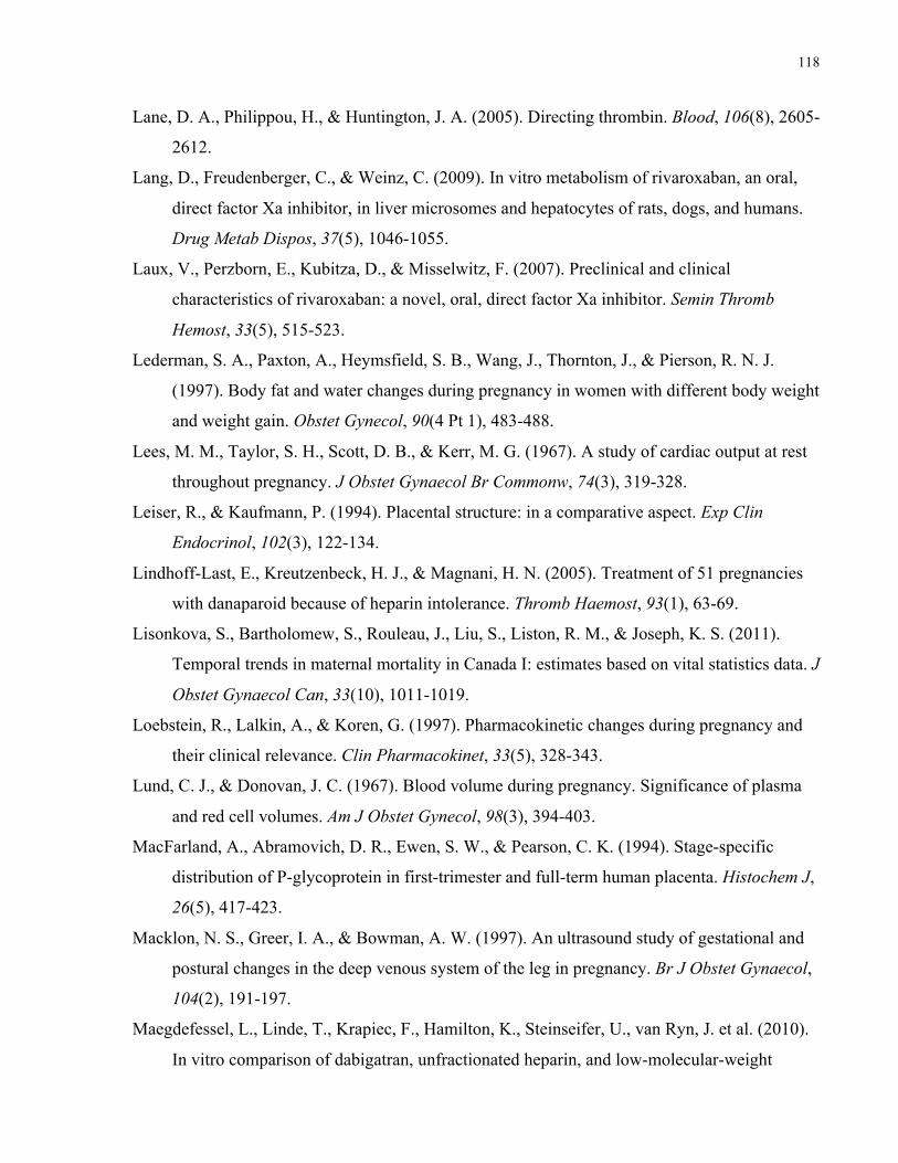
118
Lane, D. A., Philippou, H., & Huntington, J. A. (2005). Directing thrombin. Blood, 106(8), 2605-
2612.
Lang, D., Freudenberger, C., & Weinz, C. (2009). In vitro metabolism of rivaroxaban, an oral,
direct factor Xa inhibitor, in liver microsomes and hepatocytes of rats, dogs, and humans.
Drug Metab Dispos, 37(5), 1046-1055.
Laux, V., Perzborn, E., Kubitza, D., & Misselwitz, F. (2007). Preclinical and clinical
characteristics of rivaroxaban: a novel, oral, direct factor Xa inhibitor. Semin Thromb
Hemost, 33(5), 515-523.
Lederman, S. A., Paxton, A., Heymsfield, S. B., Wang, J., Thornton, J., & Pierson, R. N. J.
(1997). Body fat and water changes during pregnancy in women with different body weight
and weight gain. Obstet Gynecol, 90(4 Pt 1), 483-488.
Lees, M. M., Taylor, S. H., Scott, D. B., & Kerr, M. G. (1967). A study of cardiac output at rest
throughout pregnancy. J Obstet Gynaecol Br Commonw, 74(3), 319-328.
Leiser, R., & Kaufmann, P. (1994). Placental structure: in a comparative aspect. Exp Clin
Endocrinol, 102(3), 122-134.
Lindhoff-Last, E., Kreutzenbeck, H. J., & Magnani, H. N. (2005). Treatment of 51 pregnancies
with danaparoid because of heparin intolerance. Thromb Haemost, 93(1), 63-69.
Lisonkova, S., Bartholomew, S., Rouleau, J., Liu, S., Liston, R. M., & Joseph, K. S. (2011).
Temporal trends in maternal mortality in Canada I: estimates based on vital statistics data. J
Obstet Gynaecol Can, 33(10), 1011-1019.
Loebstein, R., Lalkin, A., & Koren, G. (1997). Pharmacokinetic changes during pregnancy and
their clinical relevance. Clin Pharmacokinet, 33(5), 328-343.
Lund, C. J., & Donovan, J. C. (1967). Blood volume during pregnancy. Significance of plasma
and red cell volumes. Am J Obstet Gynecol, 98(3), 394-403.
MacFarland, A., Abramovich, D. R., Ewen, S. W., & Pearson, C. K. (1994). Stage-specific
distribution of P-glycoprotein in first-trimester and full-term human placenta. Histochem J,
26(5), 417-423.
Macklon, N. S., Greer, I. A., & Bowman, A. W. (1997). An ultrasound study of gestational and
postural changes in the deep venous system of the leg in pregnancy. Br J Obstet Gynaecol,
104(2), 191-197.
Maegdefessel, L., Linde, T., Krapiec, F., Hamilton, K., Steinseifer, U., van Ryn, J. et al. (2010).
In vitro comparison of dabigatran, unfractionated heparin, and low-molecular-weight
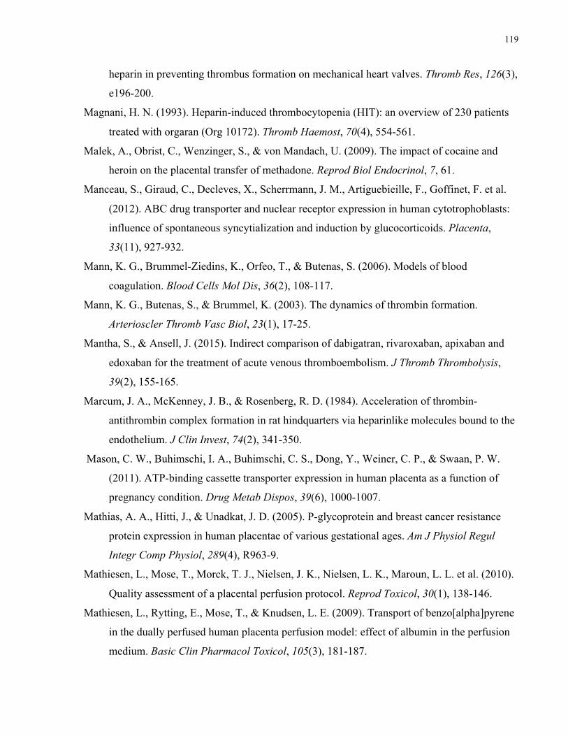
119
heparin in preventing thrombus formation on mechanical heart valves. Thromb Res, 126(3),
e196-200.
Magnani, H. N. (1993). Heparin-induced thrombocytopenia (HIT): an overview of 230 patients
treated with orgaran (Org 10172). Thromb Haemost, 70(4), 554-561.
Malek, A., Obrist, C., Wenzinger, S., & von Mandach, U. (2009). The impact of cocaine and
heroin on the placental transfer of methadone. Reprod Biol Endocrinol, 7, 61.
Manceau, S., Giraud, C., Decleves, X., Scherrmann, J. M., Artiguebieille, F., Goffinet, F. et al.
(2012). ABC drug transporter and nuclear receptor expression in human cytotrophoblasts:
influence of spontaneous syncytialization and induction by glucocorticoids. Placenta,
33(11), 927-932.
Mann, K. G., Brummel-Ziedins, K., Orfeo, T., & Butenas, S. (2006). Models of blood
coagulation. Blood Cells Mol Dis, 36(2), 108-117.
Mann, K. G., Butenas, S., & Brummel, K. (2003). The dynamics of thrombin formation.
Arterioscler Thromb Vasc Biol, 23(1), 17-25.
Mantha, S., & Ansell, J. (2015). Indirect comparison of dabigatran, rivaroxaban, apixaban and
edoxaban for the treatment of acute venous thromboembolism. J Thromb Thrombolysis,
39(2), 155-165.
Marcum, J. A., McKenney, J. B., & Rosenberg, R. D. (1984). Acceleration of thrombin-
antithrombin complex formation in rat hindquarters via heparinlike molecules bound to the
endothelium. J Clin Invest, 74(2), 341-350.
Mason, C. W., Buhimschi, I. A., Buhimschi, C. S., Dong, Y., Weiner, C. P., & Swaan, P. W.
(2011). ATP-binding cassette transporter expression in human placenta as a function of
pregnancy condition. Drug Metab Dispos, 39(6), 1000-1007.
Mathias, A. A., Hitti, J., & Unadkat, J. D. (2005). P-glycoprotein and breast cancer resistance
protein expression in human placentae of various gestational ages. Am J Physiol Regul
Integr Comp Physiol, 289(4), R963-9.
Mathiesen, L., Mose, T., Morck, T. J., Nielsen, J. K., Nielsen, L. K., Maroun, L. L. et al. (2010).
Quality assessment of a placental perfusion protocol. Reprod Toxicol, 30(1), 138-146.
Mathiesen, L., Rytting, E., Mose, T., & Knudsen, L. E. (2009). Transport of benzo[alpha]pyrene
in the dually perfused human placenta perfusion model: effect of albumin in the perfusion
medium. Basic Clin Pharmacol Toxicol, 105(3), 181-187.
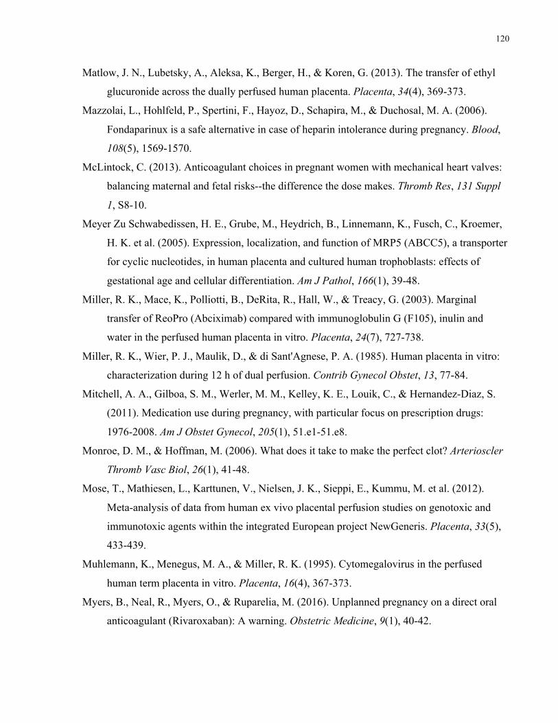
120
Matlow, J. N., Lubetsky, A., Aleksa, K., Berger, H., & Koren, G. (2013). The transfer of ethyl
glucuronide across the dually perfused human placenta. Placenta, 34(4), 369-373.
Mazzolai, L., Hohlfeld, P., Spertini, F., Hayoz, D., Schapira, M., & Duchosal, M. A. (2006).
Fondaparinux is a safe alternative in case of heparin intolerance during pregnancy. Blood,
108(5), 1569-1570.
McLintock, C. (2013). Anticoagulant choices in pregnant women with mechanical heart valves:
balancing maternal and fetal risks--the difference the dose makes. Thromb Res, 131 Suppl
1, S8-10.
Meyer Zu Schwabedissen, H. E., Grube, M., Heydrich, B., Linnemann, K., Fusch, C., Kroemer,
H. K. et al. (2005). Expression, localization, and function of MRP5 (ABCC5), a transporter
for cyclic nucleotides, in human placenta and cultured human trophoblasts: effects of
gestational age and cellular differentiation. Am J Pathol, 166(1), 39-48.
Miller, R. K., Mace, K., Polliotti, B., DeRita, R., Hall, W., & Treacy, G. (2003). Marginal
transfer of ReoPro (Abciximab) compared with immunoglobulin G (F105), inulin and
water in the perfused human placenta in vitro. Placenta, 24(7), 727-738.
Miller, R. K., Wier, P. J., Maulik, D., & di Sant'Agnese, P. A. (1985). Human placenta in vitro:
characterization during 12 h of dual perfusion. Contrib Gynecol Obstet, 13, 77-84.
Mitchell, A. A., Gilboa, S. M., Werler, M. M., Kelley, K. E., Louik, C., & Hernandez-Diaz, S.
(2011). Medication use during pregnancy, with particular focus on prescription drugs:
1976-2008. Am J Obstet Gynecol, 205(1), 51.e1-51.e8.
Monroe, D. M., & Hoffman, M. (2006). What does it take to make the perfect clot? Arterioscler
Thromb Vasc Biol, 26(1), 41-48.
Mose, T., Mathiesen, L., Karttunen, V., Nielsen, J. K., Sieppi, E., Kummu, M. et al. (2012).
Meta-analysis of data from human ex vivo placental perfusion studies on genotoxic and
immunotoxic agents within the integrated European project NewGeneris. Placenta, 33(5),
433-439.
Muhlemann, K., Menegus, M. A., & Miller, R. K. (1995). Cytomegalovirus in the perfused
human term placenta in vitro. Placenta, 16(4), 367-373.
Myers, B., Neal, R., Myers, O., & Ruparelia, M. (2016). Unplanned pregnancy on a direct oral
anticoagulant (Rivaroxaban): A warning. Obstetric Medicine, 9(1), 40-42.
121
Myllynen, P., Immonen, E., Kummu, M., & Vahakangas, K. (2009). Developmental expression
of drug metabolizing enzymes and transporter proteins in human placenta and fetal tissues.
Expert Opin Drug Metab Toxicol, 5(12), 1483-1499.
Myllynen, P., Kummu, M., Kangas, T., Ilves, M., Immonen, E., Rysa, J. et al. (2008).
ABCG2/BCRP decreases the transfer of a food-born chemical carcinogen, 2-amino-1-
methyl-6-phenylimidazo[4,5-b]pyridine (PhIP) in perfused term human placenta. Toxicol
Appl Pharmacol, 232(2), 210-217.
Myllynen, P., Pasanen, M., & Vahakangas, K. (2007). The fate and effects of xenobiotics in
human placenta. Expert Opin Drug Metab Toxicol, 3(3), 331-346.
Myllynen, P. K., Pienimaki, P. K., & Vahakangas, K. H. (2003). Transplacental passage of
lamotrigine in a human placental perfusion system in vitro and in maternal and cord blood
in vivo. Eur J Clin Pharmacol, 58(10), 677-682.
Myren, M., Mose, T., Mathiesen, L., & Knudsen, L. E. (2007). The human placenta--an
alternative for studying foetal exposure. Toxicol In Vitro, 21(7), 1332-1340.
Nagashige, M., Ushigome, F., Koyabu, N., Hirata, K., Kawabuchi, M., Hirakawa, T. et al.
(2003). Basal membrane localization of MRP1 in human placental trophoblast. Placenta,
24(10), 951-958.
Nanovskaya, T. N., Bowen, R. S., Patrikeeva, S. L., Hankins, G. D., & Ahmed, M. S. (2009).
Effect of plasma proteins on buprenorphine transfer across dually perfused placental
lobule. J Matern Fetal Neonatal Med, 22(8), 646-653.
Nau, H., Helge, H., & Luck, W. (1984). Valproic acid in the perinatal period: decreased maternal
serum protein binding results in fetal accumulation and neonatal displacement of the drug
and some metabolites. J Pediatr, 104(4), 627-634.
Novak, M., Schlagenhauf, A., Bernhard, H., Schweintzger, S., Leschnik, B., & Muntean, W.
(2011). Effect of rivaroxaban, in contrast to heparin, is similar in neonatal and adult
plasma. Blood Coagul Fibrinolysis, 22(7), 588-592.
O'Riordan, M. N., & Higgins, J. R. (2003). Haemostasis in normal and abnormal pregnancy. Best
Pract Res Clin Obstet Gynaecol, 17(3), 385-396.
Odutayo, A., & Hladunewich, M. (2012). Obstetric nephrology: renal hemodynamic and
metabolic physiology in normal pregnancy. Clin J Am Soc Nephrol, 7(12), 2073-2080.
Omarini, D., Pistotti, V., & Bonati, M. (1992). Placental perfusion. An overview of the literature.
J Pharmacol Toxicol Methods, 28(2), 61-66.
122
Osterud, B., & Rapaport, S. I. (1977). Activation of factor IX by the reaction product of tissue
factor and factor VII: additional pathway for initiating blood coagulation. Proc Natl Acad
Sci U S A, 74(12), 5260-5264.
Pacifici, G. M., & Nottoli, R. (1995). Placental transfer of drugs administered to the mother. Clin
Pharmacokinet, 28(3), 235-269.
Panigel, M., Pascaud, M., & Brun, J. L. (1967). [Radioangiographic study of circulation in the
villi and intervillous space of isolated human placental cotyledon kept viable by perfusion].
J Physiol (Paris), 59(1 Suppl), 277.
Parody, R., Oliver, A., Souto, J. C., & Fontcuberta, J. (2003). Fondaparinux (ARIXTRA) as an
alternative anti-thrombotic prophylaxis when there is hypersensitivity to low molecular
weight and unfractionated heparins. Haematologica, 88(11), ECR32.
Parry, E., Shields, R., & Turnbull, A. C. (1970). Transit time in the small intestine in pregnancy.
J Obstet Gynaecol Br Commonw, 77(10), 900-901.
Pasanen, M. (1999). The expression and regulation of drug metabolism in human placenta. Adv
Drug Deliv Rev, 38(1), 81-97.
Paxton, J. W. (1983). Alpha 1 -acid glycoprotein and binding of basic drugs. Methods Find Exp
Clin Pharmacol, 5(9), 635-648.
Peeters, L. L., Hobbelen, P. M., Verkeste, C. M., Vogel, G. M., Hasenack, H. G., Meuleman, D.
G. et al. (1986). Placental transfer of Org 10172, a low-molecular weight heparinoid, in the
awake late-pregnant guinea pig. Thromb Res, 44(3), 277-283.
Pennell, P. B., Newport, D. J., Stowe, Z. N., Helmers, S. L., Montgomery, J. Q., & Henry, T. R.
(2004). The impact of pregnancy and childbirth on the metabolism of lamotrigine.
Neurology, 62(2), 292-295.
Persson, B. L., Stenberg, P., Holmberg, L., & Astedt, B. (1980). Transamidating enzymes in
maternal plasma and placenta in human pregnancies complicated by intrauterine growth
retardation. J Dev Physiol, 2(1-2), 37-46.
Perzborn, E., Strassburger, J., Wilmen, A., Pohlmann, J., Roehrig, S., Schlemmer, K. H. et al.
(2005). In vitro and in vivo studies of the novel antithrombotic agent BAY 59-7939--an
oral, direct Factor Xa inhibitor. J Thromb Haemost, 3(3), 514-521.
Petrovic, V., Kojovic, D., Cressman, A., & Piquette-Miller, M. (2015). Maternal bacterial
infections impact expression of drug transporters in human placenta. Int
Immunopharmacol, 26(2), 349-356.
123
Pfizer Canada. (2015). Product Monograph - ELIQUIS®.
Piafsky, K. M., & Woolner, E. A. (1982). The binding of basic drugs to alpha-acid glycoprotein
in cord serum. J Pediatr, 100(5), 820-822.
Pinto, D. J., Orwat, M. J., Koch, S., Rossi, K. A., Alexander, R. S., Smallwood, A. et al. (2007).
Discovery of 1-(4-methoxyphenyl)-7-oxo-6-(4-(2-oxopiperidin-1-yl)phenyl)-4,5,6,7-
tetrahydro-1H -pyrazolo[3,4-c]pyridine-3-carboxamide (apixaban, BMS-562247), a highly
potent, selective, efficacious, and orally bioavailable inhibitor of blood coagulation factor
Xa. J Med Chem, 50(22), 5339-5356.
Pollex, E., Lubetsky, A., & Koren, G. (2008). The role of placental breast cancer resistance
protein in the efflux of glyburide across the human placenta. Placenta, 29(8), 743-747.
Pomp, E. R., Lenselink, A. M., Rosendaal, F. R., & Doggen, C. J. (2008). Pregnancy, the
postpartum period and prothrombotic defects: risk of venous thrombosis in the MEGA
study. J Thromb Haemost, 6(4), 632-637.
Raghavan, N., Frost, C. E., Yu, Z., He, K., Zhang, H., Humphreys, W. G. et al. (2009). Apixaban
metabolism and pharmacokinetics after oral administration to humans. Drug Metab Dispos,
37(1), 74-81.
Ray, J. G., & Chan, W. S. (1999). Deep vein thrombosis during pregnancy and the puerperium: a
meta-analysis of the period of risk and the leg of presentation. Obstet Gynecol Surv, 54(4),
265-271.
Reboud, P., Goulade, J., Groslambert, P., & Colomb, M. (1963). The influence of normal
pregnancy and the postpartum state on plasma proteins and lipids. Am J Obstet Gynecol,
86, 820-828.
Reverdiau-Moalic, P., Delahousse, B., Body, G., Bardos, P., Leroy, J., & Gruel, Y. (1996).
Evolution of blood coagulation activators and inhibitors in the healthy human fetus. Blood,
88(3), 900-906.
Reynolds, F., & Knott, C. (1989). Pharmacokinetics in pregnancy and placental drug transfer.
Oxf Rev Reprod Biol, 11, 389-449.
Ridd, M. J., Brown, K. F., Nation, R. L., & Collier, C. B. (1983). Differential transplacental
binding of diazepam: causes and implications. Eur J Clin Pharmacol, 24(5), 595-601.
Risau, W. (1997). Mechanisms of angiogenesis. Nature, 386(6626), 671-674.
124
Rodesch, F., Simon, P., Donner, C., & Jauniaux, E. (1992). Oxygen measurements in
endometrial and trophoblastic tissues during early pregnancy. Obstet Gynecol, 80(2), 283-
285.
Rodger, M. A., Hague, W. M., Kingdom, J., Kahn, S. R., Karovitch, A., Sermer, M. et al. (2014).
Antepartum dalteparin versus no antepartum dalteparin for the prevention of pregnancy
complications in pregnant women with thrombophilia (TIPPS): a multinational open-label
randomised trial. Lancet, 384(9955), 1673-1683.
Roehrig, S., Straub, A., Pohlmann, J., Lampe, T., Pernerstorfer, J., Schlemmer, K. H. et al.
(2005). Discovery of the novel antithrombotic agent 5-chloro-N-({(5S)-2-oxo-3- [4-(3-
oxomorpholin-4-yl)phenyl]-1,3-oxazolidin-5-yl}methyl)thiophene- 2-carboxamide (BAY
59-7939): an oral, direct factor Xa inhibitor. J Med Chem, 48(19), 5900-5908.
Royal College of Obstetricians and Gynaecologists. (2009). Guideline 37a: Reducing the Risk of
Thrombosis and Embolism During Pregnancy and the Puerperium. Clinical Green-top
Guidelines., 1-35.
Ryu, R. J., Eyal, S., Easterling, T. R., Caritis, S. N., Venkataraman, R., Hankins, G. et al. (2016).
Pharmacokinetics of metoprolol during pregnancy and lactation. J Clin Pharmacol, 56(5),
581-589.
Sattar, N., Greer, I. A., Rumley, A., Stewart, G., Shepherd, J., Packard, C. J. et al. (1999). A
longitudinal study of the relationships between haemostatic, lipid, and oestradiol changes
during normal human pregnancy. Thromb Haemost, 81(1), 71-75.
Scaglione, F. (2013). New oral anticoagulants: comparative pharmacology with vitamin K
antagonists. Clin Pharmacokinet, 52(2), 69-82.
Schaefer, C., Hannemann, D., Meister, R., Elefant, E., Paulus, W., Vial, T. et al. (2006). Vitamin
K antagonists and pregnancy outcome. A multi-centre prospective study. Thromb Haemost,
95(6), 949-957.
Schenker, S., Yang, Y., Mattiuz, E., Tatum, D., & Lee, M. (1999). Olanzapine transfer by human
placenta. Clin Exp Pharmacol Physiol, 26(9), 691-697.
Schneider, H., & Huch, A. (1985). Dual in vitro perfusion of an isolated lobe of human placenta:
method and instrumentation. Contrib Gynecol Obstet, 13, 40-47.
Schneider, H., Panigel, M., & Dancis, J. (1972). Transfer across the perfused human placenta of
antipyrine, sodium and leucine. Am J Obstet Gynecol, 114(6), 822-828.
125
Serov, A. S., Salafia, C., Grebenkov, D. S., & Filoche, M. (2016). The role of morphology in
mathematical models of placental gas exchange. J Appl Physiol (1985), 120(1), 17-28.
Serrano, M. A., Macias, R. I., Briz, O., Monte, M. J., Blazquez, A. G., Williamson, C. et al.
(2007). Expression in human trophoblast and choriocarcinoma cell lines, BeWo, Jeg-3 and
JAr of genes involved in the hepatobiliary-like excretory function of the placenta. Placenta,
28(2-3), 107-117.
Shintaku, K., Hori, S., Satoh, H., Tsukimori, K., Nakano, H., Fujii, T. et al. (2012). Prediction
and evaluation of fetal toxicity induced by NSAIDs using transplacental kinetic parameters
obtained from human placental perfusion studies. Br J Clin Pharmacol, 73(2), 248-256.
Simpson, E. L., Lawrenson, R. A., Nightingale, A. L., & Farmer, R. D. (2001). Venous
thromboembolism in pregnancy and the puerperium: incidence and additional risk factors
from a London perinatal database. BJOG, 108(1), 56-60.
Sprengers, E. D., & Kluft, C. (1987). Plasminogen activator inhibitors. Blood, 69(2), 381-387.
Spyropoulos, A. C. (2007). Investigational treatments of venous thromboembolism. Expert Opin
Investig Drugs, 16(4), 431-440.
St-Pierre, M. V., Serrano, M. A., Macias, R. I., Dubs, U., Hoechli, M., Lauper, U. et al. (2000).
Expression of members of the multidrug resistance protein family in human term placenta.
Am J Physiol Regul Integr Comp Physiol, 279(4), R1495-503.
Stangier, J., Rathgen, K., Stahle, H., Gansser, D., & Roth, W. (2007). The pharmacokinetics,
pharmacodynamics and tolerability of dabigatran etexilate, a new oral direct thrombin
inhibitor, in healthy male subjects. Br J Clin Pharmacol, 64(3), 292-303.
Stangier, J., Rathgen, K., Stahle, H., & Mazur, D. (2010). Influence of renal impairment on the
pharmacokinetics and pharmacodynamics of oral dabigatran etexilate: an open-label,
parallel-group, single-centre study. Clin Pharmacokinet, 49(4), 259-268.
Stangier, J., & Clemens, A. (2009). Pharmacology, pharmacokinetics, and pharmacodynamics of
dabigatran etexilate, an oral direct thrombin inhibitor. Clinical and Applied
Thrombosis/Hemostasis, 15(1 suppl), 9S-16S.
Stanley, E. L., Hume, R., Visser, T. J., & Coughtrie, M. W. (2001). Differential expression of
sulfotransferase enzymes involved in thyroid hormone metabolism during human placental
development. J Clin Endocrinol Metab, 86(12), 5944-5955.
Stirling, Y., Woolf, L., North, W. R., Seghatchian, M. J., & Meade, T. W. (1984). Haemostasis
in normal pregnancy. Thromb Haemost, 52(2), 176-182.
126
Sultan, A. A., Tata, L. J., West, J., Fiaschi, L., Fleming, K. M., Nelson-Piercy, C. et al. (2013).
Risk factors for first venous thromboembolism around pregnancy: a population-based
cohort study from the United Kingdom. Blood, 121(19), 3953-3961.
Sun, M., Kingdom, J., Baczyk, D., Lye, S. J., Matthews, S. G., & Gibb, W. (2006). Expression of
the multidrug resistance P-glycoprotein, (ABCB1 glycoprotein) in the human placenta
decreases with advancing gestation. Placenta, 27(6-7), 602-609.
Syme, M. R., Paxton, J. W., & Keelan, J. A. (2004). Drug transfer and metabolism by the human
placenta. Clin Pharmacokinet, 43(8), 487-514.
Taniguchi, S., Fukuda, I., Minakawa, M., Watanabe, K., Daitoku, K., & Suzuki, Y. (2008).
Emergency pulmonary embolectomy during the second trimester of pregnancy: report of a
case. Surg Today, 38(1), 59-61.
Tarrade, A., Lai Kuen, R., Malassiné, A., Tricottet, V., Blain, P., Vidaud, M. et al. (2001).
Characterization of human villous and extravillous trophoblasts isolated from first trimester
placenta. Lab Invest, 81(9), 1199-11211.
Thornton, P., & Douglas, J. (2010). Coagulation in pregnancy. Best Pract Res Clin Obstet
Gynaecol, 24(3), 339-352.
Townsley, D. M. (2013). Hematologic complications of pregnancy. Semin Hematol, 50(3), 222-
231.
Tracy, T. S., Venkataramanan, R., Glover, D. D., & Caritis, S. N. (2005). Temporal changes in
drug metabolism (CYP1A2, CYP2D6 and CYP3A Activity) during pregnancy. Am J
Obstet Gynecol, 192(2), 633-639.
Vahakangas, K., & Myllynen, P. (2006). Experimental methods to study human transplacental
exposure to genotoxic agents. Mutat Res, 608(2), 129-135.
van der Aa, E. M., Peereboom-Stegeman, J. H., Noordhoek, J., Gribnau, F. W., & Russel, F. G.
(1998). Mechanisms of drug transfer across the human placenta. Pharm World Sci, 20(4),
139-148.
van Ryn J, H. N., Waldman L, Wienen W. (2008). Dabigatran inhibits both clot-bound and fluid-
phase thrombin in vitro: comparison to heparin and hirudin [abstract 570]. Arterioscler
Thromb Vasc Biol, 28, e136-e137.
Verdy, E., Bessous, V., Dreyfus, M., Kaplan, C., Tchernia, G., & Uzan, S. (1997). Longitudinal
analysis of platelet count and volume in normal pregnancy. Thromb Haemost, 77(4), 806-
807.
127
Villani, P., Floridia, M., Pirillo, M. F., Cusato, M., Tamburrini, E., Cavaliere, A. F. et al. (2006).
Pharmacokinetics of nelfinavir in HIV-1-infected pregnant and nonpregnant women. Br J
Clin Pharmacol, 62(3), 309-315.
Wang, L., He, K., Maxwell, B., Grossman, S. J., Tremaine, L. M., Humphreys, W. G. et al.
(2011). Tissue distribution and elimination of [14C]apixaban in rats. Drug Metab Dispos,
39(2), 256-264.
Wang, J. H., Scollard, D. A., Teng, S., Reilly, R. M., & Piquette-Miller, M. (2005). Detection of
P-glycoprotein activity in endotoxemic rats by 99mTc-sestamibi imaging. J Nucl Med,
46(9), 1537-1545.
Wang, L., Zhang, D., Raghavan, N., Yao, M., Ma, L., Frost, C. E. et al. (2010). In vitro
assessment of metabolic drug-drug interaction potential of apixaban through cytochrome
P450 phenotyping, inhibition, and induction studies. Drug Metab Dispos, 38(3), 448-458.
Weinz, C., Buetehorn, U., Daehler, H. P., Kohlsdorfer, C., Pleiss, U., Sandmann, S. et al. (2005).
Pharmacokinetics of BAY 59-7939--an oral, direct Factor Xa inhibitor--in rats and dogs.
Xenobiotica, 35(9), 891-910.
Weinz, C., Schwarz, T., Kubitza, D., Mueck, W., & Lang, D. (2009). Metabolism and excretion
of rivaroxaban, an oral, direct factor Xa inhibitor, in rats, dogs, and humans. Drug Metab
Dispos, 37(5), 1056-1064.
Weitz, J. I., Leslie, B., & Hudoba, M. (1998). Thrombin binds to soluble fibrin degradation
products where it is protected from inhibition by heparin-antithrombin but susceptible to
inactivation by antithrombin-independent inhibitors. Circulation, 97(6), 544-552.
Weitz, J. I., Hudoba, M., Massel, D., Maraganore, J., & Hirsh, J. (1990). Clot-bound thrombin is
protected from inhibition by heparin-antithrombin III but is susceptible to inactivation by
antithrombin III-independent inhibitors. Journal of Clinical Investigation, 86(2), 385.
Wier, P. J., & Miller, R. K. (1985). Oxygen transfer as an indicator of perfusion variability in the
isolated human placental lobule. Contrib Gynecol Obstet, 13, 127-131.
Wijesiriwardana, A., Lees, D. A., & Lush, C. (2006). Fondaparinux as anticoagulant in a
pregnant woman with heparin allergy. Blood Coagul Fibrinolysis, 17(2), 147-149.
Yeboah, D., Sun, M., Kingdom, J., Baczyk, D., Lye, S. J., Matthews, S. G. et al. (2006).
Expression of breast cancer resistance protein (BCRP/ABCG2) in human placenta
throughout gestation and at term before and after labor. Can J Physiol Pharmacol, 84(12),
1251-1258.
128
Young, S. K., Al-Mondhiry, H. A., Vaida, S. J., Ambrose, A., & Botti, J. J. (2008). Successful
use of argatroban during the third trimester of pregnancy: case report and review of the
literature. Pharmacotherapy, 28(12), 1531-1536.
Zhang, D., He, K., Herbst, J. J., Kolb, J., Shou, W., Wang, L. et al. (2013). Characterization of
efflux transporters involved in distribution and disposition of apixaban. Drug Metab
Dispos, 41(4), 827-835.
Zilliacus, H., Ottelin, A. M., & Mattsson, T. (1966). Blood clotting and fibrinolysis in human
foetuses. Biol Neonat, 10(1), 108-112.

129
Appendices
130
Appendix A. Letter of Information and Consent Form
The Role of the Placenta in Fetal Pharmacology Page 1 of 6
Letter of Information and Consent to Participate in a Research Study Study Title: The Role of the Placenta in Fetal Pharmacology
Investigators: On Site Principal Investigator: Dr Howard Berger Perinatalogist Department of Obstetrics and Gynecology St. Michael’s Hospital Phone: (416) 867- 7460 Ext. 8408 (Available Monday - Friday 9 am – 4 pm) Off-Site Principal Investigator: Dr.Gideon Koren, Director, The Motherisk Program Division of Clinical Pharmacology and Toxicology The Hospital for Sick Children Phone: (416) 813-5781 (Available Monday – Friday 9 am – 4 pm) Introduction: Before agreeing to take part in this research study, it is important that you read the information in this research consent form. It includes details we think you need to know in order to decide if you wish to take part in the study. If you have any questions, please ask the study doctor or study staff to explain any words you don’t understand before signing this consent form. You will also have the opportunity to ask any additional questions on the day of surgery. Make sure all your questions have been answered to your satisfaction before signing this document.
All research is voluntary. You may also wish to discuss the study with your family doctor, a family member or close friend. It is important you know that this study is from the Hospital for Sick Children, and St. Michael’s Hospital is one of the sites where we are recruiting participants. Conflicts of Interest The principal investigators, Dr. Howard Berger and Dr. Gideon Koren, and the other research team members have no conflicts of interest to declare.
131
The Role of the Placenta in Fetal Pharmacology Page 2 of 6
Sponsorship The sponsor of this study is Dr. Gideon Koren and the Hospital for Sick Children. The funder of this research study is the Canadian Institutes of Health Research. Background Information: The placenta research laboratory at the Hospital for Sick Children is one of the few laboratories in North America currently studying drug transfer in the human placenta. The placenta is the special pregnancy tissue in the uterus that processes the mother's blood to give the growing baby nutrition and other important blood products. The placenta connects the developing baby to the uterine wall to allow nutrient uptake, waste elimination, and gas exchange via the mother's blood supply. In our lab, we employ a technique called “placental perfusion”. This unique technique separates the functions of the placenta from both the maternal and fetal influences. The use of this model will further our understanding of the transport and behavior of certain medications across the human placenta, throughout pregnancy. Purpose of Research: Pregnancy is a special state in which there are many physical changes that occur to both the fetus and the mother. While pregnant, some mothers need to take medication in order to maintain a healthy pregnancy. Some of these compounds can reach the growing baby by passing through the placenta. We would like to understand this process better. It is very important for researchers and doctors to better understand how medications cross the placenta, so that in the future we may help women protect their unborn fetus from harm. This is a multi-centre study and participants are recruited at St. Michael’s Hospital and Mount Sinai Hospital. As part of this study, we would like to collect whole placentaes from 100 women at St. Michael’s Hospital, and 50 women at Mount Sinai. Description of Research: Normally, once your baby is born, the umbilical cord is clamped and the baby is separated from the placenta. The placenta is then delivered and thrown away. If you agree to participate in this study, instead of the placenta being disposed of, we would like to use it as part of our study in our on-site laboratory. We will set up a placenta perfusion, which is a model that mimics the blood circulation systems for the mother and developing baby. We can use this model to study the transport of medications across the placenta without having to administer any medication to pregnant women. We will collect samples while using this model to determine if the study medication crosses the placenta, and these samples will be sent to the Hospital for Sick Children for further analysis. After our experiment, your placenta will be disposed of in the usual fashion according to the hospital ward’s guidelines.
132
The Role of the Placenta in Fetal Pharmacology Page 3 of 6
No additional procedures or modifications to your care are required to assess the placenta after delivery. If your treating doctor decides that your placenta requires special testing after delivery as part of your hospital care, we will not collect it as part of this research study and you will be immediately withdrawn from the study. Potential Harms (Injury, Discomforts or Inconveniences): Collection of these samples will not affect your labour or the delivery of your baby. The whole placenta will only be assessed for research after it has been delivered. The assessment will be done at the time the placenta is routinely disposed. The collection carries no risk to you or your baby. Potential Benefits: There are no direct benefits from participating in this study. The results from this study may improve our understanding of drug transfer across the human placenta. In addition, we hope that the information obtained in this study will allow us to develop new treatment options for women during pregnancy while protecting their unborn fetus. Alternatives to Participation If you decide that you do not want to take part in this study, your choice will not affect the care that you or your family would otherwise receive. You will continue to receive the standard of care during your pregnancy and after you deliver your baby. Protecting Your Health Information: This consent form and all data collection forms will be held in the strictest confidence. To protect your anonymity, your name will not appear on any study files except for this consent form. Information from this study will be kept in a locked filing cabinet in the locked laboratories at the St. Michael’s Hospital and on a password-protected computer database in the research laboratories at the Hospital for Sick Children for three years and only study personnel and the Research Ethics Board office will have access to this information. In addition to your placenta that will be used in our perfusion experiment, we will collect your age and the gestational age of your baby at the time of delivery. Following the experiment, your placenta will be securely disposed of as per standard hospital procedures. None of these research results will be placed in your medical records. Once the study is complete, the results will be summarized and submitted to a medical journal for publication. The outcome of this study may be presented at conferences, scientific meetings and other public forums. It is important that you are aware that you will not be identified in any of these reports and your confidentiality will be maintained. No information that discloses your identity will be released or published without consent.
133
The Role of the Placenta in Fetal Pharmacology Page 4 of 6
Experience in similar studies indicates that the greatest risk in this study to you is the unintentional release of information from your health records. The study personnel will protect your records and keep all the information in your study file confidential, including your name. The chance that this information will accidentally be given to someone else is small. You may be contacted by a representative of the Research Ethics Board to ask questions about your experience with the recruitment and consent process or regarding your experience in the study, with a view to assuring and improving the quality of these processes. Potential Cost of Participation and Reimbursement: There are no costs associated with participating in this study. You will not be reimbursed for your participation in this study. Compensation for Injury: If you become ill or are physically injured as a result of participation in this study, medical treatment will be provided to you in the same manner as you would ordinarily obtain any other medical treatment. In no way does signing this consent form waive your legal rights nor does it relieve the investigators or involved institutions from their legal and professional responsibilities. Participation and Withdrawal: Participation in any research study is voluntary. If you choose not to participate, you and your baby will continue to have access to customary care at St. Michael’s Hospital. If you decide to participate in this study you can change your mind without giving a reason, and you may withdraw from the study at any time without any effect on the care you and your baby will receive at St. Michael’s Hospital. Upon withdrawal from the study, it is within your rights to request the withdrawal of any of your data compiled up to that point. Study personnel have the right to stop your participation in the study if your doctor orders additional testing for your placenta and thinks it is not in your best interest to continue with our study. New Findings or Information We may learn new things during the study that you may need to know. We can also learn about things that might make you want to stop participating in the study. If so, you will be notified about any new information in a timely manner. You may also be asked to sign a new consent form discussing these new findings if you decide to continue in the research study.
134
The Role of the Placenta in Fetal Pharmacology Page 5 of 6
Development for Commercial Gain: Research carried out on your samples by researchers at the Hospital for Sick Children, or their collaborators, may lead to the development of marketable treatments, devices, new drugs or patentable procedures. By participating in this study you will not benefit directly from any commercial products that may arise from this research Research Ethics Board Contact: If you have any further questions about your rights as a research participant, you may contact the following individuals: Dr. David Mazer Chair, Research Ethics Board St. Michael’s Hospital 416-864-6060 ext 2557 Lorelei Nardi Team Leader, Research Ethics Office Hospital for Sick Children 416-813-5718 Further Questions: You have been given a copy of this information and consent form. If you have any questions about taking part in this study, you may contact Dr. Gideon Koren (The Hospital for Sick Children) at (416) 813-5781 or Dr. Howard Berger (St. Michael’s Hospital) at (416) 867-7460 Ext. 8408.
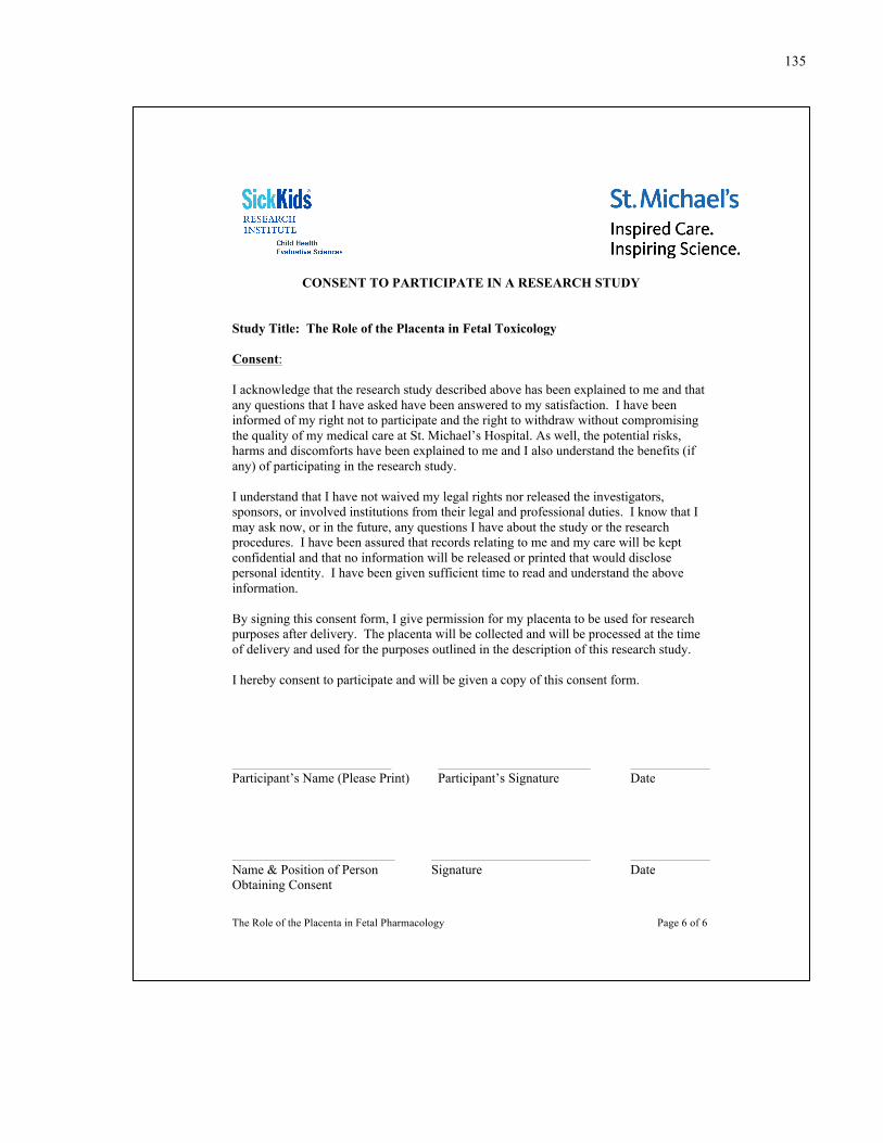
135
The Role of the Placenta in Fetal Pharmacology Page 6 of 6
CONSENT TO PARTICIPATE IN A RESEARCH STUDY
Study Title: The Role of the Placenta in Fetal Toxicology Consent: I acknowledge that the research study described above has been explained to me and that any questions that I have asked have been answered to my satisfaction. I have been informed of my right not to participate and the right to withdraw without compromising the quality of my medical care at St. Michael’s Hospital. As well, the potential risks, harms and discomforts have been explained to me and I also understand the benefits (if any) of participating in the research study. I understand that I have not waived my legal rights nor released the investigators, sponsors, or involved institutions from their legal and professional duties. I know that I may ask now, or in the future, any questions I have about the study or the research procedures. I have been assured that records relating to me and my care will be kept confidential and that no information will be released or printed that would disclose personal identity. I have been given sufficient time to read and understand the above information. By signing this consent form, I give permission for my placenta to be used for research purposes after delivery. The placenta will be collected and will be processed at the time of delivery and used for the purposes outlined in the description of this research study. I hereby consent to participate and will be given a copy of this consent form.
Participant’s Name (Please Print) Participant’s Signature Date Name & Position of Person Signature Date Obtaining Consent
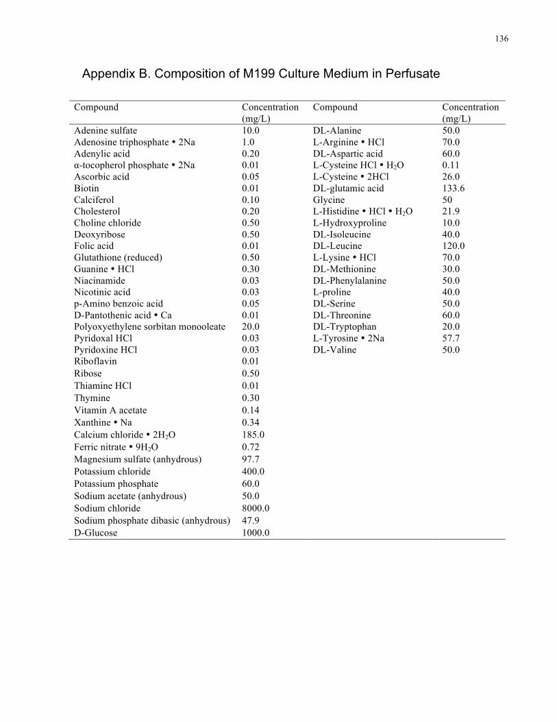
136
Appendix B. Composition of M199 Culture Medium in Perfusate
Compound Concentration (mg/L)
Compound Concentration (mg/L)
Adenine sulfate 10.0 DL-Alanine 50.0 Adenosine triphosphate � 2Na 1.0 L-Arginine � HCl 70.0 Adenylic acid 0.20 DL-Aspartic acid 60.0 α-tocopherol phosphate � 2Na 0.01 L-Cysteine HCl � H2O 0.11 Ascorbic acid 0.05 L-Cysteine � 2HCl 26.0 Biotin 0.01 DL-glutamic acid 133.6 Calciferol 0.10 Glycine 50 Cholesterol 0.20 L-Histidine � HCl � H2O 21.9 Choline chloride 0.50 L-Hydroxyproline 10.0 Deoxyribose 0.50 DL-Isoleucine 40.0 Folic acid 0.01 DL-Leucine 120.0 Glutathione (reduced) 0.50 L-Lysine � HCl 70.0 Guanine � HCl 0.30 DL-Methionine 30.0 Niacinamide 0.03 DL-Phenylalanine 50.0 Nicotinic acid 0.03 L-proline 40.0 p-Amino benzoic acid 0.05 DL-Serine 50.0 D-Pantothenic acid � Ca 0.01 DL-Threonine 60.0 Polyoxyethylene sorbitan monooleate 20.0 DL-Tryptophan 20.0 Pyridoxal HCl 0.03 L-Tyrosine � 2Na 57.7 Pyridoxine HCl 0.03 DL-Valine 50.0 Riboflavin 0.01 Ribose 0.50 Thiamine HCl 0.01 Thymine 0.30 Vitamin A acetate 0.14 Xanthine � Na 0.34 Calcium chloride � 2H2O 185.0 Ferric nitrate � 9H2O 0.72 Magnesium sulfate (anhydrous) 97.7 Potassium chloride 400.0 Potassium phosphate 60.0 Sodium acetate (anhydrous) 50.0 Sodium chloride 8000.0 Sodium phosphate dibasic (anhydrous) 47.9 D-Glucose 1000.0
137
Copyright Acknowledgements
138
139
140
141
142
143

144