Embed Size (px)
Citation preview

Placental Pathology
for general pathologists
Prof Neil Sebire
GOSH/ICH (UCL)
London UK

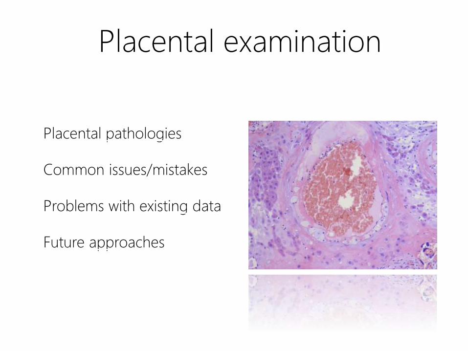
Placental examination
Placental pathologies
Common issues/mistakes
Problems with existing data
Future approaches

Routine tissue handling in
Placental PathologyDelay from delivery to
fixation / sampling (4
degrees)
1-48 hrs
Formalin fixation
Staining
Block taking
Embedding to paraffin
Inhibits autolysis (rapid)
Structurally cross links
proteins (slow)
21 3 4 5 6 7 8 9 1
0
1
1
1
2
1
3
1
4
1
5
1
6

Techniques in Placental
PathologyMorphometry
Histomorphology
Histomorphometry /
stereology
Immunostaining
Injection studies
Expression / OMICS

Placental examination
Why bother?
•Recurrence risk
•Neonatal management
•Maternal management
•Pathophysiology
•Medicolegal aspects

Value of of placental examination: Operator dependancy
Sun et al 2002
40% erroneous reports
10% overdiagnosis
90% omissions
>other subspecialities

-Poor clinical phenotypes
-Definition of lesions / use of terminology
-Interpretation of lesions
-Blinding and bias
Difficulties with placental pathology

International Placental Pathology Consensus Meeting Amsterdam 2014
Consensus on features of:
-Maternovascular malperfusion
-Fetalplacental malperfusion
-Stem vessel occlusion
-Intramural fibrin deposition
No consensus on other ‘hypoxic’ features
No consensus on clinical significance / correlates
CAP Guidelines (1997), RCOG guidelines (2000)
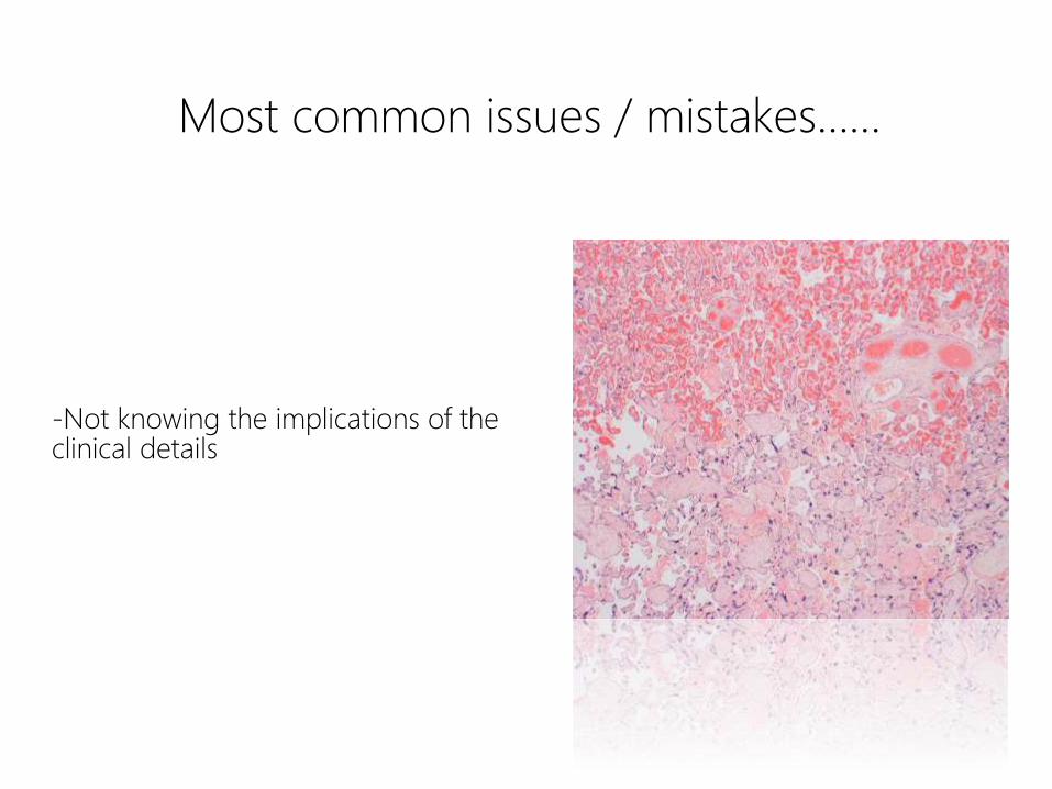
-Not knowing the implications of the clinical details
Most common issues / mistakes……

CAP Guidelines (1997), RCOG guidelines (2000)
-Not knowing the implications of the clinical details ….
-’Overfitting’ findings to the clinical details!
Most common issues / mistakes……

CAP Guidelines (1997), RCOG guidelines (2000)
-Not knowing the implications of the clinical details ….
-’Overfitting’ findings to the clinical details!
-Underestimating importance of macroscopic findings (depending on hx…….)
Most common issues / mistakes……

CAP Guidelines (1997), RCOG guidelines (2000)
-Not knowing the implications of the clinical details ….
-’Overfitting’ findings to the clinical details!
-Underestimating importance of macroscopic findings (depending on hx…….)
-Not recognising rare entities
Most common issues / mistakes……

CAP Guidelines (1997), RCOG guidelines (2000)
-Not knowing the implications of the clinical details ….
-’Overfitting’ findings to the clinical details!
-Underestimating importance of macroscopic findings (depending on hx…….)
-Not recognising rare entities
-Overcalling normal variants
Most common issues / mistakes……

Macroscopic abnormalities
Shape
Cord insertion
Abnormal vessels
Abruption
Infarcts
Jelly-like
Other (?MPVFD)


Retroplacental haematoma / abruption
Feature of uteroplacental disease
Association with:
Pre-eclampsia
Smoking
Cocaine
Thrombophilias
PROM / Chorioamnionitis

Placental abruption
•Clinical abruption - 30% histo confirmation•Retroplacental haematoma histo – 35% Hx abruption
Clinical abruption 1% pregnancies
Histology: congestion+retroplacental haem+compression+acute infarct


Placental Examination: Cambridge Study
• 1,159 singleton unselected women recruited at booking
• Objective measurements from calibrated images
• Histological exam, routine blocks
• Two paediatric pathologists
• Blinded to all clinical information-study number only
Total population
Age 29
GA 39 weeks
PET 2%
PIH 3%
SGA 6%
Pathak et al 2011-

Placental Examination: Cambridge Study
• Shape results

Aetiology of spontaneous PTB:
Studies of pathology
30-50% infection / inflamm
20-30% uteroplacental disease / ischaemic?
20-30% No pathological entity
Salafia et al 1992, Arias et al 1993
50%
20%
30%
Infection UPD Nil
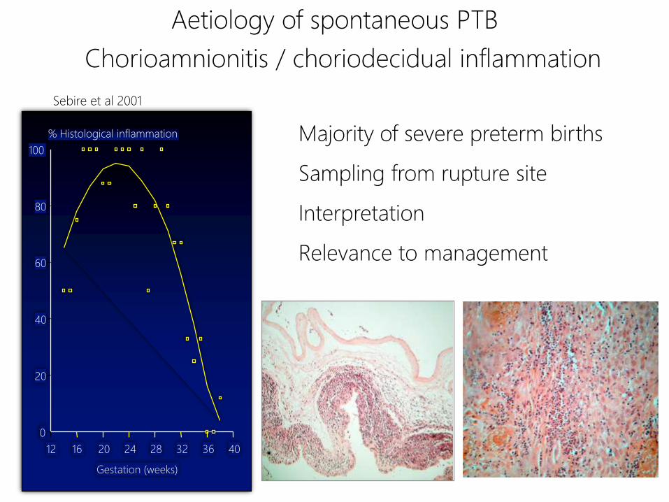
Aetiology of spontaneous PTB
Chorioamnionitis / choriodecidual inflammation
Gestation (weeks)
% Histological inflammation
12 16 20 24 28 32 36 40
0
20
40
60
80
100
Sebire et al 2001
Majority of severe preterm births
Sampling from rupture site
Interpretation
Relevance to management
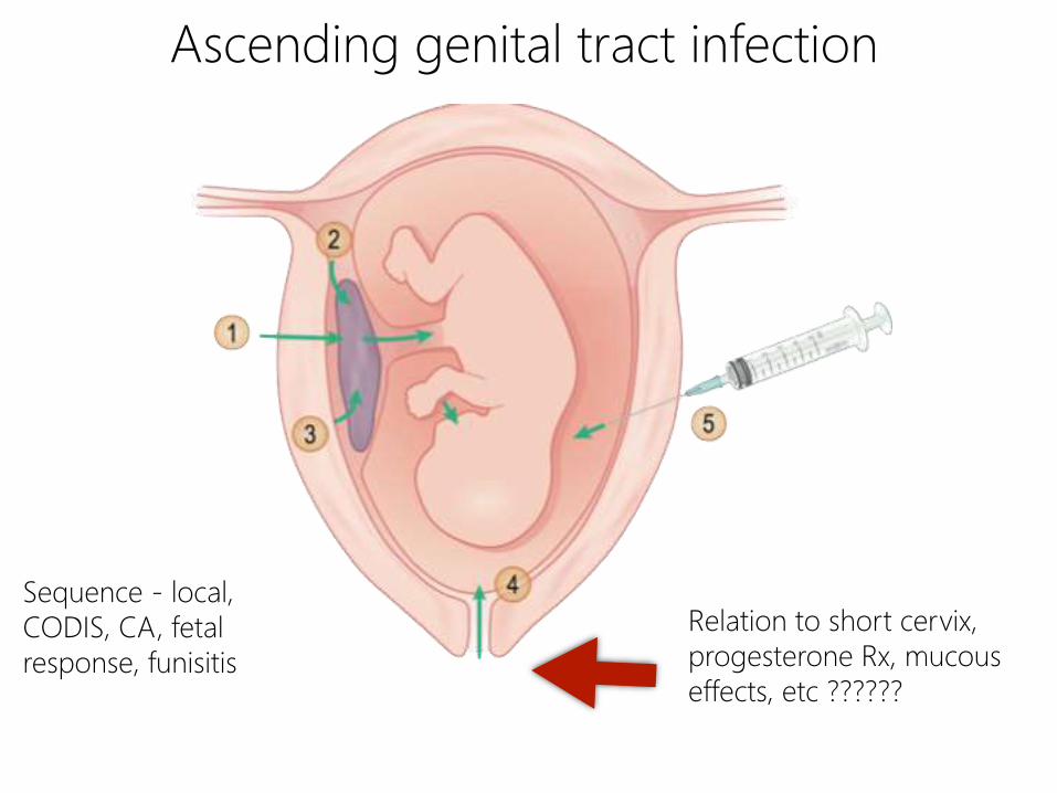
Ascending genital tract infection
Relation to short cervix,
progesterone Rx, mucous
effects, etc ??????
Sequence - local,
CODIS, CA, fetal
response, funisitis


Chorioamnionitis and brain injury
Histologic CA in term infants
relation to CP
Wu et al 2003
RR 8.9 (95% CI 1.9-40)
?direct effect or via chorionic plate thrombi

Placental changes described in FGR
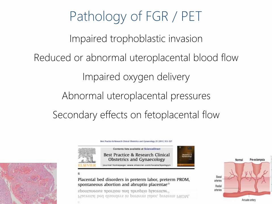
Pathology of FGR / PET
Impaired trophoblastic invasion
Reduced or abnormal uteroplacental blood flow
Impaired oxygen delivery
Abnormal uteroplacental pressures
Secondary effects on fetoplacental flow

Roberts 2008

Placental changes in FGR
Smaller size
Reduced
surface area
Villous
hypovascularity
Infarcts
Decidual
vasculopathy

Placental changes in FGR
Reduced
villous
branching
Increased
Maturation /
Terminal
villous
hypoplasia
Villitis
NRBCs
Increased
apoptosis
Shorter
telomeres
Transporter and growth
factor alterations


Other microscopic /
molecular abnormalities in
FGR
Villitis
CHI
Functional abnormalities


Villitis of unknown aetiology
Mechanism?
Mainly maternal
CD68 macrophages, CD3 T-cells
?Immune dysregulation
Redline & Patterson 1993; Kapur et al 2004;
Myerson et al 2006
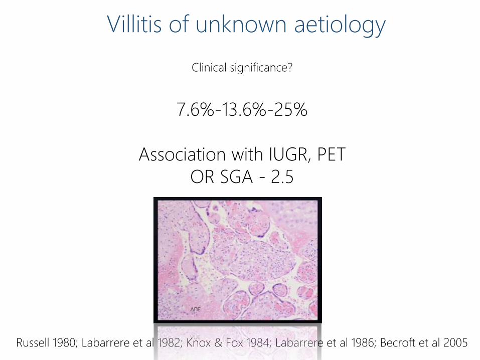
Villitis of unknown aetiology
Clinical significance?
7.6%-13.6%-25%
Association with IUGR, PET
OR SGA - 2.5
Russell 1980; Labarrere et al 1982; Knox & Fox 1984; Labarrere et al 1986; Becroft et al 2005

Chronic histiocytic intervillositis

CD68

Chronic histiocytic intervillositis
Miscarriage / IUD - 80%
20% pregnancies reach term
30% viable IUGR
High recurrence risk (70%)
?Immunological mechanism
Optimal treatment unknown
CD68 CD3


MFI / MPVFD
Bane & Gillan 2003 1/3500
Fetal loss 31%
IUGR 100%
Preterm 33%
Recurrence 18%
Katzman & Genest 2002 1/244
IUGR 31%
Recurrence 40%
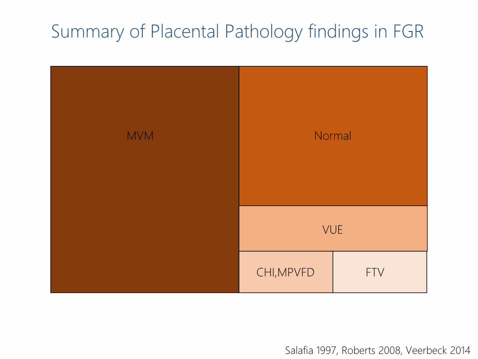
Summary of Placental Pathology findings in FGR
Salafia 1997, Roberts 2008, Veerbeck 2014
MVM Normal
VUE
FTVCHI,MPVFD

Placental changes in IUGR; problems…
Poor correlation with Doppler or severity
(infarcts and atherosis best)
No lesion pathognomonic, many are histologically normal
(approx 30-50% vs 70% controls)
Infarct 25-40 vs 10-15% controls
Villitis 8-21 vs 3-5% controls
Maturation/knots 90% vs ???
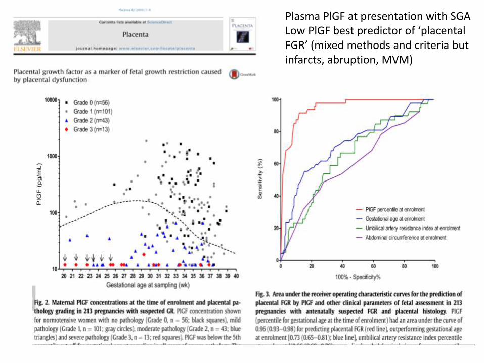
Plasma PlGF at presentation with SGALow PlGF best predictor of ‘placental FGR’ (mixed methods and criteria but infarcts, abruption, MVM)
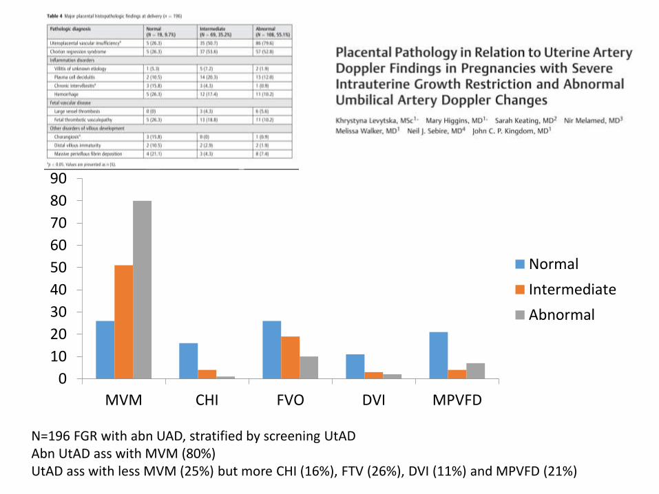
N=196 FGR with abn UAD, stratified by screening UtADAbn UtAD ass with MVM (80%)UtAD ass with less MVM (25%) but more CHI (16%), FTV (26%), DVI (11%) and MPVFD (21%)
0
10
20
30
40
50
60
70
80
90
MVM CHI FVO DVI MPVFD
Normal
Intermediate
Abnormal

Methodological issues
-Poor clinical phenotypes
-Definition of lesions
-Interpretation of lesions
-Blinding and bias

Placental Examination: Cambridge Study
• 1,159 singleton unselected women recruited at booking
• Objective measurements from calibrated images
• Histological exam, routine blocks
• Two paediatric pathologists
• Blinded to all clinical information-study number only
Total population
Age 29
GA 39 weeks
PET 2%
PIH 3%
SGA 6%
Pathak et al 2011-

Atherosis VUE
Chorioangioma FTV
OR vs PPV for placental ‘lesions

OR vs PPV for placental ‘lesions

Changes in transfer FUNCTION not detected
Pathophysiology of abnormal fetal growth complex and poorly understood
Much related to placental dysFUNCTION


Placental proteome
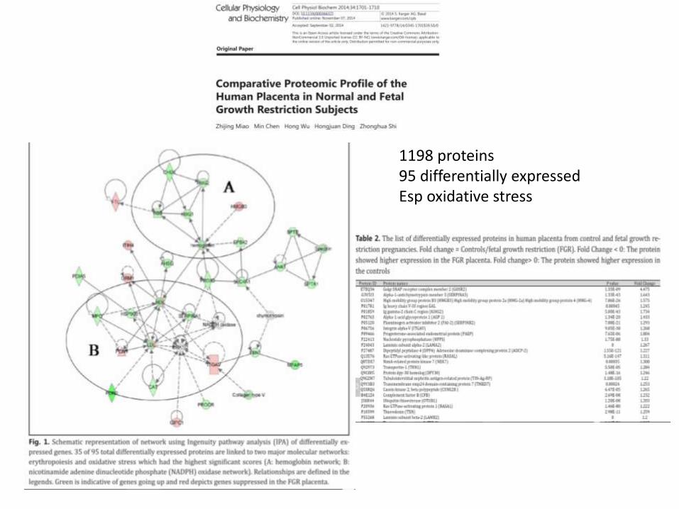
1198 proteins 95 differentially expressedEsp oxidative stress

Variation in transcriptome
0
10
20
30
40
50
60
70
Variation
Placenta
Individuals
Groups

Around 70% of FGR has associated morphological placental
pathology abnormalities
MVM represents majority (50%) of FGR, esp early onset
80+% of these are potentially detectable using USS assessment
including Doppler studies, PLGF etc
MVM Normal
VUE
FTVCHI,MPVFD

Some pathologies develop during pregnancy and unlikely to be
identifiable pre-clinically, histology only (VUE, CHI, MPVFD…)
Around 25% of FGR, more late-onset, has no clear pathology
correlates; may be amenable to future biochemical detection

Poor agreement regarding changes and poor correlation of
morphology with clinical phenotypes for individuals
Placental examination contributes to categorisation and mechanistic
grouping of FGR but is not gold standard for ‘pathology’
Future studies: clear phenotyping, blinded, objective assessments,
relationship to functional (OMIC) features