Embed Size (px)
Citation preview
Leonardo Electronic Journal of Practices and Technologies
ISSN 1583-1078
Issue 4, January-June 2004
p. 51-61
Placental, Morphological, and Ultrasonographical
Particularities in Post-Term Pregnancies
Carmen Mihaela MIHU, Dan MIHU
“Iuliu Haţieganu” University of Medicine and Pharmacy, Cluj-Napoca, Romania
Abstract
Post-term pregnancies are evaluated, using 2D and Doppler ultrasonography.
Apparition of fetal distress is certified by the increase of PRI. The current
study aimed to establish a relationship between the ultrasonographical
particularities of placentae and morphologic and morphometric aspects noticed
in placentae postpartum.
Keywords
Ultrasonograhy; Placenta; Mophometric Measurement
Introduction
Normal pregnancy lasts about 40 weeks of amenorheea (WA). The risc of fetal
complications increases during the pregnancies, which exceed this period.
After the median term of 40 WA, pregnant women can be examined using 2D and
Doppler ultrasonography, determining the placental resistivity index (PRI). After 40 WA, PRI
does not decrease; its tendency is to be increased after 42 WA.
51 http://lejpt.academicdirect.ro
Placental, Morphological, and Ultrasonographical Particularities in Post-Term Pregnancies Carmen Mihaela MIHU, Dan MIHU
Fetal distress (FD) cannot be installed if the values of PRI are lower than 0.64. In this
way, we can estimate the debut of fetal distress, by repeating each day the Doppler
examination.
Material and Methods
There were 10 cases of pregnancies, which exceeded 40 WA, included in this study.
All were normal pregnancies. The gestational age was determined correlating
anamnestic datas (first day of last menstruation) with echographic biometry datas, evaluated
before 20 WA (cranio-pelvic length, biparietal diameter, femural length).
2D ultrasonography was performed by a Ultramark 9 with transductor of 3.5 MHz,
with color and pulsatile Doppler, coupled with a real time B ultrasonograph; and a Medisson
Sonoace 8800 with transductor of 3,5 MHz, with color and pulsatile Doppler; coupled with a
real time B ultrasonograph.
Morphologic and morphometric analysis was performed on placentae. Placentae were
fixed in neutral 10% formaldehyde. They were macroscopically analysed and their weight,
thickness at the point of umbilical cordon insertion, and placental diameter were measured.
Fragments from different parts of placenta were collected, included in wax, realizing three
blocks for each placenta. They were colored in HE, PAS, red Sirius and orchid colorations. 3-
4 lamas were selected of each block, which were microscopically examined at a BX50
Olimpus microscope. Hystopathological particularities were observed and morphometric
measurements were performed.
Results
2D ultrasonography particularities
Placental maturation. 2D ultrasonographic evaluation, according to Kazzi
classification, suggested the following particularities of placentae’s structure:
• 3 intermediary placentae;
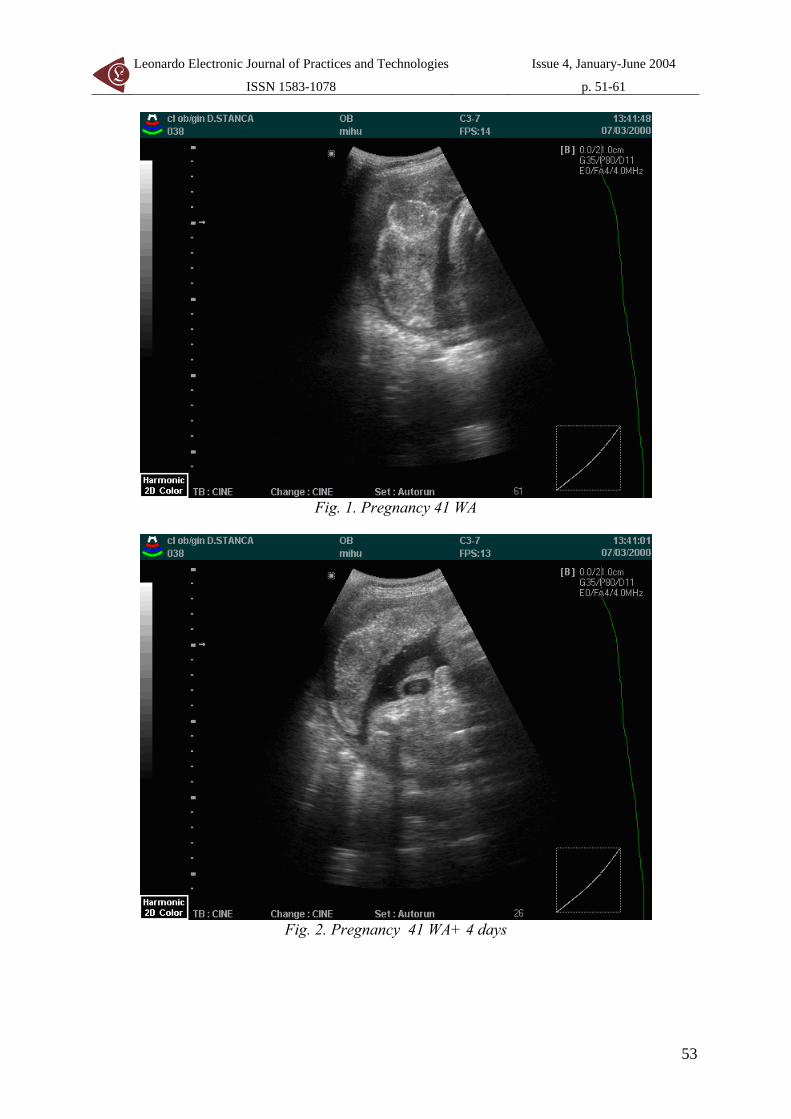
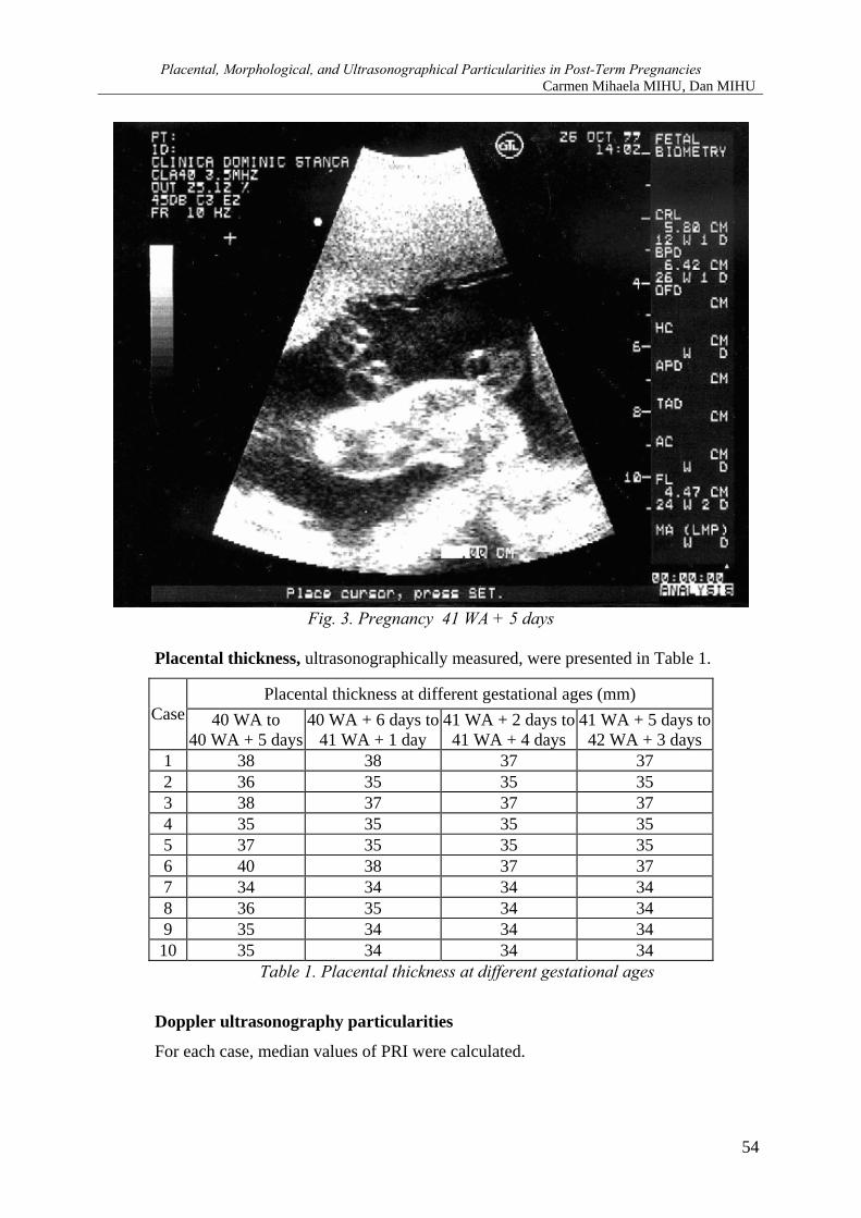
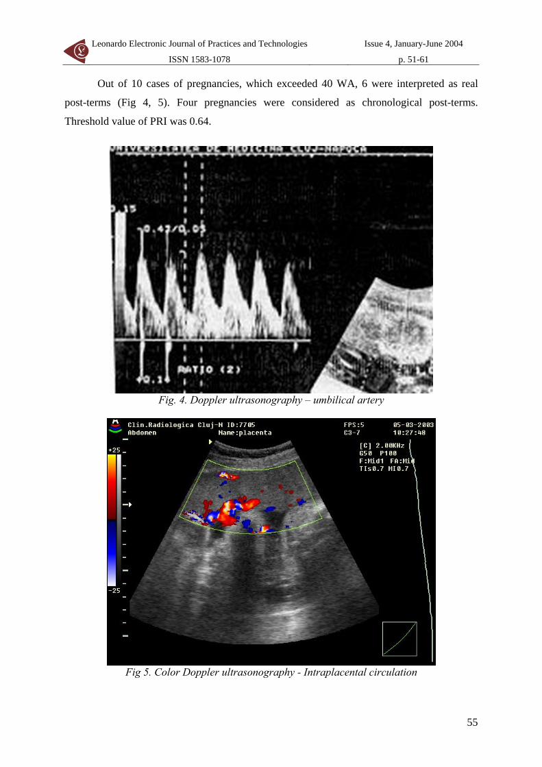
• 7 mature placentae (Fig. 1, 2, 3).
52
Leonardo Electronic Journal of Practices and Technologies
ISSN 1583-1078
Issue 4, January-June 2004
p. 51-61
Fig. 1. Pregnancy 41 WA
Fig. 2. Pregnancy 41 WA+ 4 days
53
Placental, Morphological, and Ultrasonographical Particularities in Post-Term Pregnancies Carmen Mihaela MIHU, Dan MIHU
Fig. 3. Pregnancy 41 WA + 5 days
Placental thickness, ultrasonographically measured, were presented in Table 1.
Placental thickness at different gestational ages (mm) Case 40 WA to
40 WA + 5 days 40 WA + 6 days to
41 WA + 1 day 41 WA + 2 days to
41 WA + 4 days 41 WA + 5 days to
42 WA + 3 days 1 38 38 37 37 2 36 35 35 35 3 38 37 37 37 4 35 35 35 35 5 37 35 35 35 6 40 38 37 37 7 34 34 34 34 8 36 35 34 34 9 35 34 34 34 10 35 34 34 34
Table 1. Placental thickness at different gestational ages
Doppler ultrasonography particularities
For each case, median values of PRI were calculated.
54
Leonardo Electronic Journal of Practices and Technologies
ISSN 1583-1078
Issue 4, January-June 2004
p. 51-61
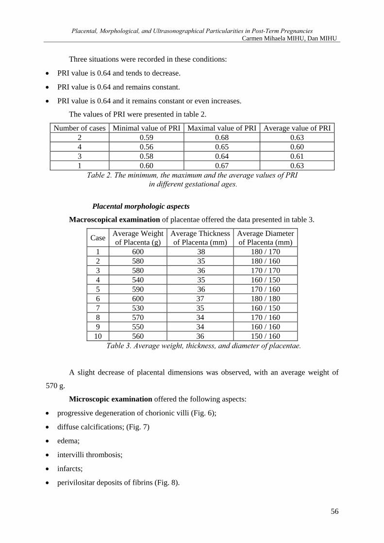
Out of 10 cases of pregnancies, which exceeded 40 WA, 6 were interpreted as real
post-terms (Fig 4, 5). Four pregnancies were considered as chronological post-terms.
Threshold value of PRI was 0.64.
Fig. 4. Doppler ultrasonography – umbilical artery
Fig 5. Color Doppler ultrasonography - Intraplacental circulation
55
Placental, Morphological, and Ultrasonographical Particularities in Post-Term Pregnancies Carmen Mihaela MIHU, Dan MIHU
Three situations were recorded in these conditions:
• PRI value is 0.64 and tends to decrease.
• PRI value is 0.64 and remains constant.
• PRI value is 0.64 and it remains constant or even increases.
The values of PRI were presented in table 2.
Number of cases Minimal value of PRI Maximal value of PRI Average value of PRI2 0.59 0.68 0.63 4 0.56 0.65 0.60 3 0.58 0.64 0.61 1 0.60 0.67 0.63
Table 2. The minimum, the maximum and the average values of PRI in different gestational ages.
Placental morphologic aspects
Macroscopical examination of placentae offered the data presented in table 3.
Case Average Weight of Placenta (g)
Average Thickness of Placenta (mm)
Average Diameter of Placenta (mm)
1 600 38 180 / 170 2 580 35 180 / 160 3 580 36 170 / 170 4 540 35 160 / 150 5 590 36 170 / 160 6 600 37 180 / 180 7 530 35 160 / 150 8 570 34 170 / 160 9 550 34 160 / 160 10 560 36 150 / 160
Table 3. Average weight, thickness, and diameter of placentae.
A slight decrease of placental dimensions was observed, with an average weight of
570 g.
Microscopic examination offered the following aspects:
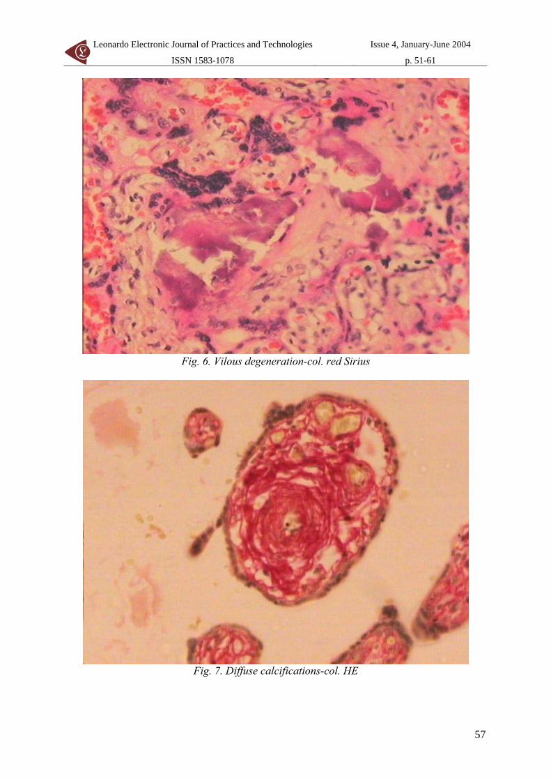
• progressive degeneration of chorionic villi (Fig. 6);
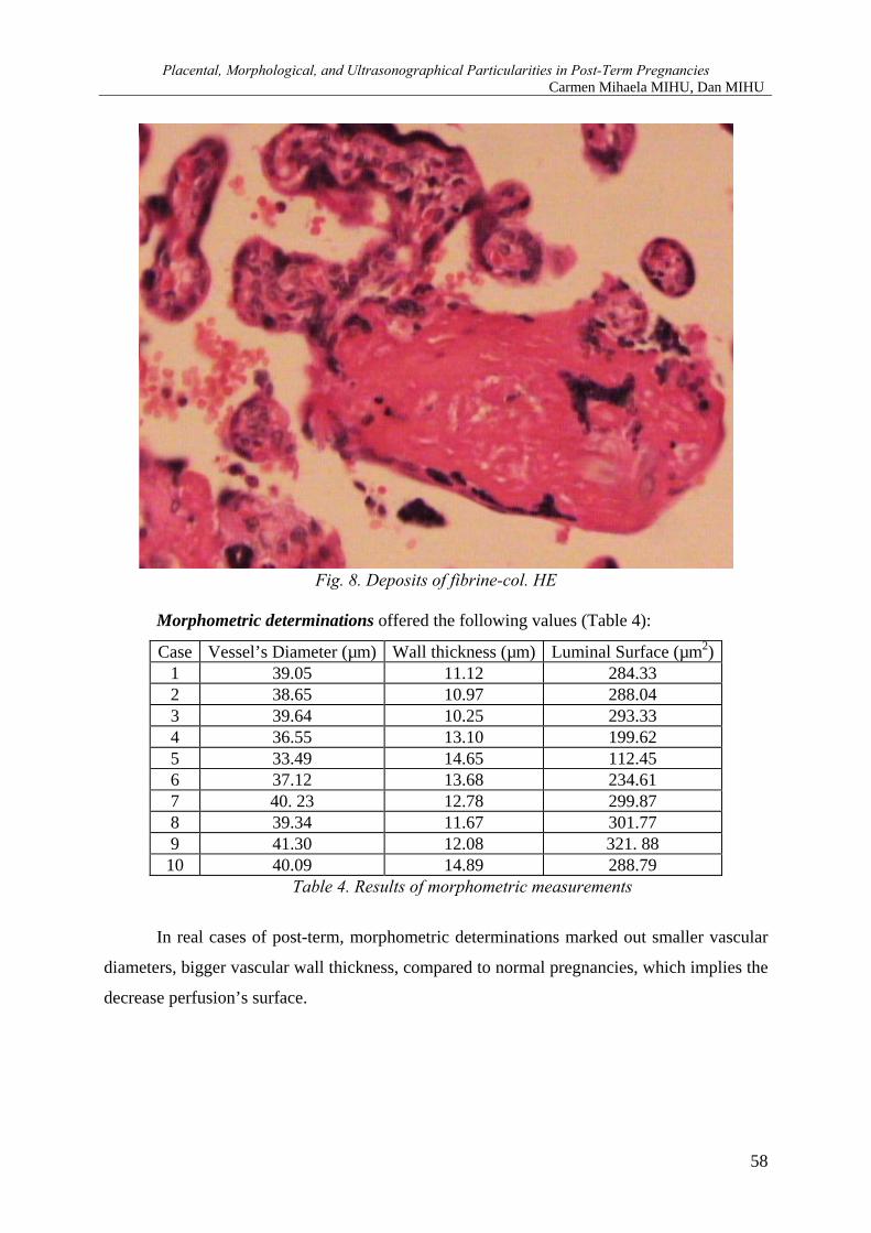
• diffuse calcifications; (Fig. 7)
• edema;
• intervilli thrombosis;
• infarcts;
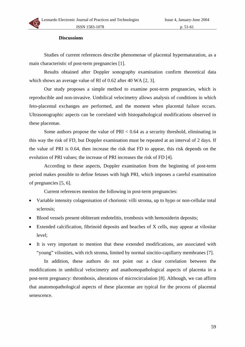
• perivilositar deposits of fibrins (Fig. 8).
56
Leonardo Electronic Journal of Practices and Technologies
ISSN 1583-1078
Issue 4, January-June 2004
p. 51-61
Fig. 6. Vilous degeneration-col. red Sirius
Fig. 7. Diffuse calcifications-col. HE
57
Placental, Morphological, and Ultrasonographical Particularities in Post-Term Pregnancies Carmen Mihaela MIHU, Dan MIHU
Fig. 8. Deposits of fibrine-col. HE
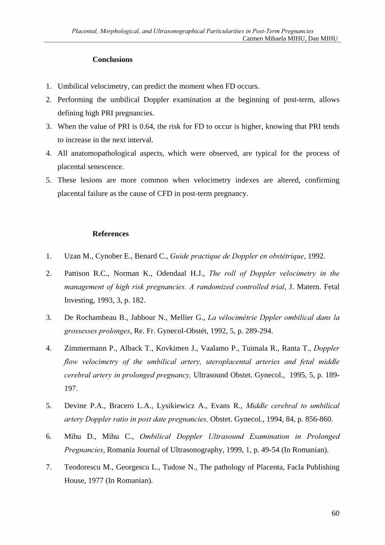
Morphometric determinations offered the following values (Table 4):
Case Vessel’s Diameter (µm) Wall thickness (µm) Luminal Surface (µm2) 1 39.05 11.12 284.33 2 38.65 10.97 288.04 3 39.64 10.25 293.33 4 36.55 13.10 199.62 5 33.49 14.65 112.45 6 37.12 13.68 234.61 7 40. 23 12.78 299.87 8 39.34 11.67 301.77 9 41.30 12.08 321. 88 10 40.09 14.89 288.79
Table 4. Results of morphometric measurements
In real cases of post-term, morphometric determinations marked out smaller vascular
diameters, bigger vascular wall thickness, compared to normal pregnancies, which implies the
decrease perfusion’s surface.
58
Leonardo Electronic Journal of Practices and Technologies
ISSN 1583-1078
Issue 4, January-June 2004
p. 51-61
Discussions
Studies of current references describe phenomenae of placental hypermaturation, as a
main characteristic of post-term pregnancies [1].
Results obtained after Doppler sonography examination confirm theoretical data
which shows an average value of RI of 0.62 after 40 WA [2, 3].
Our study proposes a simple method to examine post-term pregnancies, which is
reproducible and non-invasive. Umbilical velocimetry allows analysis of conditions in which
feto-placental exchanges are performed, and the moment when placental failure occurs.
Ultrasonographic aspects can be correlated with histopathological modifications observed in
these placentae.
Some authors propose the value of PRI < 0.64 as a security threshold, eliminating in
this way the risk of FD, but Doppler examination must be repeated at an interval of 2 days. If
the value of PRI is 0.64, then increase the risk that FD to appear, this risk depends on the
evolution of PRI values; the increase of PRI increases the risk of FD [4].
According to these aspects, Doppler examination from the beginning of post-term
period makes possible to define fetuses with high PRI, which imposes a careful examination
of pregnancies [5, 6].
Current references mention the following in post-term pregnancies:
• Variable intensity colagenisation of chorionic villi stroma, up to hypo or non-cellular total
sclerosis;
• Blood vessels present obliterant endotelitis, trombosis with hemosiderin deposits;
• Extended calcification, fibrinoid deposits and beaches of X cells, may appear at vilositar
level;
• It is very important to mention that these extended modifications, are associated with
“young” vilosities, with rich stroma, limited by normal sincitio-capillarry membranes [7].
In addition, these authors do not point out a clear correlation between the
modifications in umbilical velocimetry and anathomopathological aspects of placenta in a
post-term pregnancy: thrombosis, alterations of microcirculation [8]. Although, we can affirm
that anatomopathological aspects of these placentae are typical for the process of placental
senescence.
59
Placental, Morphological, and Ultrasonographical Particularities in Post-Term Pregnancies Carmen Mihaela MIHU, Dan MIHU
Conclusions
1. Umbilical velocimetry, can predict the moment when FD occurs.
2. Performing the umbilical Doppler examination at the beginning of post-term, allows
defining high PRI pregnancies.
3. When the value of PRI is 0.64, the risk for FD to occur is higher, knowing that PRI tends
to increase in the next interval.
4. All anatomopathological aspects, which were observed, are typical for the process of
placental senescence.
5. These lesions are more common when velocimetry indexes are altered, confirming
placental failure as the cause of CFD in post-term pregnancy.
References
1. Uzan M., Cynober E., Benard C., Guide practique de Doppler en obstétrique, 1992.
2. Pattison R.C., Norman K., Odendaal H.J., The roll of Doppler velocimetry in the
management of high risk pregnancies. A randomized controlled trial, J. Matern. Fetal
Investing, 1993, 3, p. 182.
3. De Rochambeau B., Jabbour N., Mellier G., La vélocimétrie Dppler ombilical dans la
grossesses prolonges, Re. Fr. Gynecol-Obstét, 1992, 5, p. 289-294.
4. Zimmermann P., Alback T., Kovkimen J., Vaalamo P., Tuimala R., Ranta T., Doppler
flow velocimetry of the umbilical artery, uteroplacental arteries and fetal middle
cerebral artery in prolonged pregnancy, Ultrasound Obstet. Gynecol., 1995, 5, p. 189-
197.
5. Devine P.A., Bracero L.A., Lysikiewicz A., Evans R., Middle cerebral to umbilical
artery Doppler ratio in post date pregnancies, Obstet. Gynecol., 1994, 84, p. 856-860.
6. Mihu D., Mihu C., Ombilical Doppler Ultrasound Examination in Prolonged
Pregnancies, Romania Journal of Ultrasonography, 1999, 1, p. 49-54 (In Romanian).
7. Teodorescu M., Georgescu L., Tudose N., The pathology of Placenta, Facla Publishing
House, 1977 (In Romanian).
60

Leonardo Electronic Journal of Practices and Technologies
ISSN 1583-1078
Issue 4, January-June 2004
p. 51-61
8. Nimrod C., Gruslin-Giroux A., Post term Pregnancy Chapter In: Copel J.A., Reed K.L.
Doppler Ultrasound in Obstetrics and Gynecology, New-York, Raven Press, 1995, 17,
p. 165-171.
61