Embed Size (px)
Citation preview

RESEARCH Open Access
Placenta-derived multipotent mesenchymalstromal cells: a promising potential cell-based therapy for canine inflammatorybrain diseaseRogério Martins Amorim1,2†, Kaitlin C. Clark1,3,4†, Naomi J. Walker1, Priyadarsini Kumar3,4, Kyle Herout3,Dori L. Borjesson1 and Aijun Wang1,3,4,5*
Abstract
Background: Canine inflammatory brain disease (IBD) is a severe inflammatory disorder characterized by infiltrationof activated immune cell subsets into the brain and spinal cord. Multipotent mesenchymal stromal cells (MSCs) area promising therapy for IBD, based on their potent pro-angiogenic, neuroprotective, and immunomodulatoryproperties. The aims of this study were to compare the immunomodulatory attributes of canine adipose-derivedMSCs (ASCs) and placenta-derived MSCs (PMSCs) in vitro. These data will serve as potency information to helpinform the optimal MSC cell source to treat naturally occurring canine IBD.
Methods: Indoleamine 2,3 dioxygenase (IDO) activity and prostaglandin E2 (PGE2) concentration at baseline andafter stimulation with interferon gamma (IFNγ) and/or tumor necrosis factor alpha (TNFα) were measured fromcanine ASC and PMSC cultures. Leukocyte suppression assays (LSAs) were performed to compare the ability of ASCsand PMSCs to inhibit activated peripheral blood mononuclear cell (PBMC) proliferation. IDO activity and PGE2;interleukin (IL)-2, IL-6, and IL-8; TNFα; and vascular endothelial growth factor (VEGF) concentrations were alsomeasured from co-culture supernatants. Cell cycle analysis was performed to determine how ASCs and PMSCsaltered lymphocyte proliferation.
(Continued on next page)
© The Author(s). 2020 Open Access This article is licensed under a Creative Commons Attribution 4.0 International License,which permits use, sharing, adaptation, distribution and reproduction in any medium or format, as long as you giveappropriate credit to the original author(s) and the source, provide a link to the Creative Commons licence, and indicate ifchanges were made. The images or other third party material in this article are included in the article's Creative Commonslicence, unless indicated otherwise in a credit line to the material. If material is not included in the article's Creative Commonslicence and your intended use is not permitted by statutory regulation or exceeds the permitted use, you will need to obtainpermission directly from the copyright holder. To view a copy of this licence, visit http://creativecommons.org/licenses/by/4.0/.The Creative Commons Public Domain Dedication waiver (http://creativecommons.org/publicdomain/zero/1.0/) applies to thedata made available in this article, unless otherwise stated in a credit line to the data.
* Correspondence: [email protected]†Rogério Martins Amorim and Kaitlin C. Clark contributed equally to thiswork.1Veterinary Institute for Regenerative Cures and Department of Pathology,Microbiology and Immunology, School of Veterinary Medicine, University ofCalifornia, Davis, Davis, CA, USA3Surgical Bioengineering Laboratory, Department of Surgery, School ofMedicine, University of California, Davis, 4625 2nd Ave., Research II, Suite3005, Sacramento, CA 95817, USAFull list of author information is available at the end of the article
Amorim et al. Stem Cell Research & Therapy (2020) 11:304 https://doi.org/10.1186/s13287-020-01799-0

(Continued from previous page)
Results: Activated canine MSCs from both tissue sources secreted high concentrations of IDO and PGE2, after directstimulation with IFNγ and TNFα, or indirect stimulation by activated PBMCs. Both ASCs and PMSCs inhibitedactivated PBMC proliferation in LSA assays; however, PMSCs inhibited PBMC proliferation significantly more thanASCs. Blocking PGE2 and IDO in LSA assays determined that PGE2 is important only for ASC inhibition of PBMCproliferation. Activated ASCs increased IL-6 and VEGF secretion and decreased TNFα secretion, while activatedPMSCs increased IL-6, IL-8, and VEGF secretion. ASCs inhibited lymphocyte proliferation via cell cycle arrest in theG0/G1 and PMSCs inhibited lymphocyte proliferation via induction of lymphocyte apoptosis.
Conclusion: Our results demonstrate that ASCs and PMSCs have substantial in vitro potential as a cell-basedtherapy for IBD; however, PMSCs more potently inhibited lymphocyte proliferation by inducing apoptosis ofactivated lymphocytes. These data suggest that the mechanism by which ASCs and PMSCs downregulate PBMCproliferation differs. Additional studies may elucidate additional mechanisms by which canine MSCs modulateneuroinflammatory responses.
Keywords: Multipotent progenitor cell, Mesenchymal stromal cell, Adipose tissue, Placenta, Canine, Animal model,Immunomodulation, Inflammatory brain disease, Multiple sclerosis, Translational research
BackgroundOver the past decade, stem cell therapy has become acornerstone in regenerative medicine therapies for manydiseases. However, brain and spinal cord diseases repre-sent a challenge in stem cell-based therapy, due to themultiplicity of cell types in the adult central nervous sys-tem (CNS) and the precision of cell interactions, in bothspace and time, required to enhance neuroregeneration[1]. Stem cell therapies for CNS injury are based on cellreplacement, via the transplantation of neural progenitorcells, the stimulation of endogenous CNS stem cells, oron improvement of the microenvironment mediated byanti-inflammatory/immunomodulatory paracrine cell ef-fects [2, 3]. However, challenges arise due to a lack ofstandardization of therapeutic interventions, variabilityin animal models of disease, alterations in timing andmodality of cell application, and a lack of understandingof disease pathology [4].Multipotent mesenchymal stromal cells (also known as
mesenchymal stem cells; MSCs) derived from the bonemarrow, adipose tissue, and birth-associated tissues, in-cluding umbilical cord, blood cord, amniotic fluid, andplacenta, are the most common cell type investigated incell-based therapy. Several reports have demonstratedpositive effects of MSC therapy in a large number of dis-orders, including brain and spinal cord injuries, in la-boratory animals, dogs, and humans [5, 6]. Dogs areincreasingly recognized as important animal models fortranslational medicine because they have naturally oc-curring brain and spinal cord injuries, such as canine in-flammatory brain disease (IBD), similar to multiplesclerosis (MS) in human beings [7–9].Studies involving MSCs are increasing due to their im-
munomodulatory, anti-inflammatory, and tissue regen-erative properties including the secretion of numerous
bioactive molecules leading to tissue regeneration [10,11]. However, the mechanisms by which MSCs elicitpositive effects on the damaged nervous system are notfully characterized. Mechanisms that may play an im-portant role in neuroregeneration include the secretionof growth factors, antiapoptotic factors, neurotrophicfactors, cytokines, and extracellular matrix proteins. Theimmunomodulatory and anti-inflammatory properties ofMSCs are also implicated in their ability to protect andrepair neurons. Together, these factors promote en-dogenous neuronal growth, promote neuro/gliogenesis,encourage synaptic connection from damaged neurons,recruit local oligodendrocyte precursors, reduce demye-lination, stimulate angiogenesis, decrease apoptosis, re-duce oxidative stress, modulate microglial activation,and regulate inflammation by suppressing pathologicalT, B, and natural killer (NK) cell responses [12–16].Moreover, they can accelerate a shift from a predomin-ance of pro-inflammatory Th1 cells toward an increasein the anti-inflammatory Th2 cells [17, 18]. Althoughcontroversial, some studies have suggested that MSCscan also migrate into the CNS lesion and differentiateinto neurons or astrocytes [19, 20].Canine adult and fetal/neonatal MSCs have been char-
acterized by immunophenotyping and multipotency as-says in many studies [21–27]. Fetal/neonatal MSCs,including placenta-derived stem cells, preserve some fea-tures of the primitive embryonic layers. They have thepotential to differentiate in many tissues [28], havegreater proliferative and immunomodulatory capacityand lesser immunogenicity than adult MSCs [28–30],and are neuroprotective [31, 32]. These stem cells canalso be easily harvested and expanded due to the avail-ability of a large amount of tissue which is usually dis-carded at birth. There is no ethical conflict regarding
Amorim et al. Stem Cell Research & Therapy (2020) 11:304 Page 2 of 12

placenta harvest making these stem cell sources attract-ive targets for banking and regenerative therapies [28,30]. Nonetheless, a deeper understanding of their immu-nomodulatory function is still lacking.IBD is a general term used to describe an idiopathic
disorder that can be subdivided based on histopathologyfindings, i.e., granulomatous meningoencephalomyelitis(GME), necrotizing meningoencephalitis (NME), andnecrotizing leukoencephalitis (NLE) [33, 34]. Recently,the term meningoencephalomyelitis of unknown origin(MUO) has been proposed to encompass all of these dis-eases. MUO is presumed to be an autoimmune diseasewith a genetic predisposition [33]. Immunohistochemicalphenotyping of the involved inflammatory cells demon-strates a pivotal role of MHC II+ cells, T cells, andmacrophages in GME and NLE [35–37]. GME can beidentified by CD3+ lymphocyte infiltration into sites ofgranulomatous inflammation as shown in a representa-tive clinical case shown in Fig. 1. The pattern of mRNAand protein expression of cytokines and chemokine re-ceptors may be disease specific, such as interferongamma (IFNγ) and CXCR3 in NME, and IL-17 andCCR2 in GME [34]. Cytokines have also been implicatedin the phenotype switching of microglia/macrophagesfrom a classically activated pro-inflammatory type (M1)or into an alternatively activated anti-inflammatoryphenotype (M2) in canine inflammatory CNS disease[37]. Thus, manipulating this inflammatory phenotypeby cell-based therapy might represent a promising thera-peutic approach for MUO [37].Our goal was to compare the in vitro immunomodula-
tory ability of 2 types of canine MSCs focusing on secre-tion profiles and neuroreparative assays relevant forcanine IBD. This study evaluated the in vitro potential of
canine adipose tissue-derived multipotent mesenchymalstromal cells (ASCs) and placenta-derived multipotentmesenchymal stromal cells (PMSCs) to secrete anti-inflammatory cytokines and to inhibit lymphocyteproliferation, as well as the mechanisms involved in thecanine MSC immunomodulatory process.
MethodsCanine MSC collection, isolation and cultureAdipose tissue-derived mesenchymal stromal cells (ASCs)Canine low passage (P2-P5) ASCs from 5 dogs were ob-tained from falciform fat collected from UCD William R.Pritchard Veterinary Medical Teaching Hospital healthypatients undergoing routine abdominal surgery. Fat wascollected under an approved Institutional Animal Careand Use Committee and the Clinical Trials ReviewBoard protocol at UCD (protocol number 19605). Fatwas processed and canine ASCs were isolated, expanded,cryopreserved, and phenotyped exactly as previously de-scribed [38, 39]. Briefly, adipose tissue was minced anddigested at 37 °C for 1–2 h using 0.1% collagenase type Iand 1% bovine-serum albumin (Worthington, Lakewood,NJ). Centrifugation was performed to remove theremaining lipid layer. The cell pellet was washed severaltimes before being plated in standard medium,Dulbecco’s modified Eagle’s medium (DMEM, Gibco,Invitrogen, Carlsbad, CA) supplemented with 10% FBS(HyClone, Logan, UT), 100 U/mL penicillin and 100 μg/mL streptomycin (ThermoFisher Scientific, Gibco,Pittsburgh, PA).
Placenta-derived mesenchymal stromal cells (PMSCs)Canine low passage (P2-P5) placenta-derived MSCs from5 dogs were obtained from Dr. Aijun Wang’s laboratory
Fig. 1 Canine granulomatous meningoencephalitis (GME) in a 2-year-old, female, Miniature Pinscher. a Arrow indicates area of granulomatousinflammation (hematoxylin-eosin; × 10). b CD3-positive cells in the granulomatous inflammation are indicated (arrow) (immunohistochemistry,DAB, Harris hematoxylin counter stain; × 10)
Amorim et al. Stem Cell Research & Therapy (2020) 11:304 Page 3 of 12

cell bank, University of California Davis Medical Center.Isolation, culture, and full phenotyping of these PMSClines from discarded term canine placentas was previ-ously described [40]. Briefly, at term, canine placentaswere processed by manual dissection and treated with0.25% trypsin (ThermoFisher) solution for 30 min at37 °C. Cell pellets were washed and incubated with colla-genase IA (1 mg/mL; Sigma-Aldrich, St. Louis, MO) for45 min at 37 °C. The cell pellet was resuspended andplated onto tissue culture flasks in standard medium.
Stimulation of canine MSCs with IFNγ and tumor necrosisfactor alpha (TNFα)Cryopreserved MSCs were thawed and culture expandedas previously described [38, 40]. When cells wereapproximately 70% confluent, they were trypsinized andresuspended in standard medium supplemented with L-tryptophan (Sigma-Aldrich) to a final concentration of600 μM. These MSCs were seeded at 2 × 10⁵ per well of24-well plate, with 0.75 mL total media volume per wellfor the stimulation assays. At plating, MSC was stimu-lated with IFNγ (50 ng/mL; canine recombinant IFNγ,Kingfisher, St. Paul, MN) or TNFα (50 ng/mL; canine re-combinant TNFα, Kingfisher) or a dual stimulation withboth IFNγ (50 ng/mL, Kingfisher) and TNFα (50 ng/mL,Kingfisher). Stimulated MSCs were cultured for 4 daysat which time supernatants were collected, frozen, andstored at − 80 °C for mediator quantification. Stimulationprotocols using IFNγ and TNFα were optimized byusing a dose titration strategy and collection at multipletimepoints for mediator quantification [data not shown].The optimized strategy is consistent with previous re-ports on IFNγ and TNFα secretion by activated canineMSCs [38, 39].
Canine leukocyte suppression assay (LSA)Canine leukocyte suppression assay (LSAs) were per-formed exactly as previously described [38]. In brief, per-ipheral blood was collected from healthy dogs 55pounds or larger between 1 and 8 years old, into tubescontaining sodium heparin (Vacutainer®, BD Biosci-ences) via jugular venipuncture. PBMCs were isolatedusing a discontinuous Ficoll gradient and were platedwith irradiated (10 Gy, Varian 2100C linear accelerator,Varian Medical Systems, Inc., Palo Alto, CA) allogeneiccanine ASCs or PMSCs in standard medium (DMEMwith 10% FBS, 1% penicillin/streptomycin, supplementedwith 600 μML-tryptophan) [38]. PBMCs were activatedwith 5 μg/mL concanavalin A (ConA; Sigma-Aldrich).PBMCs and irradiated MSCs were co-cocultured at a ra-tio of 5:1 in direct contact.To determine the role of contact, cells were plated in
transwell dishes (Corning 0.4 μM polycarbonate mem-brane 24-well plate; Corning, NY, USA) with MSCs
plated in the plate bottom and PBMCs in the insert. Todetermine the role of IDO and PGE2 in MSC-mediatedinhibition of lymphocyte proliferation, inhibitory agentswere used in LSA co-cultures. Indomethacin, a cyclooxy-genase (COX) inhibitor, was used to chemically blockPGE2 production (Cayman Chemical Co., Ann Arbor,MI, 1μM). Alternatively, 1-methyl-DL-tryptophan (1-MT), an IDO competitive inhibitor, was used to partiallyinhibit IDO activity. Indomethacin was added to LSA as-says during plating at a concentration of 10 μM (Sigma-Aldrich) as previously described [38] to determine therole of PGE2 on MSC-mediated immunosuppression.Additionally, at plating, 1-MT (Sigma-Aldrich) wasadded at a concentration of 1 mM as previously de-scribed [41] to determine the role of IDO activity onMSC mediated immunosuppression.After 3 days of co-culture, wells were treated with 1
mM Bromodeoxyuridine (BrdU, BD Biosciences).Twenty-four hours post BrdU treatment, leukocyteswere collected, and cells were stained with a viability dye(Fixable Viability Dye Fluor® 780; eBioscience, SanDiego, CA) and anti-canine CD3 conjugated to AlexaFluor® 488 (clone CA17.2A12; Leukocyte Antigen Biol-ogy Lab, UCD). Leukocytes were stained for nuclearBrdU incorporation (APC BrdU Flow Kit, BD Biosci-ences) per manufacturer directions and analyzed by flowcytometry (Cytomics FC500). Fold reduction ofleukocyte proliferation by MSCs was normalized to stim-ulated donor PMBCs for any given experiment. For cellcycle analysis, 7-aminoactinomycin D was added to cul-tures per manufacturer’s instructions (APC BrdU FlowKit; BD Biosciences) and analyzed by flow cytometry ondays 1–4. Flow cytometry data were analyzed usingFlowJo flow cytometry software (Tree Star, Inc.). Datawas normalized and presented as a reduction of each re-spective PBMC donor.At the time of leukocyte collection, culture super-
natant was collected, centrifuged, and stored at − 80 °Cfor the measurement of secreted mediators.
Quantification of mediator secretionFrozen aliquots of supernatants collected from MSCsstimulated with IFNγ/TNFα and from LSA co-culturesusing two technical replicates from each assay were usedto quantify PGE2 and IDO activity. Canine PGE2 wasquantified using an ELISA kit per manufacturer direc-tions (Prostaglandin E2 Express EIA kit (Monoclonal);Cayman Chemical Company, Ann Arbor, MI) [38]. Toassess IDO activity, a biochemical assay was performedon frozen supernatants as previously described [42] toquantify the conversion of tryptophan to N-formylkynurenine mediated by IDO. In brief, culture mediawas treated with 30% trichloroacetic acid (Sigma), andEhrlich’s reagent (1% p-dimethylaminobenzaldehyde in
Amorim et al. Stem Cell Research & Therapy (2020) 11:304 Page 4 of 12

glacial acetic acid, Sigma) was mixed and read at 490 nmon a microplate reader (Synergy HT Multi-Mode Gen5software) [38].Supernatants from LSAs co-cultures were used to
measure concentrations of interleukin (IL)-2, IL-6,and IL-8; vascular endothelial growth factor (VEGF);and TNFα. IL-2, IL-6, and IL-8 and were quantifiedvia QuantibodyVR Canine Cytokine Array (RayBio-tech cat# QAC-CYT-1). Negative controls were pre-pared from wells containing only media. The arraywas performed according to the manufacturer’s in-structions, and the resulting glass slide was scannedusing a GenePix 4000B microarray scanner (Molecu-lar Devices). Collected images were quantified usingGenePixVR Pro 6 acquisition and analysis software,and further plotting of standard curves and analysiswas performed using Microsoft Excel. TNFα wasmeasured from supernatants using ELISA kits (Ca-nine TNFα DuoSet, R&D Systems) per manufacturesinstructions. All ELISA samples were read on a Syn-ergy HT Multi-Mode microplate reader with Gen5software (Biotek, Winooski, VT, USA).
Statistical analysisResults are presented as mean and standard error. Datawere tested for normality using the Shapiro-Wilk nor-mality test (GraphPad InStat version 3.06 for Windows,La Jolla, CA). Data were analyzed using non-parametricMann-Whitney-Wilcoxon t test (GraphPad InStat
version 3.06 for Windows, La Jolla, CA). p < 0.05 wasconsidered statistically significant.
ResultsCanine MSCs increase IDO and PGE2 secretion in responseto IFNγ and TNFα stimulationMSC immunomodulatory functions occur in partthrough the secretion of bioactive factors. MSCs wereactivated through direct stimulation to determine theimmunomodulatory potential using recombinant pro-inflammatory mediators known to be relevant in IBD.Stimulation with IFNγ alone increased IDO activity,while stimulation with TNFα alone predominantlystimulated PGE2 production by both ASCs andPMSCs (Fig. 2a, b). The use of both stimulationagents resulted in a synergistic effect IDO activity andPGE2 production; therefore, dual stimulation usingboth IFNγ and TNFα was performed in canine ASCand PMSC cultures. Dual stimulation with both IFNγand TNFα resulted in increased PGE2 production andIDO activity (Fig. 2a, b). Canine ASCs, however, se-creted significantly more IDO than PMSCs after 4days of stimulation with canine recombinant IFNγand TNFα (Fig. 2a; p = 0.0079). Dual stimulation ofcanine ASCs and PMSCs resulted in comparable in-creases in PGE2 secretion (Fig. 2b). IDO secretionwas exclusively dependent on IFNγ with no synergis-tic increase after the addition of TNFα; however,PGE2 secretion was augmented with dual stimulation.
Fig. 2 Direct stimulation of canine ASCs and PMSCs leads to production of IDO and PGE2. Canine adipose-derived MSCs (ASCs) and placenta-derived MSCs (PMSCs) secrete increased levels of indoleamine 2,3 dioxygenase (IDO) activity and prostaglandin E2 (PGE2) in response to directstimulation using recombinant interferon gamma (IFNγ) and tumor necrosis factor alpha (TNFα). a IDO activity is directly proportional to theconversion of tryptophan to N-formyl kynurenine. ASCs and PMSCs increase IDO activity in response to dual stimulation with IFNγ and TNFα. IFNγis the main contributor to the production of IDO. IFNγ- and TNFα-stimulated ASCs promote significantly higher levels of IDO activity as comparedto PMSCs. b Canine ASCs and PMSCs produce comparable levels of prostaglandin E2 (PGE2) in response to dual stimulation with IFNγ and TNFα.TNFα is the major contributor to MSC-mediated PGE2 production. Data presented as mean and standard error. *p < 0.05, **p < 0.01, ***p < 0.001.IDO, indoleamine 2,3 dioxygenase; IFNγ, interferon gamma; MSC, mesenchymal stem cell; PGE2, prostaglandin E2; TNFα, tumor necrosisfactor alpha
Amorim et al. Stem Cell Research & Therapy (2020) 11:304 Page 5 of 12

Canine ASCs and PMSCs inhibit activated PBMCproliferation through distinct mechanisms in a contact-dependent mannerWe have previously reported that canine ASCs reducemitogen-activated PBMC proliferation in LSA co-cultures [38]. Both canine ASCs and PMSCs in directcontact with activated PBMCs inhibited lymphocyteproliferation; however, PMSCs more potently inhib-ited lymphocyte proliferation compared to ASCs(Fig. 3a, p = 0.0127). LSA co-cultures performedwithin a transwell to remove direct MSC-PBMC cellcontact resulted in marked restoration of PBMC pro-liferation regardless of MSC tissue source (Fig. 3a).These data suggest that canine MSCs reduce activatedlymphocyte proliferation in part via direct cell-cellcontact.
MSC mediated immunomodulation in dogs also oc-curs through the secretion of bioactive factors [38, 39,43, 44]. PGE2 and IDO have been implicated as crucialmechanisms by which MSCs downregulate inflammatoryresponses. To evaluate the role of PGE2, the COX in-hibitor indomethacin was used to block PGE2 synthesisand secretion. The competitive inhibitor 1-MT was usedto block the functional properties of IDO. BlockingPGE2 led to a significant reduction of ASC-mediated in-hibition as compared to PMSCs (Fig. 3b, p = 0.0043).Blocking IDO, however, lead to no alterations in ASC-or PMSC-mediated suppression of PBMC proliferation(Fig. 3b). Representative photomicrographs of stimulatedPBMCs and a PMSC LSA are shown in Fig. 3c. No mor-phological changes were noted between canine ASC andPMSC LSA conditions.
Fig. 3 Canine ASCs and PMSCs inhibit lymphocyte proliferation in a contact-dependent manner. Canine adipose-derived MSCs (ASCs) andplacenta-derived MSCs (PMSCs) possess immune-suppressive functions. a Canine ASCs and PMSCs co-incubated with stimulated peripheral bloodmononuclear cells (PBMCs) suppress lymphocyte proliferation. PMSCs significantly decrease lymphocyte proliferation more potently as comparedto ASCs. Transwells were added to remove physical contact between MSCs and mitogen (ConA) stimulated PBMCs. Lymphocyte proliferationincreased when ASCs and PMSCs were not in direct contact with activated PBMCs. b To determine the role of indoleamine 2,3 dioxygenase (IDO)and prostaglandin E2 (PGE2) in MSC-mediated immunosuppression 1-methyl-DL-tryptophan (1-MT) and indomethacin was added to co-cultures toblock each respective mediator. Blocking of IDO activity using 1-MT resulted in no alterations in MSC-mediated suppression of PBMC proliferation.Blocking PGE2 using indomethacin resulted in a loss of lymphocyte inhibition by ASCs but no effects were observed by PMSCs. PBMC stimulationis normalized and data is presented as a reduction to each respective donor. Representative photomicrographs of ConA stimulated PBMCs andPMSC LSA conditions (c). Data presented as mean and standard error. *p < 0.05, **p < 0.01, ***p < 0.001. 1-MT, 1-methyl-DL-tryptophan; ConA,concanavalin A; IDO, indoleamine 2,3 dioxygenase; LSA, leukocyte suppression assay; MSC, mesenchymal stem cell; PBMCs, peripheral bloodmononuclear cells; PGE2, prostaglandin E2
Amorim et al. Stem Cell Research & Therapy (2020) 11:304 Page 6 of 12
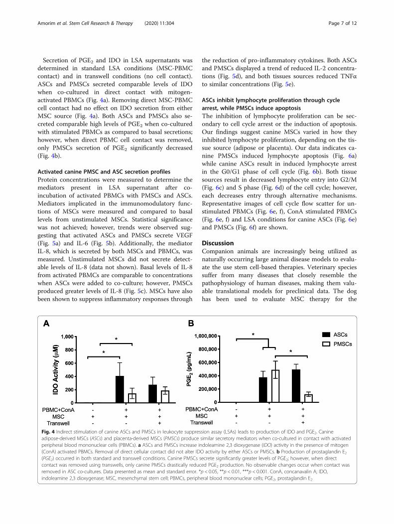
Secretion of PGE2 and IDO in LSA supernatants wasdetermined in standard LSA conditions (MSC-PBMCcontact) and in transwell conditions (no cell contact).ASCs and PMSCs secreted comparable levels of IDOwhen co-cultured in direct contact with mitogen-activated PBMCs (Fig. 4a). Removing direct MSC-PBMCcell contact had no effect on IDO secretion from eitherMSC source (Fig. 4a). Both ASCs and PMSCs also se-creted comparable high levels of PGE2 when co-culturedwith stimulated PBMCs as compared to basal secretions;however, when direct PBMC cell contact was removed,only PMSCs secretion of PGE2 significantly decreased(Fig. 4b).
Activated canine PMSC and ASC secretion profilesProtein concentrations were measured to determine themediators present in LSA supernatant after co-incubation of activated PBMCs with PMSCs and ASCs.Mediators implicated in the immunomodulatory func-tions of MSCs were measured and compared to basallevels from unstimulated MSCs. Statistical significancewas not achieved; however, trends were observed sug-gesting that activated ASCs and PMSCs secrete VEGF(Fig. 5a) and IL-6 (Fig. 5b). Additionally, the mediatorIL-8, which is secreted by both MSCs and PBMCs, wasmeasured. Unstimulated MSCs did not secrete detect-able levels of IL-8 (data not shown). Basal levels of IL-8from activated PBMCs are comparable to concentrationswhen ASCs were added to co-culture; however, PMSCsproduced greater levels of IL-8 (Fig. 5c). MSCs have alsobeen shown to suppress inflammatory responses through
the reduction of pro-inflammatory cytokines. Both ASCsand PMSCs displayed a trend of reduced IL-2 concentra-tions (Fig. 5d), and both tissues sources reduced TNFαto similar concentrations (Fig. 5e).
ASCs inhibit lymphocyte proliferation through cyclearrest, while PMSCs induce apoptosisThe inhibition of lymphocyte proliferation can be sec-ondary to cell cycle arrest or the induction of apoptosis.Our findings suggest canine MSCs varied in how theyinhibited lymphocyte proliferation, depending on the tis-sue source (adipose or placenta). Our data indicates ca-nine PMSCs induced lymphocyte apoptosis (Fig. 6a)while canine ASCs result in induced lymphocyte arrestin the G0/G1 phase of cell cycle (Fig. 6b). Both tissuesources result in decreased lymphocyte entry into G2/M(Fig. 6c) and S phase (Fig. 6d) of the cell cycle; however,each decreases entry through alternative mechanisms.Representative images of cell cycle flow scatter for un-stimulated PBMCs (Fig. 6e, f), ConA stimulated PBMCs(Fig. 6e, f) and LSA conditions for canine ASCs (Fig. 6e)and PMSCs (Fig. 6f) are shown.
DiscussionCompanion animals are increasingly being utilized asnaturally occurring large animal disease models to evalu-ate the use stem cell-based therapies. Veterinary speciessuffer from many diseases that closely resemble thepathophysiology of human diseases, making them valu-able translational models for preclinical data. The doghas been used to evaluate MSC therapy for the
Fig. 4 Indirect stimulation of canine ASCs and PMSCs in leukocyte suppression assay (LSAs) leads to production of IDO and PGE2. Canineadipose-derived MSCs (ASCs) and placenta-derived MSCs (PMSCs) produce similar secretory mediators when co-cultured in contact with activatedperipheral blood mononuclear cells (PBMCs). a ASCs and PMSCs increase indoleamine 2,3 dioxygenase (IDO) activity in the presence of mitogen(ConA) activated PBMCs. Removal of direct cellular contact did not alter IDO activity by either ASCs or PMSCs. b Production of prostaglandin E2(PGE2) occurred in both standard and transwell conditions. Canine PMSCs secrete significantly greater levels of PGE2; however, when directcontact was removed using transwells, only canine PMSCs drastically reduced PGE2 production. No observable changes occur when contact wasremoved in ASC co-cultures. Data presented as mean and standard error. *p < 0.05, **p < 0.01, ***p < 0.001. ConA, concanavalin A; IDO,indoleamine 2,3 dioxygenase; MSC, mesenchymal stem cell; PBMCs, peripheral blood mononuclear cells; PGE2, prostaglandin E2
Amorim et al. Stem Cell Research & Therapy (2020) 11:304 Page 7 of 12

Fig. 5 Bioactive factors associated with canine ASC and PMSC mediated immunosuppression differs. Canine adipose-derived MSCs (ASCs) andplacenta-derived MSCs (PMSCs) increase production of vascular endothelial growth factor (VEGF) and IL-6. a Both canine ASCs and PMSCs mildlyincreased production of VEGF when co-cultured with mitogen (ConA) activated peripheral blood mononuclear cells (PBMCs). b Canine ASCs andPMSCs upregulated IL-6 production; however, PMSCs secreted higher levels of IL-6 as compared to ASCs. c Production of IL-8 by canine ASCs wascomparable to basal levels of IL-8 produced by stimulated PBMCs; however, PMSCs increased levels of IL-8 greater than basal levels. Additionally,regulation of inflammatory mediators by canine ASCs and PMSCs was similar. d IL-2 production from stimulated PBMCs was mildly reduced byboth canine ASCs and PMSCs. e Canine ASCs and PMSCs inhibit production of tumor necrosis factor alpha (TNFα) by stimulated PBMCs tocomparable levels. Data presented as mean and standard error. ConA, concanavalin A IL, interleukin; TNFα, tumor necrosis factor alpha; VEGF,vascular endothelial growth factor
Fig. 6 Inhibition of lymphocyte proliferation by canine ASCs and PMSCs occurs through different mechanisms. Cell cycle analysis was performedusing peripheral blood mononuclear cells (PBMCs) and BrdU 5-bromo-2′-deoxyuridine and 7-aminoactinomycin D was measured. UnstimulatedPBMCs and mitogen (ConA) activated PBMCs were used as controls. a Canine PMSCs inhibit lymphocyte proliferation by inducing apoptosis.Alternatively, canine ASCs caused cell cycle arrest which is demonstrated by PBMCs accumulating in G0/G1 (b) and hindering cells from enteringG2/M (c) or DNA synthesis (S phase) (d). Representative images of cell cycle flow scatter plots and gating strategies for leukocyte DNA content (7-AAD) and proliferation via BrdU incorporation of PBMC controls (e, f) and co-incubations with canine ASCs (e) and PMSCs (f) are shown. BrdU, 5-bromo-2′-deoxyuridine and 7-aminoactinomycin D; ConA, concanavalin A; LSA, leukocyte suppression assay; MSC, mesenchymal stem cell
Amorim et al. Stem Cell Research & Therapy (2020) 11:304 Page 8 of 12

treatment of several inflammatory conditions includingosteoarthritis, spinal cord injury, inflammatory boweldisease, and graft-versus-host disease [19, 45–47].Murine experimental autoimmune encephalomyelitis
(EAE) is the most commonly used animal model tostudy MS. However, EAE does not reproduce all clinical,pathological, or immunological features of human dis-ease [48]. Canine MUO may be useful as a naturally oc-curring model of MS, given neuroimmunologicalsimilarities of these diseases, including the upregulationof IFNγ, IL-17, and MHC-II expression in the nervoussystem [5, 7, 34, 49–51]. Moreover, the genetic associ-ation of MHC-II found in dogs with MUO is present inMS [7]. MS is suggested to be mediated by Th1 andTh17 lymphocytes, leading to demyelination and axonalinjury [52, 53]. Although the demyelination noted in MSis not present in NME, fulminant or non-prototypicacute variants of MS, such as Marburg variant, Balo’sconcentric sclerosis, and acute disseminated encephalo-myelitis, closely resemble the pathological features of ca-nine NME [7]. The focal and widespread forms of GME,consistent with a delayed hypersensitivity reaction, arealso consistent with MS [5]. Cytokine expression inbrain lesions of NME and GME display increased levelsof interferon gamma (IFNγ) in NME and IL-4 and IL-17in GME [34]. IL-17 and IFNγ production by T lympho-cytes is also associated with active disease in MS patients[53]. In addition, CSF in dogs with MUO showed in-creased levels of CCL19 chemokine, also expressed inneuroinflammatory diseases such as MS / EAE, suggest-ing similar neuroimmunological events [54].MSCs are an attractive target for neurodegenerative
disease therapies due to their potent neuroprotective, re-generative, and immunomodulatory properties. Whilemost clinical studies utilize adult-derived sources ofMSCs, the placenta is a unique source of MSCs thatmaintain unique functional properties for therapeuticuse as compared to adult tissue-derived MSCs. Wefound that MSCs from adult and fetal tissue sourcesmodulate PBMC proliferation through multiple mecha-nisms. Both canine ASCs and PMSCs inhibit activatedlymphocyte proliferation through a primarily cell-cellcontact-mediated mechanism. Notably, PMSCs morepotently inhibited lymphocyte proliferation as comparedto ASCs in vitro. Inhibition of PBMC proliferation byPMSCs occurred through the induction of apoptosis,while ASCs induced cell cycle arrest. Canine ASCs andPMSCs secreted high levels of both PGE2 and IDOthrough direct stimulation with IFNγ and TNFα orthrough indirect stimulation in our mixed co-cultureassay. For PMSCs, when cell-cell contact was removed, arestoration of lymphocyte proliferation and a decrease ofPGE2 secretion were observed. Interestingly, blockingPGE2 production in PMSC cultures using a COX-
inhibitor did not restore lymphocyte proliferation. Thesefindings suggest that MSC-PBMC cellular contact is pri-marily responsible for the production of PGE2 byPMSCs; however, production of this mediator is not dir-ectly responsible for induction of lymphocyte apoptosis.Additionally, PMSCs produce IDO but regardless of re-moving cellular contact or blocking IDO via competitiveinhibitor, no effect on PBMC proliferation was observed.Collectively, this data highlights the importance of cellu-lar contact for PMSC-mediated immunosuppression andinitiation of PBMC apoptosis.Similarly, in ASCs, when direct cellular contact was re-
moved with PBMCs, lymphocyte proliferation was re-stored; however, this contact was not needed for PGE2secretion. Inhibiting PGE2 production with indometh-acin led to increased lymphocyte proliferation by ASCs.These data suggest that for canine ASCs, there are twomechanisms leading to PBMC cell cycle arrest, one thatis contact-dependent and one that is PGE2-dependent.As observed in PMSCs, inhibiting IDO with 1-MTshowed no effect on ASC mediated immunosuppression.Removing MSC-PBMC contact did not alter IDO activ-ity in canine ASC co-cultures. These findings suggestthat IDO does not play a significant role in canine MSC-mediated immunosuppression from either tissue source.PGE2 has been shown to play a key role in MSC-
mediated immunosuppression in humans, cats, dogs,and horses [18, 55]. PGE2 inhibits production of IFNγand IL-2 and induces T regulatory cells [56]. Decidualstem cells also suppress alloreactivity through inductionof T regulatory cells in a contact-dependent manner;however, involvement of programmed cell death 1 (PD-1), IDO, PGE2, and IFNγ still plays a role in thissuppression [57]. These data suggest that our findings incanine fetal PMSCs are comparable to human studies.Interestingly, we observed no alterations in immunosup-pression by the secretion of IDO, which has been re-ported in both human adult and fetal-derived tissuesources. Here we report canine MSCs produce IDOwhen activated; however, this does not seem to be a crit-ical mediator in lymphocyte suppression.Though not statistically significant, our data suggest
that both ASCs and PMSCs secrete VEGF and IL-6.Additionally, PMSCs, trended toward secreting higherlevels of IL-6 and IL-8 as compared to ASCs. Thereis also a trend toward a reduction of IL-2 productionby both ASCs and PMSCs. Both tissue sourcesreduced the production of the pro-inflammatorymediator TNFα. Canine ASC mediators closelyrecapitulate studies in human ASCs, suggesting thatthe dog will serve as a useful translational model toevaluate therapeutic applications of MSCs [58]. Theability of canine MSCs from either tissue source tosecrete immunomodulatory mediators and suppress
Amorim et al. Stem Cell Research & Therapy (2020) 11:304 Page 9 of 12

inflammatory cytokines indicates these cells are at-tractive targets for canine IBD therapeutics.MSC-immune cell contact, namely in the presence of
IFNγ and TNFα, has been shown to upregulate PDL-1,vascular cell adhesion molecule 1 (VCAM-1), and in-flammatory cytokine-induced adhesion molecule 1(ICAM-1) and augments the secretion of soluble media-tors [59–61]. Additionally, ICAM-1 by feline ASCs hasbeen shown to play a critical role in ASC-T cell adhesionand mediates T cell proliferation [61]. Contact-dependent inhibition of immune cell function has beensuggested to play a more important role in local im-munosuppression [57]. It has also been shown thatblockade of ICAM-1 and VCAM-1 ablates MSC-mediated immunosuppression which highlights the po-tential mechanistic role of adhesion molecules by MSCs[60]. The role of ICAM-1/LFA ligand has been shown toplay a critical role in feline ASC-mediated immunosup-pression through induction of G0-G1 cell cycle arrest[61]. Feline ASCs also induce cell cycle arrest by utilizingPGE2 which is comparable to our findings in dog MSCs,suggesting ICAM-1 may also play a significant role incanine MSC mediated immunosuppression. Therapiestargeting adhesion molecules have been used for numer-ous diseases including MS, using a drug that targetsα4β1-integrin [62]. Our work has demonstrated the crit-ical role of MSC-immune cell contact in immunoregula-tion; however, the exact mechanism by which theseinteractions occur will need to be addressed in futurestudies. Additionally, a mechanistic comparison of ca-nine and human PMSCs will need to be performed tofully establish the utility of this model.Taken together, our data suggest that canine ASCs
and PMSCs possess immunoregulatory properties. How-ever, the mechanisms by which this immunoregulationoccurs differs. Though both cell sources are immuno-suppressive, PMSCs displayed more potent ability to de-crease activated T cell proliferation. Additionally,PMSCs induce PBMC apoptosis while ASCs induce cellcycle arrest. This highlights the need to consider diseasepathology when selecting MSC tissue sources for se-lected therapies. More studies are needed to understandthe mechanistic differences between cell source and im-mune cell interactions. From our studies, we suggestthat PMSCs may be a novel therapeutic source for neu-rodevelopmental and neurodegenerative diseases.
ConclusionsThe findings from this study demonstrate that canineASCs and PMSCs have robust immunoregulatory poten-tial. The mechanism of immune suppression by each cellsource differs, in that PMSCs induce apoptosis of acti-vated lymphocytes and ASCs induce cell cycle arrest.Secretome profiles of activated MSCs from each source
also differed; however, PMSCs notably more potently in-hibit lymphocyte proliferation. While each tissue sourceholds great potential as a cell-based therapy for IBD,PMSCs may be an ideal tissue source for many neurode-generative diseases in both animals and humans. Add-itional studies will be needed to further elucidate themechanism by which canine MSCs modulate neuroin-flammatory responses.
Abbreviations1-MT: 1-Methyl-DL-tryptophan; ASC: Adipose-derived mesenchymal stromalcell; BrdU: 5-Bromo-2'-deoxyuridine; ConA: Concanavalin A; CNS: Centralnervous system; EAE: Experimental autoimmune encephalomyelitis; FBS: Fetalbovine serum; GME: Granulomatous meningoencephalomyelitis;IBD: Inflammatory brain disease; ICAM-1: Inflammatory cytokine-induced ad-hesion molecule 1; IDO: Indoleamine 2,3 dioxygenase; IFNγ: Interferongamma; IL: Interleukin; LSA: Leukocyte suppression assay; MS: Multiplesclerosis; MSC: Multipotent mesenchymal stromal cells;MUO: Meningoencephalomyelitis of unknown origin; NK: Natural killer cells;NLE: Necrotizing leukoencephalitis; NME: Necrotizing meningoencephalitis;PBMC: Peripheral blood mononuclear cell; PBS: Phosphate buffered saline;PD-1: Programmed cell death 1; PGE2: Prostaglandin E2; PMSC: Placenta-derived mesenchymal stromal cell; TNFα: Tumor necrosis factor alpha; VCAM-1: Vascular cell adhesion molecule 1; VEGF: Vascular endothelial growthfactor
AcknowledgementsNot applicable.
Authors’ contributionsRA developed the idea, performed experiments, analyzed data, and wrotemuch of the manuscript. KC developed canine fat ASC lines, assisted instatistical analyses and figure compilation, and wrote much of themanuscript. NJW provided oversight and training, ran flow cytometric assays,and helped analyze data. PK ran cytokine assays and helped analyze data. KHdeveloped and qualified canine placental MSC lines. AW conceived ideas,designed experiments, mentored trainees, and edited manuscript versions.DLB wrote the initial grant, designed experiments, interpreted data,mentored trainees, and edited manuscript versions. All authors read andapproved the final manuscript.
FundingFinancial support for this study was provided by the Center for CompanionAnimal Health and UC Davis Veterinary Institute of Regenerative Cures (VIRC),School of Veterinary Medicine, University of California, Davis, the NIH grant(5R01NS100761-02), the Shriners Hospitals for Children research grant (85108-NCA-19), and by a generous gift from Mr. Dick and Carolyn Randall (DLB).Rogerio M. Amorim was supported by the Sao Paulo Research Foundation-FAPESP and Sao Paulo State University-Unesp. Kaitlin Clark was supported bythe Willis W. and Ethel M. Clark Foundation Investment in Community Fel-lowship, the Lodric Maddox Graduate Fellowship, and the National Centerfor Advancing Translational Sciences, National Institutes of Health, throughgrant number UL1 TR001860 and linked award TL1 TR001861. The content issolely the responsibility of the authors and does not necessarily representthe official views of the NIH.
Availability of data and materialsThe datasets used and/or analyzed during the current study are availablefrom the corresponding author on reasonable request.
Ethics approval and consent to participateCanine fat was collected under an approved Institutional Animal Care andUse Committee and the Clinical Trials Review Board protocol at UCD(protocol number 19605). Owner informed consent was obtained.
Consent for publicationNot applicable.
Amorim et al. Stem Cell Research & Therapy (2020) 11:304 Page 10 of 12

Competing interestsThe authors declare that they have no competing interests.
Author details1Veterinary Institute for Regenerative Cures and Department of Pathology,Microbiology and Immunology, School of Veterinary Medicine, University ofCalifornia, Davis, Davis, CA, USA. 2Department of Veterinary Clinics, São PauloState University “Julio de Mesquita Filho” – UNESP, Botucatu, SP, Brazil.3Surgical Bioengineering Laboratory, Department of Surgery, School ofMedicine, University of California, Davis, 4625 2nd Ave., Research II, Suite3005, Sacramento, CA 95817, USA. 4Institute for Pediatric RegenerativeMedicine (IPRM), Shriners Hospitals Pediatric Research Center, NorthernCalifornia, Sacramento, CA, USA. 5Department of Biomedical Engineering,University of California, Davis, CA, USA.
Received: 11 December 2019 Revised: 25 June 2020Accepted: 1 July 2020
References1. Tanna T, Sachan V. Mesenchymal stem cells: potential in treatment of
neurodegenerative diseases. Curr Stem Cell Res Ther. 2014;9(6):513–21.2. Goldman S. Stem and progenitor cell-based therapy of the human central
nervous system. Nat Biotechnol. 2005;23(7):862–71.3. Lindvall O, Kokaia Z. Stem cells for the treatment of neurological disorders.
Nature. 2006;441(7097):1094–6.4. Pearse DD, Bunge MB. Designing cell- and gene-based regeneration
strategies to repair the injured spinal cord. J Neurotrauma. 2006;23(3–4):438–52.
5. Hoffman AM, Dow SW. Concise review: stem cell trials using companionanimal disease models. Stem Cells. 2016;34(7):1709–29.
6. Momin EN, Mohyeldin A, Zaidi HA, Vela G, Quinones-Hinojosa A.Mesenchymal stem cells: new approaches for the treatment of neurologicaldiseases. Curr Stem Cell Res Ther. 2010;5(4):326–44.
7. Greer KA, Wong AK, Liu H, Famula TR, Pedersen NC, Ruhe A, et al.Necrotizing meningoencephalitis of pug dogs associates with dogleukocyte antigen class II and resembles acute variant forms of multiplesclerosis. Tissue Antigens. 2010;76(2):110–8.
8. Kol A, Arzi B, Athanasiou KA, Farmer DL, Nolta JA, Rebhun RB, et al.Companion animals: translational scientist’s new best friends. Sci TranslMed. 2015;7(308):308ps21.
9. Penha EM, Meira CS, Guimaraes ET, Mendonca MV, Gravely FA, Pinheiro CM,et al. Use of autologous mesenchymal stem cells derived from bonemarrow for the treatment of naturally injured spinal cord in dogs. StemCells Int. 2014;2014:437521.
10. Chamberlain G, Fox J, Ashton B, Middleton J. Concise review: mesenchymalstem cells: their phenotype, differentiation capacity, immunological features,and potential for homing. Stem Cells. 2007;25(11):2739–49.
11. Carrade DD, Lame MW, Kent MS, Clark KC, Walker NJ, Borjesson DL.Comparative analysis of the immunomodulatory properties of equine adult-derived mesenchymal stem cells. Cell Med. 2012;4(1):1–11.
12. Hu DZ, Zhou LF, Zhu J, Mao Y, Wu XH. Upregulated gene expression oflocal brain-derived neurotrophic factor and nerve growth factor afterintracisternal administration of marrow stromal cells in rats with traumaticbrain injury. Chin J Traumatol. 2005;8(1):23–6.
13. Parr AM, Tator CH, Keating A. Bone marrow-derived mesenchymal stromalcells for the repair of central nervous system injury. Bone MarrowTransplant. 2007;40(7):609–19.
14. Maltman DJ, Hardy SA, Przyborski SA. Role of mesenchymal stem cells inneurogenesis and nervous system repair. Neurochem Int. 2011;59(3):347–56.
15. Uccelli A, Benvenuto F, Laroni A, Giunti D. Neuroprotective features ofmesenchymal stem cells. Best Pract Res Clin Haematol. 2011;24(1):59–64.
16. Paul G, Anisimov SV. The secretome of mesenchymal stem cells: potentialimplications for neuroregeneration. Biochimie. 2013;95(12):2246–56.
17. Pittenger MF, Mackay AM, Beck SC, Jaiswal RK, Douglas R, Mosca JD, et al.Multilineage potential of adult human mesenchymal stem cells. Science.1999;284(5411):143–7.
18. Aggarwal S, Pittenger MF. Human mesenchymal stem cells modulateallogeneic immune cell responses. Blood. 2005;105(4):1815–22.
19. Lee J, Kuroda S, Shichinohe H, Ikeda J, Seki T, Hida K, et al. Migration anddifferentiation of nuclear fluorescence-labeled bone marrow stromal cells
after transplantation into cerebral infarct and spinal cord injury in mice.Neuropathology. 2003;23(3):169–80.
20. Yano S, Kuroda S, Lee JB, Shichinohe H, Seki T, Ikeda J, et al. In vivofluorescence tracking of bone marrow stromal cells transplanted into apneumatic injury model of rat spinal cord. J Neurotrauma. 2005;22(8):907–18.
21. Spencer ND, Chun R, Vidal MA, Gimble JM, Lopez MJ. In vitro expansionand differentiation of fresh and revitalized adult canine bone marrow-derived and adipose tissue-derived stromal cells. Vet J. 2012;191(2):231–9.
22. Takemitsu H, Zhao D, Yamamoto I, Harada Y, Michishita M, Arai T.Comparison of bone marrow and adipose tissue-derived caninemesenchymal stem cells. BMC Vet Res. 2012;8:150.
23. Screven R, Kenyon E, Myers MJ, Yancy HF, Skasko M, Boxer L, et al.Immunophenotype and gene expression profile of mesenchymal stem cellsderived from canine adipose tissue and bone marrow. Vet ImmunolImmunopathol. 2014;161(1–2):21–31.
24. Zucconi E, Vieira NM, Bueno DF, Secco M, Jazedje T, Ambrosio CE, et al.Mesenchymal stem cells derived from canine umbilical cord vein--a novelsource for cell therapy studies. Stem Cells Dev. 2010;19(3):395–402.
25. Wenceslau CV, Miglino MA, Martins DS, Ambrosio CE, Lizier NF, Pignatari GC,et al. Mesenchymal progenitor cells from canine fetal tissues: yolk sac, liver,and bone marrow. Tissue Eng Part A. 2011;17(17–18):2165–76.
26. Vieira NM, Brandalise V, Zucconi E, Secco M, Strauss BE, Zatz M. Isolation,characterization, and differentiation potential of canine adipose-derivedstem cells. Cell Transplant. 2010;19(3):279–89.
27. Filioli Uranio M, Valentini L, Lange-Consiglio A, Caira M, Guaricci AC,L'Abbate A, et al. Isolation, proliferation, cytogenetic, and molecularcharacterization and in vitro differentiation potency of canine stem cellsfrom foetal adnexa: a comparative study of amniotic fluid, amnion, andumbilical cord matrix. Mol Reprod Dev. 2011;78(5):361–73.
28. Cremonesi F, Corradetti B, Consiglio AL. Fetal adnexa derived stem cellsfrom domestic animal: progress and perspectives. Theriogenology. 2011;75(8):1400–15.
29. Lee JM, Jung J, Lee HJ, Jeong SJ, Cho KJ, Hwang SG, et al. Comparison ofimmunomodulatory effects of placenta mesenchymal stem cells with bonemarrow and adipose mesenchymal stem cells. Int Immunopharmacol. 2012;13(2):219–24.
30. Saulnier N, Loriau J, Febre M, Robert C, Rakic R, Bonte T, et al. Canineplacenta: a promising potential source of highly proliferative andimmunomodulatory mesenchymal stromal cells? Vet ImmunolImmunopathol. 2016;171:47–55.
31. Calzarossa C, Bossolasco P, Besana A, Manca MP, De Grada L, De Coppi P,et al. Neurorescue effects and stem properties of chorionic villi andamniotic progenitor cells. Neuroscience. 2013;234:158–72.
32. Wang A, Brown EG, Lankford L, Keller BA, Pivetti CD, Sitkin NA, et al.Placental mesenchymal stromal cells rescue ambulation in ovinemyelomeningocele. Stem Cells Transl Med. 2015;4(6):659–69.
33. Coates JR, Jeffery ND. Perspectives on meningoencephalomyelitis ofunknown origin. Vet Clin North Am Small Anim Pract. 2014;44(6):1157–85.
34. Park E-S, Uchida K, Nakayama H. Th1-, Th2-, and Th17-related cytokine andchemokine receptor mRNA and protein expression in the brain tissues, Tcells, and macrophages of dogs with necrotizing and granulomatousMeningoencephalitis. Vet Pathol. 2013;50(6):1127–34.
35. Kipar A, Baumgartner W, Vogl C, Gaedke K, Wellman M.Immunohistochemical characterization of inflammatory cells in brains ofdogs with granulomatous meningoencephalitis. Vet Pathol. 1998;35(1):43–52.
36. Spitzbarth I, Schenk HC, Tipold A, Beineke A. Immunohistochemicalcharacterization of inflammatory and glial responses in a case of necrotizingleucoencephalitis in a French bulldog. J Comp Pathol. 2010;142(2–3):235–41.
37. Spitzbarth I, Baumgartner W, Beineke A. The role of pro- and anti-inflammatory cytokines in the pathogenesis of spontaneous canine CNSdiseases. Vet Immunol Immunopathol. 2012;147(1–2):6–24.
38. Clark KC, Kol A, Shahbenderian S, Granick JL, Walker NJ, Borjesson DL.Canine and equine mesenchymal stem cells grown in serum free mediahave altered immunophenotype. Stem Cell Rev. 2016;12(2):245–56.
39. Kol A, Foutouhi S, Walker NJ, Kong NT, Weimer BC, Borjesson DL.Gastrointestinal microbes interact with canine adipose-derivedmesenchymal stem cells in vitro and enhance immunomodulatoryfunctions. Stem Cells Dev. 2014;23(16):1831–43.
40. Long C, Lankford L, Kumar P, Grahn R, Borjesson DL, Farmer D, et al.Isolation and characterization of canine placenta-derived mesenchymal
Amorim et al. Stem Cell Research & Therapy (2020) 11:304 Page 11 of 12

stromal cells for the treatment of neurological disorders in dogs. CytometryA. 2018;93(1):82–92.
41. Hong J, Hueckelhoven A, Wang L, Schmitt A, Wuchter P, Tabarkiewicz J,et al. Indoleamine 2,3-dioxygenase mediates inhibition of virus-specificCD8(+) T cell proliferation by human mesenchymal stromal cells.Cytotherapy. 2016;18(5):621–9.
42. Clark KC, Fierro FA, Ko EM, Walker NJ, Arzi B, Tepper CG, et al. Human andfeline adipose-derived mesenchymal stem cells have comparablephenotype, immunomodulatory functions, and transcriptome. Stem Cell ResTher. 2017;8(1):69.
43. Chow L, Johnson V, Coy J, Regan D, Dow S. Mechanisms of immunesuppression utilized by canine adipose and bone marrow-derivedmesenchymal stem cells. Stem Cells Dev. 2017;26(5):374–89.
44. Yang HM, Song WJ, Li Q, Kim SY, Kim HJ, Ryu MO, et al. Caninemesenchymal stem cells treated with TNF-alpha and IFN-gamma enhanceanti-inflammatory effects through the COX-2/PGE2 pathway. Res Vet Sci.2018;119:19–26.
45. Perez-Merino EM, Uson-Casaus JM, Zaragoza-Bayle C, Duque-Carrasco J,Marinas-Pardo L, Hermida-Prieto M, et al. Safety and efficacy of allogeneicadipose tissue-derived mesenchymal stem cells for treatment of dogs withinflammatory bowel disease: clinical and laboratory outcomes. Vet J. 2015;206(3):385–90.
46. Black LL, Gaynor J, Adams C, Dhupa S, Sams AE, Taylor R, et al. Effect ofintraarticular injection of autologous adipose-derived mesenchymal stemand regenerative cells on clinical signs of chronic osteoarthritis of theelbow joint in dogs. Vet Ther. 2008;9(3):192–200.
47. Mielcarek M, Storb R, Georges GE, Golubev L, Nikitine A, Hwang B, et al.Mesenchymal stromal cells fail to prevent acute graft-versus-host diseaseand graft rejection after dog leukocyte antigen-haploidentical bone marrowtransplantation. Biol Blood Marrow Transplant. 2011;17(2):214–25.
48. Lassmann H, Bradl M. Multiple sclerosis: experimental models and reality.Acta Neuropathol. 2017;133(2):223–44.
49. Park SS, Lee YJ, Lee SH, Lee D, Choi K, Kim WH, et al. Functional recoveryafter spinal cord injury in dogs treated with a combination of Matrigel andneural-induced adipose-derived mesenchymal stem cells. Cytotherapy. 2012;14(5):584–97.
50. Moon JH, Jung HW, Lee HC, Jeon JH, Kim NH, Sur JH, et al. A study ofexperimental autoimmune encephalomyelitis in dogs as a disease modelfor canine necrotizing encephalitis. J Vet Sci. 2015;16(2):203–11.
51. Jeffery ND, Barker AK, Alcott CJ, Levine JM, Meren I, Wengert J, et al. Theassociation of specific constituents of the fecal microbiota with immune-mediated brain disease in dogs. PLoS One. 2017;12(1):e0170589.
52. Sospedra M, Martin R. Immunology of multiple sclerosis. Annu RevImmunol. 2005;23:683–747.
53. Atkins HL, Muraro PA, van Laar JM, Pavletic SZ. Autologous hematopoieticstem cell transplantation for autoimmune disease--is it now ready for primetime? Biol Blood Marrow Transplant. 2012;18(1 Suppl):S177–83.
54. Bartels J, Darrow BG, Schatzberg SJ, Bu L, Carlson R, Tipold A. MIP-3beta/CCL19 is associated with the intrathecal invasion of mononuclear cells inneuroinflammatory and non-neuroinflammatory CNS diseases in dogs. BMCVet Res. 2014;10:157.
55. Carrade DD, Borjesson DL. Immunomodulation by mesenchymal stem cellsin veterinary species. Comp Med. 2013;63(3):207–17.
56. Kalinski P. Regulation of immune responses by prostaglandin E2. J Immunol.2012;188(1):21–8.
57. Erkers T, Nava S, Yosef J, Ringden O, Kaipe H. Decidual stromal cellspromote regulatory T cells and suppress alloreactivity in a cell contact-dependent manner. Stem Cells Dev. 2013;22(19):2596–605.
58. Melief SM, Zwaginga JJ, Fibbe WE, Roelofs H. Adipose tissue-derivedmultipotent stromal cells have a higher immunomodulatory capacity thantheir bone marrow-derived counterparts. Stem Cells Transl Med. 2013;2(6):455–63.
59. Mohammadpour H, Pourfathollah AA, Zarif MN, Tahoori MT. TNF-alphamodulates the immunosuppressive effects of MSCs on dendritic cells and Tcells. Int Immunopharmacol. 2015;28(2):1009–17.
60. Ren G, Zhao X, Zhang L, Zhang J, L'Huillier A, Ling W, et al. Inflammatorycytokine-induced intercellular adhesion molecule-1 and vascular cell
adhesion molecule-1 in mesenchymal stem cells are critical forimmunosuppression. J Immunol. 2010;184(5):2321–8.
61. Taechangam N, Iyer SS, Walker NJ, Arzi B, Borjesson DL. Mechanisms utilizedby feline adipose-derived mesenchymal stem cells to inhibit T lymphocyteproliferation. Stem Cell Res Ther. 2019;10(1):188.
62. Hutchinson M. Natalizumab: a new treatment for relapsing remittingmultiple sclerosis. Ther Clin Risk Manag. 2007;3(2):259–68.
Publisher’s NoteSpringer Nature remains neutral with regard to jurisdictional claims inpublished maps and institutional affiliations.
Amorim et al. Stem Cell Research & Therapy (2020) 11:304 Page 12 of 12


![RESEARCH Open Access Human multipotent stromal cells ...mesenchymal lineage cell types including bone, cartilage, adipose tissue, muscle and tendon [4]. MSCs have been isolated from](https://img.pdfslide.us/doc/110x75/5e6b65b920f9b208741edf9a/research-open-access-human-multipotent-stromal-cells-mesenchymal-lineage-cell.jpg)