Embed Size (px)
Citation preview

PKR is activated by cellular dsRNAsduring mitosis and acts as a mitoticregulator
Yoosik Kim,1,2 Jung Hyun Lee,1,2 Jong-Eun Park,1,2 Jun Cho,1,2 Hyerim Yi,1,2 and V. Narry Kim1,2,3
1Center for RNA Research, Institute for Basic Science, Seoul 151-742, Korea; 2School of Biological Sciences, Seoul NationalUniversity, Seoul 151-742, Korea
dsRNA-dependent protein kinase R (PKR) is a ubiquitously expressed enzyme well known for its roles in immuneresponse. Upon binding to viral dsRNA, PKR undergoes autophosphorylation, and the phosphorylated PKR (pPKR)regulates translation and multiple signaling pathways in infected cells. Here, we found that PKR is activated inuninfected cells, specifically during mitosis, by binding to dsRNAs formed by inverted Alu repeats (IRAlus). WhilePKR and IRAlu-containing RNAs are segregated in the cytosol and nucleus of interphase cells, respectively, theyinteract during mitosis when nuclear structure is disrupted. Once phosphorylated, PKR suppresses globaltranslation by phosphorylating the a subunit of eukaryotic initiation factor 2 (eIF2a). In addition, pPKR acts as anupstream kinase for c-Jun N-terminal kinase and regulates the levels of multiple mitotic factors such as CYCLINSA and B and POLO-LIKE KINASE 1 and phosphorylation of HISTONE H3. Disruption of PKR activation via RNAior expression of a transdominant-negative mutant leads to misregulation of the mitotic factors, delay in mitoticprogression, and defects in cytokinesis. Our study unveils a novel function of PKR and endogenous dsRNAs assignaling molecules during the mitosis of uninfected cells.
[Keywords: dsRNA; PKR; translation; cell cycle]
Supplemental material is available for this article.
Received March 29, 2014; revised version accepted May 20, 2014.
Protein kinase R (PKR) was originally identified as akinase that is activated by poliovirus dsRNA and in-hibits translation (Ehrenfeld and Hunt 1971; Levin et al.1980). PKR has since been recognized as an innateimmune response factor and studied extensively for itsrole as a translational regulator during viral infection(Nallagatla et al. 2011; Dabo and Meurs 2012). TwodsRNA-binding domains located at the N terminus ofthe protein recognize a stretch of dsRNAs longer than;33 base pairs (bp), which leads to dimerization andsubsequent autophosphorylation of the enzyme (Patelet al. 1995). One of the immediate consequences ofphosphorylation/activation of PKR is the phosphoryla-tion of the a subunit of eukaryotic initiation factor2 (eIF2a) at Ser51 (Meurs et al. 1992). PhosphorylatedeIF2a (peIF2a) blocks translational initiation by prevent-ing the GDP-to-GTP exchange of eIF2 by eIF2B. Thisprocess depletes the pool of free eIF2 that is necessaryto initiate a new round of translation (Sudhakar et al.2000). peIF2a also prevents the dissociation of eIF2-GDP
from the complete initiation complex, preventing theelongation of the 80S complex (Gross et al. 1987).
In addition to eIF2a, activated PKR is known to inducephosphorylation of a number of other substrates, includingp53, inhibitor kB-b (IkB-b), and insulin receptor substrate1 (IRS-1) (Zamanian-Daryoush et al. 2000; Yang et al. 2010;Bennett et al. 2012). Furthermore, different MAPK signal-ing pathways have been shown to be either positivelyor negatively regulated by PKR (Takada et al. 2007). Hence,in addition to its role as a regulator of translation, PKR canalso act as a cue for multiple signal transduction pathwaysthat respond to infection. Indeed, using PKR-null miceor PKR knockout mouse embryonic fibroblasts, it hasbeen demonstrated that dsRNA-mediated induction ofIFN-g, which is expressed downstream from NF-kB sig-naling, was diminished in the absence of PKR (Yang et al.1995; Kumar et al. 1997).
Recent evidence, however, suggested that the physio-logical function of PKR may extend beyond antiviral
� 2014 Kim et al. This article is distributed exclusively by Cold SpringHarbor Laboratory Press for the first six months after the full-issuepublication date (see http://genesdev.cshlp.org/site/misc/terms.xhtml).After six months, it is available under a Creative Commons License(Attribution-NonCommercial 4.0 International), as described at http://creativecommons.org/licenses/by-nc/4.0/.
3Correspondence:E-mail [email protected] is online at http://www.genesdev.org/cgi/doi/10.1101/gad.242644.114.
1310 GENES & DEVELOPMENT 28:1310–1322 Published by Cold Spring Harbor Laboratory Press; ISSN 0890-9369/14; www.genesdev.org
Cold Spring Harbor Laboratory Press on April 4, 2018 - Published by genesdev.cshlp.orgDownloaded from
response. PKR might be involved in the control ofcognition, although the mechanism remains unknown(Zhu et al. 2011). PKR activity is modulated in pro-liferation and cell cycle progression, with its peakactivity at the G1/S transition in T98G glioblastomacells (Zamanian-Daryoush et al. 1999). Long-term over-expression of PKR led to G2/M-phase arrest in CHOcells (Dagon et al. 2001). Furthermore, PKR has beenshown to act as a tumor suppressor, as overexpressionof transdominant-negative PKR (TN PKR) induced ma-lignant transformation in NIH 3T3 cells (Koromilaset al. 1992). Consistent with this observation, ectopicexpression of TAR RNA-binding protein (TRBP), a cellu-lar inhibitor of PKR, also resulted in malignant trans-formation (Benkirane et al. 1997).
Although these studies implicated that PKR may reg-ulate cellular proliferation and that its down-regulationmay affect cell cycle progression, the underlying mecha-nism of PKR activation and the identity of its downstreamtargets in the cell cycle remain unknown. In this study, wefound that PKR is specifically activated during earlyphases of mitosis by binding to a double-stranded struc-ture formed by inverted Alu repeats (IRAlus) located inthe 39 untranslated region (UTR) of numerous mRNAs.Activated PKR then suppresses global translation byphosphorylating eIF2a and acts as an upstream kinase ofc-Jun N-terminal kinase (JNK). Our findings demonstratethat PKR activation is tightly regulated during mitosis,and its activity is required for proper cell division.
Results
PKR and its downstream targets are phosphorylatedduring mitosis
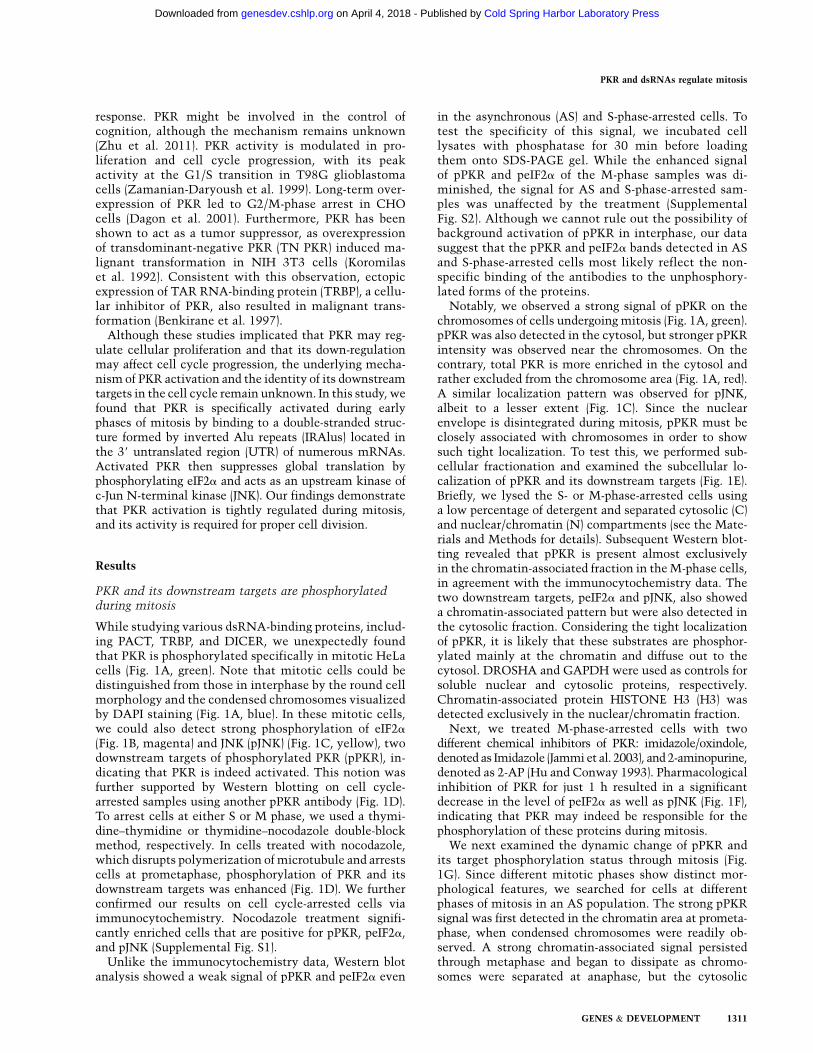
While studying various dsRNA-binding proteins, includ-ing PACT, TRBP, and DICER, we unexpectedly foundthat PKR is phosphorylated specifically in mitotic HeLacells (Fig. 1A, green). Note that mitotic cells could bedistinguished from those in interphase by the round cellmorphology and the condensed chromosomes visualizedby DAPI staining (Fig. 1A, blue). In these mitotic cells,we could also detect strong phosphorylation of eIF2a
(Fig. 1B, magenta) and JNK (pJNK) (Fig. 1C, yellow), twodownstream targets of phosphorylated PKR (pPKR), in-dicating that PKR is indeed activated. This notion wasfurther supported by Western blotting on cell cycle-arrested samples using another pPKR antibody (Fig. 1D).To arrest cells at either S or M phase, we used a thymi-dine–thymidine or thymidine–nocodazole double-blockmethod, respectively. In cells treated with nocodazole,which disrupts polymerization of microtubule and arrestscells at prometaphase, phosphorylation of PKR and itsdownstream targets was enhanced (Fig. 1D). We furtherconfirmed our results on cell cycle-arrested cells viaimmunocytochemistry. Nocodazole treatment signifi-cantly enriched cells that are positive for pPKR, peIF2a,and pJNK (Supplemental Fig. S1).
Unlike the immunocytochemistry data, Western blotanalysis showed a weak signal of pPKR and peIF2a even
in the asynchronous (AS) and S-phase-arrested cells. Totest the specificity of this signal, we incubated celllysates with phosphatase for 30 min before loadingthem onto SDS-PAGE gel. While the enhanced signalof pPKR and peIF2a of the M-phase samples was di-minished, the signal for AS and S-phase-arrested sam-ples was unaffected by the treatment (SupplementalFig. S2). Although we cannot rule out the possibility ofbackground activation of pPKR in interphase, our datasuggest that the pPKR and peIF2a bands detected in ASand S-phase-arrested cells most likely reflect the non-specific binding of the antibodies to the unphosphory-lated forms of the proteins.
Notably, we observed a strong signal of pPKR on thechromosomes of cells undergoing mitosis (Fig. 1A, green).pPKR was also detected in the cytosol, but stronger pPKRintensity was observed near the chromosomes. On thecontrary, total PKR is more enriched in the cytosol andrather excluded from the chromosome area (Fig. 1A, red).A similar localization pattern was observed for pJNK,albeit to a lesser extent (Fig. 1C). Since the nuclearenvelope is disintegrated during mitosis, pPKR must beclosely associated with chromosomes in order to showsuch tight localization. To test this, we performed sub-cellular fractionation and examined the subcellular lo-calization of pPKR and its downstream targets (Fig. 1E).Briefly, we lysed the S- or M-phase-arrested cells usinga low percentage of detergent and separated cytosolic (C)and nuclear/chromatin (N) compartments (see the Mate-rials and Methods for details). Subsequent Western blot-ting revealed that pPKR is present almost exclusivelyin the chromatin-associated fraction in the M-phase cells,in agreement with the immunocytochemistry data. Thetwo downstream targets, peIF2a and pJNK, also showeda chromatin-associated pattern but were also detected inthe cytosolic fraction. Considering the tight localizationof pPKR, it is likely that these substrates are phosphor-ylated mainly at the chromatin and diffuse out to thecytosol. DROSHA and GAPDH were used as controls forsoluble nuclear and cytosolic proteins, respectively.Chromatin-associated protein HISTONE H3 (H3) wasdetected exclusively in the nuclear/chromatin fraction.
Next, we treated M-phase-arrested cells with twodifferent chemical inhibitors of PKR: imidazole/oxindole,denoted as Imidazole (Jammi et al. 2003), and 2-aminopurine,denoted as 2-AP (Hu and Conway 1993). Pharmacologicalinhibition of PKR for just 1 h resulted in a significantdecrease in the level of peIF2a as well as pJNK (Fig. 1F),indicating that PKR may indeed be responsible for thephosphorylation of these proteins during mitosis.
We next examined the dynamic change of pPKR andits target phosphorylation status through mitosis (Fig.1G). Since different mitotic phases show distinct mor-phological features, we searched for cells at differentphases of mitosis in an AS population. The strong pPKRsignal was first detected in the chromatin area at prometa-phase, when condensed chromosomes were readily ob-served. A strong chromatin-associated signal persistedthrough metaphase and began to dissipate as chromo-somes were separated at anaphase, but the cytosolic
PKR and dsRNAs regulate mitosis
GENES & DEVELOPMENT 1311
Cold Spring Harbor Laboratory Press on April 4, 2018 - Published by genesdev.cshlp.orgDownloaded from
signal was still higher than the background. By the end oftelophase, the pPKR signal was no longer detected. Thepattern of pJNK was similar to that of pPKR, with a slightdelay (Fig. 1G, yellow); the chromosome-associated pJNKsignal was detected until metaphase and disappears grad-ually from the chromosomal region. peIF2a was also firstdetected at the prometaphase, but its dephosphorylationexhibited a clear time delay in which its phosphorylationlasted through telophase, and a weak signal still remainedeven at the end of cytokinesis (Fig. 1G, magenta).
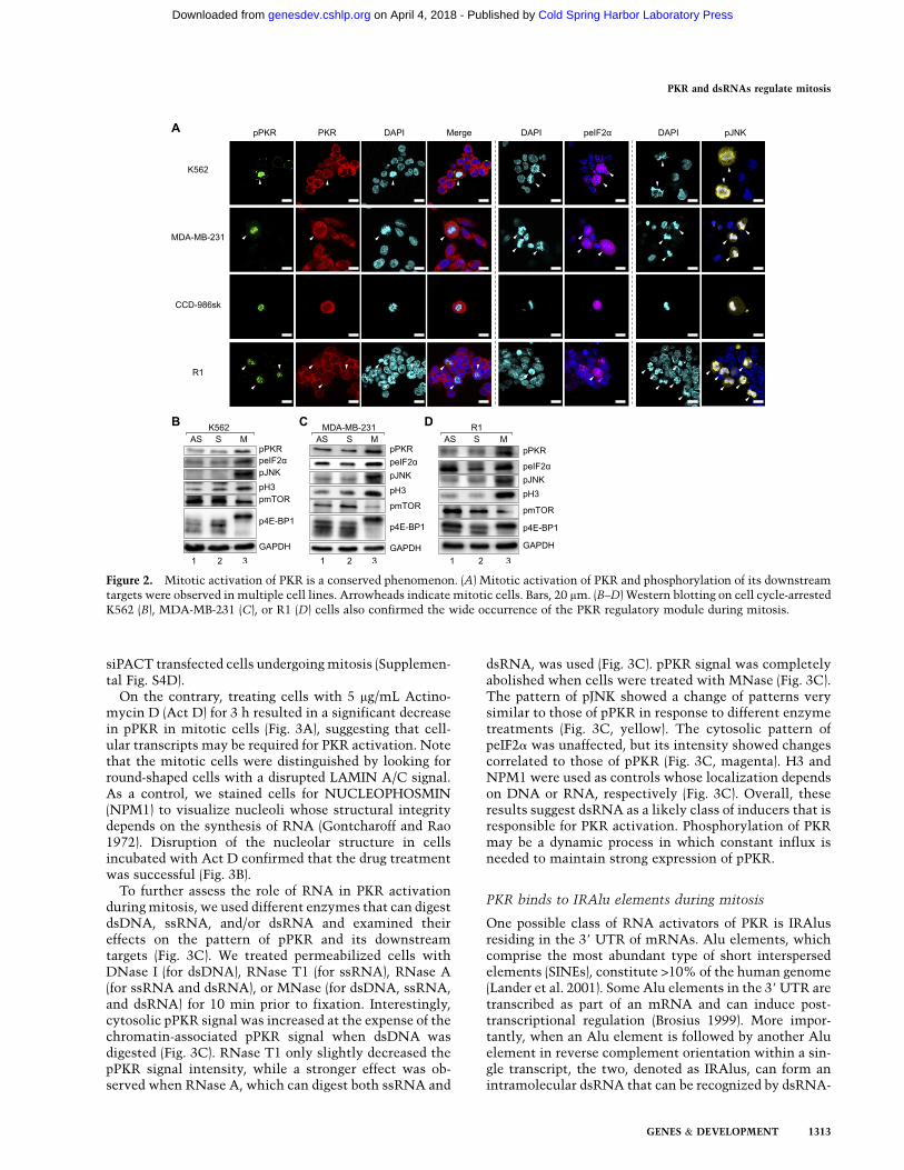
To test whether the observed activation of PKR isspecific to HeLa cells or occurs in general, we performedimmunocytochemistry on K562 leukemia cells and MDA-MB-231 breast cancer cells and asked whether PKR andits downstream targets are phosphorylated specificallyduring mitosis. We indeed detected strong signals forpPKR, peIF2a, and pJNK in cells with condensed chromo-somes that indicate cells in mitosis (Fig. 2A). Thus, theactivation of PKR and the phosphorylation of its down-stream targets also occur in different cancer cell lines.Going beyond cancer cells, we confirmed the expressionof pPKR, peIF2a, and pJNK in human primary fibroblastcells (CCD-986sk) and mouse embryonic stem cells (R1)undergoing mitosis (Fig. 2A). Taken together, the observedphenomenon occurs in noncancer and nonhuman cells as
well. The above results were also confirmed via Westernblotting and immunostaining on cells arrested at S or Mphase (Fig. 2B–D; Supplemental Fig. S3). We could notarrest CCD-986sk cells because fibroblast cells undergomitotic slippage and adaptation (Lanni and Jacks 1998).
Mitotic activation of PKR requires dsRNA
As a first step to understanding the function of PKRactivation during mitosis, we sought to identify theupstream signal that leads to phosphorylation of PKR.PKR can be activated by binding to heparin, PACT, anda stretch of dsRNA greater than ;33 bp (for review, seeCole 2007). Since the mechanism of PKR activation byheparin is not well characterized, we focused on thelatter two candidates. First, we examined the subcellularlocalization and expression level of PACT and concludedthat they remained constant through the cell cycle(Supplemental Fig S4A,B). Moreover, the interactionbetween PKR and PACT did not change between S- andM-phase-arrested cells (Supplemental Fig. S4C). Last, weknocked down PACT using siRNA and examined itseffect on pPKR. Immunocytochemistry analysis showedthat while PACT knockdown was successful, there wasno difference in the pPKR signal between siLuc and
Figure 1. M-phase-specific phosphorylation ofPKR and its downstream targets. (A) Immuno-cytochemistry revealed a strong signal of pPKR inmitotic cells (green), while total PKR (red) is pre-dominantly cytosolic. (B,C) In addition to pPKR,peIF2a (B) and pJNK (C) signals were observed incells undergoing mitosis. Bars, 20 mm. (D) Westernblotting on AS or S- or M-phase-arrested cellsconfirmed activation of PKR and phosphorylationof eIF2a and JNK during mitosis. (E) A subcellularfractionation experiment further suggested thatpPKR is closely associated with nuclear/chromatin(N). (F) Pharmacological inhibition of PKR using1 mM imidazole/oxindole (Imidazole) or 50 mM2-aminopurine (2-AP) resulted in a decrease inpeIF2a and pJNK. The numbers denote treatmenttime in minutes. (G) Close examination of patternsof pPKR (green), pJNK (yellow), and peIF2a (ma-genta) at different phases of mitosis. Bars, 20 mm.
Kim et al.
1312 GENES & DEVELOPMENT
Cold Spring Harbor Laboratory Press on April 4, 2018 - Published by genesdev.cshlp.orgDownloaded from
siPACT transfected cells undergoing mitosis (Supplemen-tal Fig. S4D).
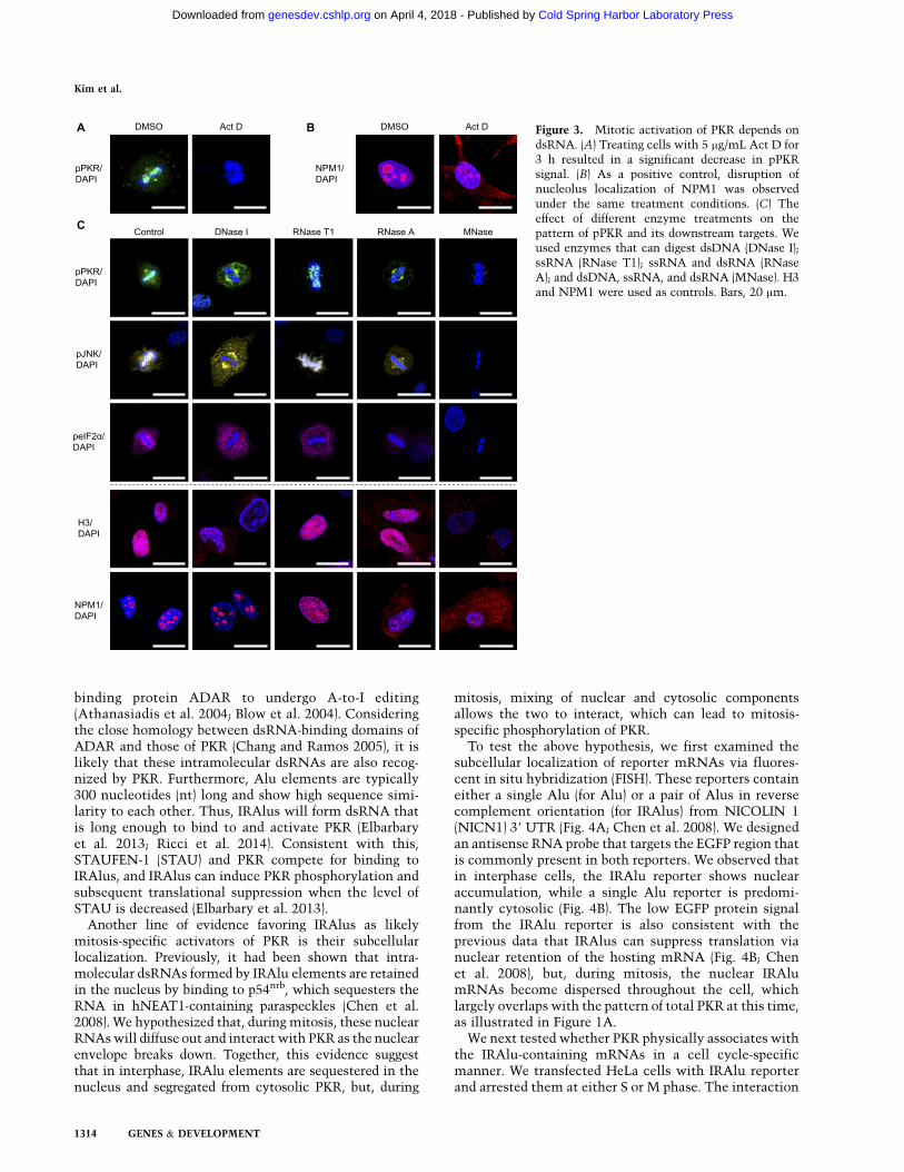
On the contrary, treating cells with 5 mg/mL Actino-mycin D (Act D) for 3 h resulted in a significant decreasein pPKR in mitotic cells (Fig. 3A), suggesting that cell-ular transcripts may be required for PKR activation. Notethat the mitotic cells were distinguished by looking forround-shaped cells with a disrupted LAMIN A/C signal.As a control, we stained cells for NUCLEOPHOSMIN(NPM1) to visualize nucleoli whose structural integritydepends on the synthesis of RNA (Gontcharoff and Rao1972). Disruption of the nucleolar structure in cellsincubated with Act D confirmed that the drug treatmentwas successful (Fig. 3B).
To further assess the role of RNA in PKR activationduring mitosis, we used different enzymes that can digestdsDNA, ssRNA, and/or dsRNA and examined theireffects on the pattern of pPKR and its downstreamtargets (Fig. 3C). We treated permeabilized cells withDNase I (for dsDNA), RNase T1 (for ssRNA), RNase A(for ssRNA and dsRNA), or MNase (for dsDNA, ssRNA,and dsRNA) for 10 min prior to fixation. Interestingly,cytosolic pPKR signal was increased at the expense of thechromatin-associated pPKR signal when dsDNA wasdigested (Fig. 3C). RNase T1 only slightly decreased thepPKR signal intensity, while a stronger effect was ob-served when RNase A, which can digest both ssRNA and
dsRNA, was used (Fig. 3C). pPKR signal was completelyabolished when cells were treated with MNase (Fig. 3C).The pattern of pJNK showed a change of patterns verysimilar to those of pPKR in response to different enzymetreatments (Fig. 3C, yellow). The cytosolic pattern ofpeIF2a was unaffected, but its intensity showed changescorrelated to those of pPKR (Fig. 3C, magenta). H3 andNPM1 were used as controls whose localization dependson DNA or RNA, respectively (Fig. 3C). Overall, theseresults suggest dsRNA as a likely class of inducers that isresponsible for PKR activation. Phosphorylation of PKRmay be a dynamic process in which constant influx isneeded to maintain strong expression of pPKR.
PKR binds to IRAlu elements during mitosis
One possible class of RNA activators of PKR is IRAlusresiding in the 39 UTR of mRNAs. Alu elements, whichcomprise the most abundant type of short interspersedelements (SINEs), constitute >10% of the human genome(Lander et al. 2001). Some Alu elements in the 39 UTR aretranscribed as part of an mRNA and can induce post-transcriptional regulation (Brosius 1999). More impor-tantly, when an Alu element is followed by another Aluelement in reverse complement orientation within a sin-gle transcript, the two, denoted as IRAlus, can form anintramolecular dsRNA that can be recognized by dsRNA-
Figure 2. Mitotic activation of PKR is a conserved phenomenon. (A) Mitotic activation of PKR and phosphorylation of its downstreamtargets were observed in multiple cell lines. Arrowheads indicate mitotic cells. Bars, 20 mm. (B–D) Western blotting on cell cycle-arrestedK562 (B), MDA-MB-231 (C), or R1 (D) cells also confirmed the wide occurrence of the PKR regulatory module during mitosis.
PKR and dsRNAs regulate mitosis
GENES & DEVELOPMENT 1313
Cold Spring Harbor Laboratory Press on April 4, 2018 - Published by genesdev.cshlp.orgDownloaded from
binding protein ADAR to undergo A-to-I editing(Athanasiadis et al. 2004; Blow et al. 2004). Consideringthe close homology between dsRNA-binding domains ofADAR and those of PKR (Chang and Ramos 2005), it islikely that these intramolecular dsRNAs are also recog-nized by PKR. Furthermore, Alu elements are typically300 nucleotides (nt) long and show high sequence simi-larity to each other. Thus, IRAlus will form dsRNA thatis long enough to bind to and activate PKR (Elbarbaryet al. 2013; Ricci et al. 2014). Consistent with this,STAUFEN-1 (STAU) and PKR compete for binding toIRAlus, and IRAlus can induce PKR phosphorylation andsubsequent translational suppression when the level ofSTAU is decreased (Elbarbary et al. 2013).
Another line of evidence favoring IRAlus as likelymitosis-specific activators of PKR is their subcellularlocalization. Previously, it had been shown that intra-molecular dsRNAs formed by IRAlu elements are retainedin the nucleus by binding to p54nrb, which sequesters theRNA in hNEAT1-containing paraspeckles (Chen et al.2008). We hypothesized that, during mitosis, these nuclearRNAs will diffuse out and interact with PKR as the nuclearenvelope breaks down. Together, this evidence suggestthat in interphase, IRAlu elements are sequestered in thenucleus and segregated from cytosolic PKR, but, during
mitosis, mixing of nuclear and cytosolic componentsallows the two to interact, which can lead to mitosis-specific phosphorylation of PKR.
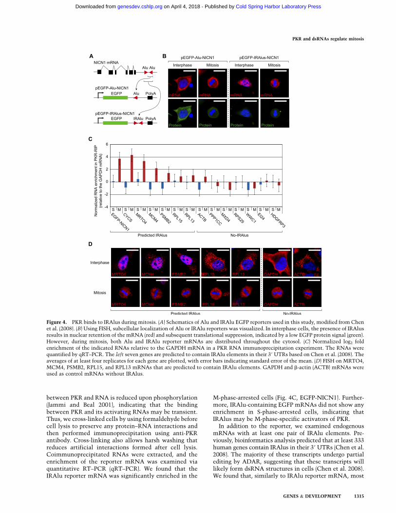
To test the above hypothesis, we first examined thesubcellular localization of reporter mRNAs via fluores-cent in situ hybridization (FISH). These reporters containeither a single Alu (for Alu) or a pair of Alus in reversecomplement orientation (for IRAlus) from NICOLIN 1(NICN1) 39 UTR (Fig. 4A; Chen et al. 2008). We designedan antisense RNA probe that targets the EGFP region thatis commonly present in both reporters. We observed thatin interphase cells, the IRAlu reporter shows nuclearaccumulation, while a single Alu reporter is predomi-nantly cytosolic (Fig. 4B). The low EGFP protein signalfrom the IRAlu reporter is also consistent with theprevious data that IRAlus can suppress translation vianuclear retention of the hosting mRNA (Fig. 4B; Chenet al. 2008), but, during mitosis, the nuclear IRAlumRNAs become dispersed throughout the cell, whichlargely overlaps with the pattern of total PKR at this time,as illustrated in Figure 1A.
We next tested whether PKR physically associates withthe IRAlu-containing mRNAs in a cell cycle-specificmanner. We transfected HeLa cells with IRAlu reporterand arrested them at either S or M phase. The interaction
Figure 3. Mitotic activation of PKR depends ondsRNA. (A) Treating cells with 5 mg/mL Act D for3 h resulted in a significant decrease in pPKRsignal. (B) As a positive control, disruption ofnucleolus localization of NPM1 was observedunder the same treatment conditions. (C) Theeffect of different enzyme treatments on thepattern of pPKR and its downstream targets. Weused enzymes that can digest dsDNA (DNase I);ssRNA (RNase T1); ssRNA and dsRNA (RNaseA); and dsDNA, ssRNA, and dsRNA (MNase). H3and NPM1 were used as controls. Bars, 20 mm.
Kim et al.
1314 GENES & DEVELOPMENT
Cold Spring Harbor Laboratory Press on April 4, 2018 - Published by genesdev.cshlp.orgDownloaded from
between PKR and RNA is reduced upon phosphorylation(Jammi and Beal 2001), indicating that the bindingbetween PKR and its activating RNAs may be transient.Thus, we cross-linked cells by using formaldehyde beforecell lysis to preserve any protein–RNA interactions andthen performed immunoprecipitation using anti-PKRantibody. Cross-linking also allows harsh washing thatreduces artificial interactions formed after cell lysis.Coimmunoprecipitated RNAs were extracted, and theenrichment of the reporter mRNA was examined viaquantitative RT–PCR (qRT–PCR). We found that theIRAlu reporter mRNA was significantly enriched in the
M-phase-arrested cells (Fig. 4C, EGFP-NICN1). Further-more, IRAlu-containing EGFP mRNAs did not show anyenrichment in S-phase-arrested cells, indicating thatIRAlus may be M-phase-specific activators of PKR.
In addition to the reporter, we examined endogenousmRNAs with at least one pair of IRAlu elements. Pre-viously, bioinformatics analysis predicted that at least 333human genes contain IRAlus in their 39 UTRs (Chen et al.2008). The majority of these transcripts undergo partialediting by ADAR, suggesting that these transcripts willlikely form dsRNA structures in cells (Chen et al. 2008).We found that, similarly to IRAlu reporter mRNA, most
Figure 4. PKR binds to IRAlus during mitosis. (A) Schematics of Alu and IRAlu EGFP reporters used in this study, modified from Chenet al. (2008). (B) Using FISH, subcellular localization of Alu or IRAlu reporters was visualized. In interphase cells, the presence of IRAlusresults in nuclear retention of the mRNA (red) and subsequent translational suppression, indicated by a low EGFP protein signal (green).However, during mitosis, both Alu and IRAlu reporter mRNAs are distributed throughout the cytosol. (C) Normalized log2 foldenrichment of the indicated RNAs relative to the GAPDH mRNA in a PKR RNA immunoprecipitation experiment. The RNAs werequantified by qRT–PCR. The left seven genes are predicted to contain IRAlu elements in their 39 UTRs based on Chen et al. (2008). Theaverages of at least four replicates for each gene are plotted, with error bars indicating standard error of the mean. (D) FISH on MRTO4,MCM4, PSMB2, RPL15, and RPL13 mRNAs that are predicted to contain IRAlu elements. GAPDH and b-actin (ACTB) mRNAs wereused as control mRNAs without IRAlus.
PKR and dsRNAs regulate mitosis
GENES & DEVELOPMENT 1315
Cold Spring Harbor Laboratory Press on April 4, 2018 - Published by genesdev.cshlp.orgDownloaded from
of the transcripts with predicted IRAlu elements alsointeract with PKR (Fig. 4C). Two of the transcripts(MRTO4 and CYCS) showed enrichment comparablewith that of IRAlu reporter EGFP mRNA. Importantly,enrichment of these mRNAs was observed only in theM-phase-arrested cells. Furthermore, among the sevencontrol RNAs that were not predicted to contain IRAlus(no-IRAlus), none of them shows significant binding toPKR. Although the enrichment levels were variable, themRNAs with predicted IRAlus tend to bind more fre-quently to PKR compared with those without IRAlus.
We then examined the subcellular localization ofendogenous mRNAs with IRAlus using FISH. We foundthat three of the predicted IRAlu mRNAs, MRTO4,MCM4, and PSMB2, show strong nuclear signals dur-ing interphase that diffuse out to cytosol during mitosis(Fig. 4D). The nuclear retention pattern of IRAlu-con-taining transcripts is clearly different from that of othertranscripts such as GAPDH and b-ACTIN mRNA,which are predominantly cytosolic (Fig. 4D). Notably,not all IRAlu-containing mRNAs were localized in thenucleus. Two ribosomal mRNAs, RPL15 and RPL13,showed highly cytosolic localization despite the pre-dicted IRAlu elements. One possible explanation is thatmultiple isoforms with different 39 UTRs are generatedby alternative processing. For example, one of the iso-forms of NICN1 mRNA localizes in the cytosol becauseit contains only one of the two Alu repeats required toform the double-stranded structure (Chen et al. 2008).We also examined the localization of CYCS mRNA, butits signal was too weak for reliable detection (data notshown). Thus, the intramolecular dsRNAs formed byIRAlu elements may bind to and activate PKR inmitosis, a period during which the nuclear envelope isabsent.
PKR suppresses bulk translation during mitosis
It is well established that mitotic cells synthesize pro-teins at a much slower rate compared with cells ininterphase (Prescott and Bender 1962; Tarnowka andBaglioni 1979). This decrease in translation has beenattributed to the inhibition of 59 cap-dependent trans-lation by 14-3-3s (Wilker et al. 2007) and hypophosphor-ylation of eIF4E-binding proteins (4E-BPs) (Pyronnetet al. 2001), which result in suppression of translationin most mRNAs, while certain mRNAs with internalribosomal entry sites (IRESs) show increased transla-tional activity. This switch to IRES-dependent trans-lation is thought to be necessary for mitotic progressionand to initiate interphase of the daughter cells. However,another line of evidence suggests that 4E-BPs are hyper-phosphorylated instead of hypophosphorylated duringmitosis and that hyperphosphorylated 4E-BPs cannotinhibit cap-dependent translation because they no lon-ger interact with eIF4E (Heesom et al. 2001; Ramirez-Valle et al. 2010). Our result is also consistent with thelatter observation that mTOR, an upstream kinase of 4E-BPs, is still phosphorylated and that 4E-BP1 is hyper-phosphorylated during mitosis in HeLa and other cell
lines examined (Figs. 1D, 2B–D; Supplemental Fig. S2).At the same time, it has been shown that eIF2a alsocontributes to translational regulation during mitosis, asit is phosphorylated in G2/M phase of U2-OS osteosar-coma cells (Datta et al. 1999), and its phosphorylationcan up-regulate the efficiency of IRES-mediated trans-lation (Gerlitz et al. 2002).
Considering that eIF2a is the most well-characterizedsubstrate of PKR, we asked whether PKR contributesto the suppression of general translation during mitosis.We started by characterizing mitotic translation of HeLacells. We performed metabolic labeling and assessedprotein synthesis rates by quantifying 35S-methionineincorporation rates. We found that during mitosis, therate of protein synthesis was ;35% of that of the S phase,which is consistent with earlier studies (Fig. 5A; Prescottand Bender 1962; Tarnowka and Baglioni 1979). Ribo-some fractionation using a sucrose gradient also showeda significant increase in the monosome peak at theexpense of polyribosome peaks in M-phase-arrested cells(Supplemental Fig. S5A,B). This increase in the 80Smonosome peak is consistent with the characteristics ofthe translation block at the initiation step (Ricci et al.2014). A similar pattern of ribosomes was observed wheneIF2a phosphorylation was induced by nutritional stress(Tonelli et al. 2011). We then treated M-phase-arrestedcells with two different PKR inhibitors at multiple timepoints and examined the changes in the peIF2a signal.The peIF2a signal was significantly down-regulatedwithin 30 min after the drug treatment and nearlycompletely abolished by 2 h (Fig. 5B,C). A decrease inthe peIF2a signal was accompanied by an increase ingeneral translation; pharmacological inhibition of PKRfor 1 h in M-phase-arrested cells increased the methio-nine incorporation rate to ;60% of that of the S phase(Fig. 5D; Supplemental Fig. S5C).
In order to rule out possible side effects of PKRinhibitors, we used RNAi to further confirm our results.When PKR was depleted in M-phase-arrested cells, thegeneral translation was increased by ;50% (Fig. 5E;Supplemental Fig. S5D). In addition, we knocked downPKR in cells synchronized at S phase via the thymidinedouble-block method and performed metabolic labelingupon releasing them from the block. Both the controland siPKR transfected cells progressed through the cellcycle in a similar manner, although the PKR-depletedcells showed moderate accumulation of the G2/M frac-tion (Supplemental Fig. S6). A metabolic labeling exper-iment revealed that down-regulation of PKR led to anincrease in the protein synthesis rate, with the mostprominent effect occurring when the cells undergomitosis (Fig. 5F; Supplemental Fig. S5E). Upon releasefrom the double-thymidine block (see SupplementalFig. S6 for FACS analysis), siPKR transfected cells showedincreased translation by <10% during S and G2 phases.However, 10 and 12 h after the release, which corre-sponds to M and early G1 phase, PKR knockdownresulted in an at least 25% increase in the proteinsynthesis rate in three biological replicates. The magni-tude of the effect of PKR knockdown was variable in each
Kim et al.
1316 GENES & DEVELOPMENT
Cold Spring Harbor Laboratory Press on April 4, 2018 - Published by genesdev.cshlp.orgDownloaded from
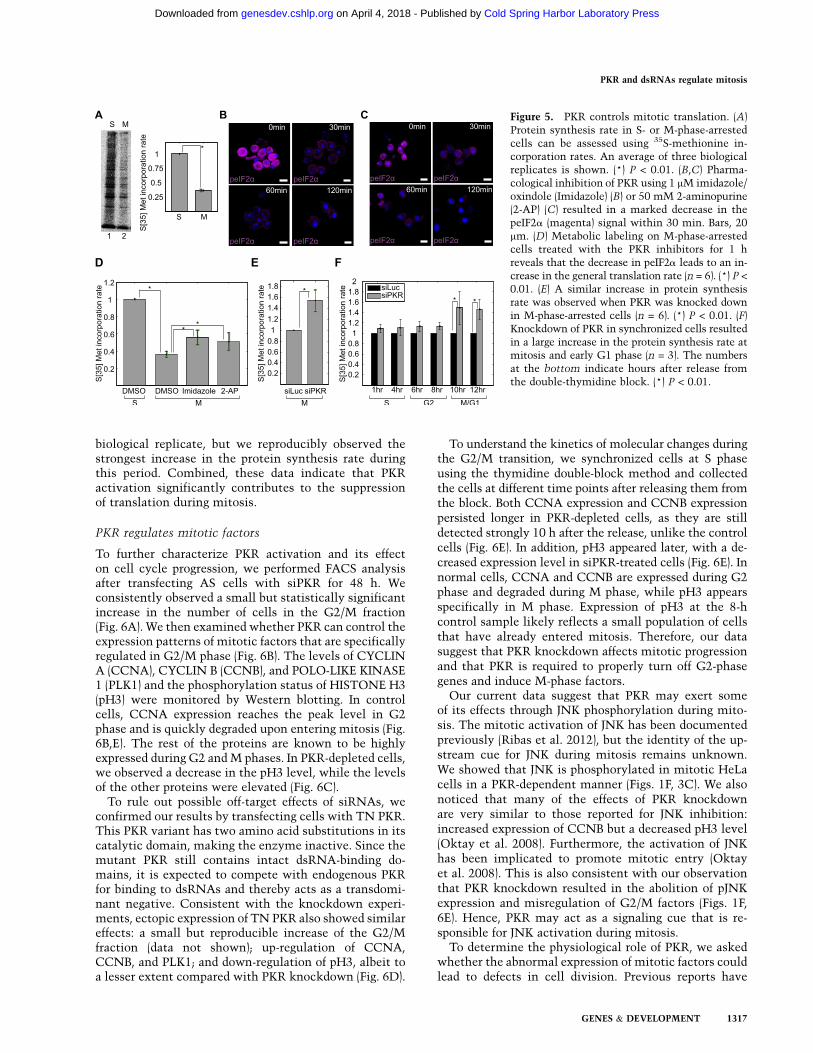
biological replicate, but we reproducibly observed thestrongest increase in the protein synthesis rate duringthis period. Combined, these data indicate that PKRactivation significantly contributes to the suppressionof translation during mitosis.
PKR regulates mitotic factors
To further characterize PKR activation and its effecton cell cycle progression, we performed FACS analysisafter transfecting AS cells with siPKR for 48 h. Weconsistently observed a small but statistically significantincrease in the number of cells in the G2/M fraction(Fig. 6A). We then examined whether PKR can control theexpression patterns of mitotic factors that are specificallyregulated in G2/M phase (Fig. 6B). The levels of CYCLINA (CCNA), CYCLIN B (CCNB), and POLO-LIKE KINASE1 (PLK1) and the phosphorylation status of HISTONE H3(pH3) were monitored by Western blotting. In controlcells, CCNA expression reaches the peak level in G2phase and is quickly degraded upon entering mitosis (Fig.6B,E). The rest of the proteins are known to be highlyexpressed during G2 and M phases. In PKR-depleted cells,we observed a decrease in the pH3 level, while the levelsof the other proteins were elevated (Fig. 6C).
To rule out possible off-target effects of siRNAs, weconfirmed our results by transfecting cells with TN PKR.This PKR variant has two amino acid substitutions in itscatalytic domain, making the enzyme inactive. Since themutant PKR still contains intact dsRNA-binding do-mains, it is expected to compete with endogenous PKRfor binding to dsRNAs and thereby acts as a transdomi-nant negative. Consistent with the knockdown experi-ments, ectopic expression of TN PKR also showed similareffects: a small but reproducible increase of the G2/Mfraction (data not shown); up-regulation of CCNA,CCNB, and PLK1; and down-regulation of pH3, albeit toa lesser extent compared with PKR knockdown (Fig. 6D).
To understand the kinetics of molecular changes duringthe G2/M transition, we synchronized cells at S phaseusing the thymidine double-block method and collectedthe cells at different time points after releasing them fromthe block. Both CCNA expression and CCNB expressionpersisted longer in PKR-depleted cells, as they are stilldetected strongly 10 h after the release, unlike the controlcells (Fig. 6E). In addition, pH3 appeared later, with a de-creased expression level in siPKR-treated cells (Fig. 6E). Innormal cells, CCNA and CCNB are expressed during G2phase and degraded during M phase, while pH3 appearsspecifically in M phase. Expression of pH3 at the 8-hcontrol sample likely reflects a small population of cellsthat have already entered mitosis. Therefore, our datasuggest that PKR knockdown affects mitotic progressionand that PKR is required to properly turn off G2-phasegenes and induce M-phase factors.
Our current data suggest that PKR may exert someof its effects through JNK phosphorylation during mito-sis. The mitotic activation of JNK has been documentedpreviously (Ribas et al. 2012), but the identity of the up-stream cue for JNK during mitosis remains unknown.We showed that JNK is phosphorylated in mitotic HeLacells in a PKR-dependent manner (Figs. 1F, 3C). We alsonoticed that many of the effects of PKR knockdownare very similar to those reported for JNK inhibition:increased expression of CCNB but a decreased pH3 level(Oktay et al. 2008). Furthermore, the activation of JNKhas been implicated to promote mitotic entry (Oktayet al. 2008). This is also consistent with our observationthat PKR knockdown resulted in the abolition of pJNKexpression and misregulation of G2/M factors (Figs. 1F,6E). Hence, PKR may act as a signaling cue that is re-sponsible for JNK activation during mitosis.
To determine the physiological role of PKR, we askedwhether the abnormal expression of mitotic factors couldlead to defects in cell division. Previous reports have
Figure 5. PKR controls mitotic translation. (A)Protein synthesis rate in S- or M-phase-arrestedcells can be assessed using 35S-methionine in-corporation rates. An average of three biologicalreplicates is shown. (*) P < 0.01. (B,C) Pharma-cological inhibition of PKR using 1 mM imidazole/oxindole (Imidazole) (B) or 50 mM 2-aminopurine(2-AP) (C) resulted in a marked decrease in thepeIF2a (magenta) signal within 30 min. Bars, 20mm. (D) Metabolic labeling on M-phase-arrestedcells treated with the PKR inhibitors for 1 hreveals that the decrease in peIF2a leads to an in-crease in the general translation rate (n = 6). (*) P <
0.01. (E) A similar increase in protein synthesisrate was observed when PKR was knocked downin M-phase-arrested cells (n = 6). (*) P < 0.01. (F)Knockdown of PKR in synchronized cells resultedin a large increase in the protein synthesis rate atmitosis and early G1 phase (n = 3). The numbersat the bottom indicate hours after release fromthe double-thymidine block. (*) P < 0.01.
PKR and dsRNAs regulate mitosis
GENES & DEVELOPMENT 1317
Cold Spring Harbor Laboratory Press on April 4, 2018 - Published by genesdev.cshlp.orgDownloaded from
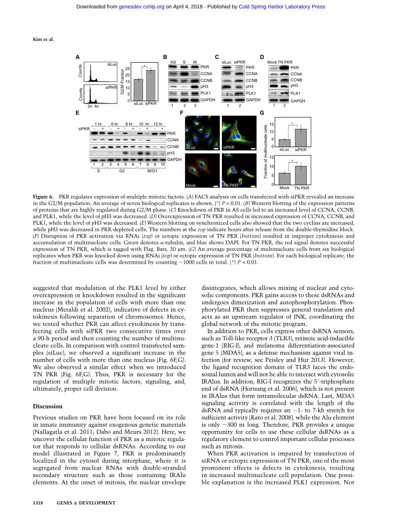
suggested that modulation of the PLK1 level by eitheroverexpression or knockdown resulted in the significantincrease in the population of cells with more than onenucleus (Meraldi et al. 2002), indicative of defects in cy-tokinesis following separation of chromosomes. Hence,we tested whether PKR can affect cytokinesis by trans-fecting cells with siPKR two consecutive times overa 90-h period and then counting the number of multinu-cleate cells. In comparison with control transfected sam-ples (siLuc), we observed a significant increase in thenumber of cells with more than one nucleus (Fig. 6F,G).We also observed a similar effect when we introducedTN PKR (Fig. 6F,G). Thus, PKR is necessary for theregulation of multiple mitotic factors, signaling, and,ultimately, proper cell division.
Discussion
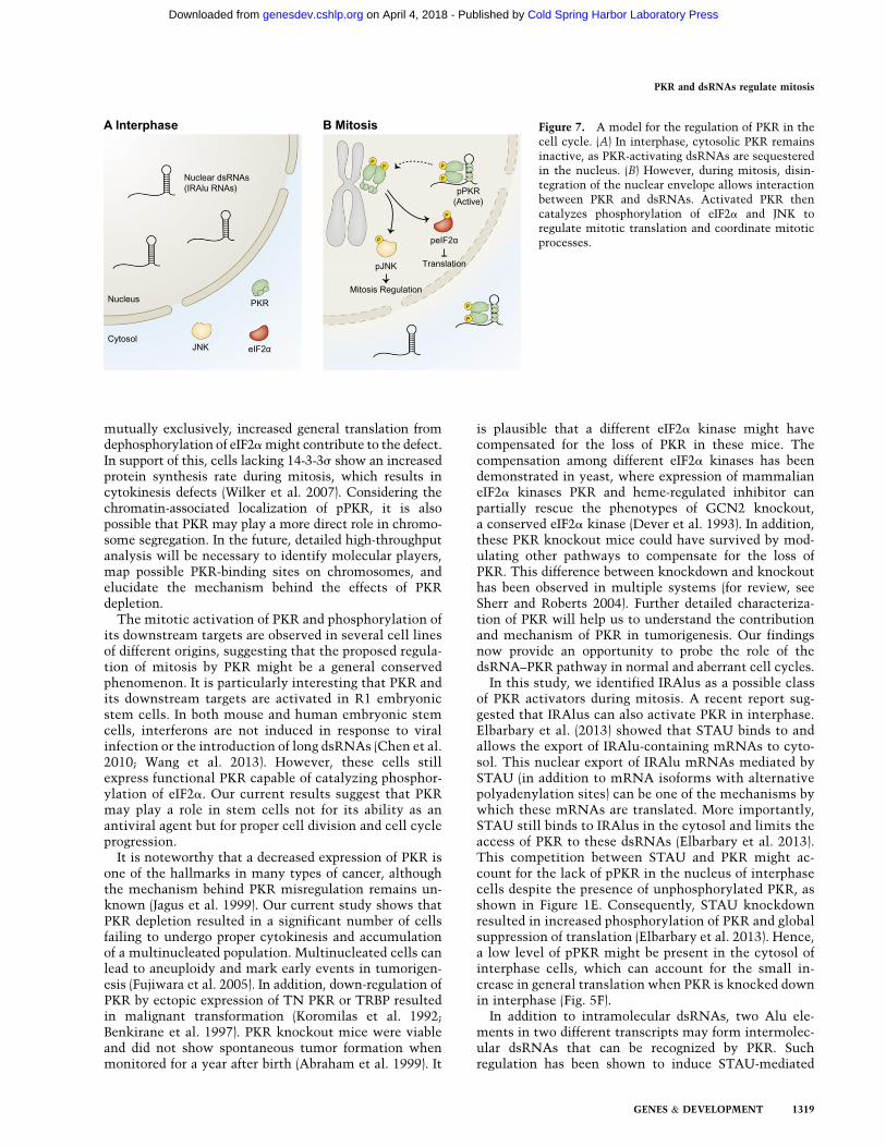
Previous studies on PKR have been focused on its rolein innate immunity against exogenous genetic materials(Nallagatla et al. 2011; Dabo and Meurs 2012). Here, weuncover the cellular function of PKR as a mitotic regula-tor that responds to cellular dsRNAs. According to ourmodel illustrated in Figure 7, PKR is predominantlylocalized in the cytosol during interphase, where it issegregated from nuclear RNAs with double-strandedsecondary structure such as those containing IRAluelements. At the onset of mitosis, the nuclear envelope
disintegrates, which allows mixing of nuclear and cyto-solic components. PKR gains access to these dsRNAs andundergoes dimerization and autophosphorylation. Phos-phorylated PKR then suppresses general translation andacts as an upstream regulator of JNK, coordinating theglobal network of the mitotic program.
In addition to PKR, cells express other dsRNA sensors,such as Toll-like receptor 3 (TLR3), retinoic acid-induciblegene-1 (RIG-I), and melanoma differentiation-associatedgene 5 (MDA5), as a defense mechanism against viral in-fection (for review, see Peisley and Hur 2013). However,the ligand recognition domain of TLR3 faces the endo-somal lumen and will not be able to interact with cytosolicIRAlus. In addition, RIG-I recognizes the 59-triphosphateend of dsRNA (Hornung et al. 2006), which is not presentin IRAlus that form intramolecular dsRNA. Last, MDA5signaling activity is correlated with the length of thedsRNA and typically requires an ;1- to 7-kb stretch forsufficient activity (Kato et al. 2008), while the Alu elementis only ;300 nt long. Therefore, PKR provides a uniqueopportunity for cells to use these cellular dsRNAs as aregulatory element to control important cellular processessuch as mitosis.
When PKR activation is impaired by transfection ofsiRNA or ectopic expression of TN PKR, one of the mostprominent effects is defects in cytokinesis, resultingin increased multinucleate cell population. One possi-ble explanation is the increased PLK1 expression. Not
Figure 6. PKR regulates expression of multiple mitotic factors. (A) FACS analysis on cells transfected with siPKR revealed an increasein the G2/M population. An average of seven biological replicates is shown. (*) P < 0.01. (B) Western blotting of the expression patternsof proteins that are highly regulated during G2/M phase. (C) Knockdown of PKR in AS cells led to an increased level of CCNA, CCNB,and PLK1, while the level of pH3 was decreased. (D) Overexpression of TN PKR resulted in increased expression of CCNA, CCNB, andPLK1, while the level of pH3 was decreased. (E) Western blotting on synchronized cells also showed that the two cyclins are increased,while pH3 was decreased in PKR-depleted cells. The numbers at the top indicate hours after release from the double-thymidine block.(F) Disruption of PKR activation via RNAi (top) or ectopic expression of TN PKR (bottom) resulted in improper cytokinesis andaccumulation of multinucleate cells. Green denotes a-tubulin, and blue shows DAPI. For TN PKR, the red signal denotes successfulexpression of TN PKR, which is tagged with Flag. Bars, 20 mm. (G) An average percentage of multinucleate cells from six biologicalreplicates when PKR was knocked down using RNAi (top) or ectopic expression of TN PKR (bottom). For each biological replicate, thefraction of multinucleate cells was determined by counting ;1000 cells in total. (*) P < 0.01.
Kim et al.
1318 GENES & DEVELOPMENT
Cold Spring Harbor Laboratory Press on April 4, 2018 - Published by genesdev.cshlp.orgDownloaded from
mutually exclusively, increased general translation fromdephosphorylation of eIF2a might contribute to the defect.In support of this, cells lacking 14-3-3s show an increasedprotein synthesis rate during mitosis, which results incytokinesis defects (Wilker et al. 2007). Considering thechromatin-associated localization of pPKR, it is alsopossible that PKR may play a more direct role in chromo-some segregation. In the future, detailed high-throughputanalysis will be necessary to identify molecular players,map possible PKR-binding sites on chromosomes, andelucidate the mechanism behind the effects of PKRdepletion.
The mitotic activation of PKR and phosphorylation ofits downstream targets are observed in several cell linesof different origins, suggesting that the proposed regula-tion of mitosis by PKR might be a general conservedphenomenon. It is particularly interesting that PKR andits downstream targets are activated in R1 embryonicstem cells. In both mouse and human embryonic stemcells, interferons are not induced in response to viralinfection or the introduction of long dsRNAs (Chen et al.2010; Wang et al. 2013). However, these cells stillexpress functional PKR capable of catalyzing phosphor-ylation of eIF2a. Our current results suggest that PKRmay play a role in stem cells not for its ability as anantiviral agent but for proper cell division and cell cycleprogression.
It is noteworthy that a decreased expression of PKR isone of the hallmarks in many types of cancer, althoughthe mechanism behind PKR misregulation remains un-known (Jagus et al. 1999). Our current study shows thatPKR depletion resulted in a significant number of cellsfailing to undergo proper cytokinesis and accumulationof a multinucleated population. Multinucleated cells canlead to aneuploidy and mark early events in tumorigen-esis (Fujiwara et al. 2005). In addition, down-regulation ofPKR by ectopic expression of TN PKR or TRBP resultedin malignant transformation (Koromilas et al. 1992;Benkirane et al. 1997). PKR knockout mice were viableand did not show spontaneous tumor formation whenmonitored for a year after birth (Abraham et al. 1999). It
is plausible that a different eIF2a kinase might havecompensated for the loss of PKR in these mice. Thecompensation among different eIF2a kinases has beendemonstrated in yeast, where expression of mammalianeIF2a kinases PKR and heme-regulated inhibitor canpartially rescue the phenotypes of GCN2 knockout,a conserved eIF2a kinase (Dever et al. 1993). In addition,these PKR knockout mice could have survived by mod-ulating other pathways to compensate for the loss ofPKR. This difference between knockdown and knockouthas been observed in multiple systems (for review, seeSherr and Roberts 2004). Further detailed characteriza-tion of PKR will help us to understand the contributionand mechanism of PKR in tumorigenesis. Our findingsnow provide an opportunity to probe the role of thedsRNA–PKR pathway in normal and aberrant cell cycles.
In this study, we identified IRAlus as a possible classof PKR activators during mitosis. A recent report sug-gested that IRAlus can also activate PKR in interphase.Elbarbary et al. (2013) showed that STAU binds to andallows the export of IRAlu-containing mRNAs to cyto-sol. This nuclear export of IRAlu mRNAs mediated bySTAU (in addition to mRNA isoforms with alternativepolyadenylation sites) can be one of the mechanisms bywhich these mRNAs are translated. More importantly,STAU still binds to IRAlus in the cytosol and limits theaccess of PKR to these dsRNAs (Elbarbary et al. 2013).This competition between STAU and PKR might ac-count for the lack of pPKR in the nucleus of interphasecells despite the presence of unphosphorylated PKR, asshown in Figure 1E. Consequently, STAU knockdownresulted in increased phosphorylation of PKR and globalsuppression of translation (Elbarbary et al. 2013). Hence,a low level of pPKR might be present in the cytosol ofinterphase cells, which can account for the small in-crease in general translation when PKR is knocked downin interphase (Fig. 5F).
In addition to intramolecular dsRNAs, two Alu ele-ments in two different transcripts may form intermolec-ular dsRNAs that can be recognized by PKR. Suchregulation has been shown to induce STAU-mediated
Figure 7. A model for the regulation of PKR in thecell cycle. (A) In interphase, cytosolic PKR remainsinactive, as PKR-activating dsRNAs are sequesteredin the nucleus. (B) However, during mitosis, disin-tegration of the nuclear envelope allows interactionbetween PKR and dsRNAs. Activated PKR thencatalyzes phosphorylation of eIF2a and JNK toregulate mitotic translation and coordinate mitoticprocesses.
PKR and dsRNAs regulate mitosis
GENES & DEVELOPMENT 1319
Cold Spring Harbor Laboratory Press on April 4, 2018 - Published by genesdev.cshlp.orgDownloaded from
mRNA decay (Gong and Maquat 2011). In this case, anAlu element in the 39 UTR of SERPINE1 mRNA forms animperfect dsRNA with another Alu element in 1/2-sbsRNA1 long noncoding RNA (lncRNA) (Gong andMaquat 2011). Thermodynamic analysis predicted thatSERPINE1 mRNA can potentially form intermoleculardsRNA with >300 lncRNAs (Gong and Maquat 2011).Considering the wide occurrence of Alu elements inthe human genome, virtually any mRNA with Alu inits 39 UTR can potentially activate PKR. In addition,functional IRAlu elements may reside in introns (Chenet al. 2008) that will also be exposed to cytosolic PKRduring mitosis. Furthermore, there might exist othertypes of dsRNAs that can be recognized by PKR. Recentstudies have shown that mRNAs contain multiple in-ternal stem–loop structures that can be recognized bydsRNA-binding proteins such as STAU (Cho et al. 2012;Ricci et al. 2014). Thus, it will be interesting to performa genome-wide search for RNA activators of PKR andstudy how they escape PKR activation in interphase.
Currently, it is unclear how pPKR is localized andassociated to the chromosome region. Nuclear localiza-tion of pPKR has been observed in limited cases, in-cluding in CD34+ cells from high-risk myelodysplasticsyndrome patients (Follo et al. 2008; Blalock et al. 2011).One possibility is that pPKR binds to an unknown proteinfactor that tethers the enzyme to the chromatin. Forexample, substrates of pPKR might be enriched near thechromosome region and recruit PKR. A recent massspectrometry analysis on PKR interactome revealed thatpPKR interacts with proteins involved in chromatinmodification and cell division, such as BUB3 and NPM1(Blalock et al. 2014). Alternatively, it is possible that theremay be different classes of dsRNAs that are localizedto the chromosome region during mitosis and tether PKR.In this case, the chromosome might serve as a center foractivating PKR as well as catalyzing phosphorylation ofPKR targets such as eIF2a and JNK. In the future, it willbe interesting to identify PKR-interacting proteins anddsRNAs and examine their localized activities at variousstages of the cell cycle.
Materials and methods
Cell cycle arrest and PKR inhibitor treatment
To arrest cells at S or M phase, the double-thymidine blockmethod was used. Briefly, cells were treated with 2 mM thymi-dine for 18 h and then released into fresh medium. After 9 h,2 mM thymidine (for S-phase arrest) or 100 ng/mL nocodazole(for M-phase arrest) was added to the medium, and cells wereincubated for an additional 17 h. When indicated, M-phase-arrested cells were treated with 1 mM imidazole/oxindole (EMDMillipore) or 50 mM 2-aminopurine (Sigma-Aldrich) for 1 h.
Immunocytochemistry
Cells cultured on a coverslip were fixed with 4% paraformalde-hyde for 10 min at room temperature. For enzyme treatment,cells were permeabilized with 0.02% Triton and incubated withdifferent enzymes for 10 min at 37°C before fixation. The
following enzymes were used in this study: 200 U/mL DNaseI (Takara), 1 mg/mL RNase A (Invitrogen), 40 U/mL RNase T1(Life Technologies), and 40,000 U/mL MNase (New EnglandBiolabs). Fixed cells were then permeabilized in 0.3% Tritonand blocked in 1% BSA for 1 h. Cells were incubated withprimary antibodies diluted in 1% BSA for 2 h. Alexa fluor-conjugated secondary antibodies were used to label the primaryantibodies. The primary antibodies used in this study were PKR(Santa Cruz Biotechnology), pPKR (Santa Cruz Biotechnology),pJNK (Promega), peIF2a (Cell signaling), NPM1 (Abcam), a-Tu-bulin (Abcam), LAMIN A/C (Santa Cruz Biotechnology), andFlag (Sigma-Aldrich). Stained cells were imaged with a ZeissLSM 700 confocal microscope using a C-Apochromat 403 lenswith an NA of 1.20.
Subcellular fractionation
For the subcellular fractionation experiment, a protocol mod-ified from Wysocka et al. (2001) was used. Cell pellets werelysed by 10 min of incubation on ice in buffer A (10 mM Tris-HCl at pH 8.0, 10 mM KCl, 1.5 mM MgCl2, 0.34 M sucrose,10% glycerol, 1 mM DTT, Protease inhibitor cocktail [Calbio-chem]) supplemented with 0.1% NP-40. Cells were centrifugedat 1300g for 5 min. The supernatant (cytosolic fraction) wascleaned by high-speed centrifugation at 20,000g for 15 min. Thepellet fraction was washed once with buffer A and resuspendedin buffer B (3 mM EDTA, 0.2 mM EGTA, 1 mM DTT, Proteaseinhibitor cocktail) for 30 min on ice. The lysate was thensonicated using a Bioruptor and analyzed via Western blotting.
Western blotting
Total cell lysates were prepared using Hepes buffer (20 mMHepes at pH 7.4, 100 mM NaCl, 20 mM KAc, 10 mM MgCl2,10 mM ZnCl2, 1% NP-40, 1 mM Na3VO4, 50 mM NaF). Forphosphatase treatment, l phosphatase (Santa Cruz Biotechnol-ogy) was used following the manufacturer’s instruction. Thirtymicrograms to 50 mg of each protein sample was separated by10% SDS-PAGE gel and transferred to PVDF membrane usingAmersham semidry transfer system. The following primaryantibodies were used in this study: anti-PKR, anti-GAPDH,anti-CCNA, anti-CCNB, and anti-PLK1 were purchased fromSanta Cruz Biotechnology; anti-pPKR was purchased fromEpitomics; anti-pJNK was purchased from Promega; anti-DROSHAwas purchased from Abcam; and anti-H3, anti-pH3, anti-peIF2a,anti-pmTOR, anti-mTOR, anti-p4E-BP1, and anti-4E-BP1 werepurchased from Cell Signaling.
Formaldehyde cross-linking and immunoprecipitation
Cells were cross-linked using 0.75% formaldehyde in 13 PBSfor 10 min at room temperature and quenched with 1 M glycinefor 5 min. Cross-linked cells were lysed in 10 mM Tris-HCl(pH 7.4), 500 mM NaCl, 2 mM EDTA, and 0.1% NP-40 andimmunoprecipitated for 3 h at 4°C. Normal rabbit IgG fromSanta Cruz Biotechnology was used as a negative control, andPKR antibody from Millipore EMD was used.
RNA extraction and qRT–PCR
Total RNA was extracted using Trizol (Life Technologies)following the manufacturer’s protocol. Purified RNA was treat-ed with DNase I (Takara) and reverse-transcribed using RevertAidreverse transcriptase (Fermentas). cDNA was amplified by SYBRGreen PCR master mix (Applied Biosystems) and analyzed by the
Kim et al.
1320 GENES & DEVELOPMENT
Cold Spring Harbor Laboratory Press on April 4, 2018 - Published by genesdev.cshlp.orgDownloaded from
StepOnePlus real-time PCR system. Primers used in this studyare provided in Supplemental Table T2.
Acknowledgments
We thank Dr. Ling-Ling Chen and Dr. Gordon G. Carmichaelfor kindly providing Alu and IRAlu reporters and appreciateDr. Lynne Maquat’s thoughtful discussion. We are also gratefulto the members of our laboratory for their input and helpfuldiscussions. This research was supported by Research CenterProgram EM1302 of the Institute for Basic Science, the T.J. ParkPost-doctoral Fellowship (to Y.K.), and the National ResearchFoundation of Korea Grant funded by the Korean Government(NRF-2012-Fostering Core Leaders of the Future Basic ScienceProgram) (to J.-E.P.).
References
Abraham N, Stojdl DF, Duncan PI, Methot N, Ishii T, Dube M,Vanderhyden BC, Atkins HL, Gray DA, McBurney MW, et al.1999. Characterization of transgenic mice with targeteddisruption of the catalytic domain of the double-strandedRNA-dependent protein kinase, PKR. J Biol Chem 274:5953–5962.
Athanasiadis A, Rich A, Maas S. 2004. Widespread A-to-I RNAediting of Alu-containing mRNAs in the human transcrip-tome. PLoS Biol 2: e391.
Benkirane M, Neuveut C, Chun RF, Smith SM, Samuel CE,Gatignol A, Jeang KT. 1997. Oncogenic potential of TARRNA binding protein TRBP and its regulatory interactionwith RNA-dependent protein kinase PKR. EMBO J 16: 611–624.
Bennett RL, Pan Y, Christian J, Hui T, May WS Jr. 2012. TheRAX/PACT–PKR stress response pathway promotes p53sumoylation and activation, leading to G(1) arrest. Cell
Cycle 11: 407–417.Blalock WL, Bavelloni A, Piazzi M, Tagliavini F, Faenza I,
Martelli AM, Follo MY, Cocco L. 2011. Multiple forms ofPKR present in the nuclei of acute leukemia cells representan active kinase that is responsive to stress. Leukemia 25:236–245.
Blalock WL, Piazzi M, Bavelloni A, Raffini M, Faenza I,D’Angelo A, Cocco L. 2014. Identification of the PKR nuclearinteractome reveals roles in ribosome biogenesis, mRNAprocessing and cell division. J Cell Physiol 229: 1047–1060.
Blow M, Futreal PA, Wooster R, Stratton MR. 2004. A survey ofRNA editing in human brain. Genome Res 14: 2379–2387.
Brosius J. 1999. RNAs from all categories generate retrosequencesthat may be exapted as novel genes or regulatory elements.Gene 238: 115–134.
Chang KY, Ramos A. 2005. The double-stranded RNA-bindingmotif, a versatile macromolecular docking platform. FEBS J
272: 2109–2117.Chen LL, DeCerbo JN, Carmichael GG. 2008. Alu element-
mediated gene silencing. EMBO J 27: 1694–1705.Chen LL, Yang L, Carmichael GG. 2010. Molecular basis for an
attenuated cytoplasmic dsRNA response in human embry-onic stem cells. Cell Cycle 9: 3552–3564.
Cho J, Chang H, Kwon SC, Kim B, Kim Y, Choe J, Ha M, KimYK, Kim VN. 2012. LIN28A is a suppressor of ER-associatedtranslation in embryonic stem cells. Cell 151: 765–777.
Cole JL. 2007. Activation of PKR: an open and shut case?. Trends
Biochem Sci 32: 57–62.Dabo S, Meurs EF. 2012. dsRNA-dependent protein kinase PKR
and its role in stress, signaling and HCV infection. Viruses 4:2598–2635.
Dagon Y, Dovrat S, Vilchik S, Hacohen D, Shlomo G, Sredni B,Salzberg S, Nir U. 2001. Double-stranded RNA-dependentprotein kinase, PKR, down-regulates CDC2/cyclin B1 andinduces apoptosis in non-transformed but not in v-mostransformed cells. Oncogene 20: 8045–8056.
Datta B, Datta R, Mukherjee S, Zhang Z. 1999. Increasedphosphorylation of eukaryotic initiation factor 2a at theG2/M boundary in human osteosarcoma cells correlateswith deglycosylation of p67 and a decreased rate of proteinsynthesis. Exp Cell Res 250: 223–230.
Dever TE, Chen JJ, Barber GN, Cigan AM, Feng L, Donahue TF,London IM, Katze MG, Hinnebusch AG. 1993. Mammalianeukaryotic initiation factor 2a kinases functionally substitutefor GCN2 protein kinase in the GCN4 translational controlmechanism of yeast. Proc Natl Acad Sci 90: 4616–4620.
Ehrenfeld E, Hunt T. 1971. Double-stranded poliovirus RNAinhibits initiation of protein synthesis by reticulocyte ly-sates. Proc Natl Acad Sci 68: 1075–1078.
Elbarbary RA, Li W, Tian B, Maquat LE. 2013. STAU1 binding 39
UTR IRAlus complements nuclear retention to protect cellsfrom PKR-mediated translational shutdown. Genes Dev 27:1495–1510.
Follo MY, Finelli C, Mongiorgi S, Clissa C, Bosi C, Martinelli G,Blalock WL, Cocco L, Martelli AM. 2008. PKR is activated inMDS patients and its subcellular localization depends ondisease severity. Leukemia 22: 2267–2269.
Fujiwara T, Bandi M, Nitta M, Ivanova EV, Bronson RT, PellmanD. 2005. Cytokinesis failure generating tetraploids promotestumorigenesis in p53-null cells. Nature 437: 1043–1047.
Gerlitz G, Jagus R, Elroy-Stein O. 2002. Phosphorylation ofinitiation factor-2a is required for activation of internaltranslation initiation during cell differentiation. Eur J Bio-
chem 269: 2810–2819.Gong C, Maquat LE. 2011. lncRNAs transactivate STAU1-
mediated mRNA decay by duplexing with 39 UTRs via Aluelements. Nature 470: 284–288.
Gontcharoff M, Rao B. 1972. Dependence of the nucleolarstructure on DNA and RNA synthesis. Chromosoma 38:441–457.
Gross M, Wing M, Rundquist C, Rubino MS. 1987. Evidencethat phosphorylation of eif-2(a) prevents the eif-2B-mediateddissociation of Eif-2�GDP from the 60 S subunit of completeinitiation complexes. J Biol Chem 262: 6899–6907.
Heesom KJ, Gampel A, Mellor H, Denton RM. 2001. Cell cycle-dependent phosphorylation of the translational repressor eIF-4E binding protein-1 (4E-BP1). Curr Biol 11: 1374–1379.
Hornung V, Ellegast J, Kim S, Brzozka K, Jung A, Kato H, PoeckH, Akira S, Conzelmann KK, Schlee M, et al. 2006. 59-triphosphate RNA is the ligand for RIG-I. Science 314: 994–997.
Hu YH, Conway TW. 1993. 2-Aminopurine inhibits the double-stranded RNA-dependent protein-kinase both in vitro and invivo. J Interferon Res 13: 323–328.
Jagus R, Joshi B, Barber GN. 1999. PKR, apoptosis and cancer.Int J Biochem Cell Biol 31: 123–138.
Jammi NV, Beal PA. 2001. Phosphorylation of the RNA-dependent protein kinase regulates its RNA-binding activ-ity. Nucleic Acids Res 29: 3020–3029.
Jammi NV, Whitby LR, Beal PA. 2003. Small molecule in-hibitors of the RNA-dependent protein kinase. BiochemBiophys Res Commun 308: 50–57.
Kato H, Takeuchi O, Mikamo-Satoh E, Hirai R, Kawai T,Matsushita K, Hiiragi A, Dermody TS, Fujita T, Akira S.2008. Length-dependent recognition of double-stranded ribo-nucleic acids by retinoic acid-inducible gene-I and melanomadifferentiation-associated gene 5. J Exp Med 205: 1601–1610.
PKR and dsRNAs regulate mitosis
GENES & DEVELOPMENT 1321
Cold Spring Harbor Laboratory Press on April 4, 2018 - Published by genesdev.cshlp.orgDownloaded from

Koromilas AE, Roy S, Barber GN, Katze MG, Sonenberg N.1992. Malignant transformation by a mutant of the IFN-inducible dsRNA-dependent protein kinase. Science 257:1685–1689.
Kumar A, Yang YL, Flati V, Der S, Kadereit S, Deb A, Haque J,Reis L, Weissmann C, Williams BR. 1997. Deficient cytokinesignaling in mouse embryo fibroblasts with a targeted de-letion in the PKR gene: role of IRF-1 and NF-kB. EMBO J 16:406–416.
Lander ES, Linton LM, Birren B, Nusbaum C, Zody MC, BaldwinJ, Devon K, Dewar K, Doyle M, FitzHugh W, et al. 2001.Initial sequencing and analysis of the human genome.Nature 409: 860–921.
Lanni JS, Jacks T. 1998. Characterization of the p53-dependentpostmitotic checkpoint following spindle disruption. Mol
Cell Biol 18: 1055–1064.Levin DH, Petryshyn R, London IM. 1980. Characterization of
double-stranded-RNA-activated kinase that phosphorylatesa subunit of eukaryotic initiation factor 2 (eIF-2a) in re-ticulocyte lysates. Proc Natl Acad Sci 77: 832–836.
Meraldi P, Honda R, Nigg EA. 2002. Aurora-A overexpressionreveals tetraploidization as a major route to centrosomeamplification in p53�/� cells. EMBO J 21: 483–492.
Meurs EF, Watanabe Y, Kadereit S, Barber GN, Katze MG,Chong K, Williams BR, Hovanessian AG. 1992. Constitutiveexpression of human double-stranded RNA-activated p68kinase in murine cells mediates phosphorylation of eukary-otic initiation factor 2 and partial resistance to encephalo-myocarditis virus growth. J Virol 66: 5805–5814.
Nallagatla SR, Toroney R, Bevilacqua PC. 2011. Regulation ofinnate immunity through RNA structure and the proteinkinase PKR. Curr Opin Struct Biol 21: 119–127.
Oktay K, Buyuk E, Oktem O, Oktay MH, Giancotti FG. 2008.The c-Jun N-terminal kinase (JNK) functions upstream ofAurora B to promote entry into mitosis. Cell Cycle 7: 533–541.
Patel RC, Stanton P, Mcmillan NMJ, Williams BRG, Sen GC.1995. The interferon-inducible double-stranded RNA-acti-vated protein-kinase self-associates in vitro and in vivo. Proc
Natl Acad Sci 92: 8283–8287.Peisley A, Hur S. 2013. Multi-level regulation of cellular
recognition of viral dsRNA. Cell Mol Life Sci 70: 1949–1963.Prescott DM, Bender MA. 1962. Synthesis of RNA and protein
during mitosis in mammalian tissue culture cells. Exp Cell
Res 26: 260–268.Pyronnet S, Dostie J, Sonenberg N. 2001. Suppression of cap-
dependent translation in mitosis. Genes Dev 15: 2083–2093.Ramirez-Valle F, Badura ML, Braunstein S, Narasimhan M,
Schneider RJ. 2010. Mitotic raptor promotes mTORC1activity, G(2)/M cell cycle progression, and internal ribosomeentry site-mediated mRNA translation. Mol Cell Biol 30:3151–3164.
Ribas VT, Goncalves BS, Linden R, Chiarini LB. 2012. Activa-tion of c-Jun N-terminal kinase (JNK) during mitosis inretinal progenitor cells. PLoS ONE 7: e34483.
Ricci EP, Kucukural A, Cenik C, Mercier BC, Singh G, Heyer EE,Ashar-Patel A, Peng L, Moore MJ. 2014. Staufen1 sensesoverall transcript secondary structure to regulate translation.Nat Struct Mol Biol 21: 26–35.
Sherr CJ, Roberts JM. 2004. Living with or without cyclins andcyclin-dependent kinases. Genes Dev 18: 2699–2711.
Sudhakar A, Ramachandran A, Ghosh S, Hasnain SE, KaufmanRJ, Ramaiah KV. 2000. Phosphorylation of serine 51 ininitiation factor 2a (eIF2a) promotes complex formationbetween eIF2a(P) and eIF2B and causes inhibition in theguanine nucleotide exchange activity of eIF2B. Biochemistry
39: 12929–12938.
Takada Y, Ichikawa H, Pataer A, Swisher S, Aggarwal BB. 2007.Genetic deletion of PKR abrogates TNF-induced activationof IkBa kinase, JNK, Akt and cell proliferation but potenti-ates p44/p42 MAPK and p38 MAPK activation. Oncogene
26: 1201–1212.Tarnowka MA, Baglioni C. 1979. Regulation of protein synthe-
sis in mitotic HeLa cells. J Cell Physiol 99: 359–367.Tonelli RR, Augusto Lda S, Castilho BA, Schenkman S. 2011.
Protein synthesis attenuation by phosphorylation of eIF2a isrequired for the differentiation of Trypanosoma cruzi intoinfective forms. PLoS ONE 6: e27904.
Wang RX, Wang JD, Paul AM, Acharya D, Bai FW, Huang FQ,Guo YL. 2013. Mouse embryonic stem cells are deficient intype I interferon expression in response to viral infectionsand double-stranded RNA. J Biol Chem 288: 15926–15936.
Wilker EW, van Vugt MATM, Artim SA, Huang PH, PetersenCP, Reinhardt HC, Feng Y, Sharp PA, Sonenberg N, WhiteFM, et al. 2007. 14-3-3s controls mitotic translation tofacilitate cytokinesis. Nature 446: 329–332.
Wysocka J, Reilly PT, Herr W. 2001. Loss of HCF-1–chromatinassociation precedes temperature-induced growth arrest oftsBN67 cells. Mol Cell Biol 21: 3820–3829.
Yang YL, Reis LF, Pavlovic J, Aguzzi A, Schafer R, Kumar A,Williams BR, Aguet M, Weissmann C. 1995. Deficientsignaling in mice devoid of double-stranded RNA-dependentprotein kinase. EMBO J 14: 6095–6106.
Yang X, Nath A, Opperman MJ, Chan C. 2010. The double-stranded RNA-dependent protein kinase differentially regu-lates insulin receptor substrates 1 and 2 in HepG2 cells. Mol
Biol Cell 21: 3449–3458.Zamanian-Daryoush M, Der SD, Williams BR. 1999. Cell cycle
regulation of the double stranded RNA activated proteinkinase, PKR. Oncogene 18: 315–326.
Zamanian-Daryoush M, Mogensen TH, DiDonato JA, WilliamsBR. 2000. NF-kB activation by double-stranded-RNA-acti-vated protein kinase (PKR) is mediated through NF-kB-in-ducing kinase and IkB kinase. Mol Cell Biol 20: 1278–1290.
Zhu PJ, Huang W, Kalikulov D, Yoo JW, Placzek AN, Stoica L,Zhou H, Bell JC, Friedlander MJ, Krnjevic K, et al. 2011.Suppression of PKR promotes network excitability andenhanced cognition by interferon-g-mediated disinhibition.Cell 147: 1384–1396.
Kim et al.
1322 GENES & DEVELOPMENT
Cold Spring Harbor Laboratory Press on April 4, 2018 - Published by genesdev.cshlp.orgDownloaded from

10.1101/gad.242644.114Access the most recent version at doi: 28:2014, Genes Dev.
Yoosik Kim, Jung Hyun Lee, Jong-Eun Park, et al. mitotic regulatorPKR is activated by cellular dsRNAs during mitosis and acts as a
Material
Supplemental
http://genesdev.cshlp.org/content/suppl/2014/06/17/28.12.1310.DC1
Related Content
Sci. Signal. July , 2014 7: ec180
Nancy R. GoughPKR, Not Just for Infected Cells
References
http://genesdev.cshlp.org/content/28/12/1310.full.html#related-urls
Articles cited in:
http://genesdev.cshlp.org/content/28/12/1310.full.html#ref-list-1This article cites 61 articles, 24 of which can be accessed free at:
License
Commons Creative
.http://creativecommons.org/licenses/by-nc/4.0/at Creative Commons License (Attribution-NonCommercial 4.0 International), as described
). After six months, it is available under ahttp://genesdev.cshlp.org/site/misc/terms.xhtmlsix months after the full-issue publication date (see This article is distributed exclusively by Cold Spring Harbor Laboratory Press for the first
ServiceEmail Alerting
click here.right corner of the article or
Receive free email alerts when new articles cite this article - sign up in the box at the top
© 2014 Kim et al.; Published by Cold Spring Harbor Laboratory Press
Cold Spring Harbor Laboratory Press on April 4, 2018 - Published by genesdev.cshlp.orgDownloaded from


![Received: 2016.02.21 The Specific Protein Kinase R (PKR ...shown that PKR participates in neurodegenerative processes with neurotoxicity [12,13]. Peel and Couturier considered PKR](https://img.pdfslide.us/doc/110x75/5e45e3e2e3e94073247c9161/received-20160221-the-specific-protein-kinase-r-pkr-shown-that-pkr-participates.jpg)