Embed Size (px)
Citation preview
PIGMENTED TUMORS OF THE CENTRAL NERVOUS SYSTEM
J O H N T . B A K O D Y , M.D.* Former Fellow in Neurosurgery
J O H N B. H A Z A R D , M.D. Department of Pathology
and W . JAMES G A R D N E R , M.D.
Department of Neurosurgery
PI G M E N T E D tumors of the central nervous system include the pig-mented meningioma, primary leptomeningeal melanoma, and secondary
malignant melanoma. A l though the last g r o u p is c o m m o n l y recognized, the first two are not so well known. In 1940, Ray and Foot 1 reported two in-stances of primary melanotic tumors of the meninges resembling meningi -omas. O n e case in this category will be presented. W h i l e there has been some controversy over the existence of primary leptomeningeal melanoma, several wel l -documented cases have been described 2.3.4>® and n o w appear in some texts.8 O n e instance will be described briefly, a lthough it has already been reported in detail by Netherton. 7 Secondary malignant melanoma of the central nervous system makes u p the third group ; this is by far the most c o m m o n of the p igmented tumors and similar series have been reported by other authors. 8.9.10.11.12,13 N o attempt to debate the various theories of or ig in f o r the me lanoma will be made , as this aspect of the p r o b l e m has been extensively dealt with previously. u.ibm.w
Pigmented Meningioma
Schnitker and Ayer1 1 in 1938 described a case which they stated re-sembled an "atypical mening ioma f r o m examination of the surgical speci-men a lone" , but which they designated malignant melanoma. Ray and Foot 1
reported a tumor of the spinal cord and a posterior fossa neoplasm, with surgical removals and 5 and 2 year survivals respectively at the time. Both tumors were said to resemble p igmented meningiomas and Foot 1 8 later classified these two atypical growths as such. In the present case, the post-operative course and survival were characteristic of a benign neoplasm and the histologic configuration was in all ways compatible . So far as can be determined f r o m a review of the literature, this represents the third reported instance of p igmented meningioma. T h e importance of recognit ion o f this group of p igmented tumors is obvious.
*Now practicing in Des Moines, Iowa.
89
All other uses require permission. on February 15, 2022. For personal use only.www.ccjm.orgDownloaded from
J O H N T . B A K O D Y , J O H N B . H A Z A R D A N D W . J A M E S G A R D N E R
(a)
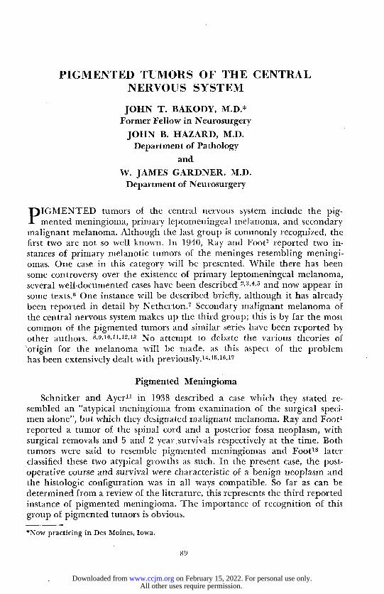
I ig. 1. (a) Pigmented meningioma, showing intracytoplasmic pigment in the meningo-thelial cells, (b) Psammoma bodies in the pigmented meningioma.
Case Repor t A white laborer, aged 45, entered the Cleveland Clinic complaining of low back pain
and left sciatica. Two years previously he first noted pain down the left lower extremity and numbness along the outer aspect of the left lower leg and foot. The leg pain followed an injury to the back after having lifted a 100 pound sack of potatoes; at the time he felt something snap in his back. The leg pain was aggravated by coughing and sneezing. Shortly thereafter the patient developed numbness of the right foot, and some pain behind the right knee when he coughed or sneezed. The strength of the left lower extremity was impaired. There were no sphincter disturbances except for a short period of urinary retention following a novocain injection (caudal block?) given elsewhere for the relief of
9 0
All other uses require permission. on February 15, 2022. For personal use only.www.ccjm.orgDownloaded from
P I G M E N T E D T U M O R S OK N E R V O U S S Y S T E M
pain. The weakness of lhe left leg had progressed so that he required crutches when walking.
The general physical examination was essentially normal. Blood pressure 150/90. There was a draining sinus above the thyroid gland which had been present since the opening of a cyst some time previously. Neurologic examination was normal except for the back and lower extremities. There was severe weakness and and wasting of the left thigh, calf, and foot, absence of the left patellar reflex and of both Achilles reflexes. There were paresthesias radiating into both feet, and hyperesthesia of the anterolateral aspects of the left lower leg and foot. The pain in the leg was not intensified by compression of the jugular veins. Routine laboratory studies were entirely normal. The spinal fluid exam-ination showed no cells, a trace of globulin, and 30 mg. per cent of protein. Lumbosacral x-rays were interpreted as normal.
The patient was admitted to the hospital and lumbar myelograms showed obstruction of the dye medium at the level of the joint space between the second and third lumbar vertebrae. With a working diagnosis of either protruded intervertebral disk, or spinal cord tumor such as neurofibroma, lumbar laminectomy was carried out. No extradural mass
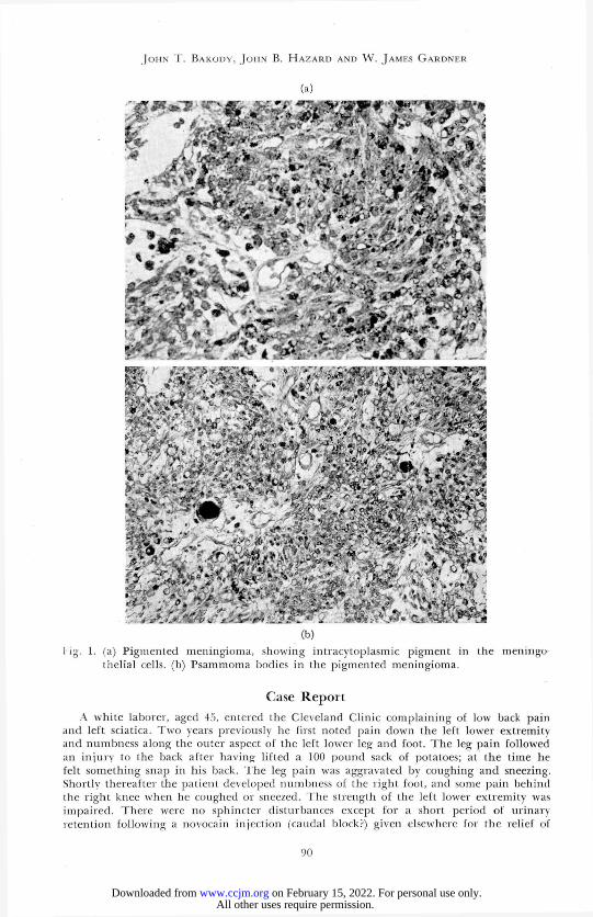
l ig. '¿. Primary leptomeningeal melanoma, showing the diffuse leptomeningeal pigmenta-tion of the brain, the subdural mantle of melanoma about the lower cord and cauda equina.
9 1
All other uses require permission. on February 15, 2022. For personal use only.www.ccjm.orgDownloaded from
J O H N T . B A K O D Y , J O H N B . H A Z A R D A N D W . J A M E S G A R D N E R
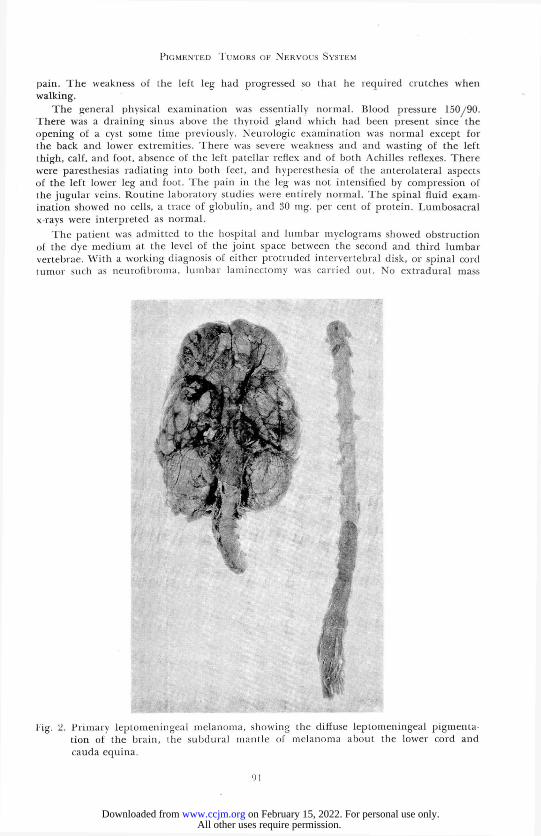
1-ig. 3. Primary leptomeningeal melanoma. Cross section of the spinal cord, showing ihe annular subdural disposition of the melanoma.
" MtsiVÎSÎA v ; t *
1
It.* "*'* •<f «
•» 9 •* f
*" M g * * *
# *
W
Y
r* . ^ZjL „ . C t 5
* » '
.-•i- - v*
J ¡c1-
i 'Vfc-*
<
•jiff* it,
ï v / IP ' L *
w* • p
- ->
, V i S
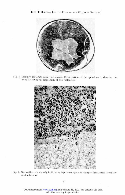
Fig. 4. Nevus-like cells densely infiltrating leptomeninges and sharply demarcated from the cord substance.
9 2
All other uses require permission. on February 15, 2022. For personal use only.www.ccjm.orgDownloaded from
P I G M E N T E D T U M O R S OK N E R V O U S S Y S T E M
was found. The dura was opened to disclose a large, slate blue, rubbery tumor mass, filling the spinal canal from L-2 to L-4. The tumor appeared to be well-encapsulated, and about 5 cm. in length. To effect removal, the right third lumbar root was divided between silver clips, the tumor capsule was incised, the interior evacuated, and the capsule excised along with a circular area of dura. The tumor appeared to extend through the intervertebral foramen on the right, and to lie partly paravertebrally. Because frozen section study was reported as melanoma, the operation was abandoned at this point.
The postoperative convalescence was uneventful. There was a definite foot-drop on the left, and an orthopedic appliance was prescribed. When last heard from, 8 years after the operation, the patient's wife reported that he had gained 53 pounds over his previous best weight, and that his general condition was excellent. He had not completely regained the use of the left lower extremity but was free from pain.
In May 1947, 6 years after operation, one of us (J. B. H.) reviewed the microscopic sections to make a diagnosis of pigmented meningioma.
Pathology
T h e gross specimen consisted of mult iple small portions of velvety, blackish-brown material, portions of which were covered by a tannish-brown fibrous capsule.
Microscopically the neoplasm was f o rmed by cclls of meningothel ial type, polyhedral and spindle-shaped, 10 to 12 microns in width and u p to approximately 40 microns in length, arranged as compact dark staining sheets with irregular pale staining zones (fig. la) . T h e cell cytoplasm was moderately abundant, f o r the most part dark staining in the compact zones, and o f ten containing fine granules or irregular, small c lumps of brown, nonrefracti le pigment. T h e cell nuclei were r o u n d or oval, well dif-ferentiated, generally without prominent nucleol i , and frequently pale or vesicular as if containing a large clear vacuole. N o mitoses were apparent. T h e p igment retained its b rown appearance after stains for iron. T h e vessels were rather evenly dispersed and of simple character. Scattered, small, r o u n d psamomma bodies were present (fig. lb ) . T h e r e was a slight tendency to whor l formation. Architecturally, the neoplasm most typically resembled the type I, variant 4 mening ioma of Cushing and Eisenhardt.19
Discussion
Differentiation between the p igmented mening ioma and tumors of the melanoma group is relatively easy due to the rather typical meningothel ial appearance of the cells, the u n i f o r m and sheet-like arrangement of the tumor, and the lack of nuclear dedifferentiation. Except f o r the presence of the intracellular pigment, the neoplasm is characteristically meningioma.
Primary Leptomeningeal Melanoma
Cases of primary leptomeningeal melanoma have been described by Ford,6 Akelaitis,20 Shapiro and Kellert,4 D a Costa and Love,2 1 Wi l cox , 2 2
Winkelman, Gotten and Silverstein,17 Farnell and Globus,2 and Kessler.3
T h e f o l l owing case has been reported previously.7
93
All other uses require permission. on February 15, 2022. For personal use only.www.ccjm.orgDownloaded from
J O H N T . B A K O D Y , J O H N B . H A Z A R D A N D W . J A M E S G A R D N E R
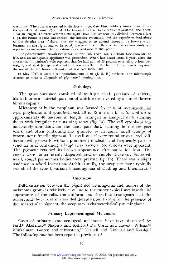
Fig. ">. Case I. Secondary malignant melanoma. Cross appearance of cortical melanotic lesion at time of left parietal craniotomy.
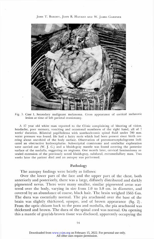
A 17 year old white man reported to the Clinic complaining of blurring of vision, headache, poor memory, vomiting and occasional numbness of the right hand, all of 7 weeks' duration. Bilateral papilledema with xanthochromic spinal fluid under 700 mm. water pressure was found. He had a hairy mole which had been present since birth cov-ering about one-third of the body surface. Observation of pneumoencephalograms indi-cated an obstructive hydrocephalus. Suboccipital craniotomy and cerebellar exploration were carried out (W. J. G.) and a bluish-gray mantle was found covering the posterior surface of the medulla, suggesting an angioma. One month later, cervical laminectomy re vealed extension of the previously noted bluish-gray, subdural, extramedullar mass. Two weeks later the patient died and an autopsy was performed.
Pathology
T h e autopsy findings were briefly as fol lows: Over the lower part of the face and the upper part of the chest, both
anteriorly and posteriorly, there was a large, diffusely distributed and darkly pigmented nevus. T h e r e were many smaller, similar pigmented areas scat-tered over the body, varying in size f r o m 1.0 to 5.0 cm. in diameter, and covered by an abundance of coarse, black hair. T h e brain weighed 1565 Gm. T h e dura was essentially normal. T h e pia arachnoid over the base of the brain was slightly thickened, opaque , and of brown appearance (fig. 2 ) . From the opt ic chiasm back to the pons and medulla , the pia arachnoid was thickened and brown. T h e dura of the spinal cord was normal. O n opening this a mantle of grayish-brown tissue was disclosed, apparently occupying the
94
All other uses require permission. on February 15, 2022. For personal use only.www.ccjm.orgDownloaded from
P I G M E N T E D T U M O R S OK N E R V O U S S Y S T E M
site of the pia arachnoid, completely surrounding the cord f r o m the level of the medulla to the cauda equina and varying in thickness f rom 1.0 to 5.0 mm. T h e greatest thickness was f r o m the midcervical to the lumbar region. The dura stripped without difficulty f r o m this tumor mantle and was not infiltrated at any point. T h e spinal nerves were surrounded by tumor tissue as they emerged f r o m the cord, but otherwise were distinct f rom it. T h e cord was of fairly normal outline, and its anatomic markings were not un-usual. T h e r e was no apparent reduct ion in the size of the spinal cord so far as the upper cervical region was concerned; the lower cervical region, however, presented some distortion of the cord with an irregularity of its shape (fig. 3) a d iminut ion in the size, and some loss of the normal anatomic markings. Grossly, however, there was little or no infiltration by tumor.
Microscopic examination of sections f r o m the cerebral cortex, olfactorv bulb, thalamus, pons, medulla, and cerebel lum showed small, round, or polyhedral cells occurring in groups or diffusely distributed in the leptomen-inges. In some areas these cells were numerous, produc ing severe thicken-
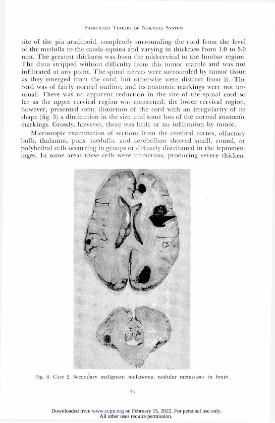
I'ig. 0. Case 2. Secondary malignant melanoma, nodular metastases in brain
9 5
All other uses require permission. on February 15, 2022. For personal use only.www.ccjm.orgDownloaded from
J O H N T . B A K O D Y , J O H N B . H A Z A R D A N D W . J A M E S G A R D N E R
ing of the leptomeninges and extending along pial vessels into the brain tissue but usually well delimited f rom this. T h e cells possessed little cytoplasm and the nuclei were of irregular configuration, dark staining and without prominent nucleoli or mitoses. T h e cells were separated in part by an increased connective tissue of the leptomeninges and in the connective tissue were occasional cells of chromatophore type laden with fine, brownish, nonrefractile pigment granules which gave a negative reaction for iron. Sections through the spinal cord presented similar but more intense infiltra-tion of the leptomeninges by the nevus-like cells described above. These surrounded nerves but did not penetrate them, and the sheath of cells was generally well demarcated f rom the spinal cord substance (fig. 4).
T h e leptomeninges were sharply delimited f rom the subdural space and in no instance was the dura infiltrated. T h e cells forming the mantle about the spinal cord revealed variations in size and staining property and rarely there were mitoses. However, nuclear dedifferentiation was never severe. T h e anatomic diagnoses were: primary leptomeningeal melanoma, diffuse, of brain and spinal cord; pigmented nevus, diffuse, of face and chest; pig-mented hairy nevi, multiple.
It is thought that a growth, such as was found in this case, arises f rom the leptomeningeal chromatophores. T h e usual clinical picture is that of increased intracranial pressure, stiff neck, sterile xanthochromic spinal fluid and mental clouding, i.e. a "tumor meningitis."
T h e cells almost exclusively infiltrate the leptomeninges with little or no penetration of brain or cord substance. Nuclear dedifferentiation is not that which is f ound in true malignant melanoma. In general, there is n o indica-tion of growth activity except for a rare mitosis. T h e aggressiveness of true malignant melanoma is not evident in any area.
A primary leptomeningeal origin cannot be accepted as such unless a careful search fails to disclose a primary malignant lesion in the eye, skin or visceral organs and even then, the presence of intracerebral melanotic nodules would challenge an interpretation of primary origin.
Secondary Malignant Melanoma
Grant1 2 reported 7 cases of melanotic sarcoma from the neurosurgical service of Peter Bent Brigham Hospital occurring f rom 1914 to 1926. There were 49 metastatic intracranial lesions in the 13 year series, making a 14 per cent incidence for the melanotic tumors. In 1932, Dunlap1 3 reported on 95 metastatic tumors of the brain, citing only 2 cases of melanoma (2.1 per cent incidence). In 1939, Courville and Schillinger9 reviewed 18 cases of metastatic melanoma of their own. Fourteen were included from a series of 1060 cases of intracranial tumors registered in the Cajal Laboratory and as part of a group of 107 metastatic tumors (12 per cent incidence for melan-omas in the group of metastatic neoplasms, and 1.3 per cent of intracranial tumors in general). In 1940, Moersch, Love, and Kernohan1 0 found 34 cases
96
All other uses require permission. on February 15, 2022. For personal use only.www.ccjm.orgDownloaded from
P I G M E N T E D T U M O R S OK N E R V O U S S Y S T E M
(in 19 of which the diagnosis was verified by operation or necropsy) of metastatic involvement of the central nervous system in a general g r o u p of 500 melanomas seen f r o m 1930 to 1939. Of those melanomas showing local recurrence or metastases, 10 per cent involved the central nervous system.
Fourteen cases of secondary malignant melanomas of the central nervous system were encountered at the Clinic in a g roup of 85 metastatic tumors occurring in 1294 intracranial neoplasms. Of these the f o l l owing 3 cases are presented.
Case Reports
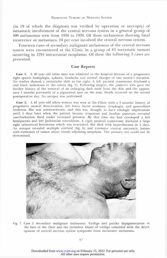
Case I. A 40 year old white man was admitted to the hospital because of a progressive right spastic hemiplegia, aphasia, headache and mental changes of one month's duration. Air studies showed a ventricular shift to the right. A left parietal craniotomy disclosed a coal black melanoma in the cortex (fig. 5). Following surgery, the patient's wife gave the further history of the removal of an enlarging dark mole from the skin and the appear-ance 4 months previously of a pigmented spot on the nose. Death occurred on the second postoperative day. No autopsy was performed.
Case 2. A 64 year old white woman was seen at the Clinic with a 3 months' history of progressive mental deterioration, left lower facial weakness, dysphagia, and generalized weakness. She was arteriosclerotic, and this was thought to have etiologic implications until 4 days later when the patient became stuporous and lumbar puncture revealed xanthochromic fluid under increased pressure. By this time she had developed a left hemiparesis and left Jacksonian convulsions. A right parietal craniotomy disclosed a large right subcortical hematoma which was evacuated. She died with hyperthermia in 2 days. An autopsy revealed multiple cerebral (fig. 6) and extensive visceral metastatic lesions with extension of tumor about vessels adjoining neoplasm. The primary site could not be determined.
tig. 7. Case 3. Secondary malignant melanoma. Vitiligo and patchy depigmentation of llie hair of the chest and the eyelashes. Onset of vitiligo coincided with the devel-opment of central nervous system symptoms from metastatic melanoma.
9 7
All other uses require permission. on February 15, 2022. For personal use only.www.ccjm.orgDownloaded from
J O H N T . B A K O D Y , J O H N B . H A Z A R D A N D W . JAMES G A R D N E R
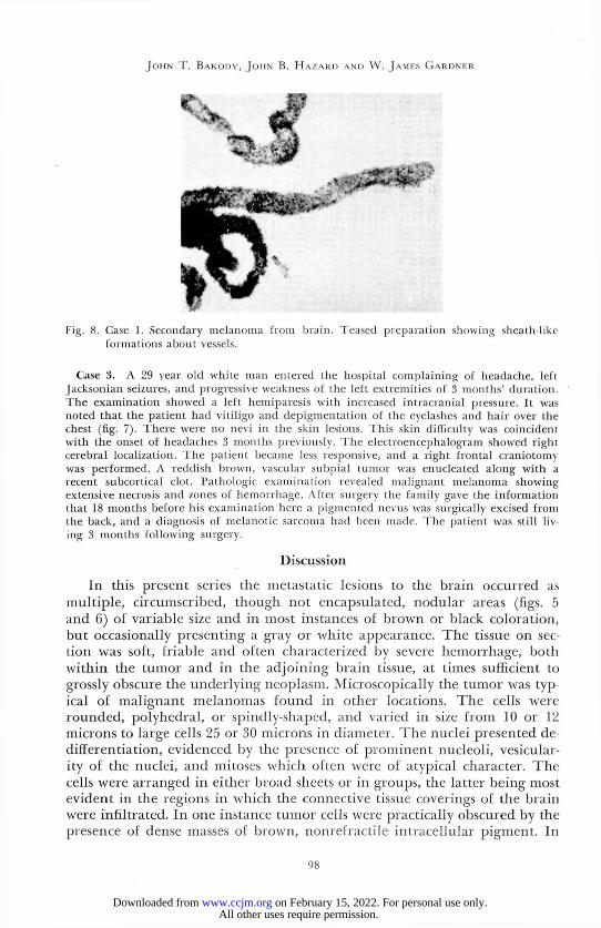
Fig. 8. Case 1. Secondary melanoma from brain. Teased preparation showing sheath-like formations about vessels.
Case 3. A 29 year old white man entered the hospital complaining of headache, left Jacksonian seizures, and progressive weakness of the left extremities of 3 months' duration. The examination showed a left hemiparesis with increased intracranial pressure. It was noted that the patient had vitiligo and depigmentation of the eyelashes and hair over the chest (fig. 7). There were no nevi in the skin lesions. This skin difficulty was coincident with the onset of headaches 3 months previously. The electroencephalogram showed right cerebral localization. The patient became less responsive, and a right frontal craniotomy was performed. A reddish brown, vascular subpial tumor was enucleated along with a recent subcortical clot. Pathologic examination revealed malignant melanoma showing extensive necrosis and zones of hemorrhage. After surgery the family gave the information that 18 months before his examination here a pigmented nevus was surgically excised from the back, and a diagnosis of melanotic sarcoma had been made. The patient was still liv-ing 3 months following surgery.
Discussion
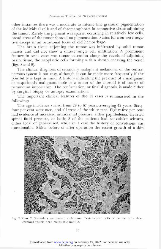
In this present series the metastatic lesions to the brain occurred as multiple, circumscribed, though not encapsulated, nodular areas (figs. 5 and 6) of variable size and in most instances of brown or black coloration, but occasionally presenting a gray or white appearance. T h e tissue on sec-tion was soft, friable and often characterized by severe hemorrhage, both within the tumor and in the ad jo in ing brain tissue, at times sufficient to grossly obscure the underlying neoplasm. Microscopical ly the tumor was typ-ical of malignant melanomas f o u n d in other locations. T h e cells were rounded , polyhedral , or spindly-shaped, and varied in size f r o m 10 or 12 microns to large cells 25 or 30 microns in diameter. T h e nuclei presented de-differentiation, evidenced by the presence of prominent nucleoli , vesicular-ity of the nuclei, and mitoses which often were of atyjoical character. T h e cells were arranged in either broad sheets or in groups, the latter be ing most evident in the regions in which the connective tissue coverings of the brain were infiltrated. In one instance tumor cells were practically obscured by the presence of dense masses of brown, nonrefracti le intracellular pigment. In
98
All other uses require permission. on February 15, 2022. For personal use only.www.ccjm.orgDownloaded from
P I G M E N T E D T U M O R S OK N E R V O U S S Y S T E M
other instances there was a moderate to intense fine granular pigmentation of the individual cells and of chromatophores in connective tissue ad jo in ing the tumor. Rarely the p igment was sparse, occurring in relatively few cells, broad areas of the tumor showed no pigmentation. Stains for iron were nega-tive except in an occasional focus of o ld hemorrhage.
T h e brain tissue ad jo in ing the tumor was infiltrated by solid tumor masses and did not show a diffuse single cell infiltration. A prominent feature in some cases was tumor extension along the vessels of ad jo in ing brain tissue, the neoplastic cells f o rming a thin sheath encasing the vessel (figs. 8 and 9).
T h e clinical diagnosis of secondary malignant melanoma of the central nervous system is not easy, although it can be made more frequently if the possibility is kept in mind. A history indicating the presence of a malignant or suspiciously malignant mole or a tumor of the choro id is of course of paramount importance. T h e confirmation, or final diagnosis, is made either by surgical biopsy or autopsy examination.
T h e important clinical features of the 14 cases is summarized in the fo l lowing:
T h e age incidence varied f r o m 29 to 67 years, averaging 42 years. Sixty-four per cent were men, and all were of the white race. Eighty-five per cent had evidence of increased intracranial pressure, either papil ledema, elevated spinal fluid pressure, or both ; 8 of the patients had convulsive seizures, either focal or generalized, while in 1 case the history of convulsions was questionable. Either before or after operation the recent growth of a skin
lig. 9. Case 2. Secondary malignant melanoma. Perivascular cuffs of tumor cells about cerebral vessels near metastatic nodule.
9 9
All other uses require permission. on February 15, 2022. For personal use only.www.ccjm.orgDownloaded from
J O H N T . B A K O D Y , J O H N B . H A Z A R D A N D W . J A M E S G A R D N E R
mole was related in 9 of the g roup and 7 patients had previous excision or cauterization of a p igmented nevus. T h e skin was k n o w n to have been the primary site in 9 patients, the left eye in 2, and in 3 the site of the primary lesion was not known. T h e onset of vitil igo, with depigmentation of the eye-lashes and hair over the chest (fig. 7) co inc ided with the onset of headaches in 1 case. Melanuria was f o u n d in 2 of the 3 cases in which attempts were made to identify melanin in the urine; the spinal fluid showed a nega-tive test f o r this p igment in the 4 where it was sought. Xanthochromic spinal fluid was present in 5 of the patients. T h e spinal fluid protein was 50 mg. „or greater in 8 of the 11 cases in which it was determined. Chest roentgeno-grams were reported normal in 8, o n e showed evidence of metastases, the diagnosis of metastasis was questionable in another, and there was one in-stance in which the chest was normal o n first examination but showed metastatic lesions 4 months later; n o record of chest x-ray examination was f o u n d for 3 patients.
Surgical therapy fails to cure the patient, and x-ray therapy is valueless.
Summary and Conclusions
Pigmented tumors of the central nervous system are presented as 3 en-tities; p igmented meningioma, primary leptomeningeal melanoma, and sec-ondary malignant melanoma.
A n example of a p igmented mening ioma is described with the histologic diagnosis made o n re-examination of the surgical specimen 6 years fo l low-ing the first and erroneous diagnosis of melanoma. If this unusual tumor had not been pigmented, it most likely w o u l d have been identified by the surgeon as a mening ioma or neur i lemmoma, but the inclusion of melanin pigment by the tumor cells made confus ion with the melanomas certain.
A single example of primary leptomeningeal melanoma is briefly presented in which the patient had an associated extensive p igmented cut-aneous nevus. Either the association of the cutaneous and neural lesions was a coincidence, or the cutaneous and neural lesions were related con-genital malformations as part of a developmental ectodermal disturbance.
Fourteen cases of secondary malignant melanoma are discussed. Atten-tion is called to the importance of melanot ic neural metastases. T h i s likeli-h o o d should constantly be considered in the differential diagnosis of cere-bral neoplasm.
References
li Ray, B. S., and Foot, N. C.: Primary melanotic tumors of meninges; resemblance to meningiomas; report of 2 cases in which operation was performed. Arch. Neurol, and Psychiat. 44:104 (July) 1940.
2- Farnell, F. J„ and Globus, J. H.: Primary melanoblastosis of leptomeninges and brain. Arch. Neurol, and Psychiat. 25:803 (April) 1931.
3. Kessler, M. M.: Melanoblastosis and melanoblastoma; primary and secondary involve-ment of the brain; anatomic study. Am. J. Cancer 30:19 (May) 1937.
1 0 0
All other uses require permission. on February 15, 2022. For personal use only.www.ccjm.orgDownloaded from
P I G M E N T E D T U M O R S OK N E R V O U S S Y S T E M
4. Shapiro, I., and Kellerl, E.: Primary melanoblastosis of meninges. New York Stale J. Med. 37:2096 (Dec. 15) 1937.
5. Bailey, P., and Bucy, 1'. C.: Origin and nature of meningeal tumors. Am. J. Cancer 15:15 (Jan.) 1931.
6. Ford, F. R.: Diseases of Nervous System in Infancy, Childhood, and Adolescence. Springfield, Charles C Thomas, 1946, p. 821.
7. Netherton, E. W.: Extensive pigmented nevus associated with primary melanoblastosis of leptomeninges of brain and spinal cord: report of case. Arch. Dermat. and Syph. 33:238 (Feb.) 1936.
8. Words, H., and Words, S. B.: Metastatic melanoma involving central nervous system. Arch. Neurol, and Psychiat. 36:601 (Sept.) 1936.
9. Courville, C. B., and Schillinger, R. J.: Metastatic melanoblastomas of brain; review of literature and survey of 18 cases. Bull. Los. Angeles Neurol. Soc. 4:8 (March) 1939.
10. Moersch, F. P., Love, J. B., and Kernolian, J. W.: Melanoma of central nervous sys-tem; report of 34 cases, in 19 of which diagnosis was verified by operation or necropsy. J. A. M. A. 115: 2148 (Dec. 21) 1940.
11. Schnitker, M. T., and Ayer, D.: Primary melanomas of leptomeninges; clinico-patlio-logic study with review of literature and report of additional case. J. Nerv. and Ment. Dis. 87:45 (Jan.) 1938.
12. Grant, F. C.: Intracranial malignant metastases; their frequency and value of surgery in their treatment. Ann. Surg. 84:635 (Nov.) 1926.
13. Dunlap, H. F.: Metastatic malignant tumors of brain. Ann. Int. Med. 5:1274 (April) 1932.
14. Ewing, J.: Neoplastic Diseases. A Treatise on Tumors, ed.4. Philadelphia, W. B. Saund-ers Company, 1940, p. 948.
15. Dawson, J. W.: Melanomata; their morphology and histogenesis; study of cell origins and transformations with critical discussion on aspects of tumour growth and clinical review. Edinburgh M. J. 32:510 (Oct.) 1925.
16. Maclachlan, W. W. G.: Extensive pigmentation of brain associated with nevi pigmen-tosi of skin. J. M. Research 29:433 (Jan.) 1914.
17. Winkelman, N. W., Gotten, N., and Silverstein, A.: Primary melanoblastosis of meninges. Arch. Neurol, and Psychiat. 35:919 (April) 1936.
18. Foot, N. C.: Pathology in Surgery. Philadelphia, J. B. Lippincott, 1945, p. 431. 19. Cushing, H., and Eisenhardt, L.: Meningiomas; Their Classification, Regional Be-
havior, Life History, and Surgical End Results. Springfield, Charles C. Thomas, 1938, p. 37.
20. Akelaitis, A. J. E.: Primary melanosarcoma of leptomeninges. Am. J. Path. 11:594 (July) 1935.
21. Da Costa, D. G., and Love, J. G.: Primary melano-epithelioma of spinal cord. Proc. Staff Meet. Mayo Clin. 14:628 (Oct. 4) 1939.
22. Wilcox, J. C.: Melanomatosis o£ skin and central nervous system in infants; congenital neurocutaneous syndrome. Am. J. Dis. Child. 57:391 (Feb.) 1939.
1 0 1
All other uses require permission. on February 15, 2022. For personal use only.www.ccjm.orgDownloaded from