Embed Size (px)
Citation preview

TECHNICAL NOTE
Elizabeth Abraham,1 M.Sc.; Margaret Cox,2 Ph.D.; and David Quincey,3 M.Sc.
Pig-mentation: Postmortem Iris Color Changein the Eyes of Sus scrofa*
ABSTRACT: Experienced forensic pathologists and examiners may be familiar with the phenomenon of postmortem iris color change; however,only Knight, Simpson’s forensic medicine, Arnold, London, 1997; Ref. 1 and Saukko and Knight, Knight’s forensic pathology, 3rd ed., Arnold, Lon-don, 2004; Ref. 2 have referred to it in the literature, and to date, there have been no published scientific research studies on this taphonomic artifact.A controlled experiment was conducted of postmortem changes to isolated Sus scrofa eyes. The eyes (n = 137) were separated into three groups andeach sample was observed for 3-day postmortem at a different temperature. In addition, a Sus scrofa head was obtained to observe postmortemchanges of eyes in situ. All isolated blue eyes in the experiment, at room temperature and higher, changed to brown ⁄ black within 48 h. The in situblue eye, at room temperature, turned brown ⁄ black within 72 h. If iris color consistently changes postmortem in humans, then this taphonomic arti-fact must be incorporated into victim identification protocol, including disaster victim identification software, and autopsy reports to prevent inaccu-rate victim identification and inappropriate exclusion from the identification process.
KEYWORDS: forensic science, postmortem, iris color change, eye color change, victim identification, forensic taphonomy
Postmortem iris color change in light-eyed individuals has beenreported in the literature, albeit without supporting studies (1,2).Saukko and Knight (2) conclude that eye color is unreliable foridentification. The implication is that the eye color changes beforethe globe decomposes, hence the possibility of incorrect eye colorassessment.
This phenomenon has been confirmed by forensic practitioners(J. Arnold and C. Barker, pers. comm.) and by forensic pathologistswho describe the reaction of families of the deceased to erroneouseye color as stated in autopsy reports (P. Ellis and J. Fernandes,pers. comm.).
The eye has been studied for postmortem changes for potentialindicators of time of death estimates, for example, potassium ionconcentration (3–10), for the detection of alcohol or narcotics(11,12), and for the presence of individuating pathology such ashypoglycemia (13–15). However, postmortem iris color change isnot found in the literature aside from the two references citedabove. While experienced pathologists may be familiar with thisartifact, not all death investigators or coroners are, and the lattermay have jurisdiction over human remains.
Eye color is routinely described in autopsy and victim identifica-tion reports (2,11,16–18). While eye color is not heavily reliedupon for identification (sex, height, and estimated age carry moreweight) (J. Arnold, pers. comm.), victim identification software cur-rently available would exclude a potential match due to discrepan-cies, including eye color, unless parameters are preset to limit the
required number of fields for a potential match (R. Venables andD. Horgaard, pers. comm.). If Knight is correct that postmortem iriscolor change is an inevitable taphonomic artifact, then victim iden-tification protocols, from autopsy reports to disaster victim identifi-cation software, would have to accommodate this phenomenon.
Methods
To test Knight’s assertion that eye color changes before the eye-ball is fully decomposed, a series of pilot experiments were under-taken using enucleated (excised) Sus scrofa (domestic pig) eyessupplied by a local butcher 1 day after slaughter. Sus scrofa eyesare anatomically similar to those of humans (19). Isolated eyes ofthe pilot experiments were kept at room temperature in an isother-mal fume cupboard, and all of the blue eyes turned brown ⁄ blackwithin 2–3 days postmortem and prior to decomposition of the eyeitself. In addition, dissection of an eye 3-day postmortem showedthe vitreous humor, which is normally transparent and gel-like inlife, to be black and watery.
The main experiment consisted of a controlled observationalstudy of a total of 137 isolated Sus scrofa eyes collected from alocal abattoir on the day of slaughter. The eyes were observed overseveral days in three different environments, each at a differenttemperature, and monitored every 12 h, starting from their place-ment in one of the three environments:
Environment A: LMS Series Four Cooled Incubator (4–8�C).Environment B: Isothermal fume cupboard (room temperature,21–26�C).Environment C: Gallenkamp Plus II Oven (30–36�C).
In addition, one Sus scrofa head was obtained to study postmor-tem changes in situ. As in humans, blue eyes in pigs are less com-mon than brown; in total there were 13 pure blue eyes for themain experiment.
The eyes used in this experiment were obtained from intensivelyreared (nonorganic) domestic pigs bred in Southwest England and
123 Rose Avenue, Toronto, ON, Canada M4X 1N7.2Inforce Foundation, Department of Materials and Applied Sciences,
Cranfield University, Shrivenham, Swindon SN6 8LA, U.K.3School of Health and Social Care, Bournemouth University, Royal
London House, Christchurch Road, Bournemouth, Dorset BH1 3LT, U.K.*Presented at the 2006 Meeting of the American Academy of Forensic
Sciences, Seattle, WA (February) and at the 2005 Inforce Foundation Con-ference: Forensic Science and the Investigation of Atrocity Crimes andMass Fatality Incidents, Bournemouth, U.K. (December).
Received 28 Jan. 2007; and in revised form 18 July 2007; accepted 28July 2007.
J Forensic Sci, May 2008, Vol. 53, No. 3doi: 10.1111/j.1556-4029.2008.00729.x
Available online at: www.blackwell-synergy.com
626 � 2008 American Academy of Forensic Sciences
killed for consumption. As such, there were no ethical issues raisedby this project.
At the time of slaughter the animals were c. 4–5 months old.The method of slaughter was an electric shock to the ear, whichstuns the animal, followed by exsanguination by severance of oneof the carotid arteries, performed manually. All of the animals hadblue, brown, or bi-colored (mixed blue and brown) eyes; thesewere enucleated by abattoir staff.
Eyes were collected on two occasions. In Part 1 of theexperiment, 119 eyes were collected, bagged, and stored on ice c.4–5 h after slaughter. There was a total of three pure blue eyes inPart 1.
On the second occasion (Part 2), collection took place within anhour postmortem. Including the Sus scrofa head, there was a totalof 10 pure blue eyes in Part 2. Including the two in situ eyes, therewere a total of 18 eyes in Part 2 of the experiment.
In both parts of the main experiment, the eyes were transportedimmediately to the laboratory, where they were catalogued andplaced on hard plastic trays in one of three environments. Detailsof the temperature and relative humidity of each environment fromboth experiments at each 12-h interval are shown in Tables 1 and2.
The Sus scrofa head obtained in Part 2 of the experiment wasstored in the isothermal fume cupboard (Environment B, 22�C).This animal had heterchromia iridium, or two different coloredeyes: one brown and one blue.
Over the course of Parts 1 and 2 of the experiment, the follow-ing activities took place every 12 h:• Recording of temperature and humidity in each environment
(see Tables 1 and 2).• Photography of entire trays and close-ups of selected eyes in a
darkroom within the laboratory facilities using a Ricoh CaplioRR30 digital camera and two 60-watt Craftlight High QualityDaylight light bulbs.
• One milliliter of vitreous humor was withdrawn from one eyefrom each environment, using calibrated micropipettes following
incision of the sclera, and stored in the cooled incubator (theeye was subsequently disposed of, i.e., vitreous humor waswithdrawn from a separate eye on each occasion).
• Notations were made on the decomposition process and iriscolor change (if any).
Finally, the vitreous humor samples were examined under a lightmicroscope, and photomicrographs were taken of several of thesamples using a Canon EOS 20D digital camera (Fukushima,Japan).
All biological material was disposed of through autoclaving fol-lowing the pilot experiments and the main experiment.
Results
The results are presented for each of the two parts of the mainexperiment and for the changes observed in vitreous humor sam-ples. For the sake of simplicity, the mean temperatures of eachenvironment are expressed.
Part 1
One of the early postmortem changes seen in the sclera (thewhite area of the globe), is referred to as tache noire (literally,‘‘black spot’’), a result of desiccation of the sclerae when the eye-lids of the deceased are open. A striking difference among the eyescollected for Part 1 of the experiment was the proportion of thesclerae that exhibited tache noire, from none or very little, to over75% coverage of the surface of the globe; an example of whatDolinak et al. (20) refer to as ‘‘global tache noire.’’
Of 119 eyes collected in Part 1, only three were pure blue; fourof the eyes had bi-colored irises. One blue eye was placed in eachof the three environments.
A change in iris color in the blue eye of Environment C(Gallenkamp oven, 32�C) began within 12-h postmortem. Thiscommenced with a distinct darkening around the pupil, and was
TABLE 1—Temperature and humidity readings for Environments A, B, and C for Part 1 of the main experiment.
EnvironmentPMI (hours)
A B C
Temp(�C)
Humidity(%)
Temp(�C)
Humidity(%)
Temp(�C)
Humidity(%)
12 7 42 26 40 30 7024 4 45 23 57 31 5036 7 43 26 38 35 4048 8 46 24 40 n ⁄ a n ⁄ a60 6 43 n ⁄ a n ⁄ a n ⁄ a n ⁄ a
Mean 6.4 44 24.8 44 32.0 53
PMI, postmortem interval.
TABLE 2—Temperature and humidity readings for Environments A, B, and C for Part 2 of the main experiment.
EnvironmentPMI (hours)
A B C
Temp (�C) Humidity (%) Temp (�C) Humidity (%) Temp (�C) Humidity (%)
12 7 55 23 64 36 4324 6 52 22 63 35 4336 6 53 22 58 35 3948 5 53 21 50 n ⁄ a n ⁄ a60 5 55 23 50 n ⁄ a n ⁄ a
Mean 5.8 54 22.2 57 35.3 42
PMI, postmortem interval.
ABRAHAM ET AL. • POSTMORTEM IRIS COLOR CHANGE IN SUS SCROFA 627
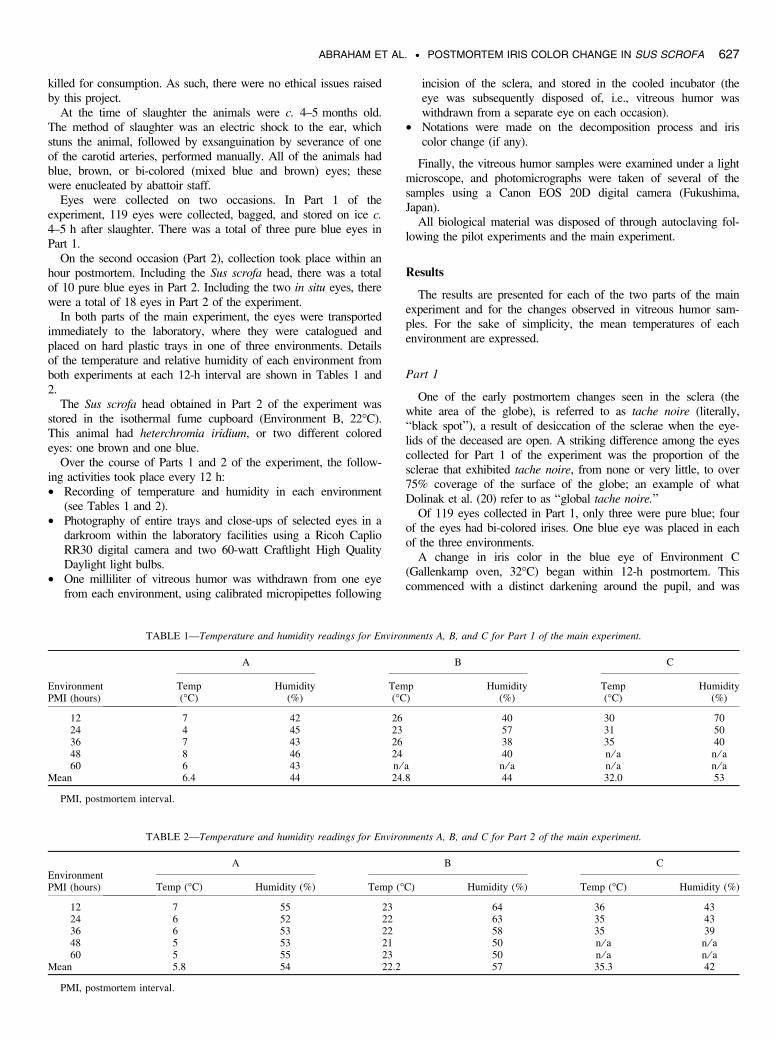
complete within 36 h, by which time the iris color was unrecogniz-able as blue. It was difficult to assess iris color in isolation becausethe rest of eye (the sclera) was almost entirely black (global tachenoire) and desiccated (Fig. 1). This is also true of the brown eyesin the same environment. At 36 h, all of the eyes were desiccatedand hard, and the iris color was undeterminable.
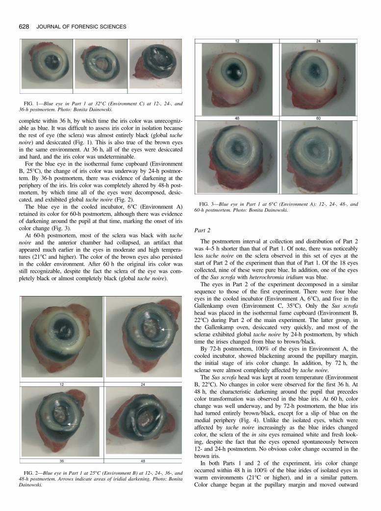
For the blue eye in the isothermal fume cupboard (EnvironmentB, 25�C), the change of iris color was underway by 24-h postmor-tem. By 36-h postmortem, there was evidence of darkening at theperiphery of the iris. Iris color was completely altered by 48-h post-mortem, by which time all of the eyes were decomposed, desic-cated, and exhibited global tache noire (Fig. 2).
The blue eye in the cooled incubator, 6�C (Environment A)retained its color for 60-h postmortem, although there was evidenceof darkening around the pupil at that time, marking the onset of iriscolor change (Fig. 3).
At 60-h postmortem, most of the sclera was black with tachenoire and the anterior chamber had collapsed, an artifact thatappeared much earlier in the eyes in moderate and high tempera-tures (21�C and higher). The color of the brown eyes also persistedin the colder environment. After 60 h the original iris color wasstill recognizable, despite the fact the sclera of the eye was com-pletely black or almost completely black (global tache noire).
Part 2
The postmortem interval at collection and distribution of Part 2was 4–5 h shorter than that of Part 1. Of note, there was noticeablyless tache noire on the sclera observed in this set of eyes at thestart of Part 2 of the experiment than that of Part 1. Of the 18 eyescollected, nine of these were pure blue. In addition, one of the eyesof the Sus scrofa with heterochromia iridium was blue.
The eyes in Part 2 of the experiment decomposed in a similarsequence to those of the first experiment. There were four blueeyes in the cooled incubator (Environment A, 6�C), and five in theGallenkamp oven (Environment C, 35�C). Only the Sus scrofahead was placed in the isothermal fume cupboard (Environment B,22�C) during Part 2 of the main experiment. The latter group, inthe Gallenkamp oven, desiccated very quickly, and most of thesclerae exhibited global tache noire by 24-h postmortem, by whichtime the irises changed from blue to brown ⁄ black.
By 72-h postmortem, 100% of the eyes in Environment A, thecooled incubator, showed blackening around the pupillary margin,the initial stage of iris color change. In addition, by 72 h, thesclerae were almost completely affected by tache noire.
The Sus scrofa head was kept at room temperature (EnvironmentB, 22�C). No changes in color were observed for the first 36 h. At48 h, the characteristic darkening around the pupil that precedescolor transformation was observed in the blue iris. At 60 h, colorchange was well underway, and by 72-h postmortem, the blue irishad turned entirely brown ⁄ black, except for a slip of blue on themedial periphery (Fig. 4). Unlike the isolated eyes, which wereaffected by tache noire increasingly as the blue irides changedcolor, the sclera of the in situ eyes remained white and fresh look-ing, despite the fact that the eyes opened spontaneously between12- and 24-h postmortem. No obvious color change occurred in thebrown iris.
In both Parts 1 and 2 of the experiment, iris color changeoccurred within 48 h in 100% of the blue irides of isolated eyes inwarm environments (21�C or higher), and in a similar pattern.Color change began at the pupillary margin and moved outward
FIG. 1—Blue eye in Part 1 at 32�C (Environment C) at 12-, 24-, and36-h postmortem. Photo: Bonita Dainowski.
FIG. 2—Blue eye in Part 1 at 25�C (Environment B) at 12-, 24-, 36-, and48-h postmortem. Arrows indicate areas of iridial darkening. Photo: BonitaDainowski.
FIG. 3—Blue eye in Part 1 at 6�C (Environment A): 12-, 24-, 48-, and60-h postmortem. Photo: Bonita Dainowski.
628 JOURNAL OF FORENSIC SCIENCES

toward the periphery of the iris. In addition, some of the eyesexhibited darkening at the periphery of the iris subsequent to theinitial darkening around the pupil (Fig. 5).
Irides of isolated eyes kept in a cool environment (4–8�C)retained their color for at least 72-h postmortem.
Brown irides tended to retain their color, regardless of the post-mortem temperature, until the eyes were fully decomposed,although the pupillary margins (and in some cases, the periphery ofthe iris) exhibited the darkening that preceded color change in blueeyes.
Changes to Vitreous Humor
In Part 1 of the experiment, vitreous humor extracted at 12- and24-h postmortem was transparent and gel-like, as expected, but by36-h postmortem the vitreous was watery and dark, particularly so
at temperatures of 21�C or higher, and became increasingly darkwith postmortem interval. In Part 2, the vitreous of the eyes in theGallenkamp oven (Environment C, 35�C) was dark by 24-hpostmortem.
Within the test tubes themselves, free melanin granules precipi-tated to the bottom of the tubes. An increase in the volume of pre-cipitated granules would appear to be correlated to postmorteminterval (Fig. 6). Although quantitative measurements were nottaken, the amount of melanin was visibly greater in the test tubes.This was most evident in vitreous collected from eyes in the warm-est environment (Environment C, 30–36�C).
Under a light microscope, it was possible to observe the foci ofmelanin-containing tissue and freed melanin granules from vitreoushumor removed from an eye in Environment C, 36-h postmortem(Fig. 7).
Discussion
This experiment demonstrated that, in Sus scrofa eyes, blue iriseschange color postmortem in warm or hot environments with mod-erate humidity: 100% of the blue irides at room temperature or
FIG. 4—Part 2, 22�C (Environment B), in situ eyes: right (brown), left(blue). Photo: Bonita Dainowski.
FIG. 5—Darkening of blue iris in pupillary margin and periphery of iris(Part 1, 25�C, Environment B, 36 h). Photo: Bonita Dainowski.
FIG. 6—Vitreous humor collected from three eyes in Part 2, 35�C (Envi-ronment C) at 12-, 24-, and 36-h postmortem. Photo: David Quincey.
ABRAHAM ET AL. • POSTMORTEM IRIS COLOR CHANGE IN SUS SCROFA 629

higher turned dark brown or black prior to the complete decompo-sition in the eye. The blue portion of the bi-colored irides under-went similar changes; areas of the iris that were originally bluebecame indistinguishable from the brown areas. If this postmortemartifact occurs in humans, it may lead to confusion of identity andpossibly prevent a potential match from being made betweenunidentified remains and a missing person report, particularly whenrelying on electronic victim identification software in the context ofmass fatality incidents.
While there is a predictable sequence of events in the decompo-sition of Sus scrofa eyes, iris color change cannot be used to deter-mine or estimate time of death. The variable of temperature alone,at least in isolated eyes, precludes postmortem iris color change asa reliable indicator of the postmortem interval.
Another important variable is humidity. While eyes may sponta-neously open some hours after death (21), some will remain closed,particularly if the death is caused by drowning or carbon monoxidepoisoning (ibid.). The position of the eyelid affects the rate atwhich the eye desiccates and, by extension, the rate of iris colorchange. Similarly, an arid or particularly humid environment willrespectively increase or decrease the rate of eye decomposition.
Apart from the artificial laboratory environment, limitations ofthis experiment include the low number of blue irides (n = 13),and the rapid desiccation of the isolated eyes. To this end, moreregular monitoring of changes (e.g., every 6 h) may be useful. Aninherent drawback to any monitoring scheme, however, is theremoval of the eyes from their respective environments, and expo-sure to hot lights during photography, both of which are likely toaffect desiccation rates. Future studies should use in situ eyes andrecord changes in relation to time of death. The fact that the one
in situ blue eye in this experiment (n = 1) turned brown may ormay not be a meaningful result. Furthermore, organic animals mayproduce different results than intensively reared animals, given thathormones, antibiotics, and other additives to feed would introducevariables which will not apply to most humans, and this needs tobe examined. Finally, intrinsic factors such as sex, diet, and exactage of the animal at death may have an effect on the rate ofdecomposition, and this information was not available for theexperiment.
The mechanism behind iris color change may be related to post-mortem changes in the vitreous humor, which changes in composi-tion, color, and viscosity after death. Free melanin granules arefound in vitreous humor as early as 24-h postmortem. The melaninis released either from the iris itself (iris pigment epithelium) or,more likely, from the retinal pigment epithelium or choroids, bothof which are heavily pigmented.
Degrading iris pigment epithelium is unlikely to contribute topostmortem iris color change for two reasons. First, melaningranules released from the iris pigment epithelium would resultin an initial lightening of the iris. It is not likely that the mela-nin is moving away from the iris; clearly, melanin content ofthe iris is increasing with the postmortem interval. Second, thereis very little iris pigment epithelium as compared with retinalpigment epithelium and choroids, which line the entire posteriorchamber.
Postmortem breakdown of melanocytes within the posterior lin-ing of the globe would result in the release of melanin granulesdirectly into the vitreous, in a process driven by autolysis, com-mencing within minutes after death (22).
While it is likely that melanin granules freed from degeneratedmelanocytes of the retinal pigment epithelium and ⁄ or the choroidlayer underlie postmortem iris color change, this hypothesis doesnot inform as to why the irides of brown eyes did not turn signifi-cantly darker brown or black in this experiment. In future studies,scanning electron microscopic examination of vitreous and iridialtissue may be useful for identifying the mechanism of thephenomenon.
Further investigation is required into the interval between deathand iris color change. Research on human remains is necessary todetermine the frequency and consistency of postmortem iris colorchange. If iridial color change occurs in humans as well as Susscrofa, then this postmortem artifact must be taken into consider-ation when comparing data sets of unidentified remains and ante-mortem data of missing individuals.
In mass fatality incidents, eye color discrepancies between ante-mortem and postmortem data can cause delays in the identificationof victims. Furthermore, in the interest of the families of deceasedwho are given access to autopsy reports, erroneous eye color dataunnecessarily upsets them.
Perhaps the protocol for victim identification should be modifiedto record only irides that fall into one of two categories: blue ⁄ greyor green ⁄hazel. Otherwise, eye color should not be recorded at all.Brown iris color, at least in the eyes of Sus scrofa, is not a reliableindicator for identification, as its original color could have beendifferent.
Acknowledgments
The authors wish to thank Dr. Bernard Wright for sharing hisobservation of the phenomenon of postmortem iris color change inhumans. They are also especially grateful to Bonita Dainowski,who photographed the eyes throughout the main experiment, aswell as taking the working shots at the abattoir. The valuable
FIG. 7—Vitreous humor from eye in Part 2, 35�C (Environment C) at36-h postmortem. Photo: David Quincey.
630 JOURNAL OF FORENSIC SCIENCES

contributions of Eat Well Butchers and Sturminster Newton Abatt-oirs Ltd., who supplied the material for the pilot and main experi-ments, are gratefully acknowledged. Thanks are also due to PaulCheetham for assistance with the experimental design and to IanHanson for his assistance with reference material. The authorsexpress gratitude to Caroline Barker, Consulting Forensic Anthro-pologist and Scientific Advisor to the INFORCE Foundation(U.K.), Jeff Arnold, Manager, Forensic Services, Office of theChief Coroner, Toronto Forensic Pathology Unit (Canada), Inspec-tor Richard Venables, recently retired from the National Crime andOperations Faculty, Central Police Training and DevelopmentAuthority of the Home Office (U.K.), Dr. Peter Ellis, Director ofForensic Medicine, Westmead Hospital (Australia), and Dr. JohnFernandes, Head, Autopsy Pathology, Regional Forensic PathologyUnit, Hamilton General Hospital (Canada) for their contributionscited in this text. For technical support, the authors are grateful toRob Haslam, Manager of Laboratory and Technical Services atBournemouth University, and technician Damien Evans, for theirassistance throughout the experimentation.
References
1. Knight B. Simpson’s forensic medicine. London: Arnold, 1997.2. Saukko P, Knight B. Knight’s forensic pathology, 3rd ed. London:
Arnold, 2004.3. Brzezinski PM, Godlewski A. Assessment of potassium and sodium ion
concentrations in the vitreous humour of swine isolated eyeballs afterorganism death. Rocz Akad Med Bialymst 2004;49(Suppl. 1):161–3.
4. Bennett MJ, Ragni MC, Hood I, Hale DE. Comparison of post-mortemurinary and vitreous humour organic acids. Ann Clin Biochem1992;29(5):541–5.
5. Bocaz-Beneventi G, Tagliaro F, Bortolotti F, Manetto G, Havel J. Capil-lary zone electrophoresis and artificial neural networks for estimation ofthe post-mortem interval (PMI) using electrolytes measurements inhuman vitreous humour. Int J Legal Med 2002;116(1):5–11.
6. Gagajewski A, Murakami MM, Kloss J, Edstrom M, Hillyer M, PetersonGF, et al. Measurement of chemical analytes in vitreous humor: stabilityand precision studies. J Forensic Sci 2004;49(2):371–4.
7. Madea B, Henssge C, Honig W, Gerbracht A. References for determin-ing the time of death by potassium in vitreous humor. Forensic Sci Int1989;40:231–3.
8. Munoz JI, Suarez-Penaranda JM, Otero XL, Rodriguez-Calvo MS, Cos-tas E, Miguens X, et al. A new perspective in the estimation of postmor-tem interval (PMI) based on vitreous. J Forensic Sci 2001;46(2):209–14.
9. Lange N, Swearer S, Sturner WQ. Human postmortem interval estima-tion from vitreous potassium: an analysis of original data from six dif-ferent studies. Forensic Sci Int 1994;66(3):159–74.
10. Stephens RJ, Richards RG. Vitreous humour chemistry: the use of potas-sium concentration for the prediction of the postmortem interval.J Forensic Sci 1987;32(2):503–9.
11. DiMaio VJ, DiMaio D. Forensic pathology, 2nd ed. Boca Raton ⁄ Lon-don ⁄ New York ⁄ Washington DC: CRC Press, 2001.
12. Jones GR, Singer PP. Forensic toxicology. In: Caplan YH, Frank RS,editors. Medicolegal death investigation: treatises in the forensic sci-ences. Colorado Springs, CO: The Forensic Sciences Foundation,1999;181–209.
13. De Letter EA, Piette MH. Can routinely combined analysis of glucoseand lactate in vitreous humour be useful in current forensic practice?Am J Forensic Med Pathol 1998;19(4):335–42.
14. Karlovsek MZ. Diagnostic values of combined glucose and lactate val-ues in cerebrospinal fluid and vitreous humour—our experiences. Foren-sic Sci Int 2004;146(Suppl.):S19–23.
15. Osuna E, Garcia-Villora A, Perez-Cerceles M, Conejero J, Maria Aben-za J, Martinez P, et al. Glucose and lactate in vitreous humor comparedwith the determination of fructosamine for the postmortem diagnosis ofdiabetes mellitus. Am J Forensic Med Pathol 2001;22(3):244–9.
16. Finkbeiner WE, Davis RL, Ursell RC. Autopsy pathology. Oxford: Else-vier Inc., 2004.
17. Graham M. The medicolegal autopsy: description of the process. In:Caplan YH, Frank RS, editors. Medicolegal death investigation: treatisesin the forensic sciences. Colorado Springs, CO: The Forensic SciencesFoundation, 1999;181–209.
18. Spitz WU. The medicolegal autopsy report. In: Spitz WU, Fisher RS,editors. Medicolegal investigation of death. Springfield, IL: Charles C.Thomas, 1980;604–14.
19. Flannery JG. Transgenic animal models for the study of inherited retinaldystrophies. ILAR J 1999;40(2). Available at: http://dels.nas.edu/ilar_n/ilarjournal/40_2/40_2Transgenic.shtml. Accessed on March 16, 2008.
20. Dolinak D, Matshes E, Lew E. Forensic pathology: principles and prac-tice. Oxford: Elsevier Inc., 2005.
21. Suzutain T, Ishibashi H, Takatori T. Studies on the estimation of thepostmortem interval. 4. Spontaneous opening and closing of the eyelidsafter death. Hokkaido Igaku Zasshi 1978;53(1):1–6.
22. Vass AA. Beyond the grave—understanding human decomposition.Microbiol Today 2001;28:190–2.
Additional information and reprint requests:Elizabeth Abraham, M.Sc.23 Rose AvenueToronto, ONCanada M4X 1N7E-mail: [email protected]
ABRAHAM ET AL. • POSTMORTEM IRIS COLOR CHANGE IN SUS SCROFA 631