Embed Size (px)
Citation preview
PI: Faubion, William A Title: Inflammatory cascades disrupt Treg function through epigenetic mechanisms
Received: 07/02/2015 FOA: PA13-302 Council: 01/2016
Competition ID: FORMS-C FOA Title: RESEARCH PROJECT GRANT (PARENT R01)
2 R01 AI089714-06A1 Dual: DK Accession Number: 3841834
IPF: 4976101 Organization: MAYO CLINIC ROCHESTER
Former Number: Department: Internal Medicine
IRG/SRG: GMPB AIDS: N Expedited: N
Subtotal Direct Costs
(excludes consortium F&A)
Year 6:
Year 7:
Year 8:
Year 9:
Year 10:
Animals: Y
Humans: N
Clinical Trial: N
Current HS Code: 10
HESC: N
New Investigator: N
Early Stage Investigator: N
Senior/Key Personnel: Organization: Role Category:
Stephen Ekker PhD Mayo Clinic Other (Specify)-Other Significant
Contributor
Thomas Smyrk MD Mayo Clinic Other (Specify)-Other Significant
Contributor
Raul Urrutia MD Mayo Clinic Other (Specify)-Other Significant
Contributor
William Faubion MD Mayo Clinic PD/PI
Chandrashekhar Pasare PhD University of Texas Southwestern
Medical Center at Dallas
Consultant

OMB Number: 4040-0001
Expiration Date: 06/30/2016
Tracking Number: GRANT11954038 Funding Opportunity Number: PA-13-302 . Received Date:2015-07-02T14:04:51.000-04:00
APPLICATION FOR FEDERAL ASSISTANCE
SF 424 (R&R)3. DATE RECEIVED BY STATE State Application Identifier
1. TYPE OF SUBMISSION* 4.a. Federal Identifier
❍ Pre-application ● Application ❍ Changed/CorrectedApplication
b. Agency Routing Number
2. DATE SUBMITTED Application Identifier c. Previous Grants.gov Tracking Number
5. APPLICANT INFORMATION Organizational DUNS*: Legal Name*: Mayo ClinicDepartment: Internal MedicineDivision: GastroenterologyStreet1*: 200 First Street SWStreet2:
City*: RochesterCounty:State*: MN: MinnesotaProvince:Country*: USA: UNITED STATESZIP / Postal Code*: 559050001
Person to be contacted on matters involving this applicationPrefix: First Name*: David Middle Name: M Last Name*: Moertel Suffix:
Position/Title: Institutional OfficialStreet1*:Street2:
City*:County:State*:
Province:Country*:ZIP / Postal Code*:
Phone Number*: Fax Number: Email:
6. EMPLOYER IDENTIFICATION NUMBER (EIN) or (TIN)*
7. TYPE OF APPLICANT* M: Nonprofit with 501C3 IRS Status (Other than Institution of HigherEducation)
Other (Specify):Small Business Organization Type ❍ Women Owned ❍ Socially and Economically Disadvantaged
8. TYPE OF APPLICATION* If Revision, mark appropriate box(es).
❍ New ● Resubmission ❍ A. Increase Award ❍ B. Decrease Award ❍ C. Increase Duration
❍ Renewal ❍ Continuation ❍ Revision ❍ D. Decrease Duration ❍ E. Other (specify) :Is this application being submitted to other agencies?* ❍Yes ●No What other Agencies?
9. NAME OF FEDERAL AGENCY*National Institute of Allergy and Infectious Diseases
10. CATALOG OF FEDERAL DOMESTIC ASSISTANCE NUMBERTITLE:
11. DESCRIPTIVE TITLE OF APPLICANT'S PROJECT*Inflammatory cascades disrupt Treg function through epigenetic mechanisms12. PROPOSED PROJECTStart Date* Ending Date*07/01/2016 06/30/2021
13. CONGRESSIONAL DISTRICTS OF APPLICANT
MN-001

SF 424 (R&R) APPLICATION FOR FEDERAL ASSISTANCE Page 2
Funding Opportunity Number: PA-13-302 . Received Date:Tracking Number: GRANT119540382015-07-02T14:04:51.000-04:00
14. PROJECT DIRECTOR/PRINCIPAL INVESTIGATOR CONTACT INFORMATIONPrefix: First Name*: William Middle Name: A Last Name*: Faubion Suffix: MDPosition/Title: Associate ProfessorOrganization Name*: Mayo ClinicDepartment: Internal MedicineDivision: GastroenterologyStreet1*:Street2:
City*:County:State*:
Province:Country*:ZIP / Postal Code*:
Phone Number*: Fax Number: Email*:
15. ESTIMATED PROJECT FUNDING 16. IS APPLICATION SUBJECT TO REVIEW BY STATEEXECUTIVE ORDER 12372 PROCESS?*
a. YES ❍ THIS PREAPPLICATION/APPLICATION WAS MADEa. Total Federal Funds Requested* $ AVAILABLE TO THE STATE EXECUTIVE ORDER 12372b. Total Non-Federal Funds* $0.00 PROCESS FOR REVIEW ON:c. Total Federal & Non-Federal Funds* $ DATE:d. Estimated Program Income* $0.00
b. NO ● PROGRAM IS NOT COVERED BY E.O. 12372; OR
❍ PROGRAM HAS NOT BEEN SELECTED BY STATE FORREVIEW
17. By signing this application, I certify (1) to the statements contained in the list of certifications* and (2) that the statements hereinare true, complete and accurate to the best of my knowledge. I also provide the required assurances * and agree to comply withany resulting terms if I accept an award. I am aware that any false, fictitious, or fraudulent statements or claims may subject me tocriminal, civil, or administrative penalties. (U.S. Code, Title 18, Section 1001)
● I agree** The list of certifications and assurances, or an Internet site where you may obtain this list, is contained in the announcement or agency specific instructions.
18. SFLLL or OTHER EXPLANATORY DOCUMENTATION File Name:
19. AUTHORIZED REPRESENTATIVEPrefix: First Name*: David Middle Name: M Last Name*: Moertel Suffix: Position/Title*: Institutional OfficialOrganization Name*: Mayo ClinicDepartment: RS-Research ServicesDivision: RS-Research ServicesStreet1*:Street2:
City*:County:State*: Province:Country*: STATESZIP / Postal Code*:
Phone Number*: Fax Number: Email*:
Signature of Authorized Representative* Date Signed*David.Moertel 07/02/2015
20. PRE-APPLICATION File Name: Mime Type:
21. COVER LETTER ATTACHMENT File Name: Mime Type:
Contact PD/PI: Faubion, William A
424 R&R and PHS-398 SpecificTable Of Contents Page Numbers
SF 424 R&R Cover Page----------------------------------------------------------------------------------------- 1
Table of Contents------------------------------------------------------------------------- 3
Performance Sites--------------------------------------------------------------------------------------------- 4
Research & Related Other Project Information------------------------------------------------------------------ 5
Project Summary/Abstract(Description)----------------------------------------------------- 6
Project Narrative------------------------------------------------------------------------- 7
Facilities & Other Resources-------------------------------------------------------------- 8
Equipment--------------------------------------------------------------------------------- 9
Research & Related Senior/Key Person-------------------------------------------------------------------------- 10
PHS398 Cover Page Supplement---------------------------------------------------------------------------------- 34
PHS 398 Modular Budget---------------------------------------------------------------------------------------- 36
Personnel Justification------------------------------------------------------------------- 42
PHS 398 Research Plan----------------------------------------------------------------------------------------- 43
Introduction------------------------------------------------------------------------------ 44
Specific Aims----------------------------------------------------------------------------- 45
Research Strategy------------------------------------------------------------------------- 46
Progress Report Publications List--------------------------------------------------------- 58
Vertebrate Animals------------------------------------------------------------------------ 60
Bibliography & References Cited----------------------------------------------------------- 63
Letters Of Support------------------------------------------------------------------------ 68
Resource Sharing Plans-------------------------------------------------------------------- 73
Table of Contents Page 3
Contact PD/PI: Faubion, William A
OMB Number: 4040-0010
Expiration Date: 06/30/2016
Tracking Number: GRANT11954038 Funding Opportunity Number: PA-13-302. Received Date:2015-07-02T14:04:51.000-04:00
Project/Performance Site Location(s)
Project/Performance Site Primary Location ❍ I am submitting an application as an individual, and not on behalf of
a company, state, local or tribal government, academia, or other type of
organization.
Organization Name: Mayo ClinicDuns Number:
Street1*:
Street2:
City*:
County:
State*:
Province:
Country*: Zip / Postal Code*:
Project/Performance Site Congressional District*: MN-001
File Name Mime Type
Additional Location(s)
Page 4
Contact PD/PI: Faubion, William A
Tracking Number: GRANT11954038 Funding Opportunity Number: PA-13-302. Received Date:2015-07-02T14:04:51.000-04:00
OMB Number: 4040-0001Expiration Date: 06/30/2016
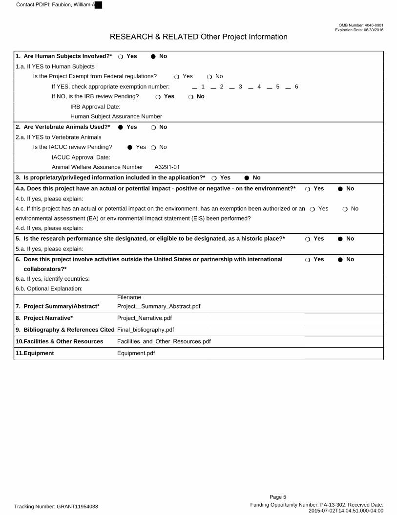
RESEARCH & RELATED Other Project Information
1. Are Human Subjects Involved?* ❍ Yes ● No
1.a. If YES to Human Subjects
Is the Project Exempt from Federal regulations? ❍ Yes ❍ No
If YES, check appropriate exemption number: 1 2 3 4 5 6
If NO, is the IRB review Pending? ❍ Yes ❍ No
IRB Approval Date:
Human Subject Assurance Number
2. Are Vertebrate Animals Used?* ● Yes ❍ No
2.a. If YES to Vertebrate Animals
Is the IACUC review Pending? ● Yes ❍ No
IACUC Approval Date:
Animal Welfare Assurance Number A3291-01
3. Is proprietary/privileged information included in the application?* ❍ Yes ● No
4.a. Does this project have an actual or potential impact - positive or negative - on the environment?* ❍ Yes ● No
4.b. If yes, please explain:
4.c. If this project has an actual or potential impact on the environment, has an exemption been authorized or an
environmental assessment (EA) or environmental impact statement (EIS) been performed?
❍ Yes ❍ No
4.d. If yes, please explain:
5. Is the research performance site designated, or eligible to be designated, as a historic place?* ❍ Yes ● No
5.a. If yes, please explain:
6. Does this project involve activities outside the United States or partnership with international
collaborators?*
❍ Yes ● No
6.a. If yes, identify countries:
6.b. Optional Explanation:
Filename
7. Project Summary/Abstract* Project__Summary_Abstract.pdf Mime Type: application/pdf
8. Project Narrative* Project_Narrative.pdf Mime Type: application/pdf
9. Bibliography & References Cited Final_bibliography.pdf Mime Type: application/pdf
10.Facilities & Other Resources Facilities_and_Other_Resources.pdfMime Type: application/pdf
11.Equipment Equipment.pdf Mime Type: application/pdf
Page 5
Contact PD/PI: Faubion, William A
PROJECT SUMMARY/ABSTRACT: The transcription factor FOXP3 is critical to the regulation of numerous debilitating human immune-mediated diseases. Very recently, the essential role for the histone methyltransferase (HMT) EZH2 in the epigenetic regulation and function of FOXP3 has been described. Inflammatory pathways modify EZH2 activity, and inflammatory signaling impairs Treg function in vivo and in vitro. The biological impact of the FOXP3-EZH2 pathway to IBD is unknown. Our long-term goal is to dissect epigenetic mechanisms regulating Treg cellular differentiation and function, particularly within the setting of GI inflammatory diseases. These discoveries will facilitate design of human cell therapy trials for IBD. The objective of this grant is to characterize the role for EZH2 in Treg suppressive function. The central hypothesis is that EZH2 plays a critical role in the homeostasis of Treg cells, and the disruption of EZH2 function by inflammatory signaling pathways contributes to IBD. Our rationale is that identification of the mechanism(s) to restore Treg suppressive function in the setting of intestinal inflammation will offer new therapeutic opportunities. Our specific aims will test the following hypotheses: (Aim1) Repression of immunoregulatory gene networks by FOXP3 requires the formation of a complex between this transcription factor and EZH2; (Aim 2) Inflammatory stimuli, such as IL6 lead to EZH2 phosphorylation and thereby disrupt the enzymatic activity of this epigenomic regulator; (Aim 3) Inhibition of the IL6 to EZH2 signaling pathway permits sustained Treg suppressive function in the setting of intestinal inflammation. Upon conclusion, we will understand the role for EZH2 in Treg loss of function in the setting of active inflammation. This contribution is significant since it will establish that several pathways targeted by available therapies (ie IL1β, IL6, TNFα) have the potential to regulate EZH2 HMT activity through post-translational modifications. Furthermore, current Treg cell therapy trials, while promising have not addressed the key issue of in vivo inflammation-induced disruption of Treg function. The proposed research is innovative because we investigate the effect of inflammatory signaling pathways on epigenetic complexes in Treg cells, a heretofore-unexamined process. Insight into epigenetic mechanisms is impactful as T cell progenitor cells inherit the parent transcriptional profile and unlike genetic change, they are modifiable by currently available therapy.
Project Summary/Abstract Page 6
Contact PD/PI: Faubion, William A

PROJECT NARRATIVE: The proposed research is relevant to the public health because IBD, increasing in prevalence, represents a major national cost measured by both patient suffering and economic burden; and despite significant advances in care, clinical trial data demonstrate remission rates at best of 40%. Upon conclusion, we will understand the role for EZH2 in Treg loss of function in the setting of active inflammation, and this discovery will stimulate the opening of a new avenue in therapeutics directed at stimulation of autologous Treg cells to function within the inflammatory milieu.
Project Narrative Page 7
Contact PD/PI: Faubion, William A

FACILITIES AND OTHER RESOURCES: Mayo Clinic’s Research Resource/Core Service Facilities
Resource Director Medical Genome Facility Includes the following Resources/Cores:
• Biospecimens Accessioning Processing (BAP) • Pathology Research Core • Gene Expression Core • Cytogenetics Shared Resource (CSR) • Genotyping Shared Resource Core Facility • Sequencing Core
Eric D. Wieben, Ph.D. • W. Edward Highsmith, Jr., Ph.D.
& Mine Cicek, Ph.D. • Thomas Flotte MD. • Jin Jen MD, PhD. • Patricia Greipp, DO. • Julie Cunningham, Ph.D. • Eric D. Wieben, Ph.D.
Analytical Nuclear Magnetic Resonance (NMR) Slobodan I. Macura, Ph.D. Mayo Antibody Core Facility Rochester (MACFR) Mr. Thomas G. Beito Biomathematics Resource Zeljko Bajzer, Ph.D. & Armando Manduca, Ph.D. Biomedical Imaging Resource Richard A. Robb, Ph.D. Electron Microscopy Resource Jeffrey L. Salisbury, Ph.D. Flow Cytometry/Optical Morphology Resource Richard G. Vile, Ph.D. Gene Targeted Mouse Core Facility (GTM-CF) Jan van Deursen, Ph.D. Immunochemical Laboratory Core Facility Ravinder J. Singh, Ph.D.; Joseph P. McConnell, Ph.D.; &
George G. Klee, M.D., Ph.D. Materials and Structural Testing Resource Kai-Nan An, Ph.D. & Kenton R. Kaufman, Ph.D. Mayo Clinic Cancer Center: Includes the following Resources/Cores:
• Biostatistics • Cancer Informatics • Clinical Research Office • Gene and Virus Therapy • Immunotherapy • Pharmacology • Pharmacy • Survey Research
Robert B. Diasio, M.D. • Daniel J. Sargent, Ph.D. & Vernon S. Pankratz, Ph.D. • James R. Cerhan, M.D. & Christopher G. Chute, M.D., • Steven R. Alberts, M.D. & Janet Olson, Ph.D. • Mark J. Federspiel, Ph.D. • Dennis A. Gastineau, M.D.; Eugene Kwon, M.D.; &
Stanimir Vuk-Pavlovic, Ph.D. • Matthew M. Ames, Ph.D. • Mr. Darryl C. Grendahl • Timothy J. Beebe, Ph.D.
Mayo Proteomics Research Center (MPRC) Daniel J. McCormick, Ph.D.
Facilities & Other Resources Page 8
Contact PD/PI: Faubion, William A

EQUIPMENT: The PI’s laboratory is equipped with most items for modern biochemistry, cell and molecular biology, and cellular immunology including 5 conventional PCR machines, 2 real time fluorescent PCR machines, a Phospholmager, 2 HPLC, FPLC, a spectrophotometer, electroporator, nucleofection unit, an ultracentrifuge, a mid-speed centrifuge and rotors, sonicator, scintillation counter, -20°C and -80°C freezers, a speed vac concentrator and a slab gel dryer. A Zeiss fluorescence confocal microscope is also available, fully equipped with an Eppendorf microinjection system for semi-automatic injection into single cells, along with an Icyte® Imaging Cytometer. There is also a cell biology and tissue processing laboratory, which has a biohood, cell incubators, centrifuge, refrigeration equipment, and purified water supplies. A computational biology laboratory with 5 Silicon Graphic Stations and 50 parallel CPU is used to model mutations. Biophysical equipment include for circular dicroism and isothermal titration calorimetry. Two fully equipped tissue culture facilities with 2 hoods each and a total of 12 incubators. The GI unit also has a fully equipped Becton Dickinson LSR II Fluorescent Activated Cell Sorter (FACS) on the floor. Kodak X-omat X-ray film processor and dark room are located on the floor.
Equipment Page 9
Contact PD/PI: Faubion, William A
OMB Number: 4040-0001
Expiration Date: 06/30/2016
Tracking Number: GRANT11954038 Funding Opportunity Number: PA-13-302 . Received Date:2015-07-02T14:04:51.000-04:00
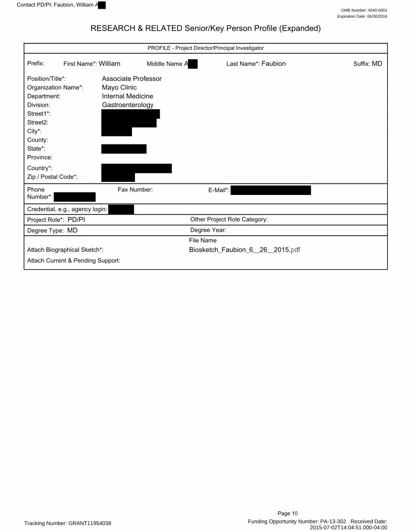
RESEARCH & RELATED Senior/Key Person Profile (Expanded)
PROFILE - Project Director/Principal Investigator
Prefix: First Name*: William Middle Name A Last Name*: Faubion Suffix: MD
Position/Title*: Associate ProfessorOrganization Name*: Mayo ClinicDepartment: Internal MedicineDivision: GastroenterologyStreet1*:Street2:City*:County:State*:Province:
Country*:Zip / Postal Code*:
PhoneNumber*:
Fax Number: E-Mail*:
Credential, e.g., agency login:
Project Role*: PD/PI Other Project Role Category:
Degree Type: MD Degree Year: File Name Mime Type
Attach Biographical Sketch*: Biosketch_Faubion_6__26__2015.pdfapplication/pdf
Attach Current & Pending Support:
Page 10
Contact PD/PI: Faubion, William A

Tracking Number: GRANT11954038 Funding Opportunity Number: PA-13-302 . Received Date:2015-07-02T14:04:51.000-04:00
PROFILE - Senior/Key Person
Prefix: FirstName*: Chandrashekhar
Middle Name Last Name*: Pasare Suffix: PhD
Position/Title*: Associate ProfessorOrganization Name*: University of Texas Southwestern Medical Center at DallasDepartment:Division:Street1*:Street2:City*:County:State*:Province:
Country*:Zip / Postal Code*:
PhoneNumber*:
Fax Number: E-Mail*:
Credential, e.g., agency login:
Project Role*: Consultant Other Project Role Category:
Degree Type: PhD Degree Year: File Name Mime Type
Attach Biographical Sketch*: Biosketch_PasareJune_2015.pdf application/pdf
Attach Current & Pending Support:
PROFILE - Senior/Key Person
Prefix: First Name*: Stephen Middle Name C Last Name*: Ekker Suffix: PhD
Position/Title*: ProfessorOrganization Name*: Mayo ClinicDepartment: Biochem & Molecular BiologyDivision: Biochem & Molecular BiologyStreet1*:Street2:City*:County:State*:Province:
Country*:Zip / Postal Code*:
PhoneNumber*:
Fax Number: E-Mail*:
Credential, e.g., agency login:
Project Role*: Other (Specify) Other Project Role Category: Other Significant ContributorDegree Type: PhD Degree Year: File Name Mime Type
Attach Biographical Sketch*: Ekker_biosketchJune__2015.pdf application/pdf
Attach Current & Pending Support:
Page 11
Contact PD/PI: Faubion, William A

Tracking Number: GRANT11954038 Funding Opportunity Number: PA-13-302 . Received Date:2015-07-02T14:04:51.000-04:00
PROFILE - Senior/Key Person
Prefix: First Name*: Thomas Middle Name C Last Name*: Smyrk Suffix: MD
Position/Title*: ProfessorOrganization Name*: Mayo ClinicDepartment: DLMPDivision: Anatomic PathologyStreet1*:Street2:City*:County:State*:Province:
Country*:Zip / Postal Code*:
PhoneNumber*:
Fax Number: E-Mail*:
Credential, e.g., agency login:
Project Role*: Other (Specify) Other Project Role Category: Other Significant ContributorDegree Type: MD Degree Year: File Name Mime Type
Attach Biographical Sketch*: Biosketch_SmyrkJune_2015.pdf application/pdf
Attach Current & Pending Support:
PROFILE - Senior/Key Person
Prefix: First Name*: Raul Middle Name Alfredo Last Name*: Urrutia Suffix: MD
Position/Title*: ProfessorOrganization Name*: Mayo ClinicDepartment: Internal MedicineDivision: GastroenterologyStreet1*:Street2:City*:County:State*:Province:
Country*:Zip / Postal Code*:
PhoneNumber*:
Fax Number: E-Mail*:
Credential, e.g., agency login:
Project Role*: Other (Specify) Other Project Role Category: Other Significant ContributorDegree Type: MD Degree Year: File Name Mime Type
Attach Biographical Sketch*: Biosketch_UrrutiaJune_2015.pdf application/pdf
Attach Current & Pending Support:
Page 12
Contact PD/PI: Faubion, William A
OMB No. 0925-0001/0002 (Rev. 08/12 Approved Through 8/31/2015)
BIOGRAPHICAL SKETCH Provide the following information for the Senior/key personnel and other significant contributors.
Follow this format for each person. DO NOT EXCEED FIVE PAGES.
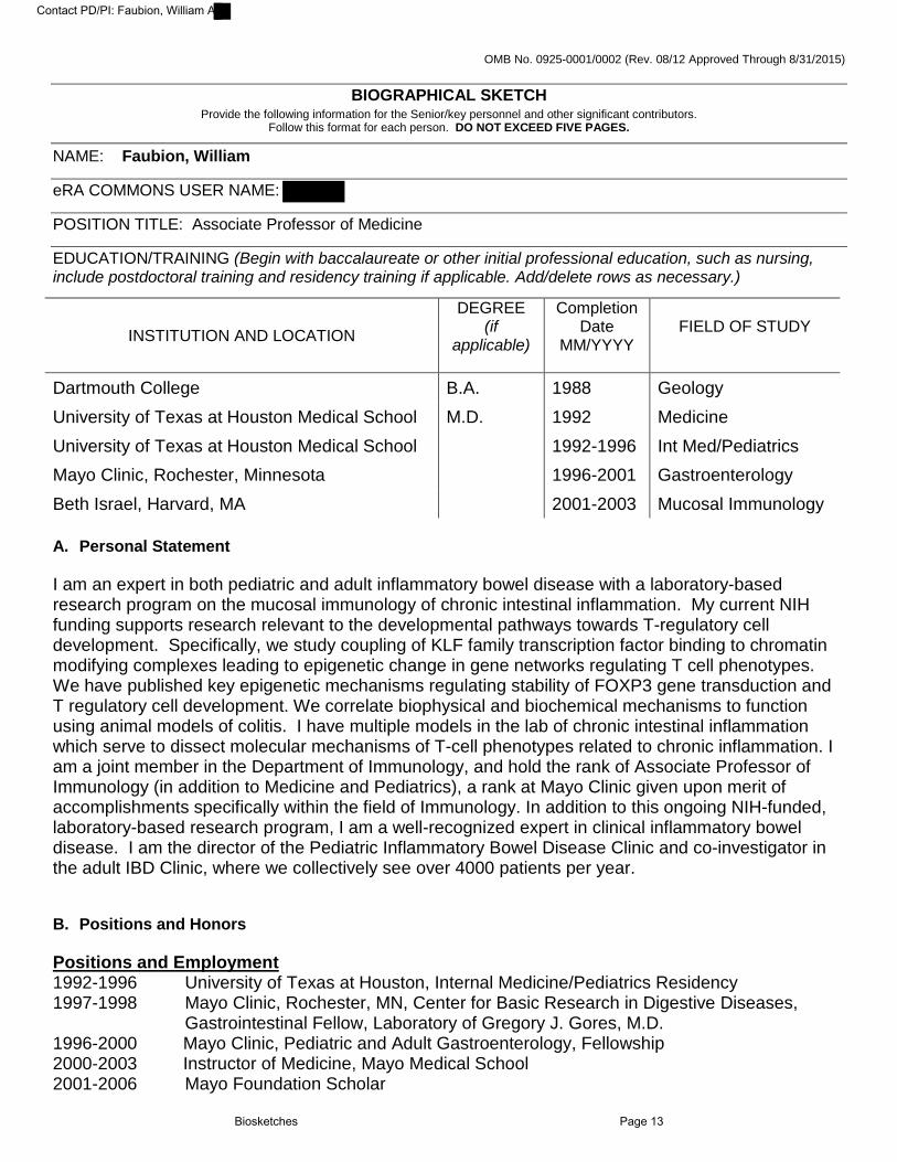
NAME: Faubion, William eRA COMMONS USER NAME: POSITION TITLE: Associate Professor of Medicine EDUCATION/TRAINING (Begin with baccalaureate or other initial professional education, such as nursing, include postdoctoral training and residency training if applicable. Add/delete rows as necessary.)
INSTITUTION AND LOCATION
DEGREE (if
applicable)
Completion Date
MM/YYYY
FIELD OF STUDY
Dartmouth College B.A. 1988 Geology University of Texas at Houston Medical School M.D. 1992 Medicine University of Texas at Houston Medical School 1992-1996 Int Med/Pediatrics Mayo Clinic, Rochester, Minnesota 1996-2001 Gastroenterology Beth Israel, Harvard, MA 2001-2003 Mucosal Immunology A. Personal Statement I am an expert in both pediatric and adult inflammatory bowel disease with a laboratory-based research program on the mucosal immunology of chronic intestinal inflammation. My current NIH funding supports research relevant to the developmental pathways towards T-regulatory cell development. Specifically, we study coupling of KLF family transcription factor binding to chromatin modifying complexes leading to epigenetic change in gene networks regulating T cell phenotypes. We have published key epigenetic mechanisms regulating stability of FOXP3 gene transduction and T regulatory cell development. We correlate biophysical and biochemical mechanisms to function using animal models of colitis. I have multiple models in the lab of chronic intestinal inflammation which serve to dissect molecular mechanisms of T-cell phenotypes related to chronic inflammation. I am a joint member in the Department of Immunology, and hold the rank of Associate Professor of Immunology (in addition to Medicine and Pediatrics), a rank at Mayo Clinic given upon merit of accomplishments specifically within the field of Immunology. In addition to this ongoing NIH-funded, laboratory-based research program, I am a well-recognized expert in clinical inflammatory bowel disease. I am the director of the Pediatric Inflammatory Bowel Disease Clinic and co-investigator in the adult IBD Clinic, where we collectively see over 4000 patients per year. B. Positions and Honors Positions and Employment 1992-1996 University of Texas at Houston, Internal Medicine/Pediatrics Residency 1997-1998 Mayo Clinic, Rochester, MN, Center for Basic Research in Digestive Diseases, Gastrointestinal Fellow, Laboratory of Gregory J. Gores, M.D. 1996-2000 Mayo Clinic, Pediatric and Adult Gastroenterology, Fellowship 2000-2003 Instructor of Medicine, Mayo Medical School 2001-2006 Mayo Foundation Scholar
Biosketches Page 13
Contact PD/PI: Faubion, William A

2001-2003 Instructor of Medicine, Division of Immunology, BIDMC, Harvard Medical School 2003-2012 Assistant Professor of Medicine, Mayo Clinic College of Medicine 2012-present Associate Professor of Medicine, Mayo Clinic College of Medicine 2012-present Associate Professor of Pediatrics, Mayo Clinic College of Medicine 2013-present Associate Professor of Immunology, Mayo Clinic College of Medicine 2013-present Director, T32 Training Grant 2014-present Research Chair, Division of Gastroenterology and Hepatology, Mayo Clinic Other Experience and Professional Memberships 2007 -2013 Member, American Board of Pediatrics 2007-present Associate Editor, Inflammatory Bowel Disease Journal 2007-present Associate Editor, Pediatric Inflammatory Bowel Disease Member, American Board of Internal Medicine 2011-present Member, American Gastroenterological Association 2011-present Member, American College of Gastroenterology Member, North American Society for Pediatric Gastroenterology and Nutrition 2011-present NIH Study Section Reviewer: Digestive Diseases and Nutrition 2012-present CCFA Career Development & Research Fellowship Committee Member 2015-2018 CCFA Research Fellowship Awards Committee Member Honors 2000 J. Arnold Bargen Award – for outstanding achievement by a fellow in gastroenterology, Mayo Clinic
2005 REGAL (Research Excellence in GI and Liver)–for third year fellows or junior faculty members who are less than five years out of their fellowship or residency in GI who demonstrate the ability to conduct important research in the areas of upper GI, lower GI, outcomes, or hepatobiliary research
2005 Berry Family Foundation Scholar Award–to support Mayo GI staff who pursue educational opportunity at other institutions and then return to Mayo to set up their own research
C. Contribution to Science 1. The SLAM family regulates colitis through both adaptive and innate immune mechanisms.
Upon joining the Terhorst laboratory in 2001, the SLAM family of immune receptors was known to be relevant to lymphoproliferative syndromes however precise mechanisms of immunoregulation were unknown. As part of this collaborative team, we defined the role for SLAM, CD48, and Ly108 in T cell and innate immune cellular function in colitis. As SLAM is a measles virus receptor, the major impact of our work on SLAM as a regulator of phagosome function is on the field of virology and retargeting of measles virus.
a. Wang, N. Satoskar, A., Faubion, W.A., Howie, D., Okamoto, S., Feske, S., Gullo, C., Clark, K., Sosa, M.R., Sharpe, A.H., Terhorst, C. (2004). The cell surface receptor SLAM controls T cell and macrophage functions. The Journal of Experimental Medicine, 199(9):1255-64. PMCID:2211908.
b. Howie, D., Laroux, F.S., Morra, M., Satoskar, A.R., Rosas, L.E., Faubion, W.A., Julien, A., Rietdijk, S., Coyle, A.J., Fraser, C., Terhorst, C. (2005). Cutting edge: the SLAM family receptor Ly108 controls T cell and neutrophil functions. Journal of Immunology, 174(10):5931-5. PMID:15879084.
c. Abadia-Molina, A.C., Ji, H., Faubion, W.A., Julien, A., Latchman, Y., Yagita, H., Sharpe, A., Bhan, A.K., Terhorst, C. (2006). CD48 controls T-cell and antigen-presenting cell functions in experimental colitis. Gastroenterology, 130(2):424-34. PMID:16472597.
d. Berger, S.B., Romero, X., Ma, C., Wang, G., Faubion, W.A., Liao, G., Compeer, E., Keszei, M., Rameh, L., Wang, N., Boes, M., Regueiro, J.R., Reinecker, H.C., Terhorst, C. (2010). SLAM is a microbial sensor that regulates bacterial phagosome functions in macrophages. Nature Immunology, 11(10):920-7. PMCID: PMC3338319.
2. Traditional innate immune receptors regulate function of human FOXP3+ Treg cells.
Biosketches Page 14
Contact PD/PI: Faubion, William A
The FOXP3+ Treg cell was defined during the time frame of my post-doctoral training with the Terhorst laboratory, and we were the first to characterize the effect of colitis on Treg thymic developmental steps. Upon my return to Mayo clinic, we continued on with Treg related research with a particular focus on human physiology. With unique experience in innate immune receptors and Treg biology, we were the first to demonstrate a role for both cell membrane (TLR10) and intracellular (NOD2) pathogen recognition receptors on human Treg cellular function. In particular, the work on TLR10 and FOXP3 led to our subsequent deeper line of investigation on FOXP3 gene transcriptional events.
a. Faubion, W.A., De Jong, Y.P., Molina, A.A., Ji, H., Clarke, K., Wang, B., Mizoguchi, E., Siimpson, S.J., Bhan, A.K., Terhorst, C. (2004). Colitis is associated with thymic destruction attenuating CD4+25+ regulatory T cells in the periphery. Gastroenterology, 126(7):1759-70.
b. Abadia-Molina, A.C., Mizoguchi, A., Faubion, W.A., De Jong, Y.P., Rietdjik, S.T., Comiskey, M., Clarke, K., Bhan, A.K., Terhorst, C. (2005). In vivo generation of oligoclonal colitic CD4+ T-cell lines expressing a distinct T-cell receptor Vbeta. Gastroenterology, 128(5):1268-77.
c. Bell, M.P., Svingen, P.A., Rahman, M.K., Xiong, Y., Faubion, W. A. Jr. (2007) FOXP3 regulates TLR10 expression in human T regulatory cells. Journal of Immunology, 173(3):1893-900. PMID:17641056.
d. Rahman, M.K., Midtling, E.H., Svingen, P.A., Xiong, Y., Bell, M.P., Tung, J., Smyrk, T., Egan, L.J., Faubion Jr, W.A. (2010). The pathogen recognition receptor NOD2 regulates human FOXP3+ T cell survival. Journal of Immunology, 184(12):7247-56. PMCID: 38886856.
3. KLF family members regulate FOXP3 and Treg function through coupling to epigenetic complexes. Careful characterization of the FOXP3 core promoter led to the earliest recognition of epigenetic
mechanisms leading to FOXP3 gene activation and Treg development. Our work on the coupling of KLF transcription factors to chromatin modifying complexes has advanced the understanding of Treg biology, CD8+ T cell function, and intestinal stem cell function. The greatest impact has been the first recognition of the role for the histone methyltransferase EZH2 in Treg biology.
a. Xiong, Y., Khanna, S., Grzenda, A.L., Sarmento, O.F., Svingen, P.A., Lomberk, G.A., Urrutia, R.A., Faubion Jr, W.A. (2012). Polycomb antagonizes p300/CREB-binding protein-associated factor to silence FOXP3 in a Kruppel-like factor-dependent manner. Journal of Biological Chemistry, 287(41):34372-85. PMCID: PMC3464543.
b. Xiong, Y., Svingen, P.A., Sarmento, O.F., Smyrk, T.C., Dave, M., Khanna, S., Lomberk, G.A., Urrutia, R.A., Faubion Jr, W.A. (2014). Differential coupling of KLF10 to Sin3-HDAC and PCAF regulates the inducibility of the FOXP3 gene. American Journal of Physiology: Regulatory, Integrative and Comparative Physiology, 307(6)R608-20. PMCID: PMC4166759.
c. Sarmento, O.F., Svingen, P.A., Xiong, Y., Xavier, R.J., McGovern, D., Smyrk, T.C., Papadakis, K.A., Urrutia, R.A., Faubion, W.A. (2015). A novel role for KLF14 in T regulatory cell differentiation. Cellular and Molecular Gastroenterology and Hepatology, 1(2):188-202. PMCID: PMC4349492.
d. Papadakis, K.A., Krempski, J., Reiter, J., Svingen, P., Xiong, Y., Sarmento, O.F., Huseby, A., Johnson, A.J., Lomberk, G.A., Urrutia, R.A., Faubion, W.A. (2015). Kruppel-like factor KLF10 regulates transforming growth factor receptor II expression and TGF-beta signaling in CD8+ T lymphocytes. The American Journal of Physiology- Cell Physiology, 308(5):C362-71. PMCID: PMC4346734.
e. Dave, M., Hayashi, Y., Gajdos, G.B., Smyrk, T.C., Svingen, P.A., Kvasha, S.M., Lorincz, A., Dong, H., Faubion, W.A., Jr., Ordog, T. (2015). Stem cells for murine interstitial cells of cajal suppress cellular immunity and colitis via prostaglandin e2 secretion, 148(5):978-90. PMCID:PMC4409492.
A full list of research-related published work can be found at: http://www.ncbi.nlm.nih.gov/sites/myncbi/william.faubion.1/bibliography/40757558/public/?sort=date&direction=ascending
Biosketches Page 15
Contact PD/PI: Faubion, William A
D. Research Support Ongoing Research Support
R01AI089714-02 Faubion (PI) 2/15/11 – 1/31/16 The goal of this study is to investigate transcriptional regulation of regulatory T cell (Treg) developed by Kruppel-like factor 10 (KLF10) and its role in immune homeostasis.
Biosketches Page 16
Contact PD/PI: Faubion, William A
OMB No. 0925-0001/0002 (Rev. 08/12 Approved Through 8/31/2015)
BIOGRAPHICAL SKETCH Provide the following information for the Senior/key personnel and other significant contributors.
Follow this format for each person. DO NOT EXCEED FIVE PAGES.
NAME: Chandrashekhar Pasare eRA COMMONS USER NAME (credential, e.g., agency login): POSITION TITLE: Associate Professor of Immunology EDUCATION/TRAINING (Begin with baccalaureate or other initial professional education, such as nursing, include postdoctoral training and residency training if applicable. Add/delete rows as necessary.)
INSTITUTION AND LOCATION
DEGREE (if
applicable)
Completion Date
MM/YYYY
FIELD OF STUDY
Bidar Veterinary College, Bidar, India B.V.Sc 06/1992 Veterinary Medicine
Indian Veterinary Research Institute, Bareilly, India M.V.Sc 05/1994 Immunology and Microbiology
National Institute of Immunology, New Delhi, India Ph.D. 09/2000 Immunology and Molecular Biology
Yale University School of Medicine, New Haven, CT Postdoctoral 05/2006 Innate control of Adaptive Immunity
A. Personal Statement I have been working in the field of innate immunity and innate control of adaptive immunity for about 14
years. In addition to a Ph.D. in Immunology, I have had post-doctoral training at Yale University, where I worked on understanding the importance of the TLR signaling pathway in activation of adaptive immune responses. This work led to understanding of TLR mediated regulation of T and B cell responses leading to publications in Science (1928 citations), Immunity (325 citations) and Nature (537 citations). My laboratory continues to work in the area of innate control of adaptive immunity and our more recent work has also focused on understanding molecular details of TLR signaling and molecular mechanisms by which the innate immune system regulates the adaptive immune responses. My lab has extensive experience in studying TLR mediated activation of adaptive immunity and our recent work has led to new understanding of regulation of immune responses in the gut (ref # 2a), discovery of a new TLR signaling adapter that links TLRs to PI3K activation (ref # 3a) and discovery of a rapid NLRP3 inflammasome activation pathway controlled by IRAK-1 (ref # 4a). We have also recently demonstrated that plasma membrane and surface TLRs have different functions in regulation of CD8 T cell responses against pathogens (ref # 1c).
B. Positions and Honors Positions and Employment 2000-2005 Post-doctoral Associate, Howard Hughes Medical Institute, Yale University School
of Medicine, New Haven, CT 2005-2006 Associate Research Scientist, Section of Immunobiology, Yale University School of
Medicine, New Haven, CT 2006-2007 Scientist, Immunology, Genentech, Inc. South San Francisco, CA 2007-2014 Assistant Professor, Department of Immunology, The University of Texas Southwestern
Medical Center, School of Medicine, Dallas, TX 2014-present Associate Professor, Department of Immunology, The University of Texas Southwestern
Medical Center, School of Medicine, Dallas, TX
Biosketches Page 17
Contact PD/PI: Faubion, William A

Professional Memberships 1999 Life member, Indian Immunology Society 2010 - Member, American Association of Immunologists
Honors/Awards 1992-1994 Indian Council of Agricultural Research’s Junior Research Fellowship 1994-1999 Senior Research Fellowship from Department of Biotechnology, India 2000-2005 Howard Hughes Medical Institute, Post-Doctoral Fellowship 2007 UT Southwestern Medical Center Endowed Scholar in Biomedical Research 2013 AAI early career faculty travel award
C. Contribution to Science 1. When I started my post-doctoral work in the laboratory of Ruslan Medzhitov about 14 years ago, TLRs
were only just discovered and not much was known about the various mechanisms by which TLRs influence adaptive immunity. In addition there was renewed interest in a population of cells called regulatory T cells or suppressor T cells. These cells, characterized by surface expression of CD4 and CD25 markers, were shown to be potent suppressors of naïve T cell activation. The general consensus was that these cells block activation of auto-reactive cells. A key question for us was: How do pathogen specific T cells over come the block induced by regulatory T cells to mount protective immune responses? We hypothesized that, since TLRs are major sensors of infections, TLR activation on DCs should be able to overcome suppression mediated by regulatory T cells (Treg cells). I used TLR-deficient and TLR-sufficient dendritic cells for in vitro suppression assays to show that TLR activation on DCs overcomes the block induced by regulatory T cells. In addition I showed that surface maturation of DCs is not sufficient to overcome Treg cell mediated suppression. Finally I demonstrated that the cytokine IL-6 secreted by DCs in response to TLR ligands is responsible for providing critical signals necessary for naïve T cells to overcome Treg cell mediates suppression. In summary, I discovered that the Toll-pathway of DC activation controls adaptive immune responses by at least two major mechanisms; 1. By induction of surface maturation of DCs and 2: By inducing secretion of IL-6, which overcomes Treg cell mediated suppression. This study was described as ground breaking in a commentary that accompanied the published article in the journal “Science”. After gaining insights into the critical requirements for TLR activation on DCs in inducing activation of naïve T cells in vitro, I set out to understand the in vivo significance of these findings and to understand the dynamic interactions between innate and adaptive immune systems in a living animal. Immunologists over the years have been using protein antigens to study induction of T and B cell responses in vivo. I showed using pure protein preparations that, naïve T cells could not be activated in vivo if immunized in the absence of a TLR ligand. Most commercial protein preparations are contaminated with TLR ligands and hence induce T and B cell activation when immunized in depot forming adjuvant such as alum. I used MyD88-deficient mice to shown the importance of different aspects of DC activation for T cell priming in vivo. I showed that DCs mature and migrate to the draining lymph node in response to LPS in MyD88-deficient mice but fail to induce naïve T cell activation. This was very contrary to the current understanding that expression of MHC and co-stimulatory molecules is sufficient to prime antigen specific T cells. I showed that this is not the case and that Treg cells block activation of naïve T cells if DCs do not secrete cytokines during priming. I was thus able to define the critical requirements for naïve T cell activation. Further, I discovered that TLR and MyD88 dependent signals are necessary for induction of memory CD4 T cells. This is an important finding and there is ongoing research in my current lab to understand the cellular and molecular mechanisms of how TLR activation induces development of CD4 memory T cells. In a related study dissecting the role of TLRs in regulating B cell responses I discovered that the poor antibody responses in MyD88 deficient mice were because of lack of TLR signaling in B cells rather than lack of T cell priming and differentiation. This study for the first time established the role of innate immune sensing in mounting T-dependent B cell responses and implicated B cell intrinsic TLR signaling in B cell activation and differentiation. I was a post-doctoral fellow for all these studies.
Biosketches Page 18
Contact PD/PI: Faubion, William A

a. Pasare, C., and Medzhitov, R. (2003). Toll pathway-dependent blockade of CD4+CD25+ T cell-
mediated suppression by dendritic cells. Science 299, 1033-1036. PMID: 12532024.
b. Pasare, C., and Medzhitov, R. (2004). Toll-dependent control mechanisms of CD4 T cell activation. Immunity 21, 733-741. PMID: 15539158.
c. Pasare, C., and Medzhitov, R. (2005). Control of B-cell responses by Toll-like receptors. Nature 438, 364-368. PMID: 16292312.
2. As an independent investigator I continued to focus on understanding the role of innate immune system in
regulating adaptive immunity. In the first study published from the laboratory we found that the cytokine requirements for induction of the inflammatory Th17 lineage cells depend on the site of priming. We found that the mucosal and systemic immune systems have different rules for inducing differentiation of naïve T cells into Th17 cells. We discovered that IL-1R mediated, MyD88 dependent signaling in CD4 T cells is critical for generation of Th17 lineage cells in all lymphoid tissues. However, while there is a requirement for IL-6 for Th17 priming in the mucosal tissues such as the lamina propria of the intestines as well as the lungs, IL-6 is not required for generation of Th17 lineage cells in the peripheral lymphoid organs such as the spleen and lymph nodes, The differential requirement for Th17 priming in the peripheral lymphoid organs versus the lamina propria of the intestines is true both during steady state as well as during exposure to pathogens via oral or systemic route. We also demonstrate that lack of IL-6 leads to higher proportion of Foxp3 positive CD4 T cells selectively only in the lamina propria of the intestines. Finally we demonstrate that gut specific need for IL-6 is dictated by DCs resident in the lamina propria of the intestines as LP DCs from IL-6 deficient mice fail to prime Th17 lineage cells. DCs resident in the spleens of IL-6 deficient mice induce normal priming of Th17 lineage cells. This is the first demonstration of selective need of different cytokines based on priming micro-environments for any kind of helper T cell lineage. Our study challenges the concept that IL-6 is a master regulator of RORgt and Th17 differentiation and provides novel insights into tissue specific requirements for T cell differentiation. Differential requirement for Th17 priming in spleen and gut lamina propria is a novel concept and has important implications for targeting systemic and mucosal tissue specific auto-immunity as well as for determining routes of vaccination. In a related study that deals with regulation of CD8 T cell responses by the innate immune system, we found that plasma membrane TLRs and endosomal TLRs have a differential role in inducing CD8 T cell priming and this is primarily dictated by population of DCs recruited to the site of priming
a. Hu, W., Troutman, T.D., Edukulla, R., and Pasare, C. (2011). Priming microenvironments
dictate cytokine requirements for T helper 17 cell lineage commitment. Immunity 35, 1010-1022. PMID: 22137454. PMCID: PMC3246047.
b. Hu, W and Pasare, C (2013). Location, location, location: tissue specific regulation of immune responses. Journal of Leukocyte Biology. 94, 409-421. PMID:23825388. PMCID: PMC3747123.
c. Mandraju R, Murray S, Forman J and Pasare C. (2014). Differential ability of surface and endosomal TLRs to induce CD8 T cell responses in vivo. J Immunol. 192, 4303-15. PMID: 24688022. PMCID: PMC4002505.
3. TIR domains are required for the initiation of TLR signaling and serve to link TLRs to their adaptors through homotypic interactions. The known adapters of TLR signaling MyD88 (and TIRAP) and TRIF (and TRAM) signal to activate NF-kB, MAP kinases and IRF proteins but it has never been clear how TLR signaling leads to PI3 kinase activation. Although NF-kB activation is important for inducing pro-inflammatory response and cytokine secretion by macrophages, PI3K activation in cells plays a critical role in regulating cell survival, cell cycling and proliferation. We have found that BCAP, via its TIR domain, links TLRs to PI3K activation, and is also responsible for negatively regulating TLR signaling. Our studies demonstrate that BCAP modulates pro-inflammatory responses and BCAP deficient mice have exaggerated inflammation in response to infections and are highly susceptible to colitis. The study of TLR mediated activation of NF-kB has yielded many important and seminal discoveries into the regulation of this inflammatory pathway. However, signaling via TLRs to PI3K is poorly characterized. Data using chemical inhibitors suggests contradictory roles for PI3K. Due to the central and important role of PI3K through many
Biosketches Page 19
Contact PD/PI: Faubion, William A

converging signaling pathways, genetic tools utilizing mice deficient for PI3K components carry the caveat of disrupted signaling through other signaling pathways. Our delineation of BCAP’s critical role in activation of the PI3K axis is important not only through our description of how BCAP regulates inflammation, but also because of the characterization of a signaling adapter critically required for the proximal bridging of TLRs to PI3K.
a. Troutman, T.D., Hu, W., Fulenchek, S., Yamazaki, T., Kurosaki, T., Bazan, J.F., and Pasare, C.
(2012). Role for B-cell adapter for PI3K (BCAP) as a signaling adapter linking Toll-like receptors (TLRs) to serine/threonine kinases PI3K/Akt. Proc Natl Acad Sci USA 109, 273-278. PMID: 22187460. PMCID: PMC3252926.
b. Troutman, T.D., Bazan, J.F., and Pasare, C. (2012). Toll-like receptors, signaling adapters and regulation of the pro-inflammatory response by PI3K. Cell Cycle 11. PMID: 22895011. PMCID: PMC3478307.
4. In the past few years, my laboratory also got interested in understanding the biogenesis of IL-1beta. The cytokines IL-1beta and IL-18 are made as pro-forms and are induced by TLR signaling. When a cell senses a toxin or virulence factor or is undergoing stress, this leads to activation of inflammasome causing caspase-1 activation, which then cleaves IL-1beta and IL-18 for their release. The scientific understanding was that there is a priming signal (TLR activation) and an activation signal (NLR activation) separated by time, necessary for inflammasome activation. We hypothesized that when there is an infection by a virulent pathogen, the cells are likely to see both of these signals at the same time and that macrophages should be equipped to induce inflammasome activation independent of priming. Consistent with this hypothesis, we discovered a rapid NLRP3 inflammasome activation pathway that leads to caspase-1 activation in as little as 20 minutes following simultaneous sensing of TLR and NLRP3 ligands. More importantly we found that this activation is directly regulated by TLR signaling component IRAK-1 and that not only does IRAK-1 form part of the inflammasome complex, but its kinase activity is important for rapid NLRP3 inflammasome assembly. We also discovered that this rapid IRAK-1 dependent inflammasome activation pathway is critical to sense Listeria monocytogenes and induce rapid IL-18 dependent IFN-gamma production by NK cells and memory CD8 T cells. We believe that this rapid inflammasome activation pathway is critical for rapid innate defense and are continuing to work on understanding the molecular mechanism of this activation and its role in protection against a variety of virulent pathogens.
a. Lin KM, Hu W, Troutman, TD, Jennings M, Brewer, T, Li, X, Nanda, S, Cohen P, Thomas J and Pasare C. (2014) IRAK-1 by passes priming and directly links TLRs to rapid NLRP3 inflammasome activation. Proc. Natl. Acad. Sci. USA. 111, 775-780. PMID: 24379360. PMCID: PMC3896167.
Complete List of Published Work in MyBibliography: http://www.ncbi.nlm.nih.gov/sites/myncbi/1pmWT2zpU9bQM/bibliograpahy/46106384/public/?sort=date&direction=ascending D. Research Support Ongoing Research Support 2015/04/15-2017/03/31 R21 AI115420, National Institute of Allergy and Infectious Diseases (NIAID, NIH) PASARE, CHANDRASHEKHAR (PI) Role of Microenvironmental cues in CD8 T cell activation and memory generation The major goals of this project are to investigate the mechanisms by which priming microenvironments influence activation and development of CD8 memory T cells.
Biosketches Page 20
Contact PD/PI: Faubion, William A

2010/08/15-2015/07/31 R01 AI082265, National Institute of Allergy and Infectious Diseases (NIAID, NIH) PASARE, CHANDRASHEKHAR (PI) Toll-like receptor mediated regulation of effector and memory CD4 T cell responses The major goals of this project are to investigate the mechanisms by which TLRs influence activation and differentiation of CD4 T cells into effector and memory cells. Competed Research Support (ended in the last 3 years) 2010/02/15-2013/01/31 R43 AI089138, National Institute of Allergy and Infectious Diseases (NIAID, NIH) PASARE, CHANDRASHEKHAR (PI) Synergistic vaccine adjuvants that stimulate both innate and adaptive immunity The major goals of this project were to develop a glycoside adjuvant that can activate innate immunity and investigate its ability to induce protective adaptive immune responses to various pathogens. Role: Principal Investigator
Biosketches Page 21
Contact PD/PI: Faubion, William A

OMB No. 0925-0001/0002 (Rev. 08/12 Approved Through 8/31/2015)
BIOGRAPHICAL SKETCH NAME: Stephen C. Ekker eRA COMMONS USER NAME (credential, e.g., agency login): POSITION TITLE: Professor of Biochemistry and Molecular Biology EDUCATION/TRAINING
INSTITUTION AND LOCATION
DEGREE (if
applicable)
Completion Date
MM/YYYY
FIELD OF STUDY
University of Illinois Urbana-Champaign, Urbana B.S. 05/1988 Electrical Engineering
University of Illinois Urbana-Champaign, Urbana B.S. 05/1988 Genetics & Dev. Biology
Johns Hopkins University, Baltimore, MD Ph.D. 05/1993 Molec. Bio. & Genetics
Johns Hopkins University, Baltimore, MD PostDoctoral 10/1995 Develop. Genetics
A. Personal Statement My background in molecular genetics and genomics started with undergraduate research with a project to restriction map the genome of the Archaebacterium Sulfolobus solfataricus. I have been helping further the zebrafish (Danio rerio) as a model vertebrate to address major issues in human health and biology since training as a post-doctoral fellow. This system has the potential for diverse genetic and behavioral studies typically restricted to the world of the fly or worm but conducted within the biological framework of a vertebrate.
The major focus of my laboratory is on understanding our genome, using the zebrafish as a rapid molecular test system. We established the rapid use of morpholino sequence-specific knockdown technology for vertebrate functional genomics applications using the zebrafish as the pioneering model system fifteen years ago. In parallel, we developed vertebrate transposon tools including our protein trap gene-breaking vectors to generate a 700+ line collection of molecularly characterized and revertible mutant zebrafish lines, the first engineered conditional alleles in any organism outside the mouse. We have deployed transposons in diverse application areas including human T cells, zebrafish, and mice. Custom restriction endonucleases offer a third leg, targeted modification using genome editing tools. We continue to develop science behind these new engineering toolkits, working with an array of laboratories as diverse as rat, pig, mouse, nematode, and fly biologists in addition to regular colleagues that work in human cells and zebrafish. As Director of the Genetics and Model Systems Core for the Center for Cell Signaling in Gastroenterology, I am delighted to support your project. B. Positions and Honors 1986-1988 Undergraduate Research: Restriction Mapping the Genome of the Archaebacterium Sulfolobus
solfataricus. University of Illinois; C.R. Woese, advisor 1988-1993 Graduate Dissertation Differential DNA Binding and the Specificity of Homeotic Gene Action,
Philip A. Beachy, Advisor, Johns Hopkins School of Medicine (SOM) and HHMI, Baltimore, MD 1990-1992 Teaching Assistant, Dept. of Mol. Biol. and Genetics, Johns Hopkins University, Baltimore, MD 1990-1993 March of Dimes Birth Defects Predoctoral Fellow 1993-1995 Postdoc, Biochemical Properties and Biological Activities of the Hedgehog Gene Family, P.A.
Beachy, Advisor, R.T. Moon, Co-Advisor, Johns Hopkins SOM and HHMI, Baltimore, MD 1995-2007 Assistant, Associate, and full Professor, University of Minnesota, Minneapolis, MN 1997-1999 March of Dimes Basil O’Connor Scholar 2000 Co-founder, Discovery Genomics, Inc. 1999-2007 Director, Arnold and Mabel Beckman Center for Transposon Research, University of Minnesota 2002-2007 Associate Head, Genetics, Cell Biology and Development, University of Minnesota 2007-present Adjunct Professor, Genetics, Cell Biology and Development, University of Minnesota 2007-present Professor Dept Biochemistry and Molecular Biology and Consultant, Mayo Clinic,
Rochester,MN Biosketches Page 22
Contact PD/PI: Faubion, William A

2007-present Director, Mayo Clinic Zebrafish Facility 2007-present Mayo Predoctoral Education Programs Committees (CTS: 2007-present; BMB 2007-12) 2008-present Editor-in-Chief, The Zebrafish journal 2009-present Director, Genetics and Model Systems Core, Mayo Center for Cell Signaling in
Gastroenterology 2010-present Director, Mayo Addiction Research Center 2012-present Founder, InSciEd Out Foundation 2014-present Chairman of the Board, InSciEd Out Foundation 2013-present Member, Faculty of 1000 2013 Co-Organizer and Founder, 1st International Zebrafish for Personalized/Precision Medicine
Conference, Toronto, Oct. 16-18, 2013 2013-present Founding member, Zebrafish Disease Models Society 2014-present Director, Model Systems Core, Mayo Translational PKD Center Professional Memberships AAAS (1992), Genetics Society of America (1992), SDB (1996), ASHG (2005), SFN (2010), IBANGS (2013) Honors (selected) Member, National Honor Society (1983); James Scholar and Dean's List, University of Illinois (1983-88); Graduation with Honors, University of Illinois (1988); Member, Phi Kappa Phi and Eta Kappa Nu Honor Societies (1988); Predoc fellow, March of Dimes (1990-93); Basil O'Connor Scholar, March of Dimes (1997-99) Major Study Panels: NSF Dev Mech (2002-2004); NCRR 10/99; NHLBI SEP 06/00; NIGMS PPG 01/02, CDF4 06/02; Craniofacial Special Emphasis Panel 11/02; NIDDK SEP 11/02; Genome 02/03, 12/03; Innov. Tox Models SEP 07/03; NDPR 10/03, 06/04; Tools for Genetic Studies in Zebrafish 04/04, 03/09; Centers for Biomedical Computing SEP, 05/05; NCI Cancer Nanotech Platform SEP (section chair) 07/05; NIH MDCN SEP 07/05; NIH CAD&O 10/05, 06/06, 10/06; NHLBI 03/06; SBIR 04/06; Neurogenesis and Cell Fate Full member, 06/05 – 06/09; RC4 05/10; NHLBI 11/10; Member of the NIH College of CSR Reviewers 01/2010-present; NIDA-K Full member 06/10-12 (end of panel); NIDA ZDA1 EXL-T Full member 06/10-present; NIH/NCATS Comparative Medicine (CM) Special Emphasis Panel (SEP) Feb 13-14, 2013; NIDA CEBRA Oct 2013, Mar 2014; NIH PPG review April 2014
Pertinent Science Education: served as Editor-in-Chief for two Special Issues of the Zebrafish on the topic ‘Zebrafish in Education.’ I solicited funding to cover all publication charges (including NIH support), making all of these issues freely available to classroom teachers and students around the world. The first issue came out in 2009, the second in December of 2012. Patents Ekker,S.C. et al. Inhibition of Gene Expression using Polynucleotide Analogues. US Patent 6,867,349. Ekker,S.C. et al., Syndecans and Angiogenesis – US Patent 7,713,925. C. Contributions to Science [125 publications; google h index 48]
1) Biochemical and biological activities of hedgehog signaling in Drosophila and vertebrates. During my post-doctoral training, I contributed to uncovering mechanistic understanding how this key pathway works in a variety of critical processes and in the cloning of vertebrate family members including the first work using zebrafish implicating shh signaling as defective in holoprosencephaly. a) Lee, J. J., Ekker, S. C., von Kessler, D. P., Porter, J. A., Sun, B. I. and Beachy, P. A. (1994).
Autoproteolysis in hedgehog protein biogenesis. Science 266, 1528-1537. b) Ekker, S .C., Ungar,A., Greenstein,P., von Kessler,D.P., Porter,J.A., Moon,R.T., and Beachy,P.A.
(1995). Patterning Activities of Vertebrate hedgehog Proteins in the Developing Eye and Brain. Current Biology, 5: 944-955.
c) Ekker, S. C., McGrew, L. L., Lai, C. J., Lee, J. J., von Kessler, D. P., Moon, R. T. and Beachy, P. A. (1995). Distinct expression and shared activities of members of the hedgehog gene family of Xenopus laevis. Development 121, 2337-2347.
d) Porter, J. A., Ekker, S. C., Park, W. J., von Kessler, D. P., Young, K. E., Chen, C. H., Ma, Y., Woods, A. S., Cotter, R. J., Koonin, E. V., et al. (1996). Hedgehog patterning activity: role of a lipophilic modification mediated by the carboxy-terminal autoprocessing domain. Cell 86, 21-34.
Biosketches Page 23
Contact PD/PI: Faubion, William A
2) Development of morpholino antisense technology for vertebrate genomics applications using the
zebrafish. a) Nasevicius, A. and Ekker, S. C. (2000). Effective targeted gene 'knockdown' in zebrafish. Nature
genetics 26, 216-220. b) Robu, M. E., Larson, J. D., Nasevicius, A., Beiraghi, S., Brenner, C., Farber, S. A. and Ekker, S. C.
(2007). p53 activation by knockdown technologies. PLoS genetics 3, e78. c) Bill, B. R., Petzold, A. M., Clark, K. J., Schimmenti, L. A. and Ekker, S. C. (2009). A primer for
morpholino use in zebrafish. Zebrafish 6, 69-77. PMCID: PMC2776066 d) Patent awarded: Ekker,S.C. et al. Inhibition of Gene Expression using Polynucleotide Analogues.
US Patent 6,867,349.
3) Development of transposons for vertebrate genomics applications in zebrafish. We deployed the first transposon system, Sleeping Beauty, for transgenesis and enhancer trapping, and then harnessed the endogenous Tol2 transposon for conditional mutagenesis and protein trapping applications. a) Davidson, A. E., Balciunas, D., Mohn, D., Shaffer, J., Hermanson, S., Sivasubbu, S., Cliff, M. P.,
Hackett, P. B. and Ekker, S. C. (2003). Efficient gene delivery and gene expression in zebrafish using the Sleeping Beauty transposon. Developmental biology 263, 191-202.
b) Balciunas, D., Davidson, A. E., Sivasubbu, S., Hermanson, S. B., Welle, Z. and Ekker, S. C. (2004). Enhancer trapping in zebrafish using the Sleeping Beauty transposon. BMC genomics 5, 62.
c) Balciunas, D., Wangensteen, K. J., Wilber, A., Bell, J., Geurts, A., Sivasubbu, S., Wang, X., Hackett, P. B., Largaespada, D. A., McIvor, R. S., et al. Ekker, S. C. (2006). Harnessing a high cargo-capacity transposon for genetic applications in vertebrates. PLoS genetics 2, e169.
d) Clark, K. J., Balciunas, D., Pogoda, H. M., Ding, Y., Westcot, S. E., Bedell, V. M., Greenwood, T. M., Urban, M. D., Skuster, K. J., Petzold, A. M., et al. Ekker, S. C. (2011). In vivo protein trapping produces a functional expression codex of the vertebrate proteome. Nature methods 8, 506-515. PMCID: PMC3306164
4) In vivo genome editing. We developed new tools and approaches for genome editing including the first targeted knockins in zebrafish. This approach also works well in in vitro systems. a) Bedell, V. M., Wang, Y., Campbell, J. M., Poshusta, T. L., Starker, C. G., Krug, R. G., 2nd, Tan, W.,
Penheiter, S. G., Ma, A. C., Leung, A. Y., et al. Ekker, S. C. (2012). In vivo genome editing using a high-efficiency TALEN system. Nature 491, 114-118. PMCID: PMC3491146
b) Neff, K. L., Argue, D. P., Ma, A. C., Lee, H. B., Clark, K. J. and Ekker, S. C. (2013). Mojo Hand, a TALEN design tool for genome editing applications. BMC bioinformatics 14, 1. PMCID: PMC3575288
c) Ma, A. C., Lee, H. B., Clark, K. J. and Ekker, S. C. (2013). High efficiency In vivo genome engineering with a simplified 15-RVD GoldyTALEN design. PloS ONE 8, e65259. PMCID: PMC3667041
d) Peng, Y., Clark, K. J., Campbell, J. M., Panetta, M. R., Guo, Y. and Ekker, S. C. (2014). Making designer mutants in model organisms. Development 141, 4042-4054. PMCID: PMC4302887
5) Establishment of Integrated Science Education Outreach (InSciEd Out), a new approach for excellence in science education for the betterment of human health for all (http://insciedout.org/). We are operational on three continents (North America, Africa and India). Our highly collaborative program focuses on core cultural change, implementing science education within the context and needs of each partner community. a) Pierret,C., Sonju,J., Leicester,J. Hoody,M., LaBounty,T., Frimannsdottir,K.,R., Ekker,S.C. (2012) Improvement in Student Science Proficiency Through InSciEd Out, Zebrafish, 9, 155-68. PMCID: PMC3529492 b) Clark, K. J. & Ekker, S. C. (2015) How Zebrafish Genetics Informs Human Biology. Nature Education 8(4):3.
URL for bibliography: http://www.ncbi.nlm.nih.gov/pubmed?Db=pubmed&Cmd=DetailsSearch&Term=ekker+sc%5BAll+Fields%5D&WebEnv=0SoUqK-GD_T4YuzbaQadqM8hNqScpHAFyiFvTbm7gSa2SDumUj4r3YHA2epX7E-HqWGGoT1pliY_npV%40255F54F3705678F0_0091SID&WebEnvRq=1
Biosketches Page 24
Contact PD/PI: Faubion, William A

D. Research Support Ongoing Research Support Genome Biology 5R01/56-GM63904-11 (Ekker, PI; Farber, Hammerschmidt, Xu and Clark as Co-I) 09/01/01 – 08/31/16 "Systematic Vertebrate Functional Genomics" This grant funds a transposon-based phenotypic screen in the zebrafish to assess the role of 500 genes in development, skin, heart and digestive system biology.
1R01 HG006431-04 (Ekker, PI) 08/01/11 – 07/30/17 “International Zebrafish Mutagenic Protein Trap” This resource grant funds the generation of 1000 new gene-break transposon lines for the zebrafish community. There is no support for biological annotation in this grant.
Supporting Grants 1P30 DK84567-06 (PI: LaRusso, N/Genetics Core PI: Ekker) 09/01/09 – 08/31/19 “Mayo Center for Cell Signaling in Gastroenterology” SCE is PI of the Genetics and Model Organism Core that is designed to bring consultation, training and state-of-the-art genetics tools and model organisms to local scientists.
P30 DK090728-5 (PI:Torres,V.E.; Ekker, Model Organism Core PI) 09/30/10 – 06/30/15 “Mayo Translational PKD Center (MTPC)” Dr. Ekker is Director of the Model Systems Core. [The competitive renewal received a score of 11 and will likely be continued.]
Education Grants 2UL1TR000135-9 (PI: Khosla, S) 07/01/11 – 06/30/16 “Mayo Clinic Center for Translational Science Activities” Dr. Ekker is Associate Director for the Predoctoral training program in translational sciences, including the K-8 InSciEd Out science education reform initiative for the healthy community program.
Completed Research Support
5R01 DA14546-10 (Ekker, PI) 05/01/01 – 06/30/14 “Intron-based Mutagenic Transposons for Zebrafish” The grant funded the development of the gene-break transposon technology for zebrafish functional genomics including the use of GBTs for behavioral genetics of the nicotine response.
Biosketches Page 25
Contact PD/PI: Faubion, William A

OMB No. 0925-0001/0002 (Rev. 08/12 Approved Through 8/31/2015)
BIOGRAPHICAL SKETCH Provide the following information for the Senior/key personnel and other significant contributors.
Follow this format for each person. DO NOT EXCEED FIVE PAGES.
NAME: Thomas C. Smyrk, M. D. eRA COMMONS USER NAME (credential, e.g., agency login): POSITION TITLE: Professor of Pathology EDUCATION/TRAINING (Begin with baccalaureate or other initial professional education, such as nursing, include postdoctoral training and residency training if applicable. Add/delete rows as necessary.)
INSTITUTION AND LOCATION
DEGREE (if
applicable)
Completion Date
MM/YYYY
FIELD OF STUDY
University of Minnesota, Minneapolis, MN BA 07/78 English
University of Minnesota, Minneapolis, MN MD 07/82
Mayo Clinic, Rochester, MN 07/86
Please refer to the Biographical Sketch sample in order to complete sections A, B, C, and D of the Biographical Sketch. A. Personal Statement: I have twenty-nine years of experience as a surgical pathologist with interest in gastrointestinal pathology. Colitis and colitis-associated dysplasia/carcinoma have been particular interests of mine. Dr. Faubion and I have successfully collaborated on several previous projects and I look forward to collaborating with him and his excellent colleagues in the near future.
1) Garrity-Park MM, Loftus EV, Bryant SC, Sandborn WJ, Smyrk TC. Tumor necrosis factor-alpha polymorphisms in ulcerative colitis-associated colorectal cancer. Am J Gastroenterol. 2008 Feb; 103(2):407-15. PMID:18289203. DOI:10.1111/j.1572-0241.2007.01572.x.
2)) Kisiel JB, Garrity-Park MM, Taylor WR, Smyrk TC, Ahlquist DA. Methylated eyes absent 4 (EYA4) gene promotor in non-neoplastic mucosa of ulcerative colitis patients with colorectal cancer: evidence for a field effect. Inflamm Bowel Dis. 2013 Sep;19(10):2079-83. PMID:23867875. PMCID:4119944. DOI:10.1097/MIB.0b013e31829b3f4d.
3) Sarmento OF, Svingen PA, Xiong Y, Xavier RJ, McGovern D, Smyrk TC, Papadakis KA, Urrutia RA, Faubion WA. A novel role for KLF14 in T regulatory cell differentiation. Cell Mol Gastroenterol Hepatol. 2015 Mar 1; 1(2):188-202.e4. PMID:25750932. PMCID:4349492. DOI:10.1016/j.jcmgh.2014.12.007.
4) Dave M, Hayashi Y, Gajdos GB, Smyrk TC, Svingen PA, Kvasha SM, Lorincz A, Dong H, Faubion WA Jr, Ordog T. Stem cells for murine interstitial cells of cajal suppress cellular immunity and colitis via prostaglandin e2 secretion. Gastroenterology. 2015 May;148(5):978-90. Epub 2015 Jan 28. PMID:25637652. PMCID:4409492.
B. Positions and Honors Positions and Employment 1986-1990 Assistant Professor of Pathology, Creighton University, Omaha, NE 1990-1997 Associate Pathologist, Clarkson Hospital, Omaha, NE 1997-2000 Associate Professor of Pathology, University of Nebraska, Omaha, NE
Biosketches Page 26
Contact PD/PI: Faubion, William A
2000-2012 Associate Professor of Pathology, Mayo Clinic, Rochester, MN 2013-present Professor of Pathology, Mayo Clinic, Rochester, MN C. Contribution to Science Lynch Syndrome: I collaborated with Henry Lynch on multiple descriptive studies covering clinical and pathologic features of Lynch syndrome, written before the genetics of that condition were known. One review has been cited more than 1000 times. (1) Jeremy Jass and I collaborated on a histology paper that got the pathology of Lynch Syndrome largely correct. (2) I attended the Bethesda conference in 1996 and collaborated on the publication of the Bethesda criteria. (3) I designed, performed and wrote the study characterizing tumor infiltrating lymphocytes as a robust predictor of microsatellite instability status. (4)
1) Lynch HT, Smyrk TC, Watson P et al. Genetics, natural history, tumor spectrum and pathology of hereditary nonpolyposis colorectal cancer: an updated review. Gastroenterology 1993;104:1535-1549. PMID: 8482467
2) Jass JR, Smyrk TC, Stewart SM, Lane MR, Lanspa SJ, Lynch HT. Pathology of hereditary non-polyposis colorectal cancer. Anticancer Res 1994;14:1631-1634. PMID: 7979198
3) Rodriguez-Bigas MA, Boland CR, Hamilton SR et al. A National Cancer Institute workshop on hereditary nonpolyposis colorectal cancer syndrome: meeting highlights and Bethesda guidelines. J Natl Cancer Inst 1997;89:1758-1762. PMID: 9392616
4) Smyrk TC, Watson P, Kaul K, Lynch HT. Tumor-infiltrating lymphocytes are a marker for microsatellite instability in colorectal carcinoma. Cancer 2001;91:2417-2422. PMID: 11413533
Eosinophilic esophagitis: We published the first description of eosinophilic esophagitis as a syndrome involving dysphagia. (1) My interest in that condition has continued over the years, resulting in contributions addressing whether diagnostic criteria are too strict (2), evaluating new diagnostic techniques (3) and elucidating the pathophysiology. (4)
1) Attwood SE, Smyrk TC, DeMeester TR, Jones JB. Esophageal eosinophilia with dysphagia. A distinct clinicopathologic syndrome. Dig Dis Sci 1993;38:109-116. PMID: 8420741
2) Ravi K, Talley NJ, Smyrk TC et al. Low grade esophageal eosinophilia in adults: an unrecognized part of the spectrum of eosinophilic esophagitis? Dis Dis Sci 2011;56:1981-1986. PMID: 21298480
3) Katzka DA, Geno DM, Ravi K et al. Accuracy, safety and tolerability of tissue collection by Cytosponge vs endoscopy for evaluation of eosinophilic esophagitis. Clin Gastroenterol Hepatol 2015;13:77-83. PMID: 24997328
4) Katzka DA, Ravi K, Geno DM et al. Endoscopic mucosal impedance measurements correlate with eosinophilia and dilation of intercellular spaces in patients with eosinophilic esophagitis. Clin Gastroenterol Hepatol 2015. PMID: 25592662
IgG4-related disease: Our 2003 paper was the first to clearly separate two forms of autoimmune pancreatitis, one IgG4-related and one not. (1) That separation is now widely accepted. We also had an early paper described the role of tissue IgG4 in the diagnosis. (2) I participated in the first international consensus conference on IgG4-related disease and contributed to the publication of its findings. (3, 4)
1) Notohara K, Burgart LJ, Yadav D, Chari S, Smyrk TC. Idiopathic chronic pancreatitis with periductal lymphoplasmacytic infiltration: clinicopathologic features of 35 cases. Am J Surg Pathol 2003;8:1119-1127. PMID: 12883244
2) Zhang L, Notohara K, Levy MJ, Chari ST, Smyrk TC. IgG4-positive plasma cell infiltration in the diagnosis of autoimmune pancreatitis. Mod Pathol 2007;20:23-28. PMID: 16980948
3) Stone JH, Khosroshahi A, Despande V et al. Recommendations for the nomenclature of IgG4-related disease and its individual organ system manifestations. Arthritis Rheum 2012;64:3061-3067. PMID: 22736240
4) Deshpande V, Zen Y, Chan JK et al. Consensus statement on the pathology of IgG4-related disease. Mod Pathol 2012;25:1181-1192. PMID: 22596100
Complete List of Published Work in MyBibliography: http://www.ncbi.nlm.nih.gov/sites/myncbi/thomas.smyrk.1/bibliography/47997356/public/?sort=date&direction=ascending
Biosketches Page 27
Contact PD/PI: Faubion, William A
D. Research Support Ongoing Research Support 8702.02-A1 P50CA 102701 Petersen (PD/PI) 9/18/14-8/31/19 NCI Mayo Clinic SPORE in Pancreatic Cancer – Core 2: Tissue Core Build and administrate the pancreatic tissue core. Role: Co-Investigator
FP00051409 P30DK 84567 LaRusso (OP) 9/1/14-8/31/19 NIDDK Mayo Center for Cell Signaling in Gastroenterology This center core grant correlates research requests with appropriate tissue sources. Role: Other Professional FP00062460 CA 163059 Wang (CI) 9/21/11-8/31/16 NIH Validation Core C ? U MI Betmet Core This study assesses new imaging techniques for dysplastic Barrett’s esophagus. Role: Co-Investigator 18248.02 U01DK 74008 Farrugia (CI) 9/1/12-8/31/16 NIDDK Gastroparesis Clinical Research Consortium – Data Coordinating This major goal is to study the pathology gastroparesis. Role: Co-Investigator FP00064454 HHSN2612012000421
Limburg (CI) 9/24/12-9/23/19
NIH Cancer Prevention Agent Development Program: Early Phase Clinical Research The major goal is to use pathology to assess response to chemoprevention. Role: Co-Investigator FP00067546 U01AA 21788 Shah (CI) 9/20/12-6/30/17 NIH TREAT – Mayo This major goal is to determine prognostic markers for alcoholic liver disease. Role: Co-Investigator FP00062052; CA 163004 Wang (CI) 9/26/11-8/31/16 NIH Stem Cells and the Origins of Barrett’s Esophagus This major goal is to identify esophageal stem cells and delineate their role in Barrett’s esophagus. Role: Co-Investigator Completed Research Support n/a
Biosketches Page 28
Contact PD/PI: Faubion, William A

BIOGRAPHICAL SKETCH Provide the following information for the Senior/key personnel and other significant contributors.
Follow this format for each person. DO NOT EXCEED FIVE PAGES.
NAME: Urrutia, Raul eRA COMMONS USER NAME (credential, e.g., agency login): POSITION TITLE: Professor of Medicine, Biophysics, and Biochemistry & Molecular Biology EDUCATION/TRAINING (Begin with baccalaureate or other initial professional education, such as nursing, include postdoctoral training and residency training if applicable. Add/delete rows as necessary.)
INSTITUTION AND LOCATION
DEGREE (if
applicable)
Completion Date
MM/YYYY
FIELD OF STUDY
University of Cordoba, Cordoba, Argentina Fellowship 05/1984 Biochemistry
University of Cordoba, Cordoba, Argentina M.D. 05/1987 Tumor Biology
A. Personal Statement: I received my basic science training at the University of Cordoba, Argentina where I performed my first studies on pancreatic cancer. Subsequently, I came to the United States to work in the area of cell motility and migration at the National Institute of Health (NIH), Bethesda, MD. Subsequently, I joined the faculty of the Mayo Clinic where I still continue my research in pancreatic cancer. Thus, my scientific dedication to cancer has remained a continuum for almost 30 years. Our laboratory has pioneered basic science studies in the area of transcription, chromatin, and epigenetics in Gastrointestinal cancer and other human diseases, including metabolic, neuropsychiatric, immunological, vascular, and auditory. Relevant to the current application, our work on KLF transcription factors extended into the characterization of many chromatin co-factors, which function as writer, reader, and erasers of the histone code. In particular, I have an active line of investigation on EZH2, because of which I have become collaborator of the current proposal. I have mentored more than 30 graduate and postgraduate fellows who have gone on to have independent laboratories. Over the years at Mayo, I have served as Director of the GI Research Unit, Director of the Ph.D. Program in Tumor Biology, Associate Director for Genomics at the Mayo Clinic General Clinical Research Center (GCRC), and Director of the GI Cancer Research Program at the Mayo Cancer Center and member of the NIDDK GI Center, the Mayo Pancreatic Cancer Spore, and the Mayo Epigenetic Interest group. Currently, I am part of the Translational Epigenomic Program in the Center for Individualized Medicine, for which I serve as Director for Education and Academic Relationships. Our mission is to promulgate the study and application of epigenetics and epigenomics to medicine, mechanistically and translationally. Regarding interactions with the extramural GI and pancreatic community, I have been council of the American Pancreatic Association, serving also as its President in 2008. I have also been on the council of the International Association of Pancreatology (IAP) and American Gastroenterology Association (AGA). Furthermore, I have served as the Vice-Chair and later chair of the pancreatic section of American Gastroenterological Association. I have served as the Editor-in-Chief for three journals in the field, including Pancreatology, International Journal of Gastrointestinal Cancer, and Case Reports in Gastroenterology. In summary, I have an established record of successful and productive research projects regarding transcriptional regulation, epigenetics, and chromatin dynamics. My laboratory is located in the highly collaborative environment of the GI Research Unit at Mayo in adjacency to the PI of the current grant, with whom I have the pleasure to collaborate intensively. Thus, I will be helpful for planning and interpreting experiments included in the current grant.
1. Grzenda A, Lomberk G, Svingen P, Mathison A, Calvo E, Iovanna J, Xiong Y, Faubion W, Urrutia R. Functional Characterization of EZH2beta reveals the increased complexity of EZH2 isoforms involved in the regulation of mammalian gene expression. Epigenetics Chromatin. 2013; 6(1):3. PMCID: PMC3606351
Biosketches Page 29
Contact PD/PI: Faubion, William A
2. Xiong Y, Khanna S, Grzenda AL, Sarmento OF, Svingen PA, Lomberk GA, Urrutia RA, Faubion WA Jr. Polycomb Antagonizes p300/CREB-binding Protein-associated Factor to Silence FOXP3 in a Kruppel-like Factor-dependent Manner. J Biol Chem. 2012 Oct 5;287(41):34372-85.
3. Mitic T, Caporali A, Floris I, Meloni M, Marchetti M, Urrutia R, Angelini GD, and Emanueli C. EZH2 modulates angiogenesis in vitro and in a mouse model of limb ischemia. Mol Ther. 2015 Jan;23(1):32-42
4. Floris I, Descamps B, Vardeu A, Miticc T, Posadino AM, Shantikumar S, Sala-Newby G, Capobianco G, Mangialardy G, Howard L, Dessole S, Urrutia R, Pintus G, Emanueli C. Gestational diabetes mellitus impairs fetal endothelial cell functions through a mechanism involving microRNA-101 and histone methyltransferase enhancer of zester homolog-2. Arterioscler Thromb Vasc Biol. 2015 Mar;35(3):664-74.
B. Positions and Honors Positions and Employment 1987-1989 Visiting Fellow, Laboratory of Molecular Otology, NIDCD, National Institutes of Health,
Bethesda, MD 1989-1990 Visiting Fellow, Laboratory of Cell Biology, NHLBI, National Institutes of Health, Bethesda, MD 1990-1991 Visiting Associate at the Laboratory of Cell Biology, NHLBI, National Institutes of Health,
Bethesda, MD 1991-1992 Visiting Associate, Laboratory of Cellular Biology, NIDCD National Institutes of Health,
Bethesda, MD 1992-1994 Research Associate, Center for Basic Research in Digestive Diseases, Mayo Clinic,
Rochester, MN 1994-2000 Assistant Professor of Medicine, College of Medicine, Mayo Clinic, Rochester, MN 1995- Assistant Professor, Tumor Biology, Mayo Graduate School, Mayo, Clinic, Rochester, MN 1996- Assistant Professor of Biochemistry and Molecular Biology, College of Medicine, Mayo Clinic,
Rochester, MN 1997-2004 Council, International Association of Pancreatology 1998-2002 Senior Associate Consultant, Mayo Clinic Cancer Center and GI Research Unit, Mayo Clinic,
Rochester, MN 1998-2007 Council, American Pancreatic Association 2000-2011 Chair, GI Research Unit, Mayo Clinic, Rochester, MN 2000-2004 Education Coordinator, Tumor Biology Program, Mayo Graduate School, Mayo Clinic,
Rochester, MN 2000- Professor of Medicine, College of Medicine, Mayo Clinic, Rochester, MN 2001-2002 Genomics Program Director, General Clinical Research Center, Mayo Clinic, Rochester, MN 2002- Consultant, Mayo Clinic Cancer Center and GI Research Unit, Mayo Clinic, Rochester, MN 2002-2005 Director, GI Cancer Group, Mayo Clinic Cancer Center, Mayo Clinic, Rochester, MN 2003-2004 Vice-chair, Pancreatic Diseases Section, American Gastroenterological Association 2005-2010 Editor-in-Chief, Pancreatology 2005- Professor of Biophysics, College of Medicine, Mayo Clinic, Rochester, MN 2004-2006 Chair, Pancreatic Diseases Section, American Gastroenterological Association 2006-2007 President, American Pancreatic Association 2006-2009 Editor-in-Chief, Journal of Gastrointestinal Cancer 2007- Editor-in-Chief, Case Reports in Gastroenterology 2008-2011 Director, ACS/IRG, Mayo Clinic Cancer Center, Mayo Clinic, Rochester, MN 2012- Director for Education and Academic Relationships, Translational Epigenomic Program, Center
for Individualized Medicine, Mayo Clinic, Rochester, MN Honors and Awards 1987 Magna Cum Laude, Medical Doctor 1995 Cancer Award from Fraternal Order of Eagles
Biosketches Page 30
Contact PD/PI: Faubion, William A

1996-1997 Cancer Award from Fraternal Order of Eagles 1994-1999 NIH First Award, R-29, National Institutes of Health 1998-2002 NIDDK R01 Award 1999 Charlotte Geyer Foundation Award 2000 Charlotte Geyer Foundation Award 2000 NIH R01, National Institutes of Health 2002 The Lustgarten Foundation for Pancreatic Cancer Research 2002-2008 NIDDK R01 Renewal, National Institutes of Health 2009-2014 NIDDK R01 Renewal, National Institutes of Health 2014-2019 NIDDK R01 Renewal, National Institutes of Health C. Contribution to Science 1. I graduated as an M.D. Magna Cum Laude in 1987 from the University of Cordoba Medical School in
Cordoba, Argentina, where I initiated my studies in the area of pancreatic cancer. To pursue training in this field, I joined the National Institutes of Health from where I published work that lead to seminal discoveries in the area of mechanochemical translocators. My basic science studies on the molecular properties of these proteins demonstrated for the first time that kinesin transports organelles along microtubules. Subsequently, I co-discovered dynamin II and demonstrated that this protein, which was thought to be only a neuronal microtubule motor, was indeed an endocytic GTPase universally conserved form yeast to vertebrates. I also discovered myosin I E, which today is dubbed as “super myosin” due to its ability to support rapid actin-based movement. Together, these proteins are the best known ATP and GTP hydrolyzing enzymes that support cancer-associated processes by critically regulating cell motility, migration, organelle transport, chromosome movements, and signaling.
a. Urrutia R, McNiven MA, Albanesi JP, Murphy DB, Kachar B. Purified kinesin promotes vesicle motility and induces active sliding between microtubules in vitro. Proc Natl Acad Sci USA. 1991; 88:6701-6705. PMCID: PMC52156
b. Torre E, McNiven MA, Urrutia R. Dynamin 1 antisense oligonucleotide treatment prevents neurite formation in cultured hippocampal neurons. J Biol Chem. 1994; 269(51):32411-7. PMID: 7798241
c. Cook TA, Urrutia R, McNiven MA. Identification of Dynamin 2, an isoform ubiquitously expressed in rat tissues. Proc Natl Acad Sci USA. 1994; 91:644-648. PMCID: PMC43005
d. Urrutia R, Jung G, Hammer JA III. The Dictyostelium myosin IE heavy chain gene encodes a truncated isoform that lacks sequences corresponding to the actin binding site in the tail. Biochim Biophys Acta. 1993; 1173:225-229. PMID: 8504170
2. With the initial goal of defining how these proteins regulate cancer processes, I began my studies on transcriptional regulation, which led us to change gears in our research and pioneer the field of KLF proteins. While KLFs have gained significant attention as transcription factor proteins that play a significant role in nuclear reprogramming (KLF4 is one of the best known Yamanaka factors for iPS cells), these proteins play a pleotropic role in the pathobiology of many diseases. My laboratory discovered a family of KLF proteins that work at the intersection of metabolism and cancer. Specifically, we cloned KLF10, KLF11, KLF13, KLF14, and KLF16, which function as metabolic regulators and tumor suppressor proteins. We demonstrated that while germ line mutation of these proteins cause metabolic diseases, such as diabetes, their epigenetic silencing contributes to the process of pancreatic carcinogenesis.
a. Fernandez-Zapico ME, Mladek A, Ellenrieder V, Folch-Puy E, Miller L, Urrutia R. An mSin3A interaction domain links the transcriptional activity of KLF11 with its role in growth regulation. EMBO J. 2003; 22(18):4748-58. PMCID: PMC212736
b. Neve B, Fernandez-Zapico ME, Ashkenazi-Katalan V, Dina C, Hamid YH, Joly E, Vaillant E, Benmezroua Y, Durand E, Bakaher N, Delannoy V, Vaxillaire M, Cook T, Dallinga-Thie GM, Jansen H, Charles MA, Clement K, Galan P, Hercberg S, Helbecque N, Charpentier G, Prentki M, Hansen T, Pedersen O, Urrutia R, Froguel P. Role of transcription factor KLF11 and its diabetes-associated gene variants in pancreatic beta cell function. Proc Natl Acad Sci USA. 2005; 102(13):4807-12. PMCID: PMC554843
Biosketches Page 31
Contact PD/PI: Faubion, William A

c. Bonnefond A, Lomberk G, Buttar N, Busiah K, Vaillant E, Lobbens S, Yengo L, Dechaume A, Mignot B, Simon A, Scharfmann R, Neve B, Tanyolac S, Hodoglugil U, Pattou F, Cave H, Iovanna J, Stein R, Polak M, Vaxillaire M, Froguel P, Urrutia R. Disruption of a novel KLF-p300-regulated pathway for insulin biosynthesis revealed by studies of the c.-331 INS mutation found in neonatal diabetes mellitus. J Biol Chem. 2011; 286(32):28414-24. PMCID: PMC3151084
d. Lomberk G, Grzenda A, Mathison A, Escande C, Zhang JS, Calvo E, Miller LJ, Iovanna J, Chini EN, Fernandez-Zapico ME, Urrutia R. Krüppel-like factor 11 regulates the expression of metabolic genes via an evolutionarily conserved protein interaction domain functionally disrupted in maturity onset diabetes of the young. J Biol Chem. 2013; 288(24): 17745-58. PMCID: PMC3682574
3. Attempts to better understand how KLF proteins work, my research began to characterize on how they couple to chromatin and mediate epigenetic signaling. We focused on testing the histone code hypothesis postulated by David Allis, which proposed that the type and combination of posttranslational modifications, known as histone marks, were sufficient to function as a predictive epigenetic code. As postulated, this hypothesis did not take into consideration the impact that the regulation of writers, readers, and erasers of these marks by themselves had on epigenetic inheritance. However, my studies demonstrated that posttranslational modifications in the readers of these marks acted as additional “subcodes”, which were necessary for gene silencing. By working on histone code erasers, we discovered Sin3-Interacting Domains, which are protein motifs that function as a link between DNA-bound transcription factors and histone deacetylases (HDACs). Subsequent work established that these pathways are antagonized by writer enzymes such as HATs and HMTs, including the identification of novel EZH2 proteins, which are among the best epigenetic oncogenes known to date.
a. Lomberk G, Bensi DE, Fernandez-Zapico ME, Urrutia R. Evidence for the existence of an HP1-mediated subcode within the histone code. Nat Cell Biol. 2006; 8: 407-15. PMID: 16531993
b. Zhang J-S, Moncrieffe MC, Kaczynski J, Ellenrieder V, Prendergast FG, Urrutia R. A conserved α-helical motif mediates the interaction of Sp1-like transcriptional repressors with the corepressor mSin3A. Mol Cell Biol. 2001; 21:5041-5049. PMCID: PMC87230
c. Grzenda A, Lomberk G, Svingen P, Mathison A, Calvo E, Iovanna J, Xiong Y, Faubion W, Urrutia R. Functional Characterization of EZH2beta reveals the increased complexity of EZH2 isoforms involved in the regulation of mammalian gene expression. Epigenetics Chromatin. 2013; 6(1):3. PMCID: PMC3606351
d. Lomberk G, Mathison AJ, Grzenda A, Seo S, DeMars CJ, Rizvi S, Bonilla-Velez J, Calvo E, Fernandez-Zapico ME, Iovanna J, Buttar NS, Urrutia R. Sequence-specific recruitment of heterochromatin protein 1 via interaction with Krüppel-like factor 11, a human transcription factor involved in tumor suppression and metabolic diseases. J Biol Chem. 2012; 287(16):13026-39. PMCID: PMC3339955
Complete List of Published Work in MyBibliography: http://www.ncbi.nlm.nih.gov/sites/myncbi/raul.urrutia.1/bibliography/40845204/public/?sort=date&direction=ascending D. Research Support Ongoing Research Support R01 DK052913 (Urrutia, PI) 09/01/2014-08/31/2019 NIDDK The Role of Zinc Finger Co-Factors in Pancreatic Cell Growth The major goal of this project is to determine the role of novel zinc finger transcription factors, KLFs, and their co-factors in pancreatic cell growth. Role: Principal Investigator
Biosketches Page 32
Contact PD/PI: Faubion, William A
P50 CA102701 (Petersen, PI) 09/01/2014-08/31/2015 NIH-NCI – Mayo Clinic Pancreatic Cancer SPORE Pilot Project Award: EZH2-Based Experimental Therapeutics in Pancreatic Cancer The major goal of this project is to characterize the effect of EZH2 downstream of oncogenic pathways on growth-promoting gene networks to facilitate PDAC initiation and promotion in a manner that is amenable to pharmacological inhibition. Role: Principal Investigator of Pilot Project
Biosketches Page 33
Contact PD/PI: Faubion, William A

Tracking Number: GRANT11954038 Funding Opportunity Number: PA-13-302. Received Date:2015-07-02T14:04:51.000-04:00
PHS 398 Cover Page SupplementOMB Number: 0925-0001
1. Project Director / Principal Investigator (PD/PI)
Prefix: First Name*: WilliamMiddle Name: ALast Name*: FaubionSuffix: MD
2. Human Subjects
Clinical Trial? ● No ❍ YesAgency-Defined Phase III Clinical Trial?* ● No ❍ Yes
3. Permission Statement*
If this application does not result in an award, is the Government permitted to disclose the title of your proposed project, and the name,address, telephone number and e-mail address of the official signing for the applicant organization, to organizations that may beinterested in contacting you for further information (e.g., possible collaborations, investment)?
❍ Yes ● No
4. Program Income*Is program income anticipated during the periods for which the grant support is requested? ❍ Yes ● NoIf you checked "yes" above (indicating that program income is anticipated), then use the format below to reflect the amount and source(s).Otherwise, leave this section blank.
Budget Period* Anticipated Amount ($)* Source(s)*
Page 34
Contact PD/PI: Faubion, William A
Tracking Number: GRANT11954038 Funding Opportunity Number: PA-13-302. Received Date:2015-07-02T14:04:51.000-04:00
PHS 398 Cover Page Supplement5. Human Embryonic Stem Cells
Does the proposed project involve human embryonic stem cells?* ● No ❍ YesIf the proposed project involves human embryonic stem cells, list below the registration number of the specific cell line(s) from the followinglist: http://grants.nih.gov/stem_cells/registry/current.htm. Or, if a specific stem cell line cannot be referenced at this time, please check the boxindicating that one from the registry will be used:Cell Line(s): ❏ Specific stem cell line cannot be referenced at this time. One from the registry will be used.
6. Inventions and Patents (For renewal applications only)
Inventions and Patents*: ❍ Yes ● No
If the answer is "Yes" then please answer the following:
Previously Reported*: ❍ Yes ❍ No
7. Change of Investigator / Change of Institution Questions
❏ Change of principal investigator / program directorName of former principal investigator / program director:Prefix: First Name*: Middle Name: Last Name*: Suffix:
❏ Change of Grantee Institution
Name of former institution*:
Page 35
Contact PD/PI: Faubion, William A
Tracking Number: GRANT11954038 Funding Opportunity Number: PA-13-302. Received Date: 2015-07-02T14:04:51.000-04:00
OMB Number: 0925-0001
PHS 398 Modular Budget
Budget Period: 1
Start Date: 07/01/2016 End Date: 06/30/2017
A. Direct Costs Funds Requested ($)Direct Cost less Consortium F&A*
Consortium F&A 0.00Total Direct Costs*
B. Indirect Costs
Indirect Cost Type Indirect Cost Rate (%) Indirect Cost Base ($) Funds Requested ($)
1. MTDC
2.
3.
4.
Cognizant Agency(Agency Name, POC Name and Phone Number)
DHHS, Arif Karim and Narendra Gandhi,
Indirect Cost Rate Agreement Date 01/20/2015 Total Indirect Costs
C. Total Direct and Indirect Costs (A + B) Funds Requested ($)
Page 36
Contact PD/PI: Faubion, William A

Tracking Number: GRANT11954038 Funding Opportunity Number: PA-13-302. Received Date: 2015-07-02T14:04:51.000-04:00
PHS 398 Modular Budget
Budget Period: 2
Start Date: 07/01/2017 End Date: 06/30/2018
A. Direct Costs Funds Requested ($)Direct Cost less Consortium F&A*
Consortium F&A 0.00Total Direct Costs*
B. Indirect Costs
Indirect Cost Type Indirect Cost Rate (%) Indirect Cost Base ($) Funds Requested ($)
1. MTDC
2.
3.
4.
Cognizant Agency(Agency Name, POC Name and Phone Number)
DHHS, Arif Karim and Narendra Gandhi,
Indirect Cost Rate Agreement Date 01/20/2015 Total Indirect Costs
C. Total Direct and Indirect Costs (A + B) Funds Requested ($)
Page 37
Contact PD/PI: Faubion, William A

Tracking Number: GRANT11954038 Funding Opportunity Number: PA-13-302. Received Date: 2015-07-02T14:04:51.000-04:00
PHS 398 Modular Budget
Budget Period: 3
Start Date: 07/01/2018 End Date: 06/30/2019
A. Direct Costs Funds Requested ($)Direct Cost less Consortium F&A*
Consortium F&A 0.00Total Direct Costs*
B. Indirect Costs
Indirect Cost Type Indirect Cost Rate (%) Indirect Cost Base ($) Funds Requested ($)
1. MTDC
2.
3.
4.
Cognizant Agency(Agency Name, POC Name and Phone Number)
DHHS, Arif Karim and Narendra Gandhi,
Indirect Cost Rate Agreement Date 01/20/2015 Total Indirect Costs
C. Total Direct and Indirect Costs (A + B) Funds Requested ($)
Page 38
Contact PD/PI: Faubion, William A

Tracking Number: GRANT11954038 Funding Opportunity Number: PA-13-302. Received Date: 2015-07-02T14:04:51.000-04:00
PHS 398 Modular Budget
Budget Period: 4
Start Date: 07/01/2019 End Date: 06/30/2020
A. Direct Costs Funds Requested ($)Direct Cost less Consortium F&A*
Consortium F&A 0.00Total Direct Costs*
B. Indirect Costs
Indirect Cost Type Indirect Cost Rate (%) Indirect Cost Base ($) Funds Requested ($)
1. MTDC
2.
3.
4.
Cognizant Agency(Agency Name, POC Name and Phone Number)
DHHS, Arif Karim and Narendra Gandhi,
Indirect Cost Rate Agreement Date 01/20/2015 Total Indirect Costs
C. Total Direct and Indirect Costs (A + B) Funds Requested ($)
Page 39
Contact PD/PI: Faubion, William A
Tracking Number: GRANT11954038 Funding Opportunity Number: PA-13-302. Received Date: 2015-07-02T14:04:51.000-04:00
PHS 398 Modular Budget
Budget Period: 5
Start Date: 07/01/2020 End Date: 06/30/2021
A. Direct Costs Funds Requested ($)Direct Cost less Consortium F&A*
Consortium F&A 0.00Total Direct Costs*
B. Indirect Costs
Indirect Cost Type Indirect Cost Rate (%) Indirect Cost Base ($) Funds Requested ($)
1. MTDC
2.
3.
4.
Cognizant Agency(Agency Name, POC Name and Phone Number)
DHHS, Arif Karim and Narendra Gandhi,
Indirect Cost Rate Agreement Date 01/20/2015 Total Indirect Costs
C. Total Direct and Indirect Costs (A + B) Funds Requested ($)
Page 40
Contact PD/PI: Faubion, William A

Tracking Number: GRANT11954038 Funding Opportunity Number: PA-13-302. Received Date: 2015-07-02T14:04:51.000-04:00
PHS 398 Modular Budget
Cumulative Budget Information
1. Total Costs, Entire Project Period
Section A, Total Direct Cost less Consortium F&A for Entire Project Period ($)Section A, Total Consortium F&A for Entire Project Period ($) 0.00Section A, Total Direct Costs for Entire Project Period ($)Section B, Total Indirect Costs for Entire Project Period ($)Section C, Total Direct and Indirect Costs (A+B) for Entire Project Period ($)
2. Budget Justifications
Personnel Justification Personnel_Justification_1_.pdfConsortium Justification Additional Narrative Justification
Page 41
Contact PD/PI: Faubion, William A
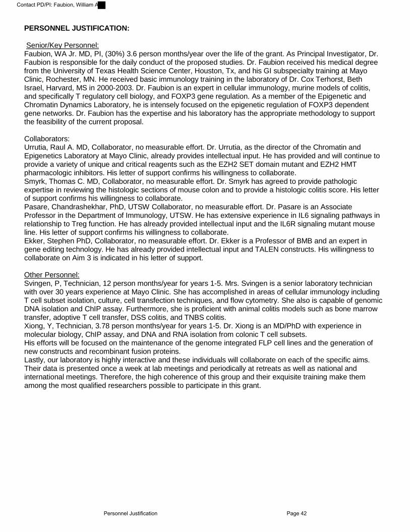
PERSONNEL JUSTIFICATION:
Senior/Key Personnel: Faubion, WA Jr. MD, PI, (30%) 3.6 person months/year over the life of the grant. As Principal Investigator, Dr. Faubion is responsible for the daily conduct of the proposed studies. Dr. Faubion received his medical degree from the University of Texas Health Science Center, Houston, Tx, and his GI subspecialty training at Mayo Clinic, Rochester, MN. He received basic immunology training in the laboratory of Dr. Cox Terhorst, Beth Israel, Harvard, MS in 2000-2003. Dr. Faubion is an expert in cellular immunology, murine models of colitis, and specifically T regulatory cell biology, and FOXP3 gene regulation. As a member of the Epigenetic and Chromatin Dynamics Laboratory, he is intensely focused on the epigenetic regulation of FOXP3 dependent gene networks. Dr. Faubion has the expertise and his laboratory has the appropriate methodology to support the feasibility of the current proposal.
Collaborators: Urrutia, Raul A. MD, Collaborator, no measurable effort. Dr. Urrutia, as the director of the Chromatin and Epigenetics Laboratory at Mayo Clinic, already provides intellectual input. He has provided and will continue to provide a variety of unique and critical reagents such as the EZH2 SET domain mutant and EZH2 HMT pharmacologic inhibitors. His letter of support confirms his willingness to collaborate. Smyrk, Thomas C. MD, Collaborator, no measurable effort. Dr. Smyrk has agreed to provide pathologic expertise in reviewing the histologic sections of mouse colon and to provide a histologic colitis score. His letter of support confirms his willingness to collaborate. Pasare, Chandrashekhar, PhD, UTSW Collaborator, no measurable effort. Dr. Pasare is an Associate Professor in the Department of Immunology, UTSW. He has extensive experience in IL6 signaling pathways in relationship to Treg function. He has already provided intellectual input and the IL6R signaling mutant mouse line. His letter of support confirms his willingness to collaborate. Ekker, Stephen PhD, Collaborator, no measurable effort. Dr. Ekker is a Professor of BMB and an expert in gene editing technology. He has already provided intellectual input and TALEN constructs. His willingness to collaborate on Aim 3 is indicated in his letter of support.
Other Personnel: Svingen, P, Technician, 12 person months/year for years 1-5. Mrs. Svingen is a senior laboratory technician with over 30 years experience at Mayo Clinic. She has accomplished in areas of cellular immunology including T cell subset isolation, culture, cell transfection techniques, and flow cytometry. She also is capable of genomic DNA isolation and ChIP assay. Furthermore, she is proficient with animal colitis models such as bone marrow transfer, adoptive T cell transfer, DSS colitis, and TNBS colitis. Xiong, Y, Technician, 3.78 person months/year for years 1-5. Dr. Xiong is an MD/PhD with experience in molecular biology, ChIP assay, and DNA and RNA isolation from colonic T cell subsets. His efforts will be focused on the maintenance of the genome integrated FLP cell lines and the generation of new constructs and recombinant fusion proteins. Lastly, our laboratory is highly interactive and these individuals will collaborate on each of the specific aims. Their data is presented once a week at lab meetings and periodically at retreats as well as national and international meetings. Therefore, the high coherence of this group and their exquisite training make them among the most qualified researchers possible to participate in this grant.
Personnel Justification Page 42
Contact PD/PI: Faubion, William A
PHS 398 Research PlanPlease attach applicable sections of the research plan, below. OMB Number: 0925-0001
Tracking Number: GRANT11954038 Funding Opportunity Number: PA-13-302. Received Date:2015-07-02T14:04:51.000-04:00
1. Introduction to Application(for RESUBMISSION or REVISION only)
Introduction_Final.pdf
2. Specific Aims HYPOTHESIS_AND_SPECIFIC_AIMS.pdf
3. Research Strategy* Research_Plan.pdf
4. Progress Report Publication List Publication_List3.pdf
Human Subjects Sections5. Protection of Human Subjects
6. Inclusion of Women and Minorities
7. Inclusion of Children
Other Research Plan Sections8. Vertebrate Animals Vertebrate_Animals.pdf
9. Select Agent Research
10. Multiple PD/PI Leadership Plan
11. Consortium/Contractual Arrangements
12. Letters of Support letters_combined.pdf
13. Resource Sharing Plan(s) Resource_Sharing_Plan.pdf
Appendix (if applicable)14. Appendix
Page 43
Contact PD/PI: Faubion, William A

INTRODUCTION TO APPLICATION We appreciate the highly laudatory review on 2/19/2015 of our proposal (21st percentile). The constructive critique has resulted in the addition of extensive new preliminary data and the development of new reagents and animal models, which have improved the proposal in novelty, relevance, feasibility, design, and methodology. Below, we address both convergent suggestions as well as individual points not shared by all reviewers (revisions highlighted in yellow in new proposal).
Productivity: Since the last review, 3 additional senior authored manuscripts have been published totaling 10 peer reviewed publications generated through the experiments defined in the first four years of this award. Regarding the concern that “this new proposal is a major departure from the directions proposed in the previous proposal,” we have provided in detail (progress report) the continuum of logic flowing from the previous aims (KLF binding to DNA, coupling to epigenetic machinery, and with relevance to colitis) to the current hypothesis. In fact, this grant is focused on the epigenetic machinery (EZH2) identified in previous Aim3 and the relevance to Treg biology and colitis.
Key preliminary data: There was consensus among reviewers on the need for additional data. (i) It was noted that, “Although the colitis T cell transfer model is primarily used in the proposal, the preliminary results use the DSS model.” We have developed a new tamoxifen-inducible CRE-EZH2 KO animal and demonstrated a requirement for EZH2 in FOXP3+ cells for prevention of Rbhi mediated colitis, a critical control for the in vivo mutational analysis (Aim3). (ii) We were queried whether the “FOXP3dEZH2 mice show spontaneous colitis?” We have yet to age mice beyond 8 weeks; yet we show the susceptibility to subclinical (1%DSS) colitis in new experiments. (iii) More data in support of A2 was required, as “The model that EZH2 phosphorylation … is critical in disrupting Treg cell function is highly speculative.” We have developed an experimental pipeline that consists of mass spectrophotometry, linear motif analyses, molecular modeling, mutational screens, and functional assays to definitively test the hypothesis of Aim 2. First, mass spectrophotometry identified phosphorylated residues predicted by molecular modeling to regulate EZH2 HMT activity. Extensive linear motif analysis predicted 6 such residues to be regulated by MAPK and Jak kinases. We generated 12 mutant EZH2 constructs and a CRISPR/Cas9 EZH2 mutant cell line in which to test the effect of phosphorylated residues on EZH2 HMT activity. Exemplary of this pipeline, we discovered phosphorylation of Y641 by Jak2 to regulate EZH2 activity. As we were queried in several ways for the effect of IL10 in this mechanism, we have incorporated IL10 into the tightly focused dissection of the critical inflammatory pathways leading to reversible modifications of EZH2 HMT activity. Individual points not shared by all reviewers and not addressed above: Reviewer 1 (R1) noted previous data exists regarding IL6 disruption of Treg function. Indeed, IL6 dependent T effector cell resistance to Treg suppression is well reported (Pasare and Medzhitov 2003, Viglietta, Baecher-Allan et al. 2004, Haas, Hug et al. 2005, Venken, Hellings et al. 2006); however purported mechanisms involve intrinsic T effector cell pathways, not Treg cell biology (Schenten, Nish et al. 2014, Bhela, Kempsell et al. 2015). The conversion of FOXP3+ cells to TH17 cells downstream of IL6 signaling has been reported (Komatsu, Okamoto et al. 2014); yet again, the mechanism(s) leading to Treg cellular conversion is unknown, represents a critical gap in knowledge, and addressed in this proposal. R1 also requested a broader description of outcome variables to assess in vivo studies and a discussion regarding the role for EZH2 in Stat3 activity. Both are provided in the revised proposal. Reviewer 2 (R2) queried “whether EZH2 may repress cytokines in other lymphocytes,” and that we “examine cell surface molecules and cytokines, such as IL-10, … to determine whether they are also affected by EZH2 blockade.” We demonstrate flow analysis of surface molecules and the results of multiplex ELISA for cytokine production. Furthermore, we have explored a role for EZH2 in T effector cells in alternative approaches. R2 noted A3 to be “wide ranging with superficial discussion of pitfalls and alternative approaches.” We have streamlined the aim by exclusion of the neonatal transfer experiment (feasibility was questioned) and have greatly enhanced the Pitfalls and Alternative Approaches. Summary: In response to this excellent critique, we have provided evidence of continued productivity, preliminary data from three new major lines of experimentation, developed two new mouse models, and addressed each review point by point with modifications to the proposal.
Introduction Page 44
Contact PD/PI: Faubion, William A

HYPOTHESIS AND SPECIFIC AIMS: The transcription factor FOXP3 is critical to the regulation of numerous debilitating human
immune-mediated diseases, the prevalence of which together affect over 8.5 million people (1 in 31 U.S. residents). In Inflammatory Bowel Disease (IBD) chronic intestinal inflammation indicates aberrant in vivo FOXP3+ T regulatory (Treg) cell function (1). Similarly, proinflammatory signals in vitro impair Treg function (2). Our lab was the first to characterize the essential role for the histone methyltransferase (HMT) EZH2 in the epigenetic regulation of FOXP3 (3). Recent published work extended our observations indicating a key role for EZH2 in FOXP3 repressor function (4); however the regulation and biological impact of the FOXP3-EZH2 pathway to IBD is unknown. This knowledge is important given the apparent loss of function of Treg cells in inflammation.
Our long-term goal is to dissect epigenetic mechanisms regulating Treg cellular differentiation and function, particularly within the setting of GI inflammatory diseases; as these discoveries will facilitate design of human cell therapy trials for IBD. Consequently, the objective of this grant is to characterize the role for the epigenetic regulator EZH2 in Treg suppressive function. These investigations are strongly supported by preliminary data demonstrating that: 1) EZH2 is required for Treg suppressive function; 2) IL6 signaling leads to phosphorylation and inhibition of EZH2; 3) lymphocytes isolated from the intestine of IBD patients demonstrate activation of IL6-induced gene networks and loss of EZH2 HMT function; and 4) conditional knockout of EZH2 in FOXP3+ T cells leads to in vivo immune dysfunction. Based upon these compelling data we propose the CENTRAL HYPOTHESIS that EZH2 plays a critical role in the homeostasis of Treg cells, and the disruption of EZH2 function by inflammatory signaling pathways contributes to IBD. Our rationale is that identification of the mechanism(s) to restore Treg suppressive function in the setting of intestinal inflammation will offer new therapeutic opportunities within the field of IBD. Our specific aims will test the following hypotheses:
Aim 1: Repression of immunoregulatory gene networks by FOXP3 requires the formation of a complex between this transcription factor and EZH2.
Aim 2: Inflammatory stimuli, such as IL6 lead to EZH2 phosphorylation and thereby disrupt the enzymatic activity of this epigenomic regulator.
Aim 3: Inhibition of the IL6 to EZH2 signaling pathway permits sustained Treg suppressive function in the setting of intestinal inflammation.
Upon conclusion, we will understand the role for EZH2 in Treg loss of function in the setting of active inflammation. This discovery will stimulate new areas for experimental therapeutics in human chronic inflammatory diseases. Our environment in the Epigenetic and Chromatin Dynamics Laboratory combined with the Department of Immunology at the Mayo Clinic makes us uniquely qualified to pursue this objective given the extensive collective experience of histone methyltransferase biology,
proinflammatory signaling networks, and FOXP3 gene regulation.! !
!"#$%& '()*&
!"#$%&!"#$%&!"#$%&!"#$%&'()*&
+,-,&.,/0,1123-&
!"#$%&
!"#$%&
'()*&
$&
42-51,&
+,-,&6789583-&
:;<&12=-5>2-=&
:-?5@@583-&
'()*&
$&
!&
!&
!!!!42-51,&
A0,5B@,-B&
!!62@&C&
62@&*&
62@&%&
+,-,&6789583-&
Figure 1: Conceptual framework. Through the mechanistic experiments designed in the following aims we will identify the role for FOXP3 in the recruitment of EZH2 to core target genes required for Treg function (Aim 1). We will define the signaling network responsible for phosphorylation of EZH2 and disrupted HMT function (Aim 2). Finally, we will perform pre-clinical studies of innovative therapy designed to generate Treg cells resistant to disruptive modifications in the setting of inflammation (Aim 3).
Specific Aims Page 45
Contact PD/PI: Faubion, William A

RESEARCH STRATEGY Significance: FOXP3 is a transcription factor critical to the development, maintenance, and
function of Treg cells. FOXP3 repression of key target gene networks is required for maintenance of Treg function (4,5). Key observations performed during the last funding cycle of this grant led us to identify for the first time a key role for EZH2 in the regulation of FOXP3 expression (3). The significance of this discovery is evident in the fact that subsequently three groups have confirmed and extended our observations on the import of EZH2 in the differentiation and maintenance of Treg cells (4,6,7). Yet, actionable mechanistic insight into 1) the FOXP3 EZH2 protein complex, 2) inflammatory signaling regulating EZH2 activity, and 3) the potential for EZH2-based therapy in IBD represents a critical gap in knowledge addressed in this application.
The canonical function of EZH2 is trimethylation of lysine 27 (K27) of histone 3 (H3) (H3K27me3). This “histone mark” leads to chromatin inaccessibility and gene silencing (8). EZH2 undergoes post-translational modifications downstream of metabolic, cell proliferative, and inflammatory signaling pathways (9-13). We have now discovered that proinflammatory pathways lead to EZH2 phosphorylation and inhibit EZH2 histone methyltransferase (HMT) activity in primary lymphocytes. This discovery is significant, as inflammatory signals can disrupt Treg function (2,14-16), which is apparent within the inflamed environment of IBD and characterized by the co-expression of the proinflammatory cytokines IFNγ and IL17 (17,18). However, little still is known about the mechanism(s) of inflammation-induced disruption of Treg function. Thus, the current study will characterize the role for inflammation-induced modification of EZH2, the inhibition of HMT activity, and the subsequent disruption of Treg suppressive function. This contribution is significant since it will establish that several pathways targeted by available therapies (i.e. IL1β, IL6, TNFα) have the potential to regulate EZH2 HMT activity through post-translational modifications. Furthermore, current Treg cell therapy trials (19), while promising have not addressed the key issue of in vivo inflammation-induced disruption of Treg function. This proposal seeks to address this knowledge gap.
IBD, increasing in prevalence, represents a major national cost measured by both patient suffering and economic burden (as evidenced by over 600 original studies on cost of IBD). Despite significant advances in care (i.e. biologic therapeutics), clinical trial data demonstrate remission rates at best of 40%; thus there remains an enormous unmet medical need. The clinical significance of our study will be the opening of a new avenue in therapeutics directed at stimulation of autologous Treg cells to function within the inflammatory milieu. Our results will impact not only IBD, but also human immune mediated disease broadly.
Innovation: Previous work on epigenetic mechanisms in Treg function has largely focused on
genome wide associations using ChIP-Seq methodology (4). The proposed research is innovative because we investigate the effect of inflammatory signaling pathways on epigenetic complexes in Treg cells, a heretofore-unexamined process. Our experimental approach dives more deeply, biochemically and biologically into the mechanistic impact of this pathway beyond the simple catalog of gene targets provided by the previous ChIP-Seq methodology. Insight into epigenetic mechanisms is impactful as T cell progenitor cells inherit the parent transcriptional profile and unlike genetic change, they are modifiable by currently available therapy. In fact, anti-EZH2 drugs are currently being tested in clinical trials and knowing the role of this HMT in T cells will become imperative as they become widely available in the clinical setting. Conceptual innovations include: 1) the novel role for proinflammatory signaling networks in the regulation of the HMT EZH2 pathway in lymphocytes and IBD; 2) the discovery of the required partnership between FOXP3 and EZH2 in Treg function; and 3) ex vivo genetic engineering of Treg cells to function in the setting of inflammation. Methodologically, there are four new animal models used in this application including the conditional EZH2 KO mouse line, two inducible EZH2 KO mouse lines, and the conditional IL6Rα knockout mouse line. The in vivo experimental techniques utilized in this proposal, particularly the cell transfer experiments utilizing adenoviral replacement of EZH2 and mutant constructs are state-of-the-art. Additional state-of-the-art methodology is provided by the use of CAS/CRISPR mutant cell lines and proteomic experiments, molecular mechanics and molecular dynamic techniques for studying the effects of post-translational modifications on the EZH2 protein, which combined are highly innovative in the field of immunology. In summary, the hypotheses guiding the proposal are novel and will be tested using a comprehensive approach with sophisticated methodology also giving rise to reagents, including new cell and animal models which will be widely shared with the scientific community.
Research Strategy Page 46
Contact PD/PI: Faubion, William A

Approach: PROGRESS REPORT General Statement: The beginning and ending dates of the current period of funding are
2/15/2011-1/31/2016. In this grant’s previous cycle, 10 relevant original articles have been published. Progress Towards Prior Specific Aims: Published Results. Our original hypothesis was that KLF
family members, through coupling with chromatin modifying complexes regulate FOXP3 expression, and ultimately T reg cell function and colitis. Our most salient accomplishments during the prior funding period relevant to our prior three Specific Aims are provided below. The resulting publications are listed within the specified section of the application (see progress report publication list).
Prior Aim #1: KLF10 expression in T cells mediates a novel mechanism of resistance to colitis. As KLF10 is regulated by TGFβ and required for inducible FOXP3+ Treg cell (iTreg) development, we postulated that KLF10 is required in T cells to prevent immune mediated disease. KLF10 deficient animals are susceptible to colitis (#1 publication list) (3). Colitis susceptibility is transferred with the bone marrow indicating the requisite role for KLF10 in immune cells to prevent colitis (#2 publication list) (20). In lymphocytes, KLF10 regulates both FOXP3 and TGFβRII transcription generating a positive feedback loop in Treg development (3,21) (#3 publication list). This regulatory loop is relevant in murine models of colitis and encephalitis (20,21). Thus, KLF10 required for TGFβ induction of iTreg cells, functions in a positive feedback loop between TGFβ signaling and the TGFβRII promoter and is critical to immune regulatory function.
Prior Aim #2: KLF10 selectively regulates distinct genomic control regions of Foxp3. Because suspected KLF binding sites exist within the FOXP3 promoter locus, we postulated that distinct KLF binding domains regulate FOXP3 transcription. Using genome integrated cell lines, mutant mouse lines, and primary human cells, we identified novel KLF binding sites in both the core promoter and key enhancer domains. KLF10 binding to the core promoter recruits Sin3-HDAC or the HAT PCAF to repress or activate FOXP3 gene transcription, respectively (#1 and 2 publication list) (3,20). KLF14 binding to an enhancer domain represses FOXP3 through the heterochromatin protein 1 gene-silencing pathway (22)(#4 publication list), a relationship found to be generalizable to other model systems (#5 publication list) (23). FOXP3 domain functions discovered in the conduct of this aim led to the functional characterization of a private mutation causing familial IBD (#6 publication list) (24) and innovative methodology to quantify in vivo human Treg kinetics (#7 publication list) (25). Collectively, these observations identify distinct genomic control regions of FOXP3 through which KLF family members regulate FOXP3 transcription.
Prior Aim #3: KLF10 works via distinct chromatin remodeling required for Foxp3 activation. Because KLF family members function to couple DNA binding to chromatin remodeling complexes, we examined the potential for KLF10 to regulate FOXP3 in an epigenetic fashion. We observed that KLF10 binding to the core promoter blocks recruitment of the HMT, EZH2 thus preventing gene repression (#1 publication list) (3). We discovered two structurally and functionally distinct EZH2 HMTs; and both are capable of permanently silencing FOXP3 (#8 publication list) (26). Thus, KLF10 alternatively represses or activates FOXP3 through coupling with Sin3 or PCAF, and in the absence of KLF10, FOXP3 is permanently silenced by EZH2. In summary, we have successfully completed work on our previous specific aims demonstrating fidelity to the proposal. Additionally, our work into TGFβ signaling upstream of the FOXP3 promoter locus resulted in novel insights into hedgehog signaling pathways (#9 publication list) (27) and the function of an epithelial derived stem cell in colitis (28) (#10 publication list). In addition, by lending critical expertise gained from the funded studies, we were able to contribute to additional collaborative publications, further attesting to the success of our program during the past funding cycle (29-34).
This grant proposal represents the continuum of logic flowing from the previous aims (KLF binding to DNA and coupling to epigenetic machinery to regulate colitis) to the current hypothesis. In the process of characterizing the role for EZH2 isoforms at the FOXP3 promoter locus (previous Aim3) we discovered a profound role for EZH2 in FOXP3 repressor function in both murine and human T regulatory cells. This critical and modifiable function within Treg cells in the setting of IBD is the subject of this grant proposal.
Research Strategy Page 47
Contact PD/PI: Faubion, William A

Aim 1: Repression of immunoregulatory gene networks by FOXP3 requires the formation of a complex between this transcription factor and EZH2.
Introduction: FOXP3 represses gene networks (4); however the mechanism is not well established. The objective of this aim is to define the role for EZH2 in FOXP3-mediated gene repression. We will test the working hypothesis that EZH2 is recruited to target genes in complex with FOXP3 (Figure 2). Our approach will be domain mutational analyses of EZH2 and FOXP3. We will test the function of these mutations by both molecular (target gene repression) and cellular (Treg suppressor function) assays. The rationale for this aim is that understanding the mechanism of FOXP3 repressor function will lead to therapy to enhance Treg cell function in vivo. Our
expectation is to identify the protein domains within EZH2 and FOXP3 required to repress target genes and enhance Treg function. This knowledge is important because EZH2 modifying agents (EPZ-6438 and GSK-126) are currently in clinical trials for lymphoma; thus insights derived from this grant into in vivo Treg function are significant and impactful.
Justification, feasibility and preliminary data: We have identified a critical role for EZH2 HMT
activity in mediating Treg suppressor function. We have generated a conditional, functional knockout of EZH2 through deletion of the enzymatically active SET domain under the control of the FOXP3 promoter (FOXP3$EZH2). By 35 days, 80% of FOXP3$EZH2 animals succumb to a lethal inflammatory disorder similar to the scurfy mouse characterized by intense inflammatory infiltrate of multiple organs (survival curve, n=107 total mice, Figure 3A). FOXP3$EZH2 animals surviving to 8 weeks exhibit significantly increased susceptibility to colitis upon exposure to subclinical (1%) DSS
challenge (histology, n=16 total mice, Figure 3B, C). The histologic activity index represents blinded scoring for 9 criteria as previously published (28). Note lamina propria expansion, goblet cell loss, crypt destruction, inflammatory cell infiltration and ulcer in severely affected FOXP3$EZH2 mice (Figure 3C). FOXP3$EZH2 lymphocytes develop at expected frequency, and upon isolation, the cells express the typical cell surface repertoire of regulatory proteins (Figure 4); however they lack IL10 and produce proinflammatory cytokines including significant upregulation of IFN! and IL17 (Figure 5A). FOXP3$EZH2 cells function poorly in vitro (Figure 5B). Furthermore, we discovered a direct interaction between EZH2 and FOXP3 by proximity ligation assay (PLA) in the nucleus of WT Treg cells (Figure 6). Thus, we conclude that FOXP3 and EZH2 co-localize in the nucleus and that Treg specific EZH2 HMT activity is required for normal immune homeostasis and in vitro FOXP3+ Treg function. This discovery is significant as the regulation of this complex through cell signaling networks will be an important modifiable mechanism to target for the treatment of immunological disorders.
!"#$%& '()*&
!"#$%&'()*&
+,+&-./01234.53&67&'()*&36&!"#$%&3809.3&+,! :64825&;138<65=&*,! >6?@$&%,! $06A2423B&C298<65&
D==8B&&
+,*&'()*?&3809.3&9.5.&0.E0.==265&+,! -.E603.0&D==8B&*,! >F@$&D==8B&%,! $>-&
+,%&-.G120.4.53&67&'()*&0./01234.53&760&H0.9&715/<65&+,! @5&I2306&=1EE0.==265&8==8B&*,! @5&I2I6&8==8B&67&715/<65&
J.5.&-.E0.==265&
Aim 1 Schema: Our model to be tested is that EZH2, FOXP3 complex formation leads to recruitment of EZH2 to target genes, resultant H3K27 trimethylation, gene silencing, and appropriate facilitation of Treg cell function. Domain mutations of both EZH2 and FOXP3 will test the requirement of complex creation (Co-IP and PLA) for H3K73me3 (ChIP), gene repression (reporter assay and PCR), and Treg function (in vitro/vivo assays).
Figure 2 (AIM1): Our model to be tested is that EZH2, FOXP3 complex formation leads to recruitment of EZH2 to target genes, resultant H3K27 trimethylation, gene silencing, and appropriate facilitation of Treg cell function.!!!
Figure 3: FOXP3!EZH2 mouse. (A) Survival curve of WT (black), Heterozygous (blue), and Homozygous (green) littermates for EZH2 deletion. (B, C) Colitis index and representative histology demonstrating severe colitis in 8 week old FOXP3!EZH2 mice in response to subclinical (1%) DSS challenge (n=16 total mice).
0 50 1000
50
100
Days
Per
cent
sur
viva
l
Survival proportions
WT (n=51)
EZH2-/- (n=27) EZH2+/- (n=31)
Total=27
MaleFemale
Total=31
MaleFemale
WT
EZH2KO
0
2
4
6
8
His
tolo
gic
Inde
x
*p<0.05!" #" $"
Research Strategy Page 48
Contact PD/PI: Faubion, William A

Research Design: The general strategy of Aim 1 is to map interaction domains (1.1) and test
domain function through assays of gene repression (1.2) and Treg cell suppressor function (1.3). The laboratory has published experience with co-immunoprecipitation (Co-IP) assays, luciferase reporter systems, ChIP assay, in vitro suppression assays, and in vivo colitis models (3,20,26). Overexpression of mutant constructs will be performed in cell lines by nucleofection and primary cells by adenoviral transduction. In our published experience, nucleofection efficiency in cell lines approximates 20% and adenoviral transduction efficiency is greater than 75%.
1.1 Biochemical approach to define the FOXP3-EZH2 complex
We will determine the interacting domains between EZH2 and FOXP3 using Co-IP with validation by PLA. We will transfect FLAG-EZH2 and HIS-FOXP3 domain mutants in primary cells and T cell lines. The deletion mutations (Figure 7) were selected on the basis of protein interacting domains suggesting direct EZH2, FOXP3 interaction (35-37). Results will be validated by PLA in primary Treg cells (Figure 6). In this assay, antibodies directed to either EZH2 or FOXP3 are tagged with oligonucleotide probes that upon
binding in proximity (30-40nm) hybridize to form circular DNA strands (38). DNA circles serve as template to amplify fluorescent probes indicating proximity. We expect EZH2 to complex with FOXP3 in a direct fashion, and the PLA results support this expectation. We expect to identify the specific domains relevant to this
interaction. As both EZH2 and FOXP3 have additional functions independent of each other, only with knowledge of precise interaction domains can we separate the biological effects of FOXP3-associated EZH2 function from EZH2 function broadly in lymphocytes.
Figure 6: FOXP3 and EZH2 co-localize in primary Treg cells. Proximity ligation assay showing direct interaction of EZH2 and FOXP3 (red punctate staining) in FOXP3+ cells (green nuclei). No hybridization was evident upon addition of ligation mix, probe, and control Ab.!
Figure 7: N and C terminal mutant constructs for FOXP3 (green) and EZH2 (blue) proteins. Regions of interest are the Zinc finger and forkhead (FH) domains of FOXP3. Disease inducing variants (human IPEX) are present in the FH domain. Similarly, the WD-binding domain and the enzymatic SET domain are shown in EZH2. Lymphoma associated variants are demonstrated. !
!"#$% !&'()*% +!,-%
.+&/%!"01% !"20%
Figure 4: Cell surface staining of FOXP3!EZH2 Treg cells (blue histogram) demonstrates upregulation of established immunoregulatory proteins. Marked upregulation over WT Treg cells (red histogram) suggests an activated Treg phenotype. Green histogram represents IgG control (representative of 3 independent experiments).
Tresp WT Treg EZH2 KO Treg0
2,000
4,000
6,000
8,000
Thym
idin
e C
ount
s
p<0.05
WT IFNg
EZH2_KO IF
Ng
WTIL17
EZH2_KO IL
17
WT IL10
EZH2_KO IL
100
200
400
600
800
pg/m
l
*
**
!" #"
Figure 5: FOXP3!EZH2 Treg cells express pro-inflammatory cytokines and function poorly in vitro. (A) ELISA of supernatants of FOXP3+ cells. (B) Proliferation assay shows that FOXP3!EZH2 Treg cells (closed histogram) fail to suppress WT T responder (WT Tresp, 1:1 ratio) when compared to WT Treg cells (*p<0.05, n=6 mice).
Research Strategy Page 49
Contact PD/PI: Faubion, William A

1.2 The functional relevance of EZH2 in target gene repression. We have performed RNA-Seq using CD4+ cells isolated from resection specimens in patients with active Crohn’s disease and ileal
biopsies of healthy age and gender matched control subjects. We analyzed existing ChIP-Seq datasets and have discovered 260 overlapping targets of FOXP3 and EZH2 (39-41). We then compared gene expression of these overlapping targets between samples from patients with CD and controls. From this broad dataset we have chosen RORA (RAR Orphan Receptor A) and IRF4 (Interferon Regulatory Factor-4) as models to study the functional relevance of EZH2 in Treg cells. RORA and IRF4 are established targets of EZH2 (42,43) and both are significantly upregulated in our Crohn’s disease sample (Figure 8). Moreover, both RORA and IRF4 are key transcription factors in the
development of Th17 cells, an immune subset responsible for human and murine colitis (44-49). Thus, we will use transcriptional regulation of these genes to test in vitro functional relevance of EZH2 and EZH2, FOXP3 complex formation using reporter assays (1.21) and ChIP assay (1.22).
1.21: Luciferase reporter assay: We will use luciferase reporter assays of the RORA and IRF4
promoters in T cell lines. We will nucleofect EZH2 with titrated quantity of FOXP3 into reporter cell lines. We will determine the relevance of FOXP3 to the function of EZH2 through quantitative luciferase assay. We will perform identical assays utilizing EZH2 and FOXP3 domain mutants (Figure 7) to test the requirement of EZH2 and FOXP3 physical interaction for repressor function.
1.22: ChIP assay: The mechanism of EZH2-mediated silencing is H3K27 trimethylation
(H3K27me3) and chromatin compaction at target promoter loci. The presence and function of EZH2 at a particular promoter locus can be assayed using Chromatin Immunoprecipitation (ChIP) for the H3K27me3 histone “mark.” Utilizing FOXP3$EZH2 Treg cells, we will transduce WT or EZH2 mutant constructs and perform ChIP assay of the RORA and IRF4 promoter for the presence of H3K27me3 marks, EZH2, and FOXP3. This animal line has been crossed to a mutant transgenic mouse line expressing the coxsackie adenovirus receptor (CAR) to allow efficient gene transduction in resting lymphocytes (26). Essential reagents include FOXP3$EZH2 mouse colony and adenoviral WT or EZH2 mutant constructs (Figure 7). Our laboratory has published experience in ChIP and adenoviral transduction (1,20,26). We expect WT EZH2 but not mutants predicted to disrupt FOXP3-mediated recruitment to repress RORA and IRF4 through deposition of H3K27me3 histone marks.
1.3 The in vitro and in vivo functional relevance of EZH2 target gene repression. Finally, we will test the significance of EZH2 target gene repression on Treg suppressor function
in vitro (1.31) and in vivo (1.32). 1.31: The in vitro assay consists of co-culture of naïve FOXP3 CD4+ T cells (T responder cells)
with titrated amounts of Treg cells in the presence of conditions to stimulate T responder cell proliferation (anti-CD3, anti-CD28) (1). We will transduce FOXP3$EZH2 Treg cells with WT EZH2 and EZH2 mutant constructs (Figure 7) to test dependence of in vitro suppressor function upon EZH2 and FOXP3 complex formation.
1.32 In vivo colitis assay: We will test in vivo function of FOXP3!EZH2
Treg cells in colitis prevention. We will utilize the CD45Rbhi transfer model of
Figure 8: RORA and IRF4 are significantly upregulated in CD4+ lymphocytes isolated from Crohn’s lesions. The histograms represent reads per kilobase of transcript per million reads mapped (RPKM) for RORA and IRF4 in RNA-Seq analysis of CD4+ lymphocytes isolated from Crohn’s disease resection specimens or ileal biopsies of healthy age, sex match control subjects (n=12 subjects).
p<0.001 p<0.001
Figure 9: FOXP3!EZH2 Treg cells do not suppress Rbhi mediated colitis. Recipient mice (n=4) of mutant Treg cells lost more weight (A, black histogram) and demonstrated severe colitis (B, representative histology from Rbhi transfer) when compared to recipients of WT Treg cells (n=4). Note goblet cell loss, lamina propria expansion and intense inflammatory cell infiltration.
WT
EZH2 KO
0
10
20
30
Perc
ent W
eigh
t Los
s
*p<0.05
!" #"
Research Strategy Page 50
Contact PD/PI: Faubion, William A

colitis, a model in which we have extensive experience, mimics well the phenotype of human IBD, and in which preclinical trials have translated well to human therapy (50-52). For the in vivo model, 350,000 naïve CD4+ lymphocytes are transferred into immunodeficient RAGnull mice alone, or along with 100,000 FOXP3+ Treg cells. Our experience with this system is well published (52-55). We have established that FOXP3!EZH2 Treg cells do not prevent colitis upon co-transfer with WT naïve CD4+ cells (Figure 9). We have developed a new model of inducible EZH2 deletion through a tamoxifen-inducible CRE-mediated recombination system driven by the chicken beta actin promoter/enhancer. Upon cross with the EZH2 flox mouse, EZH2 deletion can be obtained in any mouse cell upon treatment with tamoxifen. We harvested Treg cells from this mutant animal (and WT control), and co-transferred mutant (or WT) Treg cells along with WT CD45Rbhi cells into RAGnull mice. All animals were treated with a 5-day tamoxifen injection protocol. At 6 weeks, RAGnull mice injected with mutant Treg cells exhibited extreme weight loss and colitis while RAGnull mice treated with control Treg cells appeared healthy (Figure 9). We will now transduce WT EZH2 or mutant constructs into FOXP3!EZH2
Treg cells to test the requirement for EZH2 to associate with FOXP3 for the prevention of murine IBD. Colitis severity will be assessed by disease activity index (DAI), blinded histology assessment, cytokine expression profile of T cells isolated from mesenteric lymph nodes, and cytokine assessment of colonic supernatants (28). We expect adenoviral delivery of the WT EZH2 to rescue in vivo function of FOXP3!EZH2 Treg cells. Furthermore, we expect that EZH2 domain mutants, disrupted in their capacity to complex with FOXP3 will not prevent colitis. This finding would indicate the critical nature of this complex to prevent inflammation.
Anticipated Results, potential pitfalls, and alternative approaches: The objective of this aim
is to define the role for EZH2 in FOXP3-induced gene repression and subsequent Treg function. We expect EZH2 to complex with FOXP3 in a direct fashion (1.1). We expect WT EZH2 to repress RORA and IRF4, but only in the presence of FOXP3 (1.2). We expect the functional relevance of the EZH2 and FOXP3 interaction to be in vitro suppression of T responder cells and colitis prevention (1.3). These exciting experiments will be the first to test a modifiable epigenetic mechanism in the treatment of IBD.
The first alternative hypothesis considered is an indirect rather than direct EZH2, FOXP3 interaction through obligate EZH2 binding partners EED and Suz12 (35,36). To directly test this possibility we could perform siRNA knockdown of EED and Suz12, and repeat Co-IP assay for WT EZH2 and FOXP3. A second alternative hypothesis is that rather than a requirement for FOXP3, EZH2 utilizes additional mechanisms for target gene localization. Non coding RNAs are increasingly recognized to directly bind with chromatin modifying complexes and play a role in altered gene expression in many tissues and particularly cancer (56). Should EZH2 not require FOXP3 for target gene localization we will perform cross-linking and immunoprecipitation methodology combined with RNAseq to identify EZH2-interacting RNA. Functional knockdown of long noncoding RNA could reveal this potential function.
Aim 2: Inflammatory stimuli, such as IL6 lead to EZH2 phosphorylation and thereby disrupt the
enzymatic activity of this epigenomic regulator. !!
Figure 10 (AIM2): Our model is that a kinase (JAK2) downstream of an inflammatory signal (IL6) modifies EZH2 (Y641 phosphorylation) resulting in impaired HMT activity at FOXP3 target genes (RORA, IRF4) and subsequent Treg cell dysfunction. We will use mutant mouse/cell lines and kinase inhibition to identify the mechanism(s) of EZH2 phosphorylation (by Mass Spec, MS and WB) and resultant HMT function (HMT assay and in vitro/vivo assays of suppressor function).
!"#$%&
!"#$%&
'()&*+,-./012&34&567'&8-318-39:;.<3/&*=>&?@&439&$A567'B&)(! *CD./D&23C1+&;0/+1&'(! E0/.1+&0/-0F0<3/B&
•! $-.92.,3;3G0,&•! 10HIJ&•! K320/./D&I+G.<L+&
'(%&!C/,<3/.;&,3/1+MC+/,+&34&567'A$&!"! #$%&'()*%1C889+1103/&.11.:&+"! #$%&'&*%.11.:&34&4C/,<3/&
567'&
$&
E0/.1+& '('&$-318-39:;.<3/A0/NC,+N&N019C8<3/&34&7*O&4C/,<3/&5PQ=JAF.1+N&7*O&.11.:B&)(! *CD./D&23C1+&;0/+1&'(! E0/.1+&0/-0F0<3/&%(! *CD.<3/.;&./.;:101&34&567'&&
R+/+&J,<L.<3/&
Aim 2 Schema: Our model to be tested is that a kinase (Akt) downstream of an inflammatory signal (IL6) modifies EZH2 (S21 phosphorylation) resulting in impaired histone methyltransferase (HMT) activity at FOXP3 target genes (RORA) and subsequent Treg cell dysfunction. We will use mutant mouse lines and kinase inhibition to identify the mechanism(s) of EZH2 phosphorylation (by western blot, WB) and resultant HMT function (HMT assay and in vitro/vivo assays of suppressor function).
Research Strategy Page 51
Contact PD/PI: Faubion, William A
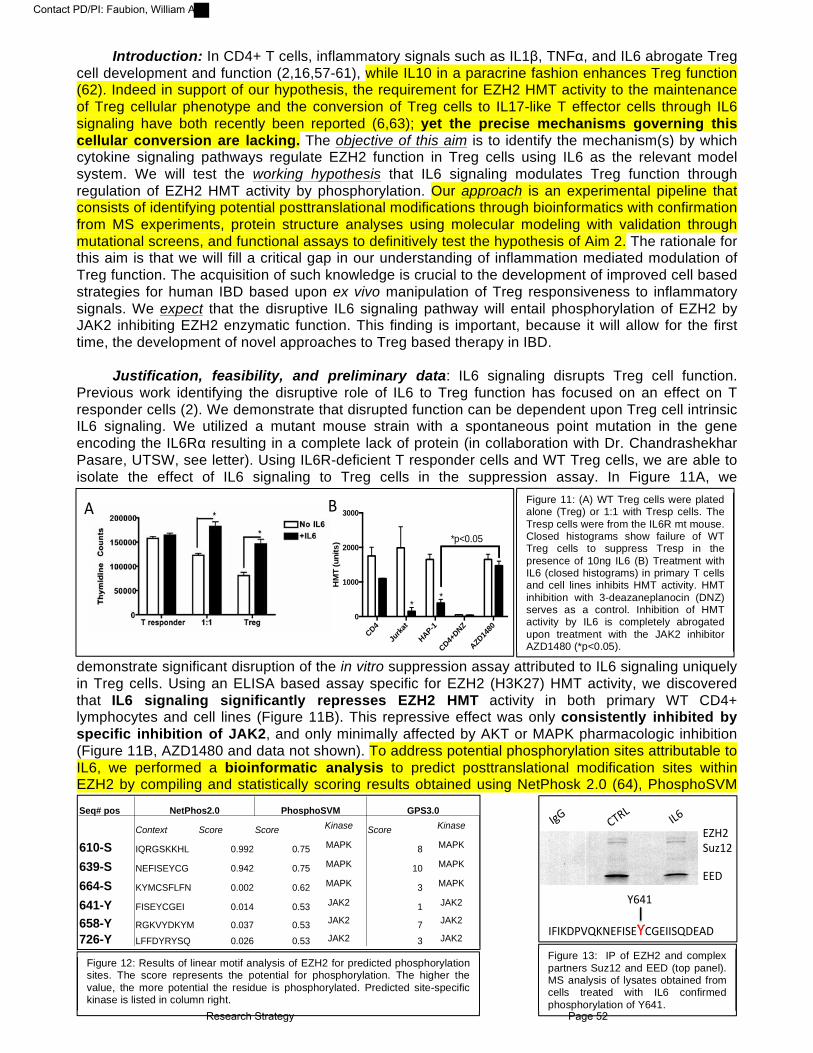
Introduction: In CD4+ T cells, inflammatory signals such as IL1", TNF#, and IL6 abrogate Treg cell development and function (2,16,57-61), while IL10 in a paracrine fashion enhances Treg function (62). Indeed in support of our hypothesis, the requirement for EZH2 HMT activity to the maintenance of Treg cellular phenotype and the conversion of Treg cells to IL17-like T effector cells through IL6 signaling have both recently been reported (6,63); yet the precise mechanisms governing this cellular conversion are lacking. The objective of this aim is to identify the mechanism(s) by which cytokine signaling pathways regulate EZH2 function in Treg cells using IL6 as the relevant model system. We will test the working hypothesis that IL6 signaling modulates Treg function through regulation of EZH2 HMT activity by phosphorylation. Our approach is an experimental pipeline that consists of identifying potential posttranslational modifications through bioinformatics with confirmation from MS experiments, protein structure analyses using molecular modeling with validation through mutational screens, and functional assays to definitively test the hypothesis of Aim 2. The rationale for this aim is that we will fill a critical gap in our understanding of inflammation mediated modulation of Treg function. The acquisition of such knowledge is crucial to the development of improved cell based strategies for human IBD based upon ex vivo manipulation of Treg responsiveness to inflammatory signals. We expect that the disruptive IL6 signaling pathway will entail phosphorylation of EZH2 by JAK2 inhibiting EZH2 enzymatic function. This finding is important, because it will allow for the first time, the development of novel approaches to Treg based therapy in IBD.
Justification, feasibility, and preliminary data: IL6 signaling disrupts Treg cell function.
Previous work identifying the disruptive role of IL6 to Treg function has focused on an effect on T responder cells (2). We demonstrate that disrupted function can be dependent upon Treg cell intrinsic IL6 signaling. We utilized a mutant mouse strain with a spontaneous point mutation in the gene encoding the IL6R# resulting in a complete lack of protein (in collaboration with Dr. Chandrashekhar Pasare, UTSW, see letter). Using IL6R-deficient T responder cells and WT Treg cells, we are able to isolate the effect of IL6 signaling to Treg cells in the suppression assay. In Figure 11A, we
demonstrate significant disruption of the in vitro suppression assay attributed to IL6 signaling uniquely in Treg cells. Using an ELISA based assay specific for EZH2 (H3K27) HMT activity, we discovered that IL6 signaling significantly represses EZH2 HMT activity in both primary WT CD4+ lymphocytes and cell lines (Figure 11B). This repressive effect was only consistently inhibited by specific inhibition of JAK2, and only minimally affected by AKT or MAPK pharmacologic inhibition (Figure 11B, AZD1480 and data not shown). To address potential phosphorylation sites attributable to IL6, we performed a bioinformatic analysis to predict posttranslational modification sites within EZH2 by compiling and statistically scoring results obtained using NetPhosk 2.0 (64), PhosphoSVM
Figure 11: (A) WT Treg cells were plated alone (Treg) or 1:1 with Tresp cells. The Tresp cells were from the IL6R mt mouse. Closed histograms show failure of WT Treg cells to suppress Tresp in the presence of 10ng IL6 (B) Treatment with IL6 (closed histograms) in primary T cells and cell lines inhibits HMT activity. HMT inhibition with 3-deazaneplanocin (DNZ) serves as a control. Inhibition of HMT activity by IL6 is completely abrogated upon treatment with the JAK2 inhibitor AZD1480 (*p<0.05).
CD4
Jurka
t
HAP-1
CD4+DNZ
AZD14
800
1000
2000
3000
HM
T (u
nits
)
**
*p<0.05
!" #"
Seq# pos NetPhos2.0 PhosphoSVM GPS3.0
Context Score Score Kinase Score Kinase
610-S IQRGSKKHL 0.992 0.75 MAPK 8 MAPK
639-S NEFISEYCG 0.942 0.75 MAPK 10 MAPK
664-S KYMCSFLFN 0.002 0.62 MAPK 3 MAPK
641-Y FISEYCGEI 0.014 0.53 JAK2 1 JAK2
658-Y RGKVYDKYM 0.037 0.53 JAK2 7 JAK2
726-Y LFFDYRYSQ 0.026 0.53 JAK2 3 JAK2
!"#$%&'()$%
!!*%
+,-%
./01%
+12%
+3+4*56748!3+&!9.-!++&7*!:*%
92;)%
9Figure 13: IP of EZH2 and complex partners Suz12 and EED (top panel). MS analysis of lysates obtained from cells treated with IL6 confirmed phosphorylation of Y641.
Figure 12: Results of linear motif analysis of EZH2 for predicted phosphorylation sites. The score represents the potential for phosphorylation. The higher the value, the more potential the residue is phosphorylated. Predicted site-specific kinase is listed in column right. Research Strategy Page 52
Contact PD/PI: Faubion, William A

(65), and GPS 3.0 (66). We focused on putative phosphorylation sites within the enzymatically active SET domain (AA 605-729), as they should have clear impact on the function of this HMT. The demonstrated motifs are predicted MAPK and JAK2 phosphorylation sites (Figure 12) and confirmed by MS to be phosphorylated upon pull down of native EZH2 (Figure 13). One of the key residues modified by phosphorylation is Y641, which mutations have previously shown to alter human B cell function (67). We gained useful insight into how Y641 phosphorylation may affect the activity of EZH2 through molecular modeling. Briefly, we developed a homology-based model using the SET domain of EZH2, which had been previously crystallized in the absence of both the SAM cofactors and the target histone peptide. These two molecules, which are key for the full HMT activity were obtained by homology superimposition with the SET domain of EHMT1/G9a and SETD2; for both are known to have an EZH2-like enzymatic mechanism (60). Figure 14 demonstrates the modeled
structure of the EZH2 methyltransferase domain. Note Y641 engages in a hydrogen bond with the de-protonated $-Nitrogen of the dimethylated lysine that will be tri-methylated, (dash line and yellow arrow, Figure 14 left panel). Using in silico mutagenesis, we investigated the potential effects of phosphorylation, by using the
well-characterized phosphomimetics (Y to E) and
non-phosphorylatable mutations (Y to F) methodology. We find that the Y641E mutation disrupts the
aforementioned hydrogen bond (Figure 14, right panel). Combined these studies suggest that phosphorylation of this Y residue may regulate the histone methyltransferase activity of EZH2. We next performed biochemical experiments to test the validity of these data. We developed a novel system based on using a CRISPR/CAS-mediated EZH2KO cell in which the enzymatic activity of transfected WT EZH2 was directly compared with both the phosphomimetic (Y641E) and nonphosphorylated (Y641F) Y641 mutants. As shown in Figure 15, the phosphomimetic mutant Y641E is inactive when compared to WT or the non-phosphorylatable Y641F mutant. Thus, given this strong preliminary data, in Aim 2 we will confirm the key modifications relevant to cytokine signaling and test the functional relevance of the biochemical assays through cellular assays and gene expression.
Research Design: The strategy of Aim 2 is to identify functionally relevant post-translational modifications of EZH2
downstream of IL6 through a mass spectrophotometry screen (2.1) followed by a point mutational analysis consisting of functional assays of enzymatic activity (2.2), gene regulation and Treg suppressor function (2.3). We use Y641 as a powerful example of feasibility and significance; however we will systematically assess all 6 phosphorylation sites of the SET domain identified by the mass spectrophotometry screen.
2.1 Establish the mechanism of modifications to the EZH2 SET domain. The preliminary data (bioinformatic analysis and pharmacologic inhibitor screens) are supportive
of the substantial role for JAK2 in regulation of EZH2 enzymatic activity. To confirm this mechanism, we will stimulate primary Treg cells and cell lines (Jurkat and Hap-1) with IL6 (10ng/ml) in the
Figure 14: Molecular Modeling of EZH2 SET domain (blue), SAM cofactor (green), and the dimethylated K27 residue of histone protein (yellow). Note Y641E mutation, right.
CTRL WTY64
1EY64
1F0
50
100
150
OD
/hr/m
g
* p<0.05
Figure 15: Using ELISA, we show reduction in HMT activity upon mutation of Y641 to the phosphomimetic, Y641E.
Research Strategy Page 53
Contact PD/PI: Faubion, William A

presence of JAK2 modulation and measure phosphorylation of the EZH2 SET domain by mass spectrophotometry and western blot. The rationale for both outcome assessments is the enhanced data acquisition of MS balanced with the speed and low cost of western blot analysis. We have developed and optimized a rabbit polyclonal Phospho-Y641 antibody.
We will inhibit JAK2 in three ways: pharmacologic JAK2 inhibition (AZD1480), JAK2 siRNA treatment, and overexpression of a JAK2 dominant negative construct (Y792F) (68). Constitutively active JAK2 V617F (69) will be used as an important gain-of-function control. We will use primary cells from our recently developed FOXP3-specific, IL6R conditional KO (FOXP3ΔIL6Rα) as a relevant negative control. We expect IL6 to lead to phosphorylation of EZH2 and subsequent impairment of HMT activity through activation of JAK2. Furthermore, we expect the FOXP3ΔIL6Rα Treg cells to be resistant to EZH2 phosphorylation.
2.2 Test the functional significance of EZH2 SET domain phosphorylation. IL6 signaling leads to both EZH2 phosphorylation and reduced HMT activity in our system
(Figure 11 and 13); thus we will now establish causality of impairment of EZH2 HMT activity to EZH2 phosphorylation. The outcome variable for the subsequent experimental set is IL6-induced impairment of HMT activity in nuclear extracts of test cells. We have generated a library of EZH2 mutant constructs designed to mimic (S to D; Y to E) or prevent (S to A; Y to F) phosphorylation. We have established an ideal in vitro cell system in which to screen the HMT activity of EZH2 mutant constructs. Utilizing a haploid fibroblast cell line (HAP-1) (70) in which the CAS9 system has induced a frameshift mutation in exon 7 of human EZH2, we demonstrate significant reduction of HMT activity in the Y641E mutant (mimic phosphorylation) when compared to WT EZH2 control (Figure 15). We have produced our first candidate mutant (Y641E) adenovirus, which will be used to confirm functional relevance in primary T cells.
HMT assay will be run using nuclear extracts from FOXP3ΔEZH2 Treg cells upon adenoviral transduction of either WT-EZH2 or mutant EZH2. We expect that Y641F (unable to be phosphorylated and widely noted to be hyperactive in human lymphoproliferative conditions) will function well in the presence of IL6 as measured by HMT activity. Conversely, we anticipate, the Y641E mutant to have an impaired HMT activity when compared to the WT construct due to the phosphomimetic properties of the glutamic acid substitution. Our team has extensive experience in mutational analyses to identify modifiable residues (20,71).
2.3 Establish the functional relevance of SET P-Y641-EZH2 within the whole cell system. IL6 stimulation reduces HMT activity in nuclear extracts of primary lymphocytes (Figure 11B).
Thus in this subaim we will extend our preliminary findings to whole cell assays of functional relevance including: 2.31) luciferase reporter and 2.32) in vitro suppression assays.
2.31: Luciferase reporter assays using genomically integrated T cell lines. As we have done
previously, we generated genomically integrated T cell lines to study EZH2-dependent events in a non-episomal system (3,20). Using the Jurkat FLP system we created two reporter cell lines with the RORA and IRF4 promoters genomically integrated in frame with a luciferase reporter. RORA and IRF4 are ideal targets as both are established targets of EZH2 (42,43) and upregulated in CD (Figure 8). Using this system we will overexpress WT EZH2 or EZH2 Y641 mutants to test the dependence of IL6 induction of RORA/IRF4 on EZH2 Y641 phosphorylation.
2.32: In vitro suppression assays. We will test the capacity of WT and mutant EZH2 constructs
to rescue in vitro suppressor function in the setting of IL6. Utilizing adenoviral transduction of FOXP3ΔEZH2 Treg cells, we will introduce WT or EZH2 Y641 mutant constructs and perform suppression assays in the presence or absence of IL6. As EZH2 Y641E mimics phosphorylation downstream of IL6 signaling, we expect the Y641E mutant to be a loss of function mutation, even without IL6 stimulation. Loss of function would be evident in the de-repression of luciferase expression and the inability to suppress T responder cell proliferation. We expect the EZH2 Y641F mutant (unable to be phosphorylated) to be a gain of function mutation, evident in the presence of IL6. Gain of function in this context will be demonstrated through the repression of luciferase and retained capacity to suppress in the setting of IL6 stimulation.
Research Strategy Page 54
Contact PD/PI: Faubion, William A
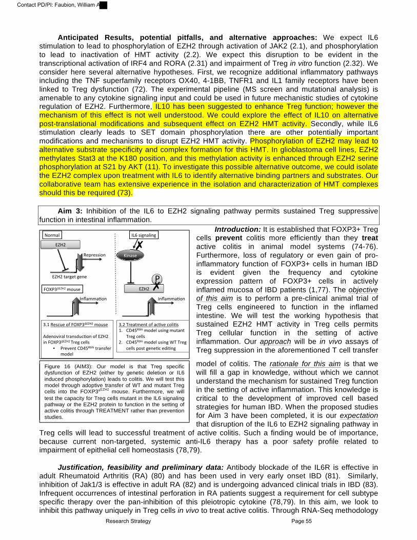
Anticipated Results, potential pitfalls, and alternative approaches: We expect IL6
stimulation to lead to phosphorylation of EZH2 through activation of JAK2 (2.1), and phosphorylation to lead to inactivation of HMT activity (2.2). We expect this disruption to be evident in the transcriptional activation of IRF4 and RORA (2.31) and impairment of Treg in vitro function (2.32). We consider here several alternative hypotheses. First, we recognize additional inflammatory pathways including the TNF superfamily receptors OX40, 4-1BB, TNFR1 and IL1 family receptors have been linked to Treg dysfunction (72). The experimental pipeline (MS screen and mutational analysis) is amenable to any cytokine signaling input and could be used in future mechanistic studies of cytokine regulation of EZH2. Furthermore, IL10 has been suggested to enhance Treg function; however the mechanism of this effect is not well understood. We could explore the effect of IL10 on alternative post-translational modifications and subsequent effect on EZH2 HMT activity. Secondly, while IL6 stimulation clearly leads to SET domain phosphorylation there are other potentially important modifications and mechanisms to disrupt EZH2 HMT activity. Phosphorylation of EZH2 may lead to alternative substrate specificity and complex formation for this HMT. In glioblastoma cell lines, EZH2 methylates Stat3 at the K180 position, and this methylation activity is enhanced through EZH2 serine phosphorylation at S21 by AKT (11). To investigate this possible alternative outcome, we could isolate the EZH2 complex upon treatment with IL6 to identify alternative binding partners and substrates. Our collaborative team has extensive experience in the isolation and characterization of HMT complexes should this be required (73).
Aim 3: Inhibition of the IL6 to EZH2 signaling pathway permits sustained Treg suppressive
function in intestinal inflammation. Introduction: It is established that FOXP3+ Treg
cells prevent colitis more efficiently than they treat active colitis in animal model systems (74-76). Furthermore, loss of regulatory or even gain of pro-inflammatory function of FOXP3+ cells in human IBD is evident given the frequency and cytokine expression pattern of FOXP3+ cells in actively inflamed mucosa of IBD patients (1,77). The objective of this aim is to perform a pre-clinical animal trial of Treg cells engineered to function in the inflamed intestine. We will test the working hypothesis that sustained EZH2 HMT activity in Treg cells permits Treg cellular function in the setting of active inflammation. Our approach will be in vivo assays of Treg suppression in the aforementioned T cell transfer
model of colitis. The rationale for this aim is that we will fill a gap in knowledge, without which we cannot understand the mechanism for sustained Treg function in the setting of active inflammation. This knowledge is critical to the development of improved cell based strategies for human IBD. When the proposed studies for Aim 3 have been completed, it is our expectation that disruption of the IL6 to EZH2 signaling pathway in
Treg cells will lead to successful treatment of active colitis. Such a finding would be of importance, because current non-targeted, systemic anti-IL6 therapy has a poor safety profile related to impairment of epithelial cell homeostasis (78,79).
Justification, feasibility and preliminary data: Antibody blockade of the IL6R is effective in
adult Rheumatoid Arthritis (RA) (80) and has been used in very early onset IBD (81). Similarly, inhibition of Jak1/3 is effective in adult RA (82) and is undergoing advanced clinical trials in IBD (83). Infrequent occurrences of intestinal perforation in RA patients suggest a requirement for cell subtype specific therapy over the pan-inhibition of this pleiotropic cytokine (78,79). In this aim, we look to inhibit this pathway uniquely in Treg cells in vivo to treat active colitis. Through RNA-Seq methodology
Figure 16 (AIM3): Our model is that Treg specific dysfunction of EZH2 (either by genetic deletion or IL6 induced phosphorylation) leads to colitis. We will test this model through adoptive transfer of WT and mutant Treg cells into the FOXP3ΔEZH2 mouse. Furthermore, we will test the capacity for Treg cells mutant in the IL6 signaling pathway or the EZH2 protein to function in the setting of active colitis through!TREATMENT rather than prevention studies.!!!!
!"#$%&'
()*+'
,-.#-//0"1'
()*+'2%#3-2'3-1-'
401%/-'
567',-/89-'":';<=>5?()*+'$"9/-''@A-1"B0#%&'2#%1/A98C"1'":'()*+'01';<=>5?()*+'D#-3'8-&&/'
•! >#-B-12'EFGH,IJ0'2#%1/:-#'$"A-&'
56+'D#-%2$-12'":'%8CB-'8"&0C/'76! EFGH,IJ0'$"A-&'9/013'$92%12'
D#-3'8-&&/'+6! EFGH,IJ0'$"A-&'9/013'KD'D#-3'
8-&&/'."/2'3-1-C8'-A0C13'
;<=>5?()*+'$"9/-'
L1M%$$%C"1'
LNO'/031%&013'
L1M%$$%C"1'
()*+'
>'
!'
!'
!!
Research Strategy Page 55
Contact PD/PI: Faubion, William A

using CD4+ T cells isolated from Crohn’s lesions, we have identified marked upregulation of IL6 induced signaling networks (Figure 17A). IL6 signaling leads to phosphorylation of EZH2 and impaired HMT activity. Thus, it is important to note significant variation in the expression of EZH2 targets between health and disease in the same cells (Figure 17B). Furthermore, the significant majority (379/457 target genes) is upregulated, a finding suggestive of impaired EZH2 HMT function (Figure 17B). Congruent with these observations, microarray data obtained from FOXP3$EZH2 Treg cells demonstrate marked upregulation of
proinflammatory pathways regulated by IL6 signaling (Figure 17C). Finally, we demonstrate that inhibition of EZH2 (DZNep 1mg/kg ip) HMT activity significantly exacerbates DSS colitis (Figure 17D, E). Thus, we have observed IL6 to inhibit EZH2, a protein that itself is a negative regulator of IL6-induced proinflammatory pathways relevant to human IBD. Moreover, pharmacologic inhibition of EZH2 (mimicking the IL6 effect) worsens DSS induced colitis. We now propose to treat active colitis through genetically engineered Treg cells. Our model system will be the CD45Rbhi transfer model of colitis (discussed in Aim 1 and demonstrated in Figure 9), and Treg cell therapy will be applied to treat rather than prevent active inflammation. We have recently utilized this precise methodology to demonstrate the enhanced function of murine Treg cells engineered to maintain an active FOXP3 promoter locus in the inflammatory setting (84). Focusing now on the IL6 to EZH2 pathway, we will perform identical experiments (genetic manipulation of Treg cells and co-transfer in the Rbhi mediated model of colitis) to utilize Treg cellular therapy in active colitis.
Research Design: The general strategy of Aim 3 is to utilize adoptive transfer systems and genetically engineered
Treg cells to demonstrate the capacity for Treg cells to function in the inflamed intestine and treat active colitis. We will use the naïve T cell into RAGnull colitis model as our model system, and we will genetically engineer Treg cells using adenoviral transduction (3.1) and TALEN methodology (3.2). Our efficient use of adenoviral constructs has been addressed above, and our genetic editing will be performed in collaboration with the Genetics and Model Systems Core with the Center for GI Signaling, Mayo Clinic (Dr. Stephen Ekker, see letter of collaboration).
3.1 Treatment of active colitis with EZH2 mutant cell lines. We have demonstrated loss of Treg suppressor function in vitro upon treatment of co-culture
assays with IL6. We now will test the effect of therapy directed to this pathway through in vivo assays of Treg function. We will use the IL6R"-deficient mice as the donor of Treg cells. We will harvest Treg cells from the FOXP3$IL6R" mouse line and test the in vivo regulatory capacity of these cells to treat
Figure 17: EZH2 controls IL6 inflammatory pathways. (A) Heat map of RNAseq of CD4+ cells isolated from Crohn’s lesions. The heat map is the IL6 pathway and shows marked variance between control subjects (CTR) and disease (Dz). (B) Volcano plot of relative expression of EZH2 targets comparing DZ to CTR. Red dots show significant variance. (C) Heat map of microarray from FOXP3!EZH2 Treg cells vs control. Red cells show upregulation of genes. Note (arrow) genes of the Th17 and Th1 pathway. (D and E) 2% DSS colitis in WT mice treated with the EZH2 inhibitor DZNep (1mg/kg) at Day 2 and 5. Colon length and DAI show severe colitis upon EZH2 inhibition (n=8).
!"#$
!"#$%&'()*+',&
!"#$%&'()*+,%
!""#$%&'()*+,'%-%./%012-*3#"4.5*"%067/*0(-8-**.5%9,:)9,);*<=;>;$3#5**?-;>#%3#[email protected]@.5*"5*.#,838-%.5*%A?"B;$C,):$37A:A&,";.5*D.5*E;C,85*F,#8*.5*D"*+G/%.5B";05"4HIA."-DJ7A*C;85
-% .% /%
0% !%
Research Strategy Page 56
Contact PD/PI: Faubion, William A

colitis. As per our published data (84), we will inject titrations of 100-300,000 cells of WT or IL6R" mutant Treg cells into recipient RAGnull animals with established colitis (4 weeks post T effector cell transfer). We expect IL6R" mutant cells to robustly treat colitis, as compared to WT Treg cells. We will rescue FOXP3$EZH2 Treg cells with EZH2 WT, Y641E, or Y641F mutant constructs. We expect the EZH2 Y641F (gain of function) but not the EZH2 Y641E (loss of function) to effectively treat established colitis.
3.2 Treatment of active colitis with genetically edited cells. Finally, we will test ex vivo cellular therapy with clear translational potential to human IBD. Our
department of Biochemistry and Molecular Biology has extensive experience with transcription activator-like effector nucleases (TALENs) and has recently published successful genetic editing of primary T cells (85,86). We will isolate WT Treg cells from C57/BL6 mice and perform genetic editing/deletion of IL6R" using TALENs (see letter of collaboration). As per previous study, 20 mcg of TALEN construct (left arm and right arm) and pEGFP-N1 (Clontech, Mountain View, CA, USA) DNAs will be transiently nucleofected by electroporation into primary Treg cells (RFP+). Forty-eight hours after nucleofection, GFP-expressing cells will be selected by fluorescence-activated cell sorting. Deletion of the IL6R" will be confirmed by flow cytometry. The TALEN edited IL6R" null cells will be expanded using Treg ex vivo cell expansion methods we have optimized (87), and injected into recipient RAGnull animals with established colitis as above. We expect genetic editing ex vivo of WT Treg cells to enhance suppressor function in the setting of active colitis, and this finding to thus provide a clear roadmap for translational cell therapy studies in patients with IBD.
Anticipated Results, potential pitfalls, and alternative approaches: We expect in 3.1 robust
treatment of active colitis with FOXP3+ Treg cells impaired in IL6R to EZH2 signaling either at the level of the receptor (IL6R" KO Treg cells) or EZH2 (EZH2 Y641F mutant) when compared to WT Treg cells. We expect in 3.2 genetic editing ex vivo of WT Treg cells to enhance suppressor function in the setting of active colitis, and this finding to provide pre-clinical data for translational cell therapy studies in patients with IBD. As the Y641F mutant in whole lymphocyte populations has been associated with lymphoma, this particular mutation uniquely within the Treg subset will require extensive analysis. To test the long-term behavior of mutant Treg cells, we could perform rescue experiments of the scurfy mouse (no functional Treg cells) to study function and toxicity. A second alternative hypothesis is that impaired Treg function in the setting of inflammation results not from intrinsic Treg dysfunction but the resistance to Treg suppressive mechanisms by T effector cells. This hypothesis is readily testable using our current reagents through the treatment of colitis induced by IL6R" mutant T effector cells with WT Treg cells. We can now dissect the role for EZH2 in any cell population using tamoxifen-inducible Cre-ER(T) mutant mouse under the promotional control of actin. A third alternative hypothesis is that EZH1, not EZH2 is the primary target of IL6 signaling in Treg cells. Beyond initiation of the H3K27me3 mark (EZH2-containing Polycomb Repressor Complex 2 function), maintenance of the mark by the EZH1 HMT may well be involved in maintained Treg function. Given the strength of the preliminary data and the phenotype of the conditional EZH2 KO mouse, we have necessarily chosen to focus on EZH2; however our laboratory and collaborative partners have the necessary tools and experience to dissect additional HMT pathways should these investigations become necessary.
Conclusion: We are studying an innovative membrane to nucleus signaling pathway connecting
environmental inflammatory signals (IL6R) to cell differentiation machinery (EZH2) responsible for Treg cell fate and function. Our work has clear biomedical relevance to patients with IBD. Moreover, our work represents a continuum beginning with basic mechanisms of kinase regulation of EZH2 (this proposal) to the ex vivo manipulation of Treg cells for cell therapy of IBD. The work is significant as it should lead to first-in-man studies of engineered Treg cells in human IBD.
Aims Year 1 Year 2 Year 3 Year 4 Year 5 SA1 SA2 SA3
Year 1 Year 2 Year 3
Research Strategy Page 57
Contact PD/PI: Faubion, William A

PROGRESS REPORT PUBLICATION LIST: AI89714 Competitive Renewal Publication List: Original research publications emanating directly from experiments designed in previous aims:
1. Xiong, Y., Khanna, S., Grzenda, A.L., Sarmento, O.F., Svingen, P.A., Lomberk, G.A., Urrutia, R.A.,
Faubion, W.A., Jr. (2012). Polycomb antagonizes p300/CREB-binding protein-associated factor to silence FOXP3 in a Kruppel-like factor-dependent manner. Journal of Biological Chemisry, 287(41):34372-85. PMCID: PMC3464543. (AIM1 and AIM3)
2. Xiong, Y., Svingen, P.A., Sarmento, O.O., Smyrk, T.C., Dave, M., Khanna, S., Lomberk, G.A., Urrutia,
R.A., Faubion, W.A., Jr .(2014) Differential coupling of KLF10 to Sin3-HDAC and PCAF regulates the inducibility of the FOXP3 gene. American Journal of Physiology Regulatory, Integrative and Comparative Physiology, 307(6):R608-20. PMCID: PMC4166759 (AIM1)
3. Papadakis, K.A., Krempski, J., Reiter, J., Svingen, P., Xiong, Y., Sarmento, O.F., Huseby, A., Johnson,
A.J., Lomberk, G.A., Urrutia, R.A., Faubion, W.A.. (2015) Krüppel-like factor KLF10 regulates transforming growth factor receptor II expression and TGF-β signaling in CD8+ T lymphocytes, American Journal of Physiology Cell Physiology, 308(5):C362-71. PMCID: PMC4346734
4. Sarmento, O.F., Svingen, P.A., Xiong, Y., Xavier, R.J., McGovern, D., Smyrk, T.C., Papadakis, K.A.,
Urrutia, R.A., Faubion, W.A..(2015). A novel role for KLF14 in T regulatory cell differentiation. Cellular and Molecular Gastroenterology and Hepatology, 1(2):188-202. PMCID: PMC4349492.
5. Mathison, A., Grzenda, A., Lomberk, G., Velez, G., Buttar, N., Tietz, P., Hendrickson, H., Liebl, A.,
Xiong, Y.Y., Gores, G., Fernandez-Zapico, M., Larusso, N.F., Faubion, W. , Shah, V.H., Urrutia, R.. (2013) Role for Kruppel-like transcription factor 11 in mesenchymal cell function and fibrosis. PLoS One. 8(9):e75311. PMCID: PMC3775729. (AIM2)
6. Okou, D.T., Mondal, K., Faubion, W.A. , Kobrynski, L.J., Denson, L.A., Mulle, J.G., Ramachandran, D.,
Xiong, Y.,Svingen, P., Patel, V., Bose, P., Waters, J.P., Prahalad, S., Cutler, D.J., Zwick, M.E., Kugathasan, S. (2014) Exome Sequencing Identifies a Novel FOXP3 Mutation in a 2-Generation Family With Inflammatory Bowel Disease. Journal of Pediatric Gastroenterology and Nutrition, 58(5):561-8. PMCID: 4277865. (AIM2)
7. Rahman, M.K., Offord, C.P., Khanna, S., Ford, G.C., Perrson, X.M., Svingen, P.A., Xiong, Y., Bajzer,
Z., Faubion, W.A. .(2013) Regulatory T cell kinetics in the peripheral blood of patients with Crohn's disease. Human Immunology, 74(2):145-50. PMCID: PMC4224946 (AIM2)
8. Grzenda, A., Lomberk, G., Svingen, P., Mathison, A., Calvo, E., Iovanna, J., Xiong, Y., Faubion, W. ,
Urrutia, R. (2013) Functional characterization of EZH2beta reveals the increased complexity of EZH2 isoforms involved in the regulation of mammalian gene expression. Epigenetics and Chromatin, 6(1):3. PMCID: PMC3606351. (AIM3)
9. Mills, L.D., Zhang, L., Marler, R., Svingen, P., Fernandez-Barrena, M.G., Dave, M., Bamlet, W.,
McWilliams, R.R., Petersen, G.M., Faubion, W, Fernandez-Zapico, M.E.. (2014) Inactivation of the Transcription Factor GLI1 Accelerates Pancreatic Cancer Progression. Journal of Biological Chemistry, 289(23):16516-25. PMCID: PMC4047418. (AIM1 and AIM3)
10. Dave, M., Hayashi, Y., Gajdos, G.B., Smyrk, T.C., Svingen, P.A., Kvasha, S.M., Lorincz, A., Dong, H.,
*Faubion, W.A., Jr, *Ordog, T. (2015) Stem cells for murine interstitial cells of cajal suppress cellular immunity and colitis via prostaglandin e2 secretion. Gastroenterology,148(5):978-90. PMCID: PMC4409492 (* Co-corresponding authors)
Collaborative projects not directly related to prior aims:
1. Li, H., Chan, L., Bartuzi, P., Melton, S.D,, Weber, A., Ben-Shlomo, S., Varol ,C., Raetz, M., Mao, X., Starokadomskyy, P., van Sommeren, S., Mokadem, M., Schneider, H., Weisberg, R., Westra, H.J., Progress Report Publications List Page 58
Contact PD/PI: Faubion, William A

Esko, T., Metspalu, A., Kumar, V., Faubion, W.A., Yarovinsky, F., Hofker, M.,Wijmenga, C., Kracht, M, Franke, L., Aguirre, V., Weersma, R.K., Gluck, N., van de Sluis, B.,Burstein, E.(2014). Copper metabolism domain-containing 1 represses genes that promote inflammation and protects mice from colitis and colitis-associated cancer. Gastroenterology, 147(1):184-195.e3.
Epub 2014 Apr 13. PubMed PMID: 24727021; PubMed Central PMCID: PMC4086320.
2. Kauder, S.E., Santell, L., Mai, E., Wright, L.Y., Luis, E., N'Diaye, E.N., Lutman, J., Ratti, N., Sa, S.M., Maun, H.R., Stefanich, E., Gonzalez, L.C., Graham, R.R., Diehl, L., Faubion, W.A.. Jr., Keir, M.E., Young, J., Chaudhuri, A., Lazarus, R.A., Egen, J.G. (2013). Functional consequences of the macrophage stimulating protein 689C inflammatory bowel disease risk allele. PLoS One, 23;8(12):e83958. PMCID: PMC3884107. 3. Dave, M., Papadakis, K.A., Faubion, W.A., Jr. (2014). Immunology of inflammatory bowel
disease and molecular targets for biologics. Gastroenterology Clinics of North America., 43(3):405-24. PMID:25110250.
4. Faubion, W.A., Jr, Fletcher, J.G., O'Byrne, S., Feagan, B.G., de Villiers, W.J., Salzberg, B., Plevy, S., Proctor, D.D., Valentine, J.F., Higgins, P.D., Harris, J.M., Diehl, L., Wright, L., Tew, G.W., Luca, D., Basu, K., Keir, M.E. (2014). EMerging BiomARKers in Inflammatory Bowel Disease (EMBARK) study identifies fecal calprotectin, serum MMP9, and serum IL-22 as a novel combination of biomarkers for Crohn's disease activity: role of cross-sectional imaging. American Journal of Gastroenterology,108(12):1891-900. Epub 2013 Oct 15. Erratum in: Am J Gastroenterol. 2014 May;109(5):780. PubMed PMID: 24126633. 5. Cayatte, C., Joyce-Shaikh, B., Vega, F., Boniface, K., Grein, J., Murphy, E., Blumenschein, W.M., Chen, S.,Malinao, M.C., Basham, B., Pierce, R.H., Bowman, E.P., McKenzie, B.S., Elson, C.O., Faubion, W.A., Malefyt Rde, W., Kastelein, R.A., Cua, D., McClanahan, T.K., Beaumont, M. (2012). Biomarkers of Therapeutic Response in the IL-23 Pathway in Inflammatory Bowel Disease. Clinical and translational Gastroenterology, ;3:e10. PubMed Central PMCID: PMC3365674.
6. van Driel, B,, Liao, G., Romero, X., O'Keeffe, M.S., Wang, G., Faubion, W.A., Berger, S.B., Magelky, E.M., Manocha, M., Azcutia, V., Grisham, M., Luscinskas, F.W., Mizoguchi, E., de Waal Malefyt, R., Reinecker, H.C., Bhan, A.K., Wang, N., Trehorst, C. (2012) Signaling lymphocyte activation molecule regulates development of colitis in mice. Gastroenterology, 143(6):1544- 1554.e7. PubMed Central: PMCID: PMC3578298.
Progress Report Publications List Page 59
Contact PD/PI: Faubion, William A

1
VERTEBRATE ANIMALS: The mice used in this proposal will be from protocols approved by the Mayo Clinic Institutional Animal Care and Use Committee (IACUC). Mayo Clinic assures strict compliance with all federal regulations and guidelines involving the use of laboratory animals in biomedical research. Animal use assurances have been filed with the IACUC and the NIH. These procedures are closely adhered to and monitored within the institution. All protocols presently being conducted and those proposed, regardless of funding sources, are reviewed and must be approved before their initiation in accordance with these assurance statements. All protocols are active for 3 years after the approval date and adhere to Mayo Clinic and NIH policies on the humane care of animals. All animals will be maintained in an Association for Assessment and Accreditation of Laboratory Animal Care–accredited and centralized facility located in the Guggenheim Building.
1. Provide a detailed description of the use of animals in the research. Identify species, strains, ages, sex, and numbers of animals to be used.
This proposal will use the following mouse lines: 1. FOXP3ΔEZH2 2. B6129S-Tg(Foxp3-EGFP/cre)1aJbs/J 3. Foxp3tm9(EGFP/cre/ERT2)Ayr/J 4. FOXP3ΔIL6R 5. C57/BL6CAR 6. C57BL/6-Foxp3tm1Flv/J (FOXP3 RFP mouse) 7. B6.129S7-Rag1tm1Mom/J 8. C57/BL6 EZH2flox/flox
These animals are currently being bred in our lab under IACUC# A31013. Below we iterate the animal requirements by experimental protocol. Total anticipated animal requirements are listed below each protocol. 1.1, 2.1, and 2.2: Co-IP, PLA, and HMT assay of WT and mutant constructs nucleofected or transduced into WT CD4+ lymphocytes, IL6Rα mutant Treg cells, and FOXP3ΔEZH2 Treg cells. Each spleen provides 4x10⁶ naïve CD4+ T cells and 1x10⁵ Treg cells. In review of the recent conduct of similar experiments generating preliminary data, our laboratory utilizes approximately 3-5 WT donor mice per week (approximately 150 per year), 2 IL6Rα mutant mice (approximately 100 per year), and 2 FOXP3ΔEZH2 mice (approximately 100 per year). We have performed experiments on both male and female WT mice with similar results thus plan to use both. Age is 4-6 weeks. Total WT=150/yr Total IL6Rα mutant=100/yr Total FOXP3ΔEZH2=100/yr 1.2: ChIP assay post viral transduction of mutant contructs into FOXP3ΔEZH2 mice. We perform ChIP assay on transduced cells approximately 2x/month, or approximately 25 mice per year. Gender and age as above. Total FOXP3ΔEZH2=25/yr 1.3, 2.3, 3.1, and 3.2: In vitro suppression assay and CD45Rbhi transfer model. The in vitro suppression model requires approximately 1x10⁵ Treg cells per assay, performed in triplicate (acquired from approximately 3 spleens). One donor WT animal is all that’s required to provide the T responder cells. Given at least three mutant EZH2 constructs to test and the requirement to perform the experiment in triplicate to maintain scientific integrity, we anticipate using 27 FOXP3ΔEZH2 mice in protocol 1.3 (3 spleens*3 constructs*triplicate=27) and 9 WT mice (1 spleen*3*triplicate=9). Total FOXP3ΔEZH2=27 For the in vivo colitis experiments, one requires 4 B6.129S7-Rag1tm1Mom/J recipients per treatment group. This number of mice was calculated based upon a one-way ANOVA analysis to allow 90% power when the differences between colitis are 1.25 standard deviations more than the controls. Given rescue with at least three constructs to test and the requirement to perform the experiment in triplicate to maintain scientific integrity, we anticipate using 36 RAGnull recipients and 10-15 donor mice for a total of approximately 50 mice in protocol 1.3. Similarly for the active colitis treatment protocol of 3.2, we will rescue RAGnull recipients with IL6R mutant Treg cells or FOXP3ΔEZH2 Treg cells transduced with three EZH2 constructs. Using the same calculations and justification as above, each colitis permutation requires 4 RAGnull recipients, repeated 3 times=12 total RAGnull animals. As protocol 3.2 tests 4 Treg variants, we will require 48 total RAGnull animals. The addition of 10-15 donor mice to the 48 total brings the total anticipated animal requirements to approximately 65 animals. Total RAGnull mice: 84 mice
Vertebrate Animals Page 60
Contact PD/PI: Faubion, William A

2
Total IL6R mutant donor mice: 10 Total FOXP3ΔEZH2 donor mice: 30 Neonatal transfer experiments of 3.1: Litters of mouse pairs predicted by genotype to produce FOXP3ΔEZH2 pups will be injected ip with 1x10⁶ WT Treg cells labeled with RFP (C57BL/6-Foxp3tm1Flv/J donor mice). Experience demonstrates 1-2 appropriate FOXP3ΔEZH2 pups per litter; thus we will need to perform this experiment on 3 litters, requiring 12 FOXP3 RFP donor mice. We will also need to maintain breeding pairs to continuously generate the lines being used in the experiments. The breeding of the FOXP3ΔEZH2 mouse is intense, as CRE+, heterozygous EZH2WT/flox mice need to be bred to the C57/BL6 EZH2flox/flox mouse to produce the appropriate genotype (typically 20% of any litter). This is the primary rationale to the generation of the tamoxifen inducible FOXP3ΔEZH2 mouse line. These adult animals, upon tamoxifen induction of CRE, will serve as donors of EZH2-deficient Treg cells. For every 6 animals used in an experiment, typically 1 breeder pair is required. Thus total mouse requirements by strain: WT=150/yr * 5yr=750 WT breeding=125 IL6mt=100/yr*5=500 IL6mt breeding=83 FOXP3ΔEZH2=100/yr*5=500 FOXP3ΔEZH2 breeding=83 RAGnull=84 RAGnull breeding=14 TOTAL=2139 mice over the 5 year period of the grant (428 mice per year). All newborn animals with the incorrect genotype will be euthanized before they are 3 weeks old, unless they can be used for breeding. The health of the animals will be monitored on a regular basis. Any animals showing signs of excessive stress, pain, or discomfort during the treatment period will be euthanized as described below. At the end of the treatment cycles, colons will be excised and embedded for histological analysis of inflammation.
2. Justify the use and number of animals and choice of species with additional justification if animals are in short supply or are costly or if you plan to use large numbers.
As principal investigator, I have determined, by means of the following sources, searches, or methods, that alternatives to the procedures which may cause animal pain or distress proposed in this protocol are not available and that this protocol does not unnecessarily duplicate previous experiments. Sources searched include: Medline Current Research Information Services (CRIS) Animal Welfare Information Center (AWIC - National Agriculture Library) PUBMED. The key words that were used in the search (e.g., MESH headings): animal model; colitis; FOXP3; IL6; EZH2. We covered the years 1980 – 2014. Mice are required for the above experiments as there is no safe human options or in vitro options. The complex questions regarding the development of Treg cells in the intestinal milieu cannot be answered in vitro. The justification of animal numbers is above with experimental procedures. There is currently no other experimental system that looks at epigenetic changes resulting in colitis prevention that is comparable to the combination of animals brought together in these studies. Mice are needed because the focus of the project is to determine the role of EZH2 in the development of coitis. These studies cannot be performed without a few key animal models with selective gene deletions. We will limit the number of mice required. To maintain breeding of the various animals, as well as longitudinal studies, we estimate it will be necessary to carry approximately 85 cages of mice on a regular basis (5 adult, same gender mice/cage).
3. Provide information on veterinary care for the animals. Animals are housed in a pathogen-free laminar flow facility located in the Mayo Clinic Animal Facility on the 19th and 20th floors of the Guggenheim Research Building. The animal facility has controlled temperature, humidity and lighting. Three full-time veterinarians certified in laboratory animal medicine and veterinary pathology will oversee all aspects of animal care in this facility. Beyond the provision of food, water and
Vertebrate Animals Page 61
Contact PD/PI: Faubion, William A
3
housing (that is inspected daily), no specific veterinary care is anticipated. The animal care facilities and use program meets all federal regulations and guidelines. Mayo Foundation is registered with the USDA (41-R-006) as an animal research facility and maintains an NIH animal assurance statement (A3291-01) with the Office of Laboratory Animal Welfare.
4. Describe procedures for ensuring that discomfort, pain, and injury will be limited to what is unavoidable. Describe the use of analgesic, anesthetic, tranquilizing drugs, and restraining devices to minimize discomfort, distress, pain, and injury.
Mayo Clinic strives to ensure that the institutional facilities and procedures adhere in all respects to USDA regulations and NIH guidelines for care and use of laboratory animals. Investigators are required to administer appropriate analgesics to all animals associated with a procedure that would normally require pain medication in humans. The mice are cared for in a clean, climate-controlled environment, with a controlled light-dark cycle. They are fed and watered daily, and cages cleaned weekly. Experiments are ended at pre-defined time points OR when animals have lost more than 20% of the starting body weight or exhibit a moribund appearance. Animals are weighed and examined by the study team daily while in study. Mice will be maintained in accordance with the applicable portions of the Animal Welfare Act and the DHHS Guide for the Care and Use of Laboratory Animals. During adoptive cell transfer, mice are briefly restrained in a clean, warm plexiglass cylinder to allow safe transfer of cells by tail vein injection. Mice are treated with Tylenol overnight following adoptive transfer.
5. Describe any euthanasia method to be used and the reasons for its selection. State whether this method is consistent with the recommendations of the American Veterinary Medical Association Guidelines on Euthanasia. If not, justify not following the recommendations.
Requests to perform euthanasia are reviewed and approved by our IACUC before initiation. Euthanasia may be performed as required by protocol to terminate a study, or as a means to relieve pain or distress that cannot be alleviated by analgesics, or if deemed necessary by the consulting veterinarian. We utilize CO(2) inhalation for euthanasia for the following reasons. (1) The rapid depressant, analgesic, and anesthetic effects of CO2 are well established. (2) Carbon dioxide is readily available and can be purchased in compressed gas cylinders. (3) Carbon dioxide is inexpensive, nonflammable, nonexplosive, and poses minimal hazard to personnel when used with properly designed equipment. This method is consistent with the recommendations of the Panel on Euthanasia of the American Veterinary Medical Association.
Vertebrate Animals Page 62
Contact PD/PI: Faubion, William A
BIBLIOGRAPHY AND REFERENCES CITED: 1. Rahman, M. K., Midtling, E. H., Svingen, P. A., Xiong, Y., Bell, M. P., Tung, J., Smyrk, T., Egan, L. J.,
and Faubion, W. A., Jr. (2010) The pathogen recognition receptor NOD2 regulates human FOXP3+ T cell survival. J Immunol 184, 7247-7256
2. Pasare, C., and Medzhitov, R. (2003) Toll pathway-dependent blockade of CD4+CD25+ T cell-mediated suppression by dendritic cells. Science 299, 1033-1036
3. Xiong, Y., Khanna, S., Grzenda, A. L., Sarmento, O. F., Svingen, P. A., Lomberk, G. A., Urrutia, R. A., and Faubion, W. A., Jr. (2012) Polycomb antagonizes p300/CREB-binding protein-associated factor to silence FOXP3 in a Kruppel-like factor-dependent manner. J Biol Chem 287, 34372-34385
4. Arvey, A., van der Veeken, J., Samstein, R. M., Feng, Y., Stamatoyannopoulos, J. A., and Rudensky, A. Y. (2014) Inflammation-induced repression of chromatin bound by the transcription factor Foxp3 in regulatory T cells. Nat Immunol 15, 580-587
5. Zheng, Y., Josefowicz, S. Z., Kas, A., Chu, T. T., Gavin, M. A., and Rudensky, A. Y. (2007) Genome-wide analysis of Foxp3 target genes in developing and mature regulatory T cells. Nature 445, 936-940
6. DuPage, M., Chopra, G., Quiros, J., Rosenthal, W. L., Morar, M. M., Holohan, D., Zhang, R., Turka, L., Marson, A., and Bluestone, J. A. (2015) The chromatin-modifying enzyme Ezh2 is critical for the maintenance of regulatory T cell identity after activation. Immunity 42, 227-238
7. Yang, X.-P., Jiang, K., Hirahara, K., Vahedi, G., Afzali, B., Sciume, G., Bonelli, M., Sun, H.-W., Jankovic, D., Kanno, Y., Sartorelli, V., O’Shea, J. J., and Laurence, A. (2015) EZH2 is crucial for both differentiation of regulatory T cells and T effector cell expansion. Sci. Rep. 5
8. Cao, R., Wang, L., Wang, H., Xia, L., Erdjument-Bromage, H., Tempst, P., Jones, R. S., and Zhang, Y. (2002) Role of Histone H3 Lysine 27 Methylation in Polycomb-Group Silencing. Science 298, 1039-1043
9. Cha, T. L., Zhou, B. P., Xia, W., Wu, Y., Yang, C. C., Chen, C. T., Ping, B., Otte, A. P., and Hung, M. C. (2005) Akt-mediated phosphorylation of EZH2 suppresses methylation of lysine 27 in histone H3. Science 310, 306-310
10. Chen, B., Liu, J., Chang, Q., Beezhold, K., Lu, Y., and Chen, F. (2013) JNK and STAT3 signaling pathways converge on Akt-mediated phosphorylation of EZH2 in bronchial epithelial cells induced by arsenic. Cell Cycle 12, 112-121
11. Kim, E., Kim, M., Woo, D. H., Shin, Y., Shin, J., Chang, N., Oh, Y. T., Kim, H., Rheey, J., Nakano, I., Lee, C., Joo, K. M., Rich, J. N., Nam, D. H., and Lee, J. (2013) Phosphorylation of EZH2 activates STAT3 signaling via STAT3 methylation and promotes tumorigenicity of glioblastoma stem-like cells. Cancer Cell 23, 839-852
12. Palacios, D., Mozzetta, C., Consalvi, S., Caretti, G., Saccone, V., Proserpio, V., Marquez, V. E., Valente, S., Mai, A., Forcales, S. V., Sartorelli, V., and Puri, P. L. (2010) TNF/p38alpha/polycomb signaling to Pax7 locus in satellite cells links inflammation to the epigenetic control of muscle regeneration. Cell Stem Cell 7, 455-469
13. Wei, Y., Chen, Y. H., Li, L. Y., Lang, J., Yeh, S. P., Shi, B., Yang, C. C., Yang, J. Y., Lin, C. Y., Lai, C. C., and Hung, M. C. (2011) CDK1-dependent phosphorylation of EZH2 suppresses methylation of H3K27 and promotes osteogenic differentiation of human mesenchymal stem cells. Nat Cell Biol 13, 87-94
14. Raffin, C., Pignon P Fau - Celse, C., Celse C Fau - Debien, E., Debien E Fau - Valmori, D., Valmori D Fau - Ayyoub, M., Ayyoub, M., Kimura, A., Naka T Fau - Kishimoto, T., Kishimoto, T., Valencia, X., Stephens G Fau - Goldbach-Mansky, R., Goldbach-Mansky R Fau - Wilson, M., Wilson M Fau - Shevach, E. M., Shevach Em Fau - Lipsky, P. E., Lipsky, P. E., Pasare, C., and Medzhitov, R. Human memory Helios- FOXP3+ regulatory T cells (Tregs) encompass induced Tregs that express Aiolos and respond to IL-1beta by downregulating their suppressor functions IL-6-dependent and -independent pathways in the development of interleukin 17-producing T helper cells TNF downmodulates the function of human CD4+CD25hi T-regulatory cells Toll pathway-dependent blockade of CD4+CD25+ T cell-mediated suppression by dendritic cells.
15. Kimura, A., Naka, T., and Kishimoto, T. (2007) IL-6-dependent and -independent pathways in the development of interleukin 17-producing T helper cells. Proc Natl Acad Sci U S A 104, 12099-12104
16. Valencia, X., Stephens, G., Goldbach-Mansky, R., Wilson, M., Shevach, E. M., and Lipsky, P. E. (2006) TNF downmodulates the function of human CD4+CD25hi T-regulatory cells. Blood 108, 253-261
References Cited Page 63
Contact PD/PI: Faubion, William A
17. Voo, K. S., Wang, Y. H., Santori, F. R., Boggiano, C., Wang, Y. H., Arima, K., Bover, L., Hanabuchi, S., Khalili, J., Marinova, E., Zheng, B., Littman, D. R., and Liu, Y. J. (2009) Identification of IL-17-producing FOXP3+ regulatory T cells in humans. Proc Natl Acad Sci U S A 106, 4793-4798
18. Dominguez-Villar, M., Baecher-Allan, C. M., and Hafler, D. A. (2011) Identification of T helper type 1-like, Foxp3+ regulatory T cells in human autoimmune disease. Nat Med 17, 673-675
19. Desreumaux, P., Foussat, A., Allez, M., Beaugerie, L., Hebuterne, X., Bouhnik, Y., Nachury, M., Brun, V., Bastian, H., Belmonte, N., Ticchioni, M., Duchange, A., Morel-Mandrino, P., Neveu, V., Clerget-Chossat, N., Forte, M., and Colombel, J. F. (2012) Safety and efficacy of antigen-specific regulatory T-cell therapy for patients with refractory Crohn's disease. Gastroenterology 143, 1207-1217
20. Xiong, Y., Svingen, P. A., Sarmento, O. O., Smyrk, T. C., Dave, M., Khanna, S., Lomberk, G. A., Urrutia, R. A., and Faubion, W. A., Jr. (2014) Differential Coupling of KLF10 to Sin3-HDAC and PCAF Regulates the Inducibility of the FOXP3 Gene. Am J Physiol Regul Integr Comp Physiol 18
21. Papadakis, K. A., Krempski, J., Reiter, J., Svingen, P., Xiong, Y., Sarmento, O. F., Huseby, A., Johnson, A. J., Lomberk, G. A., Urrutia, R. A., and Faubion, W. A. (2015) Kruppel-like factor KLF10 regulates transforming growth factor receptor II expression and TGF-beta signaling in CD8+ T lymphocytes. Am J Physiol Cell Physiol 308, 3
22. Sarmento, O. F., Svingen, P. A., Xiong, Y., Xavier, R. J., McGovern, D., Smyrk, T. C., Papadakis, K. A., Urrutia, R. A., and Faubion, W. A. (2015) A novel role for KLF14 in T regulatory cell differentiation. Cell Mol Gastroenterol Hepatol 1, 188-202
23. Mathison, A., Grzenda, A., Lomberk, G., Velez, G., Buttar, N., Tietz, P., Hendrickson, H., Liebl, A., Xiong, Y. Y., Gores, G., Fernandez-Zapico, M., Larusso, N. F., Faubion, W., Shah, V. H., and Urrutia, R. (2013) Role for Kruppel-like transcription factor 11 in mesenchymal cell function and fibrosis. PLoS One 8
24. Okou, D. T., Mondal, K., Faubion, W. A., Kobrynski, L. J., Denson, L. A., Mulle, J. G., Ramachandran, D., Xiong, Y., Svingen, P., Patel, V., Bose, P., Waters, J. P., Prahalad, S., Cutler, D. J., Zwick, M. E., and Kugathasan, S. (2014) Exome sequencing identifies a novel FOXP3 mutation in a 2-generation family with inflammatory bowel disease. J Pediatr Gastroenterol Nutr 58, 561-568
25. Rahman, M. K., Offord, C. P., Khanna, S., Ford, G. C., Perrson, X. M., Svingen, P. A., Xiong, Y., Bajzer, Z., and Faubion, W. A., Jr. (2013) Regulatory T cell kinetics in the peripheral blood of patients with Crohn's disease. Hum Immunol 74, 145-150
26. Grzenda, A., Lomberk, G., Svingen, P., Mathison, A., Calvo, E., Iovanna, J., Xiong, Y., Faubion, W., and Urrutia, R. (2013) Functional characterization of EZH2beta reveals the increased complexity of EZH2 isoforms involved in the regulation of mammalian gene expression. Epigenetics Chromatin 6, 1756-8935
27. Mills, L. D., Zhang, L., Marler, R., Svingen, P., Fernandez-Barrena, M. G., Dave, M., Bamlet, W., McWilliams, R. R., Petersen, G. M., Faubion, W., and Fernandez-Zapico, M. E. (2014) Inactivation of the Transcription Factor GLI1 Accelerates Pancreatic Cancer Progression. J Biol Chem 289, 16516-16525
28. Dave, M., Hayashi, Y., Gajdos, G. B., Smyrk, T. C., Svingen, P. A., Kvasha, S. M., Lorincz, A., Dong, H., Faubion, W. A., Jr., and Ordog, T. (2015) Stem cells for murine interstitial cells of cajal suppress cellular immunity and colitis via prostaglandin e2 secretion. Gastroenterology 148, 978-990
29. Cayatte, C., Joyce-Shaikh, B., Vega, F., Boniface, K., Grein, J., Murphy, E., Blumenschein, W. M., Chen, S., Malinao, M. C., Basham, B., Pierce, R. H., Bowman, E. P., McKenzie, B. S., Elson, C. O., Faubion, W. A., Malefyt Rde, W., Kastelein, R. A., Cua, D., McClanahan, T. K., and Beaumont, M. (2012) Biomarkers of Therapeutic Response in the IL-23 Pathway in Inflammatory Bowel Disease. Clin Transl Gastroenterol, 2
30. Dave, M., Papadakis, K. A., and Faubion, W. A., Jr. (2014) Immunology of inflammatory bowel disease and molecular targets for biologics. Gastroenterol Clin North Am 43, 405-424
31. Faubion, W. A., Jr., Fletcher, J. G., O'Byrne, S., Feagan, B. G., de Villiers, W. J., Salzberg, B., Plevy, S., Proctor, D. D., Valentine, J. F., Higgins, P. D., Harris, J. M., Diehl, L., Wright, L., Tew, G. W., Luca, D., Basu, K., and Keir, M. E. (2013) EMerging BiomARKers in Inflammatory Bowel Disease (EMBARK) study identifies fecal calprotectin, serum MMP9, and serum IL-22 as a novel combination of biomarkers for Crohn's disease activity: role of cross-sectional imaging. Am J Gastroenterol 108, 1891-1900
32. Kauder, S. E., Santell, L., Mai, E., Wright, L. Y., Luis, E., N'Diaye, E. N., Lutman, J., Ratti, N., Sa, S. M., Maun, H. R., Stefanich, E., Gonzalez, L. C., Graham, R. R., Diehl, L., Faubion, W. A., Jr., Keir, M. E.,
References Cited Page 64
Contact PD/PI: Faubion, William A

Young, J., Chaudhuri, A., Lazarus, R. A., and Egen, J. G. (2013) Functional consequences of the macrophage stimulating protein 689C inflammatory bowel disease risk allele. PLoS One 8
33. Li, H., Chan, L., Bartuzi, P., Melton, S. D., Weber, A., Ben-Shlomo, S., Varol, C., Raetz, M., Mao, X., Starokadomskyy, P., van Sommeren, S., Mokadem, M., Schneider, H., Weisberg, R., Westra, H. J., Esko, T., Metspalu, A., Kumar, V., Faubion, W. A., Yarovinsky, F., Hofker, M., Wijmenga, C., Kracht, M., Franke, L., Aguirre, V., Weersma, R. K., Gluck, N., van de Sluis, B., and Burstein, E. (2014) Copper metabolism domain-containing 1 represses genes that promote inflammation and protects mice from colitis and colitis-associated cancer. Gastroenterology 147, 184-195
34. van Driel, B., Liao, G., Romero, X., O'Keeffe, M. S., Wang, G., Faubion, W. A., Berger, S. B., Magelky, E. M., Manocha, M., Azcutia, V., Grisham, M., Luscinskas, F. W., Mizoguchi, E., de Waal Malefyt, R., Reinecker, H. C., Bhan, A. K., Wang, N., and Terhorst, C. (2012) Signaling lymphocyte activation molecule regulates development of colitis in mice. Gastroenterology 143, 1544-1554
35. Rudra, D., deRoos, P., Chaudhry, A., Niec, R. E., Arvey, A., Samstein, R. M., Leslie, C., Shaffer, S. A., Goodlett, D. R., and Rudensky, A. Y. (2012) Transcription factor Foxp3 and its protein partners form a complex regulatory network. Nat Immunol 13, 1010-1019
36. Denisenko, O., Shnyreva, M., Suzuki, H., and Bomsztyk, K. (1998) Point mutations in the WD40 domain of Eed block its interaction with Ezh2. Mol Cell Biol 18, 5634-5642
37. Cardoso, C., Timsit, S., Villard, L., Khrestchatisky, M., Fontès, M., and Colleaux, L. (1998) Specific Interaction between the XNP ATR-X Gene Product and the SET Domain of the Human EZH2 Protein. Human Molecular Genetics 7, 679-684
38. Soderberg, O., Gullberg, M., Jarvius, M., Ridderstrale, K., Leuchowius, K. J., Jarvius, J., Wester, K., Hydbring, P., Bahram, F., Larsson, L. G., and Landegren, U. (2006) Direct observation of individual endogenous protein complexes in situ by proximity ligation. Nat Methods 3, 995-1000
39. Sadlon, T. J., Wilkinson, B. G., Pederson, S., Brown, C. Y., Bresatz, S., Gargett, T., Melville, E. L., Peng, K., D'Andrea, R. J., Glonek, G. G., Goodall, G. J., Zola, H., Shannon, M. F., and Barry, S. C. (2010) Genome-wide identification of human FOXP3 target genes in natural regulatory T cells. J Immunol 185, 1071-1081
40. Zheng, Y., Josefowicz, S. Z., Kas, A., Chu, T. T., Gavin, M. A., and Rudensky, A. Y. (2007) Genome-wide analysis of Foxp3 target genes in developing and mature regulatory T. Nature 445, 936-940
41. Li, H., Bitler, B. G., Vathipadiekal, V., Maradeo, M. E., Slifker, M., Creasy, C. L., Tummino, P. J., Cairns, P., Birrer, M. J., and Zhang, R. (2012) ALDH1A1 is a novel EZH2 target gene in epithelial ovarian cancer identified by genome-wide approaches. Cancer Prev Res 5, 484-491
42. Du, J., Huang, C., Zhou, B., and Ziegler, S. F. (2008) Isoform-specific inhibition of ROR alpha-mediated transcriptional activation by human FOXP3. J Immunol 180, 4785-4792
43. Zheng, Y., Chaudhry, A., Kas, A., deRoos, P., Kim, J. M., Chu, T. T., Corcoran, L., Treuting, P., Klein, U., and Rudensky, A. Y. (2009) Regulatory T-cell suppressor program co-opts transcription factor IRF4 to control. Nature 458, 351-356
44. Izcue, A., Hue, S., Buonocore, S., Arancibia-Carcamo, C. V., Ahern, P. P., Iwakura, Y., Maloy, K. J., and Powrie, F. (2008) Interleukin-23 restrains regulatory T cell activity to drive T cell-dependent colitis. Immunity 28, 559-570
45. Moritz, L., Christoph, B., Ivaylo, I. I., Sebastian, H., Stefan, W., Clemens, N., Sandrine, P., Andrew, J. M., David, M. V., George, D. Y., Burkhard, B., Dan, R. L., and Markus, F. N. (2009) RORγ-Expressing Th17 Cells Induce Murine Chronic Intestinal Inflammation via Redundant Effects of IL-17A and IL-17F. Gastroenterology 136, 257-267
46. Kleinschek, M. A., Boniface, K., Sadekova, S., Grein, J., Murphy, E. E., Turner, S. P., Raskin, L., Desai, B., Faubion, W. A., de Waal Malefyt, R., Pierce, R. H., McClanahan, T., and Kastelein, R. A. (2009) Circulating and gut-resident human Th17 cells express CD161 and promote intestinal inflammation. J. Exp. Med. 206, 525-534
47. Yang, X. O., Pappu, B. P., Nurieva, R., Akimzhanov, A., Kang, H. S., Chung, Y., Ma, L., Shah, B., Panopoulos, A. D., Schluns, K. S., Watowich, S. S., Tian, Q., Jetten, A. M., and Dong, C. (2008) T helper 17 lineage differentiation is programmed by orphan nuclear receptors ROR alpha and ROR gamma. Immunity 28, 29-39
48. Brustle, A., Heink, S., Huber, M., Rosenplanter, C., Stadelmann, C., Yu, P., Arpaia, E., Mak, T. W., Kamradt, T., and Lohoff, M. (2007) The development of inflammatory T(H)-17 cells requires interferon-regulatory factor 4. Nat Immunol 8, 958-966
References Cited Page 65
Contact PD/PI: Faubion, William A

49. Huber, M., Brustle, A., Reinhard, K., Guralnik, A., Walter, G., Mahiny, A., von Low, E., and Lohoff, M. (2008) IRF4 is essential for IL-21-mediated induction, amplification, and stabilization of the Th17 phenotype. Proc Natl Acad Sci U S A 105, 20846-20851
50. Liu, Z., Geboes, K., Colpaert, S., Overbergh, L., Mathieu, C., Heremans, H., de Boer, M., Boon, L., D’Haens, G., Rutgeerts, P., and Ceuppens, J. L. (2000) Prevention of Experimental Colitis in SCID Mice Reconstituted with CD45RBhigh CD4+ T Cells by Blocking the CD40-CD154 Interactions. The Journal of Immunology 164, 6005-6014
51. Holm, T. L., Poulsen, S. S., Markholst, H., and Reedtz-Runge, S. (2012) Pharmacological Evaluation of the SCID T Cell Transfer Model of Colitis: As a Model of Crohn's Disease. International Journal of Inflammation 2012
52. Abadia-Molina, A. C., Ji, H., Faubion, W. A., Julien, A., Latchman, Y., Yagita, H., Sharpe, A., Bhan, A. K., and Terhorst, C. (2006) CD48 controls T-cell and antigen-presenting cell functions in experimental colitis. Gastroenterology 130, 424-434
53. Abadia-Molina, A. C., Mizoguchi, A., Faubion, W. A., De Jong, Y. P., Rietdijk, S. T., Comiskey, M., Clarke, K., Bhan, A. K., and Terhorst, C. (2005) In vivo generation of oligoclonal colitic CD4+ T-cell lines expressing a distinct T-cell receptor Vbeta. Gastroenterology 128, 1268-1277
54. de Jong, Y. P., Abadia-Molina, A. C., Satoskar, A. R., Clarke, K., Rietdijk, S. T., Faubion, W. A., Mizoguchi, E., Metz, C. N., Alsahli, M., ten Hove, T., Keates, A. C., Lubetsky, J. B., Farrell, R. J., Michetti, P., van Deventer, S. J., Lolis, E., David, J. R., Bhan, A. K., and Terhorst, C. (2001) Development of chronic colitis is dependent on the cytokine MIF. Nat Immunol 2, 1061-1066
55. Faubion, W. A., De Jong, Y. P., Molina, A. A., Ji, H., Clarke, K., Wang, B., Mizoguchi, E., Simpson, S. J., Bhan, A. K., and Terhorst, C. (2004) Colitis is associated with thymic destruction attenuating CD4+25+ regulatory T cells in the periphery. Gastroenterology 126, 1759-1770
56. Guil, S., Soler, M., Portela, A., Carrere, J., Fonalleras, E., Gomez, A., Villanueva, A., and Esteller, M. (2012) Intronic RNAs mediate EZH2 regulation of epigenetic targets. Nat Struct Mol Biol 19, 664-670
57. Dominitzki, S., Fantini, M. C., Neufert, C., Nikolaev, A., Galle, P. R., Scheller, J., Monteleone, G., Rose-John, S., Neurath, M. F., and Becker, C. (2007) Cutting edge: trans-signaling via the soluble IL-6R abrogates the induction of FoxP3 in naive CD4+CD25 T cells. J Immunol 179, 2041-2045
58. Chung, Y., Chang, S. H., Martinez, G. J., Yang, X. O., Nurieva, R., Kang, H. S., Ma, L., Watowich, S. S., Jetten, A. M., Tian, Q., and Dong, C. (2009) Critical regulation of early Th17 cell differentiation by interleukin-1 signaling. Immunity 30, 576-587
59. Li, L., Kim, J., and Boussiotis, V. A. (2010) IL-1beta-mediated signals preferentially drive conversion of regulatory T cells but not conventional T cells into IL-17-producing cells. J Immunol 185, 4148-4153
60. Nie, H., Zheng, Y., Li, R., Guo, T. B., He, D., Fang, L., Liu, X., Xiao, L., Chen, X., Wan, B., Chin, Y. E., and Zhang, J. Z. (2013) Phosphorylation of FOXP3 controls regulatory T cell function and is inhibited by TNF-alpha in rheumatoid arthritis. Nat Med 19, 322-328
61. Nadkarni, S., Mauri, C., and Ehrenstein, M. R. (2007) Anti-TNF-alpha therapy induces a distinct regulatory T cell population in patients with rheumatoid arthritis via TGF-beta. J Exp Med 204, 33-39
62. Murai, M., Turovskaya, O., Kim, G., Madan, R., Karp, C. L., Cheroutre, H., and Kronenberg, M. (2009) Interleukin 10 acts on regulatory T cells to maintain expression of the transcription factor Foxp3 and suppressive function in mice with colitis. Nat Immunol 10, 1178-1184
63. Komatsu, N., Okamoto, K., Sawa, S., Nakashima, T., Oh-hora, M., Kodama, T., Tanaka, S., Bluestone, J. A., and Takayanagi, H. (2014) Pathogenic conversion of Foxp3+ T cells into TH17 cells in autoimmune arthritis. Nat Med 20, 62-68
64. Blom, N., Gammeltoft, S., and Brunak, S. (1999) Sequence and structure-based prediction of eukaryotic protein phosphorylation sites. J Mol Biol 294, 1351-1362
65. Dou, Y., Yao, B., and Zhang, C. (2014) PhosphoSVM: prediction of phosphorylation sites by integrating various protein sequence attributes with a support vector machine. Amino Acids 46, 1459-1469
66. Xue, Y., Ren, J., Gao, X., Jin, C., Wen, L., and Yao, X. (2008) GPS 2.0, a tool to predict kinase-specific phosphorylation sites in hierarchy. Mol Cell Proteomics 7, 1598-1608
67. Sahasrabuddhe, A. A., Chen, X., Chung, F., Velusamy, T., Lim, M. S., and Elenitoba-Johnson, K. S. (2015) Oncogenic Y641 mutations in EZH2 prevent Jak2/beta-TrCP-mediated degradation. Oncogene 34, 445-454
68. McDoom, I., Ma, X., Kirabo, A., Lee, K.-Y., Ostrov, D. A., and Sayeski, P. P. (2008) Identification of Tyrosine 972 as a Novel Site of Jak2 Tyrosine Kinase Phosphorylation and its Role in Jak2 Activation. Biochemistry 47, 8326-8334
References Cited Page 66
Contact PD/PI: Faubion, William A
69. James, C., Ugo, V., Le Couedic, J. P., Staerk, J., Delhommeau, F., Lacout, C., Garcon, L., Raslova, H., Berger, R., Bennaceur-Griscelli, A., Villeval, J. L., Constantinescu, S. N., Casadevall, N., and Vainchenker, W. (2005) A unique clonal JAK2 mutation leading to constitutive signalling causes polycythaemia vera. Nature 434, 1144-1148
70. Essletzbichler, P., Konopka, T., Santoro, F., Chen, D., Gapp, B. V., Kralovics, R., Brummelkamp, T. R., Nijman, S. M., and Burckstummer, T. (2014) Megabase-scale deletion using CRISPR/Cas9 to generate a fully haploid human cell line. Genome Res 24, 2059-2065
71. Buttar, N. S., DeMars, C. J., Lomberk, G., Rizvi, S., Bonilla-Velez, J., Achra, S., Rashtak, S., Wang, K. K., Fernandez-Zapico, M. E., and Urrutia, R. (2010) Distinct Role of Kruppel-like Factor 11 in the Regulation of Prostaglandin E2 Biosynthesis. Journal of Biological Chemistry 285, 11433-11444
72. Smigiel, K. S., Srivastava, S., Stolley, J. M., and Campbell, D. J. (2014) Regulatory T-cell homeostasis: steady-state maintenance and modulation during inflammation. Immunol Rev 259, 40-59
73. Lomberk, G., Mathison, A. J., Grzenda, A., Seo, S., DeMars, C. J., Rizvi, S., Bonilla-Velez, J., Calvo, E., Fernandez-Zapico, M. E., Iovanna, J., Buttar, N. S., and Urrutia, R. (2012) Sequence-specific recruitment of heterochromatin protein 1 via interaction with Kruppel-like factor 11, a human transcription factor involved in tumor suppression and metabolic diseases. J Biol Chem 287, 13026-13039
74. Endharti, A. T., Okuno, Y., Shi, Z., Misawa, N., Toyokuni, S., Ito, M., Isobe, K., and Suzuki, H. (2011) CD8+CD122+ regulatory T cells (Tregs) and CD4+ Tregs cooperatively prevent and cure CD4+ cell-induced colitis. J Immunol 186, 41-52
75. Liu, H., Hu, B., Xu, D., and Liew, F. Y. (2003) CD4+CD25+ regulatory T cells cure murine colitis: the role of IL-10, TGF-beta, and CTLA4. J Immunol 171, 5012-5017
76. Uhlig, H. H., Coombes, J., Mottet, C., Izcue, A., Thompson, C., Fanger, A., Tannapfel, A., Fontenot, J. D., Ramsdell, F., and Powrie, F. (2006) Characterization of Foxp3+CD4+CD25+ and IL-10-secreting CD4+CD25+ T cells during. J Immunol 177, 5852-5860
77. Loddenkemper, C., Maul, J., Berg, E., Stein, H., Zeitz, M., and Duchmann, R. (2006) Analysis of FOXP3 protein expression in human CD4(+)CD25(+) regulatory T cells at. Eur J Immunol 36, 246
78. Gout, T., Ostor, A. J., and Nisar, M. K. (2011) Lower gastrointestinal perforation in rheumatoid arthritis patients treated with conventional DMARDs or tocilizumab: a systematic literature review. Clin Rheumatol 30, 1471-1474
79. Iwasa, T., Nakamura, K., Ogino, H., Itaba, S., Akiho, H., Okamoto, R., Iboshi, Y., Aso, A., Murao, H., Kanayama, K., Ito, T., and Takayanagi, R. (2011) Multiple ulcers in the small and large intestines occurred during tocilizumab therapy for rheumatoid arthritis. Endoscopy 43, 70-72
80. Smolen, J. S., Beaulieu, A., Rubbert-Roth, A., Ramos-Remus, C., Rovensky, J., Alecock, E., Woodworth, T., and Alten, R. (2008) Effect of interleukin-6 receptor inhibition with tocilizumab in patients with rheumatoid arthritis (OPTION study): a double-blind, placebo-controlled, randomised trial. Lancet 371, 987-997
81. Helbling, R., Nyddeger, A., Angelini, F., Von Scheven Gete, A., and Hofer, M. (2013) P03-024 - Early onset IBD treated by tocilizumab. Pediatric Rheumatology 11, A222
82. Lee, E. B., Fleischmann, R., Hall, S., Wilkinson, B., Bradley, J. D., Gruben, D., Koncz, T., Krishnaswami, S., Wallenstein, G. V., Zang, C., Zwillich, S. H., and van Vollenhoven, R. F. (2014) Tofacitinib versus methotrexate in rheumatoid arthritis. N Engl J Med 370, 2377-2386
83. Sandborn, W. J., Ghosh, S., Panes, J., Vranic, I., Su, C., Rousell, S., and Niezychowski, W. (2012) Tofacitinib, an oral Janus kinase inhibitor, in active ulcerative colitis. N Engl J Med 367, 616-624
84. Sarmento, O. F., Svingen, P. A., Xiong, Y., Xavier, R. J., McGovern, D., Smyrk, T. C., Papadakis, K. A., Urrutia, R. A., and Faubion, W. A. A novel role for KLF14 in T regulatory cell differentiation.
85. Ma, A. C., Lee, H. B., Clark, K. J., and Ekker, S. C. (2013) High efficiency In Vivo genome engineering with a simplified 15-RVD GoldyTALEN design. PLoS One 8
86. Wu, X., Blackburn, P. R., Tschumper, R. C., Ekker, S. C., and Jelinek, D. F. (2014) TALEN-mediated genetic tailoring as a tool to analyze the function of acquired mutations in multiple myeloma cells. Blood Cancer J 9, 32
87. Cao, T., Wenzel, S. E., Faubion, W. A., Harriman, G., and Li, L. (2010) Enhanced suppressive function of regulatory T cells from patients with immune-mediated diseases following successful ex vivo expansion. Clinical Immunology 136, 329-337
References Cited Page 67
Contact PD/PI: Faubion, William A

Stephen C. Ekker, Ph.D. Department of Biochemistry &Molecular Biology 200 First St SW 1342C Guggenheim Rochester, MN 55905
William Faubion, M.D. Mayo Clinic
June 18, 2015 Dear Bill,
I am happy to provide this letter of collaboration in support of your R01 proposal entitled “Inflammato- ry cascades disrupt Treg function through epigenetic mechanisms.” As the director of the Genetics and Model Systems Core, my passion has been to develop genetic editing tools as a service line for our investigators engaged in the NIH-funded Center for Cell Signaling, Mayo Clinic. As we discussed, we have deep experience in several tools that will be of use in your system including CRISPRs, TALENs, and the Sleeping Beauty and Tol2 transposon systems. Your choice to utilize TALENs to edit primary T cells makes sense given our published experience in the genetic editing of lymphoid cells lines with this system. We are also ready to help you try other tools such as the rapidly developing CRISPR/Cas9 sys- tems as they become available. I look forward to our collaboration, particularly as it relates to the genet- ic editing of WT lymphocytes and the treatment of active colitis with Treg cellular therapy.
I wish you every success with your application and look forward to continuing collaboration on this in- teresting project.
Sincerely,
Stephen C. Ekker, Ph.D. Professor of Biochemistry and Molecular Biology Director, Mayo Addiction Research Center Editor-in-chief, the Zebrafish journal
Letters Of Support Page 68
Contact PD/PI: Faubion, William A

Ch a n d r a s h e k h a r P a s a r e , P h . D . Louise W. Kahn Scholar in Biomedical Research Associate Professor, Dept of Immunology UT Southwestern Medical Center 5323 Harry Hines Boulevard Dallas, TX 75930-9093
June 18, 2015
William A. Faubion, M.D. Division of Gastroenterology and Hepatology Assistant Professor of Medicine Mayo Clinic 200 First Street, SW Rochester, MN 55905
Dear Bill,
I am happy to provide this letter of collaboration in support of your R01 proposal entitled “Inflammatory cascades disrupt Treg function through epigenetic mechanisms.” As we recently discussed in person, I am looking forward to our collaboration on the role of IL6 signaling and Treg function. As you know, we have developed an animal line with a spontaneous point mutation in the IL6 receptor leading to an absence in functional protein. I think this mouse line will be of interest to you, as you develop your assays of EZH2 post-translational modification downstream of IL6, and the treatment protocols of active colitis. We have been happy to provide fresh cells for your studies and look forward to the development of a breeding colony in your laboratory. Congratulations on your proposal and I look forward to our continued collaboration.
Sincerely,
Chandrashekhar Pasare, Ph.D.
Letters Of Support Page 69
Contact PD/PI: Faubion, William A
MAYO CLINIC
Mayo Clinic200 First Street SW Rochester, MN 55905
Thomas C. Smyrk, M. D.Division of Anatomic Pathology
June 18, 2015
William A. Faubion, M. D. Gastroenterology and Hepatology Mayo 9 East
Dear Bill:
I am happy to provide this letter of collaboration in support of your ROl proposal entitled "Inflammatory cascades disrupt Treg function through epigenetic mechanisms." In my role as an expert GI pathologist, I look forward to our continued collaboration as it relates to interpretation of colitisin your murine models. We have used our blinded histologic scoring assay successfully in our recentcollaborative papers. I look forward to our continued work in your new studies ofthe treatment of activecolitis with Treg cellular therapy. Congratulations on your proposal and I look forward to our continuedwork together,
Sincerely,
Thomas C. Smyrk, M.D.
TCS/cjh
Letters Of Support Page 70
Contact PD/PI: Faubion, William A

Raul Urrutia, M.D.Chair, Gastroenterology Research Unit Professor of Biophysics and Medicine
Guggenheim Building 10-42CMayo Clinic College of Medicine
Rochester, MN 55905
June 18, 2015
Dear Bill,
As you know, my habit is not to write support letters for NIH proposal unless I really believe that they have the potential to reach the outstanding category.
For this reason, it is with great enthusiasm that I put my pen to paper to express my excitement about this proposal. I have read it carefully and I believe that you focus on a remarkably exciting are of research, which is currently underrepresented though of great biomedical significance. This proposal logically addresses critical mechanistic questions of high implications, and the data provided indicate that it is highly feasible. Therefore, I congratulate you for your efforts.
Noteworthy, however, I would also like to complement this letter by expressing that it has been a pleasure for our laboratory to collaborate with you on the epigenetics, a subject on which my laboratory has made significant discoveries. We have also enjoyed our discussions and your presentations and open sharing of the data. Therefore, I assure you that you can count with all the conceptual framework, expertise, reagents, personnel and equipment from my laboratory to successfully help to achieve the goals outlined by you in the grant. Specifically, our work on EZH2 is in line with your proposed investigations. As our previous collaborative work suggests, I think your investigation into the regulation of this 3MeK27 writer HMT and immune cell function is mechanistically insightful and biomedically significant. As always, you have full access to our extensive collection of constructs, inhibitors, animal models, and other reagents you will find useful in your work. In particular, our previous work on AKT, STAT3, and EZH2 led to a large inventory of reagents (inhibitors, gain of function mutants, etc) relevant to your work. Additionally, I am happy to provide expertise in the use of our protein chromatography system as you look into the EZH2, FOXP3 protein complex.
Lastly, the most important aspect of your proposal, I believe is that it will generate fundamental data, which can be applied to other systems in addition to your area of current focus. Therefore, this is what I call, a “proposal with a significant added value” which can benefit more areas than the one studied in the grant.
Letters Of Support Page 71
Contact PD/PI: Faubion, William A
For all these reasons, I am looking forward to our collaborations, discussions, and planning of additional experiments. In simple words, my commitment to this proposal is the strongest that I can express. Good Luck with your thoughtful and exciting experiments and I look forward to continuing with our already ongoing fruitful interactions. Best Regards,
Raul Urrutia, MD Professor of Biophysics and Medicine Director, Epigenetics and Chromatin Dynamics Laboratory Director, Epigenomics Education, Mayo Clinic
Letters Of Support Page 72
Contact PD/PI: Faubion, William A

RESOURCE SHARING PLAN: Sharing of Data and Model Organisms with the Broader Research Community: -We will comply with all NIH Public Access Policies including depositing peer-reviewed publications resulting from this research into National Library of Medicine PubMed Central within 12 months after the official date of publication. -Additionally, all cell and mouse lines that are generated will be made available as requested by other investigators pursuant to Mayo Clinic and NIH guidelines.
Resource Sharing Plans Page 73
Contact PD/PI: Faubion, William A