Embed Size (px)
Citation preview

Anti-Obesity Drug Discovery and Development, 2011, 1, 00-00 1
Atta-ur-Rahman / M. Iqbal Choudhary (Eds.)
All rights reserved – © 2011 Bentham Science Publishers.
Phytochemicals as Potential Agents for Prevention and Treatment of Obesity and Metabolic Diseases
Taesun Park* and Yunjung Kim
Department of Food and Nutrition, Yonsei University, 262 Seongsanno, Seodaemun-gu,
Seoul 120-749, Republic of Korea
Abstract: The high incidence of obesity and the lack of safe pharmaceutical
agents have fuelled an increase in researches related to anti-obesity drugs.
Although a number of pharmacological approaches have been investigated in
recent years, few safe and therapeutically effective products have been developed.
Phytochemicals are components of plants that convey healthful properties beyond
their use as macronutrients or micronutrients. These compounds have biological
properties such as antioxidant, modulation of detoxification enzymes, stimulation
of the immune system, reduction of platelet aggregation, and modulation of
hormone metabolism. Furthermore, the latest discoveries and studies on the
cellular and molecular mechanisms of such phytochemicals suggest that they are
potential agents in the treatment of obesity and associated diseases, and may be
incorporated in food ingredients, dietary supplements, or drug components. The
main focus of this chapter is to review the available information on various aspects
of phytochemicals, with special reference to their effectiveness in reducing obesity
and obesity-related diseases. The bioactives that have been derived from plants,
including flavonoids, terpenoids, phenolic acids, and other categories of
phytochemicals based on their structure, have shown interesting effects on adipose
tissue such as the induction of apoptosis, the inhibition of adipocyte differentiation
and lipid accumulation, and the induction of lipolysis. Besides the ample evidence
of the anti-obesity effects of these phytochemicals in literature, the
characterization of their properties and the accumulation of preclinical data could
raise the possibility of a new application of these interesting phytochemicals as
novel drug candidates or dietary supplements.
Keywords: Phytochemical, obesity, metabolic diseases, mechanism, drug candidate.
INTRODUCTION
Common obesity is a complex disease that results from the inappropriate control of the body’s energy balance due to overfeeding and/or a sedentary way of life. In this context, both hypocaloric diets (decreased energy intake) and increased physical activity (increased energy output) result in loss of body weight and body fat. With these traditional approaches to weight loss, potential therapeutic agents could be important tools in preventing and/or treating obesity and associated metabolic diseases. Although a number of pharmacological
*Corresponding author: Tel: 82 2 2123 3123; Fax: 82 2 365 3118; E-mail: [email protected]

2 Anti- Obesity Drug Discovery and Development, 2011, Vol. 1 Park and Kim
approaches have been investigated in recent years, few therapeutically effective and safe products have been developed [1]. From ancient to modern times, some plants have been utilized as medicinal agents [2]. These medicinal agents initially took the form of crude drugs such as tinctures, teas, poultices, powders, and other herbal formulations [2, 3]. The specific plants to be used and the methods of application for particular ailments were passed down through oral history. Eventually information regarding medicinal plants was recorded in herbals. In more recent history, the use of plants as medicines has involved the isolation of bioactive compounds, beginning with the isolation of morphine from opium in the early 19th century [2, 4]. Many important bioactive compounds have been discovered from natural sources using bioactivity-directed fractionation and isolation [5]. These bioactive compounds are mostly secondary plant metabolites, and many naturally occurring pure compounds have become medicine, dietary supplements, and other useful commercial products.
Phytochemicals are the bioactive compounds of plants that do not deliver energy and are not yet classified as essential nutrients but possess healthful properties beyond their use as macronutrients or micronutrients. Plants usually produce such low-molecular-weight ingredients for their protection against pests and diseases, for the regulation of their growth, or as pigments, essence, or odor [6]. Scientists have identified thousands of phytochemicals, including flavonoids, glucosinolates (isothiocyanates and indoles), phenolic acids, phytates, and phytoestrogens (isoflavones and lignans), in vegetables, fruits, grains, legumes, and other plant sources. A vast variety of phytochemicals that are present in the daily human diet have been found to possess substantial antimutagenic and anticarcinogenic properties [7]. The chemopreventive effects of the majority of edible phytochemicals are often attributed to their antioxidative or anti-inflammatory activities [7-9]. Besides the edible chemopreventives in vegetables, fruits, herbs, and spices, some phytochemicals in diverse plants also have other beneficial health effects such as anti-obesity, lipid-lowering, and/or antidiabetic properties. Considering the above-mentioned facts, in this chapter, an overview of the present understanding of what and how phytochemicals prevent and treat obesity and associated metabolic diseases is provided. The bioactive compounds of plant origin that are discussed in this Chapter include flavonoids, phenolic acids, terpenes, and some other categories of phytochemicals. The biochemical and cellular events that are induced or modulated by these anti-obesity phytochemicals are summarized in Tables 1-4.
FLAVONOIDS
Flavonoids are the most common phenolic compounds that are widely distributed in nature and are ubiquitous in plants, fruits, seeds, and vegetables. Flavonoids have the basic chemical structure of diphenylpropanes (C6–C3–C6), and are most often found attached to sugars (glycosides) or aglycones (e.g., quercetin or kaempferol) [10]. Flavonoids can be categorized into 13 classes that comprise more than 5,000 compounds. The most common flavonoids are flavones, flavonols, and their glycosides [11]. They possess anti-inflammatory, antiviral, antioxidant, hepatoprotective, antithrombotic, anticarcinogenic, and other biological effects [10]. A number of studies have been carried out to investigate the anti-obesity effects of flavonoids such as rutin, quercetin, kaempferol, myricetin, hesperidin, hesperetin, naringenin, naringin, green tea catechins (catechin, epigallocatechin gallate, and epicatechin gallate), theaflavins, cyanidins, and isoflavones (genistein, diadzein, and glycitien). The food sources, reported biological activities, and action mechanisms of these flavonoids are summarized in Table 1.

Phytochemicals as Potential Agents Anti-Obesity Drug Discovery and Development, 2011, Vol. 1 3
Catechins: Catechin, Epigallocatechin Gallate and Epicatechin Gallate
Green tea, a beverage that is commonly consumed in Asian countries, is a significant source of a type of polyphenolic flavonoid known as (-)-catechin. Green tea catechins are mainly composed of catechin, epigallocatechin gallate (EGCG), epicatechin gallate (ECG), epigallocatechin, and epicatechin [12]. Among the numerous beneficial effects ascribed to tea consumption are anti-angiogenic, anticarcinogenic, antioxidant, anti-obesity, antihyperlipidemic, and antidiabetic effects [13-16]. For example, regular consumption of 5-6 or more cups of green tea or 200-300 mg of EGCG per day has demonstrated their usefulness in preventing cardiovascular and metabolic diseases [17]. Catechin-enriched tea has been found to reduce dietary lipid absorption in humans [18]. It has also been shown that green tea consumption increases energy expenditure and reduces the respiratory quotient in healthy humans [19]. Many of the aforementioned beneficial effects of green tea were recently attributed to its most abundant catechin, EGCG which accounts for up to 30% of the solids in green tea [20]. EGCG attenuated diet-induced obesity in animal models by reducing leptin levels and energy absorption, and by enhancing fat oxidation [20, 21]. A cross-over pilot study has indicated that diet supplementation with 300 mg of EGCG may also have beneficial effects on obese subjects, since it lowered the subjects’ postprandial respiratory quotient [22]. Brown et al. investigated the effect of diet supplementation with EGCG among overweight or obese men aged 40-65 years [23]. Each tested participant was asked to take 400 mg capsules of EGCG twice daily for eight weeks. The study showed that EGCG has antihypertensive effects, which may be associated with the cardiovascular benefits of the consumption of green tea.
A recent report indicated that (-)-catechin may have some beneficial effects in the treatment of obesity-related diseases, especially type 2 diabetes [24]. (-)-Catechin enhanced the expression and secretion of adiponectin protein in a dose- and time-dependent manner. Furthermore, treatment with (-)-catechin increased insulin-dependent glucose uptake in differentiated adipocytes and augmented the expression of adipogenic marker genes, including peroxisome proliferator-activated receptor gamma (PPAR ), CCAAT-enhancer-binding protein alpha (C/EBP ), fatty acid synthase (FAS), and stearoyl-coenzymeA desaturase 1 (SCD1), when (-)-catechin was used in the treatment during the adipocyte differentiation. In a search for the molecular mechanism that is responsible for the inducible effect of (-)-catechin on adiponectin expression, it was found that (-)-catechin markedly suppresses the expression of Kruppel-like factor 7 (KLF7) protein, which has recently been reported as inhibiting the expression of adiponectin and other adipogenesis-related genes, including leptin, PPAR , CEBP , and aP2 in adipocytes [24]. Adiponectin is an adipocytokine that have anti-atherogenic, anti-inflammatory, and antidiabetic roles [25]. Thus, it represents a novel treatment target for insulin resistance and type-2 diabetes.
Bose et al. found that EGCG treatment attenuates hyperlipidemia and fatty liver in mice that were given a high-fat diet (HFD) [26]. In another study, EGCG and epicatechin gallate (ECG) attenuated hepatic lipid accumulation in human HepG2 cells [27]. EGCG is an in vitro and in vivo inhibitor of fatty acid synthase and acetyl-CoA carboxylase 1 (ACC1) [21, 28]. ACC1 is localized in the cytosol and inhibits the -oxidation of fatty acids through malonyl-CoA formation, which results in the inhibition of fatty acid transport, mediated by carnitine palmitoyltransferase (CPT1), into the mitochondria [29]. EGCG also induced apoptosis in preadipocytes and inhibited adipogenesis in mature adipocytes. The apoptotic effects were dependent on cyclin-dependent kinase 2 (Cdk2) and caspase 3, and could be attributed to the inhibition of cell mitogenesis [30]. AMP-activated protein kinase (AMPK) is another target molecule for anti-obesity treatments, and it has been shown that EGCG

4 Anti- Obesity Drug Discovery and Development, 2011, Vol. 1 Park and Kim
inhibits adipocyte differentiation by activating AMPK [34]. EGCG has been known to stimulate the release of the intracellular reactive oxygen species (ROS), which rapidly activates AMPK and leads to apoptosis [31].
EGCG has also been shown to alleviate diabetes in genetically obese mice and rats [32]. EGCG reduced the glucose uptake in the rat intestine and significantly inhibited the sodium-dependent glucose transporter. In this experiment, ECG was even more potent than EGCG. Presently available in vitro data suggest that green tea catechins could reduce glucose absorption by inhibiting the gastro-intestinal enzymes that are involved in nutrient digestion. Furthermore, studies on animals have shown a significant increase in the glucose uptake in the skeletal muscle and a significant decrease in the glucose uptake in the adipose tissue following the ingestion of green tea catechins.
Theaflavins: Theaflavin, Theaflavin-3-Gallate, Theaflavin-3’-Gallate and Theaflavin-
3, 3’-Digallate
Theaflavins are a group of natural polyphenol pigments found in black teas, which are among the world’s most popular beverages. Theaflavins are categorized into theaflavin (TF), theaflavin-3-gallate (TF3G), theaflavin-3’-gallate (TF3’G), and theaflavin-3, 3’-digallate (TF3DG). Teas are classified into three major categories according to their manufacturing process: unfermented green tea, fully fermented black tea, and partially fermented oolong tea. Approximately 75% of teas consumed worldwide is black tea [33]. Theaflavins are formed from the polymerization of catechins at the fermentation stage during the manufacture of black tea. They contribute to the characteristic bright orange-red color of black tea, and account for 1-2% of the dried water extract from black tea [34]. In the production of black tea, EGCG is converted, together with epicatechin, during the enzymatic “fermentation” induced by polyphenol oxidase, into the most active TF3G. Teaflavins and catechins have recently received much attention as protective agents against cancer and cardiovascular disease [35, 36]. They are also believed to have a wide range of other pharmaceutical benefits, including antioxidative [37], hypolipidemic [38, 39], hypoglycemic [40], antihypertensive [41], anti-fatty liver, and anti-obesity [27] benefits.
A number of studies suggest that black tea consumption can lower total and low-density lipoprotein (LDL) cholesterol, as reviewed by Davies et al. [42]. The underlying mechanisms behind this beneficial effect might be the interference of black tea in the formation of dietary mixed micelles in the intestine [39] and its reduction of intestinal cholesterol absorption through its inhibition of pancreatic lipase activity [38]. Theaflavins inhibit cholesterol incorporation in soluble micelles, especially TF3G, which is the most active theaflavin in terms of micelle formation [39]. Similar in vitro effects on decreased cholesterol incorporation in mixed micelles were reported by Ikeda et al. [43, 44], Raederstorff et al. [45], and others for green tea catechins, especially EGCG. In the study of Kobayashi et al., the effect of theaflavins on postprandial hypertriacylglycerolemia was examined in rats, and the underlying mechanism of the action of the theaflavins was elucidated both in vitro and in vivo [38]. The administration of the theaflavins at 100 and 200 mg/kg of the body weight of the rats suppressed postprandial hypertriacylglycerolemia in a dose-dependent manner. Additionally, TF3G, TF3’G, and TF3DG, which have galloyl moieties, inhibited the activity of the pancreatic lipase in a dose-dependent manner, but TF did not, as it does not have a galloyl moiety [38]. Therefore, the presence of a gallate moiety in tea polyphenols seems to play a major inhibitory role in the formation of mixed micelles and in the inhibition of the pancreatic lipase activity.

Phytochemicals as Potential Agents Anti-Obesity Drug Discovery and Development, 2011, Vol. 1 5
In addition to the hypolipidemic effects of theaflavins, their hypoglycemic effect was revealed by Matsui et al. [40]. To clarify the postprandial glucose suppression effect of theaflavins, the researchers tested the inhibitory effects of theaflavins against -glucosidase using an immobilized -glucosidase assay system [46], which mimics the intestinal membrane-bound state of the sucrose-isomaltase complex [47]. The results indicated that theaflavins have a more potent antihyperglycemic effect than catechins by inhibiting -glucosidase (maltase) activity, and subsequently inhibiting glucose production in the intestine [40]. To determine the maltase-inhibitory effects of theaflavins, the effects were observed in descending order of potency, as follows: TF3G, TF3DG, TF3´G, and TF. This suggests that the maltase inhibition induced by theaflavins is closely associated with the presence of a free hydroxyl group at the 3´-position of the TF as well as with the esterification of TF with a mono-gallate group [40].
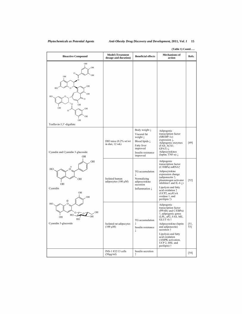
Moreover, theaflavins may be active in the prevention of fatty liver and obesity. Rats that were treated with diets rich in theaflavins (TF, TF3G, or TF3DG, 50 mg/kg body weight) had more markedly reduced body weights than the HFD-fed controls [27]. Theaflavin-supplemented rats ate more but weighed less than the HFD controls, which suggests that theaflavins might increase energy expenditure. Similarly, the relative liver weight, epididymal fat mass, and plasma lipid levels were significantly reduced in the theaflavin-rich diet group than in the control rats. The researchers also provided evidence that theaflavins likely play a significant role in reducing cellular lipid accumulation by increasing AMPK phosphorylation in HepG2 cells, and that a ROS/serine-threonine kinase liver kinase B1/AMPK signaling module may be involved in this process [27].
Cyanidins
Cyanidins are considered the most widely spread anthocyanin in the plant kingdom. They are largely distributed in the human diet through crops, beans, fruits, vegetables, and red wines, which suggests that humans ingest daily significant amounts of these compounds from plant-based diets [48]. It has been reported that cyanidins have several pharmacological properties, including antioxidant, anticarcinogenic, anti-inflammatory, neuroprotective, skin-photoprotective, gastroprotective, anti-obesity, and antidiabetic effects [48].
Tsuda et al. investigated the potency of the therapeutic implications of cyanidin 3-glucoside in the prevention of obesity and diabetes. They assessed the potential role of purple corn color (PCC), which is rich in cyanidin 3-glucoside, in the prevention of obesity and in the amelioration of diabetes [49]. Cyanidin-3-glycoside, also known as kuromanin, is probably the most notorious and the most widely investigated among the cyanidin-glycosides [50]. Tsuda et al. found that dietary PCC significantly suppressed the HFD-induced increase in the body weight gain, the white and brown adipose tissue weights, along with hyperglycemia, hyperinsulinemia, and hyperleptinemia, by suppressing the mRNA levels of the enzymes involved in fatty acid and triacylglycerol synthesis, lowering the mRNA level of the sterol regulatory element binding protein-1 (SREBP-1), and normalizing the increase in the mRNA level of the tumor necrosis factor (TNF)- in the visceral adipose tissue [49]. In another study, the researchers found that rat and human adipocytes that were treated with cyanidin 3-glucoside [51] enhanced adipocytokine (adiponectin and leptin) secretion and upregulated the adipocyte-specific gene expression [52], and that the isolated
6 Anti- Obesity Drug Discovery and Development, 2011, Vol. 1 Park and Kim
rat adipocytes that were treated with cyanidin 3-glucoside and cyanidin demonstrated an up-regulation of hormone-sensitive lipase (HSL) and perilipin as well as enhancement of the lipolytic activity [53].
Cyanidin 3-glucoside stimulated in vitro insulin secretion from rodent pancreatic beta-cells (INS-1 832/13) at 4 and 10 mM glucose concentrations [54]. Among cyanidin glycosides, cyanidin 3-rutinoside, has been proposed as a new non-competitive -glucosidase inhibitor because of its ability to inhibit - glucosidase from baker’s yeast [55]. Another cyanidin glycoside, cyanidin 3-galactoside, has also been proposed as a new competitive -glucosidase inhibitor due to its ability to inhibit - glucosidase from rat intestinal acetone powder [56]. These outcomes suggest that cyanidins have a uniquely potent therapeutic advantage and important implications in the prevention of obesity and diabetes.
Isoflavones: Genistein, Daidzein, and Glycitein
Isoflavones are commonly found in soybeans and soy products. Isoflavones exist primarily in plants in an inactive form as -D-glycosides (genistin, daidzin and glycitin). After ingested, isoflavone glycosides are hydrolyzed by interstinal glucosidases and are converted to corresponding bioactive aglycones (genistein, daidzein, and glycitein). The aglycones are then absorbed from the intestinal tract and conjugated mainly in the liver into glucuronides, which are either re-excreted through the bile and then reabsorbed through enterohepatic recycling, or excreted unchanged in the urine. The chemical structure of isoflavones is very similar to that of human estrogen. Because of this similarity in structure, they can interfere with the action of human estrogen [57]. Depending on the type of estrogen receptor in the cells, isoflavones may reduce or activate the activity of estrogen. The consumption of soy products has many health benefits, including protection against breast cancer, prostate cancer, menopausal symptoms, heart disease, and osteoporosis. Many of the health benefits of soy are derived from its isoflavones [57, 58].
A number of studies suggest that isoflavones (genistein, daidzein, and glycitein) may favorably affect adiposity, glucose homeostasis, insulin secretion, and lipid metabolism (Table 1). Several epidemiological studies have reported that isoflavone consumption by postmenopausal women was correlated with lower body mass index and fasting insulin concentrations, and higher HDL levels [59-61]. Additionally, Japanese postmenopausal women who were treated with isoflavones for 24 weeks exhibited a lower body fat mass [62]. These results indicate the beneficial effects of isoflavones on excess body weight, hyperinsulinemia, and hyperlipidemia, which are the major cardiovascular risk factors commonly associated with obesity. Plasma triglycerides were more significantly reduced in Sprague-Dawley rats that were fed a mixture of synthetic daidzein and genistein (23.6 mg/kg body weight/day) than in those that were fed casein [63]. Hamsters that were fed pure synthetic daidzein (16 mg/kg body weight/day) had significantly lower plasma total cholesterol levels than those that were fed casein [64]. In another study, glycitein (13 mg/kg body weight/day) showed a more marked cholesterol-lowering effect than other isoflavones [65]. The researchers suggested that glycitein's greater cholesterol-lowering effect was due to its greater bioavailability, as reflected in its urinary recovery, than that of the other isoflavones [65].
In addition to the hypolipidemic effects of isoflavones, dietary supplementation with 500-1,500 ppm of genistein, with a serum equivalent of about 2 μM, decreases fat-pad
Phytochemicals as Potential Agents Anti-Obesity Drug Discovery and Development, 2011, Vol. 1 7
weights by 50% in C57/BL6 mice [66]. Similarly, subcutaneous injections of genistein (8-200 mg/kg/day) in C57/BL6 ovariectomized mice for 21 days decreased the adipose tissue gain [66]. In contrast, in a study wherein male mice were exposed to daily oral doses of genistein of up to 50 mg/kg/day, the fat mass increased [67], whereas it decreased at doses of 200 mg/kg/day [67]. These results suggest that high pharmacological doses of isoflavones that yield serum levels in the micromolar range inhibit adipose tissue deposition. The mechanisms by which isoflavones reduce adiposity in vivo are still unclear. In vitro studies in which isolated rat adipocytes were used, however, showed that genistein and daidzein stimulate lipolysis by inhibiting cAMP phosphodiesterases [68, 69]. Furthermore, the exposure of cultured 3T3-L1 adipocytes to genistein has been reported to significantly activate AMPK and ACC [31]. AMPK is another target molecule for anti-obesity treatments, and genistein has been shown to inhibit adipocyte differentiation by activating AMPK. Genistein stimulated intracellular ROS release, which rapidly activated AMPK and led to apoptosis. The protein expressions of PPARs and C/EBP decreased in adipocytes that were treated with genistein [70]. Genistein also inhibited the mitotic clonal expansion of two-day post-confluent 3T3-L1 preadipocytes [70]. In an isolated perfused liver preparation, however, genistein reduced the incorporation of [
14C] glucose into lipids and increased the
output of fatty acids into the medium [71]. These changes were accompanied by a decrease in the hepatic triacylglycerol content. Thus, genistein appears to have direct effects on lipid metabolism in the liver and the adipose tissue by affecting both lipogenesis and lipolysis.
Fewer studies have evaluated the effects of isoflavones on glucose homeostasis. In mice models of obesity and diabetes, treatment with genistein lowered glycemia [72]. Potential mechanisms by which isoflavones may improve glucose metabolism have been recently proposed. Recent reports indicate that genistein may itself have a direct beneficial effect on pancreatic -cells. For example, genistein has been shown to stimulate insulin secretion in the insulin-secreting cell lines INS-1 and MIN6 and in mouse pancreatic islets, at nano- and micromolar concentrations [73]. Genistein and daidzein have been shown to prevent diabetes onset in non-obese diabetic mice by preserving the pancreatic -cell function [74]. Similarly, Lee et al. (2006) recently reported that in rats that had streptozotocin-induced diabetis, diet supplementation with genistein (600 mg/kg of the diet) caused a twofold increase in the plasma insulin levels [75]. In additional studies, genistein was shown to inhibit islet tyrosine kinase activities and glucose- and sulfonylurea-stimulated insulin release without affecting the glucose metabolism [68]. Collectively, these data suggest that dietary soy and phytoestrogens may have therapeutic significance in reducing the severity of diabetes, and in improving -cell survival and function. Further research is needed, however, first, to confirm, and then to analyze, in detail, how phytoestrogens exert antihyperglycemic actions through the pancreatic -cells.
Kaempferol
Kaemferol is a natural flavonoid that has been isolated from tea, broccoli, strawberries, gooseberries, cranberries, Euonymus alatus, Sauroupus androgynus, Delphinium, witch hazel, grapefruit, brussel sprouts, apples, and other plant sources [76, 77]. A number of studies have been carried out to investigate the anti-obesity effects of kaempferol in vitro and in vivo (Table 1). Yu et al. tested the toxicity and anti-obesity features of the compound 3-O- -D-glucosyl-(1 6)- -D-glucosyl-kaempferol (GGK) isolated from Sauropus androgynus, in rats fed an HFD. Sixty milligrams of GGK per kilogram of body weight, which was administered via gastric intubation, significantly reduced the food intake of rats, and the reduced food intake corresponded to decreases in the body weight gain [76].

8 Anti- Obesity Drug Discovery and Development, 2011, Vol. 1 Park and Kim
The clinical relevance of PPAR is highlighted by the currently marketed antidiabetic drugs, rosiglitazone and pioglitazone. As full PPAR antagonists, they promote adipocyte differentiation and lipid storage. Kaempferol also significantly improved insulin-stimulated glucose uptake in mature 3T3-L1 adipocytes [77]. The potential mechanism by which kaempferol ameliorates hyperglycemia may partly be by serving as a ligand of PPAR , and thus, improving insulin-stimulated glucose uptake in mature adipocytes, while lacking the capability to induce preadipocyte differentiation. Thus, kaempferol may potentially be developed as a safe and novel compound for anti-obesity and antidiabetes treatment.
Myricetin
Myricetin is a flavonol that is commonly found in teas, wines, berries, fruits, and medicinal plants (Abelmoschus moschatus). Its therapeutic potential is seen both in its anticarcinogenic, anti-inflammatory, antioxidative, antiviral, and antimicrobial properties and in its prevention of platelet aggregation [78-82]. The therapeutic benefits of myricetin in cardiovascular diseases associated with diabetes mellitus have also been reported [83-86]. Additionally, the insulinomimetic effect of myricetin on lipogenesis and glucose transport in the adipocytes of rats with non-insulin-dependent diabetes mellitus has been observed [87].
Liu et al. investigated the effects of myricetin on the healing of insulin resistance [83, 88]. Intravenous injection of myricetin (1 mg per kg of body weight) into genetically obese diabetic rats and rats that were fed high-fructose diets three times a day for one week and two weeks, respectively, reduced the value of the glucose-insulin index, an index of insulin resistance. In addition, myricetin treatment reversed the inability of insulin to increase the expression of the glucose transporter 4 (GLUT 4) and to increase the protein levels and phosphorylation of the insulin receptor substrate 1 (IRS1) in the soleus muscle of the obese rats. Myricetin treatment also increased the expression of the p85 regulatory subunit of phosphatidylinositol 3-kinase (PI3K) and promoted Akt serine phosphorylation in the soleus muscle of the diabetic rats [83, 88]. In another study, after two days of treatment with myricetin (3 mg per 12 h), hyperglycemia in diabetic rats was reduced by 50%, and the hypertriglyceridemia that is often associated with diabetes normalized [89]. Treatment with myricetin increased the hepatic glycogen synthase I activity and the glucose-6-phosphate content [89]. Myricein also stimulated glucose uptake in isolated rat adipocytes and enhanced insulin-stimulated lipogenesis in adipocytes at concentrations that ranged from 200 to 250 μM [87]; and at low doses (10-100 μM), inhibited the transport of glucose in isolated rat adipocytes stimulated with insulin [90]. The increase in the lipogenesis might have been a consequence of increased glucose transport. Besides, myricetin prevented diabetic cataracts by inhibiting lens aldose reductase [91]. Taking all these together shows that myricetin may improve insulin sensitivity and reverse insulin resistance. Such effects may be mediated through enhanced insulin-mediated glucose transport signaling in the muscles and adipose tissues of obese diabetic rats.
Hesperetin and Hesperidin
Hesperetin belongs to the class of flavonoids called flavanones and is derived from the hydrolysis of its aglycone, hesperidin (hesperetin 7-rhammnoglucoside). Hesperetin, together with hesperidin, is abundant in citrus fruits, such as grapefruit and oranges. In Finland, the average daily intake of hesperetin has been estimated as 28.3 mg [92]. Hesperetin has been found to exhibit estrogenic, anticarcinogenic, antioxidative, anti-inflammatory and lipid-lowering properties [92-96].

Phytochemicals as Potential Agents Anti-Obesity Drug Discovery and Development, 2011, Vol. 1 9
With regard to the lipid-lowering propensity of hesperetin, Cha et al. [96] found that this compound, at a dietary level of 1%, attenuated the orotic-acid-dependent increase in the hepatic triacylglycerol level and activity of microsomal phosphatidate phosphohydrolase, an enzyme involved in the synthesis of triacylglyceride, in rat liver. Hesperetin, at a dietary level of 0.02%, lowered the plasma levels of cholesterol and triacylglyceride and the activity of 3-hydroxy-3-methylglutaryl-coenzyme (HMG-CoA) reductase, the rate-limiting enzyme in the cholesterol biosynthesis, and acyl CoA: cholesterol acyltransferase (ACAT), another key cholesterol-regulating enzyme involved in the esterification and absorption of cholesterol, in rats fed a high-cholesterol diet [96]. Studies of Wilcox et al. [94] have shown that hesperetin decreased the secretion of apolipoprotein B (apoB), an apolipoprotein that abounds in low-density and very-low-density lipoproteins, in human hepatoma cells (HepG2). Their study results also showed that hesperetin decreases the activities of ACAT2 and microsomal triacylglycerol transfer protein (MTP), and increases the LDL receptor activity [94]. MTP plays a key role in apoB secretion by catalyzing the transfer of lipids to the nascent apoB molecule as it is cotranslationally translocated across the endoplasmic reticulum membrane [97, 98]. Furthermore, the increased LDL receptor activity contributed to the reduced accumulation of apoB in the media [94]. It has been seen that hesperetin’s blockade of the cholesterol biosynthesis results in a lower intracellular supply of cholesterol, which triggers an over-expression of hepatic LDL receptors and enhances the clearance of circulating LDL particles [99]. Thus, hesperetin may affect various aspects of lipid metabolism and exert lipid-lowering activity in vivo.
Hesperidin is a flavanone glycoside that consists of the flavone hesperitin, which is bound to the disaccharide rutinose (Table 1). The sugar causes hesperidin to be more soluble than hesperitin. Hesperidin is also mainly found in citrus fruits such as lemons and oranges. The highest concentration of hesperidin can be found in the white parts and pulps of the citrus peels. Hesperidin can also be found in green vegetables. Among naturally occurring flavonoids, hesperidin has been empirically proven to have no side-effects, as humans have been ingesting grapes and citrus fruits for a long time [100]. Hesperidin exhibits biological and pharmacological properties, such as anti-inflammatory, anticarcinogenic, antioxidant, lipid-lowering, and antidiabetic properties [101-104].
Hesperidin supplementation improves hyperglycemia in db/db mice by regulating the activities of the hepatic glucose metabolic enzymes involved in glycolysis and gluconeogenesis [102]. In another study, this compound was found to be beneficial in the lowering of the blood glucose level because it upregulates the hepatic glucokinase, PPAR , and adipocyte GLUT4 [101]. Hesperidin was also found to be very effective in improving the lipid metabolism in db/db mice by suppressing the activities of hepatic lipid metabolizing enzymes, such as fatty acid synthase, glucose-6-phosphate dehydrogenase (G6PDH), and phosphatidate phosphohydrolase, and by increasing fecal triglyceride excretion [101]. Plasma cholesterol levels are regulated by cholesterol biosynthesis, the removal of cholesterol from the body’s circulation, and the excretion of cholesterol via faecal sterols or bile acids. Numerous previous researches have reported the beneficial effects of hepatic HMG-CoA reductase and ACAT inhibitors on hypercholesterolaemia and atherosclerosis [105, 106]. In one study, hesperidin also led to a decrease in the the plasma and hepatic cholesterol levels, which may have been partly due to the decreased activities of hepatic HMG-CoA reductase and ACAT and the increased fecal cholesterol excretion [101]. These coordinated responses of the hesperidin supplement seemed to play an important role in regulating the lipid and glucose metabolism in db/db mice [101].

10 Anti- Obesity Drug Discovery and Development, 2011, Vol. 1 Park and Kim
Naringenin and Naringin
Naringenin is the predominat flavone in citrus fruits and grapefruit. It can also be absorbed from cooked tomato paste. This compound is considered to have a bioactive effect on human health as an antioxidant, a free radical scavenger, an anti-inflammatory agent, a carbohydrate metabolism promoter, and an immune system modulator. The average daily intake of naringenin in Finland has been estimated as 8.3 mg [92]. Harmon and Harp investigated the anti-obesity effects of naringenin [70]. They found that the adipose tissue mass can be reduced by inhibiting preadipocyte proliferation. Naringenin at 50-100 μM inhibited the proliferation of preconfluent preadipocytes in a time- and dose-dependent manner [70], but the presence of naringenin during the induction of differentiation had no adverse effect on the subsequent adipogenesis [107].
Huong et al. demonstrated that naringenin has the physiologic effect of increasing hepatic fatty acid oxidation by up-regulating the gene expression of the enzymes involved in peroxisomal -oxidation in mice [108]. The change may account for the propensity of this compound to lower serum lipid levels. This compound also caused significant increases in the activity levels of various enzymes involved in hepatic fatty acid oxidation, such as carnitine octanoyltransferase, acyl-coenzyme A oxidase, and 3-ketoacyl–coenzyme A thiolase [108]. Lee et al. also reported that naringenin at a 0.1% dietary level decreased the plasma cholesterol level and the hepatic levels of triacylglycerol and cholesterol in rats fed a high-cholesterol diet, which was accompanied by a decrease in the activity of HMG-CoA reductase and ACAT [109]. Naringenin decreased the secretion of apoB, an apolipoprotein that abounds in low-density and very-low-density lipoproteins, upregulated the mRNA expression of low-density lipoprotein receptors, and decreased the activities of ACAT and MTP, a key enzyme in apoB secretion, in HepG2 cells [94].
Harmon and Patel found that the grapefruit flavanone naringenin inhibited insulin-stimulated glucose uptake in 3T3-L1 adipocytes in a dose-dependent manner [107]. Naringenin inhibited the activity of PI3K, a key regulator of insulin-induced GLUT4 translocation. Although naringenin did not alter the phosphotyrosine status of the insulin receptor, insulin receptor substrate proteins, or PI3K, it inhibited the phosphorylation of the downstream signaling molecule, Akt. The physiologically attainable dose of 6 μM of naringenin reduced the insulin-stimulated glucose uptake by approximately 20%. This inhibitory effect remained 24 h after the removal of naringenin from the culture medium. Collectively, the findings suggest that regular consumption of naringenin may exacerbate insulin resistance in susceptible individuals via impaired glucose uptake in the adipose tissue. These adverse effects of naringenin on glucose homeostasis would depend on the ability of an individual to absorb and metabolize naringenin [107].
Naringin is the aglycone of naringenin (Table 1). It is metabolized into the flavonone naringenin in humans. Naringin is the major flavonoid glycoside in citrus fruits and grapefruit, and which is what gives such fruits their bitter taste. Naringin exhibits biological and pharmacological properties, such as anti-inflammatory, anticarcinogenic, antioxidant, lipid-lowering, and antidiabetic properties [101-104]. Some studies have demonstrated that naringin may act as a regulator of plasma lipids in hypercholesterolemic subjects, such as db/db mice or rats fed a HFD [101, 102, 110, 111]. Diet supplementation with 0.02% naringin for three weeks did not exhibit a hypolipidemic effect on rats fed a high-fat and high-cholesterol diet [113, 114]. Naringin improved the plasma and hepatic lipid metabolism in db/db mice by altering the hepatic-lipid-metabolizing enzyme activities and

Phytochemicals as Potential Agents Anti-Obesity Drug Discovery and Development, 2011, Vol. 1 11
fecal lipids [101]. In addition, naringin improved hyperglycemia in type 2 diabetes mellitus mice by regulating the activities and expressions of the hepatic enzymes involved in glycolysis and gluconeogenesis and by up-regulating the hepatic and adipocyte PPAR and GLUT4 [101, 102]. Naringin consumption (at 400 mg/day for eight weeks) lowered the plasma total and LDL-cholesterol concentrations in hypercholesterolemic subjects [111]. Naringin supplementation had no effect, however, on the plasma lipid levels in healthy control subjects [111]. These coordinated responses of the naringin supplement seemed to play an important role in regulating the lipid and glucose metabolism in hyperlipidemic or hyperglycemic subjects [101]. Naringin can, however, lower the hepatic cholesterol biosynthesis and levels of plasma lipids and glucose when supplemented for at least six weeks in these subjects [110, 111].
Quercetin
Quercetin the most abundant of the flavonoids, forms the backbone for many other flavonoids, including rutin (a glycosylated quercetin), hesperidin, and naringenin. Quercetin is found in various human foods such as red onions, grapes, apples, berries, cherries, broccoli, citrus fruits, and tea (Camellia sinensis), and in particularly high concentrations (180 mg per 100 g) in capers and lovage. The average daily human intake of quercetin ranges from 10 to 100 mg depending on eating habits [112]. Quercetin has a wide range of biological effects such as lowering of blood pressure [113, 114], reduction of body weight [113], and amelioration of hyperglycemia-related diseases [77, 115], as shown in animal models and, to some extent, in humans.
Edwards et al. recently published a randomized, double-blind, and placebo-controlled cross-over study that included patients with prehypertension (n = 19) and stage 1 hypertension (n = 22) [114]. The blood pressure of the prehypertensive patients did not change after their diet was supplemented with 730 mg of quercetin/day for 28 days. In contrast, significant reductions in systolic, diastolic, and mean arterial blood pressure were observed in the stage 1 hypertensive patients after quercetin treatment. Interestingly, quercetin did not affect the indices of oxidant stress that were measured in the plasma and urine, unlike in previous animal-based studies. Although many questions remain (on the optimal dosage, long-term-effects, efficacy compared with other antihypertensive agents, etc.), the study of Edwards et al. was the first to show that quercetin supplementation reduces blood pressure in hypertensive patients [114]. The increased nitric oxide (NO) availability caused by the elevated NO synthase (NOS) activity and the antioxidative activity in the DIO rats fed with quercetin is suggested as one of the causes of the suppressed elevation of the blood pressure [113].
Quercetin induces apoptosis in 3T3-L1 preadipocytes by decreasing the mitochondria membrane potential, down-regulating poly (ADP-ribose) polymerase (PARP) and Bcl-2, and activating caspase 3, Bax, and Bak [116]. In maturing preadipocytes, quercetin at 25 μM suppressed intracellular lipid accumulation by 15.9±2.5% and significantly decreased the expression of PPAR , which acts as a key adipogenic transcription factor [117]. The breakdown of triglycerides in adipocytes and the release of glycerol and fatty acids are important in the regulation of energy homeostasis [118]. Quercetin caused a dose- and time-dependent increase in lipolysis in rat adipocytes, which was synergistic with epinephrine; and this effective lipolytic flavonoid was also reported to be a potent phosphodiesterase (PDE) inhibitor [119]. Taken together, these reports indicate that quercetin can have

12 Anti- Obesity Drug Discovery and Development, 2011, Vol. 1 Park and Kim
potential anti-obesity effects by inhibiting the differentiation of preadipocytes and inducing
the apoptosis of mature adipocytes.
Strobel et al. [90] reported that quercetin interacts directly with GLUT4 to inhibit its uptake of glucose into the isolated rat adipocytes. In other in vitro studies, quercetin also directly worked on multiple targets within the insulin signaling pathway by inhibiting PI3K at relatively low concentrations [120-122] and modulating the activity of Akt/protein kinase B (PKB) and protein kinase C (PKC) [123-125]. In contrast, however, quercetin significantly improved insulin-stimulated glucose uptake in mature 3T3-L1 adipocytes [77]. Further experiments have proven that quercetin serves as a weak partial antagonist of PPAR , and that unlike the traditional antagonist, quercetin cannot induce the differentiation of the preadipocyte 3T3-L1. Therefore, the potential mechanism by which quercetin ameliorates hyperglycemia may in part be by serving as a ligand of PPAR , and thus, improving insulin-stimulated glucose uptake in mature adipocytes, while lacking the
capability to induce preadipocyte differentiation, as shown in kaempferol [77].
Additionally, initial clinical data support the role of quercetin in the prevention of obesity and type 2 diabetes. A pilot study showed that QR-333, a topical compound that contains quercetin, safely induces relief from symptoms of diabetic neuropathy and improves quality of life [126]. The randomized, placebo-controlled, double-blind trial included 34 men and women with type 1 or 2 diabetes and diabetic neuropathy, who applied QR-333 or a placebo three times daily for four weeks to each foot wherein symptoms were experienced. QR-333 reduced the severity of the numbness, jolting pain, and irritation from
the baseline values, was well tolerated, and improved quality-of-life measures.
Rutin
Rutin (3, 3 , 4 , 5, 7-pentahydrohyflavone-3-rhamnoglucoside) is a flavonol-type flavo-noid that is found in many typical plants such as buckwheat, apples, teas, citrus fruits, parsley, mulberry, and tomatoes. It is also an important dietary constituent of other foods and plant-based beverages [127]. It has been reported that rutin has several pharmacological properties including antioxidant, anticarcinogenic, cytoprotective, neuroprotective, anti-platelet, antithrombic, vasoprotective and cardioprotective activities [128-132]. Besides, rutin was shown to prevent diet-induced obesity [133, 134], to reduce plasma leptin and insulin levels, and to alleviate dyslipidemia in obese mice and rats [133-135]. Choi et al. [134] and Hsu and Yen [136] reported that rutin efficiently suppresses adipogenesis in 3T3-L1 adipocytes. It appears to be mediated through the downregulated expression of adipo-genic transcription factors, such as PPAR and C/EBP , and adipocyte-specific proteins, such as leptin; and then through the upregulated expression of adiponectin. Many studies have shown that rutin inhibits glycerol-3-phosphate dehydrogenase (GPDH), the cytosolic enzyme that plays an important role in the conversion of glycerol into triglyceride, in 3T3-L1 preadipocytes [134, 136] and in the fat and liver tissues of rats with HFD-induced obesity [133].

Phytochemicals as Potential Agents Anti-Obesity Drug Discovery and Development, 2011, Vol. 1 13
Table 1. Beneficial Effects of Flavonoids on Obesity and Associated Diseases
Bioactive Compound Model (Treatment
dosage and duration) Beneficial effects
Mechanisms of action
Refs.
(-)-Catechin
O
OH
OH
HO
OH
OH
3T3-L1 adipocytes
(50 μM)
Insulin-stimulated glucose uptake
Adiponectin expression and secretion
Adipogenic gene expression (PPAR , C/EBP , aP2)
[24]
(-)-Epigallocatechin gallate Obese men (300 mg/day, 3 days)
Postprandial RQ
[22]
Obese men (800 mg/ day, 8wk)
Blood pressure [23]
DIO mice and rats (0.32 1% wt/wt in diet, 16 20 wk)
Body weight
Visceral fat weight
Blood lipids
Blood glucose
Insulin resistance improved
Fatty liver improved
Leptin secretion
Fatty acid synthesis (FAS, G6PDH)
Fatty acid oxidation (SCD1)
Fecal lipids
[21, 26]
HO O
O
O
OH
OHOH
OH
OH
OH
OH
DIO mice (0.5 1% wt/wt in diet, 4 wk)
Body weight
Visceral fat weight
Blood lipids
Blood glucose
Hepatic fatty acid synthesis [malic enzyme, glucokinase (GK)]
Hepatic fatty acid oxidation (SCD1)
Epididymal leptin expression and fatty acid oxidation (SCD1)
[28]
db/db Mice and ZDF rats (0.5 1% wt/wt in diet, 7 wk)
Blood lipids
Blood glucose
Insulin resistance improved
Insulin secretion
Fatty acid oxidation (ACO-1 and CPT-1)
Hepatic glucose uptake (GK)
Gluconeogenesis
[32]
3T3-L1 adipocytes (100 μM)
TG accumulation
Apoptosis (apoptotic cell number ; Cdk2 and caspase-3 )
Adipogenic transcription factors (PPAR 1/2, C/EBP and LXR ) expression
Fatty acid oxidation (pACC/ACC and pAMPK/AMPK)
GPDH activity
[30, 31, 137, 138]

14 Anti- Obesity Drug Discovery and Development, 2011, Vol. 1 Park and Kim
(Table 1) Contd…..
Bioactive Compound Model (Treatment
dosage and duration) Beneficial effects
Mechanisms of action
Refs.
HepG2 cells (50 μM) TG accumulation
Adipogenic transcription factors (PPAR 2, C/EBP ) expression
[27]
Epicatechin gallate
HO O
O
O
OH
OHOH
OH
OH
OH
Everted rat intestinal sacs and rabbit intestinal brush-border membrane vesicles
(0.5 1 mM)
Glucose uptake Glucose transporter antagonist
[139]
DIO rats (50 mg/kg BW)
Body weight
Visceral fat weight
Fatty liver
Blood lipids
Hepatic fatty acid oxidation (AMPK phosphorylation)
[27]
Rats (10 mg/ kg BW with maltose, 0 120 min)
In vitro immobilized AGH assay system
Postprandial blood glucose
-Glucosidase (maltase) activity in the gut
[40]
Rats (100 or 200 mg/ kg BW with a lipid emulsion, 0 8 h)
In vitro pancreatic lipase activity assay
Postprandial blood triglyceride
Pancreatic lipase activity
Intestinal triglyceride absorption
[38]
HepG2 cells (50μM) TG accumulation
Hepatic fatty acid oxidation (AMPK phosphorylation) and fatty acid synthesis (FAS)
[27]
Theaflavins: TF, TF3G, TF3´G, TF3DG
HO O
OH
OH
OH
OH
O
OH
OHO
OH
OH
Teaflavin
HO O
O
OH
OH
OH
O
OH
OHO
OH
OH
O
OH
OH
OH
Teaflavin-3-gallate
HO O
OH
OH
OH
OH
O
OH
OHO
OH
O
O
OH
OH
OH
Teaflavin-3’-gallate
In vitro micelle formation assay
Dietary mixed micelles formation
Cholesterol incorporation into mixed micelles
[39]
Phytochemicals as Potential Agents Anti-Obesity Drug Discovery and Development, 2011, Vol. 1 15
(Table 1) Contd…..
Bioactive Compound Model (Treatment
dosage and duration) Beneficial effects
Mechanisms of action
Refs.
HO O
O
OH
OH
OH
O
OH
OHO
OH
O
O
OH
OH
OH
O
OH
OH
OH
Teaflavin-3,3’-digallate
DIO mice (0.2% wt/wt in diet, 12 wk)
Body weight
Visceral fat weight
Blood lipids
Fatty liver improved
Insulin resistance improved
Adipogenic transcription factor (SREBP-1c) expression , Adipogenic enzymes (FAS; ACS1; GPAT) Adipocytokines (leptin; TNF- )
[49]
Isolated human adipocytes (100 μM)
TG accumulation
Normalizing adipocytokine secretion
Inflammation
Adipogenic transcription factor (C/EBP ) mRNA
Adipocytokine expression change (adiponectin ; plasminogen activator inhibitor1 and IL-6 )
Lipolysis and fatty acid oxidation (UCP2, acylCoA oxidase 1, and perilipin )
[52]
Cyandin and Cyanidin 3-glucoside
O
OH
OH
OH
OH
HO
Cyanidin
OHO
OH
OH
OH
O OOH
OHHO
HO
Cyanidin 3-glucoside Isolated rat adipocytes (100 μM)
TG accumulation
Insulin resistance
Adipogenic transcription factor (PPAR and C/EBP )
, adipogenic genes (LPL, aP2, FAS, ME, GLUT 4)
Adipocytokine (leptin and adiponectin) secretion
Lipolysis and fatty acid oxidation (AMPK activation, UCP 2, HSL and perilipin)
[51, 53]
INS-1 832/13 cells (50μg/ml)
Insulin secretion
[54]

16 Anti- Obesity Drug Discovery and Development, 2011, Vol. 1 Park and Kim
(Table 1) Contd…..
Bioactive Compound Model (Treatment
dosage and duration) Beneficial effects
Mechanisms of action
Refs.
Cyanidin-3-rutinoside
OHO
OH
OH
OH
OO
OH
HO
OH
O
H3C
HO
HOOH
In vitro assay -Glucosidase
activity Competitive inhibitor of of -glucosidase
[55]
Cyanidin-3-galactoside
OHO
OH
OH
OH
OO
OH
HO
HOOH
In vitro assay -Glucosidase
activity
Competitive inhibitor of substrate of -glucosidase
[56]
Soy isoflavones mixture Postmenopausal women (75 mg/day, 6 mo)
BMI Body fat mass Blood HDL
[62]
Rats (0.038% wt/wt in diet, 3 wk)
Blood lipids (TG)
[63]
Hasmters (0.03% wt/wt in diet, 10 wk)
Blood TC Blood HDL
[64]
Postmenopausal women ( 1 mg/day)
BMI Body fat mass Waist size Blood HDL
[59]
Postmenopausal women (0.001-0.999 mg/day)
Blood HDL Fasting insulin
[59]
Mice (0.05-0.15% wt/wt in diet, 12 days)
Body fat mass LPL activity in adipose tissue
[66]
Mice (200 mg/kg, 15 days)
Body weight Body fat mass
[67]
Genistein
HO O
OH
OOH
Rats (0.01, 0.1% wt/wt in diet, 2 wk)
Blood lipids Fatty liver improved Lipogenesis
[71, 140]
db/db mice (0.02% wt/wt in diet, 5 wk)
Blood glucose Glucose tolerance
Blood lipids Fatty liver improved
Hepatic gluconeogenesis (G6Pase and PEPCK activities) Hepatic glycolysis (GK activity) Hepatic fatty acid synthesis (FAS and G6PDH activity)
[72]

Phytochemicals as Potential Agents Anti-Obesity Drug Discovery and Development, 2011, Vol. 1 17
(Table 1) Contd…..
Bioactive Compound Model (Treatment
dosage and duration) Beneficial effects
Mechanisms of action
Refs.
STZ-induced diabetic rats (0.06% wt/wt in diet, 3 wk)
Blood insulin
Blood lipids Fatty liver improved
Hepatic gluconeogenesis (G-6-Pase activities) Hepatic glycolysis (GK activity)
[75]
Isolated rat adipocytes (0.1, 1 mM)
Lypolysis Insulin sensitivity
cAMP phosphodiesterase
Tyrosine kinase activity
[68]
INS-1 and MIN6 cells (0.01-10 μM)
Insulin secretion
cAMP/PKA activity [73]
Non-obese diabetic mice (0.02% wt/wt in diet, 9 wk)
Blood glucose
Blood insulin
Blood lipids
Fatty liver improved
Pancreatic -cell function (C-peptide secretion , immune-histochemical insulin staining ) Hepatic gluconeogenesis (G6Pase and PEPCK activities) Hepatic fatty acid synthesis (malic enayme and G6PDH activity)
Hepatic fatty acid oxidation (CPT activity)
[74]
Daidzein
HO O
O
OH
Rat adipocytes
(0.1, 1 mM) Lypolysis PKA activity [69]
3T3-L1 adipocytes
(20-200 μM)
TG accumulation
Adipocyte apoptosis
Lypolysis (AMPK activity ) ROS release PPAR expression
[31, 70]
Ovariectomized rats (50 mg/kg BW)
Blood lipids [141] Glycitein
HO
H3CO
O
OH
O
DIO Hamsters (0.025% wt/wt in diet, 4 wk)
Blood lipids (Cholesterol)
[65]
DIO rats (6 60 mg/kg BW, 4 wk)
Body weight gain
Blood lipids Food intake [76]
Kaempferol
HO O
OH O
OH
OH
3T3-L1 adipocytes (20 50 μM)
Glucose uptake TG accumulation
Adipogenesis Partial agonist of PPAR
[77]
Obese Zucker rats (I.V. injection, 3 mg/kg BW/day, 1 wk)
Blood glucose Insulin resistance
Post-receptor insulin signaling in muscle (GLUT 4, IRS-1, PI3-kinase, and Akt)
[88]
Myricetin
HO
OH
O
OH
OH
OH
OH
O
Diet-induced diabetic rats (I.V. injection, 3 mg/kg BW/day, 2 wk)
Blood glucose Insulin resistance
Blood lipid (TG)
Post-receptor insulin signalings in muscle (GLUT 4, IRS-1, PI3-kinase and Akt) Hepatic glycogen synthesis
[83]

18 Anti- Obesity Drug Discovery and Development, 2011, Vol. 1 Park and Kim
(Table 1) Contd…..
Bioactive Compound Model (Treatment
dosage and duration) Beneficial effects
Mechanisms of action
Refs.
Streptozotocin -induced diabetic rats (3 mg/12 h, 4 days)
Blood glucose
Insulin resistance improved
Blood lipid (TG)
Hepatic glycogen synthesis and glucose-6-phosphate content
[89]
Isolated rat adipocytes (200 250 μM)
Glucose uptake
TG accumulation
GLUT 4’ activity [87]
Hypercholesterolaemic rats (0.02% wt/wt in diet, 5 wk)
Blood lipids
Atherogenic index
HMG-CoA reductase and ACAT activities
Fecal cholesterol excretion
[95]
Fatty liver rats (1% wt/wt in diet, 10 day)
Fatty liver improved
Hepatic triglyceride synthesis (phosphatidate phosphohydrolase, G6PDH, malic enzyme, DG acyltransferase activities )
[96]
Hesperetin
HO
OH
O
OCH3
OH
O
HepG2 cells (100 200 μM)
Lipids secretion
ApoB-containing lipoprotein assembly (expression and activity of ACAT2 and MTP; LDL receptor expression )
[94]
Hesperidin
O O
OH
OCH3
OOH
O
HOHO
O
OH
OH3C
HO
HOOH
db/db Mice (0.02% wt/wt in diet, 5 wk)
Blood glucose
Blood lipids
Fatty liver improved
Body weight
Insulin and leptin secretion
Hepatic glycolysis and glycogen synthesis
HMG-CoA reductase and ACAT activities
Fecal cholesterol excretion
[101, 102]
Mice fed normal diet (1% wt/wt in diet)
Blood lipids Hepatic peroxisomal fatty acid oxidation
[108]
Hypercholesterolaemic rats (0.1% wt/wt in diet, 3 wk)
Blood lipids
Fatty liver improved
HMG -CoA reductase and ACAT activities
[109, 142]
3T3-L1 adipocytes (50 100 μM)
Fat accumulation
Adipocyte apoptosis (Preconfluent- preadipocytes growh
)
[70]
Naringenin
HO
OH
O
OH
O HepG2 cells (100 200 μM)
Lipids secretion
ApoB-containing lipoprotein assembly (expression and activity of ACAT2 and MTP; LDL receptor expression)
[94]
3T3-L1 adipocytes (6 100 μM)
Insulin-stimulated glucose uptake
PI3K activity and Akt signaling
[107]

Phytochemicals as Potential Agents Anti-Obesity Drug Discovery and Development, 2011, Vol. 1 19
(Table 1) Contd…..
Bioactive Compound Model (Treatment
dosage and duration) Beneficial effects
Mechanisms of action
Refs.
Hypercholesterolemic subjects (400 mg/ day, 8 wk)
Blood lipids Apolipoprotein B [111]
Naringin
O O
OH
OOH
OHO
H3C
OH
O
HO
HO
OH
O
OH
db/db Mice (0.02% wt/wt in diet, 5wk)
Blood glucose
Blood lipids
Fatty liver
Body weight
Insulin and leptin secretion
Hepatic glycolysis and glycogen synthesis
Gluconeogenesis
HMG-CoA reductase and ACAT activities
Fecal lipids excretion
[101, 102]
DIO rats (0.02 % wt/wt in diet, 6wk)
Blood lipids HMG-CoA reductase and ACAT activities
[110]
Men and women, stage 1 hypertension (730 mg/ day)
Blood pressure [114]
DIO rats
(0.5% wt/wt in diet, 4 wk)
Body weight gain
Visceral fat weight Blood pressure Lipid peroxidation
Nitric oxide synthase activity
Antioxidative activity
[113]
Isolated rat adipocytes (10 100 μM)
Glucose uptake Direct interaction with GLUT4
[90]
3T3-L1 cell (20 50 μM)
Glucose uptake Fat accumulation
Partial agonist of PPAR
PPAR 1 expression
[87, 117]
Quercetin
HO
OH
O
OH
OH
O
OH
3T3-L1 cell (50 250 μM)
Adipocyte apoptosis
Caspase-3 dependent apoptotic cascade
[116]
DIO mice (25 50 mg/kg BW, 4 wk)
Body weight Body weight gain
Blood lipids Fatty liver
Adipogenic transcription factors (PPAR and C/EBP ) expression
[134]
DIO rats (50 100 mg/ kg BW, 8 wk)
Body weight Body weight gain
Visceral fat weights Blood lipids Fatty liver
Food efficiency
GPDH activity [133]
Hypercholesterolaemic rats (100 mg/kg BW, 4 wk)
Blood lipids Fatty liver
[135]
Rutin
O
O
OHO
OH O
OH
OH
OHO
OH
OH
O
OHHO
HO
H3C
3T3-L1
(50 1, 650 μM)
Fat accumulation
Adipogenic transcription factors expression (PPAR and C/EBP )
GPDH activity
[134, 136]

20 Anti- Obesity Drug Discovery and Development, 2011, Vol. 1 Park and Kim
PHENOLIC ACIDS
Among plant materials, fruits and vegetables contain not only phenolic compounds that belong mainly to the flavonoids family, but also phenolic acids. The name “phenolic acids” generally describes phenols that possess only one carboxylic acid functionality [143]. Phenolic acids exhibit acidic properties due to the presence of the carboxylic acid group. Phenolic acids are aromatic secondary plant metabolites that are widely spread throughout the plant kingdom [144]. Phenolic acids are present in plant-based foods such as fruits, vegetables, grains, tea, coffee, and spices, and are consumed by most humans everyday. Hydroxybenzoic acids (C6–C1) and hydroxycinnamic acids (C6–C3) are phenolic acids that are predominantly found in plants [144]. The estimated daily consumption of phenolic acids ranges from 25 mg to 1 g, depending on the diet [143]. Most phenolic acids have shown excellent scavenging activity with respect to active oxygens such as superoxide anion radicals, hydroxyl radicals, and singlet oxygen. They have also been reported to exert anti-inflammatory, antimutagenic, and anticarcinogenic activities [143, 145]. Phenolic acids are currently being investigated for their potential anti-obesity activities. Some of them (gallic acid, capsaicin, curcumin, and coumaric acid) have anti-obesity properties (Table 2).
Gallic Acid
Gallic acid (3,4,5-trihydroxybenzoic acid) is a phenolic compound that naturally abounds in tea leaves and vegetables, such as asparagus, broccoli, and aubergine [146]. It has been reported to have antioxidant, antimutagenic, anticarcinogenic, anti-allergic, and anti-inflammatory activities. It is expected to reduce the risk of disease and brings health benefits through daily intake [147]. In addition, gallic acid has shown antiproliferative activity in 3T3-L1 preadipocytes. Gallic acid increased the number of early and late apoptotic cells in a dose-dependent manner [148]. Hsu et al. investigated the effect of gallic acid on the apoptotic pathway in 3T3-L1 pre-adipocytes, and found that gallic acid triggers apoptosis by regulating Fas/FasL, p53, and Bcl-2 family members (Bad, Bak, Bax, Bcl-2 and Bcl-XL) and activating caspase cascade (caspase-3 and -9) [149]. Gallic acid’s induction of cell apoptosis may prove to be a pivotal mechanism for decreased pre-adipocyte proliferation.
Gallic acid has a potential anti-obesity and hypolipidemic effect on HFD-fed mice. The addition of gallic acid to the HFD decreased the body weight gain; the weights of the liver and the adipose tissue; and the serum triglyceride, total cholesterol, insulin, and leptin levels; and alleviated hepatic steatosis in rats. Gallic acid also reduced oxidative stress in rats with HFD-induced obesity [150]. More recently, Jang et al. found that the synthetic gallic acid - linoleic acid ester more effectively decreases the size of the adipocytes of mice with HFD-induced obesity relative to those of groups treated with gallic acid or linoleic acid [151]. Based on these observations, gallic acid may be useful in the treatment of obesity and raises the possibility of a new application as a dietary supplement.
Capsaicin
Capsaicin (8-methyl-N-vanillyl-trans-6-nonenamide) is a major pungent ingredient of red pepper, which is widely used as a spice [152]. Pepper is a potent analgesic and anti-inflammatory agent, and desensitizes different chemical irritants in long-term treatments [7]. Capsaicin has been reported to decrease energy intake [153], decrease the adipose tissue weight, and lower the serum triacylglycerol concentration by enhancing energy metabolism [154]. Capsaicin inhibits the growth of various immortalized and malignant cells [155] and induces apoptosis in transformed cells [156]. Capsaicin induces adipocyte apoptosis by

Phytochemicals as Potential Agents Anti-Obesity Drug Discovery and Development, 2011, Vol. 1 21
collapsing the mitochondria membrane potential; activating caspase-3, Bax, and Bak; and cleaving PARP and down-regulating Bcl-2. The researchers also suggested that capsaicin stimulated intracellular ROS release, which activated AMPK that rapidly led to apoptosis [31].
Hsu and Yen demonstrated that capsaicin efficiently suppresses adipogenesis in 3T3-L1 preadipocytes, and adipocytes [157]. Capsaicin also inhibited lipid accumulation and the protein expression of PPAR , C/EBP , and leptin, but induced the up-regulation of adiponectin in 3T3-L1 adipocytes. In another study of Hwang et al., capsaicin was shown to inhibit the adipocyte differentiation process by activating AMPK [31], which is known to play a major role in energy homeostasis. It has been shown that dietary supplementation with capsaicin (0.014%) lowers the perirenal adipose tissue weight and the serum triglyceride level in obese rats [154]. These results indicate that the beneficial anti-adipogenesis activity of capsaicin that has been observed in vitro can be relevant in vivo.
In addition to anti-obesity properties, dietary capsaicin (0.015%) lowered the fasting glucose and insulin levels, and markedly reduced the impairment of glucose tolerance in obese mice [158]. Obesity-induced inflammation plays a crucial role in the development of metabolic diseases such as insulin resistance, type 2 diabetes, hepatic steatosis, and cardiovascular diseases [159, 160]. Studies have shown that the dysregulation of proteins derived from adipose tissue, such as cytokines/chemokines and adipocytokines, results in impaired insulin signaling and lipid metabolism [161, 162]. Both the intraperitoneal injection of capsaicin (2 mg per kg of BW) and the dietary supplementation with capsaicin (0.015%) significantly decreased the expression levels of the TNF , monocyte chemoattractant protein-1 (MCP-1), and interleukin (IL)-6 genes in the adipose tissue of obese mice, as did macrophage infiltration and transient receptor potential vanilloid (TRPV)1 expression in adipose tissue [158, 163]. At the same time, the mRNA/protein of adiponectin in the adipose tissue and the PPAR / PPAR -coactivator 1 (PGC-1 ) mRNA in the liver were increased. The levels of TNF , MCP-1, and IL-6 mRNAs and of proteins in the liver were also markedly decreased by capsacin treatment. Taking all these together shows that dietary capsaicin appears to reduce obesity-induced glucose intolerance by not only suppressing inflammatory responses but also enhancing fatty acid oxidation in the adipose tissue and/or the liver, both of which are important peripheral tissues that affect insulin resistance. Hence, capsaicin may be useful as a dietary additive for reducing obesity-induced metabolic disorders.
Curcumin
Curcumin, a member of the curcuminoid family of compounds, is a yellow phenolic pigment that is obtained from the powdered rhizome of Curcuma longa. The dried ground rhizome of the perennial herb Curcuma longa, called turmeric in English, is a popular dietary spice in Asia that is used in curry. It is also an integral part of the ancient Hindu medicinal system called Ayurveda. In contrast to the maximum dietary consumption of 1.5 g per person per day in certain Southeast Asian communities, smaller quantities of turmeric tend to be used for medicinal purposes such as pain relief or wound healing [164]. Curcumin has no known dose-limiting toxicities and has been consumed by humans in dosages of up to 12 g/day without significant side-effects [165]. Curcumin has multiple pharmacological actions, such as antioxidant [166], anti-inflammatory [167], and anticancer [168] properties, which likely affect many of the interconnected pathological processes involved in apoptotic death.

22 Anti- Obesity Drug Discovery and Development, 2011, Vol. 1 Park and Kim
A number of studies have been carried out to investigate the anti-obesity, antidiabetic, and lipid-lowering effects of curcumin [169-172]. The possible hypolipidemic effect of curcumin has been investigated in diet-induced hypercholesterolemic rats [169]. Curcumin mixed with the diet (0.5% wt/wt) decreased the serum total and LDL cholesterol levels, and increased the serum HDL cholesterol level. Curcumin also decreased the enzyme activities of serum aspartate aminotransferase (AST) and alanin aminotransferase (ALT), which increased in rats fed a high-cholesterol diet. Curcumin attenuated diet-induced hypercholesterolemia [169] by increasing the rate of cholesterol catabolism [173], increasing fecal excretion [174] and inducing changes in the expression of the genes involved in cholesterol homeostasis [175]. In addition, curcumin inhibited LDL oxidation in rabbits with experimental atherosclerosis [176].
Two independent dietary feeding studies indeed showed the beneficial effect of curcumin on body weight reduction and glucose metabolism [170, 171]. Ejaz et al. demonstrated that curcumin at cellular (3T3-L1 adipocytes) and whole organism levels (mice) displays remarkable potential health benefits in the prevention of obesity and associated metabolic disorders by suppressing angiogenesis in the adipose tissue, upregulating adipocyte energy metabolism and apoptosis, and downregulating preadipocyte differentiation [170]. In that study, curcumin at 5-20 μM suppressed 3T3-L1 differentiation, caused apoptosis, and inhibited adipokine-induced angiogenesis of human umbilical vein endothelial cells. The supplementation of the HFD with curcumin (0.05% wt/wt in the diet) did not affect food intake but reduced body weight gain, adiposity, and the microvessel density in the adipose tissue of mice. Curcumin increased AMPK phosphorylation, reduced glycerol-3-phosphate acyltransferase (GPAT)-1, and increased CPT-1 expression, which led to increased fatty acid oxidation and decreased fatty acid esterification. In addition, curcumin significantly lowered the serum cholesterol and the expression of PPAR and C/EBP , two key transcription factors in adipogenesis and lipogenesis. A relatively high dose of curcumin supplementation (3% wt/wt in the diet) resulted in a significant increase in the mice’s food intake but to a small but significant decrease in their body weight and fat content [171]. Furthermore, compared with the control obese animals (both the DIO and ob/ob mice), the curcumin-treated obese mice had significantly decreased macrophage infiltration in their adipose tissue, increased forkhead box O-class (FoxO)-1 and adiponectin expression in their adipose tissue, and higher circulating adiponectin levels. Taking all these together shows that curcumin or related compounds warrant further investigation as novel adjunctive therapies for obesity and type 2 diabetes in humans.
Coumaric Acid
Coumaric acids are organic compounds that are hydroxy derivatives of cinnamic acid. They have three isomers: o-coumaric acid, m-coumaric acid, and p-coumaric acid, which differ in the position of the hydroxy substitution of the phenyl group. p-Coumaric acid is the most abundant isomer in nature. It can be found in a wide variety of edible plants such as peanuts, tomatoes, carrots, and garlic, and is an intermediate in the synthesis of many phenols. p-Coumaric acid has marked antioxidant and anti-inflammatory properties in rat colonic mucosa [177, 178] and inhibits platelet activity in vitro and in vivo [179].
The inhibitory effects of o-coumaric acid on 3T3-L1 adipocytes, as indicated by the decrease in the intracellular triglyceride content (61.3% decrease) and the GPDH activity (54.2% decrease), have been elucidated [136]. Anti-adipogenic effect of O-coumaric acid appears to be mediated through the downregulated expression of adipogenic transcription factors (PPAR and C/EBP ) and adipocyte-specific proteins (leptin), and through the

Phytochemicals as Potential Agents Anti-Obesity Drug Discovery and Development, 2011, Vol. 1 23
upregulated expression of adiponectin. The oil red stained material showed, however, that the o-coumaric acid did not influence the cell number in the 3T3-L1 adipocytes. o-Coumaric acid has an anti-obesity effect on, and suppresses dyslipidemia, hepatosteatosis and oxidative stress in, obese rats [133]. The feeding of the rats with o-coumaric acid (50 and 100 mg per kg of body weight) for eight weeks significantly deceased their body weight gain, the weights of their liver and adipose tissue, their serum parameters (triglyceride, phospholipid, total cholesterol, LDL-cholesterol, insulin, and leptin), and their hepatic steatosis.
Table 2. Beneficial Effects of Phenolic Acids on Obesity and Associated Diseases
Bioactive Compound Model (Treatment
dosage and duration) Beneficial effects Mechanisms of action Ref.
DIO mice (0.1-0.2%
wt/wt in diet, 10 wk)
Body weight
Visceral fat weights
Blood lipids
Fatty liver improved
Antioxidant enzyme (GPx,
GRd, GST)
Blood insulin, leptin
[150]
DIO mice (1% wt/wt
in diet, 7 wk)
Body weight
Fatty liver improved
Blood glucose
Blood lipids
Adipogenesis [151]
3T3-L1
(50-250 μM)
Adipogenesis
Adipocyte apoptosis
Cell population and growth
Antioxidant activity [148]
Gallic acid
OH
OH
HO
HO O
3T3-L1 cells
(10-250 μM) Fat accumulation
Cell growth
Apotosis (FAS, FasL, p53,
Bax, Bad, Bak, caspase-3, -9)
Anti-Apotosis (Bcl-2, Bcl-XL)
HDAC activity
[149]
DIO rats
(0.014% wt/wt in diet,
10 day)
Visceral fat weights
Blood lipids (TG)
Hepatic G6PDH
LPL activity in adipose tissue
[154]
DIO mice
(0.015% wt/wt in diet,
10 wk)
Body weight gain
Visceral fat weights
Blood glucose,
insulin
Blood FFA, leptin ,
adiponectin
Fatty liver improved
Adipocyte size
Adipogenesis
Glucose tolerance
(GLUT4, IRS-1 )
Adipokine expression
(adiponectin)
Inflammation in adipose tissue
and liver (TNF , MCP1, IL-6,
TRPV-1)
Hepatic steatosis (PPAR ,
PGC-1 , CPT-1 )
[158]
DIO mice
(I.P. injection: 2
mg/kg BW, 10 day)
3T3-L1 (100 μM)
Obesity-induced
inflammation
Adipokine expression (MCP1,
COX-2, adiponectin)
Inflammation (TNF , IL-6,
NF- B)
[163]
Capsaicin
OHO
NH
O
DIO rats
(I.P. injection: 6
mg/kg BW)
Blood glucose,
insulin
Blood FFA
-Adrenergic activator [180]

24 Anti- Obesity Drug Discovery and Development, 2011, Vol. 1 Park and Kim
(Table 2) Contd…..
Bioactive Compound Model (Treatment
dosage and duration) Beneficial effects Mechanisms of action Ref.
3T3-L1 cells
(10-250 μM) TG accumulation
Apotosis (Bax, Bak, caspase-
3, PARP) and Antiapotosis
(Bcl-2)
Adipogenic transcription
factors (PPAR and C/EBP ,
leptin) expression
AMPK activity
[31,
157]
DIO mice,
ob/ob mice
(3% wt/wt in diet, 40-
60 day)
Blood glucose
Body weight
Body fat
Liver weight
Adipocyte size
Adipokine expression
(adiponectin , F4-80 )
Stress response gene
expression (Sirt1, HSP-70,
90, FoxO-1a)
Hepatic NF- B activity
[171]
DIO mice
(0.05% wt/wt in diet,
12 wk)
3T3-L1 cells
(5-20 μmol/L)
Body weight
Liver weight
Blood cholesterol
Cell growth
Fatty acid oxidation (pACC
and pAMPK)
Adipokine-induced
angiogenesis (VEGF- ,
VEGFR-2)
Adipogenic transcription
factors (PPAR and C/EBP )
expression
[170]
Hypercholesterolaemic
rats
(0.5% wt/wt in diet)
Blood lipids
Fatty liver improved Antioxidant activity [169]
Curcumin
OH
OCH3
OCH3
O
O
OH
Streptozotocin -
induced diabetic rats
(0.5% wt/wt in diet, 8
wk)
Blood lipids
Fatty liver improved
Cholesterol-7 -hydroxylase
activity
Hepatic HMG-CoA
reductase acitivity
[173]
Hepatic stellate cells
(10-30 μmol/L) Fat accumulation
TG, FFA, cholesterol
accumulation
LDL receptor
SREBPs trans-activity
[172]
HepG2 cells
(25-100 mM) Fat accumulation
LDL receptor
Hepatic gene expression
change (LXR , SREBP2 ,
FAT/CD36, FABP-1 )
[175]
DIO mice
(100 mg/kg BW, 8
wk)
Body weight
Visceral fat weight
Blood lipids
Fatty liver
Food intake and food
efficiency
Hepatic GPDH activity
Oxidative stress
[133] Coumaric aicd
HO
OH
O
3T3-L1 cells
(50-250 μM) Fat accumulation
Adipogenic transcription
factor (PPAR and C/EBP )
expression
Adipocyte-specific protein
(leptin)
Adiponecton
[136]

Phytochemicals as Potential Agents Anti-Obesity Drug Discovery and Development, 2011, Vol. 1 25
TERPENES
Terpenes are widespread in nature, mainly in plants as constituents of essential oils that are used widely as natural flavor additives in food, as fragrance in perfumes, and in traditional and alternative medicine such as for aromatherapy. Many terpenes are hydrocarbons, but they also have oxygen-containing compounds such as alcohols, aldehydes, and ketones (terpenoids) [181]. Their building block is the hydrocarbon isoprene, CH2=C(CH3)-CH=CH2. As chains of isoprene units are built up, the resulting terpenes are classified sequentially by size as hemiterpenes, monoterpenes, sesquiterpenes, diterpenes, sesterterpenes, triterpenes, and tetraterpenes [182]. The biological and ecochemical functions of terpenes have not yet been fully investigated. Some of them (astaxanthin, gamma linolenic acid, ginsenosides, oleanolic acid, and ursolic acid) have been known to have anti-obesity properties (Table 3).
Astaxanthin
Astaxanthin belongs to the xanthophyll class of carotenoids and is closely related to -carotene, lutein, and zeaxanthin, sharing with them many of the general metabolic and physiological functions attributed to carotenoids. Astaxanthin is contained in some seafoods, such as salmons (1.7-2.6 mg/100 g), crabs, and shrimps [183]. Several previous studies have demonstrated astaxanthin’s wide variety of biological activities, including antioxidant [184, 185], antitumor [186], antidiabetic [187], and anti-inflammation activities [188]. Its antioxidant activity is greater than that of -carotene or -tocopherol [189]. Astaxanthin has inhibited LDL oxidation in vitro and ex vivo [190]. Ikeuchi et al. observed that astaxanthin alleviated HFD-induced increases in body weight and adipose tissue weight, and reversed the hepatic accumulation of triglyceride and hyperlipidemia in HFD-fed mice [191]. Furthermore, astaxanthin has beneficial effects on endurance exercise capacity by increasing the supply of free fatty acids in the blood in the early phase of exercise. The administration of astaxanthin decreases the utilization of glucose and increases the utilization of fatty acid as an energy source, which spares glycogen, during endurance exercise [191].
Gamma Linolenic Acid
Gamma linolenic acid (GLA, all cis 6, 9, 12-Octadecatrienoic acid, C18:3, n-6) is produced in the body from linoleic acid (all cis 6,9-octadecadienoic acid), an essential fatty acid in the omega-6 series, by the enzyme delta-6-desaturase [192]. Preformed GLA is present in trace amounts in green leafy vegetables and nuts. The most significant source of GLA for infants is breast milk. GLA is further metabolized into dihomogamma linlenic acid, which undergoes oxidative metabolism by cyclooxygenases and lipoxygenases to produce anti-inflammatory eicosanoids (series 1 prostaglandins and series 3 leukotrienes).
The availability of varying essential fatty acids (EFA) influences lipogenesis [193, 194], insulin sensitivity [195], and adipocyte differentiation [196]. These processes, in turn, provide credible links between the EFA status and the storage of dietary energy as fat. Schirmer et al. reported a distortion in the EFA distribution in obese humans, which is not resolved despite major weight loss [197]. There are many reports of similar distortions in the EFA metabolism in single-gene [198, 199] and multiple-gene [200] animal models of obesity. The consistent manifestation of this distorted EFA distribution in obese animals is a reduction in the proportion of arachidonate in the serum or membrane phospholipids and an increase in the proportion of arachidonate in the serum or liver cholesteryl esters. When the reduction in the phospholipid arachidonic acid in obese Zucker rats is corrected by
26 Anti- Obesity Drug Discovery and Development, 2011, Vol. 1 Park and Kim
administering dietary GLA to them to increase the arachidonic acid production in their liver, food intake and weight of the obese genotype were reduced. When the lean genotype of the Zucker rat was given the same dose of GLA, however, there was no effect on its food intake or body weight [199, 201]. It was further observed that GLA reduced weight regain in humans following major weight loss, which suggests the role of essential fatty acids in fuel partitioning in obesity-prone humans.
Ginsenosides (Rh2, Rg3 and Re)
Ginsenosides are triterpenoid glycosides of the dammarane series [202]. They are found exclusively in the plant genus Panax, such as in Panax ginseng (Asian ginseng) and Panax quinquefolius (American ginseng), and the so-called ginseng saponins. Ginseng has been widely used as a tonic and a restorative remedy in oriental countries. Crude extracts of ginseng have also been traditionally used as remedies for lifestyle-related diseases such as arteriosclerosis, diabetes mellitus, hyperlipidemia, hypertension and obesity [203-206]. More than 30 ginsenosides have already been identified in Panax ginseng [202]. The adjuvanticity of saponins is assumed to be dependent on their amphipathic structure and on their functional groups such as aldehyde, carboxyl, acylate and glycoside [207, 208].
Hwang et al. recently examined the anti-obesity effects of ginsenoside Rh2 [209] and Rg3 [210], which are rich in Ginseng Radix rubra (red ginseng), using cell culture systems, and investigated their anti-obesity molecular mechanisms. These two ginsenosides, at 20-40 μM, effectively inhibited adipocyte differentiation via PPAR- inhibition and AMPK activation [209, 210]. PPAR- is involved in adipogenesis, glucose and lipid homeostasis, and inflammatory responses. AMPK is a protein kinase that can be activated by stress, and its signaling plays an important role in the regulation of metabolic disorders. Ginsenoside Rh2 can also regulate the expression of CPT-1 and UCP-2 in adipocytes [209], which are lipolysis molecules that are associated with obesity and diabetes. These study results indicated that ginsenoside Rh2 and Rg3 can be potent agents in the prevention of metabolic disorders, including obesity, through the AMPK signaling pathway. Another group obtained opposite results, however, using 3T3-L1 adipocytes. Niu et al. found that ginsenoside Rh2, at markedly lower concentrations (0.01-1 μM) than in previous studies [209], can induce adipogenetic differentiation by activating the glucocorticoid receptor [211]. Glucocorticoids regulate lipid metabolism by promoting lipogenesis in the adipose tissue. The authors deemed it possible for ginsenoside Rh2 to have multiple effects on adipocyte differentiation, but the detailed mechanisms for such need further investigation.
In relation to insulin sensitivity, Lai et al. reported increased gene expression in the mRNA and protein levels of GLUT4 in the soleus muscle of streptozotocin-induced diabetic rats when they were intravenously treated with ginsenoside Rh2 [212]. Based on the cell culture system, ginsenoside Re was also found to enhance glucose uptake in 3T3-L1 adipocytes by enhancing insulin-induced GLUT4 translocation [213]. The inhibition of intracellular inflammatory molecules, including c-Jun N-terminal kinase (JNK) and the nuclear transcription factor B (NF- B), may be involved in this mechanism [213]. Serine phosphorylation of the insulin receptor substrate (IRS), which leads to the interruption of the signal transduction from the insulin receptor to the downstream molecules such as PI3K, is a major mechanism of insulin resistance. The phosphorylation is catalyzed by many serine kinases, such as JNK, I B kinase (IKK), ribosomal protein S6 kinase (S6K), and PKC, in the inflammatory signaling pathways [214]. The inhibition of JNK and IKK will reduce the serine phosphorylation in the IRS proteins and prevent the inhibition of the IRS function. It is likely that ginseng extracts increase the insulin signaling activity by

Phytochemicals as Potential Agents Anti-Obesity Drug Discovery and Development, 2011, Vol. 1 27
suppressing inflammatory responses. In another study, ginsenoside Rg3 enhanced glucose-stimulated insulin secretion from pancreatic -cells (HIT-T15) and activated AMPK and ACC in skeletal muscle cells (C2C12) [215]. All these show that ginsenoside Rg3 activation of AMPK inhibits ACC, decreases the malonyl-CoA levels, and stimulates fatty acid oxidation. The mechanisms of the antidiabetic and anti-obesity actions of ginsenosides appear to be related to insulin sensitization, insulin secretion, thermogenesis, and fatty acid oxidation.
Oleanolic Acid
3 -Hydroxyolean-12-en-28-oic acid, commonly called oleanolic acid, is one of the functional components of grape skin. Oleanolic acid belongs to the family of oleanane-type triterpenoids, which have been extensively studied in the oncology field. Triterpenoic acids, including oleanolic acid, together with its analogue and glycoside as well as free form, are widely distributed in plants [216, 217]. These triterpenoids have been known to exhibit multiple biological properties, such as hepatoprotection [218, 219], anti-inflammation [216, 220], and antitumor activities [221]. Oleanolic acid at 100 mg/kg/day showed some toxicity, however, since unlike in the non-treated HFD-fed mice, the transaminases (ALT and AST) and the alkaline phosphatase (ALP) increased in the treated mice [222]. The increases in these liver enzymes probably indicate cholestasis, since the plasma total bilirubin levels also increased [222]. Liu [223] reviewed all the pharmacological properties of oleanolic acid.
Oleanolic acid supplementation lowered the serum levels of total cholesterol and triglyceride and improved the fatty liver of rats that were fed a high-cholesterol diet [224]. The possible mechanism of this effect may be related to the lowering of the ACAT gene expression and the resulting decrease in the intestinal absorption, synthesis, or storage of exogenous and endogenous cholesterol [225]. The micro-array analysis of the hepatic mRNA revealed reduced expression levels of lipogenic genes, including ACC and GPAT-1, probably due to the suppression of the transcription factor SREBP-1c expression [224]. The suppression of the lipogenic gene expression was particularly due to the down-regulation of SREBP-1c, which could prevent obesity.
Oleanolic acid lowers the serum glucose and insulin levels and enhances glucose tolerance in HFD-fed mice [222]. Oleanolic acid, a triterpenoid from olive leaves, is a highly specific and potent G protein-coupled receptor (TGR5) antagonist. TGR5 is a member of the G-protein coupled receptor group that is activated by bile acids and that mediates some of the various cellular and physiological effects of bile acids. TGR5 slows HFD-induced weight increase and has a potent antihyperglycemic activity that contributes to the antidiabetic effect of O. europaea leaves, which was earlier thought to be mainly due to the activity of oleuropein. Moreover, the down-regulation of gluconeogenic genes and their transcription regulation factor, FoxO-1a, as well as of inflammatory cytokines, and the up-regulation of IRS and leptin were observed in the liver, which suggests the promotion of insulin sensitivity in obese rats.
Ursolic Acid
Ursolic acid (3 -hydroxy-urs-12-en-28-oic acid), a natural pentacyclic triterpenoid carboxyl acid, is present in many fruits and herbs, including apples, bilberries, cranberries, Actinidia arguta, Cornus mas, basil, peppermint, rosemary, lavender, oregano, and thyme [226-228]. It is the major component of some traditional medicinal herbs and is well known to have a wide range of biological functions, including anticancer, antioxidative, anti-inflammatory, hepatoprotective, anti-allergic, gastroprotective, hypolipidemic, anti-

28 Anti- Obesity Drug Discovery and Development, 2011, Vol. 1 Park and Kim
atherosclerotic, and immunoregulatory functions [216, 223, 229-231]. Ursolic acid has been found to suppress NF- B activation by inhibiting IKK, a kinase that is needed for NF- B activation. This leads to the suppression of cyclin D1, cyclooxygenase-2, and matrix metalloproteinase-9 [232]. Thus, NF- B has been linked to the above mentioned wide variety of diseases caused by dysregulated inflammation.
Several recent studies have investigated the promising effects of ursolic acid on the prevention and/or treatment of obesity and diabetes. Ursolic acid prevents lipid emulsion-induced increases in the plasma triacylglycerol by inhibiting the intestinal absorption of dietary fat through the inhibition of the pancreatic lipase activity [226]. Moreover, in one study, ursolic acid stimulated lipolysis in adipocytes by inhibiting cAMP-dependent phosphodiesterase. In another in vivo study, ursolic acid prevented HFD-induced glucose intolerance [228]. Zhang et al. reported that ursolic acid and its derivatives inhibit protein tyrosine phosphatase 1B (PTP1B), a key negative regulator of insulin signal transduction, which enhances insulin receptor phosphorylation and stimulates glucose uptake [233]. Compounds that inhibit PTP1B have potential use in therapeutic strategies for the treatment of diabetes and obesity. As competitive inhibitors of PTP1B, ursolic acid and its derivatives also inhibit T-cell protein tyrosine phosphatase and src homology phosphatase-2, but not leucocyte antigen-related phosphatase or protein tyrosine phosphatase and , which are all possibly involved in the insulin pathway [233]. Furthermore, ursolic acid enhanced the pancreatic -cell function in streptozotocin-induced diabetic mice that were fed an HFD [234]. Further studies could help to elucidate the potential role of this compound in weight loss and the treatment of diabetes.
Table 3. Beneficial Effects of Terpenes on Obesity and Associated Diseases
Bioactive Compound Model (Treatment
dosage and duration)
Beneficial
effects
Mechanisms of
action Ref.
Astaxanthin O
OH
O
HO
DIO mice
(6 and 30 mg/kg BW,
60 day)
Body weight
Visceral fat
weight
Blood lipids
Fatty liver
Respiratory
quotient [191]
Obese humans
(890 mg/d, 1 year)
Body weight
regain [235] Gamma linoleic acid
OH
O
18:3n-6
Obese Zucker rats
(49 mg/ day) Body weight Food intake [201]
Streptozotocin -
induced diabetic rats
(1 mg/kg, 0-150 min)
Blood glucose
-Endorphin-like
immunoreactivity
GLUT4
translocation
[212]
3T3-L1 cells
(20-40 μM)
Fat
accumulation
Adipogenesis
(PPAR activity )
AMPK signaling
pathway (pAMPK,
UCP2, CPT-1)
[209]
Ginsenoside Rh2
HO
O O
OH
OH
H
OH
H
H
H
OH
HO
3T3-L1 cells
(0.01-1 μM)
Fat
accumulation
Cell growth
Glucocorticoid
receptor activity
[211]

Phytochemicals as Potential Agents Anti-Obesity Drug Discovery and Development, 2011, Vol. 1 29
(Table 3) Contd…..
Bioactive Compound Model (Treatment
dosage and duration)
Beneficial
effects
Mechanisms of
action Ref.
Hamster pancreatic -
cell line (HIT-T15)
C2C12 myoblasts
(10-100 μM)
Blood glucose
Insulin
secretion
Fatty acid
oxidation (pACC
and pAMPK)
[215]
Ginsenoside Rg3
O
HO
O O
O
HO
OH
OH
OH
HO
HO
OH
HO
3T3-L1 cells
(40-80 μM)
Fat
accumulation
Adipogenesis
(PPAR activity
)
pAMPK
[210]
Ginsenoside Re
H
O
O
OH
O
O
OH
OH
OH
H
HO
OH
HO
H
O
OH
H
HO
O
OH
HO
OH
DIO rats
(0.0.04% wt/wt in
diet, 2 wk)
3T3-L1
(10 μM)
Glucose
uptake
GLUT4
translocation
Insulin signaling
molecules
activities (tyr
pIRS-1, PI3-
kinase,
Akt/PKC / )
NF- B, JNK,
MAPK, pERK,
TNF
[213]
DIO mice
(100 mg/kg BW, 14
day)
Body weight
Visceral fat
weight
Blood glucose
Blood insulin
Anti-
hyperglycemic
gene (TGR5)
Hepatic toxicity
(ALT, AST,
ALP)
[222]
DIO mice
(50 mg/kg BW, 4 wk)
Body weight
Blood lipids
Fatty liver
Lipid metabolism-
related genes
(Acacb, Gpd2,
GPam, SREBP-1c)
Inflammatory
cytokine-related
gene (IL-1 )
Gluconeogenesis-
related gene
(G6pc)
Insulin signaling-
related genes (Irs2,
Lep, Prkab2)
[224]
Oleanolic acid
HO
H
H
H
O
OH
Hypercholesterolemic
rats (100 mg/kg BW,
15 day)
Blood lipids
Fatty liver
Intestinal
absorption,
synthesis or
storage of
cholesterol
(ACAT)
[225]

30 Anti- Obesity Drug Discovery and Development, 2011, Vol. 1 Park and Kim
(Table 3) Contd…..
Bioactive Compound Model (Treatment
dosage and duration)
Beneficial
effects
Mechanisms of
action Ref.
DIO mice (0.05%
wt/wt in diet, 8 wk)
Fatty liver
Blood glucose
Blood insulin
Lipid
accumulation [228]
STZ-induced mice
fed a HFD (0.05%
wt/wt in diet, 4 wk)
Blood glucose
Blood insulin
Blood C-peptide
Splenic cytokine
production
(IL-2, IFN- ,
TNF )
[234]
Ursolic acid
HO
H
O
OH
H
H
Rats (50 and 100 mg/
kg BW with a lipid
emulsion, 0 4 h)
Isolated rat
adipocytes (1 100
μM)
Blood lipids
(TG)
Pancreatic lipase
activity
cAMP/PDE
activity
Lipolysis (FFA
release )
Phosphodiesterase
(PDE) inhibition
(in vitro)
[226]
Jurkat, 293, KBM-5,
H1299, U937 cells
(100 μM)
Tumorigenesis
NF- B, IKK,
TNF , PMA,
TRAF2, COX-2,
MMP-9, cyclin
D1 activity
[232]
CHO/hIR cells, L6
myotubes (2 μM)
Glucose
uptake
Insulin signaling
molecules
(PTP1B , pIR )
[233]
OTHERS
In addition to the above-mentioned three classes of phytochemicals (flavonoids, phenolic acids and terpenes), other types of phytochemicals (e.g. alkaloid, stilbenoid, and lignan) also have been reported to possess anti-obesity properties. Alkaloids are naturally occurring chemical compounds containing basic nitrogen atoms. Many alkaloids are toxic to other organisms. They often have pharmacological effects and are used as medications, as recreational drugs, or in entheogenic rituals [236]. Examples are the local anesthetic and stimulant cocaine, caffeine, nicotine, the analgesic morphine, or the antimalarial drug quinine. Stilbenoids, hydroxylated derivatives of stilbene, are secondary products of heartwood formation in trees that can act as phytoalexins and antibiotics produced by plants that are under attack [237]. An example of stilbenoids is resveratrol which is found in grapes and which has been suggested to have many health benefits [238]. Lignans are one of the major classes of phytoestrogens, which are estrogen-like chemicals and also act as antioxidants. The principal lignan precursor found in flaxseed is secoisolariciresinol diglucoside, the first plant lignans identified in foods [239]. The biological effects and food sources of these compounds are summarized in Table 4.
Berberine
Berberine is a botanical alkaloid found in the roots and barks of several plants, such as berberis, goldenseal (Hydrastis canadensis), and Coptis chinensis French, an ancient Chinese herb that has been used for thousands of years in China to treat diabetes. Berberine

Phytochemicals as Potential Agents Anti-Obesity Drug Discovery and Development, 2011, Vol. 1 31
is the main active compound of these herbs. Berberine has various pharmacological properties, including glucose-lowering [240, 241] and antimicrobial properties [242]. Many research groups [240, 243] have reported that berberine reduces body weight and significantly improves glucose tolerance and insulin action in obese and/or diabetic subjects. There is much evidence that berberine enhances insulin sensitivity, reduces hyperlipidemia [240, 243], and relieves fatty liver [244].
Berberine has been shown to reduce weight gain in both DIO rats and db/db mice [245]. It is well known that the inhibition of adipogenesis may contribute to the anti-obesity activity of berberine. A number of studies have shown that berberine suppresses adipocyte differentiation and reduces lipid accumulation in 3T3-L1 adipocytes [245-247]. In cells treated with berberine, the expressions of several lipogenic genes, including PPAR , C/EBP , SREBP-1c, fatty acid synthase (FAS), ACC, acyl-CoA synthase, lipoprotein lipase (LPL), aP2, and CD36, were suppressed. Since PPAR and C/EBP are master transcription regulators of adipogenesis, the inhibition of these two transcription factors may be the key mechanism in the inhibition of adipogenesis by berberine. Berberine has also been shown to markedly decrease the level of triglycerides, total cholesterol and LDL-cholesterol, and hepatic triglycerides in the blood of hamsters or diabetic rats fed with high-cholesterol diets [248, 249]. Two clinical trials demonstrated that berberine decreased the serum levels of triglycerides by 35%, of serum cholesterol by 29%, and of LDL-cholesterol by 25% in subjects with dyslipidemia [249]. One of the mechanisms by which berberine reduces cholesterol is related to the increased expression of the LDL receptor in both the mRNA and the protein in the liver [249]. Additionaly, Kim et al. suggested that BBR would relieve fatty liver and hyperlipidemia by modulating both the peripheral and central AMPK activities to increase the whole-body energy homeostasis [244].
The clinical study of Zhang et al. [250] indicated noteworthy reductions in the fasting and postprandial plasma glucose and hemoglobin A1c levels due to diet supplementation with 1.0 g of berberine daily for three months, compared with the placebo. Kim et al. [241] suggested that berberine’s effects on cellular glucose metabolism may be indirect in that it may activate glucose transport in 3T3-L1 adipocytes by increasing the activity of GLUT1, with no appreciable effect on Akt/PKB or GLUT4 or response to PI3K inhibition. In contrast to the results of this study, Ko et al. [251] noted that berberine at 5 or 50 mM increased the tyrosine phosphorylation of IRS1 and the Akt/PKB phosphorylation to levels comparable to that produced with 10nM insulin in 3T3-L1 adipocytes. Besides, many published studies consistently demonstrate that berberine is a strong inducer of Thr-172 phosphorylation in AMPK [240, 241, 243, 245, 248]. Phosphorylation of Thr-172 within the activation loop of the catalytic domain of the alpha sub-unit of AMPK is necessary for AMPK activity. Therefore, AMPK might be a target for berberine in the regulation of glucose metabolism.
Resveratrol
Resveratrol (trans-resveratrol, 3, 4’, 5-trihydroxystilbene) is a polyphenol that is thought to be a phytoalexin, one of a group of compounds produced during times of environmental stress or pathogenic attack [238]. It is found principally in the skin of grapes, mulberry, Polygonum cuspidatum, and peanuts [252, 253]. Red wine is also a rich source of resveratrol and is thought to have cardioprotective effects associated with moderate consumption of wine. Besides resveratrol’s cardioprotective property, it has been reported to have chemopreventive, antioxidant, antithrombotic, and anti-inflammatory properties [253-255].

32 Anti- Obesity Drug Discovery and Development, 2011, Vol. 1 Park and Kim
Two independent dietary feeding studies with resveratrol indeed showed a beneficial effect on energy balance and health [256, 257]. Resveratrol increased the survival and longevity of mice that were fed a high-caloric diet and produced changes that mimicked caloric restriction, including improved insulin sensitivity and decreased fat accumulation and body weight. These changes were accompanied by an increased mitochondrial oxidative capacity [257] and increased mitochondrial density in the examined tissues [256, 257]. They were also associated with improved metabolic biomarkers (insulin, glucose, and the insulin-like growth factor 1) [256], reduced adiposity [256, 257], and improved endurance [257]. In another recent study on 3T3-L1 adipocytes, resveratrol down-regulated the expression of PPAR , C/EBP , SREBP-1c, FAS, HSL and LPL, and up-regulated the expression of the genes that regulate mitochondrial activity, such as sirtuin 3 (SIRT3), UCP1, and mitofusin 2 (Mfn2) [258]. The observation that resveratrol stimulated SIRT1 in vitro and in yeast [259] and the discovery of other members of this family with roles in energy metabolism [260] led to the speculation that sirtuins are potential targets for the treatment of the metabolic syndrome [261]. Activated SIRT1 further deacetylates the transcriptional coactivator PGC-1 at the promoter regions to induce the expression of the genes involved in mitochondrial biogenesis and fatty acid oxidation [262].
In addition to the anti-adipogenenic activity of resveratrol, it also decreased the severity of non-alcoholic fatty liver diseases (NAFLDs) in rats [263]. This effect was mediated, at least in part, by TNF- inhibition and antioxidant activities. Resveratrol increased superoxide dismutase, glutathione peroxidase, and catalase, and decreased nitric oxide synthase in the liver of the rodents. There is evidence that resveratrol inhibits platelet aggregation and eicosanoid synthesis [264]. Resveratrol has also been shown to inhibit the expression of the tissue factor gene [265], the product of which initiates the coagulation cascade that results in thrombus formation. The perfusion of resveratrol prevents reperfusion-induced arrhythmias and mortality in rats, possibly due to its antioxidant and free-radical-scavenging activities as well as its ability to increase nitric oxide release [266]. Thus, the evidence to date suggests that resveratrol may alter the fat mass in maturing preadipocytes and may decrease the risk of cardiovascular disease via multiple mechanisms.
Secoisolariciresinol Diglucoside
Secoisolariciresinol is one of the most abundant dietary lignans in various foods, such as plant seeds, whole grains, legumes, vegetables, and fruits [58]. Secoisolariciresinol is the major lignin found in flaxseed and is present in a polymer that contains secoisolariciresinol diglucoside. Secoisolariciresinol and secoisolariciresinol diglucoside are known to have a number of health benefits, including reduction of the serum cholesterol levels, delaying of the onset of type 2 diabetes, and reduction of the formation of hormone-sensitive cancers such as breast, prostate, and colon cancers [267]. Following the consumption of secoisolariciresinol diglucoside, it is further converted by the bacteria in the colon of humans and other animals into aglycone secoisolariciresinol and the mammalian lignans, enterodiol and enterolactone [268]. The structures of enterodiol and enterolactone are similar to that of estradiol, an endogenous estrogen. This structural similarity accounts for the ability of these compounds to bind to estrogen receptors and to exert weak estrogenic or anti-estrogenic effects, depending on the presence of stronger estrogen [58]. It is well known that secoisolariciresinol has an estrogen-like activity and stimulates the cell growth of human breast cancer MCF-7 cells [269, 270].
Organic chemists are now studying the synthesis of secoisolariciresinol compounds. Stereoisomeric forms of secoisolariciresinol, such as (+)-, (-)-, and meso-secoisolariciresi-

Phytochemicals as Potential Agents Anti-Obesity Drug Discovery and Development, 2011, Vol. 1 33
nol, have been synthesized, and several of their biological activities have been compared, to determine the relationship between the structure and activity of secoisolariciresinol. Tominaga et al. found that based on this relationship, all three stereoisomeric forms of secoisolariciresinol significantly suppressed triacylglycerol accumulation in 3T3-L1 adipocytes. Especially, ( )-secoisolariciresinol, the only isomer that accelerated adiponectin production among the socoisolariciresinol isomers, strongly inhibited triacylglycerol accumulation in 3T3-L1 adipocytes [271]. Furthermore, among the socoisolariciresionl isomers, only ( )-secoisolariciresinol had an estrogen-like activity [271].
Secoisolariciresinol diglucoside lowered the serum total and LDL cholesterol levels and reduced hypercholesterolemic atherosclerosis in rabbits by reducing their oxidative stress. Secoisolariciresinol diglucoside [272] and its mammalian metabolites, secoisolariciresinol, enterodiol, and enterolactone [273], have been shown to have antioxidant properties. The antioxidant activity of secoisolariciresinol and enterodiol is higher than that of vitamin E or the parent glucoside present in flaxseed [273]. Oxidative stress has been shown to be among the causes of both type 1 and type 2 diabetes. The administration of secoisolariciresinol diglucoside was reported to have effectively inhibited the development of diabetes in streptozotocin-induced diabetic rats [274]; diabetes-prone biobreeding rats, which are models of type 1 diabetes [275] and Zucker rats, which are models of type 2 diabetes [276].
Fukumitsu et al. recently examined the effects of the secoisolariciresinol diglucoside isolated from flaxseeds on HFD-induced obesity in C57BL/6 mice, and concluded that purified secoisolariciresinol diglucoside (0.5 and 1 % wt per wt in the diet) reduced HFD-induced visceral and liver fat accumulation, and improved hypercholesterolaemia, hyperinsulinaemia and hyperleptinaemia [277]. In the liver, the HFD that contained secoisolariciresinol diglucoside significantly reduced the mRNA level of the gene that is related to fatty acid synthesis, SREBP-1c. Secoisolariciresinol diglucoside administration reduces fatty acid synthesis by suppressing SREBP-1c expression in the liver, and promotes
-oxidation in the muscle by inducing adiponectin expression. The induction of adiponectin may also improve hyperinsulinaemia [277]. In another study of Felmlee et al., secoisolariciresinol diglucoside and secoisolariciresinol decreased the rate of weight gain of female Wistar rats that were fed a standard diet and a diet with 1% cholesterol [278]. Taken together, these reports suggest that secoisolariciresinol and secoisolariciresinol diglucoside are potentially useful in preventing and improving obesity and its associated diseases.
Table 4. Beneficial Effects of Other Phytochemicals on Obesity and Associated Diseases
Bioactive Compound Model (Treatment
dosage and duration) Beneficial effects
Mechanisms of
action Ref.
Hypercholesteromic
patients (1 g/day, 3
month)
Blood lipids [249]
DIO rats (250 mg/kg
bw/day, 5 wk)
Fasting insulin
Insulin sensitivity [243]
DIO rats (380 mg/kg
BW, 2 wk)
Body weight gain
Blood lipids [245]
Berberine
N+
O
O
O
CH3 O
CH3 DIO hamsters (50 100
mg/kg BW, 10 days)
Blood lipids
Fatty liver improved
Hepatic LDL
receptor
expression
Lipogenesis
[248,
249]

34 Anti- Obesity Drug Discovery and Development, 2011, Vol. 1 Park and Kim
(Table 4) Contd…..
Bioactive Compound Model (Treatment
dosage and duration) Beneficial effects
Mechanisms of
action Ref.
db/db Mice (5 mg/kg
BW, 26 days)
Body weight
Visceral fat weights
Blood lipids
TG accumulation
Adipogenic
transcription factors
(PPAR , C/EBP ,
SREBP1c, aP2,
FAS)
Fatty acid oxidation
(pACC and
pAMPK )
Energy expenditure
GLUT4 translocation
[245]
db/db Mice (5 mg/kg
BW, 3 wk)
Body weight
Liver weight
Fatty liver improved
Blood lipids
Fatty acid oxidation
(pACC, pAMPK,
CPT-1, PGC-1 )
Lipogenesis
(PPAR and FAS)
Inflammation
(TNF , IL-6, PAI-
1, SAA3)
[244]
3T3-L1 adipocytes
(1-20 μM)
Glucose uptake
Insulin sensivity
GLUT1 activity
ERK and AMPK
activity
Fatty acid oxidation
(pAMPK )
AMP/ATP ratio
Anaerobic
respiration (lactic
acid release )
[279,
241
243]
3T3-L1 adipocytes
(3-15 μM) TG accumulation
Adipogenic
transcription factors
(PPAR , C/EBP ,
SREBP1c, aP2,
FAS) expression
Fatty acid oxidation
(pACC and
pAMPK)
GLUT4
translocation
[245]
3T3-L1 adipocytes
(50 μM)
TG accumulation
Glucose uptake
TG accumulation
GLUT4 transloca-
tion pathway (pIRS-
1, pAkt/PKB,
GLUT4)
[251]
HepG2 cells
(14-24 μg/ml)
Fatty acid oxidation
(pAMPK) [248]
- L6 myotubes, C2C12
myotubes, (3-20
μmol/L)
Insulin sensivity
Glucose uptake
Fatty acid oxida-
tion (pAMPK)
AMP/ATP ratio
Anaerobic respira-
tion (lactic acid
release)
[240,
243,
245]

Phytochemicals as Potential Agents Anti-Obesity Drug Discovery and Development, 2011, Vol. 1 35
(Table 4) Contd…..
Bioactive Compound Model (Treatment
dosage and duration) Beneficial effects
Mechanisms of
action Ref.
DIO Mice
(0.002% wt/wt in diet,
110 wk)
Insulin sensitivity
Glucose metabo-
lism improved
(insulin secretion,
fasting blood
glucose level,
insulin-like growth
factor-1 )
Mitochondria
function (PGC1 )
Fatty acid oxidation
(AMPK)
[256]
DIO Mice
(0.02-0.04% wt/wt in
diet, 9 wk)
Body weight gain
Visceral fat weights
Insulin resistance
Mitochondria
function (PGC1 ,
NRF1, UCP3,
myoglobin)
[257]
Resveratrol
OH
HO
OH
Human platelet
(0-100 μM) Platelet aggregation [264]
HUVEC
(100-200 μmol/L) Inflammation
LPS, TNF , IL-1 ,
PMA activity [265]
3T3-L1
(33.2 μM)
Adipogenesis
Antioxidant activity Cell growth [116]
3T3-L1
(25-100 μM)
Adipogenesis
Mitochondrial
activity
Adipogenic genes
expression (PPAR ,
C/EBP , SREBP-1c,
FAS, HSL, LPL)
Energy expenditure
(Sirt 3, UCP1, Mfn
2)
[258]
- Deacetylation
assay (HeLa cells)
- Cellular survival assay
(HEK293 cells, 0.5 μM)
Cell survival Sirt1 activity [259]
In vitro DPPH assay Free radical
scavenging activity
Antioxidant activity
NO release [265]
DIO mice
(1% wt/wt in diet, 4 wk)
Blood lipids
Fatty liver improved
Visceral fat weights
Adipogenic trans-
cription factor
expression (SREBP-
1c)
Fatty acid oxidation
(CPT-1)
Adipocytokine secre-
tion (adiponectin)
Blood insulin and
leptin
[277]
Secoisolariciresinol diglucoside
O
OH
OCH3
H3CO
HO
H
H
O
O
O
HO OH
OH
OH
OH
OH
OHHO
ZDF rats
(40 mg/kg BW, 10 wk)
Blood glucose
Blood cholesterol
Glycated hemoglobin
Blood and
pancreatic MDA [276]
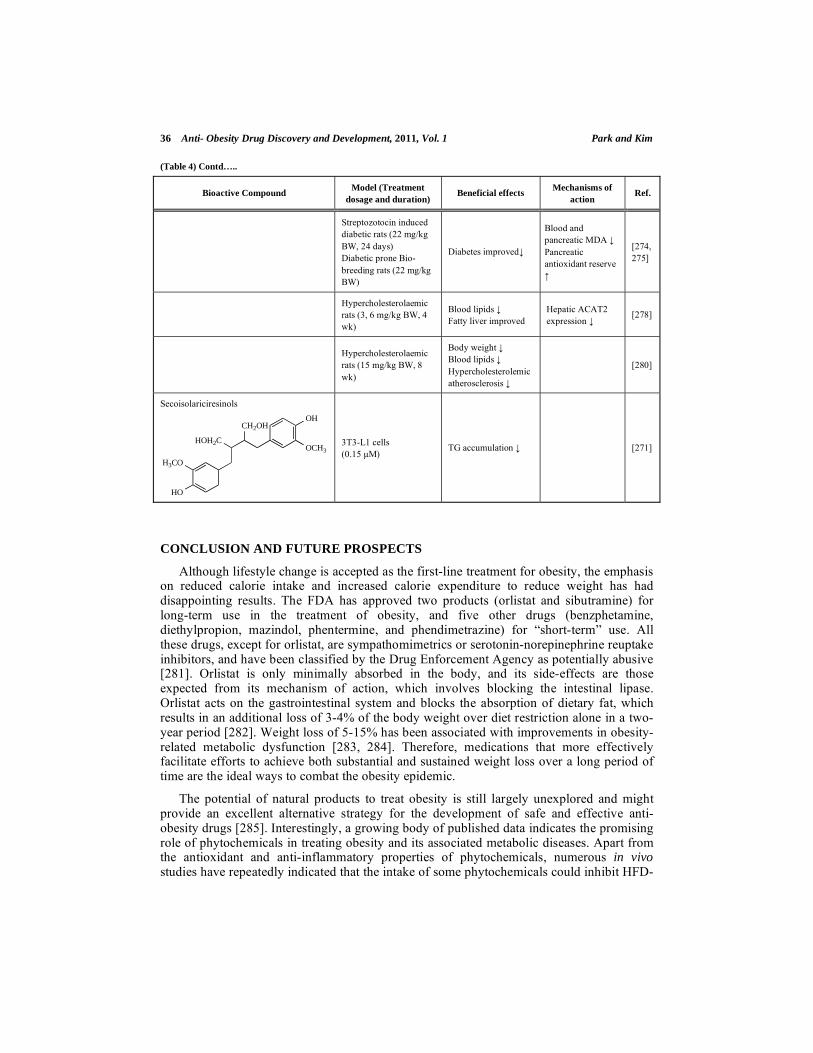
36 Anti- Obesity Drug Discovery and Development, 2011, Vol. 1 Park and Kim
(Table 4) Contd…..
Bioactive Compound Model (Treatment
dosage and duration) Beneficial effects
Mechanisms of
action Ref.
Streptozotocin induced
diabetic rats (22 mg/kg
BW, 24 days)
Diabetic prone Bio-
breeding rats (22 mg/kg
BW)
Diabetes improved
Blood and
pancreatic MDA
Pancreatic
antioxidant reserve
[274,
275]
Hypercholesterolaemic
rats (3, 6 mg/kg BW, 4
wk)
Blood lipids
Fatty liver improved
Hepatic ACAT2
expression [278]
Hypercholesterolaemic
rats (15 mg/kg BW, 8
wk)
Body weight
Blood lipids
Hypercholesterolemic
atherosclerosis
[280]
Secoisolariciresinols
HO
H3CO
HOH2C
CH2OH
OCH3
OH
3T3-L1 cells
(0.15 μM) TG accumulation [271]
CONCLUSION AND FUTURE PROSPECTS
Although lifestyle change is accepted as the first-line treatment for obesity, the emphasis on reduced calorie intake and increased calorie expenditure to reduce weight has had disappointing results. The FDA has approved two products (orlistat and sibutramine) for long-term use in the treatment of obesity, and five other drugs (benzphetamine, diethylpropion, mazindol, phentermine, and phendimetrazine) for “short-term” use. All these drugs, except for orlistat, are sympathomimetrics or serotonin-norepinephrine reuptake inhibitors, and have been classified by the Drug Enforcement Agency as potentially abusive [281]. Orlistat is only minimally absorbed in the body, and its side-effects are those expected from its mechanism of action, which involves blocking the intestinal lipase. Orlistat acts on the gastrointestinal system and blocks the absorption of dietary fat, which results in an additional loss of 3-4% of the body weight over diet restriction alone in a two-year period [282]. Weight loss of 5-15% has been associated with improvements in obesity-related metabolic dysfunction [283, 284]. Therefore, medications that more effectively facilitate efforts to achieve both substantial and sustained weight loss over a long period of time are the ideal ways to combat the obesity epidemic.
The potential of natural products to treat obesity is still largely unexplored and might provide an excellent alternative strategy for the development of safe and effective anti-obesity drugs [285]. Interestingly, a growing body of published data indicates the promising role of phytochemicals in treating obesity and its associated metabolic diseases. Apart from the antioxidant and anti-inflammatory properties of phytochemicals, numerous in vivo studies have repeatedly indicated that the intake of some phytochemicals could inhibit HFD-

Phytochemicals as Potential Agents Anti-Obesity Drug Discovery and Development, 2011, Vol. 1 37
induced obesity in mice, hamsters, rats, or even humans. The adipose tissue mass can be reduced by both inhibiting adipogenesis and inducing the apoptosis of adipocytes. Natural products that specifically target both pathways therefore have potential use in the treatment and prevention of obesity. In vitro studies have shown that some phytochemicals, such as quercetin, naringenin, EGCG, gallic acid, capsaicin, and curcumin, efficiently induce apoptosis in 3T3-L1 pre-adipocytes and adipocytes. A possible molecular mechanism for the anti-obesity action of these phytochemicals in 3T3 preadipocytes was proposed to involve the mitochondria and mitogen-activated protein kinase (MAPK) pathway, with the subsequent induction of apoptosis. Apart from inducing apoptosis, several natural compounds such as rutin, quercetin, kaempferol, ( )-catechin, EGCG, theaflavins, cyanidin, daidzein, capsaicin, curcumin, coumaric acid, ginsenosides, secoisolariciresinol diglucoside and berberine inhibited adipogenesis by downregulating the adipogenic transcription factors in the 3T3-L1 adipocytes or in the adipose tissues of obese animals. Besides these two separate mechanisms of anti-obesity action, several phytochemicals stimulate lipolysis and increase fatty acid oxidation in adipocytes. For example, EGCG, cyanidin, cyaniding 3-glucoside, curcumin, ginsenosides, ursolic acid, resveratrol, secoisolariciresinol diglucoside and berberine stimulate a dose-dependent increase in lipolysis in the adipose tissue of rodents. Numerous phytochemicals discussed in this chapter have a potential benefit in preventing both obesity and metabolic disorder, such as hyperlipidemia, fatty liver, insulin resistance and hyperglycemia, and the actions for these compounds may involve multiple independent mechanisms, including decreased adipogenesis or lipogenesis, increased fatty acid oxidation, regulated hepatic glucose metabolism along with antioxidant activities.
The FDA mandates that an effective weight-management product should provide improvements in blood pressure, lipids, glycemia, or other beneficial outcomes that are commensurate with the degree of weight loss [286]. Drugs that act ‘purely’ on one end of the energy balance equation (that is, on energy intake or expenditure) may also fail to achieve significant long-term efficacy. Because body fat is likely to be regulated homeostatically, a change in either intake or expenditure alone will face resistance as compensatory adjustments are made. In the future, it is possible that phytochemicals having multiple mechanisms simultaneously may save this dilemma. Although there is much evidence of the anti-obesity effects of phytochemicals, characterizations of their properties as drug candidates or dietary supplements have been poorly explored. Further studies that focus on understanding their pharmacokinetics and identifying their target proteins, and the accumulation of preclinical data on these interesting phytochemicals, could raise the possibility of a new application of these natural compounds as novel drugs and/or dietary supplements.
ACKNOWLEDGEMENTS
This research was supported by the SRC program (Center for Food and Nutritional Genomics: grant number 2010-0001886) of the National Research Foundation (NRF) of Korea funded by Ministry of Education, Science and Technology. The authors thank Soomin Cho and Soyoung Park for their assistance in preparing the manuscript and illustrations.
REFERENCES
[1] Jandacek RJ, Woods SC. Pharmaceutical approaches to the treatment of obesity. Drug Discov Today 2004; 9: 874-80.

38 Anti- Obesity Drug Discovery and Development, 2011, Vol. 1 Park and Kim
[2] Samuelsson G, Farah MH, Claeson P, et al. Inventory of plants used in traditional medicine in Somalia. I.
Plants of the families Acanthaceae-Chenopodiaceae. J Ethnopharmacol 1991; 35: 25-63. [3] Cox PA, Balick MJ. The ethnobotanical approach to drug discovery. Sci Am 1994; 270: 82-7.
[4] Pharmacognosy in the 21st century. J Pharm Pharmacol 2001; 53: 135-48. [5] Itokawa H, Morris-Natschke SL, Akiyama T, Lee KH. Plant-derived natural product research aimed at
new drug discovery. Nat Med (Tokyo) 2008; 62: 263-80. [6] Perez-Vizcaino F, Duarte J, Andriantsitohaina R. Endothelial function and cardiovascular disease: effects
of quercetin and wine polyphenols. Free Radic Res. 2006; 40: 1054-65. [7] Surh YJ. Anti-tumor promoting potential of selected spice ingredients with antioxidative and anti-
inflammatory activities: a short review. Food Chem Toxicol 2002; 40: 1091-7. [8] Surh Y. Molecular mechanisms of chemopreventive effects of selected dietary and medicinal phenolic
substances. Mutat Res. 1999; 428: 305-27. [9] Surh YJ, Chun KS, Cha HH, et al. Molecular mechanisms underlying chemopreventive activities of anti-
inflammatory phytochemicals: down-regulation of COX-2 and iNOS through suppression of NF-kappa B activation. Mutat Res 2001; 480: 243-68.
[10] Havsteen BH. The biochemistry and medical significance of the flavonoids. Pharmacol Ther 2002; 96: 167-202.
[11] Bravo L. Polyphenols: chemistry, dietary sources, metabolism, and nutritional significance. Nutr Rev 1998; 56: 317-33.
[12] Hu E, Liang P, Spiegelman BM. AdipoQ is a novel adipose-specific gene dysregulated in obesity. J Biol Chem 1996; 271: 10697-703.
[13] Sartippour MR, Shao ZM, Heber D, et al. Green tea inhibits vascular endothelial growth factor (VEGF) induction in human breast cancer cells. J Nutr 2002; 132: 2307-11.
[14] Kavanagh KT, Hafer LJ, Kim DW, et al. Green tea extracts decrease carcinogen-induced mammary tumor burden in rats and rate of breast cancer cell proliferation in culture. J Cell Biochem 2001; 82: 387-98.
[15] Osada K, Takahashi M, Hoshina S, Nakamura M, Nakamura S, Sugano M. Tea catechins inhibit cholesterol oxidation accompanying oxidation of low density lipoprotein in vitro. Comp Biochem Physiol
C Toxicol Pharmacol 2001; 128: 153-64. [16] Kao YH, Hiipakka RA, Liao S. Modulation of endocrine systems and food intake by green tea
epigallocatechin gallate. Endocrinology 2000; 141: 980-7. [17] Wolfram S. Effects of green tea and EGCG on cardiovascular and metabolic health. J Am Coll Nutr 2007;
26: 373S-88S. [18] Hsu TF, Kusumoto A, Abe K, et al. Polyphenol-enriched oolong tea increases fecal lipid excretion. Eur J
Clin Nutr 2006; 60: 1330-6. [19] Dulloo AG, Duret C, Rohrer D, et al. Efficacy of a green tea extract rich in catechin polyphenols and
caffeine in increasing 24-h energy expenditure and fat oxidation in humans. Am J Clin Nutr 1999; 70: 1040-5.
[20] Yang CS, Chen L, Lee MJ, Balentine D, Kuo MC, Schantz SP. Blood and urine levels of tea catechins after ingestion of different amounts of green tea by human volunteers. Cancer Epidemiol Biomarkers Prev
1998; 7: 351-4. [21] Wolfram S, Raederstorff D, Wang Y, Teixeira SR, Elste V, Weber P. TEAVIGO (epigallocatechin gallate)
supplementation prevents obesity in rodents by reducing adipose tissue mass. Ann Nutr Metab 2005; 49: 54-63.
[22] Boschmann M, Thielecke F. The effects of epigallocatechin-3-gallate on thermogenesis and fat oxidation in obese men: a pilot study. J Am Coll Nutr 2007; 26: 389S-95S.
[23] Brown AL, Lane J, Coverly J, et al. Effects of dietary supplementation with the green tea polyphenol epigallocatechin-3-gallate on insulin resistance and associated metabolic risk factors: randomized
controlled trial. Br J Nutr 2009; 101: 886-94. [24] Cho SY, Park PJ, Shin HJ, et al. (-)-Catechin suppresses expression of Kruppel-like factor 7 and increases
expression and secretion of adiponectin protein in 3T3-L1 cells. Am J Physiol Endocrinol Metab 2007; 292: E1166-72.
[25] Pajvani UB, Du X, Combs TP, et al. Structure-function studies of the adipocyte-secreted hormone Acrp30/adiponectin. Implications fpr metabolic regulation and bioactivity. J Biol Chem 2003; 278: 9073-
85. [26] Bose M, Lambert JD, Ju J, Reuhl KR, Shapses SA, Yang CS. The major green tea polyphenol, (-)-
epigallocatechin-3-gallate, inhibits obesity, metabolic syndrome, and fatty liver disease in high-fat-fed mice. J Nutr 2008; 138: 1677-83.
[27] Lin CL, Huang HC, Lin JK. Theaflavins attenuate hepatic lipid accumulation through activating AMPK in human HepG2 cells. J Lipid Res 2007; 48: 2334-43.
[28] Klaus S, Pultz S, Thone-Reineke C, Wolfram S. Epigallocatechin gallate attenuates diet-induced obesity in mice by decreasing energy absorption and increasing fat oxidation. Int J Obes (Lond) 2005; 29: 615-23.

Phytochemicals as Potential Agents Anti-Obesity Drug Discovery and Development, 2011, Vol. 1 39
[29] Tong L, Harwood HJ, Jr. Acetyl-coenzyme A carboxylases: versatile targets for drug discovery. J Cell
Biochem 2006; 99: 1476-88. [30] Wu BT, Hung PF, Chen HC, Huang RN, Chang HH, Kao YH. The apoptotic effect of green tea (-)-
epigallocatechin gallate on 3T3-L1 preadipocytes depends on the Cdk2 pathway. J Agric Food Chem 2005; 53: 5695-701.
[31] Hwang JT, Park IJ, Shin JI, et al. Genistein, EGCG, and capsaicin inhibit adipocyte differentiation process via activating AMP-activated protein kinase. Biochem Biophys Res Commun 2005; 338: 694-9.
[32] Wolfram S, Raederstorff D, Preller M, et al. Epigallocatechin gallate supplementation alleviates diabetes in rodents. J Nutr 2006; 136: 2512-8.
[33] Graham HN. Green tea composition, consumption, and polyphenol chemistry. Prev Med 1992; 21: 334-50. [34] Subramanian N, Venkatesh P, Ganguli S, Sinkar VP. Role of polyphenol oxidase and peroxidase in the
generation of black tea theaflavins. J Agric Food Chem 1999; 47: 2571-8. [35] Bushman JL. Green tea and cancer in humans: a review of the literature. Nutr Cancer 1998; 31: 151-9.
[36] Yang CS. Tea and health. Nutrition 1999; 15: 946-9. [37] Leung LK, Su Y, Chen R, Zhang Z, Huang Y, Chen ZY. Theaflavins in black tea and catechins in green
tea are equally effective antioxidants. J Nutr 2001; 131: 2248-51. [38] Kobayashi M, Ichitani M, Suzuki Y, et al. Black-tea polyphenols suppress postprandial
hypertriacylglycerolemia by suppressing lymphatic transport of dietary fat in rats. J Agric Food Chem 2009; 57: 7131-6.
[39] Vermeer MA, Mulder TP, Molhuizen HO. Theaflavins from black tea, especially theaflavin-3-gallate, reduce the incorporation of cholesterol into mixed micelles. J Agric Food Chem 2008; 56: 12031-6.
[40] Matsui T, Tanaka T, Tamura S, et al. alpha-Glucosidase inhibitory profile of catechins and theaflavins. J Agric Food Chem 2007; 55: 99-105.
[41] Lorenz M, Urban J, Engelhardt U, Baumann G, Stangl K, Stangl V. Green and black tea are equally potent stimuli of NO production and vasodilation: new insights into tea ingredients involved. Basic Res Cardiol
2009; 104: 100-10. [42] Davies MJ, Judd JT, Baer DJ, et al. Black tea consumption reduces total and LDL cholesterol in mildly
hypercholesterolemic adults. J Nutr 2003; 133: 3298S-302S. [43] Ikeda I, Kobayashi M, Hamada T, et al. Heat-epimerized tea catechins rich in gallocatechin gallate and
catechin gallate are more effective to inhibit cholesterol absorption than tea catechins rich in epigallocatechin gallate and epicatechin gallate. J Agric Food Chem 2003; 51: 7303-7.
[44] Ikeda I, Imasato Y, Sasaki E, et al. Tea catechins decrease micellar solubility and intestinal absorption of cholesterol in rats. Biochim Biophys Acta 1992; 1127: 141-6.
[45] Raederstorff DG, Schlachter MF, Elste V, Weber P. Effect of EGCG on lipid absorption and plasma lipid levels in rats. J Nutr Biochem 2003; 14: 326-32.
[46] Oki T, Matsui T, Matsumoto K. Evaluation of alpha-glucosidase inhibition by using an immobilized assay system. Biol Pharm Bull 2000; 23: 1084-7.
[47] Hauri HP, Wacker H, Rickli EE, Bigler-Meier B, Quaroni A, Semenza G. Biosynthesis of sucrase-isomaltase. Purification and NH2-terminal amino acid sequence of the rat sucrase-isomaltase precursor
(pro-sucrase-isomaltase) from fetal intestinal transplants. J Biol Chem 1982; 257: 4522-8. [48] Galvano F, La Fauci L, Vitaglione P, Fogliano V, Vanella L, Felgines C. Bioavailability, antioxidant and
biological properties of the natural free-radical scavengers cyanidin and related glycosides. Ann Ist Super Sanita 2007; 43: 382-93.
[49] Tsuda T, Horio F, Uchida K, Aoki H, Osawa T. Dietary cyanidin 3-O-beta-D-glucoside-rich purple corn color prevents obesity and ameliorates hyperglycemia in mice. J Nutr 2003; 133: 2125-30.
[50] Galvano F, La Fauci L, Lazzarino G, et al. Cyanidins: metabolism and biological properties. J Nutr Biochem 2004; 15: 2-11.
[51] Tsuda T, Ueno Y, Aoki H, et al. Anthocyanin enhances adipocytokine secretion and adipocyte-specific gene expression in isolated rat adipocytes. Biochem Biophys Res Commun 2004; 16: 149-57.
[52] Tsuda T, Ueno Y, Yoshikawa T, Kojo H, Osawa T. Microarray profiling of gene expression in human adipocytes in response to anthocyanins. Biochem Pharmacol 2006; 71: 1184-97.
[53] Tsuda T, Ueno Y, Kojo H, Yoshikawa T, Osawa T. Gene expression profile of isolated rat adipocytes treated with anthocyanins. Biochim Biophys Acta 2005; 1733: 137-47.
[54] Jayaprakasam B, Vareed SK, Olson LK, Nair MG. Insulin secretion by bioactive anthocyanins and anthocyanidins present in fruits. J Agric Food Chem 2005; 53: 28-31.
[55] Adisakwattana S, Ngamrojanavanich N, Kalampakorn K, Tiravanit W, Roengsumran S, Yibchok-Anun S. Inhibitory activity of cyanidin-3-rutinoside on alpha-glucosidase. J Enzyme Inhib Med Chem 2004; 19:
313-6. [56] Adisakwattana S, Charoenlertkul P, Yibchok-Anun S. alpha-Glucosidase inhibitory activity of cyanidin-3-
galactoside and synergistic effect with acarbose. J Enzyme Inhib Med Chem 2009; 24: 65-9.

40 Anti- Obesity Drug Discovery and Development, 2011, Vol. 1 Park and Kim
[57] Cederroth CR, Nef S. Soy, phytoestrogens and metabolism: A review. Mol Cell Endocrinol 2009; 304: 30-
42. [58] Bhathena SJ, Velasquez MT. Beneficial role of dietary phytoestrogens in obesity and diabetes. Am J Clin
Nutr 2002; 76: 1191-201. [59] Goodman-Gruen D, Kritz-Silverstein D. Usual dietary isoflavone intake is associated with cardiovascular
disease risk factors in postmenopausal women. J Nutr 2001; 131: 1202-6. [60] Goodman-Gruen D, Kritz-Silverstein D. Usual dietary isoflavone intake and body composition in
postmenopausal women. Menopause 2003; 10: 427-32. [61] Guthrie JR, Ball M, Murkies A, Dennerstein L. Dietary phytoestrogen intake in mid-life Australian-born
women: relationship to health variables. Climacteric 2000; 3: 254-61. [62] Wu J, Oka J, Higuchi M, et al. Cooperative effects of isoflavones and exercise on bone and lipid
metabolism in postmenopausal Japanese women: a randomized placebo-controlled trial. Metabolism 2006; 55: 423-33.
[63] Demonty I, Lamarche B, Deshaies Y, Jacques H. Role of soy isoflavones in the hypotriglyceridemic effect of soy protein in the rat. J Nutr Biochem 2002; 13: 671-7.
[64] Song T, Lee SO, Murphy PA, Hendrich S. Soy protein with or without isoflavones, soy germ and soy germ extract, and daidzein lessen plasma cholesterol levels in golden Syrian hamsters. Exp Biol Med
(Maywood) 2003; 228: 1063-8. [65] Lee SO, Renouf M, Ye Z, Murphy PA, Hendrich S. Isoflavone glycitein diminished plasma cholesterol in
female golden Syrian hamsters. J Agric Food Chem 2007; 55: 11063-7. [66] Naaz A, Yellayi S, Zakroczymski MA, et al. The soy isoflavone genistein decreases adipose deposition in
mice. Endocrinology 2003; 144: 3315-20. [67] Penza M, Montani C, Romani A, et al. Genistein affects adipose tissue deposition in a dose-dependent and
gender-specific manner. Endocrinology 2006; 147: 5740-51. [68] Szkudelska K, Nogowski L, Szkudelski T. Genistein affects lipogenesis and lipolysis in isolated rat
adipocytes. J Steroid Biochem Mol Biol 2000; 75: 265-71. [69] Szkudelska K, Szkudelski T, Nogowski L. Daidzein, coumestrol and zearalenone affect lipogenesis and
lipolysis in rat adipocytes. Phytomedicine 2002; 9: 338-45. [70] Harmon AW, Harp JB. Differential effects of flavonoids on 3T3-L1 adipogenesis and lipolysis. Am J
Physiol Cell Physiol 2001; 280: C807-13. [71] Nogowski L, Mackowiak P, Kandulska K, Szkudelski T, Nowak KW. Genistein-induced changes in lipid
metabolism of ovariectomized rats. Ann Nutr Metab 1998; 42: 360-6. [72] Ae Park S, Choi MS, Cho SY, et al. Genistein and daidzein modulate hepatic glucose and lipid regulating
enzyme activities in C57BL/KsJ-db/db mice. Life Sci 2006; 79: 1207-13. [73] Liu D, Zhen W, Yang Z, Carter JD, Si H, Reynolds KA. Genistein acutely stimulates insulin secretion in
pancreatic beta-cells through a cAMP-dependent protein kinase pathway. Diabetes 2006; 55: 1043-50. [74] Choi MS, Jung UJ, Yeo J, Kim MJ, Lee MK. Genistein and daidzein prevent diabetes onset by elevating
insulin level and altering hepatic gluconeogenic and lipogenic enzyme activities in non-obese diabetic (NOD) mice. Diabetes Metab Res Rev 2008; 24: 74-81.
[75] Lee JS. Effects of soy protein and genistein on blood glucose, antioxidant enzyme activities, and lipid profile in streptozotocin-induced diabetic rats. Life Sci 2006; 79: 1578-84.
[76] Yu SF, Shun CT, Chen TM, Chen YH. 3-O-beta-D-glucosyl-(1-->6)-beta-D-glucosyl-kaempferol isolated from Sauropus androgenus reduces body weight gain in Wistar rats. Biol Pharm Bull 2006; 29: 2510-3.
[77] Fang XK, Gao J, Zhu DN. Kaempferol and quercetin isolated from Euonymus alatus improve glucose uptake of 3T3-L1 cells without adipogenesis activity. Life Sci 2008; 82: 615-22.
[78] Hiermann A, Schramm HW, Laufer S. Anti-inflammatory activity of myricetin-3-O-beta-D-glucuronide and related compounds. Inflamm Res 1998; 47: 421-7.
[79] Hertog MG, Hollman PC, Katan MB, Kromhout D. Intake of potentially anticarcinogenic flavonoids and their determinants in adults in The Netherlands. Nutr Cancer 1993; 20: 21-9.
[80] Ono K, Nakane H, Fukushima M, Chermann JC, Barre-Sinoussi F. Differential inhibitory effects of various flavonoids on the activities of reverse transcriptase and cellular DNA and RNA polymerases. Eur J
Biochem 1990; 190: 469-76. [81] el-Gammal AA, Mansour RM. Antimicrobial activities of some flavonoid compounds. Zentralbl
Mikrobiol 1986; 141: 561-5. [82] Landolfi R, Mower RL, Steiner M. Modification of platelet function and arachidonic acid metabolism by
bioflavonoids. Structure-activity relations. Biochem Pharmacol 1984; 33: 1525-30. [83] Liu IM, Tzeng TF, Liou SS, Lan TW. Myricetin, a naturally occurring flavonol, ameliorates insulin
resistance induced by a high-fructose diet in rats. Life Sci 2007; 81: 1479-88. [84] Ong KC, Khoo HE. Biological effects of myricetin. Gen Pharmacol 1997; 29(2): 121-6.
[85] Liu IM, Liou SS, Lan TW, Hsu FL, Cheng JT. Myricetin as the active principle of Abelmoschus moschatus to lower plasma glucose in streptozotocin-induced diabetic rats. Planta Med 2005; 71: 617-21.

Phytochemicals as Potential Agents Anti-Obesity Drug Discovery and Development, 2011, Vol. 1 41
[86] Liu IM, Liou SS, Cheng JT. Mediation of beta-endorphin by myricetin to lower plasma glucose in
streptozotocin-induced diabetic rats. J Ethnopharmacol 2006; 104: 199-206. [87] Ong KC, Khoo HE. Insulinomimetic effects of myricetin on lipogenesis and glucose transport in rat
adipocytes but not glucose transport translocation. Biochem Pharmacol 1996; 51: 423-9. [88] Liu IM, Tzeng TF, Liou SS, Lan TW. Improvement of insulin sensitivity in obese Zucker rats by
myricetin extracted from Abelmoschus moschatus. Planta Med 2007; 73: 1054-60. [89] Ong KC, Khoo HE. Effects of myricetin on glycemia and glycogen metabolism in diabetic rats. Life Sci
2000; 67: 1695-705. [90] Strobel P, Allard C, Perez-Acle T, Calderon R, Aldunate R, Leighton F. Myricetin, quercetin and catechin-
gallate inhibit glucose uptake in isolated rat adipocytes. Biochem J 2005; 386: 471-8. [91] Chaudhry PS, Cabrera J, Juliani HR, Varma SD. Inhibition of human lens aldose reductase by flavonoids,
sulindac and indomethacin. Biochem Pharmacol 1983; 32: 1995-8. [92] Erlund I, Meririnne E, Alfthan G, Aro A. Plasma kinetics and urinary excretion of the flavanones
naringenin and hesperetin in humans after ingestion of orange juice and grapefruit juice. J Nutr 2001; 131: 235-41.
[93] Galati EM, Monforte MT, Kirjavainen S, Forestieri AM, Trovato A, Tripodo MM. Biological effects of hesperidin, a citrus flavonoid. (Note I): antiinflammatory and analgesic activity. Farmaco 1994; 40: 709-
12. [94] Wilcox LJ, Borradaile NM, de Dreu LE, Huff MW. Secretion of hepatocyte apoB is inhibited by the
flavonoids, naringenin and hesperetin, via reduced activity and expression of ACAT2 and MTP. J Lipid Res 2001; 42: 725-34.
[95] Kim HK, Jeong TS, Lee MK, Park YB, Choi MS. Lipid-lowering efficacy of hesperetin metabolites in high-cholesterol fed rats. Clin Chim Acta 2003; 327: 129-37.
[96] Cha JY, Cho YS, Kim I, Anno T, Rahman SM, Yanagita T. Effect of hesperetin, a citrus flavonoid, on the liver triacylglycerol content and phosphatidate phosphohydrolase activity in orotic acid-fed rats. Plant
Foods Hum Nutr 2001; 56: 349-58. [97] Jamil H, Dickson JK, Jr., Chu CH, et al. Microsomal triglyceride transfer protein. Specificity of lipid
binding and transport. J Biol Chem 1995; 270: 6549-54. [98] Wetterau JR, Gregg RE, Harrity TW, et al. An MTP inhibitor that normalizes atherogenic lipoprotein
levels in WHHL rabbits. Science 1998; 282: 751-4. [99] Bilheimer DW, Grundy SM, Brown MS, Goldstein JL. Mevinolin and colestipol stimulate receptor-
mediated clearance of low density lipoprotein from plasma in familial hypercholesterolemia heterozygotes. Proc Natl Acad Sci USA 1983; 80: 4124-8.
[100] Choe SC, Kim HS, Jeong TS, Bok SH, Park YB. Naringin has an antiatherogenic effect with the inhibition of intercellular adhesion molecule-1 in hypercholesterolemic rabbits. J Cardiovasc Pharmacol 2001; 38:
947-55. [101] Jung UJ, Lee MK, Park YB, Kang MA, Choi MS. Effect of citrus flavonoids on lipid metabolism and
glucose-regulating enzyme mRNA levels in type-2 diabetic mice. Int J Biochem Cell Biol 2006; 38: 1134-45.
[102] Jung UJ, Lee MK, Jeong KS, Choi MS. The hypoglycemic effects of hesperidin and naringin are partly mediated by hepatic glucose-regulating enzymes in C57BL/KsJ-db/db mice. J Nutr 2004; 134: 2499-503.
[103] Choi MS, Do KM, Park YS, et al. Effect of naringin supplementation on cholesterol metabolism and antioxidant status in rats fed high cholesterol with different levels of vitamin E. Ann Nutr Metab 2001; 45:
193-201. [104] Bok SH, Lee SH, Park YB, et al. Plasma and hepatic cholesterol and hepatic activities of 3-hydroxy-3-
methyl-glutaryl-CoA reductase and acyl CoA: cholesterol transferase are lower in rats fed citrus peel extract or a mixture of citrus bioflavonoids. J Nutr 1999; 129: 1182-5.
[105] Byington RP, Jukema JW, Salonen JT, et al. Reduction in cardiovascular events during pravastatin therapy. Pooled analysis of clinical events of the Pravastatin Atherosclerosis Intervention Program.
Circulation 1995; 92: 2419-25. [106] Delsing DJ, Offerman EH, van Duyvenvoorde W, et al. Acyl-CoA:cholesterol acyltransferase inhibitor
avasimibe reduces atherosclerosis in addition to its cholesterol-lowering effect in ApoE*3-Leiden mice. Circulation 2001; 103: 1778-86.
[107] Harmon AW, Patel YM. Naringenin inhibits phosphoinositide 3-kinase activity and glucose uptake in 3T3-L1 adipocytes. Biochem Biophys Res Commun 2003; 305: 229-34.
[108] Huong DT, Takahashi Y, Ide T. Activity and mRNA levels of enzymes involved in hepatic fatty acid oxidation in mice fed citrus flavonoids. Nutrition 2006; 22: 546-52.
[109] Lee SH, Park YB, Bae KH, et al. Cholesterol-lowering activity of naringenin via inhibition of 3-hydroxy-3-methylglutaryl coenzyme A reductase and acyl coenzyme A:cholesterol acyltransferase in rats. Ann
Nutr Metab 1999; 43: 173-80.

42 Anti- Obesity Drug Discovery and Development, 2011, Vol. 1 Park and Kim
[110] Kim SY, Kim HJ, Lee MK, et al. Naringin time-dependently lowers hepatic cholesterol biosynthesis and
plasma cholesterol in rats fed high-fat and high-cholesterol diet. J Med Food 2006; 9: 582-6. [111] Jung UJ, Kim HJ, Lee JS, et al. Naringin supplementation lowers plasma lipids and enhances erythrocyte
antioxidant enzyme activities in hypercholesterolemic subjects. Clin Nutr 2003; 22: 561-8. [112] Bischoff SC. Quercetin: potentials in the prevention and therapy of disease. Curr Opin Clin Nutr Metab
Care 2008; 11: 733-40. [113] Yamamoto Y, Oue E. Antihypertensive effect of quercetin in rats fed with a high-fat high-sucrose diet.
Biosci Biotechnol Biochem 2006; 70: 933-9. [114] Edwards RL, Lyon T, Litwin SE, Rabovsky A, Symons JD, Jalili T. Quercetin reduces blood pressure in
hypertensive subjects. J Nutr 2007; 137: 2405-11. [115] Rivera L, Moron R, Sanchez M, Zarzuelo A, Galisteo M. Quercetin ameliorates metabolic syndrome and
improves the inflammatory status in obese Zucker rats. Obesity (Silver Spring) 2008; 16: 2081-7. [116] Hsu CL, Yen GC. Induction of cell apoptosis in 3T3-L1 pre-adipocytes by flavonoids is associated with
their antioxidant activity. Mol Nutr Food Res 2006; 50: 1072-9. [117] Yang JY, Della-Fera MA, Rayalam S, et al. Enhanced inhibition of adipogenesis and induction of
apoptosis in 3T3-L1 adipocytes with combinations of resveratrol and quercetin. Life Sci 2008; 82: 1032-9. [118] Frayn KN, Karpe F, Fielding BA, Macdonald IA, Coppack SW. Integrative physiology of human adipose
tissue. Int J Obes Relat Metab Disord 2003; 27: 875-88. [119] Kuppusamy UR, Das NP. Effects of flavonoids on cyclic AMP phosphodiesterase and lipid mobilization
in rat adipocytes. Biochem Pharmacol 1992; 44: 1307-15. [120] Matter WF, Brown RF, Vlahos CJ. The inhibition of phosphatidylinositol 3-kinase by quercetin and
analogs. Biochem Biophys Res Commun 1992; 186: 624-31. [121] Agullo G, Gamet-Payrastre L, Manenti S, et al. Relationship between flavonoid structure and inhibition of
phosphatidylinositol 3-kinase: a comparison with tyrosine kinase and protein kinase C inhibition. Biochem Pharmacol 1997; 53: 1649-57.
[122] Walker EH, Pacold ME, Perisic O, et al. Structural determinants of phosphoinositide 3-kinase inhibition by wortmannin, LY294002, quercetin, myricetin, and staurosporine. Mol Cell 2000; 6: 909-19.
[123] Williams RJ, Spencer JP, Rice-Evans C. Flavonoids: antioxidants or signalling molecules? Free Radic Biol Med 2004; 36: 838-49.
[124] Gamet-Payrastre L, Manenti S, Gratacap MP, Tulliez J, Chap H, Payrastre B. Flavonoids and the inhibition of PKC and PI 3-kinase. Gen Pharmacol 1999; 32: 279-86.
[125] Gulati N, Laudet B, Zohrabian VM, Murali R, Jhanwar-Uniyal M. The antiproliferative effect of Quercetin in cancer cells is mediated via inhibition of the PI3K-Akt/PKB pathway. Anticancer Res 200;
26: 1177-81. [126] Valensi P, Le Devehat C, Richard JL, et al. A multicenter, double-blind, safety study of QR-333 for the
treatment of symptomatic diabetic peripheral neuropathy. A preliminary report. J Diabetes Complications 2005; 19: 247-53.
[127] Kuntic V, Pejic N, Ivkovic B, et al. Isocratic RP-HPLC method for rutin determination in solid oral dosage forms. J Pharm Biomed Anal 2007; 43: 718-21.
[128] Schwedhelm E, Maas R, Troost R, Boger RH. Clinical pharmacokinetics of antioxidants and their impact on systemic oxidative stress. Clin Pharmacokinet 2003; 42: 437-59.
[129] Sheu JR, Hsiao G, Chou PH, Shen MY, Chou DS. Mechanisms involved in the antiplatelet activity of rutin, a glycoside of the flavonol quercetin, in human platelets. J Agric Food Chem 2004; 52: 4414-8.
[130] Janbaz KH, Saeed SA, Gilani AH. Protective effect of rutin on paracetamol- and CCl4-induced hepatotoxicity in rodents. Fitoterapia 2002; 73: 557-63.
[131] La Casa C, Villegas I, Alarcon de la Lastra C, Motilva V, Martin Calero MJ. Evidence for protective and antioxidant properties of rutin, a natural flavone, against ethanol induced gastric lesions. J Ethnopharmacol
2000; 71: 45-53. [132] Trumbeckaite S, Bernatoniene J, Majiene D, Jakstas V, Savickas A, Toleikis A. The effect of flavonoids
on rat heart mitochondrial function. Biomed Pharmacother 2006; 60: 245-8. [133] Hsu CL, Wu CH, Huang SL, Yen GC. Phenolic compounds rutin and o-coumaric acid ameliorate obesity
induced by high-fat diet in rats. J Agric Food Chem 2009; 57: 425-31. [134] Choi I, Park Y, Choi H, Lee EH. Anti-adipogenic activity of rutin in 3T3-L1 cells and mice fed with high-
fat diet. Biofactors 2006; 26: 273-81. [135] Ziaee A, Zamansoltani F, Nassiri-Asl M, Abbasi E. Effects of rutin on lipid profile in
hypercholesterolaemic rats. Basic Clin Pharmacol Toxicol 2009; 104: 253-8. [136] Hsu CL, Yen GC. Effects of flavonoids and phenolic acids on the inhibition of adipogenesis in 3T3-L1
adipocytes. J Agric Food Chem 2007; 55: 8404-10. [137] Moon HS, Chung CS, Lee HG, Kim TG, Choi YJ, Cho CS. Inhibitory effect of (-)-epigallocatechin-3-
gallate on lipid accumulation of 3T3-L1 cells. Obesity (Silver Spring) 2007; 15: 2571-82.

Phytochemicals as Potential Agents Anti-Obesity Drug Discovery and Development, 2011, Vol. 1 43
[138] Lin J, Della-Fera MA, Baile CA. Green tea polyphenol epigallocatechin gallate inhibits adipogenesis and
induces apoptosis in 3T3-L1 adipocytes. Obes Res 2005; 13: 982-90. [139] Kobayashi Y, Suzuki M, Satsu H, et al. Green tea polyphenols inhibit the sodium-dependent glucose
transporter of intestinal epithelial cells by a competitive mechanism. J Agric Food Chem 2000; 48: 5618-23.
[140] Mackowiak P, Nogowski L, Nowak KW. Effect of isoflavone genistein on insulin receptors in perfused liver of ovariectomized rats. J Recept Signal Transduct Res 1999; 19: 283-92.
[141] Uesugi T, Toda T, Tsuji K, Ishida H. Comparative study on reduction of bone loss and lipid metabolism abnormality in ovariectomized rats by soy isoflavones, daidzin, genistin, and glycitin. Biol Pharm Bull
2001; 24: 368-72. [142] Lee MK, Moon SS, Lee SE, et al. Naringenin 7-O-cetyl ether as inhibitor of HMG-CoA reductase and
modulator of plasma and hepatic lipids in high cholesterol-fed rats. Bioorg Med Chem 2003; 11: 393-8. [143] Stalikas CD. Extraction, separation, and detection methods for phenolic acids and flavonoids. J Sep Sci
2007; 30: 3268-95. [144] Crozier A, Jaganath IB, Clifford MN. Dietary phenolics: chemistry, bioavailability and effects on health.
Nat Prod Rep 2009; 26: 1001-43. [145] Hsu CL, Yen GC. Phenolic compounds: evidence for inhibitory effects against obesity and their
underlying molecular signaling mechanisms. Mol Nutr Food Res 2008; 52: 53-61. [146] Yeh CT, Yen GC. Effect of vegetables on human phenolsulfotransferases in relation to their antioxidant
activity and total phenolics. Free Radic Res 2005; 39: 893-904. [147] Varin L, Marsolais F, Richard M, Rouleau M. Sulfation and sulfotransferases 6: Biochemistry and
molecular biology of plant sulfotransferases. FASEB J 1997; 11: 517-25. [148] Hsu CL, Huang SL, Yen GC. Inhibitory effect of phenolic acids on the proliferation of 3T3-L1
preadipocytes in relation to their antioxidant activity. J Agric Food Chem 2006 14; 54: 4191-7. [149] Hsu CL, Lo WH, Yen GC. Gallic acid induces apoptosis in 3T3-L1 pre-adipocytes via a Fas- and
mitochondrial-mediated pathway. J Agric Food Chem 2007; 55: 7359-65. [150] Hsu CL, Yen GC. Effect of gallic acid on high fat diet-induced dyslipidaemia, hepatosteatosis and
oxidative stress in rats. Br J Nutr 2007; 98: 727-35. [151] Jang A, Srinivasan P, Lee NY, et al. Comparison of hypolipidemic activity of synthetic gallic acid-linoleic
acid ester with mixture of gallic acid and linoleic acid, gallic acid, and linoleic acid on high-fat diet induced obesity in C57BL/6 Cr Slc mice. Chem Biol Interact 2008; 174: 109-17.
[152] Cordell GA, Araujo OE. Capsaicin: identification, nomenclature, and pharmacotherapy. Ann Pharmacother 1993; 27: 330-6.
[153] Yoshioka M, St-Pierre S, Drapeau V, et al. Effects of red pepper on appetite and energy intake. Br J Nutr 1999; 82: 115-23.
[154] Kawada T, Hagihara K, Iwai K. Effects of capsaicin on lipid metabolism in rats fed a high fat diet. J Nutr 1986; 116: 1272-8.
[155] Morre DJ, Chueh PJ, Morre DM. Capsaicin inhibits preferentially the NADH oxidase and growth of transformed cells in culture. Proc Natl Acad Sci USA 1995; 92: 1831-5.
[156] Macho A, Sancho R, Minassi A, Appendino G, Lawen A, Munoz E. Involvement of reactive oxygen species in capsaicinoid-induced apoptosis in transformed cells. Free Radic Res 2003; 37: 611-9.
[157] Hsu CL, Yen GC. Effects of capsaicin on induction of apoptosis and inhibition of adipogenesis in 3T3-L1 cells. J Agric Food Chem 2007; 55: 1730-6.
[158] Kang JH, Tsuyoshi G, Han IS, Kawada T, Kim YM, Yu R. Dietary Capsaicin Reduces Obesity-induced Insulin Resistance and Hepatic Steatosis in Obese Mice Fed a High-fat Diet. Obesity (Silver Spring) 2010;
18: 780-7. [159] Xu H, Barnes GT, Yang Q, et al. Chronic inflammation in fat plays a crucial role in the development of
obesity-related insulin resistance. J Clin Invest 2003; 112: 1821-30. [160] Weisberg SP, McCann D, Desai M, Rosenbaum M, Leibel RL, Ferrante AW, Jr. Obesity is associated
with macrophage accumulation in adipose tissue. J Clin Invest 2003; 112: 1796-808. [161] Matsuzawa Y, Funahashi T, Kihara S, Shimomura I. Adiponectin and metabolic syndrome. Arterioscler
Thromb Vasc Biol 2004; 24: 29-33. [162] Hausman DB, DiGirolamo M, Bartness TJ, Hausman GJ, Martin RJ. The biology of white adipocyte
proliferation. Obes Rev 2001; 2: 239-54. [163] Kang JH, Kim CS, Han IS, Kawada T, Yu R. Capsaicin, a spicy component of hot peppers, modulates
adipokine gene expression and protein release from obese-mouse adipose tissues and isolated adipocytes, and suppresses the inflammatory responses of adipose tissue macrophages. FEBS Lett 2007; 581: 4389-
96. [164] Sharma RA, Gescher AJ, Steward WP. Curcumin: the story so far. Eur J Cancer 2005; 41: 1955-68.
[165] Lao CD, Ruffin MTt, Normolle D, et al. Dose escalation of a curcuminoid formulation. BMC Complement Altern Med 2006; 6:10.

44 Anti- Obesity Drug Discovery and Development, 2011, Vol. 1 Park and Kim
[166] Dutta S, Padhye S, Priyadarsini KI, Newton C. Antioxidant and antiproliferative activity of curcumin
semicarbazone. Bioorg Med Chem Lett 2005; 15: 2738-44. [167] Lim CS, Jin DQ, Mok H, et al. Antioxidant and antiinflammatory activities of xanthorrhizol in
hippocampal neurons and primary cultured microglia. J Neurosci Res 2005; 82: 831-8. [168] Singh S, Khar A. Biological effects of curcumin and its role in cancer chemoprevention and therapy.
Anticancer Agents Med Chem 2006 ; 6: 259-70. [169] Arafa HM. Curcumin attenuates diet-induced hypercholesterolemia in rats. Med Sci Monit 2005; 11:
BR228-34. [170] Ejaz A, Wu D, Kwan P, Meydani M. Curcumin inhibits adipogenesis in 3T3-L1 adipocytes and
angiogenesis and obesity in C57/BL mice. J Nutr 2009 ; 139: 919-25. [171] Weisberg SP, Leibel R, Tortoriello DV. Dietary curcumin significantly improves obesity-associated
inflammation and diabetes in mouse models of diabesity. Endocrinology 2008; 149: 3549-58. [172] Kang Q, Chen A. Curcumin suppresses expression of low-density lipoprotein (LDL) receptor, leading to
the inhibition of LDL-induced activation of hepatic stellate cells. Br J Pharmacol 2009; 157: 1354-67. [173] Babu PS, Srinivasan K. Hypolipidemic action of curcumin, the active principle of turmeric (Curcuma
longa) in streptozotocin induced diabetic rats. Mol Cell Biochem 1997; 166: 169-75. [174] Rao DS, Sekhara NC, Satyanarayana MN, Srinivasan M. Effect of curcumin on serum and liver
cholesterol levels in the rat. J Nutr 1970 ; 100: 1307-15. [175] Peschel D, Koerting R, Nass N. Curcumin induces changes in expression of genes involved in cholesterol
homeostasis. J Nutr Biochem 2007; 18: 113-9. [176] Ramirez-Tortosa MC, Mesa MD, Aguilera MC, et al. Oral administration of a turmeric extract inhibits
LDL oxidation and has hypocholesterolemic effects in rabbits with experimental atherosclerosis. Atherosclerosis 1999 ; 147: 371-8.
[177] Guglielmi F, Luceri C, Giovannelli L, Dolara P, Lodovici M. Effect of 4-coumaric and 3,4-dihydroxybenzoic acid on oxidative DNA damage in rat colonic mucosa. Br J Nutr 2003; 89: 581-7.
[178] Luceri C, Guglielmi F, Lodovici M, Giannini L, Messerini L, Dolara P. Plant phenolic 4-coumaric acid protects against intestinal inflammation in rats. Scand J Gastroenterol 2004 ; 39: 1128-33.
[179] Luceri C, Giannini L, Lodovici M, et al. p-Coumaric acid, a common dietary phenol, inhibits platelet activity in vitro and in vivo. Br J Nutr 2007; 97: 458-63.
[180] Kawada T, Watanabe T, Takaishi T, Tanaka T, Iwai K. Capsaicin-induced beta-adrenergic action on energy metabolism in rats: influence of capsaicin on oxygen consumption, the respiratory quotient, and
substrate utilization. Proc Soc Exp Biol Med 1986; 183: 250-6. [181] Paduch R, Kandefer-Szerszen M, Trytek M, Fiedurek J. Terpenes: substances useful in human healthcare.
Arch Immunol Ther Exp (Warsz) 2007; 55: 315-27. [182] Kohlert C, van Rensen I, Marz R, Schindler G, Graefe EU, Veit M. Bioavailability and pharmacokinetics
of natural volatile terpenes in animals and humans. Planta Med 2000; 66: 495-505. [183] Kishimoto Y, Tani M, Uto-Kondo H, et al. Astaxanthin suppresses scavenger receptor expression and
matrix metalloproteinase activity in macrophages. Eur J Nutr 2010; 49: 119-26. [184] Jacobsson LS, Yuan XM, Zieden B, Olsson AG. Effects of alpha-tocopherol and astaxanthin on LDL
oxidation and atherosclerosis in WHHL rabbits. Atherosclerosis 2004; 173: 231-7. [185] Jyonouchi H, Sun S, Iijima K, Gross MD. Antitumor activity of astaxanthin and its mode of action. Nutr
Cancer 2000; 36: 59-65. [186] Chew BP, Park JS, Wong MW, Wong TS. A comparison of the anticancer activities of dietary beta-
carotene, canthaxanthin and astaxanthin in mice in vivo. Anticancer Res 1999; 19: 1849-53. [187] Naito Y, Uchiyama K, Aoi W, et al. Prevention of diabetic nephropathy by treatment with astaxanthin in
diabetic db/db mice. Biofactors 2004; 20: 49-59. [188] Guerin M, Huntley ME, Olaizola M. Haematococcus astaxanthin: applications for human health and
nutrition. Trends Biotechnol. 2003; 21: 210-216. [189] Naguib YM. Antioxidant activities of astaxanthin and related carotenoids. J Agric Food Chem 2000; 48:
1150-4. [190] Iwamoto T, Hosoda K, Hirano R, Kurata H, Matsumoto A, Miki W, et al. Inhibition of low-density
lipoprotein oxidation by astaxanthin. J Atheroscler Thromb 2000; 7: 216-22. [191] Ikeuchi M, Koyama T, Takahashi J, Yazawa K. Effects of astaxanthin in obese mice fed a high-fat diet.
Biosci Biotechnol Biochem 2007; 71: 893-9. [192] Kapoor R, Huang YS. Gamma linolenic acid: an antiinflammatory omega-6 fatty acid. Curr Pharm
Biotechnol 2006; 7: 531-4. [193] Strum-Odin R, Adkins-Finke B, Blake WL, Phinney SD, Clarke SD. Modification of fatty acid
composition of membrane phospholipid in hepatocyte monolayer with n-3, n-6 and n-9 fatty acids and its relationship to triacylglycerol production. Biochim Biophys Acta 1987; 921: 378-91.
[194] Williams MA, Tinoco J, Yang YT, Bird MI, Hincenbergs I. Feeding pure docosahexaenoate or arachidonate decreases plasma triacylglycerol secretion in rats. Lipids 1989; 24: 753-8.

Phytochemicals as Potential Agents Anti-Obesity Drug Discovery and Development, 2011, Vol. 1 45
[195] Borkman M, Storlien LH, Pan DA, Jenkins AB, Chisholm DJ, Campbell LV. The relation between insulin
sensitivity and the fatty-acid composition of skeletal-muscle phospholipids. N Engl J Med 1993; 328: 238-44.
[196] Serrero G, Lepak NM, Goodrich SP. Paracrine regulation of adipose differentiation by arachidonate metabolites: prostaglandin F2 alpha inhibits early and late markers of differentiation in the adipogenic cell
line 1246. Endocrinology 1992 ; 131: 2545-51. [197] Phinney SD, Davis PG, Johnson SB, Holman RT. Obesity and weight loss alter serum polyunsaturated
lipids in humans. Am J Clin Nutr 1991; 53: 831-8. [198] Blond JP, Henchiri C, Bezard J. Delta 6 and delta 5 desaturase activities in liver from obese Zucker rats at
different ages. Lipids. 1989; 24: 389-395. [199] Phinney SD, Tang AB, Thurmond DC, Nakamura MT, Stern JS. Abnormal polyunsaturated lipid
metabolism in the obese Zucker rat, with partial metabolic correction by gamma-linolenic acid administration. Metabolism 1993; 42: 1127-40.
[200] Phinney SD, Fisler JS, Tang AB, Warden CH. Liver fatty acid composition correlates with body fat and sex in a multigenic mouse model of obesity. Am J Clin Nutr 1994; 60: 61-7.
[201] Thurmond DC, Tang AB, Nakamura MT, Stern JS, Phinney SD. Time-dependent effects of progressive gamma-linolenate feeding on hyperphagia, weight gain, and erythrocyte fatty acid composition during
growth of Zucker obese rats. Obes Res 1993; 1: 118-25. [202] Liu CX, Xiao PG. Recent advances on ginseng research in China. J Ethnopharmacol 1992; 36: 27-38.
[203] Han KH, Choe SC, Kim HS, et al. Effect of red ginseng on blood pressure in patients with essential hypertension and white coat hypertension. Am J Chin Med 1998; 26: 199-209.
[204] Inoue M, Wu CZ, Dou DQ, Chen YJ, Ogihara Y. Lipoprotein lipase activation by red ginseng saponins in hyperlipidemia model animals. Phytomedicine 1999; 6: 257-65.
[205] Kim JH, Hahm DH, Yang DC, Lee HJ, Shim I. Effect of crude saponin of Korean red ginseng on high-fat diet-induced obesity in the rat. J Pharmacol Sci 2005; 97: 124-31.
[206] Sotaniemi EA, Haapakoski E, Rautio A. Ginseng therapy in non-insulin-dependent diabetic patients. Diabetes Care 1995; 18: 1373-5.
[207] Soltysik S, Wu JY, Recchia J, et al. Structure/function studies of QS-21 adjuvant: assessment of triterpene aldehyde and glucuronic acid roles in adjuvant function. Vaccine 1995; 13: 1403-10.
[208] Kensil CR, Soltysik S, Wheeler DA, Wu JY. Structure/function studies on QS-21, a unique immunological adjuvant from Quillaja saponaria. Adv Exp Med Biol 1996; 404: 165-72.
[209] Hwang JT, Kim SH, Lee MS, et al. Anti-obesity effects of ginsenoside Rh2 are associated with the activation of AMPK signaling pathway in 3T3-L1 adipocyte. Biochem Biophys Res Commun 2007; 364:
1002-8. [210] Hwang JT, Lee MS, Kim HJ, et al. Antiobesity effect of ginsenoside Rg3 involves the AMPK and PPAR-
gamma signal pathways. Phytother Res 2009; 23: 262-6. [211] Niu CS, Yeh CH, Yeh MF, Cheng JT. Increase of adipogenesis by ginsenoside (Rh2) in 3T3-L1 cell via an
activation of glucocorticoid receptor. Horm Metab Res 2009; 41: 271-6. [212] Lai DM, Tu YK, Liu IM, Chen PF, Cheng JT. Mediation of beta-endorphin by ginsenoside Rh2 to lower
plasma glucose in streptozotocin-induced diabetic rats. Planta Med 2006; 72: 9-13. [213] Zhang Z, Li X, Lv W, et al. Ginsenoside Re reduces insulin resistance through inhibition of c-Jun NH2-
terminal kinase and nuclear factor-kappaB. Mol Endocrinol 2008; 22: 186-95. [214] Ye J. Role of insulin in the pathogenesis of free fatty acid-induced insulin resistance in skeletal muscle.
Endocr Metab Immune Disord Drug Targets. 2007; 7: 65-74. [215] Park MW, Ha J, Chung SH. 20(S)-ginsenoside Rg3 enhances glucose-stimulated insulin secretion and
activates AMPK. Biol Pharm Bull 2008; 31: 748-51. [216] Banno N, Akihisa T, Tokuda H, et al. Triterpene acids from the leaves of Perilla frutescens and their anti-
inflammatory and antitumor-promoting effects. Biosci Biotechnol Biochem 2004; 68: 85-90. [217] Li JX, Hareyama T, Tezuka Y, Zhang Y, Miyahara T, Kadota S. Five new oleanolic acid glycosides from
Achyranthes bidentata with inhibitory activity on osteoclast formation. Planta Med 2005; 71: 673-9. [218] Liu Y, Kreppel H, Liu J, Choudhuri S, Klaassen CD. Oleanolic acid protects against cadmium
hepatotoxicity by inducing metallothionein. J Pharmacol Exp Ther 1993; 266: 400-6. [219] Kim JY, Lee KJ, Kim DH, et al. Cytokine-mediated induction of metallothionein in Hepa-1c1c7 cells by
oleanolic acid. Biochem Biophys Res Commun 2004; 325: 792-7. [220] Zhang Y, Jayaprakasam B, Seeram NP, Olson LK, DeWitt D, Nair MG. Insulin secretion and
cyclooxygenase enzyme inhibition by cabernet sauvignon grape skin compounds. J Agric Food Chem 2004; 52: 228-33.
[221] Hsu HY, Yang JJ, Lin CC. Effects of oleanolic acid and ursolic acid on inhibiting tumor growth and enhancing the recovery of hematopoietic system postirradiation in mice. Cancer Lett 1997; 111: 7-13.
[222] Sato H, Genet C, Strehle A, et al. Anti-hyperglycemic activity of a TGR5 agonist isolated from Olea europaea. Biochem Biophys Res Commun 2007; 362: 793-8.

46 Anti- Obesity Drug Discovery and Development, 2011, Vol. 1 Park and Kim
[223] Liu J. Pharmacology of oleanolic acid and ursolic acid. J Ethnopharmacol 1995; 49: 57-68.
[224] Yunoki K, Sasaki G, Tokuji Y, et al. Effect of dietary wine pomace extract and oleanolic acid on plasma lipids in rats fed high-fat diet and its DNA microarray analysis. J Agric Food Chem 2008; 56: 12052-8.
[225] Liu J. Oleanolic acid and ursolic acid: research perspectives. J Ethnopharmacol 2005; 100: 92-4. [226] Kim J, Jang DS, Kim H, Kim JS. Anti-lipase and lipolytic activities of ursolic acid isolated from the roots
of Actinidia arguta. Arch Pharm Res 2009; 32: 983-7. [227] Aggarwal BB, Shishodia S. Suppression of the nuclear factor-kappaB activation pathway by spice-derived
phytochemicals: reasoning for seasoning. Ann N Y Acad Sci 2004; 1030: 434-41. [228] Jayaprakasam B, Olson LK, Schutzki RE, Tai MH, Nair MG. Amelioration of obesity and glucose
intolerance in high-fat-fed C57BL/6 mice by anthocyanins and ursolic acid in Cornelian cherry (Cornus mas). J Agric Food Chem 2006; 54: 243-8.
[229] Sohn KH, Lee HY, Chung HY, Young HS, Yi SY, Kim KW. Anti-angiogenic activity of triterpene acids. Cancer Lett 1995; 94: 213-8.
[230] Balanehru S, Nagarajan B. Protective effect of oleanolic acid and ursolic acid against lipid peroxidation. Biochem Int 1991; 24: 981-90.
[231] Somova LO, Nadar A, Rammanan P, Shode FO. Cardiovascular, antihyperlipidemic and antioxidant effects of oleanolic and ursolic acids in experimental hypertension. Phytomedicine 2003; 10: 115-21.
[232] Shishodia S, Majumdar S, Banerjee S, Aggarwal BB. Ursolic acid inhibits nuclear factor-kappaB activation induced by carcinogenic agents through suppression of IkappaBalpha kinase and p65
phosphorylation: correlation with down-regulation of cyclooxygenase 2, matrix metalloproteinase 9, and cyclin D1. Cancer Res 2003; 63: 4375-83.
[233] Zhang W, Hong D, Zhou Y, et al. Ursolic acid and its derivative inhibit protein tyrosine phosphatase 1B, enhancing insulin receptor phosphorylation and stimulating glucose uptake. Biochim Biophys Acta 2006;
1760: 1505-12. [234] Jang SM, Yee ST, Choi J, et al. Ursolic acid enhances the cellular immune system and pancreatic beta-cell
function in streptozotocin-induced diabetic mice fed a high-fat diet. Int Immunopharmacol 2009; 9: 113-9. [235] Schirmer MA, Phinney SD. Gamma-linolenate reduces weight regain in formerly obese humans. J Nutr
2007; 137: 1430-5. [236] Ziegler J, Facchini PJ. Alkaloid biosynthesis: metabolism and trafficking. Annu Rev Plant Biol 2008; 59:
735-69. [237] Joyce SA, Brachmann AO, Glazer I, et al. Bacterial biosynthesis of a multipotent stilbene. Angew Chem
Int Ed Engl 2008; 47: 1942-5. [238] Jang M, Cai L, Udeani GO, et al. Cancer chemopreventive activity of resveratrol, a natural product
derived from grapes. Science 1997; 275: 218-20. [239] Smeds AI, Eklund PC, Sjoholm RE, et al. Quantification of a broad spectrum of lignans in cereals,
oilseeds, and nuts. J Agric Food Chem 2007; 55: 1337-46. [240] Cheng Z, Pang T, Gu M, et al. Berberine-stimulated glucose uptake in L6 myotubes involves both AMPK
and p38 MAPK. Biochim Biophys Acta 2006; 1760: 1682-9. [241] Kim SH, Shin EJ, Kim ED, Bayaraa T, Frost SC, Hyun CK. Berberine activates GLUT1-mediated glucose
uptake in 3T3-L1 adipocytes. Biol Pharm Bull 2007; 30: 2120-5. [242] Lau CW, Yao XQ, Chen ZY, Ko WH, Huang Y. Cardiovascular actions of berberine. Cardiovasc Drug
Rev 2001; 19: 234-44. [243] Yin J, Gao Z, Liu D, Liu Z, Ye J. Berberine improves glucose metabolism through induction of glycolysis.
Am J Physiol Endocrinol Metab 2008; 294: E148-56. [244] Kim WS, Lee YS, Cha SH, et al. Berberine improves lipid dysregulation in obesity by controlling central
and peripheral AMPK activity. Am J Physiol Endocrinol Metab 2009; 296: E812-9. [245] Lee YS, Kim WS, Kim KH, et al. Berberine, a natural plant product, activates AMP-activated protein
kinase with beneficial metabolic effects in diabetic and insulin-resistant states. Diabetes 2006; 55: 2256-64.
[246] Wang SH, Wang WJ, Wang XF, Chen W. Effect of Astragalus polysaccharides and berberine on carbohydrate metabolism and cell differentiation in 3T3-L1 adipocytes. Zhongguo Zhong Xi Yi Jie He Za
Zhi 2004; 24: 926-8. [247] Huang C, Zhang Y, Gong Z, et al. Berberine inhibits 3T3-L1 adipocyte differentiation through the
PPARgamma pathway. Biochem Biophys Res Commun 2006; 348: 571-8. [248] Brusq JM, Ancellin N, Grondin P, et al. Inhibition of lipid synthesis through activation of AMP kinase: an
additional mechanism for the hypolipidemic effects of berberine. J Lipid Res 2006; 47: 1281-8. [249] Kong W, Wei J, Abidi P, et al. Berberine is a novel cholesterol-lowering drug working through a unique
mechanism distinct from statins. Nat Med 2004; 10: 1344-51. [250] Zhang Y, Li X, Zou D, et al. Treatment of type 2 diabetes and dyslipidemia with the natural plant alkaloid
berberine. J Clin Endocrinol Metab 2008; 93: 2559-65.

Phytochemicals as Potential Agents Anti-Obesity Drug Discovery and Development, 2011, Vol. 1 47
[251] Ko BS, Choi SB, Park SK, Jang JS, Kim YE, Park S. Insulin sensitizing and insulinotropic action of
berberine from Cortidis rhizoma. Biol Pharm Bull 2005; 28: 1431-7. [252] Chen L, Han Y, Yang F, Zhang T. High-speed counter-current chromatography separation and purification
of resveratrol and piceid from Polygonum cuspidatum. J Chromatogr A 2001; 907: 343-6. [253] Signorelli P, Ghidoni R. Resveratrol as an anticancer nutrient: molecular basis, open questions and
promises. J Nutr Biochem 2005; 16: 449-66. [254] Das S, Cordis GA, Maulik N, Das DK. Pharmacological preconditioning with resveratrol: role of CREB-
dependent Bcl-2 signaling via adenosine A3 receptor activation. Am J Physiol Heart Circ Physiol 2005; 288: H328-35.
[255] Fremont L. Biological effects of resveratrol. Life Sci 2000; 66: 663-73. [256] Baur JA, Pearson KJ, Price NL, et al. Resveratrol improves health and survival of mice on a high-calorie
diet. Nature 2006; 444: 337-42. [257] Lagouge M, Argmann C, Gerhart-Hines Z, et al. Resveratrol improves mitochondrial function and protects
against metabolic disease by activating SIRT1 and PGC-1alpha. Cell 2006; 127: 1109-22. [258] Rayalam S, Yang JY, Ambati S, Della-Fera MA, Baile CA. Resveratrol induces apoptosis and inhibits
adipogenesis in 3T3-L1 adipocytes. Phytother Res 2008; 22: 1367-71. [259] Howitz KT, Bitterman KJ, Cohen HY, et al. Small molecule activators of sirtuins extend Saccharomyces
cerevisiae lifespan. Nature 2003; 425: 191-6. [260] Michan S, Sinclair D. Sirtuins in mammals: insights into their biological function. Biochem J 2007; 404:
1-13. [261] Guarente L. Sirtuins as potential targets for metabolic syndrome. Nature 2006; 444: 868-74.
[262] Gerhart-Hines Z, Rodgers JT, Bare O, et al. Metabolic control of muscle mitochondrial function and fatty acid oxidation through SIRT1/PGC-1alpha. EMBO J 2007; 26: 1913-23.
[263] Bujanda L, Hijona E, Larzabal M, et al. Resveratrol inhibits nonalcoholic fatty liver disease in rats. BMC Gastroenterol 2008; 8: 40.
[264] Pace-Asciak CR, Hahn S, Diamandis EP, Soleas G, Goldberg DM. The red wine phenolics trans-resveratrol and quercetin block human platelet aggregation and eicosanoid synthesis: implications for
protection against coronary heart disease. Clin Chim Acta 1995; 235: 207-19. [265] Pendurthi UR, Williams JT, Rao LV. Resveratrol, a polyphenolic compound found in wine, inhibits tissue
factor expression in vascular cells : A possible mechanism for the cardiovascular benefits associated with moderate consumption of wine. Arterioscler Thromb Vasc Biol 1999; 19: 419-26.
[266] Hung LM, Chen JK, Huang SS, Lee RS, Su MJ. Cardioprotective effect of resveratrol, a natural antioxidant derived from grapes. Cardiovasc Res 2000; 47: 549-55.
[267] Thompson LU. Experimental studies on lignans and cancer. Baillieres Clin Endocrinol Metab 1998; 12: 691-705.
[268] Axelson M, Sjovall J, Gustafsson BE, Setchell KD. Origin of lignans in mammals and identification of a precursor from plants. Nature 1982; 298: 659-60.
[269] Falany JL, Macrina N, Falany CN. Regulation of MCF-7 breast cancer cell growth by beta-estradiol sulfation. Breast Cancer Res Treat 2002; 74: 167-76.
[270] Schafer TE, Lapp CA, Hanes CM, Lewis JB, Wataha JC, Schuster GS. Estrogenicity of bisphenol A and bisphenol A dimethacrylate in vitro. J Biomed Mater Res 1999; 45: 192-7.
[271] Tominaga S, Sugahara T, Nishimoto S, et al. The Effect of Secoisolariciresinol on 3T3-L1 Adipocytes and the Relationship between Molecular Structure and Activity. Biosci Biotechnol Biochem 2009; 73: 35-9.
[272] Prasad K. Hydroxyl radical-scavenging property of secoisolariciresinol diglucoside (SDG) isolated from flax-seed. Mol Cell Biochem 1997; 168: 117-23.
[273] Prasad K. Antioxidant Activity of Secoisolariciresinol Diglucoside-derived Metabolites, Secoisolariciresinol, Enterodiol, and Enterolactone. Int J Angiol 2000; 9: 220-5.
[274] Prasad K, Mantha SV, Muir AD, Westcott ND. Protective effect of secoisolariciresinol diglucoside against streptozotocin-induced diabetes and its mechanism. Mol Cell Biochem 2000; 206: 141-9.
[275] Prasad K. Oxidative stress as a mechanism of diabetes in diabetic BB prone rats: effect of secoisolariciresinol diglucoside (SDG). Mol Cell Biochem 2000; 209: 89-96.
[276] Prasad K. Secoisolariciresinol diglucoside from flaxseed delays the development of type 2 diabetes in Zucker rat. J Lab Clin Med 2001; 138: 32-9.
[277] Fukumitsu S, Aida K, Ueno N, Ozawa S, Takahashi Y, Kobori M. Flaxseed lignan attenuates high-fat diet-induced fat accumulation and induces adiponectin expression in mice. Br J Nutr 2008; 100: 669-76.
[278] Felmlee MA, Woo G, Simko E, Krol ES, Muir AD, Alcorn J. Effects of the flaxseed lignans secoisolariciresinol diglucoside and its aglycone on serum and hepatic lipids in hyperlipidaemic rats. Br J
Nutr 2009;102: 361-9. [279] Zhou L, Yang Y, Wang X, et al. Berberine stimulates glucose transport through a mechanism distinct from
insulin. Metabolism. 2007; 56: 405-412.

48 Anti- Obesity Drug Discovery and Development, 2011, Vol. 1 Park and Kim
[280] Prasad K. Reduction of serum cholesterol and hypercholesterolemic atherosclerosis in rabbits by
secoisolariciresinol diglucoside isolated from flaxseed. Circulation 1999; 99: 1355-62. [281] Bray GA. Are non-prescription medications needed for weight control? Obesity (Silver Spring) 2008; 16:
509-14. [282] Foxcroft DR, Milne R. Orlistat for the treatment of obesity: rapid review and cost-effectiveness model.
Obes Rev 2000; 1: 121-6. [283] Douketis JD, Macie C, Thabane L, Williamson DF. Systematic review of long-term weight loss studies in
obese adults: clinical significance and applicability to clinical practice. Int J Obes (Lond) 2005; 29: 1153-67.
[284] Avenell A, Broom J, Brown TJ, et al. Systematic review of the long-term effects and economic consequences of treatments for obesity and implications for health improvement. Health Technol Assess
2004; 8: 1-182. [285] Birari RB, Bhutani KK. Pancreatic lipase inhibitors from natural sources: unexplored potential. Drug
Discov Today 2007; 12: 879-89. [286] Food and Drug Administration (FDA), Center for Drug Evaluation and Research (CDER). Draft.
Guidance for industry. Developing products for weight management. 2007. (Available at http://www.fda.gov/cder/guidance/7544dft.pdf (last accessed January 2009).