Embed Size (px)
Citation preview
PHYTOCHEMICAL SCREENING
OF UNKNOWN DRUGS
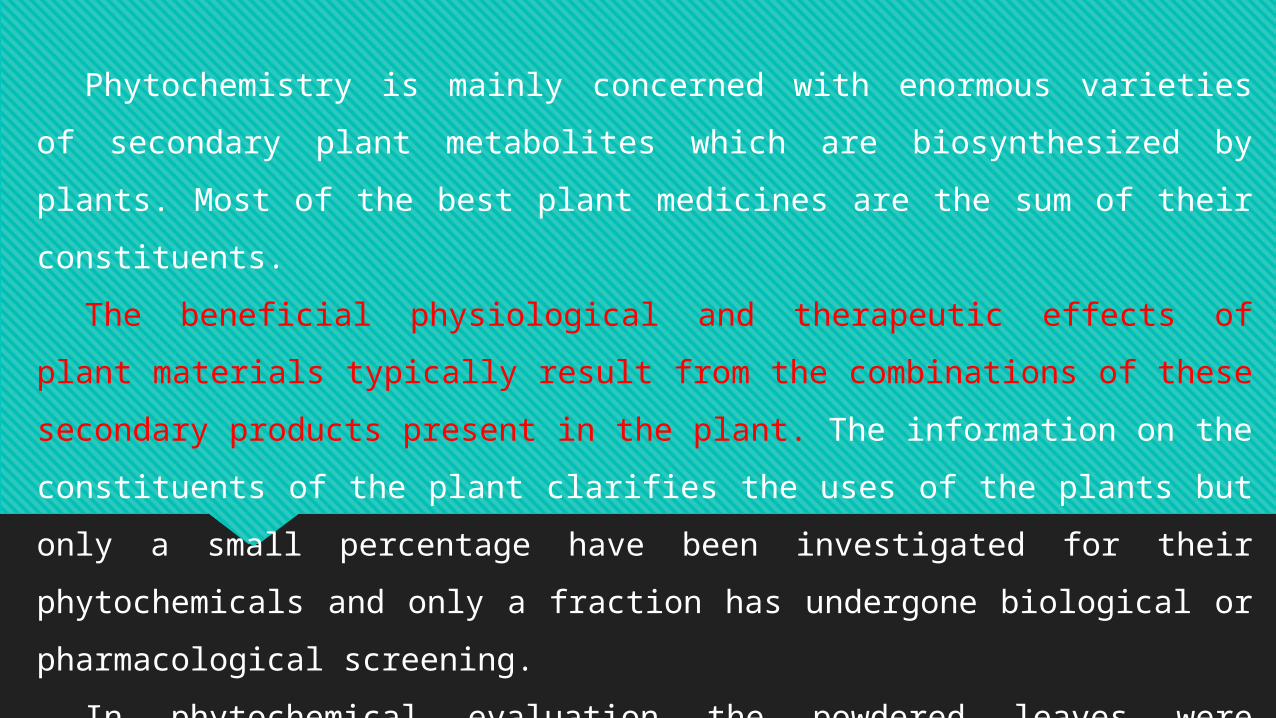
Phytochemistry is mainly concerned with enormous varieties of secondary plant
metabolites which are biosynthesized by plants. Most of the best plant medicines are the sum
of their constituents.
The beneficial physiological and therapeutic effects of plant materials typically result from
the combinations of these secondary products present in the plant. The information on the
constituents of the plant clarifies the uses of the plants but only a small percentage have been
investigated for their phytochemicals and only a fraction has undergone biological or
pharmacological screening.
In phytochemical evaluation the powdered leaves were subjected to phytochemical
screening for the detection of various plant constituents, characterized for their possible
bioactive compounds, which have been separated and subjected to detailed structural analysis.
Phytochemical analysis procedure:
After authentication, the fresh, healthy plant dry in
shade for 2-3weeks then pulverize in a blender, sieve and
use for further studies.
Subject the powdered drugs under investigation to
the following tests:
1. Detection of alkaloids:
Dissolve the extract individually in dilute Hydrochloric acid and filter.
1. a) Mayer’s Test: Treat the filtrate with Mayer’s reagent (Potassium Mercuric Iodide). Formation of a yellow colored
precipitate indicates the presence of alkaloids.
2. b) Wagner’s Test: Treat the filtrate with Wagner’s reagent (Iodine in Potassium Iodide). Formation of brown/reddish
precipitate indicates the presence of alkaloids.
3. c) Dragendroff’s Test: Treat the filtrate with Dragendroff’s reagent (solution of Potassium Bismuth Iodide). Formation
of red precipitate indicates the presence of alkaloids.
Solvent free extract, 50 mg was stirred with few ml of dilute hydrochloric acid and filtered. The filtrate was tested carefully with various alkaloidal regents as follows.
2. Detection of carbohydrates:
Dissolve the extract individually in 5 ml distilled water and filter. Use the filtrate to test for the
presence of carbohydrates.
Molisch’s Test: Treat the filtrate with 2-3 drops of 1% alcoholic α-naphthol and 2ml of concentrated
sulphuric acid was added along the sides of the test tube. Formation of the violet ring at the junction
indicates the presence of Carbohydrates.
Benedict’s test: Treat the filtrate with Benedict’s reagent and heated gently. Orange red precipitate
indicates the presence of reducing sugars.
Fehling’s Test: Treat the filtrate with dil. HCl, neutralize with alkali and heated with Fehling’s A & B
solutions. Formation of red precipitate indicates the presence of reducing sugars.
3- Detection of anthraquinone glycosides:
Extracts were hydrolyzed with dil. HCl, and then subjected to test for glycosides.
Modified Borntrager’s Test: Treat the filtrate or powder with dil. HCl, ferric chloride solution
and immerse in boiling water for about 10 minutes. Cool and extract with equal volumes of benzene
or chloroform. The benzene layer will be separated and treated with ammonia solution. Formation of
rose-pink colour in the ammonical layer indicates the presence of anthranol glycosides.
4- Detection of cardiac glycosides (Keller Kelliani’s test):
2ml of filtrate will be treated with 2ml of glacial acetic acid in a test tube and a drop of ferric
chloride solution will be added to it. This will be carefully underlayed with 1ml concentrated
sulphuric acid. A brown ring at the interface indicated the presence of deoxy sugar characteristic of
cardenolides. A violet ring may appear below the ring while in the acetic acid layer, a greenish ring
may form.
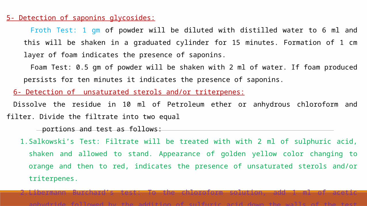
5- Detection of saponins glycosides:
Froth Test: 1 gm of powder will be diluted with distilled water to 6 ml and this will be shaken in a graduated cylinder
for 15 minutes. Formation of 1 cm layer of foam indicates the presence of saponins.
Foam Test: 0.5 gm of powder will be shaken with 2 ml of water. If foam produced persists for ten minutes it indicates
the presence of saponins.
6- Detection of unsaturated sterols and/or triterpenes:
Dissolve the residue in 10 ml of Petroleum ether or anhydrous chloroform and filter. Divide the filtrate into two equal
portions and test as follows:
1. Salkowski’s Test: Filtrate will be treated with with 2 ml of sulphuric acid, shaken and allowed to stand. Appearance of
golden yellow color changing to orange and then to red, indicates the presence of unsaturated sterols and/or
triterpenes.
2. Libermann Burchard’s test: To the chloroform solution, add 1 ml of acetic anhydride followed by the addition of
sulfuric acid down the walls of the test tube to form a layer, the formation of a reddish-violet color at the junction of
the two layers, and a green color in the chloroform solution indicates the presence of unsaturated sterols and/or
triterpenes.
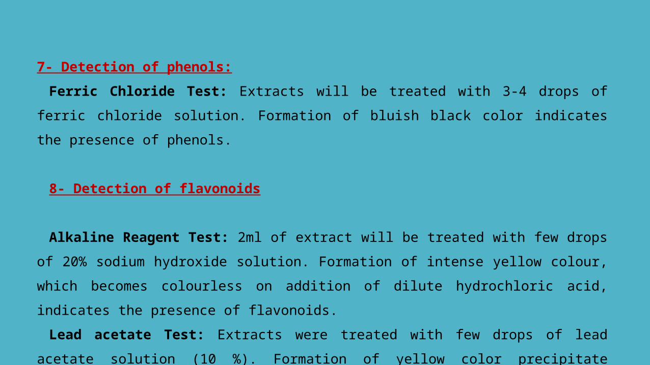
7- Detection of phenols:
Ferric Chloride Test: Extracts will be treated with 3-4 drops of ferric chloride solution. Formation of
bluish black color indicates the presence of phenols.
8- Detection of flavonoids
Alkaline Reagent Test: 2ml of extract will be treated with few drops of 20% sodium hydroxide
solution. Formation of intense yellow colour, which becomes colourless on addition of dilute
hydrochloric acid, indicates the presence of flavonoids.
Lead acetate Test: Extracts were treated with few drops of lead acetate solution (10 %). Formation of
yellow color precipitate indicates the presence of flavonoids.
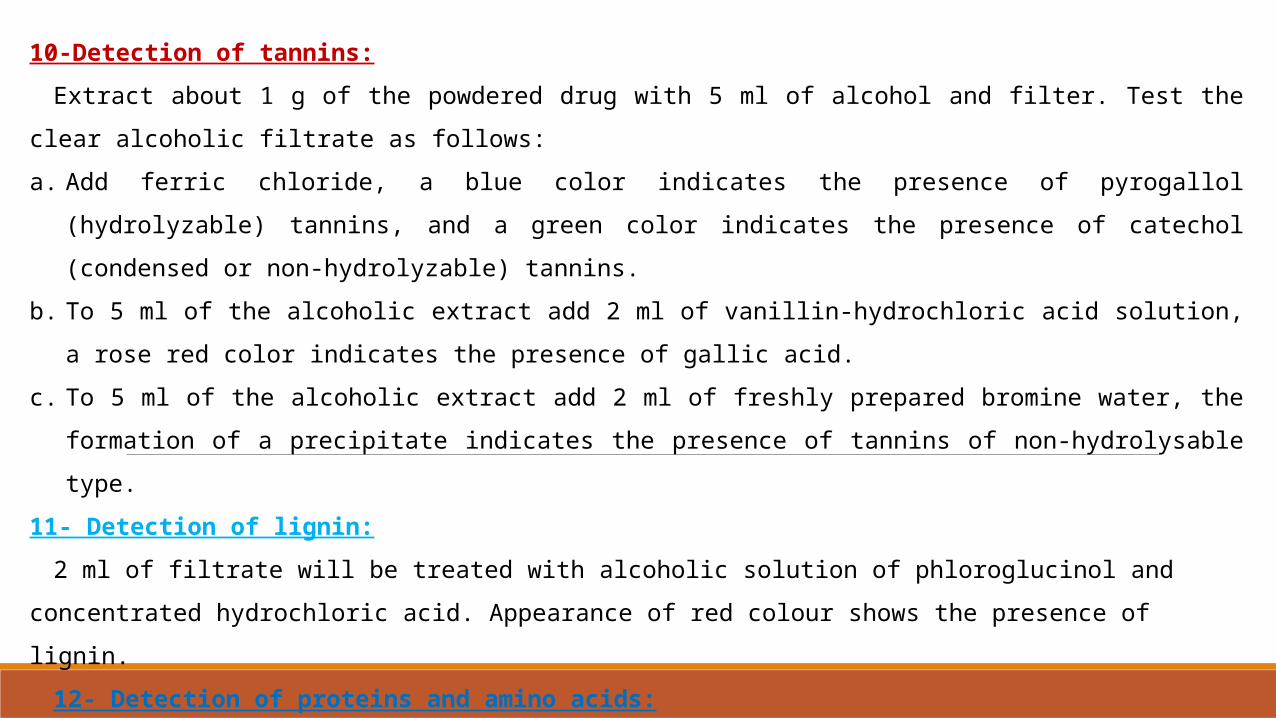
10-Detection of tannins:
Extract about 1 g of the powdered drug with 5 ml of alcohol and filter. Test the clear alcoholic filtrate as follows:
a. Add ferric chloride, a blue color indicates the presence of pyrogallol (hydrolyzable) tannins, and a green color indicates
the presence of catechol (condensed or non-hydrolyzable) tannins.
b. To 5 ml of the alcoholic extract add 2 ml of vanillin-hydrochloric acid solution, a rose red color indicates the presence of
gallic acid.
c. To 5 ml of the alcoholic extract add 2 ml of freshly prepared bromine water, the formation of a precipitate indicates the
presence of tannins of non-hydrolysable type.
11- Detection of lignin:
2 ml of filtrate will be treated with alcoholic solution of phloroglucinol and concentrated hydrochloric acid. Appearance of
red colour shows the presence of lignin.
12- Detection of proteins and amino acids:
Ninhydrin Test: (1% ninhydrin solution in acetone): 2ml of filtrate will be treated with 2-5 drops of ninhydrin solution
placed in a boiling water bath for few minutes and observed for the formation of blue to purple color. Formation of the color
indicates the presence of amino acid.