Embed Size (px)
Citation preview
MANAGEMENT OF PAIN 0195-5616/00 $15.00 + .OO
PHYSIOLOGY OF PAIN
Leigh A. Lamont, DVM, William J. Tranquilli, DVM, MS, and Kurt A. Grimm, DVM
In 1900, Sherringtongl, 92 was among the first modern neural scientists to define pain as the psychical adjunct to an imperative protective reflex. This is a concise definition, and it underlines the urgent primitive dimension of pain-the motor response that is teleologically oriented to remove tissue from potentially damaging insults. More recently, the focus has expanded to encompass the subjective emotional and motivational-affective components of pain. The Inter- national Association for the Study of Pain has proposed the following definition: pain is an unpleasant sensory and emotional experience associated with actual or potential tissue damage, or described in terms of such damage.5, 69 Thus, even though traditionally viewed as an entirely sensory phenomenon, pain differs fundamentally from other conventional sensory modalities in that numer- ous and diverse types of stimuli are capable of initiating a complex multifaceted pain response. In many ways, pain transcends attempts to define it and is best regarded as an experience involving both a physiologic sensation and an emo- tional or, as is the case for nonverbal animals, behavioral reaction to that sensation.
During the last decade, there has been an explosion in our collective knowl- edge of pain processing, and the implications for clinical practice have been substantial. The development of rational and effective pain management strate- gies requires a basic understanding of pain physiology, including (1) an apprecia- tion of the different types of inciting stimuli, (2) the neural pathways involved in processing noxious stimuli, (3) the response of the nervous system to repeated or sustained noxious input, and (4) the systemic consequences of pain. With this knowledge, the anticipation and recognition of pain are facilitated, and the use of pharmacologic agents and various hypoalgesic techniques can be optimized to better manage a variety of pain syndromes.
From the Department of Veterinary Clinical Medicine, College of Veterinary Medicine, University of Illinois, Urbana, Illinois
~ ~ ~
VETERINARY CLINICS OF NORTH AMERICA SMALL ANIMAL PRACTICE
VOLUME 30 * NUMBER 4 * JULY 2000 703
704 LAMONT et a1
PHYSIOLOGIC PAIN
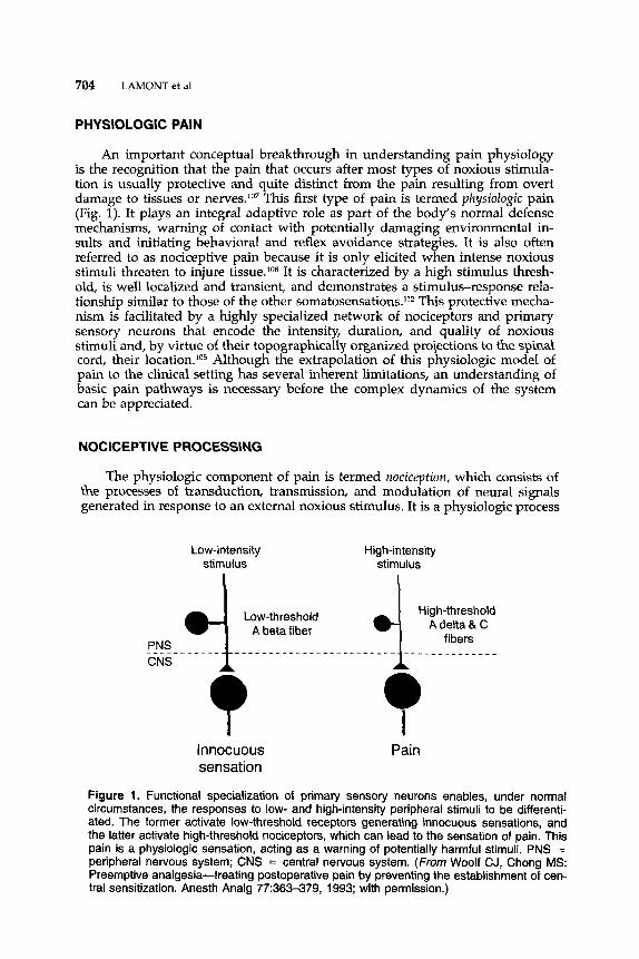
An important conceptual breakthrough in understanding pain physiology is the recognition that the pain that occurs after most types of noxious stimula- tion is usually protective and quite distinct from the pain resulting from overt damage to tissues or nerves.Io7 This first type of pain is termed physiologic pain (Fig. 1). It plays an integral adaptive role as part of the body's normal defense mechanisms, warning of contact with potentially damaging environmental in- sults and initiating behavioral and reflex avoidance strategies. It is also often referred to as nociceptive pain because it is only elicited when intense noxious stimuli threaten to injure tissue.lo8 It is characterized by a high stimulus thresh- old, is well localized and transient, and demonstrates a stimulus-response rela- tionship similar to those of the other somatosensations.1*2 This protective mecha- nism is facilitated by a highly specialized network of nociceptors and primary sensory neurons that encode the intensity, duration, and quality of noxious stimuli and, by virtue of their topographcally organized proiections to the spinal cord, their 10cation.l~~ Although the extrapolation of this physiologic model of pain to the clinical setting has several inherent limitations, an understanding of basic pain pathways is necessary before the complex dynamics of the system can be appreciated.
NOCICEPTIVE PROCESSING
The physiologic component of pain is termed nociception, which consists of the processes of transduction, transmission, and modulation of neural signals generated in response to an external noxious stimulus. It is a physiologic process
Low-intensity stimulus
High-intensity stimulus
PNS CNS - _ _ _ _
High-threshold A delta & C
fibers
I
Innocuous sensation
Pain
Figure 1. Functional specialization of primary sensory neurons enables, under normal circumstances, the responses to low- and high-intensity peripheral stimuli to be differenti- ated. The former activate low-threshold receptors generating innocuous sensations, and the latter activate high-threshold nociceptors, which can lead to the sensation of pain. This pain is a physiologic sensation, acting as a warning of potentially harmful stimuli. PNS = peripheral nervous system; CNS = central nervous system. (From Woolf CJ, Chong MS: Preemptive analgesia-treating postoperative pain by preventing the establishment of cen- tral sensitization. Anesth Analg 77:363-379, 1993; with permission.)
PHYSIOLOGY OF PAIN 705
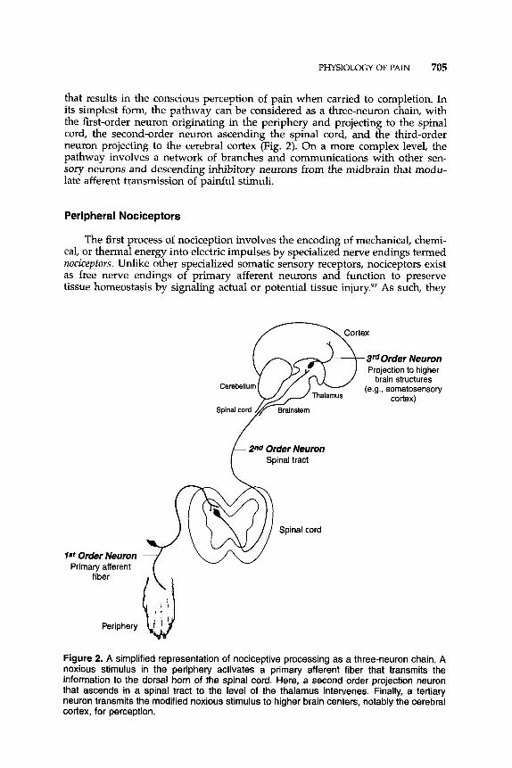
that results in the conscious perception of pain when carried to completion. In its simplest form, the pathway can be considered as a three-neuron chain, with the first-order neuron originating in the periphery and projecting to the spinal cord, the second-order neuron ascending the spinal cord, and the third-order neuron projecting to the cerebral cortex (Fig. 2). On a more complex level, the pathway involves a network of branches and communications with other sen- sory neurons and descending inhibitory neurons from the midbrain that modu- late afferent transmission of painful stimuli.
Peripheral Nociceptors
The first process of nociception involves the encoding of mechanical, chemi- cal, or thermal energy into electric impulses by specialized nerve endings termed nociceptors. Unlike other specialized somatic sensory receptors, nociceptors exist as free nerve endings of primary afferent neurons and function to preserve tissue homeostasis by signaling actual or potential tissue injury.97 As such, they
1st Order Neuron Primary afferent
fiber
Periphery
( - &+ 3d Order Neuron . . .
(e.g., somatosarisory cortex)
2nd Order Neuron Spinal tract
Spinal cord
Figure 2. A simplified representation of nociceptive processing as a three-neuron chain. A noxious stimulus in the periphery activates a primary afferent fiber that transmits the information to the dorsal horn of the spinal cord. Here, a second order projection neuron that ascends in a spinal tract to the level of the thalamus intervenes. Finally, a tertiary neuron transmits the modified noxious stimulus to higher brain centers, notably the cerebral cortex, for perception.
706 LAMONT et a1
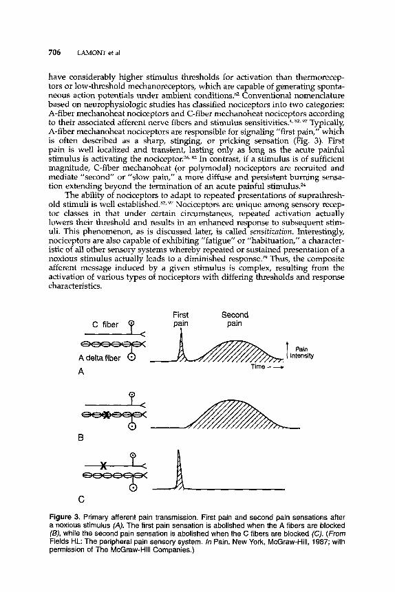
have considerably higher stimulus thresholds for activation than thermorecep- tors or low-threshold mechanoreceptors, which are capable of generating sponta- neous action potentials under ambient Conventional nomenclature based on neurophysiologic studies has classified nociceptors into two categories: A-fiber mechanoheat nociceptors and C-fiber mechanoheat nociceptors according to their associated afferent nerve fibers and stimulus sensitivities.” 97 Typically, A-fiber mechanoheat nociceptors are responsible for signaling ”first pain,” which is often described as a sharp, stinging, or pricking sensation (Fig. 3). First pain is well localized and transient, lasting only as long as the acute painful stimulus is activating the n ~ c i c e p t o r . ~ ~ , ~ ~ In contrast, if a stimulus is of sufficient magnitude, C-fiber mechanoheat (or polymodal) nociceptors are recruited and mediate “second or ”slow pain,” a more diffuse and persistent burning sensa- tion extending beyond the termination of an acute painful
The ability of nociceptors to adapt to repeated presentations of suprathresh- old stimuli is well established!*, 97 Nociceptors are unique among sensory recep- tor classes in that under certain circumstances, repeated activation actually lowers their threshold and results in an enhanced response to subsequent stim- uli. This phenomenon, as is discussed later, is called sensitization. Interestingly, nociceptors are also capable of exhibiting “fatigue” or ”habituation,” a character- istic of all other sensory systems whereby repeated or sustained presentation of a noxious stimulus actually leads to a diminished response.79 Thus, the composite afferent message induced by a given stimulus is complex, resulting from the activation of various types of nociceptors with differing thresholds and response characteristics.
First Second pain pain
I Pain
Intensity
A
P,
B
C
Time -
Figure 3. Primaty afferent pain transmission. First pain and second pain sensations after a noxious stimulus (A). The first pain sensation is abolished when the A fibers are blocked (B), while the second pain sensation is abolished when the C fibers are blocked (C). (From Fields HL: The peripheral pain sensory system. ln Pain. New York, McGraw-Hill, 1987; with permission of The McGraw-Hill Companies.)
PHYSIOLOGY OF PAIN 707
Afferent Nerve Fibers
The nociceptive signals generated by nociceptor activation are transmitted to the central nervous system by their associated afferent axons, which corre- spond to the subclasses of nociceptors outlined previously. A6 fibers are large- diameter thinly myelinated axons and consequently conduct impulses rapidly5Q 82 thereby facilitating the first pain signaled by the A fiber mechanoheat nociceptors. In contrast, transmission in the smaller unmyelinated C fibers is much slower50 and acts to reinforce the immediate response of the A fibers, becoming increasingly important as the duration of the stimulus persists. Both A6 and C fibers are located throughout the skin, peritoneum, pleura, periosteum, subchondral bone, joint capsules, blood vessels, muscles, tendons, fascia, and viscera, although their distribution density varies depending on the species and anatomic location.
Dorsal Horn Neurons
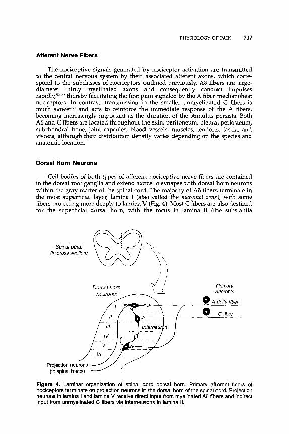
Cell bodies of both types of afferent nociceptive nerve fibers are contained in the dorsal root ganglia and extend axons to synapse with dorsal horn neurons within the gray matter of the spinal cord. The majority of A6 fibers terminate in the most superficial layer, lamina I (also called the marginal zone), with some fibers projecting more deeply to lamina V (Fig. 4). Most C fibers are also destined for the superficial dorsal horn, with the focus in lamina I1 (the substantia
Primary affei ’
A delta fiber
Dorsal horn venrs:
L
Projection neurons (to spinal tracts)
Figure 4. Laminar organization of spinal cord dorsal horn. Primary afferent fibers of nociceptors terminate on projection neurons in the dorsal horn of the spinal cord. Projection neurons in lamina I and lamina V receive direct input from myelinated A6 fibers and indirect input from unmyelinated C fibers via interneurons in lamina I I .
708 LAMONTetal
gelatinosa).5°, 96, Io8 It is in the dorsal horn that initial integration and modulation of nociceptive input occur. Primary afferent axons may form direct or indirect connections with one of three functional populations of dorsal horn neurons: (1) intemeurons, frequently divided into excitatory and inhibitory subtypes, which serve as relays and participate in local processing; (2) propriospinal neurons, which extend over multiple spinal segments and are involved in segmental reflex activity and interactions among stimuli acting at separate loci; and (3) projection neurons, which participate in rostral transmission by extending axons beyond the spinal cord to terminate in supraspinal centers such as the midbrain and the cortex.5o,% All three components are interactive and are essential for the processing of nociceptive information, which facilitates the generation of an organized and appropriate pain response.
Projection neurons have been subclassified into three groups. Nociceptive- specific (NS) neurons are concentrated in lamina I and are excited solely by noxious mechanical or thermal input from both AS and C fibers.9'j They are arranged somatotopically and respond to afferent impulses originating from discrete topographic areas.50,81, 97 Wide dynamic range (WDR) neurons predomi- nate in lamina V and receive innocuous input from low-threshold mechanorecep- tors as well as nociceptive information. They respond in a graded manner over a larger receptive field than do NS neurons and often receive convergent deep and visceral input. Although WDR neurons are considered to be ambiguous with regard to modality, they generate their strongest response to noxious stimuli, and their selective activation is capable of producing a painful sensa- tion.% A third and less well-studied group of dorsal horn neurons are termed complex neurons and are typically located in lamina VII. It is believed that these cells function in the integration of somatic and visceral afferent activity.I6, 21, 2* 96
Dorsal Horn Neurochemistry
Within the dorsal horn, the communication of nociceptive information be- tween various neurons occurs via chemical signaling mediated by excitatory and inhibitory amino acids and neuropeptides which are produced, stored, and released in the terminals of afferent nerve fibers and dorsal horn neurons.so~ 81* 96
Electrophysiologic studies have shown that the release of the excitatory amino acids glutamate and aspartate, acting as neurotransmitters, evokes fast synaptic potentials in the superficial dorsal horn neurons, thereby facilitating nociceptive transmission.z* 52, 70, % Nociceptive afferent neurons (in particular C fibers) also release a variety of other neuropeptides, including substance P, neurotensin, vasoactive intestinal peptide, calcitonin gene-related peptide, and cholecystoki- nin, which are capable of eliciting slow excitatory postsynaptic potentials in ascending projection 70, 96 Just as a barrage of nociceptive input is capable of sensitizing peripheral nociceptors, so too can sustained afferent im- pulses produce altered response characteristics in dorsal horn neurons. Indeed, the recognition of the phenomenon of "central sensitization" has had a signifi- cant impact on the development of modern pain management strategies.
Ascending Spinal Tracts
Dorsal horn nociceptive in ut is conveyed to supraspinal centers by projec- tion neurons extending througL one of several ascending pathways. f i e spino- thalamic tract (STT) is the most prominent nociceptive pathway in the spinal
PHYSIOLOGY OF PAIN 709
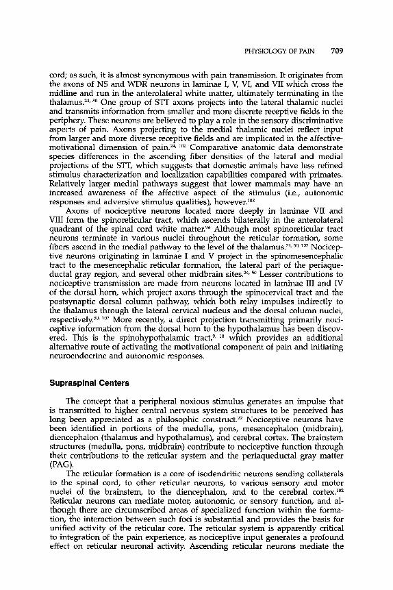
cord; as such, it is almost synonymous with pain transmission. It originates from the axons of NS and WDR neurons in laminae I, V, VI, and VII which cross the midline and run in the anterolateral white matter, ultimately terminating in the thalamus.24, 50 One group of STT axons projects into the lateral thalamic nuclei and transmits information from smaller and more discrete receptive fields in the periphery. These neurons are believed to play a role in the sensory discriminative aspects of pain. Axons projecting to the medial thalamic nuclei reflect input from larger and more diverse receptive fields and are implicated in the affective- motivational dimension of ~ain.2~. Io2 Comparative anatomic data demonstrate species differences in the ascending fiber densities of the lateral and medial projections of the STT, which suggests that domestic animals have less refined stimulus characterization and localization capabilities compared with primates. Relatively larger medial pathways suggest that lower mammals may have an increased awareness of the affective aspect of the stimulus (i.e., autonomic responses and adversive stimulus qualities), however.102
Axons of nociceptive neurons located more deeply in laminae VII and VIII form the spinoreticular tract, which ascends bilaterally in the anterolateral quadrant of the spinal cord white Although most spinoreticular tract neurons terminate in various nuclei throughout the reticular formation, some fibers ascend in the medial pathway to the level of the thalam~s?~, 50, Io2 Nocicep- tive neurons originating in laminae I and V project in the spinomesencephalic tract to the mesencephalic reticular formation, the lateral part of the periaque- ductal gray region, and several other midbrain sitesz4, 50 Lesser contributions to nociceptive transmission are made from neurons located in laminae I11 and IV of the dorsal horn, which project axons through the spinocervical tract and the postsynaptic dorsal column pathway, which both relay impulses indirectly to the thalamus through the lateral cervical nucleus and the dorsal column nuclei, respecti~ely.~~, lo2 More recently, a direct projection transmitting primarily noci- ceptive information from the dorsal horn to the hypothalamus has been discov- ered. This is the spinohypothalamic tract? lo which provides an additional alternative route of activating the motivational component of pain and initiating neuroendocrine and autonomic responses.
Supraspinal Centers
The concept that a peripheral noxious stimulus generates an impulse that is transmitted to higher central nervous system structures to be perceived has long been appreciated as a philosophic construct.22 Nociceptive neurons have been identified in portions of the medulla, pons, mesencephalon (midbrain), diencephalon (thalamus and hypothalamus), and cerebral cortex. The brainstem structures (medulla, pons, midbrain) contribute to nociceptive function through their contributions to the reticular system and the periaqueductal gray matter (PAG).
The reticular formation is a core of isodendritic neurons sending collaterals to the spinal cord, to other reticular neurons, to various sensory and motor nuclei of the brainstem, to the diencephalon, and to the cerebral cortex.1o2 Reticular neurons can mediate motor, autonomic, or sensory function, and al- though there are circumscribed areas of specialized function within the forma- tion, the interaction between such foci is substantial and provides the basis for unified activity of the reticular core. The reticular system is apparently critical to integration of the pain experience, as nociceptive input generates a profound effect on reticular neuronal activity. Ascending reticular neurons mediate the
710 L.AMONTeta1
affective and motivational aspects of pain through their projections to the medial thalamus and limbic system.
The PAG of the midbrain is a major locus of integration for homeostatic control. Although noted for its importance in the descending modulation of nociceptive information, it also extends ascending projections to the thalamus and hypothalamus, thereby providing an indirect alternative pathway for noci- ceptive sensory activity to reach diencephalic structures.2z
The thalamus serves as the relay point for sensory information en route to the cerebral cortex and is composed of numerous complex nuclei, several of which play key roles in nociception." 24, Io2 As mentioned, ascending pathways that mediate the sensory-discriminative aspects of pain terminate in the laterally located thalamic nuclei, and pathways contributing to the affective dimension of pain are destined for the medial thalamic nuclei.
The limbic system, also called the paleocortex, is derived from phylogeneti- c d y antiquated telencephalic structures as well as components of the diencepha- lon and mesencephalon. It consists of the amygdala, hippocampus, septa1 nuclei, preoptic region, hypothalamus, and certain thalamic components. Limbic struc- tures mediate aversive drive and thus influence the motivational component of pain and determine purposeful behavior.lo2
Impulse transmission to the cerebral cortex is believed to play a vital role in integrating pain perception. Imaging studies in human beings indicate that several discrete regions of cortex are activated by noxious stimulation: the first and second somatosensory cortices, the anterior insular cortex, and the anterior cingulate (a component of the limbic-associated cortex), providing convincing evidence that cortical regions are in fact targets for noxious inp~ t .9~ Although the functional and structural species differences occurring at this level are undoubtedly more significant than at any other points along the nociceptive pathway, it seems clear that the cortex is able to modulate both the cognitive and aversive affective aspects of pain sensation and to mediate increasingly complex behavior patterns.m* lo2
Thalamocortical Neurochemistry
In comparison to the primary afferent and spinal cord terminal systems, relatively little is known concerning the neurotransmitters and receptors em- ployed by nociceptive neurons or by the modulatory inputs to these neurons at the thalamic and cortical levels.22 It is believed that as is the case in the dorsal horn, glutamate and aspartate constitute the prinapal exatatory mediators in- volved in signal transmission and processing in thalamocortical systems. The inhibitory amino acids (gamma-aminobutyric acid [GABA], glycine), the mono- amines (norepinephrine, serotonin, dopamine), acetylcholine, and histamine af- fect the overall excitability of the thalamocortex in a state-dependent manner and function as part of the descending modulatory control system.
Descending Modulatory Pathways
It has been recognized for the better part of a century that descending inhibitory pathways modulate all types of sensory input. It has been established that nociceptive transmission in particular is subject to diverse and powerful inhibitory influences acting at many levels of the neuraxis. The descending modulatory system has been described as having four tiers: (1) the cortical and
PHYSIOLOGY OF PAIN 711
thalamic structures, (2) the PAG of the midbrain, (3) the rostral medulla and pons of the brainstem, and (4) the medullary and spinal cord dorsal horn.50. lM
Perhaps the most important and well-studied anatomic area contributing to the endogenous analgesia system is the mesencephalic PAG. The PAG is a cell- rich region surrounding the cerebral aqueduct and is considered by some to be a caudal extension of the limbic system into the midbrain.22, 47 The PAG receives descending input from the cortex, amygdala, and hypothalamus, and is modified by ascending projections from the medulla, reticular formation (including the locus coeruleus), and spinal lo* As noted previously, the PAG is also involved in ascending transmission via rostral connections to thalamic, hypothal- amic, and limbic structures, and caudal efferents project to the rostral ventrome- dial medulla. The antinociceptive effects observed by direct stimulation of PAG neuronal cell bodies are thought to be mediated largely by opioid activation of PAG outflow, likely operating through a GABA-containing internemma The dense concentration of opioid peptides and receptors found throughout the PAG underscores its importance as a substrate for opioid antinociception.62
The descending nociceptive inhibition arising from PAG activation is medi- ated through a relay in the rostral ventromedial medulla, facilitating projection caudally to the level of the dorsal horn. Several distinct rostral ventromedial medulla nuclei are implicated in antinociception, and all receive input from the PAG, send fibers to the spinal cord, and contribute to endogenous opioid analgesia.a* lo*
The final site involved in the descending modulation of nociceptive informa- tion is at the level of the spinal cord. Just as dorsal horn processing is vital to the integration of ascending noxious input, its role in antinociception is equally crucial. Dense concentrations of GABA, lycine, serotonin, norepinephrine and
been identified in dorsal horn neurons, and all produce inhibitory effects on nociceptive transmi~sion.~~, 29, 98, lo2 Specifically, the spinal opioid system fine- tunes descending control mechanisms by acting presynaptically (by blocking release of substance P) as well as po~tsynaptically.~~, lo2 Communication among dorsal horn neurons involves complex interactions, and it is now apparent that a single neuron may be influenced by many neurotransmitters, that each neurotransmitter may have numerous actions in a given region, and that multi- ple neurotransmitters may exist within a single -, lM Simply stated on a more global level, nociceptive processing is a three-neuron chain with dual input at each level. Discriminative and affective aspects of pain are transmitted in related but distinct ascending pathways, with modifications made by both segmental and descending modulatory systems.
the endogenous opioid peptides (enkep a alins, endorphins, dynorphins) have
PATHOLOGIC PAIN
The traditional stimulus-response model of physiologic pain is conceptually appealing and has laid the foundation for a more comprehensive understanding of nociceptive pathways. Nevertheless, it must be recognized that physiologic pain alone is a rare entity in the clinical setting. In most situations, the noxious stimulus is not transient and may be associated with significant tissue inflamma- tion and nerve injury. Under such circumstances, the classic "hard-wired system becomes less relevant, and dynamic changes in the processin of noxious input
called pathologic pain (because it implies that the tissue damage has already occurred) or clinical pain, as ongoing discomfort and abnormal sensitivity are
are evident in both peripheral and central nervous systems. 4 is type of pain is
712 LAMONT et al
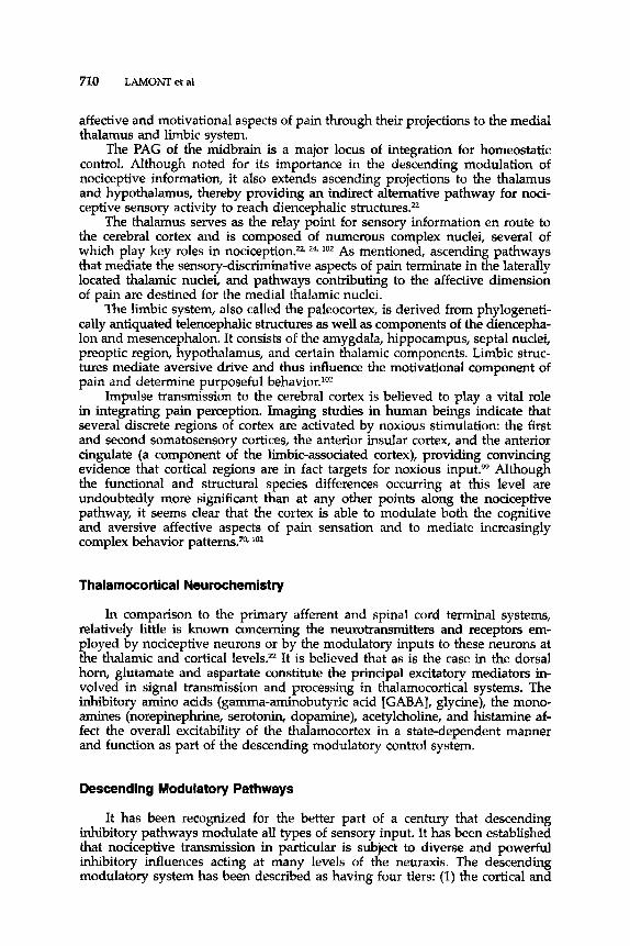
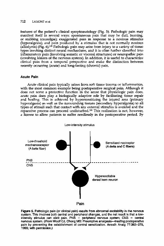
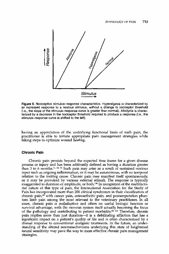
features of the patient's clinical symptomatology (Fig. 5). Pathologic pain may manifest itself in several ways: spontaneous pain that may be dull, burning, or stabbing (causalgia); exaggerated pain in response to a noxious stimulus (hyperalgesia); and pain produced by a stimulus that is not normally noxious (allodynia) (Fig. 6).loS Pathologic pain may arise from injury to a variety of tissue types invoking distinct neural mechanisms, and it is often further classified into inflammatory pain (involving somatic or visceral structures) or neuropahc pain (involving lesions of the nervous system). In addition, it is useful to characterize clinical pain from a temporal perspective and make the distinction between recently occurring (acute) and long-lasting (chronic) pain.
Acute Pain
Acute clinical pain typically arises from soft tissue trauma or inflammation, with the most common example being postoperative surgical pain. Although it does not serve a protective function in the sense that physiologic pain does, acute pain does play a biologically adaptive role by facilitating tissue repair and healing. This is achieved by hypersensitizing the injured area (primary hyperalgesia) as well as the surrounding tissues (secondary hyperalgesia) to all types of stimuli such that contact with any external stimulus is avoided and the reparative process can proceed undisturbed.*'18 This realization is not, however, a license to allow patients to suffer needlessly in the postoperative period. By
Low-intensity stimulus
Low-threshold mechanoreceptor
(A beta fiber)
Pain
Figure 5. Pathologic pain (or clinical pain) results from abnormal excitability in the nervous system. This involves both central and peripheral changes, and the net result is that a Iow- intensity stimulus can elicit pain. PNS = peripheral nervous system; CNS = central nervous system. (From Woolf CJ, Chong MS: Preemptive analgesia-treating postoperative pain by preventing the establishment of central sensitization. Anesth Analg 77:363-379, 1993; with permission.)
PHYSIOLOGY OF PAIN 713
/
Stimulus - Figure 6. Nociceptive stimulus-response characteristics. Hyperalgesia is characterized by an increased response to a noxious stimulus, without a change in nociceptor threshold (i.e., the slope of the stimulus-response cuwe is greater than normal). Allodynia is charac- terized by a decrease in the nociceptor threshold required to produce a response (i.e., the stimulus-response curve is shifted to the left).
having an appreciation of the underlying functional basis of such pain, the practitioner is able to initiate appropriate pain management strategies while taking steps to optimize wound healing.
Chronic Pain
Chronic pain persists beyond the expected time frame for a given disease process or injury and has been arbitrarily defined as having a duration greater than 3 to 6 months.37, 68, 98 Such pain may arise as a result of sustained noxious input such as ongoing inflammation, or it may be autonomous, with no temporal relation to the inciting cause. Chronic pain may manifest itself spontaneously, or it may be provoked by various external stimuli. The response is typically exaggerated in duration or amplitude, or both.lo8 In recognition of the multifacto- rial nature of t h s type of pain, the International Association for the Study of Pain has incorporated more than 200 clinical syndromes in their classification of chronic pain,@ with cancer pain, osteoarthritic pain, and postamputation phan- tom limb pain among the most relevant to the veterinary practitioner. In all cases, chronic pain is maladaptive and offers no useful biologic function or survival advantage, with the nervous system itself actually becoming the focus of the pathology and contributing to patient morbidity.@, ll1 Therefore, chronic pain implies more than just duration-it is a debilitating affliction that has a significant impact on a patient's quality of life and is often characterized by a dismal response to conventional analgesic treatments. In the future, an under- standing of the altered neuromechanisms underlying this state of heightened neural sensitivity may pave the way to more effective chronic pain management strategies.
714 LAMONT et a1
NERVOUS SYSTEM PLASTICITY
Hypersensitivity is a cardinal feature of acute and chronic pathologic pain. This phenomenon occurs as a direct result of dramatically altered nervous system function, with dynamic changes seen peripherally as a reduction in the threshold of nociceptor activation at the site of injury and centrally as an increased responsiveness of spinal neurons to sensory input.
Peripheral Sensitization
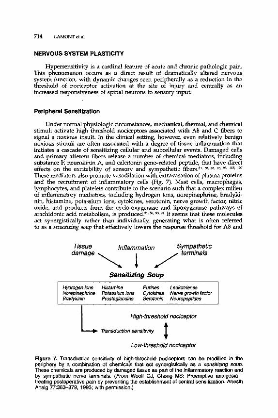
Under normal physiologic circumstances, mechanical, thermal, and chemical stimuli activate high threshold nociceptors associated with AS and C fibers to signal a noxious insult. In the clinical setting, however, even relatively benign noxious stimuli are often associated with a degree of tissue inflammation that initiates a cascade of sensitizing cellular and subcellular events. Damaged cells and primary afferent fibers release a number of chemical mediators, including substance P, neurokinin A, and calcitonin gene-related peptide, that have direct effects on the excitability of sensory and sympathetic fibers.31, 58, 84, 93, 95, Io5, Io7
These mediators also promote vasodilation with extravasation of plasma proteins and the recruitment of inflammatory cells (Fig. 7). Mast cells, macrophages, lymphocytes, and platelets contribute to the scenario such that a complex milieu of inflammatory mediators, including hydrogen ions, norepinephrine, bradyki- nin, histamine, potassium ions, cytokines, serotonin, nerve growth factor, nitric oxide, and products from the cyclo-oxygenase and lipoxygenase pathways of arachidonic acid metabolism, is 58a 93* 94 It seems that these molecules act synergistically rather than individually, generating what is often referred to as a sensitizing soup that effectively lowers the response threshold for AS and
Tissue lnflamma fion Sympathetic damage terminals
Sensitizing Soup
Hydrogen ions Histamine Purines Leukotrienes Norepinephrine Potassium ions Cyfokines Nerve growth factor Sradykinin Prostaglandins Serotonin Neuropeptides
1 High- threshold nociceptor
I Transduction sensitivity
Lo w-threshold nociceptor
Figure 7. Transduction sensitivity of high-threshold nociceptors can be modified in the periphery by a combination of chemicals that act synergistically as a sensitizing soup. These chemicals are produced by damaged tissue as part of the inflammatory reaction and by sympathetic nerve terminals. (From Woolf CJ, Chong MS: Preemptive analgesia- treating postoperative pain by preventing the establishment of central sensitization. Anesth Analg 77:363379, 1993; with permission.)
PHYSIOLOGY OF PAIN 715
C fiber activation.’03, Io8 Although changes in the afferent transduction threshold characterizing peripheral sensitization are responsible for the zone of primary hyperalgesia surrounding the site of tissue injury, they cannot explain all the behavioral aspects of pain hypersensitivity seen in the clinical lo* Fur- thermore, the identification of a subpopulation of afferent nerve terminals called ”silent” nociceptors has also contributed to our understanding of the phenome- non of peripheral ~ensitization.~~ These nociceptors are a class of unmyelinated polymodal C fibers that demonstrate little if any activity even when subjected to extreme stimulation; however, they are exquisitely sensitive to the effects of local inflammation and may discharge vigorously under such ~onditions.9~ Although they apparently exist in a variety of tissue types and species, the significance of these silent nociceptors in clinical pain syndromes has not yet been elucidated.
Central Sensitization
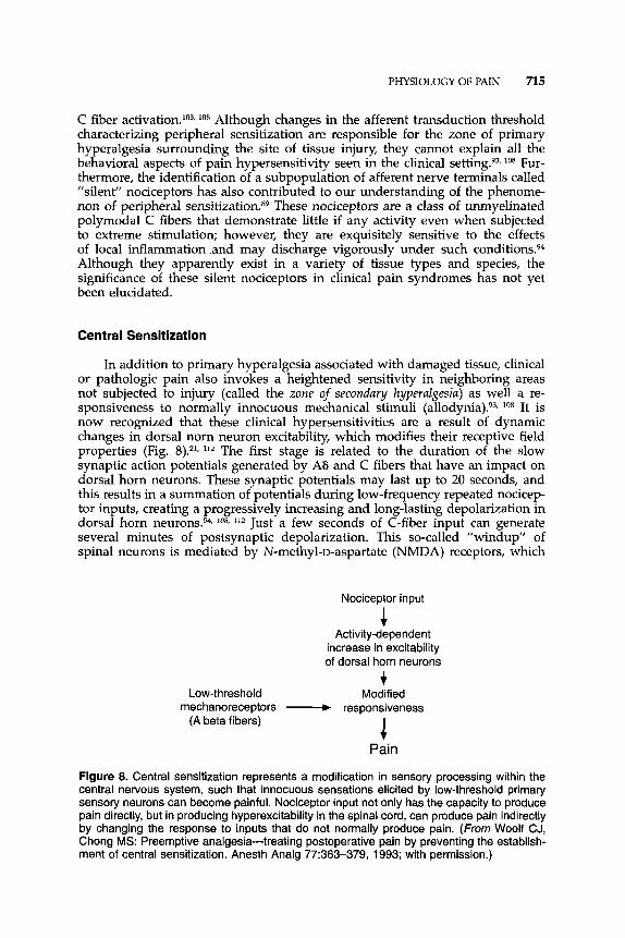
In addition to primary hyperalgesia associated with damaged tissue, clinical or pathologic pain also invokes a heightened sensitivity in neighboring areas not subjected to injury (called the zone of secondary hyperalgesia) as well a re- sponsiveness to normally innocuous mechanical stimuli (all~dynia).~~, lo* It is now recognized that these clinical hypersensitivities are a result of dynamic changes in dorsal norn neuron excitability, which modifies their receptive field properties (Fig. 8).*’, 112 The first stage is related to the duration of the slow synaptic action potentials generated by A6 and C fibers that have an impact on dorsal horn neurons. These synaptic potentials may last up to 20 seconds, and this results in a summation of potentials during low-frequency repeated nocicep- tor inputs, creating a progressively increasing and long-lasting depolarization in dorsal horn neurons.94, Just a few seconds of C-fiber input can generate several minutes of postsynaptic depolarization. This so-called “windup” of spinal neurons is mediated by N-methy1-D-aspartate (NMDA) receptors, which
Nociceptor input
Activity-dependent increase in excitability of dorsal horn neurons
4
t Low-threshold Modified
(A beta fibers) mechanoreceptors - responsiveness
4 Pain
Figure 8. Central sensitization represents a modification in sensory processing within the central nervous system, such that innocuous sensations elicited by low-threshold primary sensory neurons can become painful. Nociceptor input not only has the capacity to produce pain directly, but in producing hyperexcitability in the spinal cord, can produce pain indirectly by changing the response to inputs that do not normally produce pain. (From Woolf CJ, Chong MS: Preemptive analgesia-treating postoperative pain by preventing the establish- ment of central sensitization. Anesth Analg 77:363-379, 1993; with permission.)
716 LAMONTetal
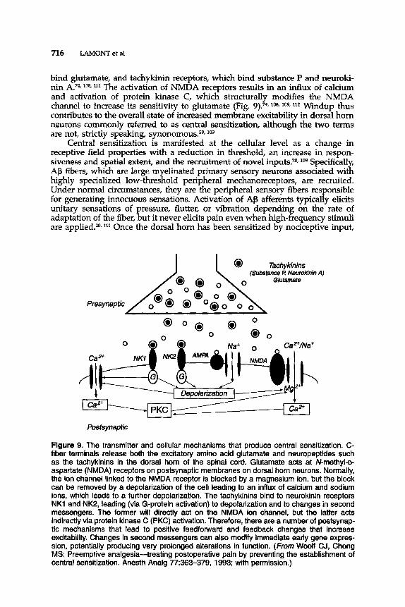
bind glutamate, and tachykinin receptors, which bind substance P and neumki- nin log, 112 The activation of NMDA receptors results in an influx of calcium and activation of protein kinase C, which structurally modifies the NMDA channel to increase its sensitivity to glutamate (Fig. 9).'** 11* Windup thus contributes to the overall state of increased membrane excitability in dorsal horn neurons commonly referred to as central sensitization, although the two terms are not, strictly speaking, synonomo~s.~~, Io9
Central sensitization is manifested at the cellular level as a change in receptive field properties with a reduction in threshold, an increase in respon- siveness and spatial extent, and the recruitment of novel inputs.m, lo9 Specifically, AP fibers, which are large myelinated primary sensory neurons associated with highly specialized low-threshold peripheral mechanoreceptors, are recruited. Under normal circumstances, they are the peripheral sensory fibers responsible for generating innocuous sensations. Activation of A$ afferents typically elicits unitary sensations of pressure, flutter, or vibration depending on the rate of adaptation of the fiber, but it never elicits pain even when high-frequency stimd are applied.20# Once the dorsal horn has been sensitized by nociceptive input,
Tachykinins (Substance F: Neumkinin A)
Qlufamate
Presynaptic
0 o n
Cap NK
0 0 0 0 0 0
Postsynap fic
Figure 9. The transmitter and cellular mechanisms that produce central sensitization. C- fiber terminals release both the excitatory amino acid glutamate and neuropeptides such as the tachykinins in the dorsal horn of the spinal cord. Glutamate acts at N-methyl-o- aspartate (NMDA) receptors on postsynaptic membranes on dorsal horn neurons. Normally, the ion channel linked to the NMDA receptor is blocked by a magnesium ion, but the block can be removed by a depolarization of the cell leading to an influx of calcium and sodium ions, which leads to a further depolarization. The tachykinins bind to neurokinin receptors NK1 and NK2, leading (via G-protein activation) to depolarization and to changes in second messengers. The former will directly act on the NMDA ion channel, but the latter acts indirectly via protein kinase C (PKC) activation. Therefore, there are a number of postsynap- tic mechanisms that lead to positive feedfornard and feedback changes that increase excitability. Changes in second messengers can also modify immediate early gene expres- sion, potentially producing vely prolonged alterations in function. (From Woolf CJ, Chong MS: Preemptive analgesia-treating postoperative pain by preventing the establishment of central sensitization. Anesth Analg 77:363-379, 1993; with permission.)
PHYSIOLOGY OF PAIN 717
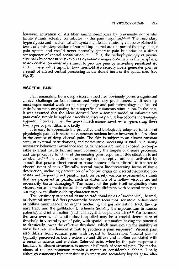
however, activation of A@ fiber mechanoreceptors by previously nonpainful tactile stimuli actually contributes to the pain response.108, ll1 The secondary hyperalgesia and mechanical allodynia manifested clinically can be explained in terms of a misinterpretation of normal inputs that are not part of the physiologic pain system and would never normally generate pain but arise as a direct consequence of central sensitization.'08, Thus, the pathophysiology of postin- jury pain hypersensitivity involves dynamic changes occurring in the periphery, which enable low-intensity stimuli to produce pain by activating sensitized A6 and C fibers, while input in low-threshold AP sensory fibers generates pain as a result of altered central processing in the dorsal horn of the spinal cord (see Fig. 8).
VISCERAL PAIN
Pain emanating from deep visceral structures obviously poses a significant clinical challenge for both human and veterinary practitioners. Until recently, most experimental work on pain physiology and pathophysiology has focused entirely on pain originating from superficial cutaneous stimulation. In the past, it was assumed that the ideas derived from a somatic model of inflammatory pain could simply be applied directly to visceral pain. It has become increasingly apparent, however, that the neural mechanisms involved in generating these two types of pain differ markedly.
It is easy to appreciate the protective and biologically adaptive function of physiologic pain as it relates to cutaneous noxious input; however, it is less clear in the context of deep visceral pain. The skin is subject to an almost constant array of external perturbations, and nociceptive processing is vital in initiating necessary behavioral avoidance strategies. Viscera are rarely exposed to compa- rable external insults but are more commonly the targets of disease processes, and the protective function of the ensuing pain response in this situation is not as obvious.67, 75 In addition, the concept of nociceptive afferents activated by stimuli that pose a direct threat to tissue homeostasis is difficult to transfer to visceral types of pain. Clinically, several major life-threatening forms of tissue destruction, including perforation of a hollow organ or visceral neoplastic pro- cesses, are frequently not painful, and, conversely, various experimental stimuli that are perceived as painful such as distention of a hollow viscous are not necessarily tissue damaging.67 The nature of the pain itself originating from visceral verses somatic tissues is significantly different, with visceral pain pos- sessing several distinguishing characteristics.
The sensitivity of visceral tissue to traditional types of mechanical, thermal, or chemical stimuli differs profoundly. Viscera seem most sensitive to distention of hollow muscular-walled organs (including the gastrointestinal tract, the uri- nary tract, and the gallbladder), ischemia (notably the myocardium in human patients), and inflammation (such as in cystitis or pancreatitis)." 67 Furthermore, the area over which a stimulus is applied may be a crucial determinant of threshold in visceral types of pain, with spatial summation having the potential to drastically lower the effective threshold, which may explain the inability of most localized mechanical stimuli to produce a pain response.67 Visceral pain also differs from somatic pain with regard to localization. Visceral pain is typically perceived as being extensive and diffuse and is often associated with a sense of nausea and malaise. Referred pain, whereby the pain response is localized to distant structures, is another hallmark of visceral pain. The mecha- nisms of this phenomenon remain a matter of considerable debate. Finally, although cutaneous hypersensitivity (primary and secondary hyperalgesia, allo-
718 LAMONT et a1
dynia) has been well characterized and repeatedly documented, few reports of similar changes occurring in viscera are available, although it does seem that inflammatory states in particular may predispose to visceral hypersensitivity.67
NEUROPATHIC PAIN
Neuropathc pain is produced as a consequence of damage to the nervous system. Like inflammatory pain states, neuropathic pain is characterized by altered sensory processing of stimuli and results in several distinct and unique manifestations of hypersensitivity.108 Patients suffering from neuropathic pain typically experience persistent burning sensations, partial or focal loss of sensi- tivity, allodynia, and hyperresponsiveness to multiple stimuli (hyperpathia).Io6 Multiple mechanisms are undoubtedly at work here, but two general categories of pathologic changes seem to contribute to neuropathc pain: abnormal periph- eral input and abnormal central processing.1ffi,
Abnormal peripheral input may arise from an acute injury discharge in axotomized afferent fibers.Io4 This discharge persists for a period of 10 or more seconds, and the collective effects generate a massive and aberrant input to the central nervous system. In addition to producing intense and excruciating pain, this input seems to produce long-lasting NMDA receptor-mediated windup in dorsal horn n e ~ r o n s . ~ ~ , ' ~ ~ Several days after this acute injury discharge, a second form of abnormal peripheral input develops, with ectopic activity originating from injured axons, the proximal axonal stump (neuroma), and cell bodies in the dorsal root ganglion.25,106 This ectopic discharge is chronic and may reflect the development of abnormal sensitivity to mechanical, thermal, or chemical stimuli. The potential also exists for cross-excitation between different types of sensory fibers or between postganglionic sympathetic fibers and sensory fibers.Io6
The phenomenon of central sensitization also contributes to the persistence and hypersensitivity associated with neuropathic pain. Afferent fiber input may arise from chronic ectopic discharge in sensory neurons as described previously, or it may be driven by sympathetic neurons exciting C fibers that have devel- oped an adrenergic sensitivity secondary to axotomy (sympathetically main- tained pain).I4 An additional form of altered central processing is observed in neuropathic pain states and involves structural reorganization in the cell bodies of injured axons in the dorsal root ganglion.lo6 Studies have demonstrated that axotomized AP fibers sprout from their normal site of termination in the deeper laminae of the dorsal horn into the superficial laminae I and 11, which are normally occupied by A6 and C fibers.loo, lI3 Nerve injury also stimulates sympa- thetic fibers to sprout around large dorsal root ganglion cells, providing another mechanism whereby postaxotomy sympathetic activity may activate nociceptive afferents.66 Sympathetically maintained pain is an important and therapeutically challenging component of neuropathic pain in human patients and presumably occurs, in varying degrees, in veterinary patients as well. Abnormal central processing as a result of a persistent state of central sensitization or dorsal horn structural reorganization may provide a unifying explanation for neuropathic pain mediated by sympathetic and AP fibers.Io6
THE NEUROMATRIX THEORY OF PAIN
It is generally accepted that future avenues in the study of pain will focus on understanding the role of the brain. In a recent editorial by M e l z a ~ k , ~ ~ ~ he
PHYSIOLOGY OF PAIN 719
proposes a new theory that challenges conventional views that the brain func- tions passively to detect and analyze nociceptive input from the periphery. The neuromatrix theory of pain proposes that pain is a multidimensional experience produced from characteristic "neurosignature" patterns arising from nerve im- pulses generated by a widely distributed neural network, the "body-self neuro- matrix," located in the brain. Although these neurosignatures may be triggered by somatosensory inputs (i.e., according to nociceptive processing), they may also be generated independently of them. Thus, the neuromatrix theory of brain function breaks the Cartesian psychophysical link between injury and pain and suggests that nociceptive input is not, in fact, a prerequisite for the experience of pain. Sensory inputs merely modulate that experience; they do not directly cause it. Although sculpted by sensory input, the neuromatrix is genetically predetermined, and so provides an attractive explanation for phantom limb pain and other examples in which the perceived level of pain is poorly correlated with This new insight into the complexity of the brain's role in modulating the experience of the body as a whole points to a new and exciting future in the understanding of pain and its management.
SYSTEMIC RESPONSES TO PAIN AND INJURY
The nervous system is the principal target of nociceptive information and provides the vehicle by which an organism can react to such input. The ensuing pain response, however, is diverse and by no means confined solely to the nervous system. Pain induces segmental and suprasegmental reflex responses that result in increased sympathetic tone, vasoconstriction, increased systemic vascular resistance, increased cardiac output through increases in stroke volume and heart rate, increased myocardial work through increases in metabolic rate and oxygen consumption, decreased gastrointestinal and urinary tone, and in- creased skeletal muscle tone.lo2, 114 Endocrine responses include increased secre- tion of corticotropin, cortisol, antidiuretic hormone, growth hormone, cyclic adenosine monophosphate, catecholamines, renin, angiotensin 11, aldosterone, glucagon, and interleukin 1, with concomitant decreases in insulin and testoster- one ~ecreti0n.l~~ Metabolically, this translates into a catabolic state characterized by hyperglycemia, increased protein catabolism and lipolysis, renal retention of water and sodium with increased potassium excretion, and decreased glomeru- lar filtration rate.'14 Nociceptive stimulation of brainstem centers causes in- creased respiratory drive, although segmental hypoventilation may occur as a result of splinting or bronchospasm. At the diencephalic and cortical levels, intense anxiety and fear greatly enhance the reflex sympathetic responses out- lined previously and contribute to increased blood viscosity, prolonged clotting time, fibrinolysis, and platelet aggregation.lo2, 114
These effects constitute the classic "stress response," the magnitude and duration of which parallel the degree of tissue damage, which often persists for days or more.6, lo2 The stress response is an evolutionary adaptation designed to optimize survival in the immediate postinjury period; however, its persistence in a clinical setting can be deleterious and have an impact on patient morbidity. In many patients with severe posttraumatic or postsurgical pain, the ensuing neuroendocrine responses are sufficient to initiate and maintain a state of shock.86 Therefore, attenuation of the stress response is an important component of any pain management strategy. Indeed, the presence or absence of stress- related physiologic changes forms the foundation of most pain assessment schemes currently used in animal patients.
720 LAMONT et a1
New Markers of Pain-Induced Stress
Suppression of the classic adrenal-pituitary axis stress hormone response has long been regarded as the best objective gauge of optimal pain management. The recognition of intracellular markers of stress has fueled renewed interest in this area. These markers are generated within dorsal horn neurons of the spinal cord and are believed to contribute to phenotypic changes in peripheral sensory neurons.1o8 The following are just a few of the currently studied intracellular markers:
1. Expression of immediate-early genes (e.g., c-fos) that code for protein products involved in initiating long-term neuronal e~citability'~, 73
2. Activation of enzymes (e.g., protein kinase C and nitric oxide synthase) that play important roles in central sensitization and development of opioid tolerance3* 15, 28
3. Secretion of nerve growth factor and neuropoietic cytokines that have widespread effects, contributing to both peripheral and central sensitization', Io8
As a more thorough understanding of dorsal horn physiology evolves, it is likely that these markers of pain-related stress may prove useful in assessing pain states and the efficacy of pharmacologic interventions.
IMPLICATIONS FOR PAIN MANAGEMENT
Most clinical pain syndromes are complex and often involve more than one type of pain. It can be difficult to predict the mechanisms mediating pain associated with multiple tissue and neuronal perturbations in a given animal. Pain associated with intervertebral disk disease or invasive soft tissue neoplasias likely has inflammatory and neuropathic components. In addition, acute and chronic pain states may occur simultaneously. An animal with osteosarcoma may present with classic symptoms of chronic inflammatory pain and hypersen- sitivity, although surgery to amputate the affected limb may generate pain sensation typical of acute tissue injury. Amputation may also initiate neuropathic pain associated with large nerve transection. It should not be surprising then that a single drug administered at a "standard dose for various pain syndromes is not an effective strategy for managing pain in all patients. The clinical objec- tive should be to minimize debilitating pathologic pain while maintaining the protective and adaptive aspects associated with physiologic pain. With this in mind, various strategies can be employed to maximize the success of therapeutic interventions.
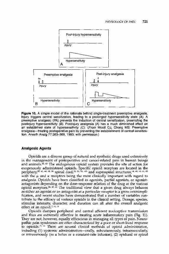
The first of these strategies is the concept of preemptive analgesia (Fig. 10). The plasticity of the nervous system in response to noxious input has been well established. Initiating treatment before acute insult is believed to inhibit periph- eral and central sensitization processes.112 The second strategy involves combin- ing analgesic drugs and techniques to achieve beneficial additive or synergistic analgesic effects (multimodal or balanced analgesia). With this approach, lower doses can usually be used, thereby reducing potential undesirable side effects.*, 54 The following is a brief review of the major classes of analgesic agents commonly used to obtund the nociceptive processes of transduction, transmission, and modulation and thus the perception of pain.
PHYSIOLOGY OF PAIN 721
Preemptive analgesia
Post-injury hypersensitivity
n Injury
Post-injury analgesia
Hypersensitivity A
Hypersensitivity B
Hypersensitivity C
I I I
Figure 10. A simple model of the rationale behind single-treatment preemptive analgesia. Injury triggers central sensitization, leading to a prolonged hypersensitivity state (A). A preemptive analgesic (PA) prevents the induction of central sensitization, preempting the postinjury hypersensitivity (B). Postinjury analgesia (A) has a much diminished effect on an established state of hypersensitivity (C). (From Woolf CJ, Chong MS: Preemptive analgesia-treating postoperative pain by preventing the establishment of central sensitiza- tion. Anesth Analg 77:363-369, 1993; with permission.)
Analgesic Agents
Opioids are a diverse group of natural and synthetic drugs used extensively in the management of postoperative and cancer-related pain in human beings and animals.45, 6o The endogenous opioid system provides the site of action for exogenously administered opioids. Specific opioid receptors are located in the periphery,35, 42, 44, 53, 58 spinal cord,15, 28, 53, 115 and supraspinal structures,44, 49, 53, 83
with the and K receptors being the most clinically important with regard to analgesia. Opioids have been classified as agonists, partial agonists, or agonist- antagonists depending on the dose-response relation of the drug at the various opioid receptors.34* 40, 45 The traditional view that a given drug always behaves as either an agonist or an antagonist at a particular receptor is a gross oversimpli- fication, and recent studies have demonstrated that a number of variables con- tribute to the efficacy of various opioids in the clinical setting. Dosage, species, stimulus intensity, character, and duration can all alter the overall analgesic effect of an ~pioid.~, 11, 72
Opioids dampen peripheral and central afferent nociceptive transmission and thus are extremely effective in treating acute inflammatory pain (Fig. 11). They are not, however, equally efficacious in managing all types of pain. Neuro- pathic pain syndromes are often characterized by a poor or short-lived response to ~pioids.'~, 26 There are several clinical methods of opioid administration, including (1) systemic administration-orally, subcutaneously, intramuscularly, or intravenously (as a bolus or a constant-rate infusion), (2) epidural or spinal
722 LAMONT et a1
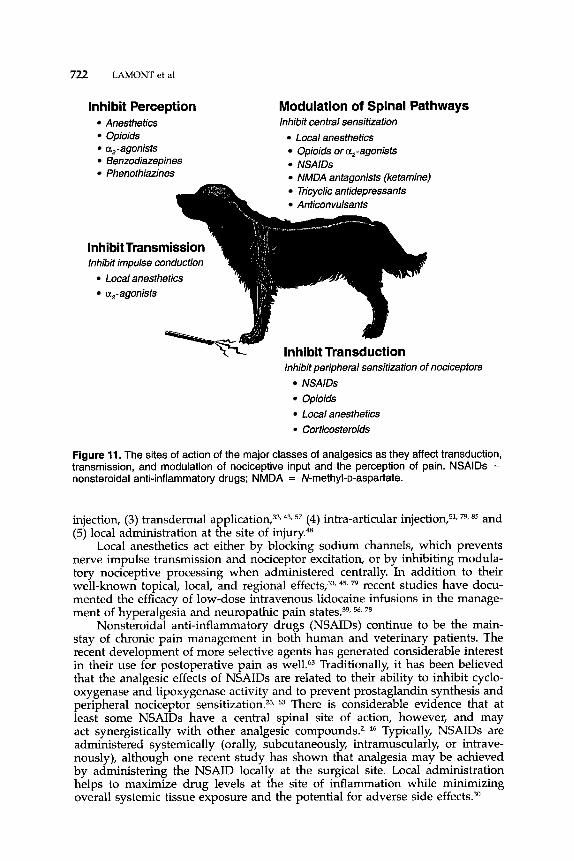
Inhibit Perception Modulation of Spinal Pathways Anesthetics Inhibit central sensitization
Opioids Local anesthetics a,-agonists Opioids or a,-agonists Benzodiazepines NSAlDs Phenothiazines NMDA antagonists (ketamine)
Trcyclic antidepressants Anticonvulsanfs
Inhibit Transmission Inhibit impulse conduction
Local anesthetics a,-agonists
Inhibit peripheral sensitization of nociceptors
NSAlDs Opioids Local anesthetics Corticosteroids
Figure 11. The sites of action of the major classes of analgesics as they affect transduction, transmission, and modulation of nociceptive input and the perception of pain. NSAlDs = nonsteroidal anti-inflammatory drugs; NMDA = N-methyl-o-aspartate.
injection, (3) transdermal appli~ation,3~, 57 (4) intra-articular 79, 85 and (5) local administration at the site of injury.@
Local anesthetics act either by blocking sodium channels, which prevents nerve impulse transmission and nociceptor excitation, or by inhibiting modula- tory nociceptive processing when administered centrally. In addition to their well-known topical, local, and regional effects:" 45, 79 recent studies have docu- mented the efficacy of low-dose intravenous lidocaine infusions in the manage- ment of hyperalgesia and neuropathic pain ~tates.3~. 56, 78
Nonsteroidal anti-inflammatory drugs (NSAIDs) continue to be the main- stay of chronic pain management in both human and veterinary patients. The recent development of more selective agents has generated considerable interest in their use for postoperative pain as well.63 Traditionally, it has been believed that the analgesic effects of NSAIDs are related to their ability to inhibit cyclo- oxygenase and lipoxygenase activity and to prevent prostaglandin synthesis and peripheral nociceptor sensitizati~n.~~, 63 There is considerable evidence that at least some NSAIDs have a central spinal site of action, however, and may act synergistically with other analgesic compounds.z, 46 Typically, NSAIDs are administered systemically (orally, subcutaneously, intramuscularly, or intrave- nously), although one recent study has shown that analgesia may be achieved by administering the NSAID locally at the surgical site. Local administration helps to maximize drug levels at the site of inflammation while minimizing overall systemic tissue exposure and the potential for adverse side effect^.^"
PHYSIOLOGY OF PAIN 723
a,-Adrenergic agonists bind to a,-receptors located in the dorsal horn of the spinal cord, modulating the release of substance P, calcitonin gene-related pep- tide, and various other neurotransmitters involved in rostra1 transmission of nociceptive inf~rmation.'~ Opioids likely exert their analgesic action through similar modulatory pathways, and coadministration may result in additive or synergistic drug interactions. a2-Agonists are used as "rescue" therapy when opioid tolerance has developed.77 The analgesic drug tramadol has both opioid and a,-agonist-like actions and has been used to treat a variety of pain syn- dromes in human patient^.'^ a2-Receptors are also located supraspinally in the locus coeruleus, thalamus, and cerebral cortex, where when activated, they inlubit norepinephrine release, resulting in profound sedation that diminishes the conscious perception of pain.I9 Although a,-receptors are notably lacking on the axons of peripheral nerves, a,-agonists are apparently capable of producing some degree of C-fiber conduction blockade. Ths action may underlie their enhancement of sensory nerve blockade when combined with local anesthetics.12, 32, 38 Thus, a,-agonists have been administered systemically, epidurally71 or periph- erally to prolong sensory nerve blockade aclrueved by local
Traditionally, ketamine has been classified as a dissociative anesthetic, but more recently, it has been recognized as an NMDA antagonist as ~e l l . 5~ . 95
Although there are multiple binding sites, it is the NMDA receptor blockade that accounts for most of the analgesic, amnestic, psychomimetic, and neuropro- tective effects of ketamine.55 At low doses, ketamine can enhance analgesia by preventing Nh4DA receptor-mediated windup and subsequent sensitization of dorsal horn neurons. It may even abolish hypersensitivity once it is already established28, 55, Ilo; therefore, the spinal cord modulatory effects of ketamine make it particularly useful in the management of chronic neuropathic types of pain that typically respond poorly to opioid treatmentF7, 28, 76, 8o Analgesic effects of ketamine may be achieved by systemic administration (either intramuscularly or intravenously as a bolus or constant-rate infusion), epidural administration, or topical application to burn injuries.
General anesthetics are not, strictly speaking, analgesics. They do, however, inhibit the perception of pain by rendering the cortex unaware of incoming nociceptive information (unconsciousness). Although a patient at a surgical plane of anesthesia is unaware of pain, it is still beneficial to inhibit peripheral transduction and transmission while enhancing spinal modulation processes so as to prevent intense noxious input from sensitizing the nervous system. For this reason, the concepts of preemptive and multimodal analgesia are relevant in this context and should be incorporated into the overall anesthetic plan when general anesthesia for surgical trauma is anticipated.
95
References
1.
2.
3.
4.
5.
6.
Anand P: Nerve growth factor regulates nociception in human health and disease. Br J Anaesth 75201-208, 1995 Bannworth 8, Demotes-Mainard F, Schaeverbeke T, et al: Central analgesic effects of aspirin-like dmgs. Fundam Clin Pharmacol 9:l-7, 1995 Basbaum AI: Insights into the development of morphine tolerance. Pain 61:349-352, 1995 Behbehani MM: Physiology and mechanisms of pain. Proceedings of the Society of Critical Care Medicine 15:55-90, 1995 Bonica JJ: Definitions and taxonomy of pain. In The Management of Pain, ed 2. Philadelphia, Lea & Febiger, 1990, pp 18-27 Bonica JJ: General considerations of acute pain. In Bonica JJ (ed): The Management of Pain, ed 2. Philadelphia, Lea & Febiger, 1990, pp 159-180
724 LAMONT et a1
7. Briggs SL, Sneed K, Sawyer DC: Antinociceptive effects of oxymorphone-butorpha- nol-acepromazine combination in cats. Vet Surg 27:46&472, 1998
8. Brodner G, Pogatzki E, Van Aken H: A multimodal approach to control postoperative pathophysiology and rehabilitation in patients undergoing abdominothoracic esopha- gectomy. Anesth Analg 86:228-234, 1998
9. Burstein R, Cliffer KD, Giesler GJ, Jr: Direct somatosensory projections from the spinal cord to the hypothalamus and telencephalon. J Neurosci 74159-4164, 1987
10. Burstein R, Dado RD, Cliffer KD, et al: Physiological characterization of spinohypo- thalamic tract neurons in the lumbar enlargement of rats. J Neurophysiol 66:261- 284, 1991
11. Burton MB, Gebhart GF: Effects of kappa-opioid receptor agonists on responses to colorectal distension in rats with and without acute colonic inflammation. J Pharmacol Exp Ther 285:707-715, 1998
12. Butterworth JF, Strichartz GR The a,-adrenergic agonists clonidine and guanfacine produce tonic and phasic block of conduction in rat sciatic nerve fibers. Anesth Analg
13. Cagney B, Williams 0, Jennings L, et al: Tramadol or fentanyl analgesia for ambula- tory knee arthroscopy. Eur J Anaesthesiol 16:182-185, 1999
14. Cambell JN, Meyer RA, Davis KD, et al: Sympathetically maintained pain. A unifying hypothesis. In Willis WD, Jr (ed): Hyperalgesia and Allodynia. New York, Raven Press, 1992, pp 141-149
15. Carr DB: Spinal opioid and nonopioid analgesia. In International Anesthesia Research Society, Review Course Lectures, 1997, pp 19-23
16. Carroll GL: Analgesics and pain. Vet Clin North Am Small Anim Pract 29:701-717, 1999
17. Cesselin F: Opioid and anti-opioid peptides. Fundam Clin Pharmacol 9:409-433, 1995 18. Chaplan SR, Sorkin LS: Agonizing over pain terminology. Pain Forum 6:81-87, 1997 19. Clark TP: Alpha,-adrenergic receptor agonists and antagonists. In Clinical Pharmacol-
ogy: Principles and Practice, Proceedings from the Western Veterinary Conference, Las Vegas, 1998, pp 116-121
20. Cook AJ, Woolf CJ, Wall I'D, et al: Dynamic receptive field plasticity in rat spinal cord dorsal horn following C-primary afferent inputs. Nature 325:151-153, 1987
21. Cousins MJ: Management of postoperative pain. In Proceedings of the International Anesthesia Research Society 60th Congress, Las Vegas, 1986, pp 32-39
22. Craig AD, Dostrovsky JO: Processing of nociceptive information at supraspinal levels. In Yaksh TL, Lynch C 111, Zapol WM, et a1 (eds): Anesthesia: Biologic Foundations. Philadelphia, Lippincott-Raven, 1997, pp 625-642
23. Cribb AE: The pharmacology of non-steroidal antiinflammatory drugs. In Clinical Pharmacology: Principles and Practice, Proceedings from the Western Veterinary Conference, Las Vegas, 1998, pp 49-58
24. Cross SA: Symposium on pain management, part I: Pathophysiology of pain. Mayo Clin Proc 69:375383, 1994
25. Devor M: Neuropathic pain and injured nerve: Peripheral mechanisms. Br Med Bull 47619430, 1991
26. Dickenson AH: Neurophysiology of opioid poorly responsive pain. Cancer Surv
27. Dickenson AH: NMDA receptor antagonists: Interactions with opioids. Acta Anaes-
28. Dickenson AH: Spinal cord pharmacology of pain. Br J Anaesth 75:193-200, 1995 29. Dickenson AH, Stanfa LC, Chapman V, et al: Response properties of dorsal horn
neurons: Pharmacology of the dorsal horn. In Yaksh TL, Lynch C 111, Zapol WM, et al (eds): Anesthesia: Biologic Foundations. Philadelphia, Lippincott-Raven, 1997, pp 611424
30. Dionne RA, Gordon SM, Tahara M, et al: Analgesic efficacy and pharmacokinetics of ketoprofen administered into a surgical site. J Clin Pharmacol 39331-138, 1999
31. Dray A: Inflammatory mediators of pain. Br J Anaesth 753255133, 1995 32. Eisenach JC, DeKock M, Klimscha W a,-Adrenergic agonists for regional anesthesia:
76:295-301, 1993
21:5-16, 1994
thesiol Scand 41:112-115, 1997
A clinical review of clonidine (1984-1995). Anesthesiology 85:655474, 1996
PHYSIOLOGY OF PAIN 725
33. Faggella AM: Management of pain in the critically ill patient. Semin Vet Med Surg 12:115-121, 1997
34. Ferrante FM: Principles of opioid pharmacology: Practical implications of basic mech- anisms. J Pain Symptom Manage 11:265-273, 1996
35. Ferreira SH, Nakamura M: Prostaglandin hyperalgesia: The peripheral analgesic activity of morphine, enkephalins and opioid antagonists. Prostaglandins 18:191- 200, 1979
36. Fields HL: The peripheral pain sensory system. In Pain. New York, McGraw-Hill, 1987, pp 1340
37. Garcia J, Altman RD: Chronic pain states: Pathophysiology and medical therapy. Semin Arthritis Rheum 271-16, 1997
38. Gaumann DM, Brunet PC, Jirounek P: Hyperpolarizing afterpotentials in C fibers and local anesthetic effects of clonidine and lidocaine. Pharmacology 48:21-29, 1994
39. Groudine SB, Fisher HAG, Kaufman RP, Jr, et al: Intravenous lidocaine speeds the return of bowel function, decreases postoperative pain, and shortens hospital stay in patients undergoing radical retropubic prostatectomy. Anesth Analg 86:235-239, 1998
40. Hansen B: Analgesics and analgesic techniques for the small animal patient. In Scientific Presentations of the 66th Annual Meeting of the American Animal Hospital Association, Denver, 1999, pp 445449
41. Hardie EM: Predictable pain management: Chronic pain. In Proceedings of the North American Veterinary Conference, Orlando, 1996, pp 21-25
42. Hassan A, Ableitner A, Stein C, et al: Inflammation of the rat paw enhances axonal transport of opioid receptors in the sciatic nerve and increases their density in inflamed tissue. Neuroscience 55:185-195, 1993
43. Heinricher MM: Organizational characteristics of supraspinally mediated responses to nociceptive input. In Yaksh TL, Lynch C 111, Zapol WM, et a1 (eds): Anesthesia: Biologic Foundations. Philadelphia, Lippincott-Raven, 1997, pp 643-662
44. Heinricher MM, Morgan MM, Fields HL: Direct and indirect action of morphine on medullary neurons that modulate nociception. Neuroscience 48533-543, 1992
45. Hellyer P W Management of acute and surgical pain. Semin Vet Med Surg 12:106 114, 1997
46. Herrero JF, Headley P M Reversal by naloxone of the spinal antinociceptive actions of a systemically administered NSAID. Br J Pharmacol 118:968-972, 1996
47. Holstege G: Descending pathways from the periaqueductal gray and adjacent areas. In DePaulis A, Bandler R (eds): The Midbrain Periaqueductal Gray Matter. New York, Plenum Press, 1991, pp 239-265
48. Houghton AK, Valdez JG, Westlund KN: Peripheral morphine administration blocks the development of hyperalgesia and allodynia after bone damage in the rat. Anesthe- siology 89:190-201, 1998
49. Jensen TS: Opioids in the brain. Supraspinal mechanisms in pain control. Acta Anaesthesiol Scand 41:123-132, 1997
50. Jessell TM, Kelly DD: Pain and analgesia. In Kandel ER, Schwartz JH, Jessell TM (eds): Principles of Neural Science, ed 3. New York, Elsevier Science, 1991, pp 385-399
51. Joshi GP, McCarroll SM, Brady OH, et al: Intra-articular morphine for pain relief after anterior cruciate ligament repair. Br J Anaesth 70:87-88, 1993
52. Kangarga I, Randic M Outflow of endogenous aspartate and glutamate from rat spinal cord dorsal horn in vitro by activation of low- and high-threshold primary afferent fibers. Modulation by mu-opioids. Brain Res 553:347-352, 1991
53. Kanjhan R: Opioids and pain. Clin Exp Pharmacol Physiol22397403, 1995 54. Kehlet H, Dahl JB: The value of “multimodal” or “balanced analgesia” in postopera-
55. Kohrs R, Durieux ME: Ketamine: Teaching an old drug new tricks. Anesth Analg
56. Koppert W, Zeck S, Sittl R, et al: Low-dose lidocaine suppresses experimentally
57. Kyles AE: Transdermal fentanyl. Compend Contin Educ Pract Vet 20:721-726, 1998 58. Levine JD, Fields HL, Basbaum AI: Peptides and the primary afferent nociceptor. J
tive pain treatment. Anesth Analg 771048-1056, 1993
8711861193, 1998
induced hyperalgesia in humans. Anesthesiology 89:1345-1353, 1998
Neurosci 132273-2286, 1993
726 LAMONT et a1
59. Li J, Simone DA, Larson AA: Windup leads to characteristics of central sensitization. Pain 79:75-82, 1999
60. Lubenow TR, McCarthy RJ, Ivankovich AD: Management of acute postoperative pain. In Barash PG, Cullen BF, Stoelting RK (eds): Clinical Anesthesia, ed 2. Philadelphia, Lippincott, 1992, pp 1547-1577
61. Mansour A, Watson SJ: Anatomical distribution of opioid receptors in mammalians: An overview. In Herz A (ed): Opioids I. Berlin, Springer-Verlag, 1993
62. Mansour A, Khachaturian H, Lewis ME, et al: Anatomy of CNS opioid receptors. Trends Neurosci 11:308-314, 1988
63. Mathews KA: Nonsteroidal antiinflammatory analgesics to manage acute pain in dogs and cats. Compend Contin Educ Pract Vet 18:1117-1123, 1996
64. Mayer EA, Gebhart GF: Basic and clinical aspects of visceral hyperalgesia. Gastroen- terology 107271-293, 1994
65. Maze M, Tranquilli WJ: Alpha-2 adrenoceptor agonists: Defining the role in clinical anesthesia. Anesthesiology 74:581405, 1991
66. McLachlan EM, Janig W, Devor M, et al: Peripheral nerve root injury triggers norad- renergic sprouting within dorsal root ganglia. Nature 363:543-545, 1993
67. McMahon SB, Dmitrieva N, Koltzenburg M: Visceral pain. Br J Anaesth 75:132-144, 1995
67a. Melzack R: Pain-an overview. Acta Anesthesiol Scand 43:880-884, 1999 68. Merskev HM: Classification of chronic pain syndromes [abstract]. Pain Suppl _ _
3(suppf):S217, 1986 69. Merskev H M Pain terms: A list with definitions and notes on usage. Recommended
by the iASP Subcommittee on Taxonomy. Pain 6:249-252, 1979 70. Millan MJ: The induction of pain: An integrative review. Prog Neurobiol 571-164,
1999 71. Mogenson T, Eliasn K, Ejlersen E, et al: Epidural clonidine enhances postoperative
analgesia from a combined low-dose epidural bupivacaine and morphine regime. Anesth Analg 75:607410, 1992
72. Morgan D, Cook CD, Smith MA, et al: An examination of the interactions between the antinociceptive effects of morphine and various p-opioids: The role of intrinsic efficacy and stimulus intensity. Anesth Analg 88:407-413, 1999
"
73. Munglani R, Hunt SP: Molecular biology of pain. Br J Anaesth 75:186192, 1995 74. Nagy I, Maggi CA, Dray A, et al: The role of neurokinin and N-methyl-D-aspartate
receptors in synaptic transmission from capsaicin sensitive primary afferents in the rat spinal cord in vitro. Neuroscience 52:1029-1037, 1993
75. Ness TJ, Gebhart GF: Visceral pain: A review of experimental studies. Pain 41:167- 234, 1990
76. Nikolajsen L, Hansen CL, Nielson J, et al: The effect of ketamine on phantom pain: A central neuropathic disorder maintained by peripheral input. Pain 67:69-77, 1996
77. Ossipov MH, Suarez LJ, Spaulding TC: Antinociceptive interactions between a2- adrenergic and opiate agonists at the spinal level in rodents. Anesth Analg 683194- 200, 1989
78. Pang WW, Mok MS, Huang S, et al: The analgesic effect of fentanyl, morphine, meperidine, and lidocaine in the peripheral veins: A comparative study. Anesth Analg 86:382-386, 1998
79. Pascoe P: Local and regional anesthesia and analgesia. Semin Vet Med Surg 1294- 105, 1997
80. Rabben T, Skjelbred P, Oye I: Prolonged analgesic effect of ketamine, an N-methyla- aspartate receptor inhibitor, in patients with chronic pain. J Pharmacol Exp Ther
81. Raffe M: Recent advances in our understanding of pain: How should they affect management? Semin Vet Med Surg 12:75-79, 1997
82. Raja SN, Meyer RA, Campbell JN: Transduction properties of the sensory afferent fibers. In Yaksh TL, Lynch C 111, Zapol WM, et a1 (eds): Anesthesia: Biologic Founda- tions. Philadelphia, Lippincott-Raven, 1997, pp 515-530
83. Randic A, Gebhart GF: Vagal afferent modulation of nociception. Brain Research Reviews 17:77-99. 1992
289:1060-1066, 1999
PHYSIOLOGY OF PAIN 727
84. Rang HI', Urban L: New molecules in analgesia. Br J Anaesth 75:145-156, 1995 85. Reuben SS, Steinberg RB, Cohen MA, et al: Intraarticular morphine in the multimodal
analgesic management of postoperative pain after ambulatory anterior cruciate liga- ment repair. Anesth Analg 86:374-378, 1998
86. Roizen MF: Should we all have a sympathectorny at birth? Or at least postoperatively? [editorial] Anesthesiology 68:482, 1988
87. Rusin KI, Jiang MC, Cerne R, et al: Interactions between excitatory amino acids and tachykinins in the rat spinal cord dorsal horn. Brain Res Bull 30:329-338, 1993
88. Russo CM, Brose WG: Chronic pain. Annu Rev Med 49:123-133, 1998 89. Schaible HG, Schmidt RF: Direct observation of the sensitization of articular afferents
during an experimental arthritis. In Proceedings of the Fifth World Congress on Pain, vol 3. Amsterdam, Elsevier, 1988, pp 44-50
90. Seltzer Z, Cohn S, Ginzburg R, et al: Modulation of neuropathic pain behavior in rats by spinal disinhibition and NMDA receptor blockade of injury discharge. Pain
91. Sherrington C S Cutaneous sensations. In Schafer EA (ed): Textbook of Physiology.
92. Sherrington C S The Integrative Action of the Nervous System. New Haven, Yale
93. Siddall PJ, Cousins MJ: Neurobiology of pain. Int Anesthesiol Clin 35:l-26, 1997 94. Siddall PJ, Cousins MJ: Pain mechanisms and management An update. Clin Exp
Pharmacol Physiol 22:679-688, 1995 95. Siddall PJ, Cousins MJ: Recent advances in pain management. Aust NZ J Surg
653674485, 1995 96. Sorkin LS, Carlton SM: Spinal anatomy and pharmacology of afferent processing. In
Yaksh TL, Lynch C 111, Zapol WM, et a1 (eds): Anesthesia: Biologic Foundations. Philadelphia, Lippincott-Raven, 1997
97. Sosnowski M, Lebrun P, Fodderie L Receptors, neuropathways, and mechanisms. Anesth Clin North Am 10:211-228, 1992
98. Stamford JA: Descending control of pain. Br J Anaesth 75:217-227, 1995 99. Talbot JD, Marrett S, Evans AC, et al: Multiple representation of pain in human
cerebral cortex. Science 251:1355-1358, 1991 100. Torbejork HE, Lundberg LER, LaMotte RH. Central changes in processing of mecha-
noreceptor input in capsaicin-induced sensory hyperalgesia in humans. J Physiol (Lond) 448:765-780, 1992
101. Tranquilli WJ, Hellyer P, Faggella A, et al: A round table discussion: Rethinking your approach to sedation, anesthesia, and analgesia [insert]. Vet Med November:l-12, 1997
102. Thurmon JC, Tranquilli WJ, Benson GJ: Perioperative pain and distress. In Lumb and Jones Veterinary Anesthesia, ed 3. Baltimore, Lea & Febiger, 1996, pp 40-60
103. Treede RD, Meyer RA, Raja SN, et al: Peripheral and central mechanisms of cutaneous hyperalgesia. Prog Neurobiol 38:397-421, 1992
104. Wall I'D, Waxman S, Basbaum AI: Ongoing activity in peripheral nerve: Injury discharge. Exp Neurol43:57&589, 1974
105. Willis WD, Coggeshall RE: Sensory Mechanisms of the Spinal Cord. New York, Plenum Press, 1991
106. Woolf CJ: The pathophysiology of peripheral neuropathic pain-abnormal peripheral input and abnormal central processing. Acta Neurochir Suppl (Wien) 58:122-130,1993
107. Woolf CJ: Recent advances in the pathophysiology of acute pain. Br J Anaesth
108. Woolf CJ: Somatic pain-pathogenesis and prevention. Br J Anaesth 75:169-176, 1995 109. Woolf CJ: Windup and central sensitization are not equivalent. Pain 66:105-108, 1996 110. Woolf CJ, Thompson WN: The induction and maintenance of central sensitization is
dependant on N-methyl-D-aspartate acid receptor activation: Implications for the treatment of post-injury pain hypersensitivity states. Pain 44:292-299, 1991
111. Woolf CJ, Doubell TP: The pathophysiology of chronic pain-increased sensitivity to low threshold A beta fiber inputs. Curr Opin Neurobiol4:525-534, 1994
112. Woolf CJ, Chong MS: Preemptive analgesia-Treating postoperative pain by pre- venting the establishment of central sensitization. Anesth Analg 77363-379, 1993
45:69-75, 1991
London, Pentland, 1900
University Press, 1906
63~139-146, 1989

728 LAMONT et a1
113. Woolf CJ, Shortland P, Coggeshall RE: Periopheral nerve injury triggers central sprout- ing of myelinated afferents. Nature 355:75-77, 1992
114. Wright EM, Jr, Woodson JF: Clinical assessment of pain in laboratory animals. In Rollin BE, Kesel ML (eds): The Experimental Animal in Biologic Research. Boca Raton, FL, CRC Press, 1990, pp 205-216
115. Yaksh TL: The spinal actions of opioids. In Herz A (ed): Opioids 11. Berlin, Springer- Verlag, 1993, pp 53-90
Address reprint requests to Leigh A. Lamont, DVM
Department of Veterinary Clinical Medicine University of Illinois
1008 West Hazelwood Drive Urbana, IL 61801