Embed Size (px)
Citation preview

PHYSIOLOGY OF CARDIOVASCULAR SYSTEM
Physiology of the Heart
Heart rate = 70/min, 100 000/day, 5 1/min, 4 500 l/day
Morphology of the heart:
2 separate pumps – right/left
Each – from 2 pumps – atria/vetricle
Endocardium
Myocardium – heart muscle
Pericardium
Histology:
Arrangement of the cardiac muscle fibers (lattice-work)
Cardiac muscle – sui generis –
- striated as skeletal muscle
- syncytium – as smooth muscle
Cells – cylindric, length 50-100 µm , thickness 10-20 microns, intercalated discs (mechanical +
electrical connection = functional syncytium).
Physiological Properties of the Heart
1) Automatic (autonomic) function
2) Conductivity
3) Excitability
4) Contractility
5) Rhytmicity
1) Automatic Function
= ability to work also after an isolation
Principle - existence of primary centre of automatic function – the sino-atrial node – special
excitatory system of the heart
Necessity to fulfil some condition (temperature, humidity, supply – O2, energ. substances,
transport away- metabolites...)
2) Conductivity
The special conductive system of the heart:
SA node – Keith-Flack´s node (1907) – pacemaker
3 mm wide, 1 cm long – in the posterior wall of the right atrium (at the junction of v. cava sup.
with RA). The fibers are only 3-5 microns in diameter.
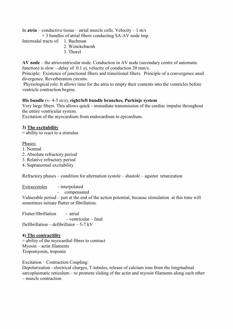
In atria – conductive tissue – atrial muscle cells. Velocity – 1 m/s
+ 3 bundles of atrial fibers conducting SA-AV node imp.
Internodal tracts of: 1. Bachman
2. Wenckebacnh
3. Thorel
AV node – the atrioventricular node. Conduction in AV node (secondary centre of automatic
function) is slow – delay of 0.1 s), velocity of conduction 20 mm/s.
Principle: Existence of junctional fibers and transitional fibers. Principle of a convergence ansd
divergence. Reverberation circuits.
Physiological role: It allows time for the atria to empty their contents into the ventricles before
ventricle contraction begins.
His bundle (v- 4-5 m/s), right/left bundle branches, Purkinje system
Very large fibers. This allows quick - immediate transmission of the cardiac impulse throughout
the entire ventricular system.
Excitation of the myocardium from endocardium to epicardium.
3) The excitability
= ability to react to a stimulus
Phases:
1. Normal
2. Absolute refractory period
3. Relative refractory period
4. Supranormal excitability
Refractory phases – condition for alternation systole – diastole – against tetanization
Extrasystoles - interpolated
- compensated
Vulnerable period – just at the end of the action potential, because stimulation at this time will
sometimes initiate flutter or fibrillation.
Flutter/fibrillation - atrial
- ventricular – fatal
Defibrillation – defibrillator – 5-7 kV
4) The contractility
= ability of the myocardial fibres to contract
Myosin – actin filaments
Tropomyosin, troponin
Excitation – Contraction Coupling:
Depolarization - electrical charges, T-tubules, release of calcium ions from the longitudinal
sarcoplasmatic reticulum – to promote sliding of the actin and myosin filaments along each other
– muscle contraction

All or Nothing Principle of the Heart
= stimulation of any single atrial muscle fiber causes the action potential in entire atrial muscle
mass. The same in ventricles.
Syncytial nature of cardiac muscle.
5) The Rhythmicity
= regular alternation of contraction and relaxation
HR – reflects metabolic rate/weight
birds 800/min
mice 500
men 70 elephant 25-30
whale 10/min
Required conditions for the heart activity
1) Temperature - optimal for humans 37º
- lower: decreasing of activity
- higher: increasing of activity + metabolic needs
2) Metabolism of the heart:
Aerobic – without possibility to cover energy demands of anaerobic pathway (only 1% of the
total energy is provided by anaerobic metabolism).Lack of the O2 debt.
Sources of energy for heart: Lactate, pyruvate, fat, FFA, AA, ketones.
3) Oxygen consumption: 10 ml/100 g/min, 35 ml/350 g/min = 10% of total O2
consumption (250 ml/min). During physical work, 5x more
4) Isoionia:
Isoionic – environment (including perfusion fluid)
Balance between: Calcium and potassium
During Ca abundance – rigor
During K abundance - inhibition
5) pH: acidosis inhibition of the heart activity – heart
stops in diastole
alkalosis – heart stops in systole – rigor
The Cardiac Cycle
- the period from the end of one cardiac systole to the end of the next heart contraction.
1) Electrical cycle – depolarisation/repolarisation
2) Mechanical cycle – contraction/relaxation of cardiac muscle
Periods of the cardiac cycle
1) Filling of the atria – during diastole = venous return
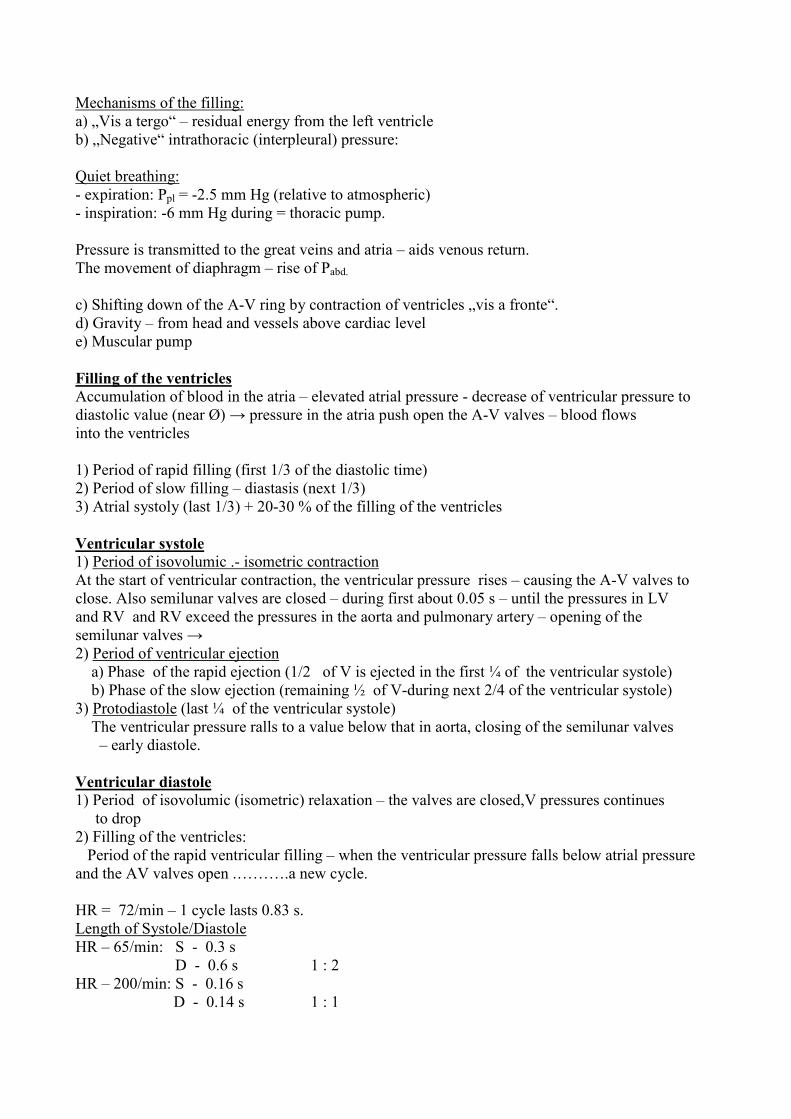
Mechanisms of the filling:
a) „Vis a tergo“ – residual energy from the left ventricle
b) „Negative“ intrathoracic (interpleural) pressure:
Quiet breathing:
- expiration: Ppl = -2.5 mm Hg (relative to atmospheric)
- inspiration: -6 mm Hg during = thoracic pump.
Pressure is transmitted to the great veins and atria – aids venous return.
The movement of diaphragm – rise of Pabd.
c) Shifting down of the A-V ring by contraction of ventricles „vis a fronte“.
d) Gravity – from head and vessels above cardiac level
e) Muscular pump
Filling of the ventricles
Accumulation of blood in the atria – elevated atrial pressure - decrease of ventricular pressure to
diastolic value (near Ø) → pressure in the atria push open the A-V valves – blood flows
into the ventricles
1) Period of rapid filling (first 1/3 of the diastolic time)
2) Period of slow filling – diastasis (next 1/3)
3) Atrial systoly (last 1/3) + 20-30 % of the filling of the ventricles
Ventricular systole
1) Period of isovolumic .- isometric contraction
At the start of ventricular contraction, the ventricular pressure rises – causing the A-V valves to
close. Also semilunar valves are closed – during first about 0.05 s – until the pressures in LV
and RV and RV exceed the pressures in the aorta and pulmonary artery – opening of the
semilunar valves →
2) Period of ventricular ejection
a) Phase of the rapid ejection (1/2 of V is ejected in the first ¼ of the ventricular systole)
b) Phase of the slow ejection (remaining ½ of V-during next 2/4 of the ventricular systole)
3) Protodiastole (last ¼ of the ventricular systole)
The ventricular pressure ralls to a value below that in aorta, closing of the semilunar valves
– early diastole.
Ventricular diastole
1) Period of isovolumic (isometric) relaxation – the valves are closed,V pressures continues
to drop
2) Filling of the ventricles:
Period of the rapid ventricular filling – when the ventricular pressure falls below atrial pressure
and the AV valves open .……….a new cycle.
HR = 72/min – 1 cycle lasts 0.83 s.
Length of Systole/Diastole
HR – 65/min: S - 0.3 s
D - 0.6 s 1 : 2
HR – 200/min: S - 0.16 s
D - 0.14 s 1 : 1
The duration of systole is more fixed.
Tachycardia is accompanied mainly by shortening of the diastole – if more than 180/min –
insufficient filling – critical frequency (HR) for adults.
Functions of the valves
The AV – valves prevent backflow of blood from V to A during systole
The semilunar valves prevent backflow from the aorta and pulmonary artery to V during
diastole.
All valves close and open passively – by pressure gradient.
The Electrical Activity of the Heart
Resting membrane potential (RMP): myocardial fibers
approximately – 90 mV
SA node: -55 to –60 mV
Conductive tissue: - 90 to –100 mV
+++++++++
––––––––––
- - - - - - - - -
- - - - - - - - -
––––––––––
+++++++++
RMP depends on differences in concentration of K+
Ki
+150 mmol/l; Ke
+ - 5 mmol/l = 30 x
RMP don´t allow to K+ to equalize concentrations.
Nai+ = 5-10 mmol/l; Nae
+ = 140 mmol/l
Depolarization: Firing level –65 mV
Initial – is due to an increase in Na+ permeability
(through fast Na+ channels)
Following – a slower increase in Ca2+ permeability
(through slow Ca2+ channels) – plateau (!)
Repolarization is due to a delayed increase in K+ permeability.
The excitation in the conductive system cells
Lower RMP (-60 mV) firing level – 35 mV
Fast Na+ channel is not activated.
Unstable RMP – open slow (nonspecific channel) – pacemaker potential = prepotential – due to
a steady decrease in K+ permeability
Effect of heart nerves on prepotential:
- vagus – acetylcholine – increase in K+ permeability – the slope of
prepotentials in decreased
- sympathetic nerves – opposite effect - decrease in K+ permeability ...
Prepotential in SA node has the slope increased in comparison to one in AV node –
- primary center. Gradient of automaticity. The slope of the prepotential determines HR.
Electrocardiography (ECG)
Registration of electrical potentials from the heart also from body surface (the tissues of
the body contain electrolytes – are conductive).
W. Einthoven (1903) – string galvanometer
A three – lead system: Leads I. II. III. – standard - frontal plane ECG – equilateral triangle
Unipolar leads – V – Wilson – potential of one site is 0.
Transverse plane, precordial leads V1-6 (12)
Augmented unipolar limb leads – aVR, aVL, aVF.
Form of the ECG
Isoelectric line
Waves:
P – atrial depolarization (0.1 – 0.3 mV, 0.1 s)
QRS – ventricular depolarization (atrial repolarization)
Q – initial depolarization (His bundle, branches)
R – activation of major portion of ventricular myocardium
S - late activation of posterobasal portion of the LV mass and the pulmonary conus
T – ventricular repolarization
U – repolarization of the papillary muscles
The duration of the waves, intervals and segments
P – wave 0.1 s
PQ – interval 0.16 s
PQ – segment 0.06 – 0.1 s
QRS complex 0.05 – 0.1 s
QT interval 0.2 – 0.4
QT segment 0.12
T wave 0.16
The voltage of ECG curve
P - 0.1 – 0.3 mV
R - 0.7 - 1.5 mV
T - 0.3 – 0.5 mV
Q, S - -0.3 - -0.5 mV
Intracellular potential 100 mV
Explanation:
1) ECG potential represents an algebraic sum of the action potentials of myocardial fibres
2) loss of voltage during spreading of potential
Special use of ECG
Esophageal leads (e.g. E-30)
Intracardial leads – RA, RV – His bundle electrogram
Monitoring:
– permanent in UIC
- Holter´s monitoring (tape recorder) diagnosis of arrhytmias
Cardiac Output
- of the LV = the quantity of blood pumped by the LV/min
- of the RV = the quantity of blood pumped by the RV/min
COLV = CORV !!!
CO = SV x f = 70 ml x 72 = 5000 ml/min
Normal values – 5-6 l/min
Cardiac Index (CI) = CO/m2 = 3 – 3.2 l/min/m
2
Stroke volume = volume of blood ejected per systole
Changes in CO
by changes in SV, or f, or both
Change in SV
End diastolic volume (EDV) – approx. 150 ml
End systolic volume (ESV) - 70 ml
More effective contractions – positive inotropic effect – ESV – functional reserve for the SV and
CO increase
Change in f – heart rate
up to a critical level (180/min – in adults)
Cardiac Output Changes
Effect of age – CI in 10 years – 4 l/min/m2
80 - 2 l/min/m2
Effect of exercise – CO can rise to 30-35 l/min
Effects of metabolism – CO proportional to M
Effect of gravity - +40 - +80 %
Effect of posture – Co falls about 20 %
Measurement of Cardiac Output
1) Direct method: electromagnetic flowmeter
2) Indirect methods:
a) Fick´s method
b) Indicator dilution methods
- using dyes
- thermal dilution - intermittent
- continuous infusion
c) Doppler´s method
d) echocardiography
e) bioimpendance method
Methods for heart examination
1) Invasive – cardiac catheterization (Forssmann 1929):
- Measurement of pressure in atria, ventricles, aorta, pulmonary
artery
- application of dyes ...
- angiocardiography, coronarography - application of X-ray contrast material
2) Noninvasive methods:
- Electrocardiography
- Heart rate variability evaluation
- Auscultation of heart sounds
- Phonocardiography
- Echocardiography
- Polycardiography
Auscultation/registration of heart sounds
1st heart sound – associated with:
1) closure of the AV valves at the beginning of systole
2) vibration of the walls of the heart and ejected blood
2nd heart sound – result from closure of the semilunar valves and from reverberation of blood
back
3rd heart sound – occasionally – at the beginning of the middle third of diastole – period of the
rapid filling of ventricles
4th heart sound – during atrial systole
The Phonocardiography
Recording of the heart sounds simultaneously with ECG
Advantages:
1) More exact analysis of the heart sounds and murmurs
2) A writing evidence
The time relationships between ECG and PhCG
Echocardiography
Pulses of ultrasonic waves are emitted and received by transducer.
Reflected ultrasound form the structures with different densities = echo
f of ultrasound > 20 kHz – in EchCG
2.25 MHz - in adults
4.4 MHz - in children
7 MHz - in newborns
Evaluation of the thickness and motions of the heart walls, septum and function of the valves
(mainly mitral) during the cardiac cycle. Valvular lesions.
Polycardiography
Simultaneous registration of: 1) ECG
2) PhCG
3) Arterial pulse
4) Venous pulse
5) Pressure curves (ao, LV, RV, PA ...)
Hemodynamics – Dynamics of Blood Circulation
A) General Hemodynamics
B) Special Hemodynamics
A) General – biophysical considerations:
Parameters:
1) Flow
2) Velocity
3) Pressure
4) Resistance
1) Blood Flow
V
F = –––– (m3/s; 1/s; ml/min ...)
t
Flow: – luminar (streamline)
- turbulent
dens.x diam. x velocity
Reynolds number = –––––––––––––––––––
Viscosity
Critical velocity – in ascending aorta
- in anemia
Systolic murmurs
Dif.P
F = ––––––
R
Critical closing pressure = P at which flow ceases.
Velocity of Blood
1
v = –– (m/s; cm/s ...)
t
F = flow cm3 x s
-1
v = ––– = ---------- = cm/s
A = cross–sectional area cm2
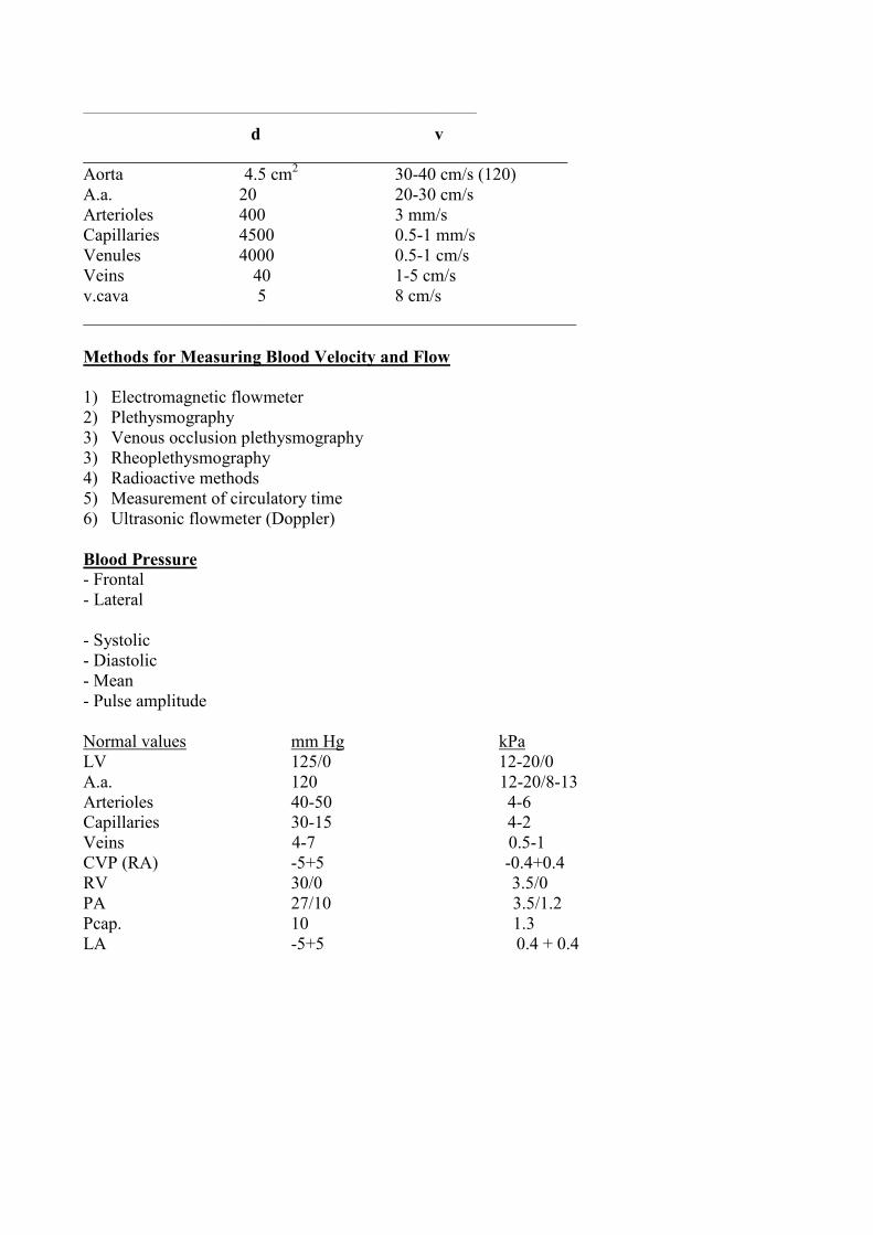
___________________________________________________________________
d v
_______________________________________________________
Aorta 4.5 cm2 30-40 cm/s (120)
A.a. 20 20-30 cm/s
Arterioles 400 3 mm/s
Capillaries 4500 0.5-1 mm/s
Venules 4000 0.5-1 cm/s
Veins 40 1-5 cm/s
v.cava 5 8 cm/s
________________________________________________________
Methods for Measuring Blood Velocity and Flow
1) Electromagnetic flowmeter
2) Plethysmography
3) Venous occlusion plethysmography
3) Rheoplethysmography
4) Radioactive methods
5) Measurement of circulatory time
6) Ultrasonic flowmeter (Doppler)
Blood Pressure
- Frontal
- Lateral
- Systolic
- Diastolic
- Mean
- Pulse amplitude
Normal values mm Hg kPa
LV 125/0 12-20/0
A.a. 120 12-20/8-13
Arterioles 40-50 4-6
Capillaries 30-15 4-2
Veins 4-7 0.5-1
CVP (RA) -5+5 -0.4+0.4
RV 30/0 3.5/0
PA 27/10 3.5/1.2
Pcap. 10 1.3
LA -5+5 0.4 + 0.4
Measurement of BP
Methods: - direct
- indirect
Direct methods: Hales 1773
Catheterization – electronic manometers with transducers
Indirect methods:
- Palpation (Riva – Rocci)
- Auscultatory (Korotkoff)
- Oscillometric (Pachon, electronic-digital)
Principles for accurate measurement of BP
Patient: should rest undisturbed in a quiet, comfortable setting at room temperature for at least 5-
15 minutes. To avoid physical activity, food consumption, smoking, caffeine ingestion and
emotional stress for at least half an hour before measurement.Full bladder or bowel can cause an
increase in BP. Nonconstring clothing – with no sleeves. Children – should be given sufficient
time to recover from crying.
„White – coat hypertension“ – physicians cause + 27/15. Measurement at home.
Recommendations for observer measuring BP
- Have normal hearing and vision, be trained in the technique for measurement BP
- Support the patient´s arm – the antecubital fossea at heart luvel
- Chair with back and arm support when the patient is sitting
- Use an appropriately sized cuff
- Check the BP by palpation before auscultation
- Deflate the cuff 2-3 mmHg/s
- Use the 1st and 5th Korotkoff sounds to determine BP syst. and BP diast.
- Allow 1-2 min. between readings
- Take readings with the patient in the lying or sitting position and in the standing position
- Assess the BP at least 3x over 3-6 months among patients with midly
elevated BP
BP depends on:
1) Heart activity
2) Vascular resistance
3) Volume and viscosity of blood
4) Compression vessels by different organs and pressures (e.g. intraabdominal)
5) Hydrostatic pressure – effect of gravity
1)Heart activity
CO = SV x f
Increase in SV → increase mainly BP syst.
Increase in f (HR) → increase mainly BP diast.
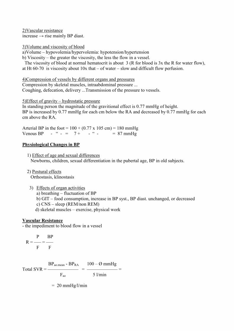
2)Vascular resistance
increase → rise mainly BP diast.
3)Volume and viscosity of blood
a)Volume – hypovolemia/hypervolemia: hypotension/hypertension
b) Viscosity – the greater the viscosity, the less the flow in a vessel.
The viscosity of blood at normal hematocrit is about 3 (R for blood is 3x the R for water flow),
at Ht 60-70 is viscosity about 10x that – of water – slow and difficult flow perfusion.
4)Compression of vessels by different organs and pressures
Compression by skeletal muscles, intraabdominal pressure ...
Coughing, defecation, delivery ...Transmission of the pressure to vessels.
5)Effect of gravity – hydrostatic pressure
In standing person the magnitude of the gravitional effect is 0.77 mmHg of height.
BP is increased by 0.77 mmHg for each cm below the RA and decreased by 0.77 mmHg for each
cm above the RA.
Arterial BP in the foot = 100 + (0.77 x 105 cm) = 180 mmHg
Venous BP - “ - = 7 + - “ - = 87 mmHg
Physiological Changes in BP
1) Effect of age and sexual differences
Newborns, children, sexual differentiation in the pubertal age, BP in old subjects.
2) Postural effects
Orthostasis, klinostasis
3) Effects of organ activities
a) breathing – fluctuation of BP
b) GIT – food consumption, increase in BP syst., BP diast. unchanged, or decreased
c) CNS – sleep (REM/non REM)
d) skeletal muscles – exercise, physical work
Vascular Resistance
- the impediment to blood flow in a vessel
P BP
R = ––– = –––
F F
BPao.mean - BPRA 100 – Ø mmHg
Total SVR = ––––––––––––– = ––––––––––––– =
Fao 5 l/min
= 20 mmHg/l/min
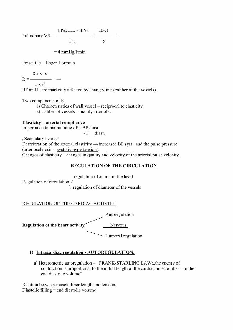
BPPA mean - BPLA 20-Ø
Pulmonary VR = ––––––––––––––– = ––––––– =
FPA 5
= 4 mmHg/l/min
Poiseuille – Hagen Formula
8 x vi x l
R = ––––––––– →
π x r4
BF and R are markedly affected by changes in r (caliber of the vessels).
Two components of R:
1) Characteristics of wall vessel – reciprocal to elasticity
2) Caliber of vessels – mainly arterioles
Elasticity – arterial compliance
Importance in maintaining of: - BP diast.
- F diast.
„Secondary hearts“
Deterioration of the arterial elasticity → increased BP syst. and the pulse pressure
(arteriosclerosis – systolic hypertension).
Changes of elasticity – changes in quality and velocity of the arterial pulse velocity.
REGULATION OF THE CIRCULATION
regulation of action of the heart
Regulation of circulation ⁄
\ regulation of diameter of the vessels
REGULATION OF THE CARDIAC ACTIVITY
Autoregulation
Regulation of the heart activity Nervous
Humoral regulation
1) Intracardiac regulation - AUTOREGULATION:
a) Heterometric autoregulation – FRANK-STARLING LAW:„the energy of
contraction is proportional to the initial length of the cardiac muscle fiber – to the
end diastolic volume“
Relation between muscle fiber length and tension.
Diastolic filling = end diastolic volume
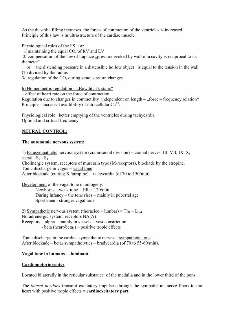
As the diastolic filling increases, the forces of contraction of the ventricles is increased.
Principle of this law is in ultrastructure of the cardiac muscle.
Physiological roles of the FS law:
1/ maintaining the equal COs of RV and LV
2/ compensation of the law of Laplace „pressure evoked by wall of a cavity is reciprocal to its
diameter“
or: the distending pressure in a distensible hollow object is equal to the tension in the wall
(T) divided by the radius
3/ regulation of the COs during venous return changes
b) Homeometric regulation – „Bowditch´s stairs“
– effect of heart rate on the force of contraction
Regulation due to changes in contractility independent on length – „force – frequency relation“
Principle - increased availibility of intracellular Ca+2.
Physiological role: better emptying of the ventricles during tachycardia
Optimal and critical frequency.
NEURAL CONTROL:
The autonomic nervous system:
1) Parasympathetic nervous system (craniosacral division) = cranial nerves: III, VII, IX, X,
sacral. S2 - S4
Cholinergic system, receptors of muscarin type (M-receptors), blockade by the atropine.
Tonic discharge in vagus = vagal tone
After blockade (cutting X./atropine) – tachycardia (of 70 to 150/min)
Development of the vagal tone in ontogeny:
Newborns – weak tone – HR = 120/min.
During infancy – the tone rises – mainly in pubertal age
Sportsmen - stronger vagal tone
2) Sympathetic nervous system (thoracico – lumbar) = Th1 – L3-4
Noradrenergic system, receptors NA(A)
Receptors – alpha – mainly in vessels – vasoconstriction
- beta (heart-beta1) – positive tropic effects
Tonic discharge in the cardiac sympathetic nerves = sympathetic tone
After blockade – beta1 sympatholytics – bradycardia (of 70 to 55-60/min).
Vagal tone in humans – dominant.
Cardiomotoric center
Located bilaterally in the reticular substance of the medulla and in the lower third of the pons.
The lateral portions transmit excitatory impulses through the sympathetic nerve fibers to the
heart with positive tropic effects = cardioexcitatory part.
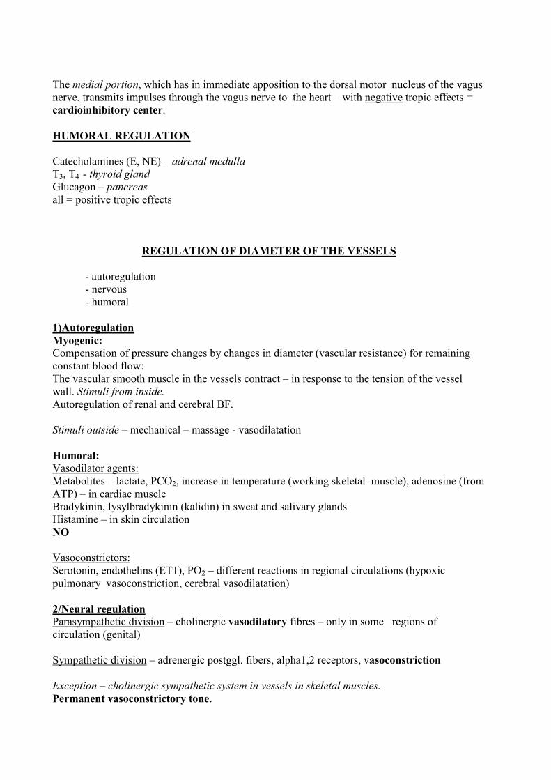
The medial portion, which has in immediate apposition to the dorsal motor nucleus of the vagus
nerve, transmits impulses through the vagus nerve to the heart – with negative tropic effects =
cardioinhibitory center.
HUMORAL REGULATION
Catecholamines (E, NE) – adrenal medulla
T3, T4 - thyroid gland
Glucagon – pancreas
all = positive tropic effects
REGULATION OF DIAMETER OF THE VESSELS
- autoregulation
- nervous
- humoral
1)Autoregulation
Myogenic:
Compensation of pressure changes by changes in diameter (vascular resistance) for remaining
constant blood flow:
The vascular smooth muscle in the vessels contract – in response to the tension of the vessel
wall. Stimuli from inside.
Autoregulation of renal and cerebral BF.
Stimuli outside – mechanical – massage - vasodilatation
Humoral:
Vasodilator agents:
Metabolites – lactate, PCO2, increase in temperature (working skeletal muscle), adenosine (from
ATP) – in cardiac muscle
Bradykinin, lysylbradykinin (kalidin) in sweat and salivary glands
Histamine – in skin circulation
NO
Vasoconstrictors:
Serotonin, endothelins (ET1), PO2 – different reactions in regional circulations (hypoxic
pulmonary vasoconstriction, cerebral vasodilatation)
2/Neural regulation
Parasympathetic division – cholinergic vasodilatory fibres – only in some regions of
circulation (genital)
Sympathetic division – adrenergic postggl. fibers, alpha1,2 receptors, vasoconstriction
Exception – cholinergic sympathetic system in vessels in skeletal muscles.
Permanent vasoconstrictory tone.
Vasomotor Centre
in medulla oblongata
Pressoric area – tonic activity – in rostral ventrolateral reticular area in medulla
Depressoric area – in the medial and caudal area
Afferents to the vasomotor centre:
- Direct stimulation by ↑ CO2, ↓ O2
- Excitatory impulses:
cortex – hypothalamus
pain pathways
peripheral chemoreceptors
- Inhibitory impulses:
cortex – hypothalamus
lung receptors
baroreceptors
Hormonal control of vessels
Vasodilatatory hormones:
VIP - in splanchnic circulation
ANP – secreted by heart – in renal ...
Vasoconstrictory hormones:
norepinephrine, angiotensin II, vasopressin (ADH)
Short review of the Autonomic Nervous System Pharmacology
Sympathomimetic drugs:
alpha: Norepinephrine
beta: Isoproterenol (Isuprel)
Epinephrine (adrenaline)
Miscellaneous: the precursors of E, NE:
Dopamine (Intropin)
Dobutamine (Dobutrex)
Amphetamine (by releasing endogenous catecholamines)
Blocking agents:
Anti alpha – phenoxybenzamine (Dibenzyline)
- tolazoline, phentolamine (Regitine)
- anti beta1 – propranolol (Inderal)
- metipranolol (Trimepranol)
- pindolol (Visken)……
Parasympathomimetic drugs:
Acetylcholine
Pilocarpine
Methacholine
Muscarine (in Amanita muscaria)
Anticholinesterases:
Physostigmine
Neostigmine
Blocking agents:
Anticholinergic - atropine
- scopolamine
CARDIOVASCULAR REFLEXES (CVR)
Reflex = regular response of the organism to stimulation mediated through central nervous
systém
Reflex arc: 1) receptor (sense organ);
2) afferent neuron;
3) center;
4) efferent neuron;
5) effector (in CVR – heart and vessels)
Receptors (for CVR):
1) Interoceptors – for perception of the internal environment:
a) Baroreceptors
b) Chemoreceptors
Baroreceptors – low – pressure receptors
- high pressure receptors
a) Low – pressure receptors – atrial stretch receptors:
2 types - „A“ – they discharge primarily during atrial systole
- „B“ - - “ - atrial filling,
peak – at the end of atrial diastole.
The discharge is increased when venous return is increased = volumoreceptors for monitoring
venous return.
Effects of the increased stimulation of the atrial baroreceptors:
Vasodilatation – an accumulation of the blood in the periphery – a fall in BP.
Bainbridge Reflex – rapid infusion to the right atrium – changes in HR.
Initial basal increased HR – effect: bradycardia
- “ - decreased HR – effect: tachycardia
Two components:- reflex – through atrial baroreceptors
- mechanical – local stretch – effect on the SA node
b) High – pressure receptors
- carotid sinus
- aortic arch
- LV
Carotid sinus receptors: spray type nerve endings – in the adventitia of the carotid bifurcation
and the internal carotid artery.
Afferent neurons: Innervation – through the branch of n. IX. – Hering´s nerve.
Center of the carotid sinus reflex – tractus solitarius in the medulla,
Efferent neurons: n.X and parasympathetic + sympathetic nerves to vessels.
Effectors: Heart, smooth muscles in the vessels wall.
Stimulation: The baroreceptors are stimulated by distension of the vessels wall – stretch
receptors.
Aortic arch receptors: in the wall of the aorta.
Affferent pathway: Innervation – through the vagus)
Center, efferent pathway, effectors and effects – the same as in CSR
Left ventricular receptors: Afferent and efferent pathways: n. vagus.
Activity and effects of the HP baroreceptors
Normal BP – r. discharge at a slow rate
Increased BP – the discharge rate increases
Decreased BP - - “ - declines
Effects:
BP increase (hypertension): The increased discharge rate evokes rising activity in
cardioinhibitory center and depressoric area of the vasomotor center – bradycardia,
vasodilatation – a fall in BP to the normal level
BP decrease (hypotension): The decreased discharge ... vice versa. Normalization of the BP in
different situations.
Baroreceptor Testing
- Pressure on the SC region – unconstant stimulus
Syndrome of the hypersensitive SC (sy HSC)
- Carotid Clamping – proximal or distal to the CS - on experimental
animals
- Cutting the CS nerves - on experiments/ treatment sy HSC
- Application of the drugs eliciting an increase in BP (NE,dopamine)
- Different maneuvers (orthostasis, klinostasis, Valsalva ...)
Orthostatic Reflex
When a person stands – venous return decreases due to hydrostatic pressure of the blood.
A decrease in CO and systemic BP occurs.
Falling BP at the baroreceptors elicits an immediate reflex, with strong sympathetic activity –
vasoconstriction, increase in BP diast., tachycardia.
The aim – to maintain an adequate perfusion of organs.
Valsalva maneuver
Forced expiratory effort against a closed glottis – Ppl + 30-50 mmHg.
5 phases:
1st. – at the onset of straining, the BP rises
2nd – a decrease of venous return, CO, BP; HR increases
3rd – reflex vasoconstriction – stop of the fall of BP, HR increases
4th – after the first inspirium, start of the breathing – decrease in BP –
- filling of the pulmonary circulation with blood
5th - an increase in BP – vasoconstricton persists, an excessive venous return – stimulation
of baroreceptors, causing bradycardia and a drop in BP to normal values.
Chemoreceptors - peripheral
- central
Peripheral chemoreceptors:
Carotid and aortic bodies – near the carotid bifurcation, and the arch of the aorta.
The highest BF/g.
Stimulation – low PaO2 (high PaCO2, acidosis)
Effects – an increasing of pressoric area of vasomotor centre, vasoconstriction (splanchnic),
redistribution of the blood, an increase in BP.
HR – primarily tachycardia, secondary bradycardia through baroreceptors
Central chemoreceptors: in the brain stem, on the ventral surface, H+ zone.
Stimulation: a decrease in pH - acidosis.
Effects – changes in respiration, without any direct effect on cardiovascular system.
2) Nonspecific receptors
A) Trigeminal endings:
1) Oculocardiac reflex:
Stimulation: pressure on the mechanoreceptors of eye and orbit –
Effects: Depressoric effects on vessels and breathing - a decrease in
HR by 5-14 / min.
2) Kratschmer apnoeic reflex:
Stimulation:Intranasal insufflation of various irritant gases (smoke,
ammonia ..)
Effects: reflex respiratory arrest + laryngoconstriction + bradycardia
+ redistribution of the blood to the vital most important organs
(from the splanchnic circulation).
3) The diving reflex:
Stimulation of the trigeminal region with cold (water).
Effects: the same as in Kratschmer.
Oxygen conserving reflex – the changes primarily safeguard the blood and oxygen supply
of the heart, the brain.
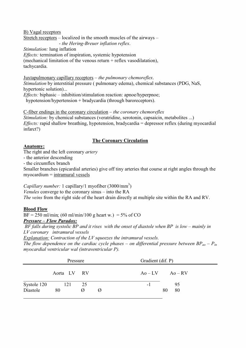
B) Vagal receptors
Stretch receptors – localized in the smooth muscles of the airways –
- the Hering-Breuer inflation reflex.
Stimulation: lung inflation
Effects: termination of inspiration, systemic hypotension
(mechanical limitation of the venous return + reflex vasodilatation),
tachycardia.
Juxtapulmonary capillary receptors – the pulmonary chemoreflex.
Stimulation by interstitial pressure ( pulmonary edema), chemical substances (PDG, NaS,
hypertonic solution)...
Effects: biphasic – inhibition/stimulation reaction: apnoe/hyperpnoe;
hypotension/hypertension + bradycardia (through baroreceptors).
C-fiber endings in the coronary circulation – the coronary chemoreflex
Stimulation: by chemical substances (veratridine, serotonin, capsaicin, metabolites ...)
Effects: rapid shallow breathing, hypotension, bradycardia = depressor reflex (during myocardial
infarct?)
The Coronary Circulation
Anatomy:
The right and the left coronary artery
- the anterior descending
- the circumflex branch
Smaller branches (epicardial arteries) give off tiny arteries that course at right angles through the
myocardium = intramural vessels
Capillary number: 1 capillary/1 myofiber (3000/mm3)
Venules converge to the coronary sinus – into the RA
The veins from the right side of the heart drain directly at multiple site within the RA and RV.
Blood Flow
BF = 250 ml/min; (60 ml/min/100 g heart w.) = 5% of CO
Pressure – Flow Paradox:
BF falls during systolic BP and it rises with the onset of diastole when BP is low – mainly in
LV coronary intramural vessels
Explanation: Contraction of the LV squeezes the intramural vessels.
The flow dependence on the cardiac cycle phases – on differential pressure between BPao – Pin
myocardial ventricular wal (intraventricular P).
Pressure Gradient (dif. P)
Aorta LV RV Ao – LV Ao – RV
_______________________________________________________
Systole 120 121 25 -1 95
Diastole 80 Ø Ø 80 80
________________________________________________________
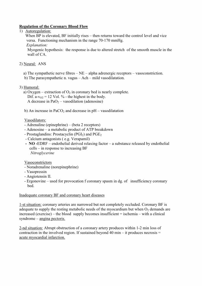
Regulation of the Coronary Blood Flow
1) Autoregulation:
When BP is elevated, BF initially rises – then returns toward the control level and vice
versa. Functioning mechanism in the range 70-170 mmHg.
Explanation:
Myogenic hypothesis: the response is due to altered stretch of the smooth muscle in the
wall of CA.
2) Neural: ANS
a) The sympathetic nerve fibres – NE – alpha adrenergic receptors – vasoconstriction.
b) The parasympathetic n. vagus – Ach – mild vasodilatation.
3) Humoral:
a) Oxygen – extraction of O2 in coronary bed is nearly complete.
Dif. a-vO2 = 12 Vol. % - the highest in the body.
A decrease in PaO2 – vasodilation (adenosine)
b) An increase in PaCO2 and decrease in pH – vasodilatation
Vasodilators:
- Adrenaline (epinephrine) – (beta 2 receptors)
- Adenosine – a metabolic product of ATP breakdown
- Prostaglandins: Prostacyclin (PGI2) and PGE2
- Calcium antagonists ( e.g. Verapamil)
- NO -EDRF – endothelial derived relaxing factor – a substance released by endothelial
cells – in response to increasing BF
Nitroglycerine
Vasoconstrictors
- Noradrenaline (norepinephrine)
- Vasopressin
- Angiotensin II.
- Ergonovine – used for provocation f coronary spasm in dg. of insufficiency coronary
bed.
Inadequate coronary BF and coronary heart diseases
1-st situation: coronary arteries are narrowed but not completely occluded. Coronary BF is
adequate to supply the resting metabolic needs of the myocardium but when O2 demands are
increased (exercise) – the blood supply becomes insufficient = ischemia – with a clinical
syndrome – angina pectoris.
2-nd situation: Abrupt obstruction of a coronary artery produces within 1-2 min loss of
contraction in the involved region. If sustained beyond 40 min – it produces necrosis =
acute myocardial infarction.
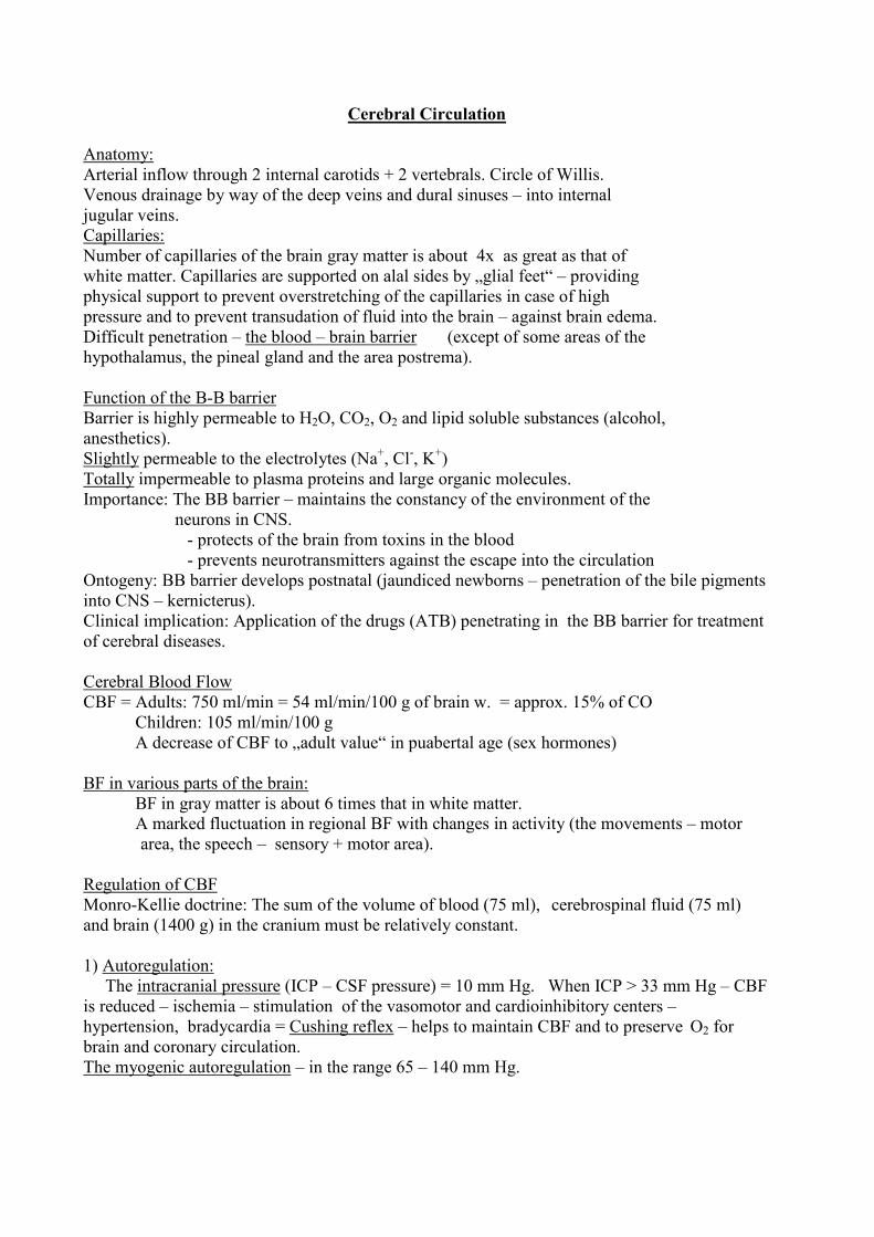
Cerebral Circulation
Anatomy:
Arterial inflow through 2 internal carotids + 2 vertebrals. Circle of Willis.
Venous drainage by way of the deep veins and dural sinuses – into internal
jugular veins.
Capillaries:
Number of capillaries of the brain gray matter is about 4x as great as that of
white matter. Capillaries are supported on alal sides by „glial feet“ – providing
physical support to prevent overstretching of the capillaries in case of high
pressure and to prevent transudation of fluid into the brain – against brain edema.
Difficult penetration – the blood – brain barrier (except of some areas of the
hypothalamus, the pineal gland and the area postrema).
Function of the B-B barrier
Barrier is highly permeable to H2O, CO2, O2 and lipid soluble substances (alcohol,
anesthetics).
Slightly permeable to the electrolytes (Na+, Cl
-, K
+)
Totally impermeable to plasma proteins and large organic molecules.
Importance: The BB barrier – maintains the constancy of the environment of the
neurons in CNS.
- protects of the brain from toxins in the blood
- prevents neurotransmitters against the escape into the circulation
Ontogeny: BB barrier develops postnatal (jaundiced newborns – penetration of the bile pigments
into CNS – kernicterus).
Clinical implication: Application of the drugs (ATB) penetrating in the BB barrier for treatment
of cerebral diseases.
Cerebral Blood Flow
CBF = Adults: 750 ml/min = 54 ml/min/100 g of brain w. = approx. 15% of CO
Children: 105 ml/min/100 g
A decrease of CBF to „adult value“ in puabertal age (sex hormones)
BF in various parts of the brain:
BF in gray matter is about 6 times that in white matter.
A marked fluctuation in regional BF with changes in activity (the movements – motor
area, the speech – sensory + motor area).
Regulation of CBF
Monro-Kellie doctrine: The sum of the volume of blood (75 ml), cerebrospinal fluid (75 ml)
and brain (1400 g) in the cranium must be relatively constant.
1) Autoregulation:
The intracranial pressure (ICP – CSF pressure) = 10 mm Hg. When ICP > 33 mm Hg – CBF
is reduced – ischemia – stimulation of the vasomotor and cardioinhibitory centers –
hypertension, bradycardia = Cushing reflex – helps to maintain CBF and to preserve O2 for
brain and coronary circulation.
The myogenic autoregulation – in the range 65 – 140 mm Hg.
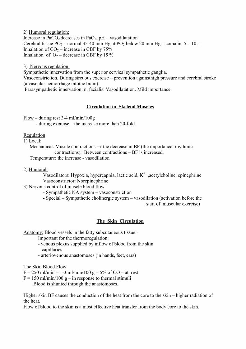
2) Humoral regulation:
Increase in PaCO2 decreases in PaO2, pH – vasodilatation
Cerebral tissue PO2 – normal 35-40 mm Hg at PO2 below 20 mm Hg – coma in 5 – 10 s.
Inhalation of CO2 – increase in CBF by 75%
Inhalation of O2 – decrease in CBF by 15 %
3) Nervous regulation:
Sympathetic innervation from the superior cervical sympathetic ganglia.
Vasoconstriction. During streuous exercise – prevention againsthigh pressure and cerebral stroke
(a vascular hemorrhage intothe brain).
Parasympathetic innervation: n. facialis. Vasodilatation. Mild importance.
Circulation in Skeletal Muscles
Flow – during rest 3-4 ml/min/100g
- during exercise – the increase more than 20-fold
Regulation
1) Local:
Mechanical: Muscle contractions → the decrease in BF (the importance rhythmic
contractions). Between contractions – BF is increased.
Temperature: the increase - vasodilation
2) Humoral:
Vasodilators: Hypoxia, hypercapnia, lactic acid, K+ ,acetylcholine, epinephrine
Vasoconstrictor: Norepinephrine
3) Nervous control of muscle blood flow
- Sympathetic NA system – vasoconstriction
- Special – Sympathetic cholinergic system – vasodilation (activation before the
start of muscular exercise)
The Skin Circulation
Anatomy: Blood vessels in the fatty subcutaneous tissue.-
Important for the thermoregulation:
- venous plexus supplied by inflow of blood from the skin
capillaries
- arteriovenous anastomoses (in hands, feet, ears)
The Skin Blood Flow
F = 250 ml/min = 1-3 ml/min/100 g = 5% of CO – at rest
F = 150 ml/min/100 g – in response to thermal stimuli
Blood is shunted through the anastomoses.
Higher skin BF causes the conduction of the heat from the core to the skin – higher radiation of
the heat.
Flow of blood to the skin is a most effective heat transfer from the body core to the skin.
Regulation of the skin BF
Autoregulation
Nervous: Sympathetic nerves: The increase of the sympathetic nerve traffic –
- vasoconstriction and vice versa. Vasoconstrictory tone.
Local axon reflexes:
Impulses initiated in sensory nerves (by injury) are relayed antidromically down by other
branches of the sensory nerve fiber. The only one situation – antidromic conduction.
Humoral: - Histamine and H-like substances – H1 – receptors – vasodilatatioin
- Bradykinin – sweat glands – kalikrein – effects on plasma proteins – bradykinin –
vasodilatation
- Serotonin, NE – vasoconstriction
Tests of the skin vascular reactibility
A) White reaction: the mechanical stimulation (pointed object is drawn lightly over the skin) –
a pale line – due to contractions of the precapillary sphincters (in 10-15 s).
B) Tripple response: the skin attacked more strongly
1) Red reaction (in 10 s) – capillary dilatation
2) Swelling (local edema) increased permeability of the capillaries – histamine, H-
substances
3) Difuse reddening around the injury – arterial dilatation – axon reflexes.
Pulmonary Circulation
Morphology:
Circulation in series to the systemic circulation. The pulmonary vessels are
short and have large diameter.The walls are thin and distensible.
Physiology
Flow: CO RV – 5.5 l/min; Velocity – 40 cm/s;
BF – is much more pulsatile than is systemic circulation (low arteriolar resistance)
Ventilation/Perfusion Ratio
Differences in various parts of the lungs.
Pressure: PA = 25/10 mm Hg. Mean = 15 mm Hg.
Pulmonary capillary pressure = 7-10 mm Hg.
Resistance: low – 2-3 mm/l/min
Volume of blood: 1 l of the blood in pulmonary bed – only about 75-100 ml is in the pulmonary
capillaries. SV = 70 ml – all the capillary blood is replaced at each heart beat.
The increase of the volume after deep inspirium, in horizontal position. Reservoir function.
Distribution of blood flow in pulmonary circulation
The hydrostatic pressure of the blood within the pulmonary capillaries influences BF in different
regions of the lungs. At the top of the lungs is little flow – in the lowest point in the lungs is max.