Embed Size (px)
Citation preview
University of Birmingham
Physiological tremor reveals how thixotropy adaptsskeletal muscle for posture and movementVernooij, Carlijn; Reynolds, Raymond; Lakie, Martin
DOI:10.1098/rsos.160065
License:Creative Commons: Attribution (CC BY)
Document VersionPublisher's PDF, also known as Version of record
Citation for published version (Harvard):Vernooij, C, Reynolds, R & Lakie, M 2016, 'Physiological tremor reveals how thixotropy adapts skeletal musclefor posture and movement', Royal Society Open Science. https://doi.org/10.1098/rsos.160065
Link to publication on Research at Birmingham portal
Publisher Rights Statement:Eligibility for repository: Checked on 10/5/2016
General rightsUnless a licence is specified above, all rights (including copyright and moral rights) in this document are retained by the authors and/or thecopyright holders. The express permission of the copyright holder must be obtained for any use of this material other than for purposespermitted by law.
•Users may freely distribute the URL that is used to identify this publication.•Users may download and/or print one copy of the publication from the University of Birmingham research portal for the purpose of privatestudy or non-commercial research.•User may use extracts from the document in line with the concept of ‘fair dealing’ under the Copyright, Designs and Patents Act 1988 (?)•Users may not further distribute the material nor use it for the purposes of commercial gain.
Where a licence is displayed above, please note the terms and conditions of the licence govern your use of this document.
When citing, please reference the published version.
Take down policyWhile the University of Birmingham exercises care and attention in making items available there are rare occasions when an item has beenuploaded in error or has been deemed to be commercially or otherwise sensitive.
If you believe that this is the case for this document, please contact [email protected] providing details and we will remove access tothe work immediately and investigate.
Download date: 01. May. 2020

rsos.royalsocietypublishing.org
ResearchCite this article: Vernooij CA, Reynolds RF,Lakie M. 2016 Physiological tremor reveals howthixotropy adapts skeletal muscle for postureand movement. R. Soc. open sci. 3: 160065.http://dx.doi.org/10.1098/rsos.160065
Received: 29 January 2016Accepted: 5 April 2016
Subject Category:Biology (whole organism)
Subject Areas:physiology/behaviour/neuroscience
Keywords:physiological tremor, mechanical resonance,thixotropy, posture, electromyography, muscle
Author for correspondence:Carlijn A. Vernooije-mail: [email protected]
Physiological tremor revealshow thixotropy adaptsskeletal muscle for postureand movementCarlijn A. Vernooij1,2, Raymond F. Reynolds1 and
Martin Lakie1
1School of Sport, Exercise and Rehabilitation Sciences, University of Birmingham,Edgbaston, Birmingham B15 2TT, UK2Institut des Sciences du Mouvement E.J. Marey (UMR 7287), Aix-Marseille Universitéand CNRS, 163 Avenue de Luminy, CP 910, Marseille 13009, France
People and animals can move freely, but they must alsobe able to stay still. How do skeletal muscles economicallyproduce both movement and posture? Humans are wellknown to have motor units with relatively homogeneousmechanical properties. Thixotropic muscle properties canprovide a solution by providing a temporary stiffening ofall skeletal muscles in postural conditions. This stiffening isalleviated almost instantly when muscles start to move. Inthis paper, we probe this behaviour. We monitor both theneural input to a muscle, measured here as extensor muscleelectromyography (EMG), and its output, measured as tremor(finger acceleration). Both signals were analysed continuouslyas the subject made smooth transitions between posture andmovement. The results showed that there were marked changesin tremor which systematically increased in size and decreasedin frequency as the subject moved faster. By contrast, the EMGchanged little and reflected muscle force requirement ratherthan movement speed. The altered tremor reflects naturallyoccurring thixotropic changes in muscle behaviour. Our resultssuggest that physiological tremor provides useful and hithertounrecognized insights into skeletal muscle’s role in posture andmovement.
1. BackgroundHuman motor activity consists of periods of immobility, enlivenedby periods of movement. How are these very different posturaland dynamic roles addressed by the neuromuscular system?In many species, there is a division of skeletal muscle intotonic and phasic types with profound mechanical differences, forexample amphibians [1], insects [2], birds [3] and reptiles [4].
2016 The Authors. Published by the Royal Society under the terms of the Creative CommonsAttribution License http://creativecommons.org/licenses/by/4.0/, which permits unrestricteduse, provided the original author and source are credited.
on May 10, 2016http://rsos.royalsocietypublishing.org/Downloaded from
2
rsos.royalsocietypublishing.orgR.Soc.opensci.3:160065
................................................Often the tonic types are arranged as bundles which parallel the phasic fibres, so that the same muscle canact in different ways depending on which bundle is activated. However, mammals, including humans,do not have specific muscle types for tonic or phasic behaviour. Instead, they draw on a commonpool of skeletal muscle motor units, invariably recruited in order of increasing motor neuron size, tosatisfy both posture and movement [5]. There are certainly mechanical differences between the initiallyrecruited slow motor units and later recruited faster motor units. One study on human extensor hallucisbrevis muscles showed contraction times with an extreme range of 35–98 ms [6]. These differences areactually rather slight, considering that posture may endure for periods of many seconds or minutes,whereas movements may be over in a fraction of a second. Thus, they seem more suited to generatingmovements of different contraction speeds rather than for the fundamentally opposed roles of movementand posture.
However, motor unit size and type are not the only determinants of muscle behaviour. There areprofound mechanical differences between muscles when they are lengthening or shortening, and whenthey are static (or very nearly so). This difference is caused by muscular thixotropy, which can be definedas ‘the dependence of muscle stiffness on the history of length changes’. Thixotropic changes greatlyalter the muscle’s mechanical response to artificial stimulation and imposed movement [7–11]. Whileat rest, muscles become stiff and resistant to lengthening or shortening. If there is a change in musclelength which exceeds the minor elasticity inherent in cross-bridges, this ‘stiction’ falls to a low level,re-establishing itself rapidly as posture returns (the phenomenon was noted as early as 1929 by Denny-Brown [12]; he called it stationary rigidity). Thixotropic stiffening will ensure stability at rest, but willalso permit unimpeded movement, thus enabling skeletal muscle to fulfil completely different posturaland dynamic roles.
The implication is clear; muscle can be expected to act very differently when stationary and whenmoving. Such changes are easily demonstrated by in vitro experiments [13]. However, the changesin muscle behaviour in vivo are relatively inscrutable and have not been much studied. In a fewrecent studies, we have explored the altered relationship between the input to a muscle, measured aselectromyography (EMG), and its output, measured as limb tremor (acceleration), as a consequence ofmuscle thixotropy (hand: [10,14] and finger: [11,15]). With movement, the peak in the tremor spectrumshifts to a lower frequency, whereas the shape of the EMG spectrum changes only slightly. Theexplanation is that the stiffness of the muscle changes (decreases), but its inertial load does not, thusaltering the tuning of the muscle/limb oscillator and the way that it responds to the buffeting of motorunit firing. However, these studies separately examined posture and movement, and did not track themechanisms underlying the transition between a period of rest and a period of movement.
In this paper, we describe the effect of transitions between posture and movement on muscleproperties. Subjects used their middle finger to track a target that evolved from a static position intoa very slow vertical sinusoidal movement before returning to a stationary position. We recorded surfaceEMG from the finger extensor muscle because it represents the neural drive to move the finger toovercome gravitational and elastic forces. We simultaneously recorded finger tremor as an indicationof muscle output. We hypothesized the following: (i) tremor frequency will alter as a limb alternatesbetween posture and movement; (ii) the frequency modulation can be parsimoniously explained bymechanical changes in muscle tissue and (iii) EMG magnitude will reflect the load but its frequencycomposition will not relate to tremor frequency.
Our results show that human physiological tremor, sometimes seen as little more than a curiosity, canilluminate the way in which muscle carries out its postural and dynamic duties and provide fundamentalinsights into the nature of posture and movement. Some of these findings have been briefly reportedelsewhere [16,17].
2. Material and methods2.1. Experimental volunteersThe experiment was carried out on 15 healthy, right-handed volunteers (23.7 ± 9.9 years old,three female) who gave written informed consent, and was undertaken in accordance with thedeclaration of Helsinki. Permission was obtained from the ethics committee of the University ofBirmingham. None of the volunteers suffered from known neurological or muscular disorders.Participants were asked to refrain from exercising and not to consume alcohol or caffeine 24 h beforeparticipation.
on May 10, 2016http://rsos.royalsocietypublishing.org/Downloaded from
3
rsos.royalsocietypublishing.orgR.Soc.opensci.3:160065
................................................position of the target and finger
average performance
target
representative performance
average performance
target
representative performance
0 20 40 60 80 100 120
0 20 40 60 80 100 120time (s)
time (s)
speed of the target and finger
posi
tion
(deg
)sp
eed
(deg
s–1)
+15
0
3.0
1.5
0
–15
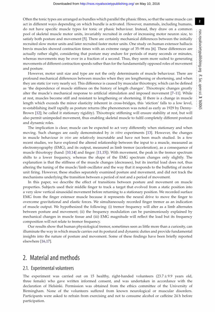
Figure 1. The target (thick black line), a representative performance (thin black line) and the average performance of all subjects/alltrials (dashed) in the 120 s tracking task. Positive values represent an upward movement of the target and finger (extension). Note, forclarity, the traces have been vertically displaced so they do not superimpose.
2.2. ApparatusThe participants sat in a comfortable chair with the right forearm pronated and supported by a plasticcurved rest. The hand as well as the index and ring finger were securely taped to a horizontal U-shapedaluminium support with a gap for the middle finger adjusted to fit individual subjects. This allowed forunhindered and isolated flexion–extension of the middle finger around metacarpophalangeal joint 3. Alight duraluminium splint underneath the middle finger prevented movement at interphalangeal joints.The arm and the hand rest were individually connected to a heavy steel table by magnetic supports, sothey could be optimally positioned.
A miniature three-axis accelerometer (model SCA3000, Active Robots, UK, 12.7 × 20.32 mm) wasattached above the nail bed of the middle finger to measure its vertical acceleration. A retroreflectivelaser rangefinder (YP11MGV80, Wenglor Sensoric, Germany) was pointed at a white plastic reflectivesurface (approx. 2 × 3 cm) placed on top of the accelerometer to record vertical finger position. Acomputer screen approximately 1.5 m in front of the subject displayed finger position as a white crossand a computer-controlled target in the form of a red ball. The target was a stereotyped positionalwaveform which included transitions between posture and movement as shown in figure 1. Specifically,the target was initially static at an individually determined comfortable neutral (neither extended norflexed) position of the finger. After 10 s, the target moved into a vertically orientated sinusoid with afrequency linearly rising from 0 to 0.05 Hz over 50 s (i.e. a chirp signal). The target then deceleratedas a mirror image of this chirp and ended stationary in the neutral position for 10 s. The entiresequence occupied 120 s. The peak-to-peak amplitude of the waveform was approximately 30° of fingermovement (15° extension (up) and 15° flexion (down)), which was well within the limits of the subjects’comfortable range of motion. Participants were asked to track the target by keeping the white crossand red ball on the computer screen aligned. It was emphasized to the subjects that this was nota test of accuracy or precision and that they should remain as relaxed as possible. The maximumangular velocity required to track was 3 degree s−1. Subjectively, this seemed a very slow movement,and this stereotyped task was easy to perform. Each subject repeated the task 10 times and refrainedfrom moving their hand or fingers 10 s before the start of every trial. Surface EMG was recordedfrom the belly of the extensor digitorum communis muscle (m. EDC) with a Bagnoli system (DelsysInc., USA).
on May 10, 2016http://rsos.royalsocietypublishing.org/Downloaded from
4
rsos.royalsocietypublishing.orgR.Soc.opensci.3:160065
................................................
4
6
8
10
10 20 30 40 50
10 20 30 40 50
4
6
8
10
10 20 30 40 50
acce
lera
tion
ampl
itude
(×
10–3
ms–2
)
frequency (Hz)10 20 30 40 50frequency (Hz)
10 20 30 40 50frequency (Hz)
10 20 30 40 50frequency (Hz)
frequency (Hz)frequency (Hz)
EM
G
120 s
0.51.01.52.02.53.03.5
0.51.01.52.02.53.03.5
0.51.01.52.02.53.03.5
0.51.01.52.02.53.03.5
10 20 30 40 50frequency (Hz)
10 20 30 40 50frequency (Hz)
4
6
8
10
4
6
8
10
ampl
itude
(×
10–5
mV
)
5 s 5 s
5 s5 s
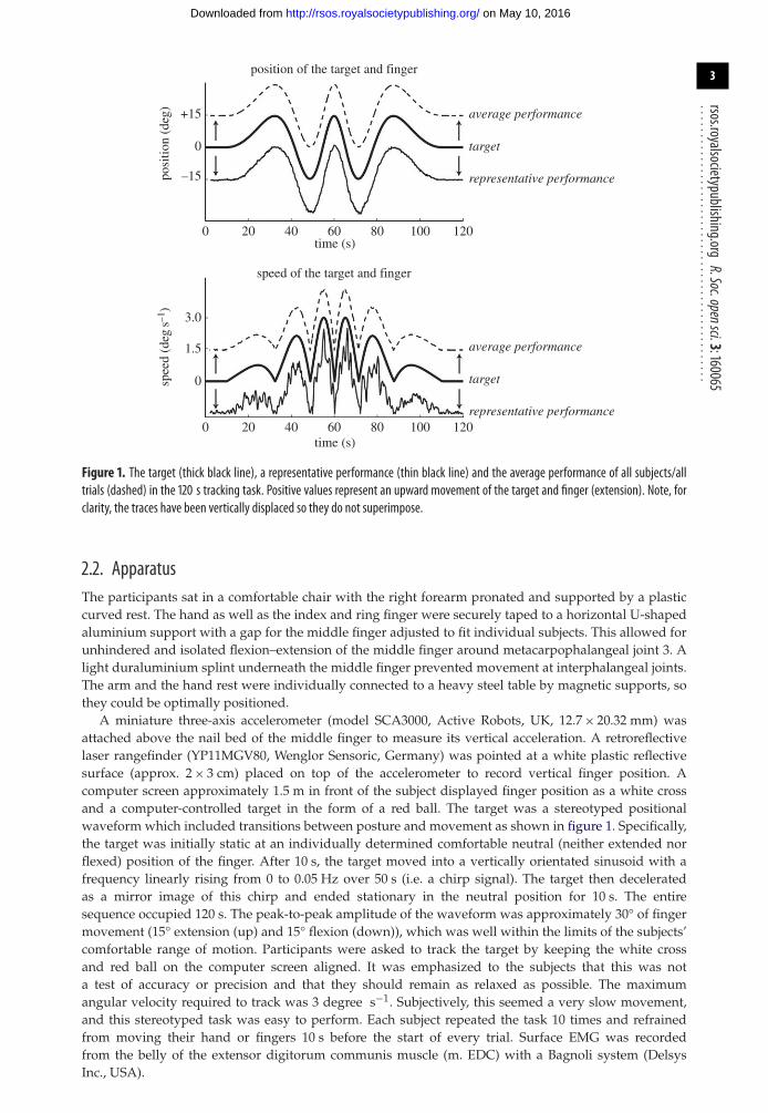
Figure 2. Frequency spectra of EMG and acceleration (tremor) at different phases of target tracking by a representative subject. Top:EMG frequency spectra. Middle: finger position. Bottom: acceleration frequency spectra. From left to right, spectra are shown for 5-ssnapshots of static posture,maximumupward finger velocity,maximumdownward finger velocity and static posture. The tremor spectraare relatively robust, whereas the EMG is much more variable. There is a considerable modulation of tremor frequency and size betweenposture and maximum velocity snapshots while there is no obvious direct correspondence with the related EMG spectra.
2.3. Data analysisEMG was amplified ×1000. EMG, acceleration and positional data were sampled at 1000 Hz anddigitized by an MC 6026 PCI card. Further analysis was carried out offline using custom MATLAB scripts(MathWorks Matlab 2011a, USA). EMG was band-pass filtered (35–200 Hz, fourth-order Butterworthzero-phase-lag filter) and rectified. Acceleration signals were high-pass filtered (0.1 Hz, fourth-orderButterworth dual filter) to correct for artefactual modulation within the range of target frequencies.For one representative subject, the amplitude spectra of the finger acceleration and EMG were obtainedby NEUROSPEC software (v. 2.0, 2008) in order to display four 5 s snapshots at different phases of the120 s target tracking (figure 2); static (second 5–10), maximum upward finger velocity (second 52.5–57.5),maximum downward finger velocity (second 62.5–67.5) and static (second 115–120).
Position, EMG and acceleration were then down-sampled to 100 Hz. To capture the non-stationaryfeatures of these signals (frequency and power) over the tracking movements, we calculated the wavelettransformation of EMG and acceleration. Wavelets depict the frequency components of a signal overtime as if it were a frequency spectrum over a sliding time-window, therefore enabling us to characterizethe frequency and power of EMG and acceleration associated with the different movement speeds andpositions of the finger. The mean wavelet power was calculated using a continuous wavelet transform(CWT) ([10,18]; for a tutorial, see [19]). The CWT scales and translates a mother wavelet shape at eachinfinitesimal time step of a continuous signal. The wavelet coefficient C of signal s over time t is calculatedas: Ca,b = ∫ s(t)(1/
√a)Ψ ∗ ((t − b)/a) dt, where ψ is the mother wavelet, a is the frequency scale of ψ , b is
the position of ψ and * is the complex conjugation. We used a complex Morlet mother wavelet with abandwidth f b of 1 Hz and a central frequency f c of 1.5 Hz, so that Ψ (x) = (1/
√π fb) e2tπ fcx e−x2/fb . The
wavelet was scaled to correspond to a frequency range of 5–37.5 Hz. Wavelet analysis allowed us toexamine the peak power and frequency of acceleration and EMG over the 120 s duration of the trackingtrial. Note that ‘power’ here is relative, as the wavelet analysis transforms the data. Absolute velocity
on May 10, 2016http://rsos.royalsocietypublishing.org/Downloaded from
5
rsos.royalsocietypublishing.orgR.Soc.opensci.3:160065
................................................0.8
0.6
0.4
0.2
0 20 40 60 80 100 120
6
10
14
18
22
time (s)
3.0
1.5
0
3.0
1.5
0
0 20 40 60 80 100 120time (s)
trem
or p
ower
(ar
b.un
its)
reve
rsed
trem
or f
requ
ency
(H
z)
postural frequency range
spee
d (d
egs–1
)sp
eed
(deg
s–1)
speed (deg s–1)
0.8
0.6
0.4
0.2
22
18
14
10
6
trem
or p
ower
(ar
b.un
its)
trem
or f
requ
ency
(H
z)
0 1.5 3.0
speed (deg s–1)0 1.5 3.0
y = 0.51x + 0.15r2 = 0.87
y = –13.65*x0.28 + 20.64r2 = –0.92
(a) (b)
(c) (d)
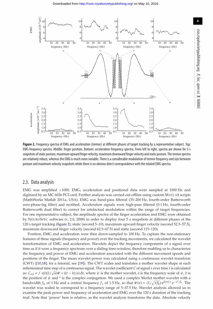
Figure 3. (a) Finger acceleration (tremor) power (arbitrary units, solid line± s.d.) as depicted by wavelet analysis is shown alongsidemovement speed (dashed line). (b) Tremor power plotted as a function ofmovement speed. A linear regression provides a satisfactory fit,although it may misrepresent the relationship when movement is slow. (c) Tremor frequency (solid line± s.d.) as depicted by waveletanalysis and movement speed (dashed line). The postural tremor frequency range is also indicated (details in text). Note that the scalefor frequency runs from 6 to 22 Hz and is reversed to emphasize the striking correspondencewithmovement speed. (d) Tremor frequencyplotted as a function of movement speed. A power function provides an excellent fit.
0 20 40 60 80 100 120
0 20 40 60 80 100 120time (s)
0.03
0.02
0.01
0.03
0.02
0.01
–15–15
0
0
+15
+15
posi
tion
(deg
)
–15 0 +15position (deg)
1514131211
y = 0.03x + 13.10
r2 = 0.34
y = 4.67e–4x + 0.01
r2 = 0.93
1514131211
EM
G f
requ
ency
(H
z)
EM
G f
requ
ency
(H
z)
EM
G p
ower
(ar
b.un
its)
EM
G p
ower
(ar
b.un
is)
posturalfrequencyrange
(b)
(d)(c)
(a)
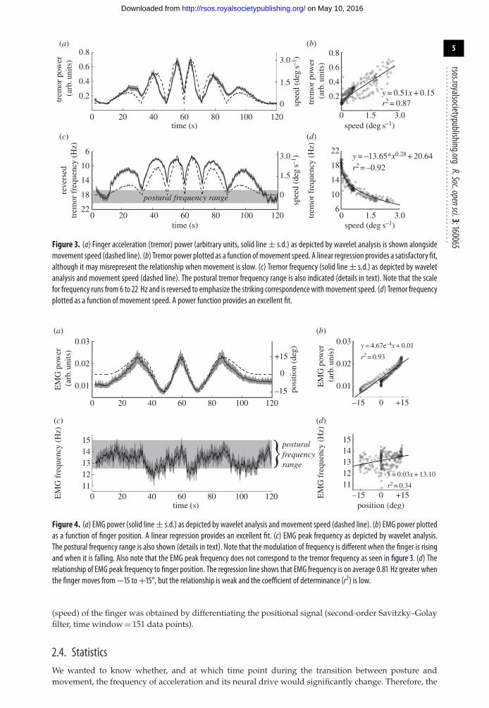
Figure 4. (a) EMG power (solid line± s.d.) as depicted by wavelet analysis and movement speed (dashed line). (b) EMG power plottedas a function of finger position. A linear regression provides an excellent fit. (c) EMG peak frequency as depicted by wavelet analysis.The postural frequency range is also shown (details in text). Note that the modulation of frequency is different when the finger is risingand when it is falling. Also note that the EMG peak frequency does not correspond to the tremor frequency as seen in figure 3. (d) Therelationship of EMG peak frequency to finger position. The regression line shows that EMG frequency is on average 0.81 Hz greater whenthe finger moves from−15 to+15°, but the relationship is weak and the coefficient of determinance (r2) is low.
(speed) of the finger was obtained by differentiating the positional signal (second-order Savitzky–Golayfilter, time window = 151 data points).
2.4. StatisticsWe wanted to know whether, and at which time point during the transition between posture andmovement, the frequency of acceleration and its neural drive would significantly change. Therefore, the
on May 10, 2016http://rsos.royalsocietypublishing.org/Downloaded from
6
rsos.royalsocietypublishing.orgR.Soc.opensci.3:160065
................................................95% confidence interval (CI) was calculated for the peak frequency of acceleration and EMG over time.For both variables, the frequencies covered by the CI over the last 5 s of the two static periods (at the startand end of the movement) were designated the ‘postural’ frequency range (figures 3c and 4c). We thenmeasured the time when the limits of the CI during the tracking movement would exceed this range, ofwhich the first and last time points were used to signify a borderline finger movement that signifies achange in frequency. Wavelet data as well as finger position and finger velocity were averaged over trialsand subjects and finally smoothed (moving average with time constant = 0.4 s).
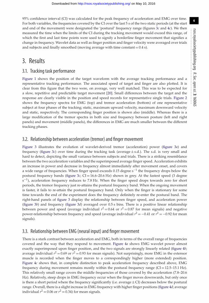
3. Results3.1. Tracking task performanceFigure 1 shows the position of the target waveform with the average tracking performance and arepresentative tracking performance. The associated speed of target and finger are also plotted. It isclear from this figure that the two were, on average, very well matched. This was to be expected fora slow, repetitive and predictable target movement [20]. Small differences between the target and theresponse are clearly visible in the position and speed records for representative single trials. Figure 2shows the frequency spectra for EMG (top) and tremor acceleration (bottom) of one representativesubject at four phases of the tracking; static, maximum upward velocity, maximum downward velocityand static, respectively. The corresponding finger position is shown also (middle). Whereas there is alarge modification of the tremor spectra in both size and frequency between posture (left and rightpanels) and movement (middle panels), the differences in EMG are much smaller between the differenttracking phases.
3.2. Relationship between acceleration (tremor) and finger movementFigure 3 illustrates the evolution of wavelet-derived tremor (acceleration) power (figure 3a) andfrequency (figure 3c) over time during the tracking task (average ± s.d.). The s.d. is very small andhard to detect, depicting the small variance between subjects and trials. There is a striking resemblancebetween the two acceleration variables and the superimposed average finger speed. Acceleration exhibitsan increase in power and decrease in frequency almost immediately after movement onset, and coversa wide range of frequencies. When finger speed exceeds 0.15 degree s−1 the frequency drops below thepostural frequency bands (figure 3c; CI = 16.6–20.6 Hz) shown in grey. At the fastest speed (3 degrees−1), acceleration frequency reduces to 7.8 Hz. When the finger speed drops towards zero for shortperiods, the tremor frequency just re-attains the postural frequency band. When the ongoing movementis faster, it fails to re-attain the postural frequency band. Only when the finger is stationary for sometime towards the end of the experiment does the frequency definitely re-enter the postural band. Theright-hand panels of figure 3 display the relationship between finger speed, and acceleration power(figure 3b) and frequency (figure 3d) averaged over 0.5 s bins. There is a positive linear relationshipbetween power and speed (average individual r2 = 0.64 or r2 = 0.87 for mean signals) and negativepower-relationship between frequency and speed (average individual r2 = −0.41 or r2 = −0.92 for meansignals).
3.3. Relationship between EMG (neural input) and finger movementThere is a stark contrast between acceleration and EMG, both in terms of the overall range of frequenciescovered and the way that they respond to movement. Figure 4a shows EMG wavelet power almostexactly superimposed upon finger position, and the two signals are strongly linearly related (figure 4b;average individual r2 = 0.69 or r2 = 0.93 for mean signals). Not surprisingly, more EMG in the extensormuscle is recorded when the finger moves to a correspondingly higher (more extended) position.Figure 4c shows that, in complete distinction to peak acceleration frequency described above, EMGfrequency during movement remains mostly within the postural frequency range (CI = 12.5–15.1 Hz).This relatively small range covers the middle frequencies of those covered by the acceleration (7.8–20.6Hz). Relatively, steep drops in EMG frequency occur when the finger moves downwards, but only onceis there a short period where the frequency significantly (i.e. average ± CI) decreases below the posturalrange. Overall, there is a slight increase in EMG frequency with higher finger positions (figure 4d; averageindividual r2 = 0.06 or r2 = 0.34) for mean signals.
on May 10, 2016http://rsos.royalsocietypublishing.org/Downloaded from
7
rsos.royalsocietypublishing.orgR.Soc.opensci.3:160065
................................................
15
14
13
12
11
0.01 0.02EMG power (arb. units)
EM
G p
ower
(ar
b.un
its)
y = 53.13x + 12.42
y = 0.07x + 12.35
y = 3.17x–0.64 + 4.45
r2 = 0.13
r2 = 0.13
r2 = –0.78
15 deg
0
–15 deg
EM
G f
requ
ency
(H
z)
15
14
13
12
11EM
G f
requ
ency
(H
z)ac
cele
ratio
n fr
eque
ncy
(Hz) 22
18
14
10
60.2 0.4 0.6 0.8
acceleration power (arb. units)
3.0 m s–2
2.5
2.0
1.5
1.0
0.5
0 m s–2
3 m s–2
2.5
2.0
1.5
1.0
0.5
0 m s–2
3.0 m s–2
2.5
2.0
1.5
1.0
0.5
0 m s–2
0.03
0.02
0.01
0.2 0.4 0.6 0.8 6 10 14 18 22acceleration power (arb. units) acceleration frequency (Hz)
line of unity
(a) (b)
(c) (d )
Figure 5. In (a), the greyscale represents finger position. In (b–d), it represents finger speed. Black always represents the lowest values.(a) EMG peak frequency as a function of EMG power. The highest finger positions are associated with the greatest EMG power andthe highest peak frequencies. (b) Acceleration frequency as a function of acceleration power. The highest finger speeds are stronglyassociated with the greatest acceleration power and the lowest frequencies. (c) EMG power as a function of acceleration power. Thehighest finger speeds are associated with the greatest acceleration power, but there is no strong relationship between speed and EMGpower or acceleration power and EMG power. (d) The relationship between EMG peak frequency and acceleration frequency. The highestfinger speeds are associatedwith the lowest frequency of both acceleration and EMGbut the two variables alter by very different amountsas indicated by the line of unity (dashed line).
3.4. Power–frequency relationship within EMG and accelerationThe average relationship between power and frequency for EMG and for acceleration is shown infigure 5. In each panel, finger kinematics are represented by the use of greyscale (finger position ina; finger speed in b–d), where a fade to black implies lower values. Figure 5a displays a significantpositive relationship between EMG power and frequency. In general, as EMG power increases, so does itsfrequency (r2 = 0.13, p< 0.01), but there is considerable variability. The superimposed greyscale capturesthe much stronger positive relationship between EMG power and finger position (also shown in figure 4b).Figure 5b shows a significant negative relationship between power and frequency of finger acceleration.The largest drop in frequency occurs when the finger begins to move, i.e. at low finger speeds wherethe acceleration is small (denoted by darker black dots). A power-curve fit gave a strong correlation ofr2 = −0.78 (p< 0.01).
As any acceleration has to be a consequence of some neural input, whether broad-band or frequency-specific input, we also studied the relationship between EMG and acceleration by correlating theirpower and frequency. Figure 5c shows an expected tendency for more acceleration with increasedEMG power, but the strength of this correlation is weak (r2 = 0.18, p< 0.01) and the relationship iscomplicated; acceleration power can be small when EMG is both high and low. This corresponds tothe situation when the finger is stationary at its top or bottom position, respectively. The greyscaleshows the previously described positive correlation between finger speed and acceleration power (seenin figure 3b). Figure 5d shows no strong modulation of acceleration frequency with EMG frequency.Although a significant positive linear relationship does exist (solid line; r2 = 0.13, p< 0.01), this line isvery different from the line of unity (dashed line) indicating that there is no direct causal relationship.Again, the strong negative relationship between acceleration frequency and finger speed is obvious in thegreyscale.
on May 10, 2016http://rsos.royalsocietypublishing.org/Downloaded from
8
rsos.royalsocietypublishing.orgR.Soc.opensci.3:160065
................................................4. DiscussionHere, we examined the behaviour of physiological finger tremor during transitions between postureand movement. This is important, because tremor during (slow) movement has been claimed to befundamentally different from postural tremor [21]. However, although the relationship of the phase oftremor with rapid voluntary movement has been studied before, the way in which postural tremor at restevolves into the kinetic tremor of movement, and vice versa, has not been characterized. Our results showthat there are predictable (large) alterations in the power and frequency of tremor as posture becomesmovement. These alterations are not shown in the concomitantly recorded EMG which drives the muscle.There has been a tendency in the literature to discuss ‘10 Hz tremor’ as though it provides ‘a windowinto the operation of the nervous system’ [22]. What this study demonstrates is that systematic and quitelarge alterations in tremor frequency can be generated by movement, and it is not easy to explain thesechanges as being produced by a neurogenic oscillator. On the other hand, it is quite easy to attributethem to mechanical causes. We discuss whether alterations in the stiffness of the muscles, which occurduring movement, produce the altered tremor power and frequency and whether physiological tremorcan provide practical and previously unrecognized insights into the stiffness of the musculature.
4.1. Acceleration and EMG changes with movementThe power and frequency of physiological finger tremor (measured as acceleration) are stronglymodulated by the speed of finger movement, with faster movement being associated with progressivelylarger and slower oscillations. The change is obvious in the initial stages of movement; accelerationfrequency drops substantially and power increases as soon as movement begins (figure 3). This is nottrue for the associated EMG power which correlated nearly linearly with finger position (figure 4a,b).Throughout the 120 s trial, the EMG frequency stays within a relatively narrow range (12.5–15.1 Hz),which is situated neither at the high nor low end of the range of acceleration frequencies (7.8–20.6 Hz;figures 3 and 4). There is no simple relationship between the frequency of the acceleration (which altersa lot) and the frequency of the EMG (which alters only a little). This suggests that finger tremor undernormal, healthy circumstances might not necessarily be generated by a specific neural input. This givesrise to an important question: are the characteristics of finger tremor determined by the mechanicalproperties of the limb?
The strong correlation of extensor EMG power with finger position (figure 4) was entirely expectedbecause it reflected the increased muscle force required to raise the finger. The activity of the main flexormuscle is not reported here as it hardly exceeded the background level and any activation present waserratic. The maximal velocity (which was attained slowly) was only 3 degree s−1, which is extremelyslow. Therefore, for the speeds that we studied the braking forces were almost certainly passive andmechanical. The downward force was mainly applied by elastic forces in the flexor muscles owing topassive stretch or, more likely, gravity. Some modulation of EMG frequency was also anticipated. Forseveral reasons, there is a generalized increase in EMG frequency as force is increased (reviewed by [23]),for instance, because EMG will be modulated according to rate coding. EMG frequency drops abruptlyas the finger commences downwards movement (flexion) and rises more slowly and progressively asthe finger is extended (figure 4c). This is probably owing to the different efficiency of the extensormuscle as it acts concentrically and eccentrically. In addition, some EMG frequency modulation couldhave been caused by measurement artefacts owing to a change in muscle fibre orientation relative to theEMG electrode [23]. Independent of its cause, and more importantly here, there were no correspondinghysteretic effects in tremor acceleration frequency (figure 3c). This divergent behaviour is additionalsimple evidence against the idea that physiological tremor frequency is mainly a reflection of theunderlying frequency of the EMG.
4.2. The resonant nature of physiological tremorIn general, physiological finger tremor is described as being composed of a number of accelerationfrequency components of distinct origins. Often a combination is proposed of two centrally determinedmodes of oscillation (a postural tremor generator and a kinetic tremor generator) or a combinationof mechanical limb properties and neural oscillations of central origin [24,25] or reflex origin [26,27].However, our previous research suggested that all frequencies reported in finger tremor could beeconomically explained by resonance [11,15,28] which differs in frequency depending on the state ofthe musculature. When the finger and muscle do not move, muscle stiffness is high, thereby generating
on May 10, 2016http://rsos.royalsocietypublishing.org/Downloaded from
9
rsos.royalsocietypublishing.orgR.Soc.opensci.3:160065
................................................a high-frequency component greater than 20 Hz. On the other hand, when muscle moves its stiffness islow and generates a frequency component approximately 8 Hz. (This concept is further discussed below.)This study adds to that research by showing clearly that acceleration power and frequency are modulatedby the speed of movement. The smooth modulation and lack of substantial change in the EMG suggestthere is a single peripheral determinant of frequency that adjusts its properties with movement.
4.3. Thixotropic stiffening of muscle raises resonant frequencyThe finding that finger tremor is modulated by the speed of finger movement ties in neatly with amechanical resonance origin. It is known that extrafusal and intrafusal muscular stiffness is dependenton the recent history of movement [8,29]. When muscle moves, its stiffness reduces greatly, for exampleby a factor of approximately 15 in the human calf muscles [30]. Although still currently debated inthe literature, conceivably the underlying mechanism consists of the elastic characteristic of cross-bridges and the number of actin–myosin attachments [8,13,31,32] with the additional contribution ofunfolding of gap–filament proteins like titin [33,34]. Whatever the mechanism, during posture, musclestiffness is high. During movement, the postural short-range elastic stiffness (SREC) is transformed into asmaller, approximately constant, frictional resistance [31]. If a steady posture is subsequently maintained,stiffness will slowly regenerate over the following seconds. This phenomenon, known as thixotropy,makes muscle stiffness very strongly dependent on length changes.
With movement, there is substantially decreased muscle stiffness and thus a decreased resonantfrequency. The muscle length, resulting from a different finger position, is not the determining factor,because thixotropic stiffening occurs at all muscle lengths [13]. In a reduced preparation (single muscleor single muscle fibre), the transition from SREC to frictional behaviour is very abrupt [13]. In a limbwhich is controlled by different synergistic and antagonistic muscles and very many muscle fibres,the transition will be expected to be less abrupt and this is what we report here. However, it is clearthat the greatest reduction in frequency occurs for very small movements (figure 3d). This behaviour isentirely consistent with a reduction in stiffness once a very small range of movement is exceeded. Hill[31] described the range of the SREC in vitro as 0.2% of muscle length. Several studies have shown thatin vivo the required muscular movement to exceed the SREC is very little [8,35]. The tiny movementsassociated with ‘posture’, i.e. the tremor itself, are accommodated within the SREC range. In contrast,even with very slow movements, the SREC is continually exceeded, and the frequency remains lowerthan the postural range. This is exactly what we find here (figure 3c).
4.4. The implications for physiological tremor recordingThese results emphasize the importance of controlling very carefully the conditions under which tremoris recorded. Often, twin peaks in the postural tremor spectrum are discussed in the literature, e.g. [36,37],but this does not necessarily imply they occur simultaneously as is always assumed. When holding thefinger extended the muscle stiffness and resonant frequency will be high. However, an occasional smallpostural adjustment will elicit a temporary drop in muscle stiffness, which will generate a temporary lowresonant frequency. A Fourier transform displays the average frequency spectrum of a tremor recording,typically lasting 30–60 s. If small postural adjustments are interspersed with posture, both high and lowfrequencies will be present in the tremor FFT, both produced by the same mechanism. If the Fouriertransform of postural tremor is taken over a few seconds only (as in figure 2), the low frequency peakmay be absent.
4.5. What is posture, what is movement?Hill [31] and others have shown very clearly that the threshold of the SREC is one of position. TheSREC will re-exert itself at any muscle length if the muscle is allowed to rest there for sufficient time. Rest,presumably, is not absolute, but is in reality a period where velocity is very low. There is no such thingas a fixed position, because there is always minor movement, positional drift and tremor. Figure 3 showsthat the confidence limits of postural tremor frequency are exceeded when movement speed reaches aspeed of 0.15 degree s−1. Although this is by definition a movement, its speed is so slow that it is notgreatly above the threshold for detection if it was to be passively applied [38]. The postural frequencyrange is only confidently re-entered when the finger is stationary for some time (110 s onwards; figure 3c).This strongly suggests posture and movement are quantitatively, rather than qualitatively, different.
on May 10, 2016http://rsos.royalsocietypublishing.org/Downloaded from
10
rsos.royalsocietypublishing.orgR.Soc.opensci.3:160065
................................................4.6. Is muscle thixotropy the basis of posture and movement?The question of the distinctiveness of movement and posture is an old one, with some authorities,but not all, suggesting that specialized neural pathways are involved [39]. Our results do not answerthat question. However, it is undeniable that in man the ‘final common pathway’ for both movementand posture is a single set of skeletal muscles which must satisfy the very different requirementsof both roles. Muscle stiffness increases greatly as muscle lengthening or shortening ceases, and ourresults suggest that tremor provides a window into this natural behaviour. They show that as muscletransitions from lengthening or shortening to posture, tremor frequency increases, reflecting an increasein muscle stiffness and resonant frequency. This thixotropic stiffening of muscle is due to the reformationof actin–myosin attachments in combination with non-cross-bridge mechanisms. It is likely that thishas a stabilizing influence, making it easy to minimize speed for long periods of time. By definition,this is posture. Man-made servo systems for controlling manufacturing processes, elevator systems,valves and the like, very commonly incorporate an electromechanical brake, so that a static positioncan be economically maintained. Thixotropic muscle stiffening, which permits the economical andeffective control of posture and movement by a common actuator, may be nature’s version of the brake.Meanwhile, with little movement, thixotropic loosening induces very large reductions in muscle stiffness(up to 15-fold; [35]), allowing unimpeded and rapid motion. An example where the functional impactof thixotropy is clear is when raising a gun to shoot a target. This task generates a conflict betweenmovement and stability. The marksman’s muscles are not very stiff when the gun is raised, to enable alarge angular momentum. His muscles then get very stiff when he takes aim to maintain joint orientation.The transition in muscle stiffness solves the conflicting requirements of flexibility and speed during fastaction, and accuracy and stability at rest. Thus, skeletal muscle is optimized for not only posture, but alsofor movement.
Ethics. Ethical approval was obtained from the ethics committee of the University of Birmingham. Volunteers gavewritten informed consent, and the experiment was undertaken in accordance with the Declaration of Helsinki. Noneof the volunteers suffered from known neurological or muscular disorders. Participants were asked to refrain fromexercising and not to consume alcohol or caffeine 24 h before participation.Data accessibility. All raw data are available through the Dryad repository: http://dx.doi.org/10.5061/dryad.r1j0s.Authors’ contributions. C.V., R.R. and M.L. contributed substantially to conception and design, or acquisition of data,or analysis and interpretation of data; C.V., R.R. and M.L. drafted the article, or revised it critically for importantintellectual content; and C.V., R.R. and M.L. have read and approved the manuscript before submission.Competing interests. We have no competing interests.Funding. No external funding is received for this study.Acknowledgements. Thanks to Ken Dawkins and Steve Allen for technical support.
References1. Sommerkamp H. 1928 Das substrat der
Dauerverkfirzung am Froschmuskel (Physiologischeund pharmakologische Sonderstellung bestimmerMuskelfasern. Arch. Exp. Path. Pharmak. 128,99–115. (doi:10.1007/BF01863244)
2. Cochrane DG, Elder HY, Usherwood PNR. 1972Physiology and ultrastructure of phasic and tonicskeletal muscle fibres in the locust, Schistocercagregaria. J. Cell Sci. 10, 419–441.
3. Ginsborg BL. 1960 Some properties of avian skeletalmuscle fibres with multiple neuromuscularjunctions. J. Physiol. 154, 581–598. (doi:10.1113/jphysiol.1960.sp006597)
4. Gleeson TT, Putnam RW, Bennett AF 1980Histochemical, enzymatic, and contractileproperties of skeletal muscle fibers in the lizardDipsosaurus dorsalis. J. Exp. Zool. 214, 293–302.(doi:10.1002/jez.1402140307)
5. Bawa PNS, Jones KE, Stein RB. 2014 Assessment ofsize ordered recruitment. Front. Hum. Neurosci. 8,532. (doi:10.3389/fnhum.2014.00532)
6. Sica REP, McComas AJ. 1971 Fast and slow twitchunits in a human muscle. J. Neurol. Neurosurg.Psychiatry 34, 113–120. (doi:10.1136/jnnp.34.2.113)
7. Lakie M, Robson LG. 1988 Thixotropic changes inhumanmuscle stiffness and the effects of fatigue. Q.J. Exp. Physiol. 73, 487–500. (doi:10.1113/expphysiol.1988.sp003169)
8. Proske U, Morgan DL, Gregory JE. 1993 Thixotropy inskeletal muscle and in muscle spindles: a review.Prog. Neurobiol. 41, 705–721. (doi:10.1016/0301-0082(93)90032-N)
9. Axelson HW, Hagbarth KE. 2001 Human motorcontrol consequences of thixotropic changes inmuscular short-range stiffness. J. Physiol. 535,279–288. (doi:10.1111/j.1469-7793.2001.00279.x)
10. Reynolds RF, Lakie M. 2010 Post-movementchanges in the frequency and amplitude ofphysiological tremor despite unchanged neuraloutput. J. Neurophysiol. 104, 2020–2023.(doi:10.1152/jn.00513.2010)
11. Vernooij CA, Lakie M, Reynolds RF. 2014 Thecomplete frequency spectrum of physiologicaltremor can be recreated by broad-bandmechanical or electrical drive. J. Neurophysiol.113, 647–656. (doi:10.1152/jn.00519.2014)
12. Denny-Brown D. 1929 On the nature of posturalreflexes. Proc. R. Soc. Lond. B 104, 252–301.(doi:10.1098/rspb.1929.0010)
13. Campbell KS, Lakie M. 1998 A cross-bridgemechanism can explain the thixotropic short-rangeelastic component of relaxed frog skeletal muscle.J. Physiol. 510, 941–962. (doi:10.1111/j.1469-7793.1998.941bj.x)
14. Lakie M, Vernooij CA, Osborne TM, Reynolds RF.2012 The resonant component of humanphysiological hand tremor is altered by slowvoluntary movements. J. Physiol. 590,2471–2483. (doi:10.1113/jphysiol.2011.226449)
15. Vernooij CA, Reynolds RF, Lakie M. 2013 A dominantrole for mechanical resonance in physiologicalfinger tremor revealed by selective minimisation ofvoluntary drive and movement. J. Neurophysiol.109, 2317–2326. (doi:10.1152/jn.00926.2012)
16. Vernooij CA, Lakie M, Reynolds RF. 2013Physiological finger tremor size reflects alteredmechanical properties of muscle resulting fromchanges in neural control. In Abstracts of Progress inMotor Control IX, 13–16 July 2013, Montréal, CA,pp. 73–74.
on May 10, 2016http://rsos.royalsocietypublishing.org/Downloaded from
11
rsos.royalsocietypublishing.orgR.Soc.opensci.3:160065
................................................17. Vernooij CA, Reynolds RF, Lakie M. 2013
Physiological finger tremor size mirrors speed offinger movement due to muscle thixotropy. In Proc.of the Physiology Society 7 : 37th Congress of IUPS2013, 22–26 July 2013, Birmingham, UK, pp. PCD252.
18. Gilbertson T, Lalo E, Doyle L, Di Lazzaro V, Cioni B,Brown P. 2005 Existing motor state is favored at theexpense of newmovement during 13-35 Hzoscillatory synchrony in the human corticospinalsystem. J. Neurosci. 25, 7771–7779. (doi:10.1523/JNEUROSCI.1762-05.2005)
19. Samar VJ, Bopardikar A, Rao R, Swartz K. 1999Wavelet analysis of neuroelectric waveforms: aconceptual tutorial. Brain Lang. 66, 7–60.(doi:10.1006/brln.1998.2024)
20. Poulton EC. 1974 Tracking skill and manual control.New York, NY: Academic Press.
21. Vallbo ÅB, Wessberg J. 1993 Organization of motoroutput in slow finger movements in man. J.Physiol. 469, 673–691. (doi:10.1113/jphysiol.1993.sp019837)
22. McAuley JH, Marsden CD. 2000 Physiological andpathological tremors and rhythmic central motorcontrol. Brain 123, 1545–1567. (doi:10.1093/brain/123.8.1545)
23. Farina D, Merletti R, Enoka RM. 2004 The extractionof neural strategies from the surface EMG. J. Appl.Physiol. 96, 1486–1495. (doi:10.1152/japplphysiol.01070.2003)
24. Bye RT, Neilson PD. 2010 The BUMPmodel ofresponse planning: intermittent predictive controlaccounts for 10 Hz physiological tremor. Hum. Mov.Sci. 29, 713–736. (doi:10.1016/j.humov.2010.01.006)
25. Vaillancourt DE, Newell KM. 2000 Amplitudechanges in the 8–12, 20–25, and 40 Hz oscillationsin finger tremor. Clin. Neurophysiol. 111, 1792–1801.(doi:10.1016/S1388-2457(00)00378-3)
26. Christakos CN, Papadimitriou NA, Erimaki S. 2006Parallel neuronal mechanisms underlyingphysiological force tremor in steady musclecontractions of humans. J. Neurophysiol. 95, 53–66.(doi:10.1152/jn.00051.2005)
27. Hagbarth KE, Young RR. 1979 Participation of thestretch reflex in human physiological tremor.Brain 102, 509–526. (doi:10.1093/brain/102.3.509)
28. Lakie M, Vernooij CA, Osler CJ, Stevenson AT, ScottJPR, Reynolds RF. 2015 Increased gravitational forcereveals the mechanical, resonance nature ofphysiological tremor. J. Physiol. 593, 4411–4422.(doi:10.1113/JP270464)
29. Proske U, Gandevia SC. 2012 The proprioceptivesenses: their roles in signaling body shape, bodyposition and movement, and muscle force. Physiol.Rev. 92, 1651–1697. (doi:10.1152/physrev.00048.2011)
30. Loram ID, Lakie M. 2002 Direct measurement ofhuman ankle stiffness during quiet standing: theintrinsic mechanical stiffness is insufficient forstability. J. Physiol. 545, 1041–1053. (doi:10.1113/jphysiol.2002.025049)
31. Hill DK. 1968 Tension due to interaction betweenthe sliding filaments in resting striated muscle.The effect of stimulation. J. Physiol. 199,637–684. (doi:10.1113/jphysiol.1968.sp008672)
32. Altman D, Minozzo FC, Rassier DE. 2015 Thixotropyand rheopexy of muscle fibers probed usingsinusoidal oscillations. PLoS ONE 10, e0121726.(doi:10.1371/journal.pone.0121726)
33. Kellermayer MSZ et al. 2008 Muscle thixotropy:more than just cross-bridges? Response tocomment by Campbell and Lakie. Biophys. J.94, 329–330. (doi:10.1529/biophysj.107.122309)
34. Powers K, Schappacher-Tilp G, Jinha A, Leonard TR,Nishikawa K, Herzog W. 2014 Titin force is enhancedin actively stretched skeletal muscle. J. Exp. Biol.217, 3629–3636. (doi:10.1242/jeb.105361)
35. Loram ID, Maganaris CN, Lakie M. 2007 The passive,human calf muscles in relation to standing: theshort range stiffness lies in the contractilecomponent. J. Physiol. 584, 677–692. (doi:10.1113/jphysiol.2007.140053)
36. Daneault J-F, Carignan B, Duval C. 2011 Fingertremor can be voluntarily reduced during a trackingtask. Brain Res. 1370, 164–174. (doi:10.1016/j.brainres.2010.11.047)
37. Stiles RN, Randall JE. 1967 Mechanical factors inhuman tremor frequency. J. Appl. Physiol. 23,324–330.
38. Fitzpatrick RC, McCloskey DI. 1994 Proprioceptive,visual and vestibular thresholds for the perceptionof sway during standing in humans. J. Physiol. 478,173–186. (doi:10.1113/jphysiol.1994.sp020240)
39. Kurtzer I, Herter TM, Scott SH. 2005 Random changein cortical load representation suggests distinctcontrol of posture and movement. Nat. Neurosci. 8,498–504. (doi:10.1038/nn1420)
on May 10, 2016http://rsos.royalsocietypublishing.org/Downloaded from