Embed Size (px)
Citation preview
Biogeosciences, 6, 2313–2331, 2009www.biogeosciences.net/6/2313/2009/© Author(s) 2009. This work is distributed underthe Creative Commons Attribution 3.0 License.
Biogeosciences
Physiological basis for high CO2 tolerance in marine ectothermicanimals: pre-adaptation through lifestyle and ontogeny?
F. Melzner1, M. A. Gutowska2, M. Langenbuch1, S. Dupont3, M. Lucassen4, M. C. Thorndyke5, M. Bleich2, andH.-O. Portner4
1Biological Oceanography, Leibniz-Institute of Marine Sciences (IFM-GEOMAR), Kiel, Germany2Institute of Physiology, Christian-Albrechts-University, Kiel, Germany3Department of Marine Ecology, Goteborg University, The Sven Loven Centre for Marine Sciences, Kristineberg, Sweden4Integrative Ecophysiology, Alfred-Wegener-Institute for Polar and Marine Research, Bremerhaven, Germany5Royal Swedish Academy of Sciences, The Sven Loven Centre for Marine Sciences, Kristineberg, Sweden
Received: 9 April 2009 – Published in Biogeosciences Discuss.: 5 May 2009Revised: 4 September 2009 – Accepted: 6 October 2009 – Published: 30 October 2009
Abstract. Future ocean acidification has the potential toadversely affect many marine organisms. A growing bodyof evidence suggests that many species could suffer fromreduced fertilization success, decreases in larval- and adultgrowth rates, reduced calcification rates, and even mortalitywhen being exposed to near-future levels (year 2100 scenar-ios) of ocean acidification. Little research focus is currentlyplaced on those organisms/taxa that might be less vulnerableto the anticipated changes in ocean chemistry; this is unfortu-nate, as the comparison of more vulnerable to more tolerantphysiotypes could provide us with those physiological traitsthat are crucial for ecological success in a future ocean. Here,we attempt to summarize some ontogenetic and lifestyletraits that lead to an increased tolerance towards high envi-ronmentalpCO2. In general, marine ectothermic metazoanswith an extensive extracellular fluid volume may be less vul-nerable to future acidification as their cells are already ex-posed to much higherpCO2 values (0.1 to 0.4 kPa, ca. 1000to 3900µatm) than those of unicellular organisms and ga-metes, for which the ocean (0.04 kPa, ca. 400µatm) is the ex-tracellular space. A doubling in environmentalpCO2 there-fore only represents a 10% change in extracellularpCO2 insome marine teleosts. High extracellularpCO2 values are tosome degree related to high metabolic rates, as diffusion gra-dients need to be high in order to excrete an amount of CO2that is directly proportional to the amount of O2 consumed.In active metazoans, such as teleost fish, cephalopods and
Correspondence to:F. Melzner([email protected])
many brachyuran crustaceans, exercise induced increases inmetabolic rate require an efficient ion-regulatory machineryfor CO2 excretion and acid-base regulation, especially whenanaerobic metabolism is involved and metabolic protons leakinto the extracellular space. These ion-transport systems,which are located in highly developed gill epithelia, form thebasis for efficient compensation of pH disturbances duringexposure to elevated environmentalpCO2. Compensationof extracellular acid-base status in turn may be important inavoiding metabolic depression. So far, maintained “perfor-mance” at higher seawaterpCO2 (>0.3 to 0.6 kPa) has onlybeen observed in adults/juveniles of active, high metabolicspecies with a powerful ion regulatory apparatus. However,while some of these taxa are adapted to cope with elevatedpCO2 during their regular embryonic development, gametes,zygotes and early embryonic stages, which lack specializedion-regulatory epithelia, may be the true bottleneck for eco-logical success – even of the more tolerant taxa.
Our current understanding of which marine animal taxawill be affected adversely in their physiological and ecolog-ical fitness by projected scenarios of anthropogenic oceanacidification is quite incomplete. While a growing amount ofempirical evidence from CO2 perturbation experiments sug-gests that several taxa might react quite sensitively to oceanacidification, others seem to be surprisingly tolerant. How-ever, there is little mechanistic understanding on what phys-iological traits are responsible for the observed differentialsensitivities (see reviews of Seibel and Walsh, 2003; Portneret al., 2004; Fabry et al., 2008; Portner, 2008). This leads usto the first very basic question of how to define general CO2tolerance on the species level.
Published by Copernicus Publications on behalf of the European Geosciences Union.
2314 F. Melzner et al.: Physiological basis for high CO2 tolerance in marine ectothermic animals
1 Defining tolerance towards elevated seawaterpCO2
When trying to classify marine organisms into CO2 sensitiveand CO2 tolerant groups, we encounter a major complica-tion: Projected ocean acidification progresses at a rate muchtoo slow to be simulated in the laboratory, and differences ingenetic adaptation potential vary at orders of magnitude be-tween taxa. Organisms with a high generation turnover time(e.g. bacteria, unicellular auto- and heterotrophs) will havetime for thousands of generations to select for genotypes thatcan cope with an ocean characterized bypCO2 values of upto ca. 0.2 kPa (ca. 2000µatm) by the year 2300 (Caldeira andWickett, 2003, 2005), while in long-lived species, such as theocean quahog (Arctica islandica; mollusca: bivalvia), witha maximum life expectancy of ca. 400 years (Abele et al.,2008), today’s genotypes may be exposed to the highpCO2values of the year 2300. Thus, species longevity/generationtime is a crucial factor that could possibly determine futuresuccess in an – on evolutionary timescales – rapidly changinghabitat.
Multi-generation experiments will be very important tounderstand the adaptation potential of a given species, how-ever, such approaches are only beginning to emerge (Kuri-hara and Ishimatsu, 2008; Dupont and Thorndyke, 2009), es-pecially in metazoans with long generation cycles (months,years). Thus, by reading the ideas we propose in this con-cept paper we should keep in mind the problems of rate ofchange in ocean carbonate chemistry and the genetic adapta-tion potential of a given species. In addition, the largely un-explored problems of species interaction and food-web feed-backs will be major factors shaping ecological performanceof marine species in a future highpCO2 ocean. For exam-ple, delayed larval development, as observed in echinodermearly life stages subjected to elevatedpCO2 (Dupont andThorndyke, 2009), probably will increase predation relatedmortality in the field, even if there is no mortality differencein experimental cultures (Elkin and Marshall, 2007).
Considering the limited availability of multi-generationexperiments, the best we can do at the moment to de-fine tolerance versus sensitivity to ocean acidification, is tolook at indicators for animal performance during long-term(weeks to months) CO2 perturbation experiments. We usethe term “animal performance” as the sum of the major rel-evant traits that ensure ecological success of a species (ona species level), i.e. among others, aerobic scope, locomo-tory scope, reproductive output, calcification and somaticgrowth, which, together, influence animal fitness. Aerobicmetabolic scope (the difference between active and standardmetabolic rates, see Fry, 1948, for a definition) is a parame-ter that can be (more or less) easily assessed in mobile ani-mals, e.g. crustaceans, cephalopods or fish (e.g. Booth et al.,1984a; Wells and Wells, 1985; Portner et al., 1991; Melzneret al., 2009), whereas it can only be approximated in ses-sile animals, sometimes via the specific dynamic action offood (e.g. Vahl, 1984; Widdows, 1973). Aerobic metabolic
scope of a species is also often directly related to locomo-tory scope and growth performance. While the measurementof standard metabolic rates can potentially indicate how thecosts for homeostatic regulation are altered under an acuteabiotic stress regime, somatic and reproductive growth per-formance can integrate cost re-allocation over a longer timeinterval. Thus, “footprints” in the energy budget in responseto an abiotic stressor regime can be detected more easily inlong-term growth trials. While somatic/reproductive growthmay be one of the best performance indicators, it has alreadybecome clear that in order to consider possible trade-offs be-tween single parameters all relevant indicators have to be in-cluded simultaneously to generate a meaningful assessmentof a given species’ vulnerability to future ocean acidification(e.g. see below, Wood et al., 2008; Kurihara et al., 2008).Unfortunately, to date there are few comprehensive perfor-mance assessments for marine metazoan species subjectedto long-term elevatedpCO2. Thus, in the following text, wewill place emphasis on those taxa where most informationis available, hoping that future studies will focus on the si-multaneous assessment of multiple performance indicatorsin long-term CO2 perturbation experiments. The aim of thepresent review paper is thus not to compare single parame-ters between different species but to pool data on higher tax-onomic levels to improve our understanding of major physio-logical characteristics that provide the basis for a high degreeof CO2 tolerance. While it is clear already now that due to thesynergistic effects of a complex set of parameters CO2 toler-ance at near-future levels of ocean acidification is difficult topredict, even for closely related species (e.g. echinoderm lar-val stages: Dupont and Thorndyke, 2009; Widdicombe andSpicer, 2008), we will make use of those studies that haveused higherpCO2 values (>0.3 to 0.5 kPa) to elucidate somefundamental tolerance mechanisms that are closely related tolifestyle and metabolic rates of more active taxa.
2 Sensitive vs. tolerant physiotypes: which taxaperform best?
If we combine evidence from the few long-term CO2 per-turbation experiments (weeks to months) until now, it ap-pears that (adult) marine ectothermic vertebrates are the mostCO2-tolerant group – various performance parameters seemnot to be compromised by chronic hypercapnia at levels>0.3 to 0.6 kPa. Teleost species studied in long-term growthtrials (wolffish, Anarhichas minor; salmon, Salmo salar)did not display reductions in somatic growth performancewhen exposed topCO2 values of up to 0.6 kPa and higher(5900µatm; Foss et al., 2003; Fivelstad et al., 1998, 2003).In addition, recent findings indicate that long-term acclima-tion of Atlantic cod (Gadus morhua) to pCO2 values of 0.3and 0.6 kPa (ca. 3000 to 5900µatm) does not seem to impactswimming performance (critical swimming speed, Ucrit),standard and active metabolism, as well as aerobic scope
Biogeosciences, 6, 2313–2331, 2009 www.biogeosciences.net/6/2313/2009/
F. Melzner et al.: Physiological basis for high CO2 tolerance in marine ectothermic animals 2315
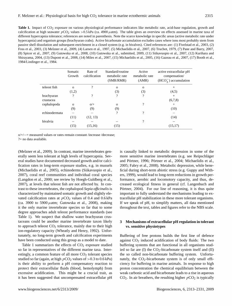
Table 1. Impact of CO2 exposure on various physiological performance indicators like metabolic rate, acid-base regulation, growth andcalcification at high seawaterpCO2 values>0.5 kPa (ca. 4900µatm). The table gives an overview on effects assessed in marine taxa ofdifferent hypercapnia tolerance; references are noted in parenthesis. Note the scarce knowledge in specific areas (active metabolic rate underhypercapnia) and organism groups (brachyuran crabs). Active bicarbonate accumulation excludes cases where ions most probably stem frompassive shell dissolution and subsequent enrichment in a closed system (e.g. in bivalves). Cited references are: (1) Fivelstad et al., 2003, (2)Foss et al., 2003, (3) Melzner et al., 2009, (4) Larsen et al., 1997, (5) Michaelidis et al., 2007, (6) Truchot, 1979, (7) Pane and Barry, 2007,(8) Spicer et al., 2007, (9) Gutowska et al., 2008, (10) Gutowska et al., submitted, 2009, (11) Siikavuopio et al., 2007, (12) Kurihara andShirayama, 2004, (13) Dupont et al., 2008, (14) Miles et al., 2007, (15) Michaelidis et al., 2005, (16) Gazeau et al., 2007, (17) Booth et al.,1984/Lindinger et al., 1984.
Somatic Rate of Standard/routine Active active extracellular pHGrowth calcification metabolic rate metabolic rate compensation/
(SMR/RMR) (AMR) (HCO−
3 ) accumulation
teleost fish o ? o o +(1,2) (3) (3) (4,5)
brachyuran ? ? ? ? +crustacea (6,7,8)cephalopoda o o/+ o ? +
(9) (9) (9) (10)echinodermata − − ? ? −
(11) (12, 13) (14)bivalvia − − − ? −
(15) (15,16) (15) (15,17)
o/+/−= measured values or rates remain constant /increase /decrease;?= no data available.
(Melzner et al., 2009). In contrast, marine invertebrates gen-erally seem less tolerant at high levels of hypercapnia. Sev-eral studies have documented decreased growth and/or calci-fication rates in long-term exposure studies, e.g. in mussels(Michaelidis et al., 2005), echinoderms (Siikavuopio et al.,2007), coral reef communities and individual coral species(Langdon et al., 2000; see review by Hoegh-Guldberg et al.,2007), at levels that teleost fish are not affected by. In con-trast to these invertebrates, the cephalopodSepia officinalisischaracterized by maintained somatic growth and slightly ele-vated calcification rates atpCO2 values of 0.4 and 0.6 kPa(ca. 3900 to 5900µatm; Gutowska et al., 2008), makingit the only marine invertebrate species so far that to somedegree approaches adult teleost performance standards (seeTable 1). We suspect that shallow water brachyuran crus-taceans could be another marine invertebrate taxon likelyto approach teleost CO2 tolerance, mainly due to their highion-regulatory capacity (Wheatly and Henry, 1992). Unfor-tunately, no long-term growth and calcification experimentshave been conducted using this group as a model to date.
Table 1 summarizes the effects of CO2 exposure studiedso far in representatives of the different marine taxa. Inter-estingly, a common feature of all more CO2 tolerant speciesstudied so far (again, at highpCO2 values of>0.3 to 0.6 kPa)is their ability to perform a pH compensatory reaction toprotect their extracellular fluids (blood, hemolymph) fromexcessive acidification. This might be a crucial trait, asit has been suggested that uncompensated extracellular pH
is causally linked to metabolic depression in some of themore sensitive marine invertebrates (e.g. see Reipschlagerand Portner, 1996; Portner et al., 2004; Michaelidis et al.,2005; Fabry et al., 2008). Metabolic depression, while bene-ficial during short-term abiotic stress (e.g. Guppy and With-ers, 1999), would lead to long-term reductions in growth per-formance, aerobic and locomotory capacity, and thus, de-creased ecological fitness in general (cf. Langenbuch andPortner, 2004). For our line of reasoning, it is thus quiteimportant to fully understand the mechanisms leading to ex-tracellular pH stabilization in these more tolerant organisms.If we speak of pH, to simplify matters, all data mentionedthroughout the text, tables and figures refer to the NBS scale.
3 Mechanisms of extracellular pH regulation in tolerantvs. sensitive physiotypes
Buffering of free protons builds the first line of defenceagainst CO2 induced acidification of body fluids: The twobuffering systems that are functional in all organisms stud-ied so far are (I) the CO2-bicarbonate system itself and (II)the so called non-bicarbonate buffering system. Unfortu-nately, the CO2-bicarbonate system is of only small effi-ciency for buffering in marine animals. In response to highproton concentration the chemical equilibrium between theweak carbonic acid and bicarbonate leads to a rise in aqueousCO2. In air breathers, the resulting higherpCO2 is typically
www.biogeosciences.net/6/2313/2009/ Biogeosciences, 6, 2313–2331, 2009
2316 F. Melzner et al.: Physiological basis for high CO2 tolerance in marine ectothermic animals
eliminated by means of increased ventilation. However, thisprocess is seriously impaired by the (comparatively) lowpCO2 values in body fluids of water breathers and the re-sulting very small diffusion gradients between organism andthe surrounding water (see Heisler, 1986, for an extended dis-cussion). Consequently, binding of respiratory protons (orig-inating from CO2 hydration) by so called non-bicarbonatebuffers is the first step to minimize pH changes under acid-ified conditions. Non-bicarbonate buffering is mainly pro-vided by partially protonated amino acid side chains (mostlyfrom histidine or cysteine at physiological pH values), N-terminal α-amino groups of proteins or organic/inorganicphosphate groups. As buffering can only mask protons dur-ing an acidotic pH shift and thus reduce pH changes com-pared to a non-buffered system, surplus protons have to beeliminated to restore the original fluid pH. This can only beachieved by means of active ion transport across specializedepithelia, such as gills, renal or digestive tissue. Althoughthe involved ion exchange mechanisms as a whole are poorlyunderstood and may vary between different marine taxa, theprocesses contributing to pH compensation are summarizedas proton equivalent ion exchange. Concerning the reductionof proton activity in body fluids, it is not important if a pHchange is realized by higher proton excretion rates, rising bi-carbonate import from the seawater or increased retention ofmetabolic bicarbonate.
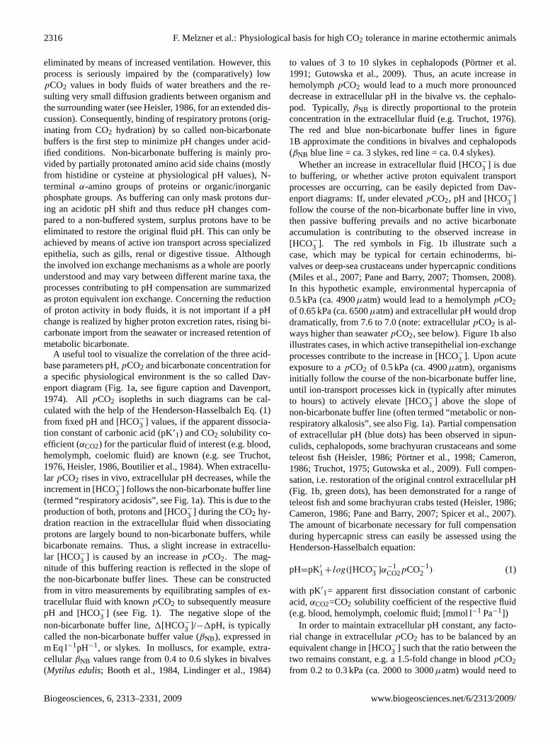
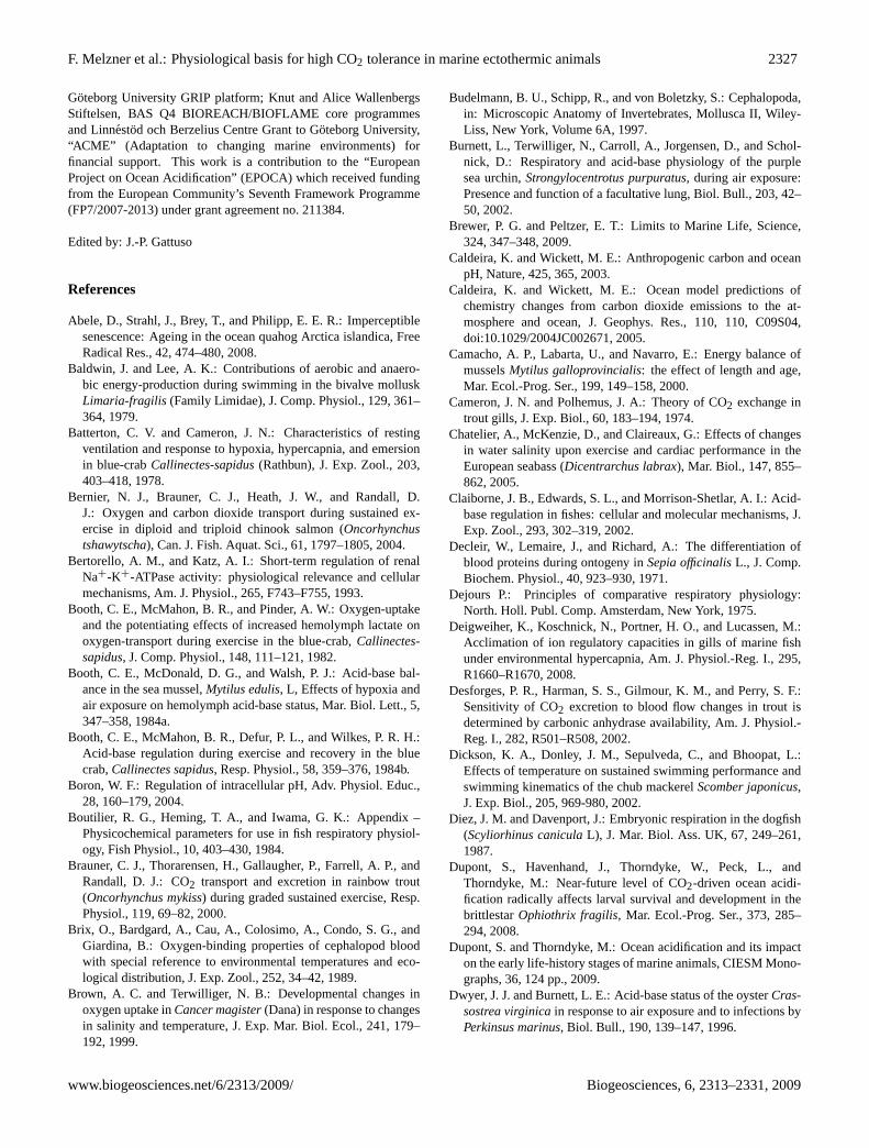
A useful tool to visualize the correlation of the three acid-base parameters pH,pCO2 and bicarbonate concentration fora specific physiological environment is the so called Dav-enport diagram (Fig. 1a, see figure caption and Davenport,1974). All pCO2 isopleths in such diagrams can be cal-culated with the help of the Henderson-Hasselbalch Eq. (1)from fixed pH and [HCO−3 ] values, if the apparent dissocia-tion constant of carbonic acid (pK’1) and CO2 solubility co-efficient (αCO2) for the particular fluid of interest (e.g. blood,hemolymph, coelomic fluid) are known (e.g. see Truchot,1976, Heisler, 1986, Boutilier et al., 1984). When extracellu-lar pCO2 rises in vivo, extracellular pH decreases, while theincrement in [HCO−3 ] follows the non-bicarbonate buffer line(termed “respiratory acidosis”, see Fig. 1a). This is due to theproduction of both, protons and [HCO−3 ] during the CO2 hy-dration reaction in the extracellular fluid when dissociatingprotons are largely bound to non-bicarbonate buffers, whilebicarbonate remains. Thus, a slight increase in extracellu-lar [HCO−
3 ] is caused by an increase inpCO2. The mag-nitude of this buffering reaction is reflected in the slope ofthe non-bicarbonate buffer lines. These can be constructedfrom in vitro measurements by equilibrating samples of ex-tracellular fluid with knownpCO2 to subsequently measurepH and [HCO−
3 ] (see Fig. 1). The negative slope of thenon-bicarbonate buffer line,1[HCO−
3 ]/−1pH, is typicallycalled the non-bicarbonate buffer value (βNB), expressed inm Eq l−1pH−1, or slykes. In molluscs, for example, extra-cellularβNB values range from 0.4 to 0.6 slykes in bivalves(Mytilus edulis; Booth et al., 1984, Lindinger et al., 1984)
to values of 3 to 10 slykes in cephalopods (Portner et al.1991; Gutowska et al., 2009). Thus, an acute increase inhemolymphpCO2 would lead to a much more pronounceddecrease in extracellular pH in the bivalve vs. the cephalo-pod. Typically,βNB is directly proportional to the proteinconcentration in the extracellular fluid (e.g. Truchot, 1976).The red and blue non-bicarbonate buffer lines in figure1B approximate the conditions in bivalves and cephalopods(βNB blue line = ca. 3 slykes, red line = ca. 0.4 slykes).
Whether an increase in extracellular fluid [HCO−
3 ] is dueto buffering, or whether active proton equivalent transportprocesses are occurring, can be easily depicted from Dav-enport diagrams: If, under elevatedpCO2, pH and [HCO−
3 ]follow the course of the non-bicarbonate buffer line in vivo,then passive buffering prevails and no active bicarbonateaccumulation is contributing to the observed increase in[HCO−
3 ]. The red symbols in Fig. 1b illustrate such acase, which may be typical for certain echinoderms, bi-valves or deep-sea crustaceans under hypercapnic conditions(Miles et al., 2007; Pane and Barry, 2007; Thomsen, 2008).In this hypothetic example, environmental hypercapnia of0.5 kPa (ca. 4900µatm) would lead to a hemolymphpCO2of 0.65 kPa (ca. 6500µatm) and extracellular pH would dropdramatically, from 7.6 to 7.0 (note: extracellularpCO2 is al-ways higher than seawaterpCO2, see below). Figure 1b alsoillustrates cases, in which active transepithelial ion-exchangeprocesses contribute to the increase in [HCO−
3 ]. Upon acuteexposure to apCO2 of 0.5 kPa (ca. 4900µatm), organismsinitially follow the course of the non-bicarbonate buffer line,until ion-transport processes kick in (typically after minutesto hours) to actively elevate [HCO−3 ] above the slope ofnon-bicarbonate buffer line (often termed “metabolic or non-respiratory alkalosis”, see also Fig. 1a). Partial compensationof extracellular pH (blue dots) has been observed in sipun-culids, cephalopods, some brachyuran crustaceans and someteleost fish (Heisler, 1986; Portner et al., 1998; Cameron,1986; Truchot, 1975; Gutowska et al., 2009). Full compen-sation, i.e. restoration of the original control extracellular pH(Fig. 1b, green dots), has been demonstrated for a range ofteleost fish and some brachyuran crabs tested (Heisler, 1986;Cameron, 1986; Pane and Barry, 2007; Spicer et al., 2007).The amount of bicarbonate necessary for full compensationduring hypercapnic stress can easily be assessed using theHenderson-Hasselbalch equation:
pH=pK′
1+ log([HCO−
3 ]α−1CO2pCO−1
2 ) (1)
with pK′1= apparent first dissociation constant of carbonic
acid,αCO2=CO2 solubility coefficient of the respective fluid(e.g. blood, hemolymph, coelomic fluid; [mmol l−1 Pa−1])
In order to maintain extracellular pH constant, any facto-rial change in extracellularpCO2 has to be balanced by anequivalent change in [HCO−3 ] such that the ratio between thetwo remains constant, e.g. a 1.5-fold change in bloodpCO2from 0.2 to 0.3 kPa (ca. 2000 to 3000µatm) would need to
Biogeosciences, 6, 2313–2331, 2009 www.biogeosciences.net/6/2313/2009/
F. Melzner et al.: Physiological basis for high CO2 tolerance in marine ectothermic animals 2317
metabolic acidosis
metabo
lic al
kalos
is
respiratory acidosis
respiratory alkalosis
7.0 7.2 7.4 7.6 7.8 8.0
0.13
0.27
0.53
0.801.332.02.8
7.0 7.2 7.4 7.6 7.8 8.00
4
8
12
16
20PCO2 (kPa):
0 h
1 h
4 h
8 h
16 h
extracellular pH (pHe) extracellular pH (pHe)
extra
cellu
lar [
HC
O3- ]
(mM
)
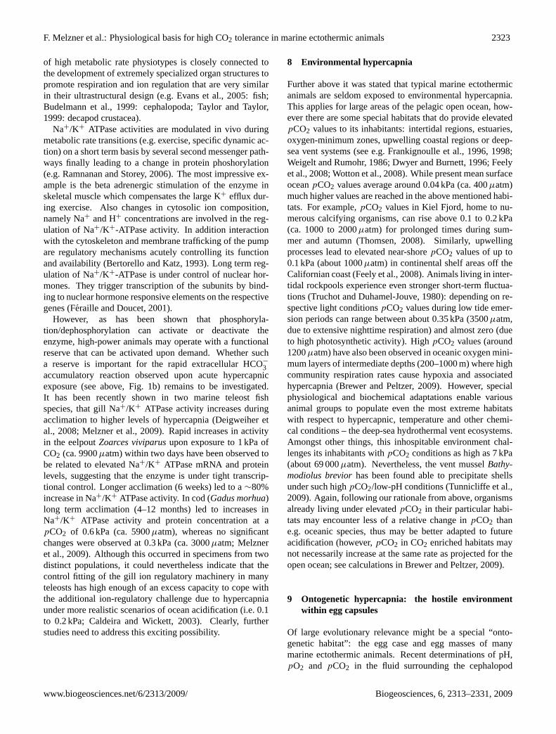
Fig. 1. Davenport diagrams.(A): Schematic illustration of non-bicarbonate buffer line, dashed green line. Arrows indicate changes inpCO2 and [HCO−
3 ] during respiratory acidosis/alkalosis and metabolic acidosis/alkalosis. See text for explanations.(B): Three differenthypothetical organisms subjected to 0.5 kPa (ca. 4900µatm) environmental hypercapnia. Red symbols: No active accumulation of bicar-bonate in the extracellular space to compensate pH, pH follows the non-bicarbonate buffer line. Blue symbols, green symbols: partial/fullpH compensation through active bicarbonate accumulation. Stars indicate control parameters, numbers indicate time (h) exposed to elevatedpCO2 (hypothetical time course!). See text for a detailed discussion.
result in a 1.5-fold increase in [HCO−3 ] to maintain extracel-lular pH at the control level.
The main prerequisite for such a rapid and efficient bi-carbonate accretion are high net proton equivalent fluxesbetween ectothermic organisms and the surrounding sea-water. Such data are currently only available for deca-pod crustaceans and for teleost/elasmobranch fish as wellas an invertebrate (sipunculid) worm. Values of about100µEq kg−1 h−1 net acid efflux have been recorded for thecrustaceanCarcinus maenasexposed to apCO2 value ofabout 0.7 kPa (ca. 6900µatm; Truchot, 1979), even highervalues have been recorded in the marine teleostConger con-ger, where exposure to 1.3 kPa CO2 (ca. 12 800µatm) pro-duced a net acid efflux of 920µEq kg−1 h−1 (Holeton et al.,1983). Rates were much lower in the sipunculid and mir-rored transiently enhanced net proton release during transi-tion to a new steady state in acid-base status under hypercap-nia (Portner et al., 1998).
In summary, it appears that a relative degree of tolerancetowards hypercapnic exposure can be found mainly in suchmarine ectothermic organisms that possess the ability to ac-tively accumulate large amounts of bicarbonate ions to sta-bilize extracellular pH. In addition, these organisms are typ-ically equipped with relatively high non-bicarbonate buffer-ing capacities, which protect extracellular pH during acuteCO2 exposure. While hypercapnia typically is not a rele-vant stressor in the natural habitat of many marine organisms(however, see Sects. 8 and 9), high capacities for net acidextrusion directly result from an active mode of life, high
metabolic rates and frequent as well as rapid metabolic ratefluctuations. We will follow this line of argument in the fol-lowing paragraphs.
4 A common denominator: metabolic rate andmetabolic rate fluctuations
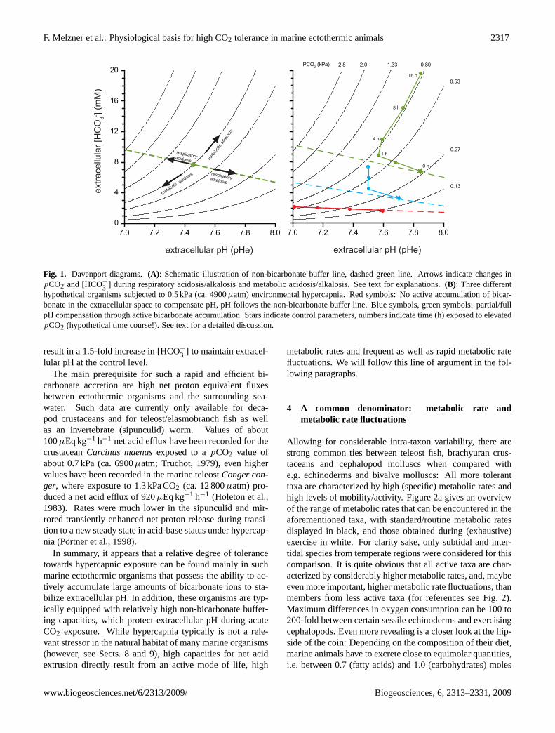
Allowing for considerable intra-taxon variability, there arestrong common ties between teleost fish, brachyuran crus-taceans and cephalopod molluscs when compared withe.g. echinoderms and bivalve molluscs: All more toleranttaxa are characterized by high (specific) metabolic rates andhigh levels of mobility/activity. Figure 2a gives an overviewof the range of metabolic rates that can be encountered in theaforementioned taxa, with standard/routine metabolic ratesdisplayed in black, and those obtained during (exhaustive)exercise in white. For clarity sake, only subtidal and inter-tidal species from temperate regions were considered for thiscomparison. It is quite obvious that all active taxa are char-acterized by considerably higher metabolic rates, and, maybeeven more important, higher metabolic rate fluctuations, thanmembers from less active taxa (for references see Fig. 2).Maximum differences in oxygen consumption can be 100 to200-fold between certain sessile echinoderms and exercisingcephalopods. Even more revealing is a closer look at the flip-side of the coin: Depending on the composition of their diet,marine animals have to excrete close to equimolar quantities,i.e. between 0.7 (fatty acids) and 1.0 (carbohydrates) moles
www.biogeosciences.net/6/2313/2009/ Biogeosciences, 6, 2313–2331, 2009
2318 F. Melzner et al.: Physiological basis for high CO2 tolerance in marine ectothermic animals
0.0
0.1
0.2
0.3
0.4
0.5
0.6
0.7
0.8
0.9
1.0
1.1
0
1
2
3
4
5
6
7
8
9
10
tele
osts
brac
hyur
a
ceph
alop
oda
biva
lvia
echi
node
rmat
a
extra
cellu
lar p
CO
2 (kP
a)
extra
cellu
lar [
HC
O3- ]
(mM
)
7.0
7.1
7.2
7.3
7.4
7.5
7.6
7.7
7.8
7.9
8.0
extra
cellu
lar p
H
0.1
1
10
100
1000
met
abol
ic ra
te (µ
mol
O2 k
g-1 m
in-1)
tele
osts
brac
hyur
a
ceph
alop
oda
biva
lvia
echi
node
rmat
a
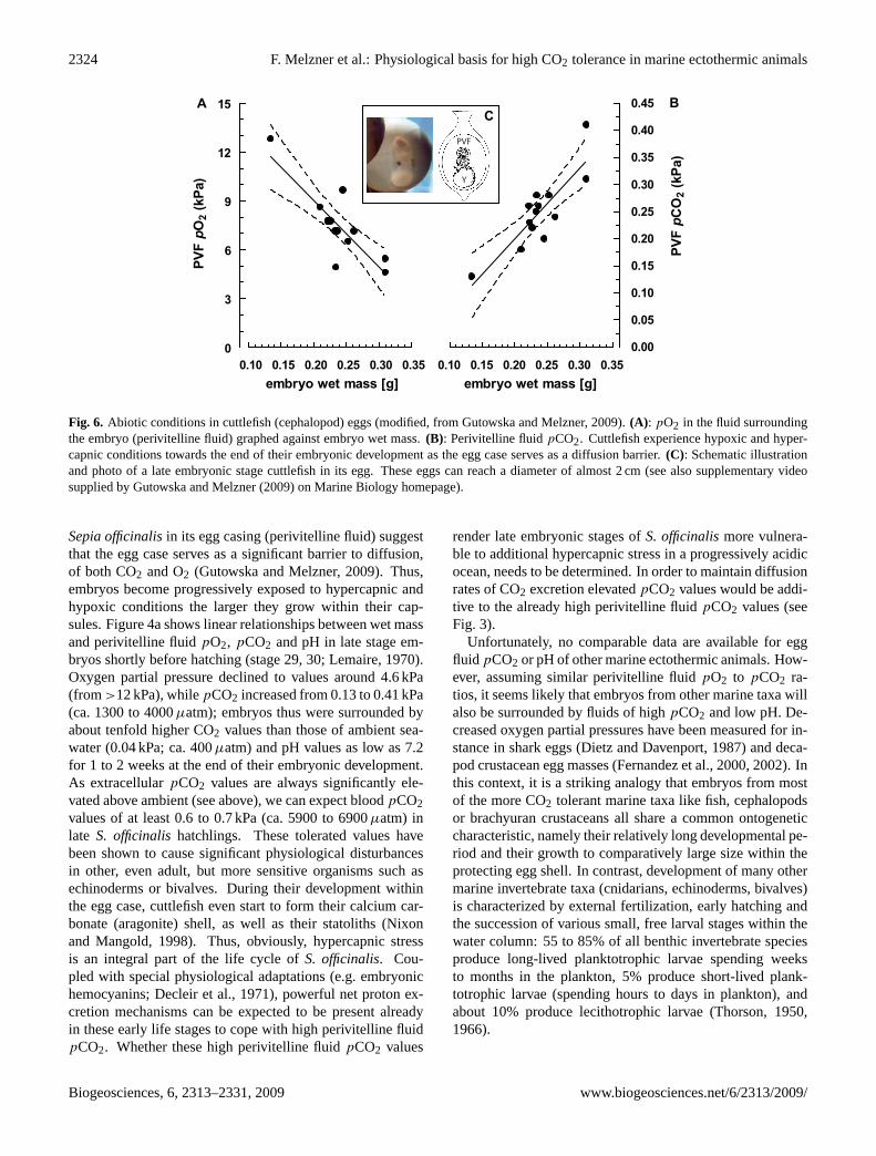
Fig. 2. (A): Routine (black symbols) and active (white symbols) metabolic rates for groups of randomly chosen marine subtidal ectothermicanimals from temperate ocean regions. To ensure comparability, all metabolic rates have been scaled to an animal weight of 20 g (total bodyweight) at 15◦C, using a Q10 value of 2.5 and a mass exponent of b=0.75 (see supplementary file:http://www.biogeosciences.net/6/2313/2009/bg-6-2313-2009-supplement.pdf). (B), (C), (D): Acid-base parameters for groups of randomly chosen marine subtidal ectothermicanimals under control (black symbols) conditions or after exercise (white symbols). (B) depictspCO2 values, 2C pHNBS values and 2-D bicarbonate concentrations determined in extracellular fluids (blood or hemolymph) of various marine taxa. In most casespCO2 andbicarbonate values have been calculated from measurements of pHNBS and dissolved inorganic carbon using the Henderson-Hasselbalchequation and appropriate constants (pK′
1, αCO2). See supplementary for more detailed information and a table of references:http://www.biogeosciences.net/6/2313/2009/bg-6-2313-2009-supplement.pdf.
of CO2 per mole of O2 consumed. Thus, the flux of CO2that active vs. more inactive marine ectotherms have to chan-nel from their mitochondria across the cell membranes intothe blood space (or coelomic fluid/hemolymph) and, finally,across respiratory epithelia, also varies at the same order of
magnitude. Exercise induced alterations in oxygen consump-tion thus are always coupled to almost equimolar changesin CO2 flux. Such 3 to 5-fold fluctuations in O2/CO2 ex-change in active species can occur within minutes, elicitedboth, by exercise and food consumption. Thus, taxa with
Biogeosciences, 6, 2313–2331, 2009 www.biogeosciences.net/6/2313/2009/
F. Melzner et al.: Physiological basis for high CO2 tolerance in marine ectothermic animals 2319
high metabolic rates must possess an advanced machineryfor the elimination of CO2 and associated acid-base distur-bances. As a consequence, this machinery might also behelpful in coping with highpCO2 values originating fromseawater hypercapnia.
5 High extracellular pCO2 in marine ectothermicmetazoans
All marine ectothermic metazoans have one feature in com-mon: their cells are surrounded by an extracellular fluid com-partment (blood, coelomic fluid or hemolymph) that is usedas a convective transport system for various substances, in-cluding dissolved gases. As with O2, CO2 exchange betweenthis fluid and the external medium (seawater) is mainly real-ized by means of diffusion according to the following equa-tion (Dejours, 1975):
MCO2=KCO2(AE−1)(pCO2e−pCO2sw) (2)
with MCO2=CO2 flux in moles, KCO2= species (and organ)specific diffusion constant,pCO2e= extracellularpCO2,pCO2sw= seawaterpCO2, A= functional diffusion area, E=thickness of the diffusion barrier.
Thus, CO2 excretion is directly proportional to the CO2partial pressure gradient from the inside (extracellular fluid)to the outside (seawater). Consequently, higher marine meta-zoan animals are characterized by extracellular fluids withseveral-fold higherpCO2 values than the surrounding sea-water in order to produce a substantial net outward flowof CO2 (see Fig. 2B), although diffusion areas also scalewith metabolic rate. Minimum extracellularpCO2 valuesin some marine metazoans (some echinoderms, bivalves)are little higher than 0.1 kPa (ca. 1000µatm), most animals,however, live with extracellularpCO2 values of 0.2 kPa(ca. 2000µatm) and greater. Highest extracellularpCO2 val-ues in those water breathers are found in teleost fish (0.3 to0.5 kPa; ca. 3000–4900µatm). Most ectothermic marine an-imals maintain relatively constant extracellularpCO2 valuesthat go along with taxon specific extracellular [HCO−
3 ] andpH (under comparable abiotic conditions). Common patternscan be observed in both, brachyuran crustaceans and teleostfish: Relatively high [HCO−3 ] values of 5 to 10 mM usu-ally help support high extracellular pH values of 7.6 to 7.95(Fig. 2c, d). On the other end of the scale, echinoderms aretypically characterized by low extracellular pH (7.0 to 7.5)and low bicarbonate concentrations that are barely higherthan those of seawater. Coleoid cephalopods, despite theirfish like performance display relatively low extracellular pHand bicarbonate values.
ExtracellularpCO2 values may be first line indicatorsof an animals’ susceptibility towards future ocean acidifica-tion. A simple example can illustrate this idea: any unicel-lular marine organism (e.g. a coccolithophorid, sperm and
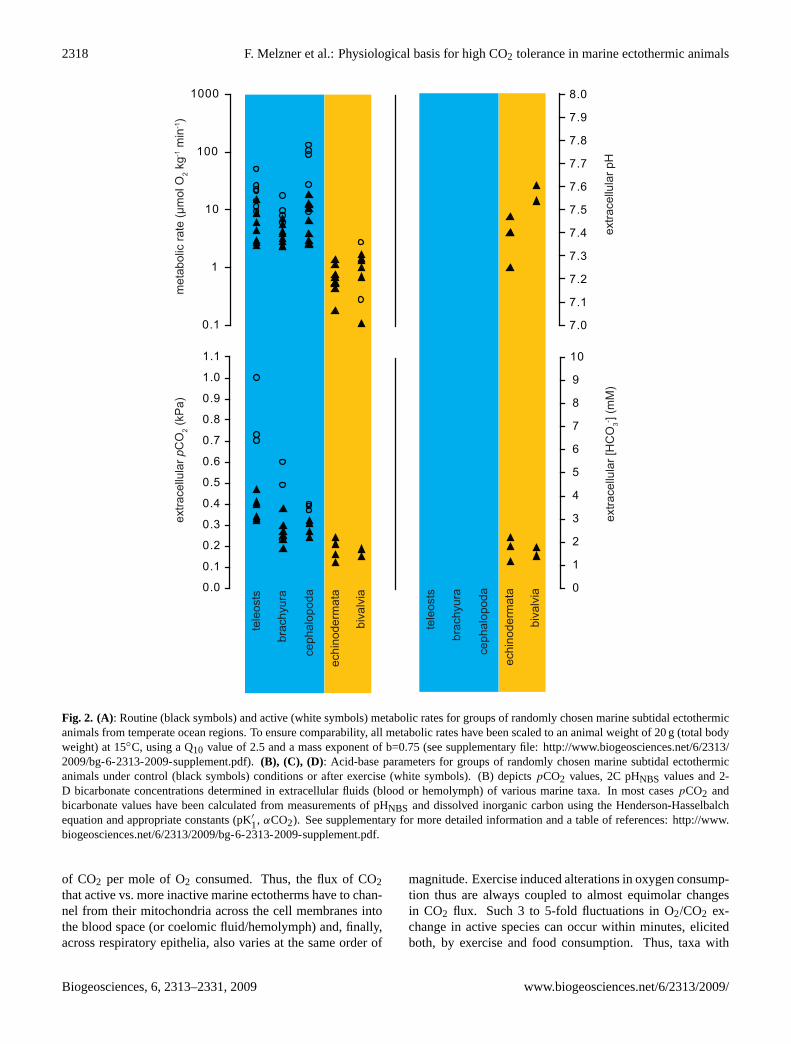
oocytes of broadcast spawners) today is surrounded by “ex-tracellular” fluid (= seawater) with apCO2 of about 0.04 kPa(ca. 400µatm). An increase in seawaterpCO2 by another0.04 kPa therefore leads to a 100% increase in “extracellu-lar” pCO2 for that organism. A similar increase in sea-water pCO2 would probably only lead to a 40% increasein coelomic fluidpCO2 of an echinoderm with a controlcoelomic fluidpCO2 of 0.1 kPa (ca. 1000µatm), and to a10% increase in bloodpCO2 of a teleost fish with a controlextracellularpCO2 of 0.4 kPa (ca. 3900µatm). In both cases,extracellularpCO2 would need to be increased by 0.04 kPain order to maintain a constant CO2 diffusion gradient. Thus,the higher thepCO2 values that cells are exposed to now, thelower the relative change that will come with future oceanacidification. Thus, fish/cephalopod/brachyuran cells will beexposed to a lower relative change inpCO2 than cells of typ-ical bivalves/echinoderms, while unicellular organisms (andlife stages) will experience the greatest relative changes intheir respective extracellular environment.
Figure 2b indicates, that following exhaustive exercise,even higher extracellularpCO2 values can be encountered:Respiratory and metabolic acidosis result in maximumpCO2values between 0.4 kPa (ca. 3900µatm, cephalopods) and>1.0 kPa (>9900µatm, teleost fish). Thus, these taxa areadapted to cope (at least occasionally) with extracellularpCO2 values that are up to five times higher than maximumvalues we might expect through ocean acidification in sur-face waters within the next few hundred years, i.e. 0.2 kPa(ca. 2000µatm: Caldeira and Wickett, 2003).
Interestingly, little information is available on extracellu-lar pCO2 values during sub-maximal (exclusively aerobic)exercise. While one would expect that animals simply in-crease their extracellularpCO2 in order to enhance CO2 dif-fusion rates across gill epithelia, the few examples availablefor teleost fish suggest thatpCO2 is not dramatically ele-vated under such conditions (van den Thillart et al., 1983;Brauner et al., 2000). For other taxa (brachyuran crustaceans,cephalopods) such measurements have not been performed.It is thus quite rewarding to take a closer look at the physio-logical basis that enables elevated O2/CO2 exchange rates inteleost fish during aerobic exercise and to look at some phys-iological consequences of exhaustive exercise in active taxain general. These mechanisms probably form the basis ofefficient pH compensation as exploited during hypercapnia.
6 High CO2 fluxes during (exhaustive) exercise
The capacity to live with elevatedpCO2 values in the extra-cellular fluid and to cope with extreme and rapid fluctuationsin pCO2 during muscular exercise is a challenge for activetaxa. In order to support high metabolic rates, active groupsdiscussed above rely on efficient circulatory systems. Thesedo not only operate at high pressure and volume flow, but alsocontain intra- (fish) or extracellular (decapod crustaceans,
www.biogeosciences.net/6/2313/2009/ Biogeosciences, 6, 2313–2331, 2009
2320 F. Melzner et al.: Physiological basis for high CO2 tolerance in marine ectothermic animals
unicell / gametezygote
seawater pCO2: 0.04 kPa
metazoan cells
seawater pCO2: 0.04 kPa
blood pCO2: 0.2 kPa
metazoan cells
seawater pCO2: 0.04 kPa
blood pCO2: 0.4 kPa
egg fluid pCO2: 0.2 kPa
unicell / gametezygote
seawater pCO2: 0.08 kPa
metazoan cells
seawater pCO2: 0.08 kPa
blood pCO2: 0.24 kPa
metazoan cells
seawater pCO2: 0.08 kPa
blood pCO2: 0.44 kPa
egg fluid pCO2: 0.24 kPa
+100 %
+20 %
+10 %
2 x CO21 x CO
2% change in pCO
2e:
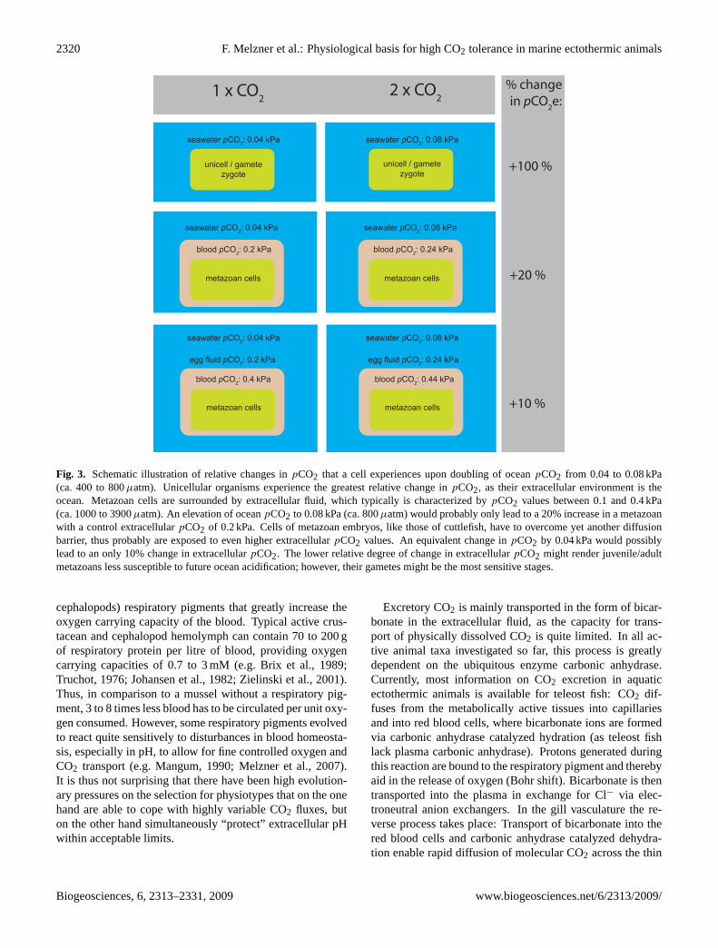
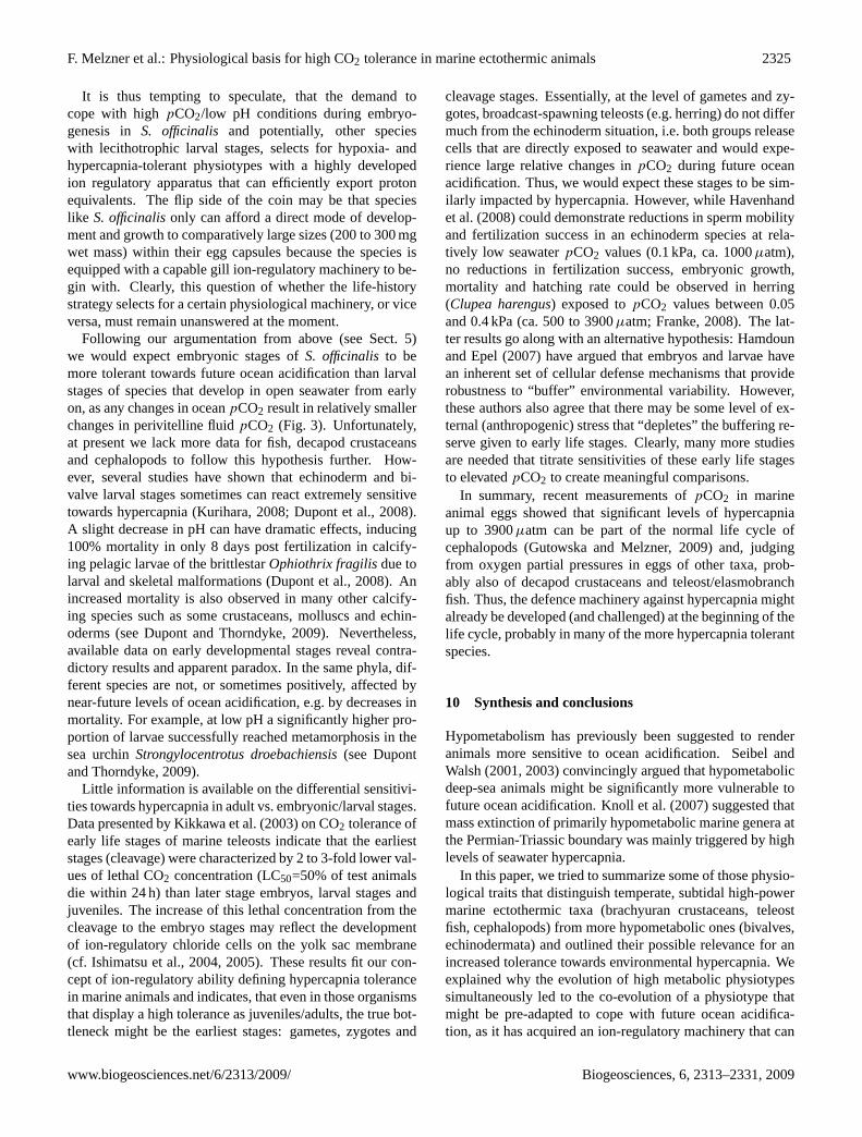
Fig. 3. Schematic illustration of relative changes inpCO2 that a cell experiences upon doubling of oceanpCO2 from 0.04 to 0.08 kPa(ca. 400 to 800µatm). Unicellular organisms experience the greatest relative change inpCO2, as their extracellular environment is theocean. Metazoan cells are surrounded by extracellular fluid, which typically is characterized bypCO2 values between 0.1 and 0.4 kPa(ca. 1000 to 3900µatm). An elevation of oceanpCO2 to 0.08 kPa (ca. 800µatm) would probably only lead to a 20% increase in a metazoanwith a control extracellularpCO2 of 0.2 kPa. Cells of metazoan embryos, like those of cuttlefish, have to overcome yet another diffusionbarrier, thus probably are exposed to even higher extracellularpCO2 values. An equivalent change inpCO2 by 0.04 kPa would possiblylead to an only 10% change in extracellularpCO2. The lower relative degree of change in extracellularpCO2 might render juvenile/adultmetazoans less susceptible to future ocean acidification; however, their gametes might be the most sensitive stages.
cephalopods) respiratory pigments that greatly increase theoxygen carrying capacity of the blood. Typical active crus-tacean and cephalopod hemolymph can contain 70 to 200 gof respiratory protein per litre of blood, providing oxygencarrying capacities of 0.7 to 3 mM (e.g. Brix et al., 1989;Truchot, 1976; Johansen et al., 1982; Zielinski et al., 2001).Thus, in comparison to a mussel without a respiratory pig-ment, 3 to 8 times less blood has to be circulated per unit oxy-gen consumed. However, some respiratory pigments evolvedto react quite sensitively to disturbances in blood homeosta-sis, especially in pH, to allow for fine controlled oxygen andCO2 transport (e.g. Mangum, 1990; Melzner et al., 2007).It is thus not surprising that there have been high evolution-ary pressures on the selection for physiotypes that on the onehand are able to cope with highly variable CO2 fluxes, buton the other hand simultaneously “protect” extracellular pHwithin acceptable limits.
Excretory CO2 is mainly transported in the form of bicar-bonate in the extracellular fluid, as the capacity for trans-port of physically dissolved CO2 is quite limited. In all ac-tive animal taxa investigated so far, this process is greatlydependent on the ubiquitous enzyme carbonic anhydrase.Currently, most information on CO2 excretion in aquaticectothermic animals is available for teleost fish: CO2 dif-fuses from the metabolically active tissues into capillariesand into red blood cells, where bicarbonate ions are formedvia carbonic anhydrase catalyzed hydration (as teleost fishlack plasma carbonic anhydrase). Protons generated duringthis reaction are bound to the respiratory pigment and therebyaid in the release of oxygen (Bohr shift). Bicarbonate is thentransported into the plasma in exchange for Cl− via elec-troneutral anion exchangers. In the gill vasculature the re-verse process takes place: Transport of bicarbonate into thered blood cells and carbonic anhydrase catalyzed dehydra-tion enable rapid diffusion of molecular CO2 across the thin
Biogeosciences, 6, 2313–2331, 2009 www.biogeosciences.net/6/2313/2009/
F. Melzner et al.: Physiological basis for high CO2 tolerance in marine ectothermic animals 2321
gill epithelium and release into the surrounding water (seeTufts and Perry, 1998, for a review). During the short transittime through the gill vasculature (0.5 to 2.5 s; Cameron andPolhemus, 1974) approximately 12 to 35% of blood [HCO−
3 ]is transformed and excreted (Perry, 1986). While sufficientcapacities of carbonic anhydrase are necessary within the redblood cells to enable a rapid dehydration of bicarbonate dur-ing the gill passage (Henry and Swenson, 2000), the rate lim-iting step in CO2 excretion in teleosts is thought to be thetransfer of plasma bicarbonate into the red blood cell via theband 3 anion exchanger (e.g. Perry and Gilmour, 1993; Woodand Munger, 1994). Recent experimental evidence couldconvincingly establish that the rate of CO2 excretion acrossgill epithelia is diffusion limited (e.g. Perry and Gilmour,2006). Each anaemia (i.e. a low content of red blood cellsin the blood) and elevated blood flow were observed to leadto elevated bloodpCO2, an effect, that could be reversedby experimentally making carbonic anhydrase available infish plasma (Desforges et al., 2002; Gilmour and MacNeill,2003).
During aerobic exercise, provision of oxygen to the work-ing muscles becomes paramount and increases in metabolicrate are compensated for by elevated rates of blood con-vection (cardiac output). Other changes in the gill vascu-lature enable more efficient gas exchange, helping to main-tain pCO2, extracellular pH and [HCO−3 ] at control lev-els. Most important are increases in the perfused gill area(A in Eq. 2) and decreases in the gill epithelial thickness(E in Eq. 2), which are caused by increases in ventral aor-tic blood pressure (e.g. Kiceniuk and Jones, 1977; Ran-dall and Daxboeck, 1984). However, elevated cardiac out-put can reduce gill transit time by a factor of three (Ran-dall, 1982). As the CO2 excretion system is already lim-ited by the capacity of the red blood cell HCO−
3 /Cl− ex-change system, higher swimming velocities can result inslightly elevated bloodpCO2, a respiratory acidosis may de-velop (e.g. Brauner et al., 2000). Brauner et al. (2000) couldalso demonstrate that when their experimental fish (sea wa-ter acclimated rainbow trout,Oncorhynchus mykiss) wereapproaching their critical swimming speed (shortly beforeexhaustion), arterial pH was protected from acidification byrapid active accumulation of HCO−3 . Extremely high bloodpCO2 values (>0.6 kPa; ca. 5900µatm) and low extracellu-lar pH values<7.5 are only encountered during and follow-ing exhaustive exercise (Fig. 2b, white symbols) in brachyu-ran crustaceans and teleost fish. These are mainly caused byanaerobic metabolism (“metabolic acidosis”): Force produc-tion by aerobic swimming muscles is complemented by therecruitment of anaerobic (“white”) fibers; lactate and pro-tons originate as metabolic end products. Both are even-tually released into the extracellular fluid, where the pro-tons can titrate plasma [HCO−3 ], thus decreasing extracellu-lar pH (see Figs. 1a, 2c). However, rapid compensation pro-cesses are occurring during exhaustive exercise and particu-larly during the recovery phase. Gill ion-regulatory epithelia
produce enormous net proton equivalent fluxes from the or-ganism into the surrounding seawater, ranging in magnitudebetween 1200µEq kg−1 h−1 (rainbow trout,O. mykiss; Ho-leton et al., 1983) and 4800µEq kg−1 h−1 (blue crab,Call-inectes sapidus; Booth et al., 1984) to restore the originalacid-base status. Clearly, a powerful ion regulatory machin-ery can be made visible under conditions of extreme physicalstress, the very same machinery that will then enable activeorganisms to compensate extracellular pH during hypercap-nic disturbances (see above). It thus makes sense to take acloser look at ion-regulatory epithelia in the more active taxa.
7 The acid-base regulatory machinery and its mainmotor
Species specific mechanisms of transepithelial ion exchangehave been reviewed, e.g. in Boron (2004), Claiborne etal. (2002), Perry and Gilmour (2006), and Wheatly andHenry (1992). However, we are far from exactly under-standing the whole system of ion exchange mechanisms, es-pecially in the invertebrate taxa. Interestingly, similar molec-ular components have been conserved in different marine an-imal groups. Gills are the primary sites of acid-base regu-latory processes in all high metabolic rate marine taxa dis-cussed in this text, in fish (Perry and Gilmour, 2006), crus-taceans (Wheatly and Henry, 1992) and probably also incephalopods (Schipp et al., 1979). In fish, specialized ep-ithelial cells, the mitochondria rich cells, contain a set ofion transporting proteins and channels that are important foracid-base regulation. Cells that are active in acid secretioncontain electroneutral Na+/H+ exchangers or V-type H+ AT-Pases, coupled energetically to apical Na+ channels. Whilethe latter system is thought to be more important for fresh-water organisms which have to absorb Na+ (e.g. Wilson etal., 2000), the former can operate on the favourable Na+ gra-dient between seawater and cytosol, shuttling one H+ out ofthe cell for each Na+ imported. While Na+/H+ exchangersdo not directly consume energy (there is no ATPase directlylinked to these proteins), they essentially operate on the en-ergy spent by the ATP consuming sodium pump (Na+/K+
ATPase). Basolateral Na+/K+ ATPase is thus commonlyconsidered the motor of the ion-regulatory machinery in ma-rine animal gills. Pumping two K+ into the cell while simul-taneously removing three Na+, it creates the low intracellular[Na+] typical for all animal cells and thus is partly respon-sible for the cell’s membrane potential. One potential mech-anism for the removal of acid during a respiratory acidosiscould be the following (established from results of studies inteleost fish; see Fig. 4): Excess CO2 diffuses into the mito-chondria rich cells and is instantly hydrated by cytosolic car-bonic anhydrase into protons and bicarbonate ions. While theprotons are exported via the Na+/H+ exchanger, bicarbon-ate could be released into the plasma by means of basolat-eral Cl−/HCO−
3 exchangers or Na+/HCO−
3 co-transporters.
www.biogeosciences.net/6/2313/2009/ Biogeosciences, 6, 2313–2331, 2009
2322 F. Melzner et al.: Physiological basis for high CO2 tolerance in marine ectothermic animals
3 Na+
2 K+Na+
H+
CAcCO2
HCO3-
H+
Cl-HCO3
-
[K+]i[K+]e
[Na+]e[Na+]i
[K+]sw
[Na+]sw
Cl-
12
34
seawater gill epithelial cell extracellular fluid
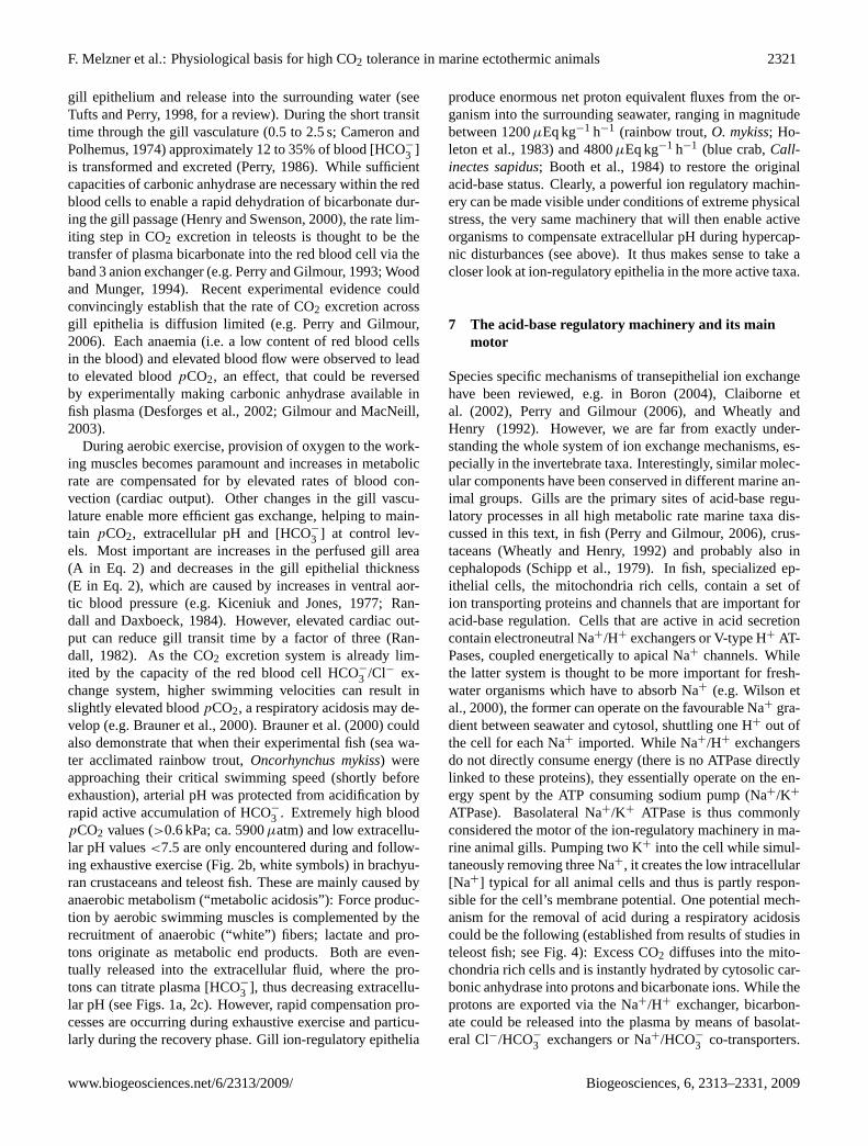
Fig. 4. Simplified schematic depiction of an epithelial gill cell(ionocyte) of a teleost fish (adapted from Perry and Gilmour, 2006).Decapod crustacean and cephalopod gill epithelia are equipped withsimilar proteins. (1)=Na+/K+ ATPase,(2)=Na+/H+ exchanger,(3)=Cl−/HCO−
3 exchanger,(4)=Cl− channel (e.g. CFTR), CAc =cytoplasmatic carbonic anhydrase. Na+/K+ ATPase is responsi-ble for the low intracellular Na+ and high K+ concentration. Sec-ondary active transporters, such as Na+/H+ exchanger can utilizethe sodium gradient to export H+. H+ are produced when CO2 ishydrated by CAc. The resulting HCO−3 can be transferred into theextracellular fluid (blood, hemolymph), while Cl− is exported to theseawater through chloride channels to maintain electroneutrality.
This plasma bicarbonate may then undergo further protona-tion/dehydration/hydration cycles leading to a net proton ex-trusion via the gills. In order to maintain electroneutrality inthe plasma, Cl− is typically excreted, possibly via apical Cl−
channels (e.g. CFTR; see Perry and Gilmour, 2006; Deig-weiher et al., 2008, for an extended discussion). However,the true mechanisms may be more complicated owing to thelarge number of transporters and channels present in gill ep-ithelia (see also Deigweiher et al., 2008). However, basicprocesses can be suspected similar for decapod crustaceansand cephalopods as well; it is known by now that similar ionexchange proteins are also expressed in gills of these inverte-brates (e.g. Schipp et al., 1979; Piermarini et al., 2007; Virkkiet al., 2003; Henry and Swenson, 2000; Wheatly and Henry,1992; Hu, Lucassen and Melzner, unpublished).
As Na+/K+ ATPase activity is the main energy sink anddriving force for gill ion exchange processes in marine ec-tothermic animals, it can serve as a useful indicator for theoverall capacity in ion and acid-base regulation. Conse-quently, gill Na+/K+ ATPase activity has been shown to cor-relate with metabolic rate in marine teleost species: Gibbsand Somero (1990) found highest Na+/K+ ATPase activi-ties in shallow water, active species, while more inactive,deep-sea species activities were an order of magnitude lower.
1
10
100
1000
M. e
dulis
S. o
ffici
nalis
tele
osts
gill
Na+
/K+
-ATP
ase
activ
ity(µ
mol
/ h
g gi
ll tis
sue)
C. m
aena
s
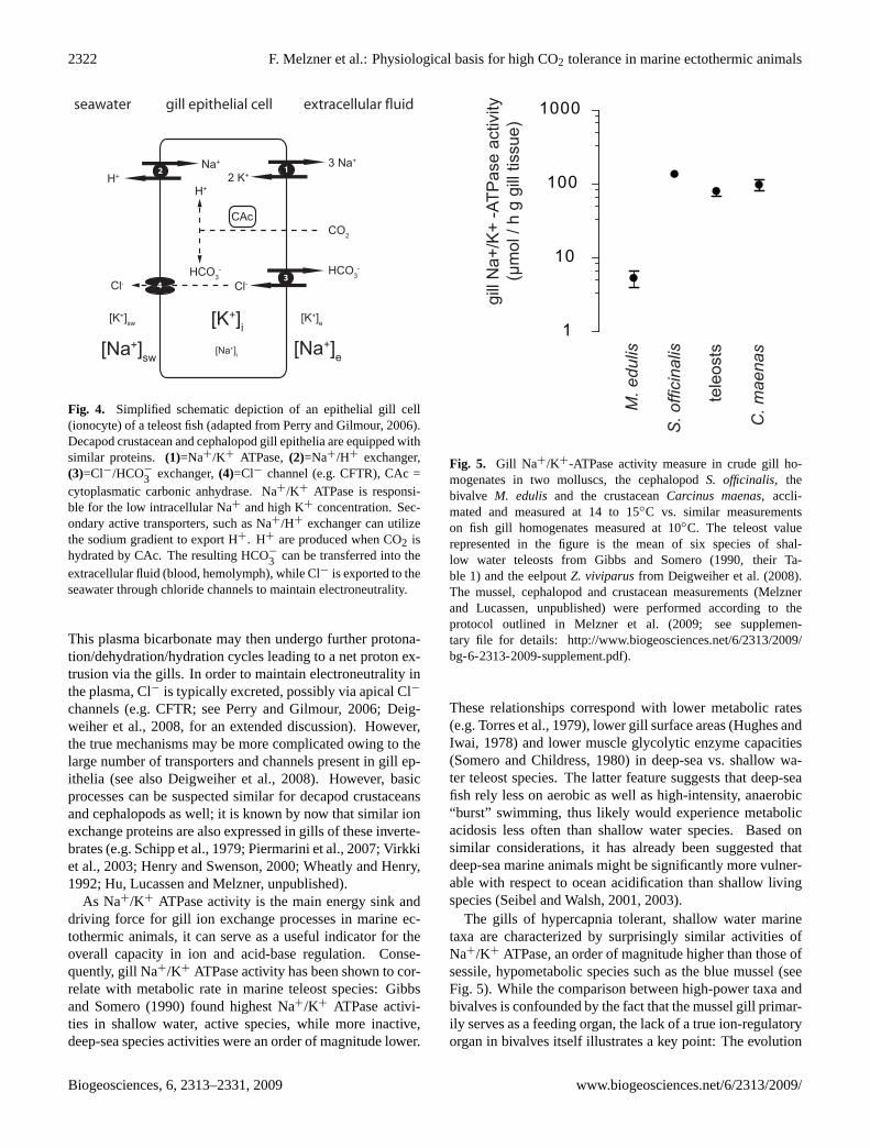
Fig. 5. Gill Na+/K+-ATPase activity measure in crude gill ho-mogenates in two molluscs, the cephalopodS. officinalis, thebivalve M. edulis and the crustaceanCarcinus maenas, accli-mated and measured at 14 to 15◦C vs. similar measurementson fish gill homogenates measured at 10◦C. The teleost valuerepresented in the figure is the mean of six species of shal-low water teleosts from Gibbs and Somero (1990, their Ta-ble 1) and the eelpoutZ. viviparusfrom Deigweiher et al. (2008).The mussel, cephalopod and crustacean measurements (Melznerand Lucassen, unpublished) were performed according to theprotocol outlined in Melzner et al. (2009; see supplemen-tary file for details: http://www.biogeosciences.net/6/2313/2009/bg-6-2313-2009-supplement.pdf).
These relationships correspond with lower metabolic rates(e.g. Torres et al., 1979), lower gill surface areas (Hughes andIwai, 1978) and lower muscle glycolytic enzyme capacities(Somero and Childress, 1980) in deep-sea vs. shallow wa-ter teleost species. The latter feature suggests that deep-seafish rely less on aerobic as well as high-intensity, anaerobic“burst” swimming, thus likely would experience metabolicacidosis less often than shallow water species. Based onsimilar considerations, it has already been suggested thatdeep-sea marine animals might be significantly more vulner-able with respect to ocean acidification than shallow livingspecies (Seibel and Walsh, 2001, 2003).
The gills of hypercapnia tolerant, shallow water marinetaxa are characterized by surprisingly similar activities ofNa+/K+ ATPase, an order of magnitude higher than those ofsessile, hypometabolic species such as the blue mussel (seeFig. 5). While the comparison between high-power taxa andbivalves is confounded by the fact that the mussel gill primar-ily serves as a feeding organ, the lack of a true ion-regulatoryorgan in bivalves itself illustrates a key point: The evolution
Biogeosciences, 6, 2313–2331, 2009 www.biogeosciences.net/6/2313/2009/
F. Melzner et al.: Physiological basis for high CO2 tolerance in marine ectothermic animals 2323
of high metabolic rate physiotypes is closely connected tothe development of extremely specialized organ structures topromote respiration and ion regulation that are very similarin their ultrastructural design (e.g. Evans et al., 2005: fish;Budelmann et al., 1999: cephalopoda; Taylor and Taylor,1999: decapod crustacea).
Na+/K+ ATPase activities are modulated in vivo duringmetabolic rate transitions (e.g. exercise, specific dynamic ac-tion) on a short term basis by several second messenger path-ways finally leading to a change in protein phoshorylation(e.g. Ramnanan and Storey, 2006). The most impressive ex-ample is the beta adrenergic stimulation of the enzyme inskeletal muscle which compensates the large K+ efflux dur-ing exercise. Also changes in cytosolic ion composition,namely Na+ and H+ concentrations are involved in the reg-ulation of Na+/K+-ATPase activity. In addition interactionwith the cytoskeleton and membrane trafficking of the pumpare regulatory mechanisms acutely controlling its functionand availability (Bertorello and Katz, 1993). Long term reg-ulation of Na+/K+-ATPase is under control of nuclear hor-mones. They trigger transcription of the subunits by bind-ing to nuclear hormone responsive elements on the respectivegenes (Feraille and Doucet, 2001).
However, as has been shown that phosphoryla-tion/dephosphorylation can activate or deactivate theenzyme, high-power animals may operate with a functionalreserve that can be activated upon demand. Whether sucha reserve is important for the rapid extracellular HCO−
3accumulatory reaction observed upon acute hypercapnicexposure (see above, Fig. 1b) remains to be investigated.It has been recently shown in two marine teleost fishspecies, that gill Na+/K+ ATPase activity increases duringacclimation to higher levels of hypercapnia (Deigweiher etal., 2008; Melzner et al., 2009). Rapid increases in activityin the eelpoutZoarces viviparusupon exposure to 1 kPa ofCO2 (ca. 9900µatm) within two days have been observed tobe related to elevated Na+/K+ ATPase mRNA and proteinlevels, suggesting that the enzyme is under tight transcrip-tional control. Longer acclimation (6 weeks) led to a∼80%increase in Na+/K+ ATPase activity. In cod (Gadus morhua)long term acclimation (4–12 months) led to increases inNa+/K+ ATPase activity and protein concentration at apCO2 of 0.6 kPa (ca. 5900µatm), whereas no significantchanges were observed at 0.3 kPa (ca. 3000µatm; Melzneret al., 2009). Although this occurred in specimens from twodistinct populations, it could nevertheless indicate that thecontrol fitting of the gill ion regulatory machinery in manyteleosts has high enough of an excess capacity to cope withthe additional ion-regulatory challenge due to hypercapniaunder more realistic scenarios of ocean acidification (i.e. 0.1to 0.2 kPa; Caldeira and Wickett, 2003). Clearly, furtherstudies need to address this exciting possibility.
8 Environmental hypercapnia
Further above it was stated that typical marine ectothermicanimals are seldom exposed to environmental hypercapnia.This applies for large areas of the pelagic open ocean, how-ever there are some special habitats that do provide elevatedpCO2 values to its inhabitants: intertidal regions, estuaries,oxygen-minimum zones, upwelling coastal regions or deep-sea vent systems (see e.g. Frankignoulle et al., 1996, 1998;Weigelt and Rumohr, 1986; Dwyer and Burnett, 1996; Feelyet al., 2008; Wotton et al., 2008). While present mean surfaceoceanpCO2 values average around 0.04 kPa (ca. 400µatm)much higher values are reached in the above mentioned habi-tats. For example,pCO2 values in Kiel Fjord, home to nu-merous calcifying organisms, can rise above 0.1 to 0.2 kPa(ca. 1000 to 2000µatm) for prolonged times during sum-mer and autumn (Thomsen, 2008). Similarly, upwellingprocesses lead to elevated near-shorepCO2 values of up to0.1 kPa (about 1000µatm) in continental shelf areas off theCalifornian coast (Feely et al., 2008). Animals living in inter-tidal rockpools experience even stronger short-term fluctua-tions (Truchot and Duhamel-Jouve, 1980): depending on re-spective light conditionspCO2 values during low tide emer-sion periods can range between about 0.35 kPa (3500µatm,due to extensive nighttime respiration) and almost zero (dueto high photosynthetic activity). HighpCO2 values (around1200µatm) have also been observed in oceanic oxygen mini-mum layers of intermediate depths (200–1000 m) where highcommunity respiration rates cause hypoxia and associatedhypercapnia (Brewer and Peltzer, 2009). However, specialphysiological and biochemical adaptations enable variousanimal groups to populate even the most extreme habitatswith respect to hypercapnic, temperature and other chemi-cal conditions – the deep-sea hydrothermal vent ecosystems.Amongst other things, this inhospitable environment chal-lenges its inhabitants withpCO2 conditions as high as 7 kPa(about 69 000µatm). Nevertheless, the vent musselBathy-modiolus breviorhas been found able to precipitate shellsunder such highpCO2/low-pH conditions (Tunnicliffe et al.,2009). Again, following our rationale from above, organismsalready living under elevatedpCO2 in their particular habi-tats may encounter less of a relative change inpCO2 thane.g. oceanic species, thus may be better adapted to futureacidification (however,pCO2 in CO2 enriched habitats maynot necessarily increase at the same rate as projected for theopen ocean; see calculations in Brewer and Peltzer, 2009).
9 Ontogenetic hypercapnia: the hostile environmentwithin egg capsules
Of large evolutionary relevance might be a special “onto-genetic habitat”: the egg case and egg masses of manymarine ectothermic animals. Recent determinations of pH,pO2 and pCO2 in the fluid surrounding the cephalopod
www.biogeosciences.net/6/2313/2009/ Biogeosciences, 6, 2313–2331, 2009
2324 F. Melzner et al.: Physiological basis for high CO2 tolerance in marine ectothermic animals
0
3
6
9
12
15
PVF
p O2
(kPa
)
0.10 0.15 0.20 0.25 0.30 0.350.00
0.05
0.10
0.15
0.20
0.25
0.30
0.35
0.40
0.45
embryo wet mass [g]
PVF
p CO
2 (k
Pa)
A B
0.10 0.15 0.20 0.25 0.30 0.35embryo wet mass [g]
PVF
Y
C
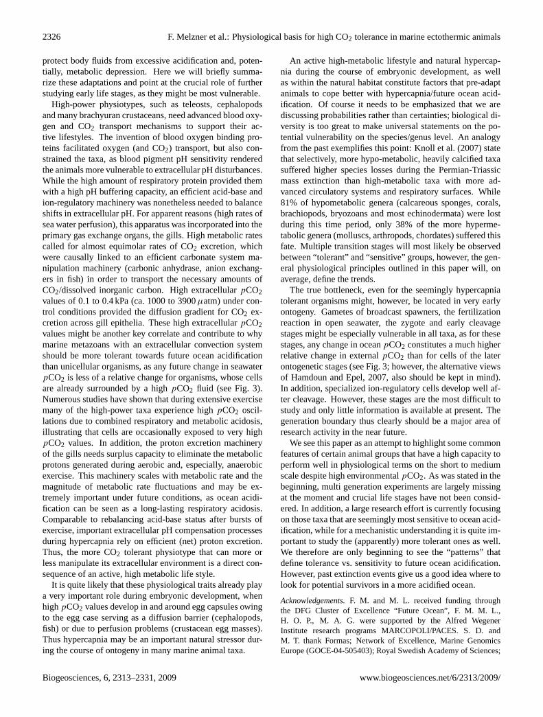
Fig. 6. Abiotic conditions in cuttlefish (cephalopod) eggs (modified, from Gutowska and Melzner, 2009).(A): pO2 in the fluid surroundingthe embryo (perivitelline fluid) graphed against embryo wet mass.(B): Perivitelline fluidpCO2. Cuttlefish experience hypoxic and hyper-capnic conditions towards the end of their embryonic development as the egg case serves as a diffusion barrier.(C): Schematic illustrationand photo of a late embryonic stage cuttlefish in its egg. These eggs can reach a diameter of almost 2 cm (see also supplementary videosupplied by Gutowska and Melzner (2009) on Marine Biology homepage).
Sepia officinalisin its egg casing (perivitelline fluid) suggestthat the egg case serves as a significant barrier to diffusion,of both CO2 and O2 (Gutowska and Melzner, 2009). Thus,embryos become progressively exposed to hypercapnic andhypoxic conditions the larger they grow within their cap-sules. Figure 4a shows linear relationships between wet massand perivitelline fluidpO2, pCO2 and pH in late stage em-bryos shortly before hatching (stage 29, 30; Lemaire, 1970).Oxygen partial pressure declined to values around 4.6 kPa(from>12 kPa), whilepCO2 increased from 0.13 to 0.41 kPa(ca. 1300 to 4000µatm); embryos thus were surrounded byabout tenfold higher CO2 values than those of ambient sea-water (0.04 kPa; ca. 400µatm) and pH values as low as 7.2for 1 to 2 weeks at the end of their embryonic development.As extracellularpCO2 values are always significantly ele-vated above ambient (see above), we can expect bloodpCO2values of at least 0.6 to 0.7 kPa (ca. 5900 to 6900µatm) inlate S. officinalishatchlings. These tolerated values havebeen shown to cause significant physiological disturbancesin other, even adult, but more sensitive organisms such asechinoderms or bivalves. During their development withinthe egg case, cuttlefish even start to form their calcium car-bonate (aragonite) shell, as well as their statoliths (Nixonand Mangold, 1998). Thus, obviously, hypercapnic stressis an integral part of the life cycle ofS. officinalis. Cou-pled with special physiological adaptations (e.g. embryonichemocyanins; Decleir et al., 1971), powerful net proton ex-cretion mechanisms can be expected to be present alreadyin these early life stages to cope with high perivitelline fluidpCO2. Whether these high perivitelline fluidpCO2 values
render late embryonic stages ofS. officinalismore vulnera-ble to additional hypercapnic stress in a progressively acidicocean, needs to be determined. In order to maintain diffusionrates of CO2 excretion elevatedpCO2 values would be addi-tive to the already high perivitelline fluidpCO2 values (seeFig. 3).
Unfortunately, no comparable data are available for eggfluid pCO2 or pH of other marine ectothermic animals. How-ever, assuming similar perivitelline fluidpO2 to pCO2 ra-tios, it seems likely that embryos from other marine taxa willalso be surrounded by fluids of highpCO2 and low pH. De-creased oxygen partial pressures have been measured for in-stance in shark eggs (Dietz and Davenport, 1987) and deca-pod crustacean egg masses (Fernandez et al., 2000, 2002). Inthis context, it is a striking analogy that embryos from mostof the more CO2 tolerant marine taxa like fish, cephalopodsor brachyuran crustaceans all share a common ontogeneticcharacteristic, namely their relatively long developmental pe-riod and their growth to comparatively large size within theprotecting egg shell. In contrast, development of many othermarine invertebrate taxa (cnidarians, echinoderms, bivalves)is characterized by external fertilization, early hatching andthe succession of various small, free larval stages within thewater column: 55 to 85% of all benthic invertebrate speciesproduce long-lived planktotrophic larvae spending weeksto months in the plankton, 5% produce short-lived plank-totrophic larvae (spending hours to days in plankton), andabout 10% produce lecithotrophic larvae (Thorson, 1950,1966).
Biogeosciences, 6, 2313–2331, 2009 www.biogeosciences.net/6/2313/2009/
F. Melzner et al.: Physiological basis for high CO2 tolerance in marine ectothermic animals 2325
It is thus tempting to speculate, that the demand tocope with highpCO2/low pH conditions during embryo-genesis in S. officinalis and potentially, other specieswith lecithotrophic larval stages, selects for hypoxia- andhypercapnia-tolerant physiotypes with a highly developedion regulatory apparatus that can efficiently export protonequivalents. The flip side of the coin may be that specieslike S. officinalisonly can afford a direct mode of develop-ment and growth to comparatively large sizes (200 to 300 mgwet mass) within their egg capsules because the species isequipped with a capable gill ion-regulatory machinery to be-gin with. Clearly, this question of whether the life-historystrategy selects for a certain physiological machinery, or viceversa, must remain unanswered at the moment.
Following our argumentation from above (see Sect. 5)we would expect embryonic stages ofS. officinalisto bemore tolerant towards future ocean acidification than larvalstages of species that develop in open seawater from earlyon, as any changes in oceanpCO2 result in relatively smallerchanges in perivitelline fluidpCO2 (Fig. 3). Unfortunately,at present we lack more data for fish, decapod crustaceansand cephalopods to follow this hypothesis further. How-ever, several studies have shown that echinoderm and bi-valve larval stages sometimes can react extremely sensitivetowards hypercapnia (Kurihara, 2008; Dupont et al., 2008).A slight decrease in pH can have dramatic effects, inducing100% mortality in only 8 days post fertilization in calcify-ing pelagic larvae of the brittlestarOphiothrix fragilisdue tolarval and skeletal malformations (Dupont et al., 2008). Anincreased mortality is also observed in many other calcify-ing species such as some crustaceans, molluscs and echin-oderms (see Dupont and Thorndyke, 2009). Nevertheless,available data on early developmental stages reveal contra-dictory results and apparent paradox. In the same phyla, dif-ferent species are not, or sometimes positively, affected bynear-future levels of ocean acidification, e.g. by decreases inmortality. For example, at low pH a significantly higher pro-portion of larvae successfully reached metamorphosis in thesea urchinStrongylocentrotus droebachiensis(see Dupontand Thorndyke, 2009).
Little information is available on the differential sensitivi-ties towards hypercapnia in adult vs. embryonic/larval stages.Data presented by Kikkawa et al. (2003) on CO2 tolerance ofearly life stages of marine teleosts indicate that the earlieststages (cleavage) were characterized by 2 to 3-fold lower val-ues of lethal CO2 concentration (LC50=50% of test animalsdie within 24 h) than later stage embryos, larval stages andjuveniles. The increase of this lethal concentration from thecleavage to the embryo stages may reflect the developmentof ion-regulatory chloride cells on the yolk sac membrane(cf. Ishimatsu et al., 2004, 2005). These results fit our con-cept of ion-regulatory ability defining hypercapnia tolerancein marine animals and indicates, that even in those organismsthat display a high tolerance as juveniles/adults, the true bot-tleneck might be the earliest stages: gametes, zygotes and
cleavage stages. Essentially, at the level of gametes and zy-gotes, broadcast-spawning teleosts (e.g. herring) do not differmuch from the echinoderm situation, i.e. both groups releasecells that are directly exposed to seawater and would expe-rience large relative changes inpCO2 during future oceanacidification. Thus, we would expect these stages to be sim-ilarly impacted by hypercapnia. However, while Havenhandet al. (2008) could demonstrate reductions in sperm mobilityand fertilization success in an echinoderm species at rela-tively low seawaterpCO2 values (0.1 kPa, ca. 1000µatm),no reductions in fertilization success, embryonic growth,mortality and hatching rate could be observed in herring(Clupea harengus) exposed topCO2 values between 0.05and 0.4 kPa (ca. 500 to 3900µatm; Franke, 2008). The lat-ter results go along with an alternative hypothesis: Hamdounand Epel (2007) have argued that embryos and larvae havean inherent set of cellular defense mechanisms that providerobustness to “buffer” environmental variability. However,these authors also agree that there may be some level of ex-ternal (anthropogenic) stress that “depletes” the buffering re-serve given to early life stages. Clearly, many more studiesare needed that titrate sensitivities of these early life stagesto elevatedpCO2 to create meaningful comparisons.
In summary, recent measurements ofpCO2 in marineanimal eggs showed that significant levels of hypercapniaup to 3900µatm can be part of the normal life cycle ofcephalopods (Gutowska and Melzner, 2009) and, judgingfrom oxygen partial pressures in eggs of other taxa, prob-ably also of decapod crustaceans and teleost/elasmobranchfish. Thus, the defence machinery against hypercapnia mightalready be developed (and challenged) at the beginning of thelife cycle, probably in many of the more hypercapnia tolerantspecies.
10 Synthesis and conclusions
Hypometabolism has previously been suggested to renderanimals more sensitive to ocean acidification. Seibel andWalsh (2001, 2003) convincingly argued that hypometabolicdeep-sea animals might be significantly more vulnerable tofuture ocean acidification. Knoll et al. (2007) suggested thatmass extinction of primarily hypometabolic marine genera atthe Permian-Triassic boundary was mainly triggered by highlevels of seawater hypercapnia.
In this paper, we tried to summarize some of those physio-logical traits that distinguish temperate, subtidal high-powermarine ectothermic taxa (brachyuran crustaceans, teleostfish, cephalopods) from more hypometabolic ones (bivalves,echinodermata) and outlined their possible relevance for anincreased tolerance towards environmental hypercapnia. Weexplained why the evolution of high metabolic physiotypessimultaneously led to the co-evolution of a physiotype thatmight be pre-adapted to cope with future ocean acidifica-tion, as it has acquired an ion-regulatory machinery that can
www.biogeosciences.net/6/2313/2009/ Biogeosciences, 6, 2313–2331, 2009
2326 F. Melzner et al.: Physiological basis for high CO2 tolerance in marine ectothermic animals
protect body fluids from excessive acidification and, poten-tially, metabolic depression. Here we will briefly summa-rize these adaptations and point at the crucial role of furtherstudying early life stages, as they might be most vulnerable.
High-power physiotypes, such as teleosts, cephalopodsand many brachyuran crustaceans, need advanced blood oxy-gen and CO2 transport mechanisms to support their ac-tive lifestyles. The invention of blood oxygen binding pro-teins facilitated oxygen (and CO2) transport, but also con-strained the taxa, as blood pigment pH sensitivity renderedthe animals more vulnerable to extracellular pH disturbances.While the high amount of respiratory protein provided themwith a high pH buffering capacity, an efficient acid-base andion-regulatory machinery was nonetheless needed to balanceshifts in extracellular pH. For apparent reasons (high rates ofsea water perfusion), this apparatus was incorporated into theprimary gas exchange organs, the gills. High metabolic ratescalled for almost equimolar rates of CO2 excretion, whichwere causally linked to an efficient carbonate system ma-nipulation machinery (carbonic anhydrase, anion exchang-ers in fish) in order to transport the necessary amounts ofCO2/dissolved inorganic carbon. High extracellularpCO2values of 0.1 to 0.4 kPa (ca. 1000 to 3900µatm) under con-trol conditions provided the diffusion gradient for CO2 ex-cretion across gill epithelia. These high extracellularpCO2values might be another key correlate and contribute to whymarine metazoans with an extracellular convection systemshould be more tolerant towards future ocean acidificationthan unicellular organisms, as any future change in seawaterpCO2 is less of a relative change for organisms, whose cellsare already surrounded by a highpCO2 fluid (see Fig. 3).Numerous studies have shown that during extensive exercisemany of the high-power taxa experience highpCO2 oscil-lations due to combined respiratory and metabolic acidosis,illustrating that cells are occasionally exposed to very highpCO2 values. In addition, the proton excretion machineryof the gills needs surplus capacity to eliminate the metabolicprotons generated during aerobic and, especially, anaerobicexercise. This machinery scales with metabolic rate and themagnitude of metabolic rate fluctuations and may be ex-tremely important under future conditions, as ocean acidi-fication can be seen as a long-lasting respiratory acidosis.Comparable to rebalancing acid-base status after bursts ofexercise, important extracellular pH compensation processesduring hypercapnia rely on efficient (net) proton excretion.Thus, the more CO2 tolerant physiotype that can more orless manipulate its extracellular environment is a direct con-sequence of an active, high metabolic life style.
It is quite likely that these physiological traits already playa very important role during embryonic development, whenhighpCO2 values develop in and around egg capsules owingto the egg case serving as a diffusion barrier (cephalopods,fish) or due to perfusion problems (crustacean egg masses).Thus hypercapnia may be an important natural stressor dur-ing the course of ontogeny in many marine animal taxa.
An active high-metabolic lifestyle and natural hypercap-nia during the course of embryonic development, as wellas within the natural habitat constitute factors that pre-adaptanimals to cope better with hypercapnia/future ocean acid-ification. Of course it needs to be emphasized that we arediscussing probabilities rather than certainties; biological di-versity is too great to make universal statements on the po-tential vulnerability on the species/genus level. An analogyfrom the past exemplifies this point: Knoll et al. (2007) statethat selectively, more hypo-metabolic, heavily calcified taxasuffered higher species losses during the Permian-Triassicmass extinction than high-metabolic taxa with more ad-vanced circulatory systems and respiratory surfaces. While81% of hypometabolic genera (calcareous sponges, corals,brachiopods, bryozoans and most echinodermata) were lostduring this time period, only 38% of the more hyperme-tabolic genera (molluscs, arthropods, chordates) suffered thisfate. Multiple transition stages will most likely be observedbetween “tolerant” and “sensitive” groups, however, the gen-eral physiological principles outlined in this paper will, onaverage, define the trends.
The true bottleneck, even for the seemingly hypercapniatolerant organisms might, however, be located in very earlyontogeny. Gametes of broadcast spawners, the fertilizationreaction in open seawater, the zygote and early cleavagestages might be especially vulnerable in all taxa, as for thesestages, any change in oceanpCO2 constitutes a much higherrelative change in externalpCO2 than for cells of the laterontogenetic stages (see Fig. 3; however, the alternative viewsof Hamdoun and Epel, 2007, also should be kept in mind).In addition, specialized ion-regulatory cells develop well af-ter cleavage. However, these stages are the most difficult tostudy and only little information is available at present. Thegeneration boundary thus clearly should be a major area ofresearch activity in the near future.
We see this paper as an attempt to highlight some commonfeatures of certain animal groups that have a high capacity toperform well in physiological terms on the short to mediumscale despite high environmentalpCO2. As was stated in thebeginning, multi generation experiments are largely missingat the moment and crucial life stages have not been consid-ered. In addition, a large research effort is currently focusingon those taxa that are seemingly most sensitive to ocean acid-ification, while for a mechanistic understanding it is quite im-portant to study the (apparently) more tolerant ones as well.We therefore are only beginning to see the “patterns” thatdefine tolerance vs. sensitivity to future ocean acidification.However, past extinction events give us a good idea where tolook for potential survivors in a more acidified ocean.
Acknowledgements.F. M. and M. L. received funding throughthe DFG Cluster of Excellence “Future Ocean”, F. M. M. L.,H. O. P., M. A. G. were supported by the Alfred WegenerInstitute research programs MARCOPOLI/PACES. S. D. andM. T. thank Formas; Network of Excellence, Marine GenomicsEurope (GOCE-04-505403); Royal Swedish Academy of Sciences;
Biogeosciences, 6, 2313–2331, 2009 www.biogeosciences.net/6/2313/2009/
F. Melzner et al.: Physiological basis for high CO2 tolerance in marine ectothermic animals 2327
Goteborg University GRIP platform; Knut and Alice WallenbergsStiftelsen, BAS Q4 BIOREACH/BIOFLAME core programmesand Linnestod och Berzelius Centre Grant to Goteborg University,“ACME” (Adaptation to changing marine environments) forfinancial support. This work is a contribution to the “EuropeanProject on Ocean Acidification” (EPOCA) which received fundingfrom the European Community’s Seventh Framework Programme(FP7/2007-2013) under grant agreement no. 211384.
Edited by: J.-P. Gattuso
References
Abele, D., Strahl, J., Brey, T., and Philipp, E. E. R.: Imperceptiblesenescence: Ageing in the ocean quahog Arctica islandica, FreeRadical Res., 42, 474–480, 2008.
Baldwin, J. and Lee, A. K.: Contributions of aerobic and anaero-bic energy-production during swimming in the bivalve molluskLimaria-fragilis (Family Limidae), J. Comp. Physiol., 129, 361–364, 1979.
Batterton, C. V. and Cameron, J. N.: Characteristics of restingventilation and response to hypoxia, hypercapnia, and emersionin blue-crabCallinectes-sapidus(Rathbun), J. Exp. Zool., 203,403–418, 1978.
Bernier, N. J., Brauner, C. J., Heath, J. W., and Randall, D.J.: Oxygen and carbon dioxide transport during sustained ex-ercise in diploid and triploid chinook salmon (Oncorhynchustshawytscha), Can. J. Fish. Aquat. Sci., 61, 1797–1805, 2004.
Bertorello, A. M., and Katz, A. I.: Short-term regulation of renalNa+-K+-ATPase activity: physiological relevance and cellularmechanisms, Am. J. Physiol., 265, F743–F755, 1993.
Booth, C. E., McMahon, B. R., and Pinder, A. W.: Oxygen-uptakeand the potentiating effects of increased hemolymph lactate onoxygen-transport during exercise in the blue-crab,Callinectes-sapidus, J. Comp. Physiol., 148, 111–121, 1982.
Booth, C. E., McDonald, D. G., and Walsh, P. J.: Acid-base bal-ance in the sea mussel,Mytilus edulis, L, Effects of hypoxia andair exposure on hemolymph acid-base status, Mar. Biol. Lett., 5,347–358, 1984a.
Booth, C. E., McMahon, B. R., Defur, P. L., and Wilkes, P. R. H.:Acid-base regulation during exercise and recovery in the bluecrab,Callinectes sapidus, Resp. Physiol., 58, 359–376, 1984b.
Boron, W. F.: Regulation of intracellular pH, Adv. Physiol. Educ.,28, 160–179, 2004.
Boutilier, R. G., Heming, T. A., and Iwama, G. K.: Appendix –Physicochemical parameters for use in fish respiratory physiol-ogy, Fish Physiol., 10, 403–430, 1984.
Brauner, C. J., Thorarensen, H., Gallaugher, P., Farrell, A. P., andRandall, D. J.: CO2 transport and excretion in rainbow trout(Oncorhynchus mykiss) during graded sustained exercise, Resp.Physiol., 119, 69–82, 2000.
Brix, O., Bardgard, A., Cau, A., Colosimo, A., Condo, S. G., andGiardina, B.: Oxygen-binding properties of cephalopod bloodwith special reference to environmental temperatures and eco-logical distribution, J. Exp. Zool., 252, 34–42, 1989.
Brown, A. C. and Terwilliger, N. B.: Developmental changes inoxygen uptake inCancer magister(Dana) in response to changesin salinity and temperature, J. Exp. Mar. Biol. Ecol., 241, 179–192, 1999.
Budelmann, B. U., Schipp, R., and von Boletzky, S.: Cephalopoda,in: Microscopic Anatomy of Invertebrates, Mollusca II, Wiley-Liss, New York, Volume 6A, 1997.
Burnett, L., Terwilliger, N., Carroll, A., Jorgensen, D., and Schol-nick, D.: Respiratory and acid-base physiology of the purplesea urchin,Strongylocentrotus purpuratus, during air exposure:Presence and function of a facultative lung, Biol. Bull., 203, 42–50, 2002.
Brewer, P. G. and Peltzer, E. T.: Limits to Marine Life, Science,324, 347–348, 2009.
Caldeira, K. and Wickett, M. E.: Anthropogenic carbon and oceanpH, Nature, 425, 365, 2003.
Caldeira, K. and Wickett, M. E.: Ocean model predictions ofchemistry changes from carbon dioxide emissions to the at-mosphere and ocean, J. Geophys. Res., 110, 110, C09S04,doi:10.1029/2004JC002671, 2005.
Camacho, A. P., Labarta, U., and Navarro, E.: Energy balance ofmusselsMytilus galloprovincialis: the effect of length and age,Mar. Ecol.-Prog. Ser., 199, 149–158, 2000.
Cameron, J. N. and Polhemus, J. A.: Theory of CO2 exchange introut gills, J. Exp. Biol., 60, 183–194, 1974.
Chatelier, A., McKenzie, D., and Claireaux, G.: Effects of changesin water salinity upon exercise and cardiac performance in theEuropean seabass (Dicentrarchus labrax), Mar. Biol., 147, 855–862, 2005.
Claiborne, J. B., Edwards, S. L., and Morrison-Shetlar, A. I.: Acid-base regulation in fishes: cellular and molecular mechanisms, J.Exp. Zool., 293, 302–319, 2002.
Decleir, W., Lemaire, J., and Richard, A.: The differentiation ofblood proteins during ontogeny inSepia officinalisL., J. Comp.Biochem. Physiol., 40, 923–930, 1971.
Dejours P.: Principles of comparative respiratory physiology:North. Holl. Publ. Comp. Amsterdam, New York, 1975.
Deigweiher, K., Koschnick, N., Portner, H. O., and Lucassen, M.:Acclimation of ion regulatory capacities in gills of marine fishunder environmental hypercapnia, Am. J. Physiol.-Reg. I., 295,R1660–R1670, 2008.
Desforges, P. R., Harman, S. S., Gilmour, K. M., and Perry, S. F.:Sensitivity of CO2 excretion to blood flow changes in trout isdetermined by carbonic anhydrase availability, Am. J. Physiol.-Reg. I., 282, R501–R508, 2002.
Dickson, K. A., Donley, J. M., Sepulveda, C., and Bhoopat, L.:Effects of temperature on sustained swimming performance andswimming kinematics of the chub mackerelScomber japonicus,J. Exp. Biol., 205, 969-980, 2002.
Diez, J. M. and Davenport, J.: Embryonic respiration in the dogfish(Scyliorhinus caniculaL), J. Mar. Biol. Ass. UK, 67, 249–261,1987.
Dupont, S., Havenhand, J., Thorndyke, W., Peck, L., andThorndyke, M.: Near-future level of CO2-driven ocean acidi-fication radically affects larval survival and development in thebrittlestarOphiothrix fragilis, Mar. Ecol.-Prog. Ser., 373, 285–294, 2008.
Dupont, S. and Thorndyke, M.: Ocean acidification and its impacton the early life-history stages of marine animals, CIESM Mono-graphs, 36, 124 pp., 2009.
Dwyer, J. J. and Burnett, L. E.: Acid-base status of the oysterCras-sostrea virginicain response to air exposure and to infections byPerkinsus marinus, Biol. Bull., 190, 139–147, 1996.
www.biogeosciences.net/6/2313/2009/ Biogeosciences, 6, 2313–2331, 2009
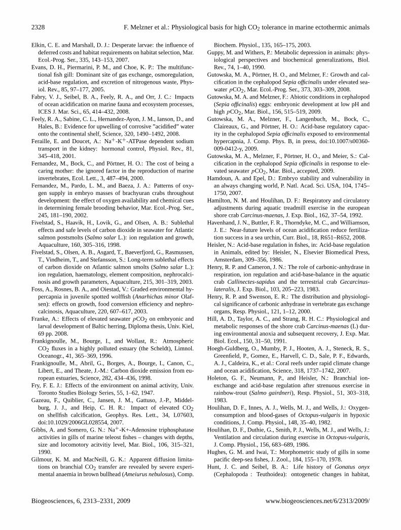
2328 F. Melzner et al.: Physiological basis for high CO2 tolerance in marine ectothermic animals
Elkin, C. E. and Marshall, D. J.: Desperate larvae: the influence ofdeferred costs and habitat requirements on habitat selection, Mar.Ecol.-Prog. Ser., 335, 143–153, 2007.
Evans, D. H., Piermarini, P. M., and Choe, K. P.: The multifunc-tional fish gill: Dominant site of gas exchange, osmoregulation,acid-base regulation, and excretion of nitrogenous waste, Phys-iol. Rev., 85, 97–177, 2005.
Fabry, V. J., Seibel, B. A., Feely, R. A., and Orr, J. C.: Impactsof ocean acidification on marine fauna and ecosystem processes,ICES J. Mar. Sci., 65, 414–432, 2008.
Feely, R. A., Sabine, C. L., Hernandez-Ayon, J. M., Ianson, D., andHales, B.: Evidence for upwelling of corrosive ”acidified” wateronto the continental shelf, Science, 320, 1490–1492, 2008.
Feraille, E. and Doucet, A.: Na+-K+-ATPase dependent sodiumtransport in the kidney: hormonal control, Physiol. Rev., 81,345–418, 2001.
Fernandez, M., Bock, C., and Portner, H. O.: The cost of being acaring mother: the ignored factor in the reproduction of marineinvertebrates, Ecol. Lett., 3, 487–494, 2000.
Fernandez, M., Pardo, L. M., and Baeza, J. A.: Patterns of oxy-gen supply in embryo masses of brachyuran crabs throughoutdevelopment: the effect of oxygen availability and chemical cuesin determining female brooding behavior, Mar. Ecol.-Prog. Ser.,245, 181–190, 2002.
Fivelstad, S., Haavik, H., Lovik, G., and Olsen, A. B.: Sublethaleffects and safe levels of carbon dioxide in seawater for Atlanticsalmon postsmolts (Salmo salarL.): ion regulation and growth,Aquaculture, 160, 305–316, 1998.
Fivelstad, S., Olsen, A. B., Asgard, T., Baeverfjord, G., Rasmussen,T., Vindheim, T., and Stefansson, S.: Long-term sublethal effectsof carbon dioxide on Atlantic salmon smolts (Salmo salarL.):ion regulation, haematology, element composition, nephrocalci-nosis and growth parameters, Aquaculture, 215, 301–319, 2003.
Foss, A., Rosnes, B. A., and Oiestad, V.: Graded environmental hy-percapnia in juvenile spotted wolffish (Anarhichas minorOlaf-sen): effects on growth, food conversion efficiency and nephro-calcinosis, Aquaculture, 220, 607–617, 2003.
Franke, A.: Effects of elevated seawaterpCO2 on embryonic andlarval development of Baltic herring, Diploma thesis, Univ. Kiel,69 pp. 2008.
Frankignoulle, M., Bourge, I., and Wollast, R.: AtmosphericCO2 fluxes in a highly polluted estuary (the Scheldt), Limnol.Oceanogr., 41, 365–369, 1996.
Frankignoulle, M., Abril, G., Borges, A., Bourge, I., Canon, C.,Libert, E., and Theate, J.-M.: Carbon dioxide emission from eu-ropean estuaries, Science, 282, 434–436, 1998.
Fry, F. E. J.: Effects of the environment on animal activity, Univ.Toronto Studies Biology Series, 55, 1–62, 1947.
Gazeau, F., Quiblier, C., Jansen, J. M., Gattuso, J.-P., Middel-burg, J. J., and Heip, C. H. R.: Impact of elevated CO2on shellfish calcification, Geophys. Res. Lett., 34, L07603,doi:10.1029/2006GL028554, 2007.
Gibbs, A. and Somero, G. N.: Na+-K+-Adenosine triphosphataseactivities in gills of marine teleost fishes – changes with depths,size and locomotory acitvitiy level, Mar. Biol., 106, 315–321,1990.
Gilmour, K. M. and MacNeill, G. K.: Apparent diffusion limita-tions on branchial CO2 transfer are revealed by severe experi-mental anaemia in brown bullhead (Ameiurus nebulosus), Comp.
Biochem. Physiol., 135, 165–175, 2003.Guppy, M. and Withers, P.: Metabolic depression in animals: phys-
iological perspectives and biochemical generalizations, Biol.Rev., 74, 1–40, 1990.
Gutowska, M. A., Portner, H. O., and Melzner, F.: Growth and cal-cification in the cephalopodSepia officinalisunder elevated sea-waterpCO2, Mar. Ecol.-Prog. Ser., 373, 303–309, 2008.
Gutowska, M. A. and Melzner, F.: Abiotic conditions in cephalopod(Sepia officinalis) eggs: embryonic development at low pH andhighpCO2, Mar. Biol., 156, 515–519, 2009.
Gutowska, M. A., Melzner, F., Langenbuch, M., Bock, C.,Claireaux, G., and Portner, H. O.: Acid-base regulatory capac-ity in the cephalopodSepia officinalisexposed to environmentalhypercapnia, J. Comp. Phys. B, in press, doi:10.1007/s00360-009-0412-y, 2009.
Gutowska, M. A., Melzner, F., Portner, H. O., and Meier, S.: Cal-cification in the cephalopodSepia officinalisin response to ele-vated seawaterpCO2, Mar. Biol., accepted, 2009.
Hamdoun, A. and Epel, D.: Embryo stability and vulnerability inan always changing world, P. Natl. Acad. Sci. USA, 104, 1745–1750, 2007.
Hamilton, N. M. and Houlihan, D. F.: Respiratory and circulatoryadjustments during aquatic treadmill exercise in the europeanshore crabCarcinus-maenas, J. Exp. Biol., 162, 37–54, 1992.
Havenhand, J. N., Buttler, F. R., Thorndyke, M. C., and Williamson,J. E.: Near-future levels of ocean acidification reduce fertiliza-tion success in a sea urchin, Curr. Biol., 18, R651–R652, 2008.
Heisler, N.: Acid-base regulation in fishes, in: Acid-base regulationin Animals, edited by: Heisler, N., Elsevier Biomedical Press,Amsterdam, 309–356, 1986.
Henry, R. P. and Cameron, J. N.: The role of carbonic-anhydrase inrespiration, ion regulation and acid-base-balance in the aquaticcrab Callinectes-sapidusand the terrestrial crabGecarcinus-lateralis, J. Exp. Biol., 103, 205–223, 1983.
Henry, R. P. and Swenson, E. R.: The distribution and physiologi-cal significance of carbonic anhydrase in vertebrate gas exchangeorgans, Resp. Physiol., 121, 1–12, 2000.
Hill, A. D., Taylor, A. C., and Strang, R. H. C.: Physiological andmetabolic responses of the shore crabCarcinus-maenas(L) dur-ing environmental anoxia and subsequent recovery, J. Exp. Mar.Biol. Ecol., 150, 31–50, 1991.
Hoegh-Guldberg, O., Mumby, P. J., Hooten, A. J., Steneck, R. S.,Greenfield, P., Gomez, E., Harvell, C. D., Sale, P. F., Edwards,A. J., Caldeira, K., et al.: Coral reefs under rapid climate changeand ocean acidification, Science, 318, 1737–1742, 2007.
Holeton, G. F., Neumann, P., and Heisler, N.: Branchial ion-exchange and acid-base regulation after strenuous exercise inrainbow-trout (Salmo gairdneri), Resp. Physiol., 51, 303–318,1983.
Houlihan, D. F., Innes, A. J., Wells, M. J., and Wells, J.: Oxygen-consumption and blood-gases ofOctopus-vulgarisin hypoxicconditions, J. Comp. Physiol., 148, 35–40, 1982.
Houlihan, D. F., Duthie, G., Smith, P. J., Wells, M. J., and Wells, J.:Ventilation and circulation during exercise inOctopus-vulgaris,J. Comp. Physiol., 156, 683–689, 1986.
Hughes, G. M. and Iwai, T.: Morphometric study of gills in somepacific deep-sea fishes, J. Zool., 184, 155–170, 1978.
Hunt, J. C. and Seibel, B. A.: Life history ofGonatus onyx(Cephalopoda : Teuthoidea): ontogenetic changes in habitat,
Biogeosciences, 6, 2313–2331, 2009 www.biogeosciences.net/6/2313/2009/
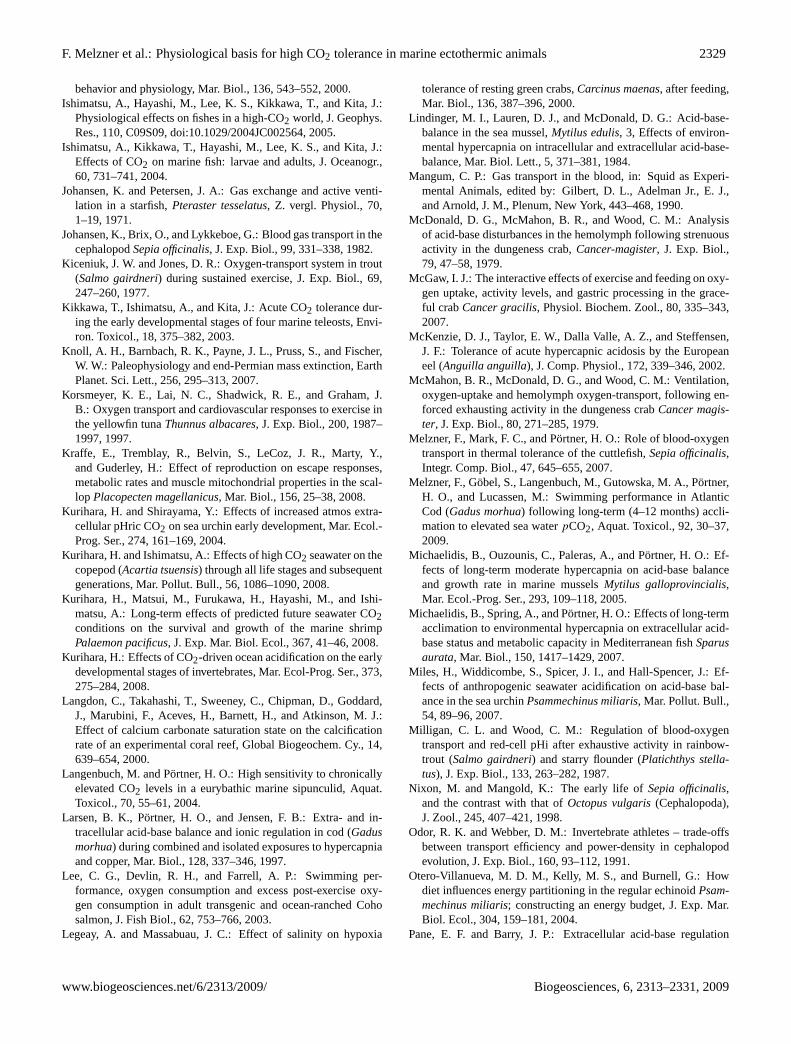
F. Melzner et al.: Physiological basis for high CO2 tolerance in marine ectothermic animals 2329
behavior and physiology, Mar. Biol., 136, 543–552, 2000.Ishimatsu, A., Hayashi, M., Lee, K. S., Kikkawa, T., and Kita, J.:
Physiological effects on fishes in a high-CO2 world, J. Geophys.Res., 110, C09S09, doi:10.1029/2004JC002564, 2005.
Ishimatsu, A., Kikkawa, T., Hayashi, M., Lee, K. S., and Kita, J.:Effects of CO2 on marine fish: larvae and adults, J. Oceanogr.,60, 731–741, 2004.
Johansen, K. and Petersen, J. A.: Gas exchange and active venti-lation in a starfish,Pteraster tesselatus, Z. vergl. Physiol., 70,1–19, 1971.
Johansen, K., Brix, O., and Lykkeboe, G.: Blood gas transport in thecephalopodSepia officinalis, J. Exp. Biol., 99, 331–338, 1982.
Kiceniuk, J. W. and Jones, D. R.: Oxygen-transport system in trout(Salmo gairdneri) during sustained exercise, J. Exp. Biol., 69,247–260, 1977.
Kikkawa, T., Ishimatsu, A., and Kita, J.: Acute CO2 tolerance dur-ing the early developmental stages of four marine teleosts, Envi-ron. Toxicol., 18, 375–382, 2003.
Knoll, A. H., Barnbach, R. K., Payne, J. L., Pruss, S., and Fischer,W. W.: Paleophysiology and end-Permian mass extinction, EarthPlanet. Sci. Lett., 256, 295–313, 2007.
Korsmeyer, K. E., Lai, N. C., Shadwick, R. E., and Graham, J.B.: Oxygen transport and cardiovascular responses to exercise inthe yellowfin tunaThunnus albacares, J. Exp. Biol., 200, 1987–1997, 1997.
Kraffe, E., Tremblay, R., Belvin, S., LeCoz, J. R., Marty, Y.,and Guderley, H.: Effect of reproduction on escape responses,metabolic rates and muscle mitochondrial properties in the scal-lop Placopecten magellanicus, Mar. Biol., 156, 25–38, 2008.
Kurihara, H. and Shirayama, Y.: Effects of increased atmos extra-cellular pHric CO2 on sea urchin early development, Mar. Ecol.-Prog. Ser., 274, 161–169, 2004.
Kurihara, H. and Ishimatsu, A.: Effects of high CO2 seawater on thecopepod (Acartia tsuensis) through all life stages and subsequentgenerations, Mar. Pollut. Bull., 56, 1086–1090, 2008.
Kurihara, H., Matsui, M., Furukawa, H., Hayashi, M., and Ishi-matsu, A.: Long-term effects of predicted future seawater CO2conditions on the survival and growth of the marine shrimpPalaemon pacificus, J. Exp. Mar. Biol. Ecol., 367, 41–46, 2008.
Kurihara, H.: Effects of CO2-driven ocean acidification on the earlydevelopmental stages of invertebrates, Mar. Ecol-Prog. Ser., 373,275–284, 2008.
Langdon, C., Takahashi, T., Sweeney, C., Chipman, D., Goddard,J., Marubini, F., Aceves, H., Barnett, H., and Atkinson, M. J.:Effect of calcium carbonate saturation state on the calcificationrate of an experimental coral reef, Global Biogeochem. Cy., 14,639–654, 2000.
Langenbuch, M. and Portner, H. O.: High sensitivity to chronicallyelevated CO2 levels in a eurybathic marine sipunculid, Aquat.Toxicol., 70, 55–61, 2004.
Larsen, B. K., Portner, H. O., and Jensen, F. B.: Extra- and in-tracellular acid-base balance and ionic regulation in cod (Gadusmorhua) during combined and isolated exposures to hypercapniaand copper, Mar. Biol., 128, 337–346, 1997.
Lee, C. G., Devlin, R. H., and Farrell, A. P.: Swimming per-formance, oxygen consumption and excess post-exercise oxy-gen consumption in adult transgenic and ocean-ranched Cohosalmon, J. Fish Biol., 62, 753–766, 2003.
Legeay, A. and Massabuau, J. C.: Effect of salinity on hypoxia
tolerance of resting green crabs,Carcinus maenas, after feeding,Mar. Biol., 136, 387–396, 2000.
Lindinger, M. I., Lauren, D. J., and McDonald, D. G.: Acid-base-balance in the sea mussel,Mytilus edulis, 3, Effects of environ-mental hypercapnia on intracellular and extracellular acid-base-balance, Mar. Biol. Lett., 5, 371–381, 1984.
Mangum, C. P.: Gas transport in the blood, in: Squid as Experi-mental Animals, edited by: Gilbert, D. L., Adelman Jr., E. J.,and Arnold, J. M., Plenum, New York, 443–468, 1990.
McDonald, D. G., McMahon, B. R., and Wood, C. M.: Analysisof acid-base disturbances in the hemolymph following strenuousactivity in the dungeness crab,Cancer-magister, J. Exp. Biol.,79, 47–58, 1979.
McGaw, I. J.: The interactive effects of exercise and feeding on oxy-gen uptake, activity levels, and gastric processing in the grace-ful crabCancer gracilis, Physiol. Biochem. Zool., 80, 335–343,2007.
McKenzie, D. J., Taylor, E. W., Dalla Valle, A. Z., and Steffensen,J. F.: Tolerance of acute hypercapnic acidosis by the Europeaneel (Anguilla anguilla), J. Comp. Physiol., 172, 339–346, 2002.
McMahon, B. R., McDonald, D. G., and Wood, C. M.: Ventilation,oxygen-uptake and hemolymph oxygen-transport, following en-forced exhausting activity in the dungeness crabCancer magis-ter, J. Exp. Biol., 80, 271–285, 1979.
Melzner, F., Mark, F. C., and Portner, H. O.: Role of blood-oxygentransport in thermal tolerance of the cuttlefish,Sepia officinalis,Integr. Comp. Biol., 47, 645–655, 2007.
Melzner, F., Gobel, S., Langenbuch, M., Gutowska, M. A., Portner,H. O., and Lucassen, M.: Swimming performance in AtlanticCod (Gadus morhua) following long-term (4–12 months) accli-mation to elevated sea waterpCO2, Aquat. Toxicol., 92, 30–37,2009.
Michaelidis, B., Ouzounis, C., Paleras, A., and Portner, H. O.: Ef-fects of long-term moderate hypercapnia on acid-base balanceand growth rate in marine musselsMytilus galloprovincialis,Mar. Ecol.-Prog. Ser., 293, 109–118, 2005.
Michaelidis, B., Spring, A., and Portner, H. O.: Effects of long-termacclimation to environmental hypercapnia on extracellular acid-base status and metabolic capacity in Mediterranean fishSparusaurata, Mar. Biol., 150, 1417–1429, 2007.
Miles, H., Widdicombe, S., Spicer, J. I., and Hall-Spencer, J.: Ef-fects of anthropogenic seawater acidification on acid-base bal-ance in the sea urchinPsammechinus miliaris, Mar. Pollut. Bull.,54, 89–96, 2007.
Milligan, C. L. and Wood, C. M.: Regulation of blood-oxygentransport and red-cell pHi after exhaustive activity in rainbow-trout (Salmo gairdneri) and starry flounder (Platichthys stella-tus), J. Exp. Biol., 133, 263–282, 1987.
Nixon, M. and Mangold, K.: The early life ofSepia officinalis,and the contrast with that ofOctopus vulgaris(Cephalopoda),J. Zool., 245, 407–421, 1998.
Odor, R. K. and Webber, D. M.: Invertebrate athletes – trade-offsbetween transport efficiency and power-density in cephalopodevolution, J. Exp. Biol., 160, 93–112, 1991.
Otero-Villanueva, M. D. M., Kelly, M. S., and Burnell, G.: Howdiet influences energy partitioning in the regular echinoidPsam-mechinus miliaris; constructing an energy budget, J. Exp. Mar.Biol. Ecol., 304, 159–181, 2004.
Pane, E. F. and Barry, J. P.: Extracellular acid-base regulation
www.biogeosciences.net/6/2313/2009/ Biogeosciences, 6, 2313–2331, 2009
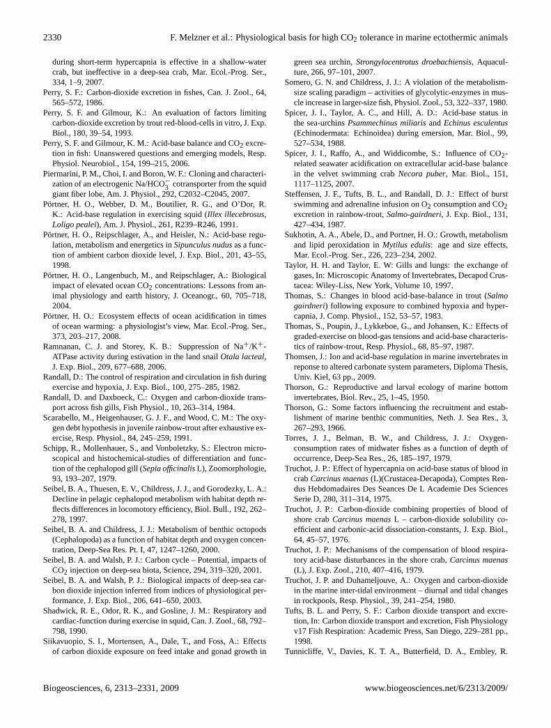
2330 F. Melzner et al.: Physiological basis for high CO2 tolerance in marine ectothermic animals
during short-term hypercapnia is effective in a shallow-watercrab, but ineffective in a deep-sea crab, Mar. Ecol.-Prog. Ser.,334, 1–9, 2007.
Perry, S. F.: Carbon-dioxide excretion in fishes, Can. J. Zool., 64,565–572, 1986.
Perry, S. F. and Gilmour, K.: An evaluation of factors limitingcarbon-dioxide excretion by trout red-blood-cells in vitro, J. Exp.Biol., 180, 39–54, 1993.
Perry, S. F. and Gilmour, K. M.: Acid-base balance and CO2 excre-tion in fish: Unanswered questions and emerging models, Resp.Physiol. Neurobiol., 154, 199–215, 2006.
Piermarini, P. M., Choi, I. and Boron, W. F.: Cloning and characteri-zation of an electrogenic Na/HCO−3 cotransporter from the squidgiant fiber lobe, Am. J. Physiol., 292, C2032–C2045, 2007.
Portner, H. O., Webber, D. M., Boutilier, R. G., and O’Dor, R.K.: Acid-base regulation in exercising squid (Illex illecebrosus,Loligo pealei), Am. J. Physiol., 261, R239–R246, 1991.
Portner, H. O., Reipschlager, A., and Heisler, N.: Acid-base regu-lation, metabolism and energetics inSipunculus nudusas a func-tion of ambient carbon dioxide level, J. Exp. Biol., 201, 43–55,1998.
Portner, H. O., Langenbuch, M., and Reipschlager, A.: Biologicalimpact of elevated ocean CO2 concentrations: Lessons from an-imal physiology and earth history, J. Oceanogr., 60, 705–718,2004.
Portner, H. O.: Ecosystem effects of ocean acidification in timesof ocean warming: a physiologist’s view, Mar. Ecol.-Prog. Ser.,373, 203–217, 2008.
Ramnanan, C. J. and Storey, K. B.: Suppression of Na+/K+-ATPase activity during estivation in the land snailOtala lacteal,J. Exp. Biol., 209, 677–688, 2006.
Randall, D.: The control of respiration and circulation in fish duringexercise and hypoxia, J. Exp. Biol., 100, 275–285, 1982.
Randall, D. and Daxboeck, C.: Oxygen and carbon-dioxide trans-port across fish gills, Fish Physiol., 10, 263–314, 1984.
Scarabello, M., Heigenhauser, G. J. F., and Wood, C. M.: The oxy-gen debt hypothesis in juvenile rainbow-trout after exhaustive ex-ercise, Resp. Physiol., 84, 245–259, 1991.
Schipp, R., Mollenhauer, S., and Vonboletzky, S.: Electron micro-scopical and histochemical-studies of differentiation and func-tion of the cephalopod gill (Sepia officinalisL), Zoomorphologie,93, 193–207, 1979.
Seibel, B. A., Thuesen, E. V., Childress, J. J., and Gorodezky, L. A.:Decline in pelagic cephalopod metabolism with habitat depth re-flects differences in locomotory efficiency, Biol. Bull., 192, 262–278, 1997.
Seibel, B. A. and Childress, J. J.: Metabolism of benthic octopods(Cephalopoda) as a function of habitat depth and oxygen concen-tration, Deep-Sea Res. Pt. I, 47, 1247–1260, 2000.
Seibel, B. A. and Walsh, P. J.: Carbon cycle – Potential, impacts ofCO2 injection on deep-sea biota, Science, 294, 319–320, 2001.
Seibel, B. A. and Walsh, P. J.: Biological impacts of deep-sea car-bon dioxide injection inferred from indices of physiological per-formance, J. Exp. Biol., 206, 641–650, 2003.
Shadwick, R. E., Odor, R. K., and Gosline, J. M.: Respiratory andcardiac-function during exercise in squid, Can. J. Zool., 68, 792–798, 1990.
Siikavuopio, S. I., Mortensen, A., Dale, T., and Foss, A.: Effectsof carbon dioxide exposure on feed intake and gonad growth in
green sea urchin,Strongylocentrotus droebachiensis, Aquacul-ture, 266, 97–101, 2007.
Somero, G. N. and Childress, J. J.: A violation of the metabolism-size scaling paradigm – activities of glycolytic-enzymes in mus-cle increase in larger-size fish, Physiol. Zool., 53, 322–337, 1980.
Spicer, J. I., Taylor, A. C., and Hill, A. D.: Acid-base status inthe sea-urchinsPsammechinus miliarisandEchinus esculentus(Echinodermata: Echinoidea) during emersion, Mar. Biol., 99,527–534, 1988.
Spicer, J. I., Raffo, A., and Widdicombe, S.: Influence of CO2-related seawater acidification on extracellular acid-base balancein the velvet swimming crabNecora puber, Mar. Biol., 151,1117–1125, 2007.
Steffensen, J. F., Tufts, B. L., and Randall, D. J.: Effect of burstswimming and adrenaline infusion on O2 consumption and CO2excretion in rainbow-trout,Salmo-gairdneri, J. Exp. Biol., 131,427–434, 1987.
Sukhotin, A. A., Abele, D., and Portner, H. O.: Growth, metabolismand lipid peroxidation inMytilus edulis: age and size effects,Mar. Ecol.-Prog. Ser., 226, 223–234, 2002.
Taylor, H. H. and Taylor, E. W: Gills and lungs: the exchange ofgases, In: Microscopic Anatomy of Invertebrates, Decapod Crus-tacea: Wiley-Liss, New York, Volume 10, 1997.
Thomas, S.: Changes in blood acid-base-balance in trout (Salmogairdneri) following exposure to combined hypoxia and hyper-capnia, J. Comp. Physiol., 152, 53–57, 1983.
Thomas, S., Poupin, J., Lykkeboe, G., and Johansen, K.: Effects ofgraded-exercise on blood-gas tensions and acid-base characteris-tics of rainbow-trout, Resp. Physiol., 68, 85–97, 1987.
Thomsen, J.: Ion and acid-base regulation in marine invertebrates inreponse to altered carbonate system parameters, Diploma Thesis,Univ. Kiel, 63 pp., 2009.
Thorson, G.: Reproductive and larval ecology of marine bottominvertebrates, Biol. Rev., 25, 1–45, 1950.
Thorson, G.: Some factors influencing the recruitment and estab-lishment of marine benthic communities, Neth. J. Sea Res., 3,267–293, 1966.
Torres, J. J., Belman, B. W., and Childress, J. J.: Oxygen-consumption rates of midwater fishes as a function of depth ofoccurrence, Deep-Sea Res., 26, 185–197, 1979.
Truchot, J. P.: Effect of hypercapnia on acid-base status of blood incrabCarcinus maenas(L)(Crustacea-Decapoda), Comptes Ren-dus Hebdomadaires Des Seances De L Academie Des SciencesSerie D, 280, 311–314, 1975.
Truchot, J. P.: Carbon-dioxide combining properties of blood ofshore crabCarcinus maenasL – carbon-dioxide solubility co-efficient and carbonic-acid dissociation-constants, J. Exp. Biol.,64, 45–57, 1976.
Truchot, J. P.: Mechanisms of the compensation of blood respira-tory acid-base disturbances in the shore crab,Carcinus maenas(L), J. Exp. Zool., 210, 407–416, 1979.
Truchot, J. P. and Duhameljouve, A.: Oxygen and carbon-dioxidein the marine inter-tidal environment – diurnal and tidal changesin rockpools, Resp. Physiol., 39, 241–254, 1980.
Tufts, B. L. and Perry, S. F.: Carbon dioxide transport and excre-tion, In: Carbon dioxide transport and excretion, Fish Physiologyv17 Fish Respiration: Academic Press, San Diego, 229–281 pp.,1998.
Tunnicliffe, V., Davies, K. T. A., Butterfield, D. A., Embley, R.
Biogeosciences, 6, 2313–2331, 2009 www.biogeosciences.net/6/2313/2009/
F. Melzner et al.: Physiological basis for high CO2 tolerance in marine ectothermic animals 2331
W., Rose, J. M., and Chadwick, W. W.: Survival of mussels inextremely acidic waters on a submarine volcano, Nature Geosci.,2, 344–348, 2009.
Vahl, O.: The relationship between specific dynamic action (SDA)and growth in the common starfish,Asterias rubensL., Oecolo-gia, 61, 122–125, 1984.
Van den Thillart, G. D., Randall, D., and Hoa-Ren, L.: CO2 and H+
excretion by swimming coho salmon,Oncorhynchus kisutch, J.Exp. Biol., 107, 169–180, 1983.
Virkki, L. V., Choi, I., Davis, B. A., and Boron, W. F.: Cloning ofa Na+-driven Cl/HCO3− exchanger from squid giant fiber lobe,Am. J. Physiol., 285, C771–C780, 2003.
Watt, A. J. S., Whiteley, N. M., and Taylor, E. W.: An in situ studyof respiratory variables in three British sublittoral crabs with dif-ferent routine rates of activity, J. Exp. Mar. Biol. Ecol., 239, 1–21, 1999.
Webber, D. M. and Odor, R. K.: Monitoring the metabolic-rate andactivity of free-swimming squid with telemetered jet pressure, J.Exp. Biol., 126, 205–224, 1986.
Webster, S. K.: Oxygen-consumption in echinoderms from severalgeographical locations, with particular reference to Echinoidea,Biol. Bull., 148, 157–164, 1975.
Weigelt, M. and Rumohr, H.: Effects of wide-range oxygen deple-tion on benthic fauna and demersal fish in Kiel Bay 1981–1983,Rep. Mar. Res., 31, 124–136, 1986.
Wells, M. J., Odor, R. K., Mangold, K., and Wells, J.: oxygen-consumption in movement by octopus, Mar. Behav. Physiol., 9,289–303, 1983.
Wheatly, M. G. and Henry, R. P.: Extracellular and intracellularacid-base regulation in crustaceans, J. Exp. Zool., 263, 127–142,1992.
Widdicombe, S. and Spicer, J. I.: Predicting the impact of oceanacidification on benthic biodiversity: What can animal physiol-ogy tell us? J. Exp. Mar. Biol. Ecol., 366, 187–197, 2008.
Widdows, J.: Effect of temperature and food on heart beat, venti-lation rate and oxygen-uptake ofMytilus edulis, Mar. Biol., 20,269–276, 1973.
Willson, L. L., and Burnett, L. E.: Whole animal and gill tissue oxy-gen uptake in the Eastern oyster,Crassostrea virginica: Effectsof hypoxia, hypercapnia, air exposure, and infection with the pro-tozoan parasitePerkinsus marinus, J. Exp. Mar. Biol. Ecol., 246,223–240, 2000.
Wilson, J. M., Laurent, P., Tufts, B. L., Benos, D. J., Donowitz, M.,Vogl, A. W., and Randall, D. J.: NaCl uptake by the branchial ep-ithelium in freshwater teleost fish: An immunological approachto ion-transport protein localization, J. Exp. Biol., 203, 2279–2296, 2000.
Wood, C. M. and Munger, R. S.: Carbonic-anhydrase injection pro-vides evidence for the role of blood acid-base status in stimulat-ing ventilation after exhaustive exercise in rainbow-trout, J. Exp.Biol., 194, 225–253, 1994.
Wood, H. L., Spicer, J. I., and Widdicombe, S.: Ocean acidificationmay increase calcification rates, but at a cost, P. Roy. Soc. B-Biol.Sci., 275, 1767–1773, 2008.
Wootton, J. T., Pfister, C. A., and Forester, J. D.: Dynamic pat-terns and ecological impacts of declining ocean pH in a high-resolution multi-year dataset, P. Natl. Acad. Sci. USA, 105,18848–18853, 2008.
Yamamoto, K.: Increase of arterial O2 content in exercised yel-lowtail (Seriola quinqueradiata), Comp. Biochem. Physiol., 98,43–46, 1991.
Zielinski, S., Sartoris, F. J., and Portner, H. O.: Temperature effectson hemocyanin oxygen binding in an Antarctic cephalopod, Biol.Bull., 200, 67–76, 2001.
www.biogeosciences.net/6/2313/2009/ Biogeosciences, 6, 2313–2331, 2009