Embed Size (px)
Citation preview
NASA/TP-2003-212054
PHYSIOLOGICAL AND MOLECULAR
ELECTROMAGNETIC FIELDS ON HUMAN NEURONAL CELLS
GENETIC EFFECTS OF TIME-VARYING
Thomas J. Goodwin, Ph.D. Lyndon 8. Johnson Space Center
September 2003
https://ntrs.nasa.gov/search.jsp?R=20030075722 2018-06-22T18:28:54+00:00Z
THE NASA STI PROGRAM OFFICE.. . IN PROFILE
Since its founding, NASA has been dedicated to the advancement of aeronautics and space science. The NASA Scientific and Technical Information (STI) Program Office plays a key part in helping NASA maintain this important role.
The NASA STI Program Office is operated by Langley Research Center, the lead center for NASA’s scientific and technical information. The NASA STI Program Office provides access to the NASA STI Database, the largest collection of aeronautical and space science STI in the world. The Program Office is also NASA’s institutional mechanism for disseminating the results of its research and development activities. These results are published by NASA in the NASA STI Report Series, which includes the following report types:
TECHNICAL PUBLICATION. Reports of completed research or a major significant phase of research that present the results of NASA programs and include extensive data or theoretical analysis. Includes compilations of significant scientific and technical data and information deemed to be of continuing reference value. NASA’s counterpart of peer-reviewed formal professional papers but has less stringent limitations on manuscript length and extent of graphic presentations.
TECHNICAL MEMORANDUM. Scientific and technical findings that are preliminary or of specialized interest, e.g., quick release reports, working papers, and bibliographies that contain minimal annotation. Does not contain extensive analysis.
CONTRACTOR REPORT. Scientific and technical findings by NASA-sponsored contractors and grantees.
CONFERENCE PUBLICATION. Collected papers from scientific and technical conferences, symposia, seminars, or other meetings sponsored or cosponsored by NASA.
SPECIAL PUBLICATION. Scientific, technical, or historical information from NASA programs, projects, and mission, often concerned with subjects having substantial public interest.
TECHNICAL TRANSLATION. English- language translations of foreign scientific and technical material pertinent to NASA’s mission.
Specialized services that complement the STI Program Office’s diverse offerings include creating custom thesauri, building customized databases, organizing and publishing research results . . . even providing videos.
For more information about the NASA STI Program Office, see the following:
Access the NASA STI Program Home Page at http://www.stinasagov
E-mail your question via the Internet to [email protected]
Fax your question to the NASA Access Help Desk at (301) 621-0134
Telephone the NASA Access Help Desk at (301) 621-0390
Write to: NASA Access Help Desk NASA Center for Aerospace Information 7121 Standard Hanover, MD 21076-1320
NASA/TP-2003-212054
PHYSIOLOGICAL AND MOLECULAR
ELECTROMAGNETIC FIELDS ON HUMAN NEURONAL CELLS
GENETIC EFFECTS OF TIME-VARYING
Thomas J. Goodwin, PH.D. Lyndon B. Johnson Space Center Houston, Texas
National Aeronautics and Space Administration
Johnson Space Center Houston, Texas 77058-3696
September 2003
Available from:
NASA Center for Aerospace Information 7121 Standard Hanover, MD 2 1076- 1320
National Technical Information Service 5285 Port Royal Road
Springfield, VA 22 16 1 This report is also available in electronic form at http://techreports.larc.nasa.gov/cgi-bin/NTRS
11
CONTENTS
Page ... Contents ....................................................................................................................................... 111
Physiological and Molecular Genetic Effects of Time-Varying Electromagnetic Fields on Human Neuronal Cells .............................................................................................................. 1
Abstract ...................................................................................................................................... 1 Background ................................................................................................................................ 1 Materials and Methods ............................................................................................................... 3
Cells ....................................................................................................................................... 3 Cell Culture Protocols ............................................................................................................ 3 Generator ................................................................................................................................ 4
Two-Dimensional Experimental Protocols ................................................................................ 5 Three-Dimensional (RWV) Experimental Protocol .................................................................. 6 Analytical Methods .................................................................................................................... 6
Cell Metabolic Parameters ..................................................................................................... 6 Light Microscopy ................................................................................................................... 6 Scanning Electron Microscopy .............................................................................................. 6 Genetic Discovery Array ........................................................................................................ 6 Statistical Analysis ................................................................................................................. 7
Results ........................................................................................................................................ 7 Two-Dimensional Experiments ............................................................................................. 7
Three-Dimensional Experiments ............................................................................................... 9 Rotating-Wall Vessel Background ....................................................................................... 12 Rotating-Wall Vessel Experimental Data ............................................................................ 12
Gene Array ............................................................................................................................... 15 Discussion and Conclusions ........................................................................................................ 26 References .................................................................................................................................... 28
. .
Figures FIGURE 1 . Notice the silver-faced box in the left of photo . The generator is attached to the
FIGURE 2 . The NHNP cells produced a whitish fingerprint on each of the electrodes under
FIGURE 3 . NHNP dishes with TVEMF applied ........................................................................ 10
FIGURE 5 . Close inspection of the edge of the Corona reveals NHNP cells attempting
FIGURE 6 . In direct contrast to the treated NHNP cells, non-waveform tissues attached
FIGURE 7 . Changes in pH between treated and control cultures in RWVs was minimal.
FIGURE 8 . Glucose consumption was identical during the course of the experiments ............. 13
experiment in the incubator by a ribbon cable ....................................................................... 4
TVEMF .................................................................................................................................. 5
FIGURE 4 . A Corona can be seen at the edge of the tissue 24 hours after TVEMF .................. 10
to grow in an oriented fashion away from the transplanted tissue ....................................... 11
but did not proliferate over the same 24-hour period ........................................................... 11
indicating the overall metabolism was nearly identical in both vessels ............................... 13
... 111
CONTENTS (CONTINUED)
Page
FIGURE 9. Oxygen consumption was sliglltly higher in TVEMF in the later stages of the
FIGURE 10. As would be expected, TVEMF COz levels were slightly higher than
FIGURE 11. Lactate production in the cultures was nearly identical. Comparison
FIGURE 12. 12A shows NHNP cells grown without TVEMF; 12B shows the dramatic
FIGURE 13. Observe the difference between 13A (untreated) and 13B. Cellular organization
experiments. ........................................................................................................................ .14
in controls ............................................................................................................................. 14
with glucose consumption indicates the expected parallel. ................................................. 15
difference in the development of neural “tubes” under these conditions ............................ .16
changed dramatically with TVEMF exposure in RWV three-dimensional culture. ........... 17
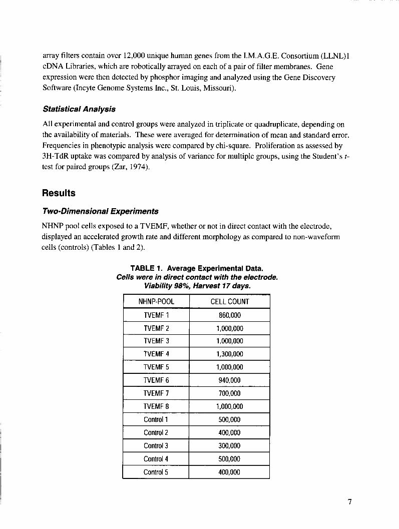
Tables TABLE 1. Average Experimental Data. Cells were in direct contact with the electrode.
TABLE 2. Average Experimental Data. Cells were not in direct contact with the
TABLE 3. Average Experimental Data. Cells were in direct contact with the electrode.
TABLE 4. Average Experimental Data. Cells were not in direct contact with the electrode.
TABLE 5. Down-Regulated Genes in Descending Order
Viability 98%, Harvest 17 days ............................................................................................. 7
electrode. Viability loo%, Harvest 18 days ........................................................................... 8
Cell Count at various times after removal of TVEMF ........................................................... 8
Cell Count and Viability at Various times post harvest. ........................................................ 9 .18
i I I ......................................................... , TABLE 6. Waveform Up-Regulated Genes in Ascending Order ............................................... 23
iv
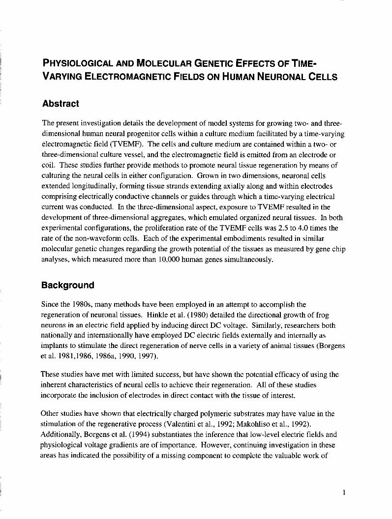
PHYSIOLOGICAL AND MOLECULAR GENETIC EFFECTS OF TIME- VARYING ELECTROMAGNETIC FIELDS ON HUMAN NEURONAL CELLS
Abstract
The present investigation details the development of model systems for growing two- and three- dimensional human neural progenitor cells within a culture medium facilitated by a time-varying electromagnetic field (TVEMF). The cells and culture medium are contained within a two- or three-dimensional culture vessel, and the electromagnetic field is emitted from an electrode or coil. These studies further provide methods to promote neural tissue regeneration by means of culturing the neural cells in either configuration. Grown in two dimensions, neuronal cells extended longitudinally, forming tissue strands extending axially along and within electrodes comprising electrically conductive channels or guides through which a time-varying electrical current was conducted. In the three-dimensional aspect, exposure to TVEMF resulted in the development of three-dimensional aggregates, which emulated organized neural tissues. In both experimental configurations, the proliferation rate of the TVEMF cells was 2.5 to 4.0 times the rate of the non-waveform cells. Each of the experimental embodiments resulted in similar molecular genetic changes regarding the growth potential of the tissues as measured by gene chip analyses, which measured more than 10,000 human genes simultaneously.
Background
Since the 1980s, many methods have been employed in an attempt to accomplish the regeneration of neuronal tissues. Hinkle et al. (1980) detailed the directional growth of frog neurons in an electric field applied by inducing direct DC voltage. Similarly, researchers both nationally and internationally have employed DC electric fields externally and internally as implants to stimulate the direct regeneration of nerve cells in a variety of animal tissues (Borgens et al. 1981,1986, 1986a, 1990, 1997).
These studies have met with limited success, but have shown the potential efficacy of using the inherent characteristics of neural cells to achieve their regeneration. All of these studies incorporate the inclusion of electrodes in direct contact with the tissue of interest.
Other studies have shown that electrically charged polymeric substrates may have value in the stimulation of the regenerative process (Valentini et al., 1992; Makohliso et al., 1992). Additionally, Borgens et al. (1994) substantiates the inference that low-level electric fields and physiological voltage gradients are of importance. However, continuing investigation in these areas has indicated the possibility of a missing component to complete the valuable work of
1
previous investigations. The missing component may well be the addition of a magnetic element.
Combining the electric and magnetic elements would seem logical, but to date may not have been employed due to traditional purist approaches to experimentation. The current studies are a composite of physiology and electromagnetic bioengineering, which relate generally to the fields of biophysics, tissue regeneration, tissue culture, and neurobiology. The present investigation relates to the use of a time-varying electromagnetic field for potentiation of the growth of mammalian cells and tissues. The preferred protocol uses two-dimensional conducting plate electrodes and may be applied to conventional two-dimensional tissue cultures or to three- dimensional tissue cultures. One may achieve three-dimensional cultures either by exposure to actual microgravity or by using rotating wall vessel (RWV) technology, which simulates some of the physical conditions of microgravity (Goodwin et al., 1993~). The time-varying electromagnetic field is achieved in the vicinity of the electrode by passing a time-varying current pulse with modulated signal through the electrode.
Growth of a variety of both normal and neoplastic mammalian tissues in both mono-culture and co-culture has been established in both batch-fed and perfused RWVs (Schwarz et al., 1991a, 1991b), and in conventional plate or flask-based culture systems. In some applications, growth of three-dimensional tissues in these culture systems has been facilitated by support of a solid matrix of biocompatible polymers and microcarriers. In the case of spheroidal growth, three- dimensional structure has been achieved without matrix support (Goodwin et al., 1992, 1993a, 1993b, 1997). The NASA RWV tissue culture technologies have extended this three- dimensional capacity for a number of tissues and have allowed the tissue to express different genes and biomolecules. Neuronal tissue has been largely refractory, in terms of controlled growth induction and three-dimensional organization, under conventional culture conditions. Actual microgravity, and to a lesser extent, rotationally simulated microgravity, have permitted some enhanced nerve growth (Lelkes and Unsworth, 1997). Previous attempts to electrically stimulate growth have used static electric fields, static magnetic fields, and the direct passage of current through the culture medium, but not the induction of an AC time-varying electromagnetic field in the culture region.
Neuronal tissue comprises elongated nerve cells composed of elongated axons, dendrites, and nuclear areas. Axons and dendrites are chiefly responsible for transmission of neural signals over distance. Longitudinal cell orientation is critical for proper tissue formation and function. The nucleus plays the typical role of directing nucleic acid synthesis for the control of cellular metabolic function, including growth. In vivo, the neuronal tissue is invariably spatially associated with a system of feeder, or glial, cells. This three-dimensional spatial arrangement has not been reproduced by conventional in vitro culture.
2
Investigators (Borgens et al., Valentini et al., and others) have used static electric fields in an attempt to enhance nerve growth in culture with some success to either alter embryonic development or achieve isolated nerve axon directional growth, but have not yet achieved actual control mechanisms for potentiation of growth or genetic activity causing growth. Mechanical devices intended to help grow and orient three-dimensional mammalian neuronal tissue are currently available. Fukuda et al. used zones formed between stainless steel shaving blades to orient neuronal cells or axons. Additionally, electrodes charged with electrical potential were employed to enhance axon response. Aebischer described an electrically charged, implantable tubular membrane for use in regenerating severed nerves within the human body.
None of these devices uses channels of cell-attractive material, nor do they apply a time-varying electromagnetic field, a static electrical, or a static magnetic field. Additionally, no use is made of simulated or actual microgravity techniques for pure neuronal or mixed neuronal and feeder cell cultures. Incorporation of an electromagnetic stimulus in conjunction with growing three- dimensional mammalian neuronal tissue in the proximity of, or directly upon the surface of, a current-carrying electrode (which may be bioattractive and directly adherent to the cells) is expected to enhance the proliferative response. Furthermore, the use of a time-varying current to induce a corresponding time-varying electromagnetic field in the vicinity of the growing culture to potentiate or spatially direct cell growth is not part of the prior art.
Materials and Methods
Cells
Normal human neuronal progenitor cells (NHNP) were pooled from three donors to diminish donor-to-donor variations in response. As controls, NHNP were grown in conventional tissue culture following standard cell culturing procedures in tissue culture flasks obtained through Clonetics Corporation, San Diego, California.
Cell Culture Protocols
For two-dimensional culture, GTSF-2 medium with 10% FBS, Ciprofloxacin and Fungizone was used to culture the cells (Goodwin et al., 1993a). 1 X PBS, Collagenase, DNase and trypsin were purchased from Clonetics San Diego, California, and used Corning T-75 flasks (Corning Inc., Corning, New York) for initial cell culture to obtain the appropriate number of cells for each experiment. Briefly, cells to be cultured were enzymatically dissociated with the referenced reagents from T-flasks, washed once with PBS-CMF and assayed for viability by trypan dye exclusion (GIBCO, Grand Island, New York). Cells were grown on 100-mm petri dishes (tissue culture treated to prevent adherence) or grown on the actual electrodes inside the petri dishes. Electrodes were made of platinum and stainless steel. Cell cultures were maintained in a humidified Forma CO2 incubator (Forma, Inc.) at 37°C at a C02 concentration of 6%.
3
For three-dimensional culture, NHNP cells were prepared as described above and an RWV was sequentially inoculated with 5 mg/ml Cytodex-3 type 1 collagen-coated microcarriers (Pharmacia) and freshly digested NHNP cells, yielding a cell density of 2.5.x lo5 cells/ml in a 55-ml vessel. Tissues were cultured for 17 to 21 days or until 3- to 5-mm diameter tissue masses formed.
Generator
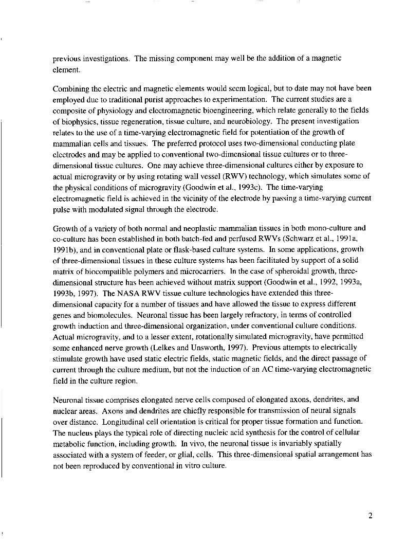
A waveform (TVEMF) generator of original design and capability was developed and used to generate the waveform in a strength of 1-6 mA (AC) square wave, 10 Hz variable duty cycle, which was pulse-width modulated (Goodwin et al. patent cases MSC 22633-1; US Patent, 6,485,963, B1 and MSC 22633-2 Notice to Issue). NHNP cells were subjected to these extremely low-level magnetic fields (ELF waves) (-10 - 200 mGauss), which are far less than the field strength of the Earth. Figure 1 illustrates the experimental arrangements for two- dimensional cell cultures.
FIGURE 1. Notice the silver-faced box in the left of photo. The generator is attached to the experiment in the incubator by a ribbon cable.
4
Two-Dimensional Experimental Protocols
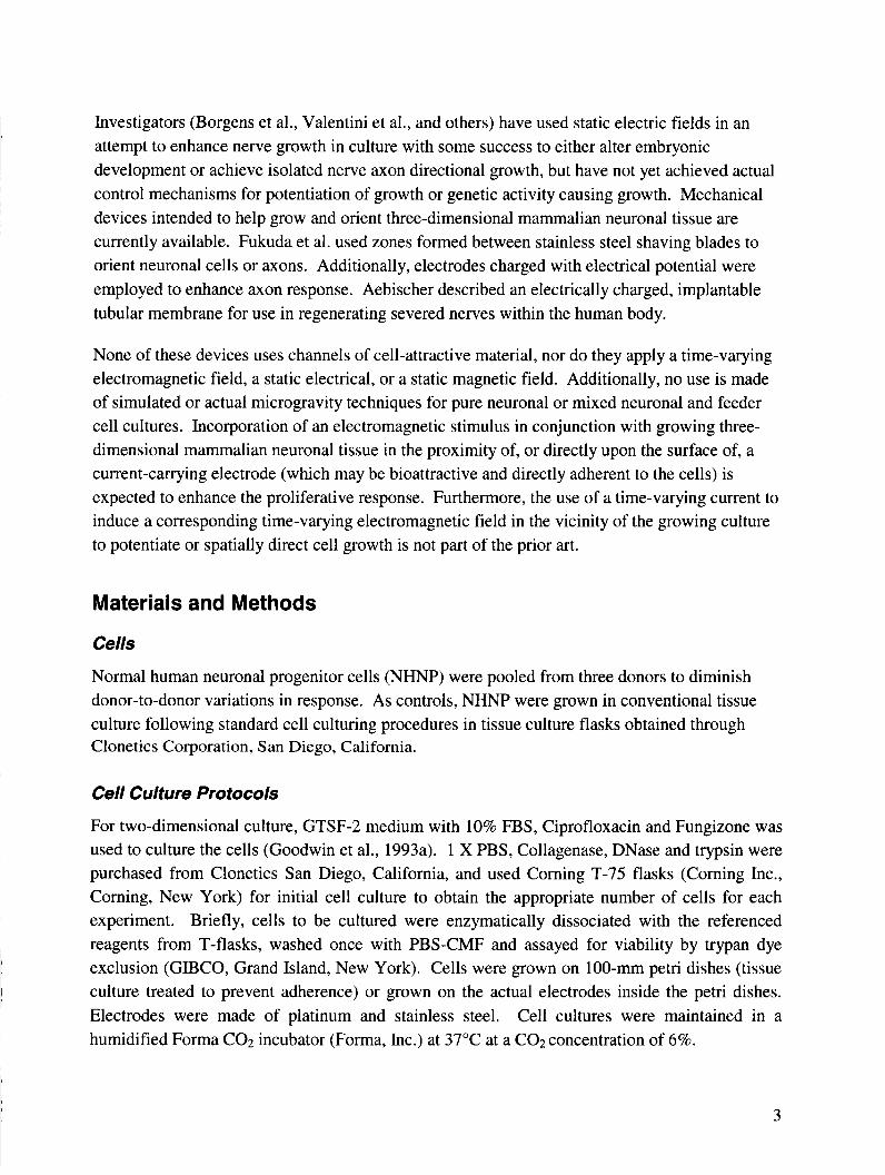
Initially, a metal electrode was placed inside a petri dish and centered. NHNP were seeded at 2.5 x 10⁵ cells in 0.7 ml of media and carefully dropped on the electrode in a concentrated
FIGURE 2. The NHNP cells produced a whitish fingerprint on each of the electrodes under TVEMF.
Cells were incubated for 2 days. The second day after cell inoculation is considered day 0 of the experiment protocol. At day 0, each dish was given 15 ml of media and waveform was applied to the electrodes. Cells were fed with 15 ml of media at day 3 and with 13 ml every three days thereafter at day 6,9, and 12. At days 14 and 17, the cells were fed again with 15 ml of media. At days 17 to 21, the cells were incubated for 10 minutes in a CollagenaseDNase cocktail, then trypsin was directly applied to the cocktail and the cells were further incubated for 3 more minutes. Before the complete media was added to deactivate the trypsin, the cocktail mix was pipetted up and down several times. The cells were washed twice with 1 X PBS, reapplied with the media, and placed on ice. The cells were observed under a dissecting microscope, counted,
~
I and assessed for viability.
An identical protocol was followed in similar experiments with the exception that, instead of the electrode being placed within the petri dishes, in media, it was attached to the underside of the TVEMF treated dishes, so that the cells had no direct contact with the metal surface.
5
Three-Dimensional (RWV) Experimental Protocol
Three-dimensional neural cells and tissues were cultured by the method described above, except that the TVEMF RWV was modified to incorporate an electromagnetic coil. The coil was wrapped around the core of the vessel so it emitted the same electromagnetic field strength as in the two-dimensional configuration. All other conditions were identical to the two-dimensional experimental conditions.
Analytical Methods
Cell Metabolic Parameters
The following metabolic parameters were monitored on a daily basis during the culture of the RWV model: pH, dissolved 0 2 , dissolved C02, glucose concentration, cell viability, and LDH (lactate dehydrogenase). Dissolved 02, C02, pH, and glucose are routinely measured in our laboratories (Goodwin et al., 1992, 1993b; Jessup et al., 1994; Scharz et al., 1992). At 24-hour intervals, pH, C02, and 0 2 were determined on a Corning 238-model clinical blood gas analyzer (Corning, Corning, New York). The glucose concentration was determined by a Beckman 2 model clinical glucose analyzer (Beckman Instruments, Inc., Fullerton, California). Samples for determination of LDH levels were collected each 24-hour period. LDH analyses were performed on a clinical Kodak Biolyzer (Kodak, Inc., New Haven, Connecticut) using dry chemistry technique.
Light Microscopy
Two-dimensional samples of each experiment were photographed by a Nikon T330 microscope equipped with advanced Hoffman Modulation optics (Nikon Inc., New York, New York).
Scanning Electron Microscopy
Samples for scanning electron microscopy were harvested every 48 hours of culture and were washed once in PBS-CMF, fixed in a solution containing 3% glutaraldehyde, 2% paraformaldehyde in 0.1 M cacodylate buffer at pH 7.4, processed by RMC, Inc., and scanned at 10 kV.
Genetic Discovery Array
Samples of human NHNP cells grown in conventional flask culture were trypsinized and placed into petri dishes in a 55-ml RWV. After 17-2 1 days of culture on 5 mg/ml cytodex-3 beads, cells were washed once with ice-cold phosphate buffered saline. The cells were lysed and mRNA selected with biotinylated oligo (dT) then separated with streptavidin paramagnetic particles (Poly A Ttract System 1000, Promega Madison, WI). 32P-labeled cDNA probes were generated by reverse transcription with 32P dCTP. The cDNA probes were hybridized to identical gene discovery array filters (Incyte Genome Systems Inc., St. Louis, Missouri). The gene discovery
6
array filters contain over 12,000 unique human genes from the I.M.A.G.E. Consortium (LLNL)l cDNA Libraries, which are robotically arrayed on each of a pair of filter membranes. Gene expression were then detected by phosphor imaging and analyzed using the Gene Discovery Software (Incyte Genome Systems Inc., St. Louis, Missouri).
~
Control 4
Control 5
Statistical Analysis
All experimental and control groups were analyzed in triplicate or quadruplicate, depending on the availability of materials. These were averaged for determination of mean and standard error. Frequencies in phenotypic analysis were compared by chi-square. Proliferation as assessed by 3H-TdR uptake was compared by analysis of variance for multiple groups, using the Student’s t- test for paired groups (Zar, 1974).
~~ ~ ~~
500,000
400,000
Resu I ts
Two-Dimensional Experiments
NHNP pool cells exposed to a TVEMF, whether or not in direct contact with the electrode, displayed an accelerated growth rate and different morphology as compared to non-waveform cells (controls) (Tables 1 and 2).
TABLE 1. Average Experimental Data. Cells were in direct contact with the electrode.
Viability 98%, Harvest 17 days.
I NHNP-POOL I CELLCOUNT I I TVEMFl I 860,000 I I TVEMF2 I 1,000,000 I I TVEMF3 I 1,000,000 I I TVEMF4 I 1,300,000 I I TVEMF5 I 1,000,000 I I TVEMFG I 940,000 I
TVEMF 7 700,000
I Control 2 I 400,000 I I Control 3 I 300,000 I
7
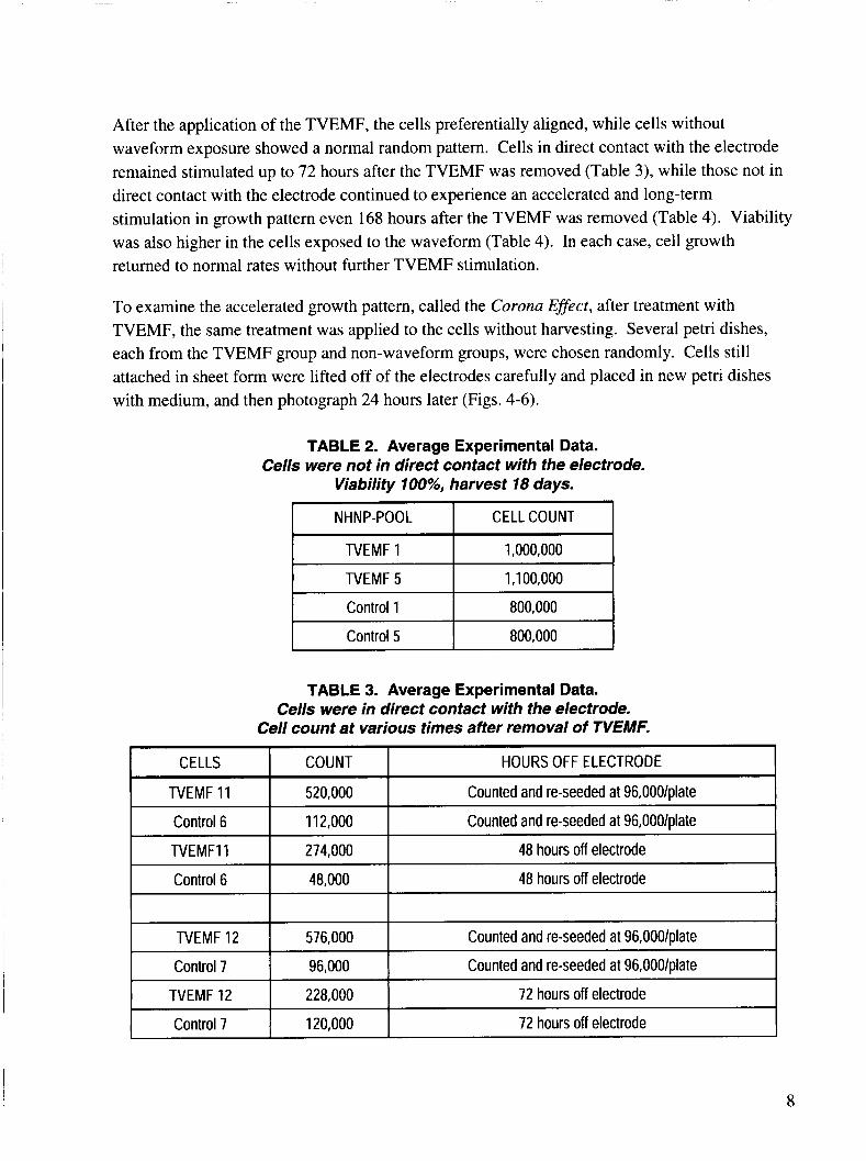
After the application of the TVEMF, the cells preferentially aligned, while cells without waveform exposure showed a normal random pattern. Cells in direct contact with the electrode remained stimulated up to 72 hours after the TVEMF was removed (Table 3), while those not in direct contact with the electrode continued to experience an accelerated and long-term stimulation in growth pattern even 168 hours after the TVEMF was removed (Table 4). Viability was also higher in the cells exposed to the waveform (Table 4). In each case, cell growth returned to normal rates without further TVEMF stimulation.
NHNP-POOL
To examine the accelerated growth pattern, called the Corona Effect, after treatment with TVEMF, the same treatment was applied to the cells without harvesting. Several petri dishes, each from the TVEMF group and non-waveform groups, were chosen randomly. Cells still attached in sheet form were lifted off of the electrodes carefully and placed in new petri dishes with medium, and then photograph 24 hours later (Figs. 4-6).
CELL COUNT
TVEMF 1
TVEMF 5
1,000,000
1,100,000
I Control 1 I 800,000
I Control 5 I 800,000
TABLE 3. Average Experimental Data. Cells were in direct contact with the electrode.
Cell count at various times after removal of TVEMF.
8
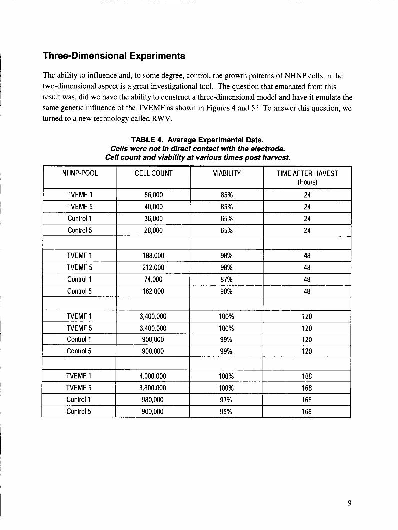
Three-Dimensional Experiments
The ability to influence and, to some degree, control, the growth patterns of NHNP cells in the two-dimensional aspect is a great investigational tool. The question that emanated from this result was, did we have the ability to construct a three-dimensional model and have it emulate the same genetic influence of the TVEMF as shown in Figures 4 and 5? To answer this question, we turned to a new technology called RWV.
9
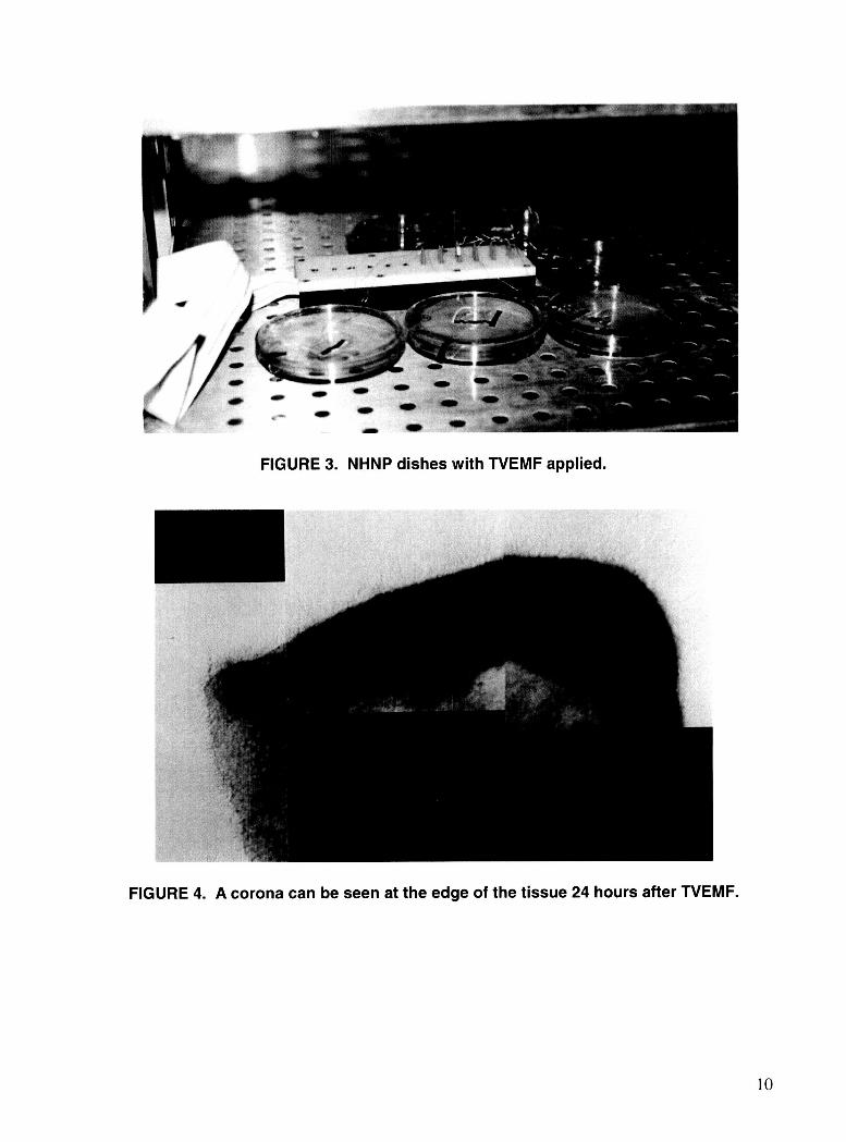
FIGURE 3. NHNP dishes with TVEMF applied.
FIGURE 4. A corona can be seen at the edge of the tissue 24 hours after TVEMF.
10
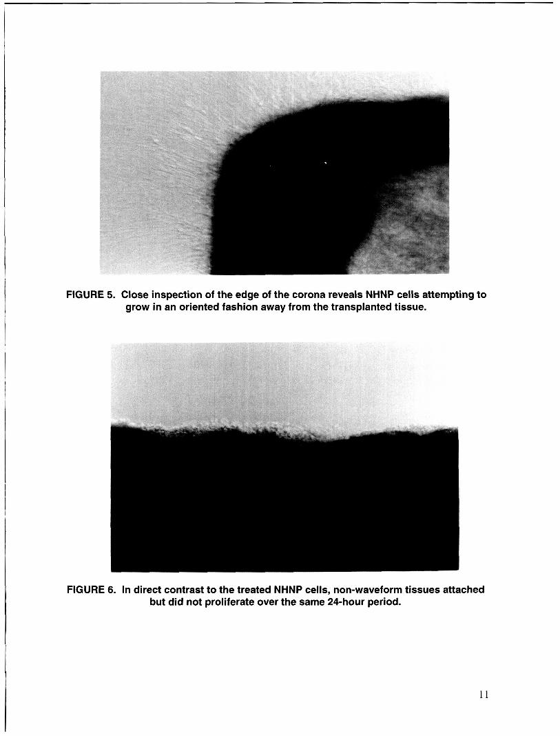
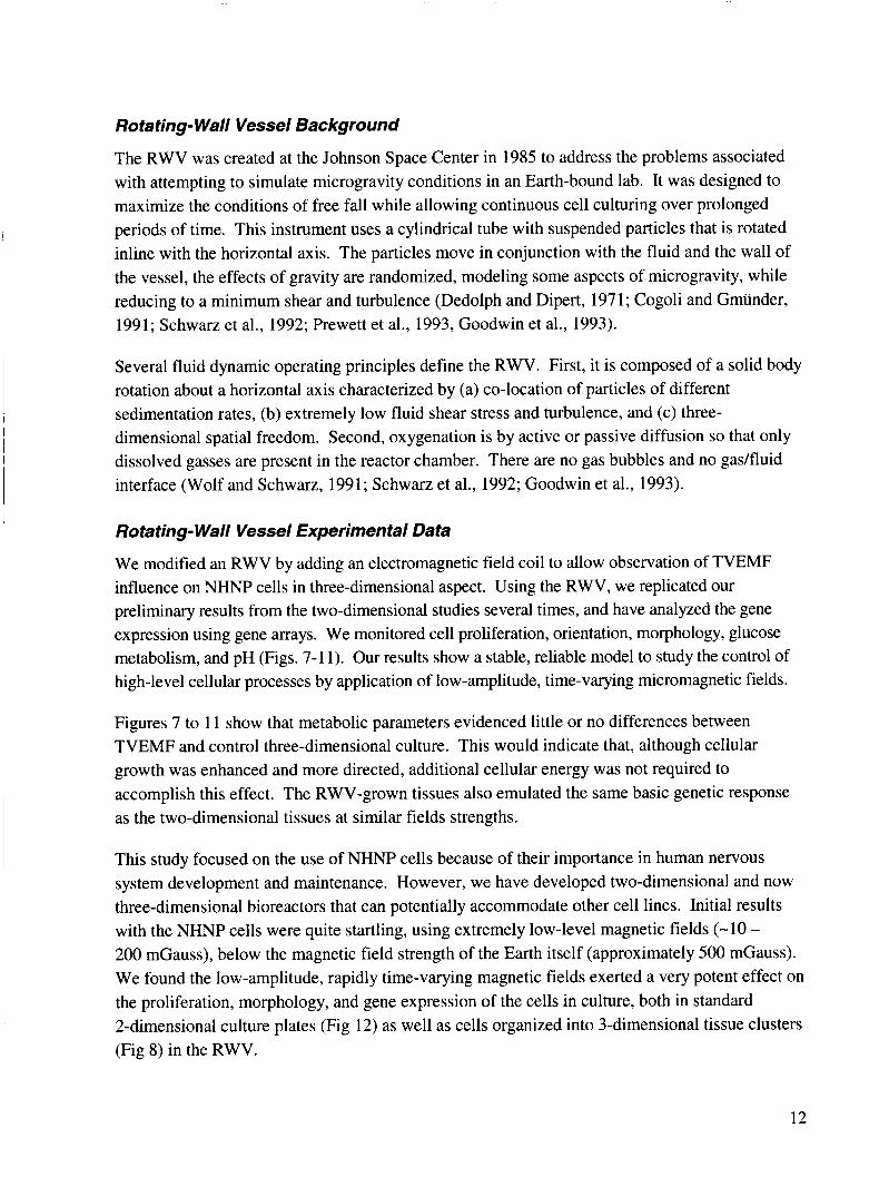
FIGURE 5. Close inspection of the edge of the corona reveals NHNP cells attempting to grow in an oriented fashion away from the transplanted tissue.
FIGURE 6. In direct contrast to the treated NHNP cells, non-waveform tissues attached but did not proliferate over the same 24-hour period.
11
Rotating- Wall Vessel Background
The RWV was created at the Johnson Space Center in 1985 to address the problems associated with attempting to simulate microgravity conditions in an Earth-bound lab. It was designed to maximize the conditions of free fall while allowing continuous cell culturing over prolonged periods of time. This instrument uses a cylindrical tube with suspended particles that is rotated inline with the horizontal axis. The particles move in conjunction with the fluid and the wall of the vessel, the effects of gravity are randomized, modeling some aspects of microgravity, while reducing to a minimum shear and turbulence (Dedolph and Dipert, 1971; Cogoli and Gmiinder, 1991; Schwarz et al., 1992; Prewett et al., 1993, Goodwin et al., 1993).
Several fluid dynamic operating principles define the RWV. First, it is composed of a solid body rotation about a horizontal axis characterized by (a) co-location of particles of different sedimentation rates, (b) extremely low fluid shear stress and turbulence, and (c) three- dimensional spatial freedom. Second, oxygenation is by active or passive diffusion so that only dissolved gasses are present in the reactor chamber. There are no gas bubbles and no gadfluid interface (Wolf and Schwarz, 1991; Schwarz et al., 1992; Goodwin et al., 1993).
Rotating- Wall Vessel Experimental Data
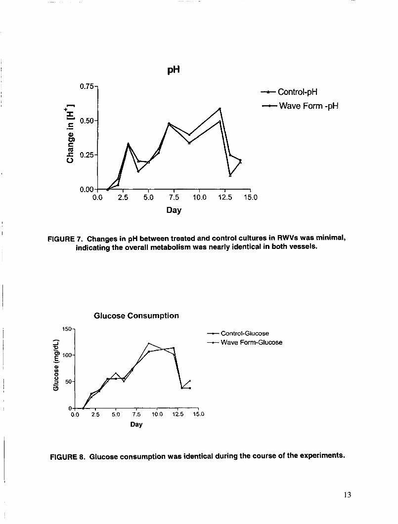
We modified an RWV by adding an electromagnetic field coil to allow observation of TVEMF influence on NHNP cells in three-dimensional aspect. Using the RWV, we replicated our preliminary results from the two-dimensional studies several times, and have analyzed the gene expression using gene arrays. We monitored cell proliferation, orientation, morphology, glucose metabolism, and pH (Figs. 7-1 1). Our results show a stable, reliable model to study the control of high-level cellular processes by application of low-amplitude, time-varying micromagnetic fields.
Figures 7 to 11 show that metabolic parameters evidenced little or no differences between TVEMF and control three-dimensional culture. This would indicate that, although cellular growth was enhanced and more directed, additional cellular energy was not required to accomplish this effect. The RWV-grown tissues also emulated the same basic genetic response as the two-dimensional tissues at similar fields strengths.
This study focused on the use of NHNP cells because of their importance in human nervous system development and maintenance. However, we have developed two-dimensional and now three-dimensional bioreactors that can potentially accommodate other cell lines. Initial results with the NHNP cells were quite startling, using extremely low-level magnetic fields (-10 - 200 mGauss), below the magnetic field strength of the Earth itself (approximately 500 mGauss). We found the low-amplitude, rapidly time-varying magnetic fields exerted a very potent effect on the proliferation, morphology, and gene expression of the cells in culture, both in standard 2-dimensional culture plates (Fig 12) as well as cells organized into 3-dimensional tissue clusters (Fig 8) in the RWV.
12
0.~7 n I
- Control-pH -Wave Form -pH
0.0 2.5 5.0 7.5 10.0 12.5 15.0
Day
FIGURE 7. Changes in pH between treated and control cultures in RWVs was minimal, indicating the overall metabolism was nearly identical in both vessels.
Glucose Consumption
- Control-Glucose -Wave Form-Glucose A 1 5 ~ 1
FIGURE 8. Glucose consumption was identical during the course of the experiments.
13
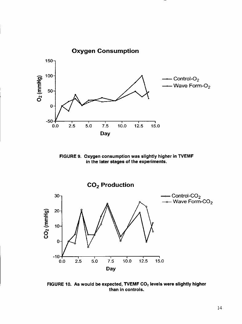
Oxygen Consumption
100- m - Control-02 E -Wave Form-02 I
E 50- v
6 0-
-50 I I I I I 1
0.0 2.5 5.0 7.5 10.0 12.5 15.0
Day
FIGURE 9. Oxygen consumption was slightly higher in N E M F in the later stages of the experiments.
COz Production
301 - Control-C02 --o- Wave Form-C02
1
0.0 2.5 5.0 7.5 10.0 12.5 15.0
FIGURE 10. As would be expected, TVEMF C02 levels were slightly higher than in controls.
14
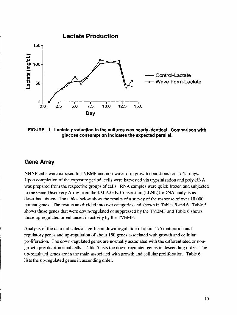
Lactate Production
5- z Y F loo - Control-Lactate
-Wave Form-Lactate
Q, m 2 50 -I
w .c,
0 0.0 2.5 5.0 7.5 10.0 12.5 15.0
Day
FIGURE 11. Lactate production in the cultures was nearly identical. Comparison with glucose consumption indicates the expected parallel.
Gene Array
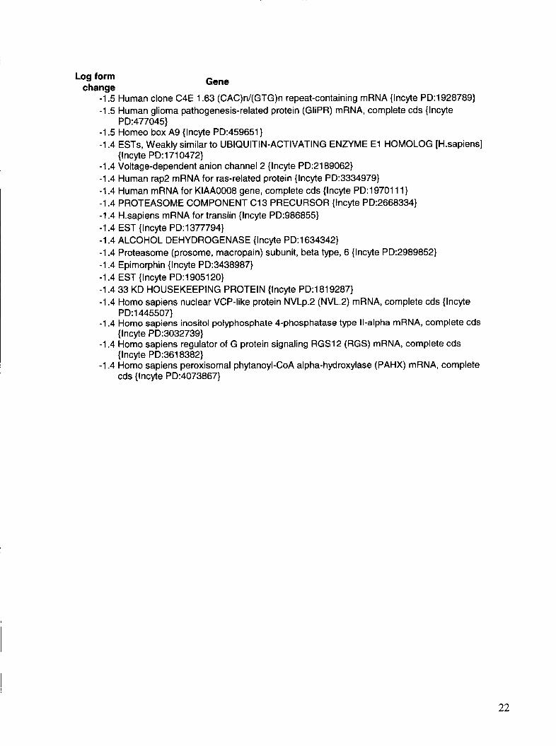
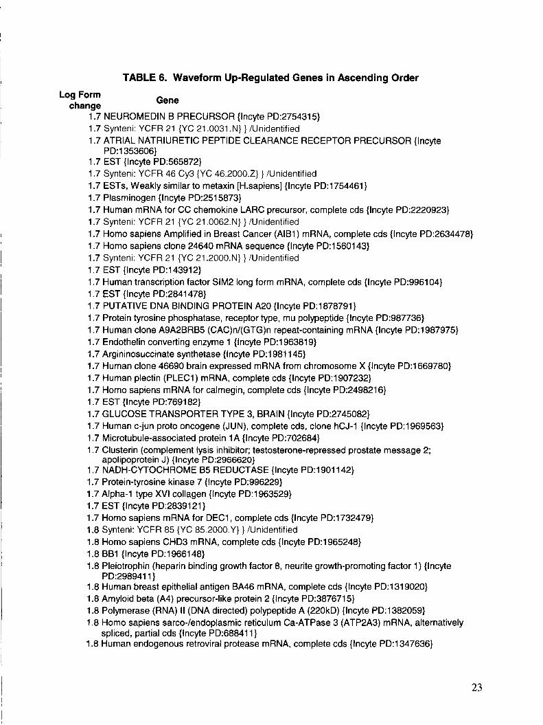
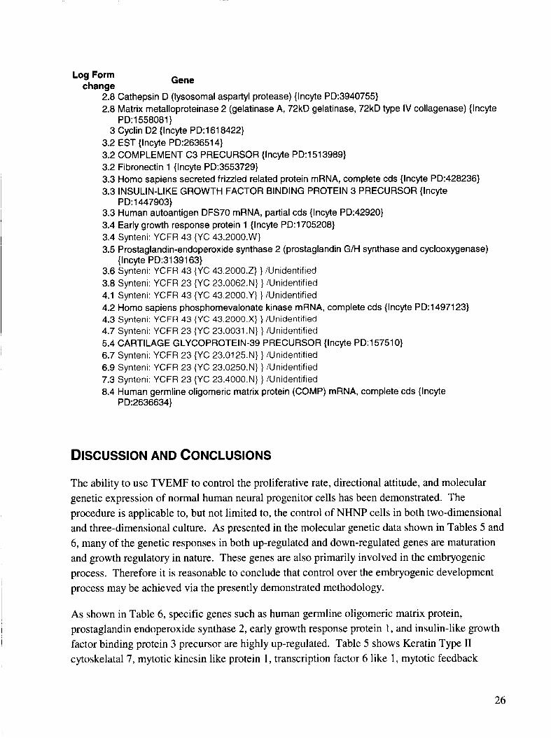
NHNP cells were exposed to TVEMF and non-waveform growth conditions for 17-21 days. Upon completion of the exposure period, cells were harvested via trypsinization and poly-RNA was prepared from the respective groups of cells. RNA samples were quick frozen and subjected to the Gene Discovery Array from the I.M.A.G.E. Consortium (LLNL) 1 cDNA analysis as described above. The tables below show the results of a survey of the response of over 10,000 human genes. The results are divided into two categories and shown in Tables 5 and 6. Table 5 shows those genes that were down-regulated or suppressed by the TVEMF and Table 6 shows those up-regulated or enhanced in activity by the TVEMF.
Analysis of the data indicates a significant down-regulation of about 175 maturation and regulatory genes and up-regulation of about 150 genes associated with growth and cellular proliferation. The down-regulated genes are normally associated with the differentiated or non- growth profile of normal cells. Table 5 lists the down-regulated genes in descending order. The up-regulated genes are in the main associated with growth and cellular proliferation. Table 6 lists the up-regulated genes in ascending order.
15
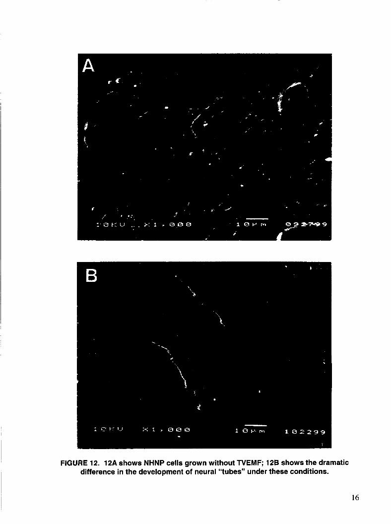
FIGURE 12. 12A shows NHNP cells grown without TVEMF; 128 shows the dramatic difference in the development of neural “tubes” under these conditions.
16
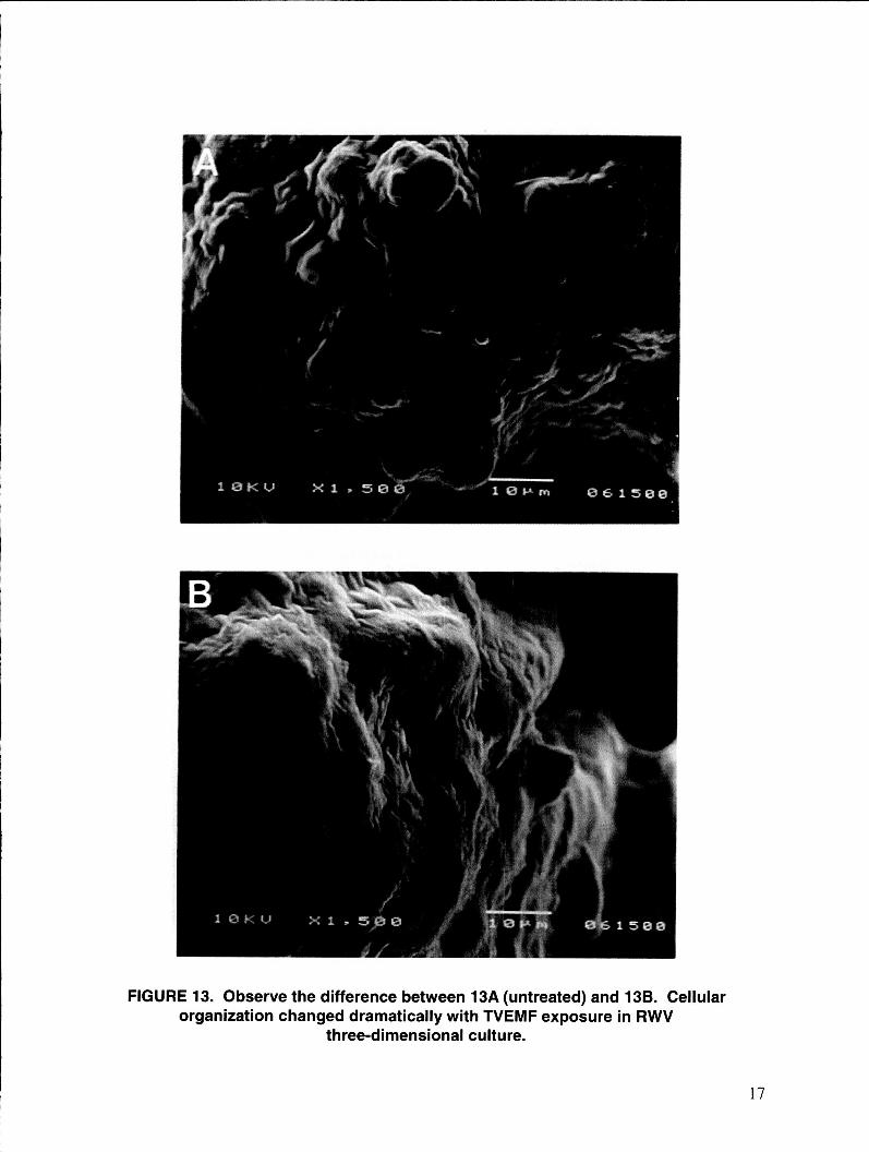
FIGURE 13. Observe the difference between 13A (untreated) and 13B. Cellular organization changed dramatically with TVEMF exposure in RWV
three-dimensional culture.
17
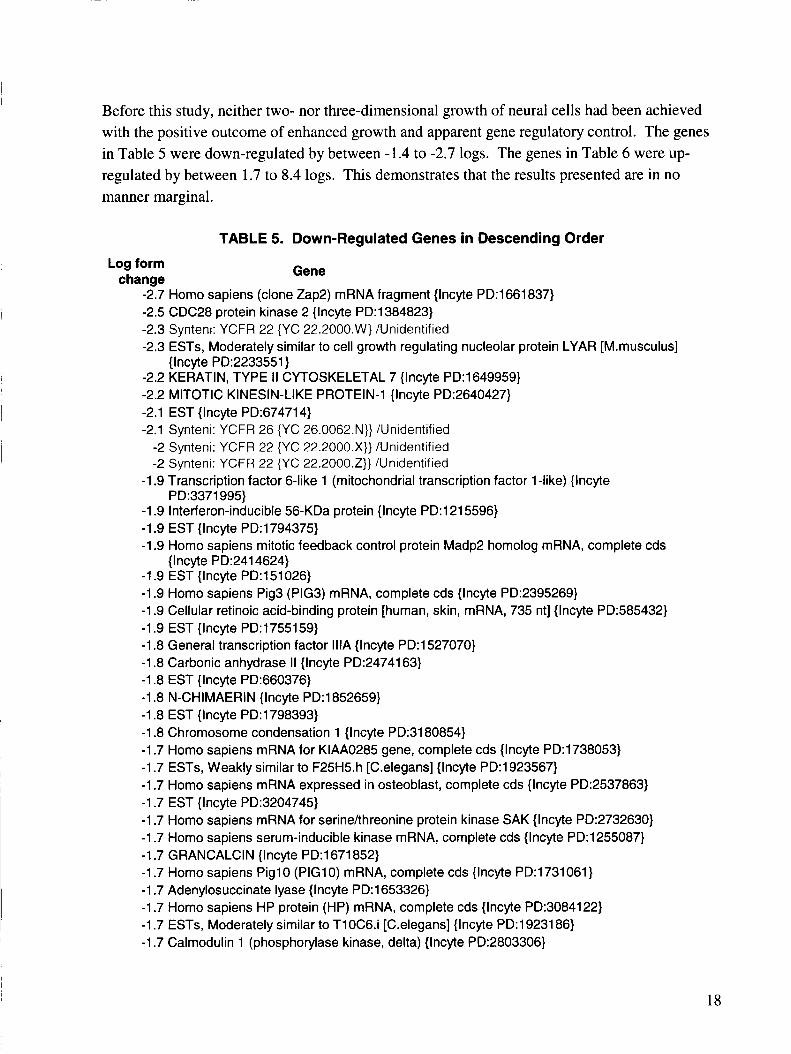
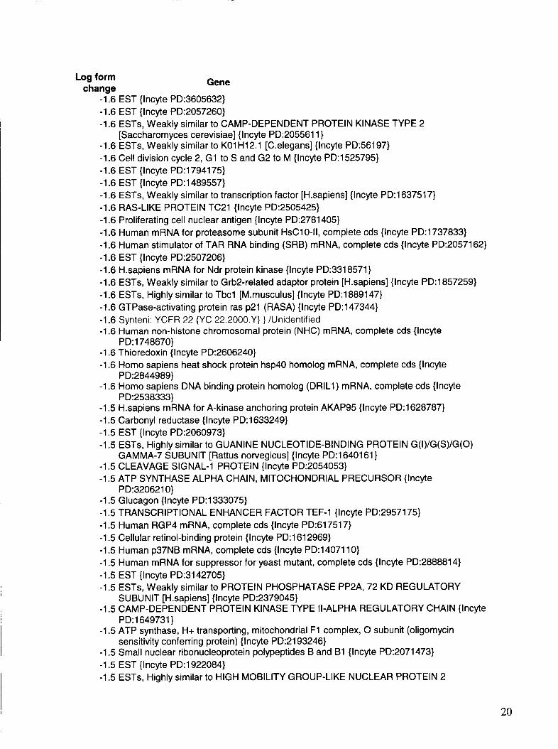
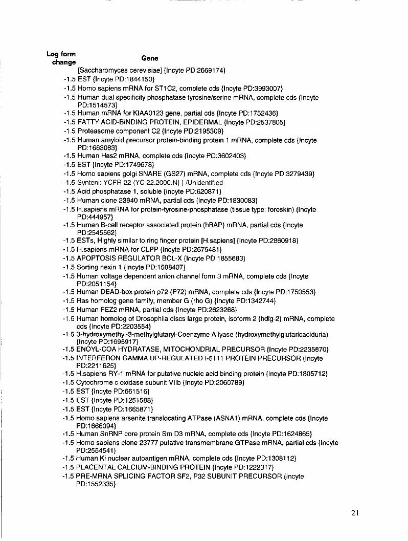
Before this study, neither two- nor three-dimensional growth of neural cells had been achieved with the positive outcome of enhanced growth and apparent gene regulatory control. The genes in Table 5 were down-regulated by between -1.4 to -2.7 logs. The genes in Table 6 were up- regulated by between 1.7 to 8.4 logs. This demonstrates that the results presented are in no manner marginal.
Log form chanae
TABLE 5. Down-Regulated Genes in Descending Order
Gene
-2.7 Homo sapiens (clone Zap2) mRNA fragment {Incyte PD:1661837} -2.5 CDC28 protein kinase 2 {Incyte PD:1384823} -2.3 Synteni: YCFR 22 {YC 22.2000.Wj /Unidentified -2.3 ESTs, Moderately similar to cell growth regulating nucleolar protein LYAR [M.musculus]
-2.2 KERATIN, TYPE I 1 CYTOSKELETAL 7 {Incyte PD:1649959} -2.2 MITOTIC KINESIN-LIKE PROTEIN-1 {lncyte PD:2640427} -2.1 EST {lncyte PD:674714} -2.1 Synteni: YCFR 26 {YC 26.0062.N}} /Unidentified
-2 Synteni: YCFR 22 {YC 22.2000.X}} /Unidentified -2 Synteni: YCFR 22 {YC 22.2000.Z}} /Unidentified
{lncyte PD:2233551}
-1.9 Transcription factor 6-like 1 (mitochondrial transcription factor 1 -like) {Incyte
-1.9 Interferon-inducible 56-KDa protein {Incyte PD:1215596} -1.9 EST {Incyte PD:1794375} -1.9 Homo sapiens mitotic feedback control protein Madp2 homolog mRNA, complete cds
{lncyte PD:2414624} -1.9 EST {Incyte PD:151026} -1.9 Homo sapiens Pig3 (PIG3) mRNA, complete cds {lncyte PD:2395269} -1.9 Cellular retinoic acid-binding protein [human, skin, mRNA, 735 nt] {Incyte PD:585432} -1.9 EST {lncyte PD:1755159} -1.8 General transcription factor IllA {lncyte PD:1527070} -1.8 Carbonic anhydrase I1 {Incyte PD:2474163} -1.8 EST {lncyte PD:660376} -1.8 N-CHIMAERIN {lncyte PD:1852659} -1.8 EST {lncyte PD:1798393} -1.8 Chromosome condensation 1 {Incyte PD:3180854} -1.7 Homo sapiens mRNA for KIAA0285 gene, complete cds {lncyte PD:1738053} -1.7 ESTs, Weakly similar to F25H5.h [C.elegans] {lncyte PD:1923567} -1.7 Homo sapiens mRNA expressed in osteoblast, complete cds {Incyte PD:2537863} -1.7 EST {Incyte PD:3204745} -1.7 Homo sapiens mRNA for serinehhreonine protein kinase SAK {Incyte PD:2732630} -1.7 Homo sapiens serum-inducible kinase mRNA, complete cds {Incyte PD:1255087} -1.7 GRANCALCIN {Incyte PD:1671852} -1.7 Homo sapiens Pig1 0 (PIG1 0) mRNA, complete cds {Incyte PD:1731061} -1.7 Adenylosuccinate lyase {lncyte PD:1653326} -1.7 Homo sapiens HP protein (HP) mRNA, complete cds {Incyte PD:3084122} -1.7 ESTs, Moderately similar to T10C6.i [C.elegans] {Incyte PD:1923186} -1.7 Calmodulin 1 (phosphorylase kinase, delta) {Incyte PD:2803306}
PD:3371995}
18
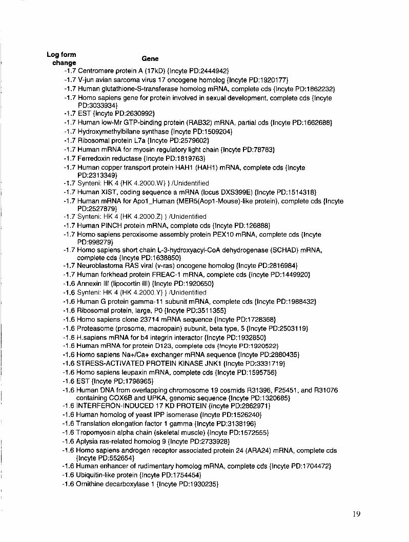
Log form change Gene
-1.7 Centromere protein A (17kD) {Incyte PD:2444942} -1.7 V-jun avian sarcoma virus 17 oncogene homolog {lncyte PD:1920177} -1.7 Human glutathione-S-transferase homolog mRNA, complete cds { lncyte PD:1862232} -1.7 Homo sapiens gene for protein involved in sexual development, complete cds {Incyte
-1.7 EST {lncyte PD:2630992} -1.7 Human low-Mr GTP-binding protein (RAB32) mRNA, partial cds {lncyte PD:1662688} -1.7 Hydroxymethylbilane synthase {lncyte PD:1509204} -1.7 Ribosomal protein L7a {Incyte PD:2579602} -1.7 Human mRNA for myosin regulatory light chain {Incyte PD:78783} -1.7 Ferredoxin reductase {Incyte PD:1819763} -1.7 Human copper transport protein HAH1 (HAH1) mRNA, complete cds {lncyte
-1.7 Synteni: HK 4 {HK 4.2000.W) } /Unidentified -1.7 Human XIST, coding sequence a mRNA (locus DXS399E) {lncyte PD:1514318} -1.7 Human mRNA for Apol-Human (MER5(Aopl -Mouse)-like protein), complete cds {Incyte
-1.7 Synteni: HK 4 {HK 4.2000.2) } /Unidentified -1.7 Human PINCH protein mRNA, complete cds {Incyte PD:126888} -1.7 Homo sapiens peroxisome assembly protein PEX10 mRNA, complete cds {Incyte
-1.7 Homo sapiens short chain L-3-hydroxyacyl-CoA dehydrogenase (SCHAD) mRNA,
-1.7 Neuroblastoma RAS viral (v-ras) oncogene homolog {Incyte PD:2816984} -1.7 Human forkhead protein FREAC-1 mRNA, complete cds {Incyte PD:1449920} -1.6 Annexin Ill (lipocortin Ill) {Incyte PD:1920650} -1.6 Synteni: HK 4 {HK 4.2000.Y) } /Unidentified -1.6 Human G protein gamma-11 subunit mRNA, complete cds {Incyte PD:1988432} -1.6 Ribosomal protein, large, PO {Incyte PD:3511355} -1.6 Homo sapiens clone 2371 4 mRNA sequence {Incyte PD:1728368} -1.6 Proteasome (prosome, macropain) subunit, beta type, 5 {lncyte PD:2503119} -1.6 Hsapiens mRNA for b4 integrin interactor {Incyte PD:1932850} -1.6 Human mRNA for protein D123, complete cds {Incyte PD:1920522} -1.6 Homo sapiens Na+/Ca+ exchanger mRNA sequence {Incyte PD:2880435} -1.6 STRESS-ACTIVATED PROTEIN KINASE JNK1 {lncyte PD:3331719} -1.6 Homo sapiens leupaxin mRNA, complete cds {Incyte PD:l595756} -1.6 EST {Incyte PD:1798965} -1.6 Human DNA from overlapping chromosome 19 cosmids R31396, F25451, and R31076
-1.6 INTERFERON-INDUCED 17 KD PROTEIN {Incyte PD:2862971} -1.6 Human homolog of yeast IPP isomerase {lncyte PD:1526240} -1.6 Translation elongation factor 1 gamma {Incyte PD:3138196} -1.6 Tropomyosin alpha chain (skeletal muscle) {Incyte PD:1572555} -1.6 Aplysia ras-related homolog 9 {lncyte PD:2733928} -1.6 Homo sapiens androgen receptor associated protein 24 (ARA24) mRNA, complete cds
-1.6 Human enhancer of rudimentary homolog mRNA, complete cds {Incyte PD:1704472} -1.6 Ubiquitin-like protein {Incyte PD:1754454} -1.6 Ornithine decarboxylase 1 {Incyte PD:1930235}
P D: 3033934)
PD:2313349}
PD:2527879}
PD:998279}
complete cds {lncyte PD:1638850}
containing COX6B and UPKA, genomic sequence {lncyte PD:l320685}
{Incyte PD:552654}
19
Log form change Gene
-1.6 EST {lncyte PD:3605632} -1.6 EST {Incyte PD:2057260} -1.6 ESTs, Weakly similar to CAMP-DEPENDENT PROTEIN KINASE TYPE 2
-1.6 ESTs, Weakly similar to KO1 H12.1 [C.elegans] {Incyte PD:56197} -1.6 Cell division cycle 2, G1 to S and G2 to M {Incyte PD:1525795} -1.6 EST {Incyte PD:1794175} -1.6 EST {Incyte PD:1489557} -1.6 ESTs, Weakly similar to transcription factor [H.sapiens] {Incyte PD:1637517} -1.6 RAS-LIKE PROTEIN TC21 {Incyte PD:2505425} -1.6 Proliferating cell nuclear antigen {Incyte PD:2781405} -1.6 Human mRNA for proteasome subunit HsC10-11, complete cds {lncyte PD:1737833} -1.6 Human stimulator of TAR RNA binding (SRB) mRNA, complete cds {lncyte PD:2057162) -1.6 EST {lncyte PD:2507206} -1.6 H.sapiens mRNA for Ndr protein kinase {lncyte PD:3318571} -1.6 ESTs, Weakly similar to Grb2-related adaptor protein [H.sapiens] {lncyte PD:1857259} -1.6 ESTs, Highly similar to Tbcl [M.musculus] {lncyte PD:1889147} -1.6 GTPase-activating protein ras p21 (RASA) {lncyte PD:147344} -1.6 Synteni: YCFR 22 {YC 22.2000.Y) } /Unidentified -1.6 Human non-histone chromosomal protein (NHC) mRNA, complete cds {Incyte
-1.6 Thioredoxin {Incyte PD:2606240} -1.6 Homo sapiens heat shock protein hsp40 homolog mRNA, complete cds {Incyte
-1.6 Homo sapiens DNA binding protein homolog (DRIL1) mRNA, complete cds {Incyte
-1.5 H.sapiens mRNA for A-kinase anchoring protein AKAP95 {Incyte PD:1628787} -1.5 Carbonyl reductase {Incyte PD:1633249} -1.5 EST {Incyte PD:2060973} -1.5 ESTs, Highly similar to GUANINE NUCLEOTIDE-BINDING PROTEIN G(I)/G(S)/G(O)
-1.5 CLEAVAGE SIGNAL-1 PROTEIN {lncyte PD:2054053} -1.5 ATP SYNTHASE ALPHA CHAIN, MITOCHONDRIAL PRECURSOR {Incyte
-1.5 Glucagon {Incyte PD:1333075} -1.5 TRANSCRIPTIONAL ENHANCER FACTOR TEF-1 {Incyte PD:2957175} -1.5 Human RGP4 mRNA, complete cds {lncyte PD:617517} -1.5 Cellular retinol-binding protein {lncyte PD:1612969} -1.5 Human p37NB mRNA, complete cds {Incyte PD:1407110} -1.5 Human mRNA for suppressor for yeast mutant, complete cds {Incyte PD:2888814} -1.5 EST {Incyte PD:3142705} -1.5 ESTs, Weakly similar to PROTEIN PHOSPHATASE PP2A, 72 KD REGULATORY
-1.5 CAMP-DEPENDENT PROTEIN KINASE TYPE II-ALPHA REGULATORY CHAIN {Incyte
-1.5 ATP synthase, H+ transporting, mitochondrial F1 complex, 0 subunit (oligomycin
-1.5 Small nuclear ribonucleoprotein polypeptides B and B1 {Incyte PD:2071473} -1.5 EST {Incyte PD:1922084} -1.5 ESTs, Highly similar to HIGH MOBILITY GROUP-LIKE NUCLEAR PROTEIN 2
[Saccharomyces cerevisiae] {Incyte PD:2055611}
PD:1748670}
PD:2844989}
PD:2538333}
GAMMA-7 SUBUNIT [Rattus norvegicus] {Incyte PD:1640161}
PD:3206210}
SUBUNIT [H.sapiens] {lncyte PD:2379045}
PD:1649731}
sensitivity conferring protein) {Incyte PD:2193246}
20
Log form change Gene
- [Saccharomyces cerevisiae] {Incyte PD:2669174}
-1.5 EST {Incyte PD:1844150} -1.5 Homo sapiens mRNA for ST1 C2, complete cds {Incyte PD:3993007} -1.5 Human dual specificity phosphatase tyrosine/serine mRNA, complete cds {Incyte
-1.5 Human mRNA for KIAAO123 gene, partial cds {Incyte PD:1752436} -1.5 FATTY ACID-BINDING PROTEIN, EPIDERMAL {Incyte PD:2537805} -1.5 Proteasome component C2 {Incyte PD:2195309} -1.5 Human amyloid precursor protein-binding protein 1 mRNA, complete cds {Incyte
-1.5 Human Has2 mRNA, complete cds {lncyte PD:3602403} -1.5 EST {Incyte PD:1749678} -1.5 Homo sapiens golgi SNARE (GS27) mRNA, complete cds {Incyte PD:3279439} -1.5 Synteni: YCFR 22 {YC 22.2000.Nj } /Unidentified -1.5 Acid phosphatase 1, soluble {Incyte PD:620871} -1.5 Human clone 23840 mRNA, partial cds {Incyte PD:1830083} -1.5 H.sapiens mRNA for protein-tyrosine-phosphatase (tissue type: foreskin) {Incyte
-1.5 Human B-cell receptor associated protein (hBAP) mRNA, partial cds {lncyte
-1.5 ESTs, Highly similar to ring finger protein [H.sapiens] {lncyte PD:2860918} -1.5 H.sapiens mRNA for CLPP {lncyte PD:2675481} -1.5 APOPTOSIS REGULATOR BCL-X {lncyte PD:1855683} -1.5 Sorting nexin 1 {Incyte PD:1508407} -1.5 Human voltage dependent anion channel form 3 mRNA, complete cds {lncyte
-1.5 Human DEAD-box protein p72 (P72) mRNA, complete cds {Incyte PD:1750553} -1.5 Ras homolog gene family, member G (rho G) {lncyte PD:1342744} -1.5 Human FEZ2 mRNA, partial cds {Incyte PD:2623268} -1.5 Human homolog of Drosophila discs large protein, isoform 2 (hdlg-2) mRNA, complete
-1.5 3-hydroxymethyl-3-methylglutaryl-Coenzyme A lyase (hydroxymethylglutaricaciduria)
-1.5 ENOYL-COA HYDRATASE, MITOCHONDRIAL PRECURSOR {Incyte PD:2235870} -1.5 INTERFERON GAMMA UP-REGULATED 1-51 11 PROTEIN PRECURSOR {Incyte
-1.5 H.sapiens RY-1 mRNA for putative nucleic acid binding protein {lncyte PD:1805712} -1.5 Cytochrome c oxidase subunit Vllb {Incyte PD:2060789} -1.5 EST {Incyte PD:661516} -1.5 EST {Incyte PD:1251588} -1.5 EST {Incyte PD:1665871} -1.5 Homo sapiens arsenite translocating ATPase (ASNA1) mRNA, complete cds {lncyte
-1.5 Human SnRNP core protein Sm D3 mRNA, complete cds {lncyte PD:1624865} -1.5 Homo sapiens clone 23777 putative transmembrane GTPase mRNA, partial cds {Incyte
-1.5 Human Ki nuclear autoantigen mRNA, complete cds {Incyte PD:1308112} -1.5 PLACENTAL CALCIUM-BINDING PROTEIN {Incyte PD:1222317} -1.5 PRE-MRNA SPLICING FACTOR SF2, P32 SUBUNIT PRECURSOR {Incyte
PD:1514573}
PD:1663083}
PD:444957}
PD:2545562}
PD:2051154}
cds {lncyte PD:2203554}
{Incyte PD:1695917}
PD:2211625}
PD:1666094}
PD:2554541}
PD:1552335}
21
Gene Log form change
-1.5 Human clone C4E 1.63 (CAC)n/(GTG)n repeat-containing mRNA {lncyte PD:1928789} -1.5 Human glioma pathogenesis-related protein (GliPR) mRNA, complete cds {lncyte
-1.5 Homeo box A9 {Incyte PD:459651} -1.4 ESTs, Weakly similar to UBIQUITIN-ACTIVATING ENZYME E l HOMOLOG [H.sapiens]
-1.4 Voltage-dependent anion channel 2 {lncyte PD:2189062} -1.4 Human rap2 mRNA for ras-related protein {Incyte PD:3334979} -1.4 Human mRNA for KIAA0008 gene, complete cds {Incyte PD:1970111} -1.4 PROTEASOME COMPONENT C13 PRECURSOR {Incyte PD:2668334} -1.4 Hsapiens mRNA for translin {lncyte PD:986855} -1.4 EST {Incyte PD:1377794} -1.4 ALCOHOL DEHYDROGENASE {Incyte PD:1634342} -1.4 Proteasome (prosome, macropain) subunit, beta type, 6 {lncyte PD:2989852} -1.4 Epimorphin {Incyte PD:3438987} -1.4 EST {Incyte PD:1905120} -1.4 33 KD HOUSEKEEPING PROTEIN {Incyte PD:1819287} -1.4 Homo sapiens nuclear VCP-like protein NVLp.2 (NVL.2) mRNA, complete cds {Incyte
-1.4 Homo sapiens inositol polyphosphate 4-phosphatase type Il-alpha mRNA, complete cds
-1.4 Homo sapiens regulator of G protein signaling RGS12 (RGS) mRNA, complete cds
-1.4 Homo sapiens peroxisomal phytanoyl-CoA alpha-hydroxylase (PAHX) mRNA, complete
PD:477045}
{Incyte PD:1710472}
PD:1445507}
{lncyte PD:3032739}
{Incyte PD:3618382}
cds {lncyte PD:4073867}
22
Log Form chanae
TABLE 6. Waveform Up-Regulated Genes in Ascending Order
Gene 1.7 NEUROMEDIN B PRECURSOR {Incyte PD:2754315} 1.7 Synteni: YCFR 21 {YC 21.0031 .N} } /Unidentified 1.7 ATRIAL NATRIURETIC PEPTIDE CLEARANCE RECEPTOR PRECURSOR {Incyte
1.7 EST {lncyte PD:565872} 1.7 Synteni: YCFR 46 Cy3 {YC 46.2000.2) } /Unidentified 1.7 ESTs, Weakly similar to metaxin [H.sapiens] {Incyte PD:1754461} 1.7 Plasminogen {lncyte PD:2515873} 1.7 Human mRNA for CC chemokine LARC precursor, complete cds {lncyte PD:2220923} 1.7 Synteni: YCFR 21 {YC 21.0062.Nj } /Unidentified 1.7 Homo sapiens Amplified in Breast Cancer (AIB1) mRNA, complete cds {Incyte PD:2634478} 1.7 Homo sapiens clone 24640 mRNA sequence {lncyte PD:1560143} 1.7 Synteni: YCFR 21 {YC 21.2000.N) } /Unidentified 1.7 EST {Incyte PD:143912} 1.7 Human transcription factor SIM2 long form mRNA, complete cds {Incyte PD:996104} 1.7 EST {lncyte PD:2841478} 1.7 PUTATIVE DNA BINDING PROTEIN A20 {Incyte PD:1878791} 1.7 Protein tyrosine phosphatase, receptor type, mu polypeptide {Incyte PD:987736} 1.7 Human clone A9A2BRB5 (CAC)n/(GTG)n repeat-containing mRNA {Incyte PD:1987975} 1.7 Endothelin converting enzyme 1 {lncyte PD:1963819} 1.7 Argininosuccinate synthetase {Incyte PD:1981145} 1.7 Human clone 46690 brain expressed mRNA from chromosome X {Incyte PD:1669780} 1.7 Human plectin (PLECl) mRNA, complete cds {lncyte PD:1907232} 1.7 Homo sapiens mRNA for calmegin, complete cds {lncyte PD:2498216} 1.7 EST {Incyte PD:769182} 1.7 GLUCOSE TRANSPORTER TYPE 3, BRAIN {Incyte PD:2745082} 1.7 Human c-jun proto oncogene (JUN), complete cds, clone hCJ-1 {lncyte PD:1969563} 1.7 Microtubule-associated protein 1 A {lncyte PD:702684} 1.7 Clusterin (complement lysis inhibitor; testosterone-repressed prostate message 2;
1.7 NADH-CYTOCHROME 85 REDUCTASE {Incyte PD:1901142} 1.7 Protein-tyrosine kinase 7 {Incyte PD:996229} 1.7 Alpha-1 type XVI collagen {Incyte PD:1963529} 1.7 EST {Incyte PD:2839121} 1.7 Homo sapiens mRNA for DEC1, complete cds {lncyte PD:1732479} 1.8 Synteni: YCFR 85 {YC 85.2000.Y} } /Unidentified 1.8 Homo sapiens CHD3 mRNA, complete cds {Incyte PD:1965248} 1.8 BB1 {lncyte PD:1966148} 1.8 Pleiotrophin (heparin binding growth factor 8, neurite growth-promoting factor 1) {Incyte
1.8 Human breast epithelial antigen BA46 mRNA, complete cds {Incyte PD:1319020} 1.8 Amyloid beta (A4) precursor-like protein 2 {Incyte PD:3876715} 1.8 Polymerase (RNA) II (DNA directed) polypeptide A (220kD) {lncyte PD:1382059} 1.8 Homo sapiens sarco-/endoplasmic reticulum Ca-ATPase 3 (ATP2A3) mRNA, alternatively
1.8 Human endogenous retroviral protease mRNA, complete cds {Incyte PD:1347636}
PD:1353606}
apolipoprotein J) {lncyte PD:2966620}
PD:2989411}
spliced, partial cds {Incyte PD:688411}
23
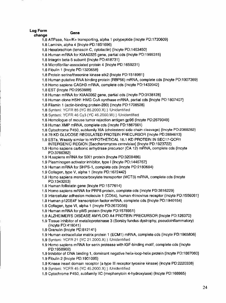
Log Form change Gene
1.8 ATPase, Na+/K+ transporting, alpha 1 polypeptide {lncyte PD:1730609} 1.8 Laminin, alpha 4 {Incyte PD:1851696} 1.8 Hexabrachion (tenascin C, cytotactin) {Incyte PD:1453450} 1.8 Human mRNA for KIAA0325 gene, partial cds {Incyte PD:1995315} 1.8 lntegrin beta-5 subunit {lncyte PD:418731} 1.8 Microfibrillar-associated protein 4 {lncyte PD:1659231} 1.8 Fibulin 1 {Incyte PD:1320658} 1.8 Protein serinehhreonine kinase stk2 {Incyte PD:1518981} 1.8 Human putative RNA binding protein (RBP56) mRNA, complete cds {Incyte PD:1907369} 1.8 Homo sapiens CAGH3 mRNA, complete cds {Incyte PD:1432042} 1.8 EST {Incyte PD:2953888} 1.8 Human mRNA for KIAA0062 gene, partial cds {lncyte PD:3138128} 1.8 Human clone HSH1 HMG CoA synthase mRNA, partial cds {Incyte PD:l807407} 1.8 Filamin 1 (actin-binding protein-280) {lncyte PD:1708528} 1.8 Synteni: YCFR 85 {YC 85.2000.Xj } /Unidentified 1.8 Synteni: YCFR 46 Cy3 {YC 46.2000.W) } /Unidentified 1.8 Homologue of mouse tumor rejection antigen gp96 {Incyte PD:2679349} 1.8 Human XMP mRNA, complete cds {lncyte PD:1887661} 1.8 Cytochrome P450, subfamily XIA (cholesterol side chain cleavage) {Incyte PD:2368282} 1.8 78 KD GLUCOSE REGULATED PROTEIN PRECURSOR {lncyte PD:2884613} 1.9 ESTs, Weakly similar to HYPOTHETICAL 16.1 KD PROTEIN IN SEC17-QCR1
INTERGENIC REGION [Saccharomyces cerevisiae] {Incyte PD:1923722} 1.9 Homo sapiens carbonic anhydrase precursor (CA 12) mRNA, complete cds {Incyte
PD:3766382} 1.9 H.sapiens mRNA for SIX1 protein {Incyte PD:3208486} 1.9 Plasminogen activator inhibitor, type I {lncyte PD:1445767} 1.9 Human mRNA for SHPS-1, complete cds {Incyte PD:2180684} 1.9 Collagen, type V, alpha 1 {lncyte PD:1672442} 1.9 Homo sapiens monocarboxylate transporter (MCT3) mRNA, complete cds {Incyte
1.9 Human follistatin gene {Incyte PD:1577614} 1.9 Homo sapiens mRNA for PRP8 protein, complete cds {Incyte PD:3616229} 1.9 Intercellular adhesion molecule 1 (CD54), human rhinovirus receptor {Incyte PD:1556061} 1.9 Human p120E4F transcription factor mRNA, complete cds {Incyte PD:1940164} 1.9 Collagen, type VI, alpha 1 {Incyte PD:2672056} 1.9 Human mRNA for pM5 protein {Incyte PD:l578951} 1.9 ALZHEIMER'S DISEASE AMYLOID A4 PROTEIN PRECURSOR {lncyte PD:126370} 1.9 Tissue inhibitor of metalloproteinase 3 (Sorsby fundus dystrophy, pseudoinflammatory)
1.9 Granulin {Incyte PD:812141} 1.9 Human extracellular matrix protein 1 (ECM1) mRNA, complete cds {Incyte PD:1965806} 1.9 Synteni: YCFR 21 {YC 21.2000.X) } /Unidentified 1.9 Homo sapiens mRNA for serin protease with IGF-binding motif, complete cds {lncyte
1.9 Inhibitor of DNA binding 1, dominant negative helix-loop-helix protein {Incyte PD:1687060} 1.9 Fibulin 2 {Incyte PD:1901095} 1.9 Kinase insert domain receptor (a type Ill receptor tyrosine kinase) {lncyte PD:2220338} 1.9 Synteni: YCFR 45 {YC 45.2000.Xj } /Unidentified 1.9 Cytochrome P450, subfamily IIC (mephenytoin 4-hydroxylase) {lncyte PD:168865}
PD:1343253}
{Incyte PD:418041}
PD:l958902}
24
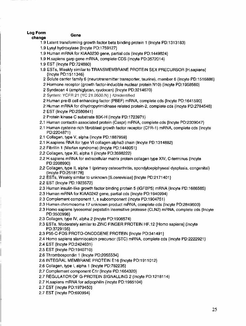
Log Form change Gene
1.9 Latent transforming growth factor beta binding protein 1 {Incyte PD:1313183} 1.9 Lysyl hydroxylase {Incyte PD:1759127} 1.9 Human mRNA for KIAA0230 gene, partial cds {Incyte PD:1449824} 1.9 H.sapiens garp gene mRNA, complete CDS {Incyte PD:3572014} 1.9 EST {Incyte PD:724880} 1.9 ESTs, Weakly similar to TRANSMEMBRANE PROTEIN SEX PRECURSOR [Hsapiens]
{lncyte PD:1511346} 2 Solute carrier family 6 (neurotransmitter transporter, taurine), member 6 {lncyte PD:1516886} 2 Hormone receptor (growth factor-inducible nuclear protein N10) {Incyte PD:1958560} 2 Syndecan 4 (amphiglycan, ryudocan) {Incyte PD:3214670} 2 Synteni: YCFR 21 {YC 21.0500.Nj } /Unidentified 2 Human pre-B cell enhancing factor (PBEF) mRNA, complete cds {Incyte PD:1641590} 2 Human mRNA for dihydropyrimidinase related protein-2, complete cds {Incyte PD:2784546} 2 EST {lncyte PD:2580841} 2 Protein kinase C substrate 80K-H {lncyte PD:1723971}
2.1 Human contactin associated protein (Caspr) mRNA, complete cds {Incyte PD:2309047} 2.1 Human cysteine-rich fibroblast growth factor receptor (CFR-1) mRNA, complete cds {Incyte
2.1 Collagen, type V, alpha {Incyte PD:1887959} 2.1 H.sapiens RNA for type VI collagen alpha3 chain {lncyte PD:1314882} 2.2 Fibrillin 1 (Marfan syndrome) {Incyte PD:1448051} 2.2 Collagen, type XI, alpha 1 {Incyte PD:3598222} 2.2 H.sapiens mRNA for extracellular matrix protein collagen type XIV, C-terminus {Incyte
PD:2208990} 2.2 Collagen, type II, alpha 1 (primary osteoarthritis, spondyloepiphyseal dysplasia, congenital)
{lncyte PD:2518178} 2.2 ESTs, Weakly similar to unknown [S.cerevisiae] {Incyte PD:2171401} 2.2 EST {Incyte PD:1923572} 2.3 Human insulin-like growth factor binding protein 5 (IGFBP5) mRNA {Incyte PD:1686585} 2.3 Human mRNA for KIAA0242 gene, partial cds {Incyte PD:1940994} 2.3 Complement component 1, s subcomponent {Incyte PD:1904751} 2.3 Human chromosome 17 unknown product mRNA, complete cds {Incyte PD:2849603} 2.3 Homo sapiens lysosomal pepstatin insensitive protease (CLN2) mRNA, complete cds { lncyte
2.3 Collagen, type IV, alpha 2 {lncyte PD:1906574} 2.3 ESTs, Moderately similar to ZINC FINGER PROTEIN HF.12 [Homo sapiens] {Incyte
2.3 P55-C-FOS PROTO-ONCOGENE PROTEIN {Incyte PD:341491} 2.4 Homo sapiens stanniocalcin precursor (STC) mRNA, complete cds {Incyte PD:2222921} 2.4 EST {Incyte PD:2424631} 2.5 EST {lncyte PD:1940710} 2.6 Thrombospondin 1 {Incyte PD:2055534} 2.6 INTEGRAL MEMBRANE PROTEIN E16 {lncyte PD:1911012} 2.6 Collagen, type I, alpha 1 {Incyte PD:782235} 2.7 Complement component C1 r {Incyte PD:1664320} 2.7 REGULATOR OF G-PROTEIN SIGNALLING 2 {lncyte PD:1218114} 2.7 H.sapiens mRNA for adipophilin {Incyte PD:1985104} 2.7 EST {Incyte PD:1979450} 2.7 EST {lncyte PD:690994}
PD:2204871}
PD:3500996}
PD:3729155}
25
Log Form change
2.8 2.8
3 3.2 3.2 3.2 3.3 3.3
3.3 3.4 3.4 3.5
3.6 3.8 4.1 4.2 4.3 4.7
Gene
Cathepsin D (lysosomal aspartyl protease) { lncyte PD:3940755} Matrix metalloproteinase 2 (gelatinase A, 72kD gelatinase, 72kD type IV collagenase) {Incyte PD:1558081} Cyclin D2 {Incyte PD:1618422} EST {Incyte PD:2636514} COMPLEMENT C3 PRECURSOR {Incyte PD:1513989} Fibronectin 1 {Incyte PD:3553729} Homo sapiens secreted frizzled related protein mRNA, complete cds {Incyte PD:428236} INSULIN-LIKE GROWTH FACTOR BINDING PROTEIN 3 PRECURSOR {Incyte PD:l447903} Human autoantigen DFS70 mRNA, partial cds {Incyte PD:42920} Early growth response protein 1 {lncyte PD:1705208} Synteni: YCFR 43 {YC 43.2000.W) Prostaglandin-endoperoxide synthase 2 (prostaglandin G/H synthase and cyclooxygenase) (Incyte PD:3139163} Synteni: YCFR 43 {YC 43.2000.2) } /Unidentified Synteni: YCFR 23 {YC 23.0062.N) } /Unidentified Synteni: YCFR 43 {YC 43.2000.Y) } /Unidentified Homo sapiens phosphomevalonate kinase mRNA, complete cds {Incyte PD:l497123} Synteni: YCFR 43 {YC 43.2000.X) } /Unidentified Synteni: YCFR 23 {YC 23.0031 .N} } /Unidentified
5.4 CARTILAGE GLYCOPROTEIN-39 PRECURSOR {Incyte PI315751 0) 6.7 Synteni: YCFR 23 {YC 23.0125.N) } /Unidentified 6.9 Synteni: YCFR 23 {YC 23.0250.N) } /Unidentified 7.3 Synteni: YCFR 23 {YC 23.4000.N) } /Unidentified 8.4 Human germline oligomeric matrix protein (COMP) mRNA, complete cds {lncyte
PD:2636634}
DISCUSSION AND CONCLUSIONS
The ability to use TVEMF to control the proliferative rate, directional attitude, and molecular genetic expression of normal human neural progenitor cells has been demonstrated. The procedure is applicable to, but not limited to, the control of NHNP cells in both two-dimensional and three-dimensional culture. As presented in the molecular genetic data shown in Tables 5 and 6 , many of the genetic responses in both up-regulated and down-regulated genes are maturation and growth regulatory in nature. These genes are also primarily involved in the embryogenic process. Therefore it is reasonable to conclude that control over the embryogenic development process may be achieved via the presently demonstrated methodology.
As shown in Table 6 , specific genes such as human germline oligomeric matrix protein, prostaglandin endoperoxide synthase 2, early growth response protein 1, and insulin-like growth factor binding protein 3 precursor are highly up-regulated. Table 5 shows Keratin Type II cytoskelatal7, mytotic kinesin like protein 1, transcription factor 6 like 1, mytotic feedback
26
control protein, and cellular retinoic acid binding protein are down-regulated. Each of these two sets is only an example from the approximately 320 genes changes expressed as a consequence of exposure to TVEMF.
There is significant precedent in the literature for the results reported above. Kepler et al. (1990) reported the effects of the neurons with oscillatory properties on the composite of neural networks. This work illustrates the likelihood that a pulse width modulated system might bring on specific responses in neural tissues. As previously discussed, Valentini et al. (1993) demonstrated the ability to enhance the outgrowth of neural fibers on materials that possess a weak electric charge. This would indicate that intense electric fields are not necessarily an essential component of this process, and that a weak and persistent stimulus might yield a measurable effect.
Additional evidence of the effects of magnetic fields exists in the work of Sandyk et al. (1992a). This communication details dramatic improvement of a patient with progressive degenerative multiple sclerosis. Briefly, the patient showed considerable improvement when subjected to treatment at a frequency of 2-7 Hz and an intensity of the magnetic field of 7.5 pic0 Tesla. These parameters marginally parallel those of this report. In a similar fashion, Sandyk et al. (1992b) reported significant improvement in patents treated with the same field strength and intensity.
The ability to suppress or stimulate the growth of non-excitable cells has been reported in mouse lymphoma cells by Lyte et al. (1991). A narrow range of electric field was found to be effective at one end to stimulate and at the other to inhibit the growth of these cells. These data might suggest cellular receptors in all cells. To sustain this notion, Briistle et al. (1996) reported the potential to use neural progenitors for recapitulation of neural tissues. As would be expected, this would require genetic control at the embryonic level. We believe this study indicates our ability to trigger these parametric events.
As is clearly demonstrated in the human body, the bioelectric, biochemical process of electrical nerve stimulation is a documented reality. The present investigation demonstrates that a similar phenomenon can be potentiated in a synthetic atmosphere, Le., two-dimensionally or in rotating wall cell culture vessels. One may use this electrical potentiation for a number of purposes, including developing tissues for transplantation, repairing traumatized tissues, and moderating some neurodegenerative diseases and perhaps controlling the degeneration of tissue as might be effected in a bioelectric stasis field.
27
REFERENCES
Aebischer, U.S. Patent 5,030,225.
Borgens RB, Roederer E, Cohen MJ (198 1): Enhanced spinal cord regeneration in lamprey by
Borgens RB (1986): The role of natural and applied electric fields in neuronal regeneration and
applied electric fields. Science 213(7):611-617.
development. In: Ionic Currents in Development (McGinnis and Vanable, eds.), Alan R. Liss (ed.) (New York), pp. 239-250.
Borgens RB, Blight AR, Murphy DJ, Stewart L (1986): Transected dorsal column axons within the guinea pig spinal cord regenerate in the presence of an applied electric field. J Comp Neurol250: 168- 180.
Borgens RB, Blight AR, Murphy DJ (1986): Axonal regeneration in spinal cord injury: A perspective and new technique. J Comp Neurol250: 157- 167.
hemisection in guinea pigs: The effects of applied electric fields. J Comp Neurol296:634- 653.
Borgens RB, Blight AR, McGinnis ME (1990): Functional recovery after spinal cord
Borgens RB, Shi R, Mohr TJ, Jaeger CB (1994): Mammalian cortical astrocytes align
Borgens RJ3, Bohnert DM (1997): The responses of mammalian spinal axons to an applied DC
Briistle 0, McKay RDG (1996): Neuronal progenitors as tools for cell replacement in the
themselves in a physiological voltage gradient. Exp. Neurol 128:4 1-49.
voltage gradient. Exp Neurol 145:376-389.
nervous system. Current Opinion in Neurobiol5:688-695.
Cogoli A and Gmiinder FK (199 1): Gravity effects on single cells. In Bonting SL (ed): Advances in Space Biology and Medicine. Vol 1, JAI Press.
rotation, Plant Physiol47:756764. Dedolph RR, Dipert MH (197 1): The physical basis of gravity nullification by clinostatic
Fukuda et al., U.S Patent 5,328,843.
Goodwin TJ, Jessup JM, Wolf DA (1992): Morphological Differentiation of Colon Carcinoma Cell Lines HT-29 and HT-29KM in Rotating-Wall Vessels. In Vitro Cell Dev Biol 28A, 47-60.
Goodwin TJ, Schroeder WF, Wolf DA, Moyer MP (1993a): Coculture of normal human small intestine cells in a rotating-wall vessel culture system. Proc. SOC. Exp Biol Med 202: 18 1 - 192.
Goodwin TJ, Prewett TL, Spaulding GF, et al. (1993b): A model for culture of primary breast
Goodwin TJ, Prewett TL, Wolf DA, et al. (1993~): Reduced shear stress: a major component in
carcinoma tissues in rotating wall vessels (rwv). Mol Biol Cell 4 Suppl, 15a.
the ability of mammalian tissues to form 3-dimensional assemblies in simulated microgravity. J Cell Biochem 51:301-3 11.
28
Goodwin TJ et al. (1997): Three-dimensional culture of a mixed mullerian tumor of the ovary:
Hinkle L, McCaig CD, Robinson KR ( 198 1): The direction of growth of differentiating neurones
Jessup JM, Brown K, Ishii S, et al. (1994): Adv Space Res 14(8):71-76.
Kepler TB, Marder E, Abbott LF (1990): The effect of electrical coupling on the frequency of
Lelkes PI, Unsworth BR (1997): Growing tissues in microgravity. Nature Med 4(8):901-907.
Lyte M, Gannon JE, O’clock GD Jr (1991): Effects of in vitro electrical stimulation on enhancement and suppression of malignant lymphoma cell proliferation. JNCI 83: 1 16- 1 19.
Makohliso SA, Valentini RF, Aebischer P (1993): Magnitude and polarity of a fluoroethylene propylene electret substrate charge influences neurite outgrowth in vitro. J Biomed Mat Res
expression of in vivo characteristics In Vitro Cell Dev BioZ - Anim 33:366-374.
and myoblasts from frog embryos in an applied electric field. J PhysioZ314: 121-135.
model neuronal oscillators. Science 24533-85.
27: 1075-1085.
Sandyk R (1992a): Clinical Case ReportAuccessful treatment of multiple sclerosis with
Prewett TL, Goodwin TJ, Spaulding GF (1993): Three-dimensional modeling of T-24 human
magnetic fields. J Neurosci 66:237-250.
bladder carcinoma cell line: A new simulated microgravity vessel. J Tissue Culture Methods Vol 15, Jan. ‘93.
Sandyk R (1992b): Magnetic fields in the therapy of Parkinsonism. J Neurosci 66:209-235.
Schwarz RP, et al. (1991a): U.S. Patent 4,988,623.
Schwarz RP, et al. (1991b): U.S. Patent 5,026,650.
Schwarz RP, Goodwin TJ, Wolf DA (1992): Cell culture for three-dimensional modeling in rotating-wall vessels: an application of microgravity. J Tiss Cult Meth 14,51-58.
Valentini RF, Vargo TG, Gardella JA Jr, Aebischer P (1992): Electrically charged polymeric substrates enhance nerve fibre outgrowth in vitro. Biomat 13(3): 183-190.
Valentini RF, Vargo TG, Gardella JA Jr, Aebischer P (1993): Patterned neuronal attachment and outgrowth on surface modified, electrically charged fluoropolymer substrates. J Biomat Sci Polymer Edn 5( 1/2): 13-36.
Zar JH ( 1974): Biostatistical Analysis. New Jersey: Prentice-Hall.
29
REPORT DOCUMENTATION PAGE
September 2003
1 ~ o m ~ p p r o v e d OM0 No. 0704-0188
NASA Technical Paper
Public reporting burden for this collection of information is estimated to average 1 hour per response, including the time for reviewing instructions, searching existing data sources, gathering and maintaining the data needed, and completing and reviewing the collection of information. Send comments regarding this burden estimate or any other aspect of this collection of information, including suaaestions for reducing this burden, to Washinaton Headauarters Services, Directorate for Information Operations and Reports, 1215 Jefferson Davis Highway. Suite 1204, Arlington. VA 222024302,
4. TITLE AND SUBTITLE Physiological and Molecular Genetic Effects of Time-Varying Electromagnetic Fields on Human Neuronal Cells
- . and-to the Office of Management and Budget. Paperwork Reduction Project (0704-0188). Washington, DC 20503
1. AGENCY USE ONLY (Leave Blank) I 2. REPORT DATE I 3. REPORT TYPE AND DATES COVERED
5. FUNDING NUMBERS
7. PERFORMING ORGANIZATION NAME(S) AND ADDRESS(ES)
Lyndon B. Johnson Space Center Houston, Texas 77058
6. AUTHOR(S) Thomas J. Goodwin, Ph.D.
8. PERFORMING ORGANIZATION
S-906 REPORT NUMBERS
12a. DISTRIBUTION/AVAILABlLlTY STATEMENT
UnclassifiedlUnlimited Available from the NASA Center for Aerospace Information (CASI) 7 12 1 Standard Hanover, MD 21076-1320 subject category 52
I
9. SPONSORlNGlMONlTORlNG AGENCY NAME(S) AND ADDRESS(ES) I 10. SPONSORlNGlMONlTORlNG
12b. DISTRIBUTION CODE
National Aeronautics and Space Administration Washington, DC 20546-0001
14. SUBJECT TERMS 15. NUMBEROF
Time-varying electromagnetic field, rotating wall vessel, three-dimensional culture, neural tissue regeneration
PAGES
37
AGENCY REPORT NUMBER TP-2003-2 12054
16. PRICE CODE
11. SUPPLEMENTARY NOTES
17. SECURITY CLASSIFICATION 18. SECURITY CLASSIFICATION 19. SECURITY CLASSIFICATION OF REPORT OF THIS PAGE OF ABSTRACT
Unclassified Unclassified Unclassified
20. LIMITATION OF ABSTRACT
Unlimited
I
13. ABSTRACT (Maximum 200 words) The present investigation details the development of model systems for growing two and three-dimensional human neural progenitor cells within a culture medium facilitated by a time-varying electromagnetic field (TVEMF). The cells and culture medium are contained within a two or three-dimensional culture vessel, and the electromagnetic field is emitted from an electrode or coil. These studies further provide methods to promote neural tissue regeneration by means of culturing the neural cells in either configuration. Grown in two-dimensions, neuronal cells extended longitudinally forming tissue strands extending axially along and within electrodes comprising electrically conductive channels or guides through which a time-varying electrical current was conducted. In the three-dimensional aspect exposure to TVEMF resulted in the development of three-dimensional aggregates, which emulated organized neural tissues. In both experimental configurations, the proliferation rate of the TVEMF cells was 2.5 to 4.0 times the rate of the non-waveform cells. Each of the experimental embodiments resulted in similar molecular genetic changes regarding the growth potential of the tissues as measured by gene chip analyses, which measured more than 10,000 human genes simultaneously