Embed Size (px)
Citation preview
Arabian Journal of Chemistry (2014) 7, 372–380
King Saud University
Arabian Journal of Chemistry
www.ksu.edu.sawww.sciencedirect.com
ORIGINAL ARTICLE
Physically crosslinked poly(vinyl alcohol)-
hydroxyethyl starch blend hydrogel membranes:
Synthesis and characterization for
biomedical applications
* Corresponding author. Tel.: +20 1283320302; fax: +20 34593414.
E-mail address: [email protected] (E.A. Kamoun).
Peer review under responsibility of King Saud University.
Production and hosting by Elsevier
1878-5352 ª 2013 Production and hosting by Elsevier B.V. on behalf of King Saud University.
http://dx.doi.org/10.1016/j.arabjc.2013.05.026
El-Refaie Kenawy a, Elbadawy A. Kamoun b,*, Mohamed S. Mohy Eldin b,
Mahmoud A. El-Meligya
a Department of Chemistry, Polymer Research Group, Faculty of Science, University of Tanta, Tanta 31527, Egyptb Polymer Materials Research Department, Advanced Technology & New Materials Research Institute (ATNMRI), City ofScientific Research and Technological Applications (SRTA-City), New Borg Al-Arab City, P.O. Box 21934, Alexandria, Egypt
Received 14 January 2013; accepted 29 May 2013Available online 7 June 2013
KEYWORDS
Poly(vinyl alcohol);
Hydroxyethyl starch;
Hydrogel membranes;
Freeze–thaw method;
Physicochemical properties;
Thermal properties
Abstract Poly(vinyl alcohol), PVA is a polymer of great importance because of its many appealing
characteristics specifically for various pharmaceutical and biomedical applications. Physically cross-
linked hydrogel membranes composed of different amounts of hydroxyethyl starch (HES) in (PVA)
and ampicillin were prepared by applying freeze–thawing method. This freezing–thawing cycle was
repeated for three consecutive cycles. Physicochemical properties of PVA–HES membrane gel such
as gel fraction, swelling, morphology, elongation, tensile strength, and protein adsorption were
investigated. Introducing HES into freeze–thawed PVA structure affected crystal size distribution
of PVA; and hence physicochemical properties and morphological structure have been affected.
Increased HES concentration decreased the gel fraction %, maximum strength and break elonga-
tion. Indeed it resulted into a significant incrementing of the swelling ability, amount of protein
adsorption, broader pore size, and pore distribution of membrane morphological structure. Fur-
thermore, an increase in HES concentration resulted in better and still lower thermal stability com-
pared to virgin PVA and freeze–thawed PVA. The maximum weight loss of PVA–HES hydrogel
membranes ranged between 18% and 60% according to HES content, after two days of degrada-
tion in phosphate buffer saline (PBS), which indicates they are biodegradable. Thus, PVA–HES
hydrogel membranes containing ampicillin could be a novel approach for biomedical application
e.g. wound dressing purposes.ª 2013 Production and hosting by Elsevier B.V. on behalf of King Saud University.
1. Introduction
PVA hydrogels have been previously used for numerous bio-
medical and pharmaceutical applications (Tanigami et al.,1995). PVA hydrogels have several advantages that make them
Physically crosslinked poly(vinyl alcohol)-hydroxyethyl starch blend hydrogel membranes: Synthesis and characterization 373
proper candidates for biomaterials. Some of these advantagesinclude their non-toxic, non-carcinogenic, biocompatible, bio-adhesive characteristics, excellent film-forming, excellent trans-
parency, and additionally their ease of processing. PVA has asimple chemical structure and its chemical modification is pos-sible using simple reaction as well. Meanwhile, PVA gels pos-
sess a high degree of swelling in water or biological fluids andan elastic or rubbery nature structure (Chan, 1999). Because oflatter advantages, PVA is capable of simulating natural tissues
and can be completely accepted into the body. PVA gels havebeen applied in different biomedical application sites such ascontact lenses, the lining for artificial hearts, wound dressing,and drug delivery applications. Peppas and Merrill (1977a,b)
have revealed in their earliest work in considering PVA hydro-gels as biomaterials. Generally, hydrogels were achieved bycrosslinking process of polymers, which may be done by a
chemical reaction (e.g. radical polymerization, chemical reac-tion of complementary groups, using high energy irradiation,or enzymatic reaction) or by physical reaction (e.g. ionic inter-
action, crystallization of the polymeric chain, hydrogen bondbetween chains, protein interaction, or design of amphiphilicblock and graft copolymers) (Hennink and Nostrum, 2002).
In recent decades, the need of physical crosslinked gels hasbeen potentially increased, (Van Tomme et al., 2005) to avoidthe use of chemical crosslinking agents and reagents. Theseagents are not only often toxic compounds which can be re-
moved or extracted from prepared gels before application,but also can affect the integrity of the substances when en-trapped (e.g. proteins, drugs, and cells). Therefore, the physical
crosslinking method has been chosen and preferred compara-ble with the chemical crosslinking method for most crosslinkedpolymers’ preparation. Several attempts have been done to
prepare crosslinked PVA-based hydrogels including radiationcrosslinking, (Park and Chang, 2003) chemical reaction withglyoxal, (Teramoto et al., 2001) bifunctional reagents with glu-
taraldehyde, (Dai and Barbari, 1999) or reaction with borates(Korsmeyer and Peppas, 1981).
Although, an aqueous solution of PVA can form lowstrength of hydrogel upon exposure to very long storage time
at room temperature, but this method did not meet any appli-cation requirements, where the mechanical properties are themost important character in hydrogel properties. The earliest
attempt for crosslinking of PVA using freezing–thawing meth-od has been pioneered by Peppas (1975). Semi-crystalline PVAgels were prepared by exposing PVA aqueous solution to
repetitive freezing–thawing cycles which induced crystalliza-tion and result in a network structure, which act as physicalcrosslinking sites in the network. The freezing–thawing meth-od is regarded the best and the preferred method for obtaining
physically crosslinked PVA hydrogel without using any tradi-tional toxic chemical crosslinking agent (Yokoyama et al.,1986). While, the obtained mechanical properties of physically
crosslinked PVA hydrogel are tunable structure and can be ad-justed by the molecular weight and concentration of PVA orthe cycle number of freeze–thaw method. Many polymers have
been previously blended to PVA to meet such clinical demandsor sometimes to develop a polymeric system suitable for spe-cific biomedical applications, such as drug delivery application,
tissue engineering or wound dressing. The blended polymerswith PVA are like PVP, (Park and Chang, 2003) chitosan,(Kim et al., 2003a) poly (N-isopropylacrylamide), (Kimet al., 2003b) carboxymethyl chitosan, (Zhao et al., 2003) algi-
nate, (Kim et al., 2008) and dextran (Cascone et al., 1999;Fathi et al., 2011).
Hydroxyethyl starch, (HES) is a synthetic polymer pre-
pared by reacting naturally occurring amylopectin with ethyl-ene oxide resulting in hydroxyethyl groups being added tooxygen at different carbon positions at glucopyranose unit
C2, C3, or C6 to be in the final form of a-1,4-linked D-gluco-pyranose residues (Kalhorn et al., 1984). HES has valuablemedical applications e.g. as blood plasma volume expander
polymers (Deitrich, 2001). Leukapheresis agent, as cryo-pre-servative (Kalhorn et al., 1984), as polymer drug delivery, (Ka-moun and Menzel, 2012) and as blood isotonic electrolytesolutions, which further evidenced its non toxicity, biodegrad-
ability, and biocompatibility with the human body.(Dorotheeet al., 1998) Thus, HES has been chosen to incorporate withPVA membranes due to its unique biomedical characteristics
mentioned earlier, additionally its appealing intrinsic proper-ties e.g. high hydrophilicity, abundance natural sources, andlow cost polymers compared to other polysaccharides e.g. so-
dium alginate and dextran, which have been previouslyblended with PVA membranes.
In the light of such contributions, the blended HES with
PVA hydrogel has not been previously reported yet, and in thiswork the results of PVA–HES blend membrane based hydro-gels are explained in detail for the first time in the literature.PVA–HES blend gel membranes were prepared and entangle-
ment physically using freeze–thaw cycle method at high con-centrations of PVA (10%, w/w) and high HES contents (0%,25%, 33%, 50%, 65%, and 75%, w/w). The PVA–HES blend
gel membranes were characterized by Fourier transformerinfrared (FT-IR), scanning electron microscope (SEM), differ-ential scanning calorimetry (DSC), and thermal gravimetric
analysis (TGA). In addition, the physicochemical propertiesof gel membranes e.g. gel fraction, swelling behavior, maxi-mum tensile strength, protein adsorption, and protein release
profile have been assessed for wound dressing polymeric mem-brane materials.
2. Experimental
2.1. Materials
PVA (typically average Mw = 72,000 g/mol; 98.9% hydro-lyzed) was obtained from Biochemica, Germany. HES (aver-age Mw = 130,000 g/mol as determined by GPC, and
DS = 0.5), albumin from bovine serum (BSA, fraction V, min-imum 96% electrophoresis, nitrogen content 16.2%), andampicillin sodium salt were purchased from Sigma–Aldrich
Chemie GmbH, Steinheim, Germany. Folin & Ciocalteu,s phe-nol reagent (FC, 2 N with respect to acid), was exported fromPark Scientific Limited, Northampton, UK. Distilled water
was used throughout this research. All other chemicals wereused without any further purification.
2.2. Instrumental analysis and measurements
2.2.1. Preparation of PVA–HES hydrogels
PVA–HES hydrogel membranes were prepared by freezing–
thawing (F–T) cycle according to the reported procedure of(Peppas and Stauffer, 1991). Briefly, aqueous solution contain-ing 10% (w/v) PVA and 1.5% (w/v) HES and 20 mg of ampi-
374 E.-R. Kenawy et al.
cillin sodium salt were carefully dissolved in deionized water.Different proportions of PVA and HES contents (0%, 25%,33%, 50%, 65%, and 75%) solutions were mixed, sonicated,
and vortexed for 3 h. Proper amounts of this mixture werepoured in standard disposable polypropylene Petri dishes (in-side dimension is 84 mm diameter · 12 mm H), followed by
freezing at �20 �C for 18 h and thawing for 6 h at 25 �C forthree continuous cycles, to provide mechanically acceptablehydrogel properties for further experiments. The obtained
PVA–HES hydrogel membranes were frozen in liquid nitrogenfor 10 min before being lyophilized fractures for SEM investi-gations. All samples were left in deionized water for 72 h to ex-tract leachable sol fraction or unconnected HES from polymer
matrix for further characterizations.
2.2.2. Gel fraction
The obtained PVA–HES hydrogel membranes were dried firstin a laminar airflow chamber under sterile conditions for 24 h,then dried again at 50 �C in an oven for 24 h and weighted(W0). The dried xerogel samples were soaked in distilled water
for 24 h up to an equilibrium swelling weight (Ws) for remov-ing the leachable or soluble HES parts from membrane. Thegel membrane then dried directly at 50 �C in an oven and
weighted again (We). The gel fraction (GF %) was calculatedby the following equation (Yang et al., 2008).
Gel fraction ðGF %Þ ¼ ðWe=W0Þ � 100
2.2.3. Swelling behavior
In order to measuring the swelling degree of PVA–HES hydro-gel membranes, membrane samples were cut into 2 · 2 cmpieces and dried at 50 �C in an oven for 24 h, the weight of
dried sample was determined (We). The dried samples weresoaked in distilled water, maintained and incubated at 37 �C,then weighted (Ws) at specific interval times. The water uptake
of PVA–HES hydrogel membranes was determined using thefollowing formula (Yang et al., 2008).
Water uptake or swelling ratio ðSRÞ % ¼ ½ðWs �WeÞ=We� � 100
2.2.4. Protein adsorption study
The amount of adsorbed bovine serum albumin (BSA) was de-tected by UV–visible spectrophotometer (Type: Ultrospec
2000, Pharmacia Biotech., Cambridge, England). In order toestablish the relationship between the visible absorbance ofBSA at 630 nm and the concentration of BSA, a calibration
curve was drawn for standard solution of BSA ranging from3–60 mg/ml. All standard solutions were prepared with dis-tilled water. From the calibration curve a study was maderestricting the curve to the linear part that followed Beer’s law
A ¼ acL
where A is the absorbance, c is the concentration, a is a pro-portionality constant, and L is the path-length which is con-stant.(Queiroz et al., 2001) Pieces of PVA hydrogel
membranes cut into 1 · 1 cm were immersed in 10 ml phos-phate buffer saline (pH 7.4), and incubated at 37 �C for 24 huntil reaching equilibrium swelling weight. The swollen hydro-
gel pieces were transferred to buffer solution containing BSA(30 mg/ml) and shacked for 4 h at 37 �C. After protein
adsorption, the hydrogel pieces were gently removed. The pro-
tein adsorption of the each sample was calculated by the differ-ence between protein concentrations before and afterimmersing hydrogel pieces in protein/phosphate buffer solu-
tion using albumin reagent kit (absorbance range at 630 nm),this procedure has been adapted and modified from the proce-dure of Lin et al. (2006)
2.2.5. Hydrolytic degradation
PVA–HES membranes contacting ampicillin have been driedunder vacuum at 50 �C for 24 h. Dried membrane sampleswith size of 15 · 8 mm, were weighted and immersed in 3 ml
phosphate buffer saline (0.1 M, pH 7.4, at 37 �C). The sampleswere removed at timed intervals and then wiped gently withsoft paper to remove surface water. The samples were dried
again under the same mentioned drying conditions aboveand finally weighted. All experiments were done in duplicate.
2.2.6. Operating procedure of in vitro drug release profiles of thehydrogel
15 ml of FC reagent was added into a series of 50 ml conicalflasks containing ampicillin (0.2–0.8 mg). The contents were
completely mixed and kept into a thermostated water bath at95 �C for 30 min. The flasks were taken out cooled at roomtemperature at �25 �C, then transferred into 25 ml slandered
volumetric flask and diluted up to the mark with distilledwater. The absorbance of the resulting blue color of dye wasmeasured against a reagent blank at 750 nm using spectropho-
tometer as previously discussed (Queiroz et al., 2001; Lin et al.,2006; Ahmad et al., 2004).
Two pieces of PVA–HES hydrogel membranes contactingampicillin (the obtained casted membrane from the Petri dish,
each piece with surface area is almost 55 cm2 and membranethickness is between 0.06 and 0.1 mm), were immersed in phos-phate buffer (pH 8, at 37 �C) and kept in continuous shaking.
200 lL of last solution was withdrawn at timed intervals each15 min and was added to 3 ml FC reagent. The last mixturewas heated at 95 �C for 30 min, and then taken out for cooling
at room temperature; 1.8 ml of FC reagent was added to thecooled mixture and carefully vortexed before measuring. Theabsorbance of ampicillin released from prepared samples wasdetected by spectrophotometer at 750 nm. All experiments
were done in triplicate.
2.2.7. Characterizations
d FT-IR
Vacuumed and dried samples of freeze–thawed PVA, HES,and freeze–thawed PVA–HES xerogels were analyzed by FT-IR on an EQUINOX 55 instrument (BRUKER, Germany).
Translucent KBr-disks were prepared by grinding the driedsample materials together with infrared grade KBr and thenpressing. The FTIR spectra were obtained by recording 64
scans between 4000 and 400 cm�1 with a resolution of2 cm�1. All samples were freeze-dried using liquid nitrogen,crushed to a fine powder (KBr: sample = 140 mg: 2 mg), and
pressed by applying a force 105 N into a transparent disk(maximum disk weight = 145 mg) with a diameter of 13 mm.All samples were measured in absorbance mode.
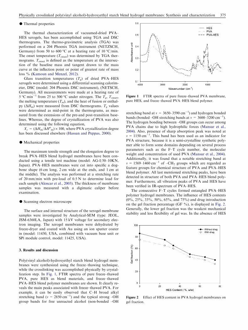
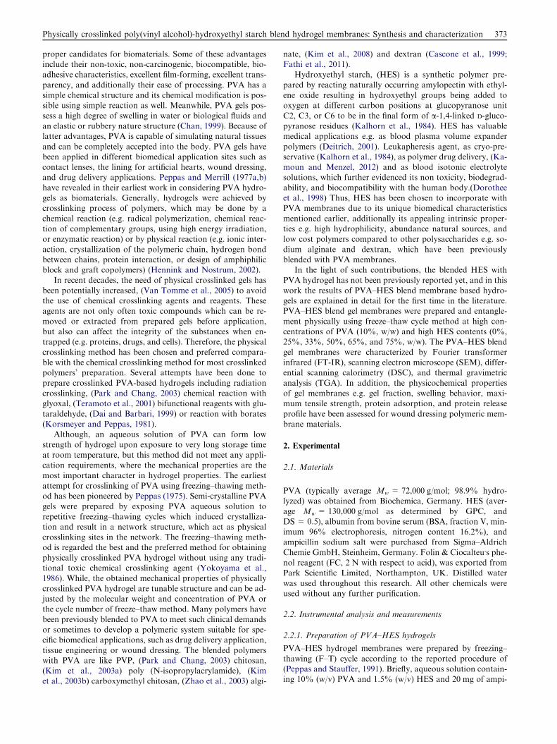
Figure 1 FTIR spectra of pure freeze–thawed PVA membrane,
pure HES, and freeze–thawed PVA–HES blend polymer.
Physically crosslinked poly(vinyl alcohol)-hydroxyethyl starch blend hydrogel membranes: Synthesis and characterization 375
d Thermal properties
The thermal characterization of vacuumed-dried PVA–HES xerogels, has been accomplished using TGA and DSC
thermograms. The thermo-gravimetric analysis (TGA) wasperformed on a 204 Phoenix TGA instrument (NETZSCH,Germany) from 50 to 600 �C at a heating rate of 10 �C/min.The onset temperature (Tonset) was determined by TGA ther-
mograms. Tonset is defined as the temperature at the intersec-tion of the baseline mass and tangent drawn to the masscurve at the inflection point or point of greatest rate of mass
loss % (Kamoun and Menzel, 2012).Glass transition temperatures (Tg) of dried PVA–HES
xerogels were determined using a differential scanning calorim-
eter, DSC (model: 204 Phoenix DSC instrument), (NETSCH,Germany). All measurements were made at a heating rate of5 �C min�1 from 25 to 500 �C under nitrogen. The Tg, Tonset,the melting temperature (Tm), and the heat of fusion or enthal-
py (DHm) were measured from DSC thermograms. Tg valueswere determined as mid-point in the thermograms, as mea-sured from the extensions of the pre-and post-transition base-
lines. Whereas, the degree of crystallization of PVA was alsodetermined using the following equation,
Xc = (DHm/DHom) · 100, where PVA crystallization degree
has been discussed elsewhere (Hassan and Peppas, 2000).
d Mechanical properties
The maximum tensile strength and the elongation degree tobreak PVA–HES blend hydrogel membranes have been con-ducted using a tensile test machine (model: AG-I/50–10KN,
Japan). PVA–HES membranes were cut into specific a dog-bone shape (6 cm long, 2 cm wide at the ends, and 1 cm atthe middle). The analysis was performed at a stretching rate
of 20 mm/min with pre-load of 0.5 N to determine load foreach sample (Alencar et al., 2003). The thickness of membranesamples was measured with a digimatic caliper before
examination.
d Scanning electron microscope
The surface and internal structure of the xerogel membranesamples were investigated by Analytical-SEM (type: JEOL,JSM-6360LA, Japan) with 15 kV voltage for secondary elec-
tron imaging. The xerogel membranes were dehydrated byfreeze-dryer and coated with Au using an ion sputter coaterin (model: 11430, USA, combined with vacuum base unit or
SPi module control, model: 11425, USA).
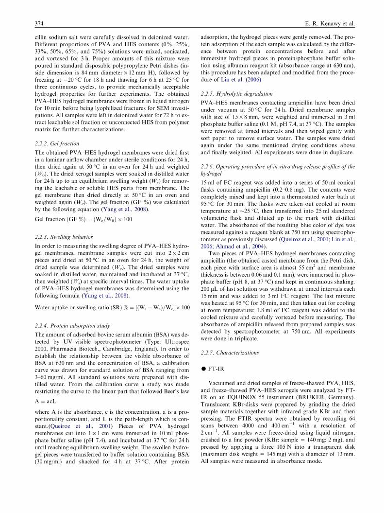
Figure 2 Effect of HES content in PVA hydrogel membranes on
gel fraction.
3. Results and discussion
Poly(vinyl alcohol)-hydroxyethyl starch blend hydrogel mem-branes were synthesized using the freeze–thawing technique,while the crosslinking was accomplished physically by crystal-
lization step. In Fig. 1, FTIR spectra of pure freeze–thawedPVA, pure HES as blend materials, and freeze–thawedPVA–HES blend polymer membranes are shown. It clearly re-veals the main peaks associated with freeze–thawed PVA. For
example, it can be easily observed that C–H broad alkylstretching band (m = 2850 cm�1) and the typical strong –OHgroup bands for free unreacted alcohol (non-bonded –OH
stretching band at m = 3650–3590 cm�1) and hydrogen bondedbands (bonded –OH stretching bands at m = 3600–3200 cm�1).
The hydrogen bonding between –OH groups can occur amongPVA chains due to high hydrophilic forces (Mansur et al.,2004). Also, presence of sharp absorption peak was noted at
m = 1150 cm�1. This band has been used as an indicator forPVA structure, because it is a semi-crystalline synthetic poly-mer able to form some domains depending on several process
parameters such as the F–T cycle number, the molecularweight and concentration of used PVA (Mansur et al., 2004).Additionally, it was found that a notable stretching band atm = 1569–1460 cm�1 of –CH2 groups which are regarded as
feature groups for chemical structure of PVA and PVA–HESblend polymer. All last mentioned stretching peaks, have beendetected in structure of both PVA and PVA–HES blend poly-
mer. Furthermore, all vibration peaks of PVA and HES havebeen verified in IR-spectrum of PVA–HES.
The consecutive F–T cycles formed entangled PVA–HES
polymer hydrogel membranes. The influence of HES contents(0%, 25%, 33%, 50%, 65%, and 75%) and drug introductionon the gel fraction percentage (GF %), is displayed in Fig. 2.Generally, the lower gel fraction was the weakest mechanical
stability and less flexibility of gel was. In the absence of HES
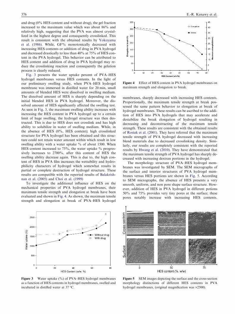
Figure 4 Effect of HES content in PVA hydrogel membranes on
maximum strength and elongation to break.
376 E.-R. Kenawy et al.
and drug (0% HES content and without drug), the gel fractionincreased to the maximum value which was about 86% andrelatively high, suggesting that the PVA was almost crystal-
lized in the highest degree and consequently crosslinked. Thisresult is consistent with the obtained results by Yokoyamaet al. (1986). While, GF% monotonically decreased with
increasing HES contents or addition of drug in PVA hydrogeland decreased drastically to less than 40% at 75% of HES con-tent in the PVA hydrogel. This behavior can be attributed to
HES content and addition of drug in PVA hydrogel may re-duce the crosslinking reaction and consequently the gelationprocess is clearly reduced.
Fig. 3 presents the water uptake percent of PVA–HES
hydrogel membranes versus HES contents. In the light ofour preliminary swelling study, when PVA–HES hydrogelmembrane was immersed in distilled water for 20 min, small
amounts of blended HES were dissolved in swelling medium.The dissolved amount of HES is sharply depending on theinitial blended HES in PVA hydrogel. Moreover, the dis-
solved amount of HES significantly affected the swelling test.As seen in Fig. 3, the maximum swelling ability increases withincreasing the HES content in PVA hydrogel up to a certain
limit of huge swelling, the hydrogel structure was then des-tructed. This is due to HES does not crosslink and has highability to solubilize in water of swelling medium. While, inthe absence of HES (0%, HES content); high crosslinked
structure for PVA hydrogel has been obtained and this struc-ture could not retain water amount within which result in lowswelling ability with a water uptake % of about 1500. When
HES content increased to 75%, the water uptake % progres-sively increases to 2700%, after this content of HES theswelling ability decrease again. This is due to, the high con-
tent of HES in PVA film increases the wettability and hydro-philicity characters of hydrogel which somewhat results inpartial or complete destruction of hydrogel structure. These
results are compatible with the reported results of Balakrish-nan et al. (2005) and Choi et al. (1999)
To investigate the additional influence of HES on themechanical properties of PVA hydrogel membranes, their
maximum tensile strength and elongation at break have beenevaluated and shown in Fig. 4. As shown, the maximum tensilestrength and elongation at break of PVA–HES hydrogel
Figure 3 Water uptake (%) of PVA–HES hydrogel membranes
as a function of HES contents in hydrogel membranes, swelled and
incubated in distilled water at 37 �C.
membranes, sharply decreased with increasing HES contents.Proportionally, the maximum tensile strength at break pos-
sessed the same pattern behavior to elongation at break ofhydrogel membranes. These results can be ascribed to the addi-tion of HES into PVA hydrogels that may accelerate and
destabilize the break elongation of hydrogel resulting indecreasing and deconstructing of the maximum tensilestrength. These results are consistent with the obtained resultsof Rosiak et al. (2001). They have referred that the maximum
tensile strength of PVA hydrogel decreased with increasingblend materials due to decreased crosslinking density. Simi-larly, our results are completely consistent with the reported
results by Hwang et al. (2010). They have demonstrated thatthe maximum tensile strength of PVA hydrogel has sharply de-creased with increasing dextran portions in the hydrogel.
The morphology structure of PVA–HES hydrogel mem-branes was investigated by SEM. The SEM micrographs ofthe surface and interior structures of PVA hydrogel mem-
branes versus HES portions are shown in Fig. 5. Accordingto SEM micrographs, the absence of HES presents a verysmooth, uniform, and non pore shape surface structure. How-ever, addition of HES in PVA hydrogel in different potions
50% and 75% provides very tiny pores at the surface; thesepores notably increase with increasing HES contents.
Figure 5 SEM images depicting the surface and the cross-section
morphology distinctions of different HES contents in PVA
hydrogel membranes, (original magnification was ·2500).
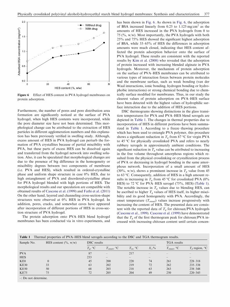
Figure 6 Effect of HES content in PVA hydrogel membranes on
protein adsorption.
Physically crosslinked poly(vinyl alcohol)-hydroxyethyl starch blend hydrogel membranes: Synthesis and characterization 377
Furthermore, the number of pores and pore distribution areaformation are significantly noticed at the surface of PVA
hydrogel, when high HES contents were incorporated, whilethe pore diameter size have not been determined. This mor-phological change can be attributed to the extraction of HES
particles in different agglomeration numbers and this explana-tion has been previously verified in swelling study. Although,excess amount of HES in PVA hydrogel can perturb the for-mation of PVA crystallites because of partial miscibility with
PVA, but these parts of excess HES can be dissolved againand transferred from the hydrogel network into swelling solu-tion. Also, it can be speculated that morphological changes are
due to the presence of big difference in the homogeneity ormiscibility degrees between two components of membrane(i.e. PVA and HES), which resulted in ordered-crystalline
phase and uniform shape structure in case 0% HES, due tohigh entanglement of PVA and disordered-crystalline phasefor PVA hydrogel blended with high portions of HES. The
morphological results and our speculation are compatible withobtained results of Cascone et al. (1999) and Fathi et al. (2011)On the other hand, layered and channeling cross-section-shapestructures were observed at 0% HES in PVA hydrogel. In
addition, pores, cracks, and somewhat caves have appearedafter incorporation of different portions of HES in cross-sec-tion structure of PVA hydrogel.
The protein adsorption onto PVA–HES blend hydrogelmembranes has been conducted via in vitro experiments, and
Table 1 Thermal properties of PVA–HES blend xerogels according
Sample No. HES content (%, w/w) DSC results
Tg, �C Tonset, �
PVA 87 –
HES 233 –
KE0 0 45 200
KE30 33 63 202
KE50 50 68 203
KE75 75 72 205
–: Do not determine.
has been shown in Fig. 6. As shown in Fig. 6, the adsorptionof BSA increased linearly from 0.25 to 1.125 mg/cm2 as theamounts of HES increased in the PVA hydrogels from 0 to
75 (%, w/w). Most importantly, the PVA hydrogels with both25% and 75% HES showed the significant highest adsorptionof BSA, while 33–65% of HES the differences in adsorption
amounts were much closed, indicating that HES content af-fected the protein adsorption behavior onto the surface ofPVA hydrogel. These results are consistent with the reported
results by Kim et al. (2008) who revealed that the adsorptionof protein increased with increasing blended alginate in PVAhydrogels. Moreover, the mechanism of protein adsorptionon the surface of PVA–HES membranes can be attributed to
various types of interaction forces between protein moleculesand the membrane surface, such as weak bonding (van derWaal interactions, ionic bonding, hydrogen bonding or hydro-
phobic interactions) or strong chemical bonding due to chem-ically surface modified for membranes. Thus, in our study theclearest values of protein adsorption on PVA–HES surface
have been detected with the highest values of hydrophilic sur-face interaction due to the addition of HES portions.
DSC thermograms showing distinctions in the glass transi-
tion temperatures for PVA and PVA–HES blend xerogels aredepicted in Table 1. The changes in thermal properties due toincorporation of HES in different portions have been summa-rized in Table 1. According to a freeze–thawing procedure
which has been used to entangle PVA polymer, this procedureshows a significant reduction in Tg from 87 �C for virgin PVAto 45 �C for physically crosslinked PVA and refers to nearly
rubbery xerogels in approximately ambient conditions. Thesignificant reduction in Tg value can be attributed to increasingin the free volume throughout amorphous regions which re-
sulted from the physical crosslinking or crystallization processof PVA or decreasing in hydrogel bonding in the same amor-phous network. Incorporation of a small amount of HES
(30%, w/w), shows a prominent increase in Tg value from 45to 63 �C. Consequently, addition of HES in a high amount re-sults in increasing in Tg from 45 �C for crosslinked PVA (0%HES) to 72 �C for PVA–HES xerogel (75%, HES) (Table 1).
The notable increase in Tg values due to blending HES, canbe ascribed to higher Tg values of HES itself, its higher misci-bility and its good homogeneity with PVA. Accordingly, the
onset temperature (Tonset) values increase progressively withincreasing the content of HES. The presented data are consis-tent with the reported data of Tg for chitosan/PVA hydrogels
(Cascone et al., 1999). Cascone et al. (1999) have demonstratedthat the Tg of the first thermogram peak for chitosan/PVA in-creased with increasing chitosan content until certain concen-
to the DSC and TGA thermogram results.
TGA results
C Tm, �C Xc,% Tonset, �C Td region, �C
217 – – –
– – – –
220 74 261 228–318
219 72 262 235–338
218 63 263 238–348
204 49 250 220–345
378 E.-R. Kenawy et al.
tration, then it remained constant. Also, the current results ofTg, are consistent with results of Fathi et al. (2011) who re-vealed that the addition of dextran to PVA increased signifi-
cantly the Tg values of xerogels in the first decompositionthermogram peak. The melting point (Tm) values were deter-mined from the third relaxation in PVA–HES xerogels, during
the melting of the crystallization region upon decomposition attemperature above 140 �C. Although, the determination of themelting temperature of PVA-based materials, is very difficult
at temperature above 130 �C, (Marten et al., 1985) but accord-ing to the popular estimated method for the equilibrium Tm,has been used (Hassan and Peppas, 2000). As shown in Table 1,relative changes in the melting temperatures have been ob-
served after the addition of HES up to 50%. While, Tm signif-icantly decreased from 220 �C for pure crosslinked PVA to204 �C for PVA–HES blend xerogel (75%, HES content).
The depicted data in Table 1 related to Tm are consistent withthose of crystallization degree (Xc). The heat of fusion and theshoulder temperature are increased significantly after freeze–
thawing process, and the Xc therefore decreased significantlyafter the addition of HES content from 74% (for 0%, HEScontent) to 49% (for 75%, HES content), this is due to the
reduction of crystallization or entanglement process and in-crease of HES content which does not have the ability forcrosslinking and perturbing the crystallization process.
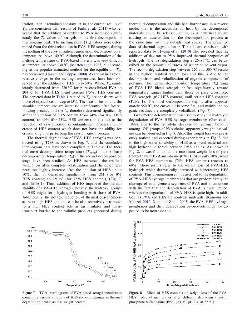
The thermal degradation of PVA–HES xerogels was con-
ducted using TGA as drawn in Fig. 7, and the concludedthermogram data have been complied in Table 1. The ther-mal onset decomposition temperature (Tonset) and the sharp
decomposition temperature (Td) in the second decompositionstage have been studied. As HES increased, the residualweight loss after complete volatilization and the onset tem-
peratures slightly increase after the addition of HES up to50%, then it decreased significantly from 261 (for 0%HES content) to 250 �C (for 75% HES content), (Fig. 7,
and Table 1). Thus, addition of HES improved the thermalstability of PVA–HES xerogels, because the hydroxyl groupsof HES might form hydrogen bonding with those of PVA.Additionally, the notable reduction of thermal onset temper-
ature at high HES content, can be also tentatively attributedto a high HES content acts as an insulator and mass-transport barrier to the volatile products generated during
Figure 7 TGA thermograms of PVA based xerogel membranes
containing various amounts of HES showing changes in thermal
degradation profile as loss weight percent.
thermal decomposition and this heat barrier acts in a reversemode, that is the accumulation heat by the decomposedmaterials could be released, acting as a new heat source
causing an acceleration on the decomposition process atthe same time with the outside heat source. The presenteddata of thermal degradation in Table 1, are consistent with
reported data by Hwang et al. (2010) who revealed that theaddition of dextran to PVA improved thermal properties ofhydrogels. The first degradation step at 26–85 �C, can be as-
cribed to the removal of traces of water or solvent vapor.The second degradation step between 220 and 500 �C resultsin the highest residual weight loss and this is due to thedecomposition and volatilization of organic components of
polymer. The thermal decomposition temperature (Td) rangeof PVA–HES blend xerogels shifted significantly towardtemperature ranges higher than those of pure crosslinked
PVA xerogels (0% HES content) until certain HES contents(Table 1). The third decomposition step is after approxi-mately 550 �C, the curves all become flat, and mainly the or-
ganic residues are completely volatilized, (Fig. 7).Gravimetric determination was used to study the hydrolytic
degradation of PVA–HES hydrogel membranes (Gan et al.,
1999). Due to the hydrolytic cleavage of hydrogen bondingamong –OH groups of PVA chains, apparently weight loss val-ues can be observed in Fig. 8. Also, this weight loss was previ-ously noticed and expected during experiments in Fig. 3, due
to the high water solubility of HES as a blend material andhigh hydrophilic forces between PVA chains. As shown inFig. 8, it was found that the maximum weight loss of pure
freeze–thawed PVA membrane (0% HES) is only 18%, whilefor PVA–HES membrane (75% HES content) reaches to60%. These results refer to the weight loss of PVA–HES
hydrogels which dramatically increased with increasing HEScontents. This phenomenon can be ascribed to the degradationof PVA–HES hydrogel membranes that are predominantly the
cleavage of entanglement segments of PVA and is consistentwith the fact that the degradation of PVA is quite limited,whereas the degradation of PVA–HES is quite high. In addi-tion, as PVA and HES are nontoxic materials, (Kamoun and
Menzel, 2012; Xiao and Zhou, 2003) the PVA–HES hydrogelmembranes and their degradation by-products might be ex-pected to be nontoxic too.
Figure 8 Effect of HES contents on weight loss of the PVA–
HES hydrogel membranes after different degrading times in
phosphate buffer saline (PBS) (0.1 M, pH 7.4, at 37 �C).
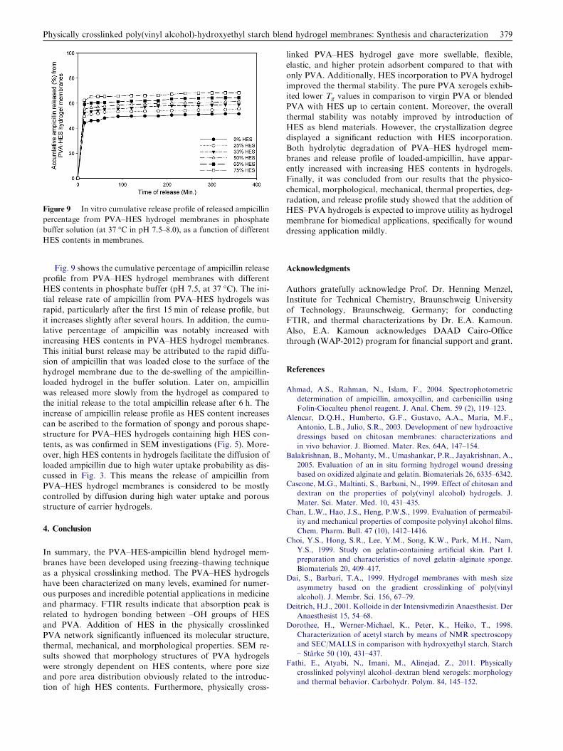
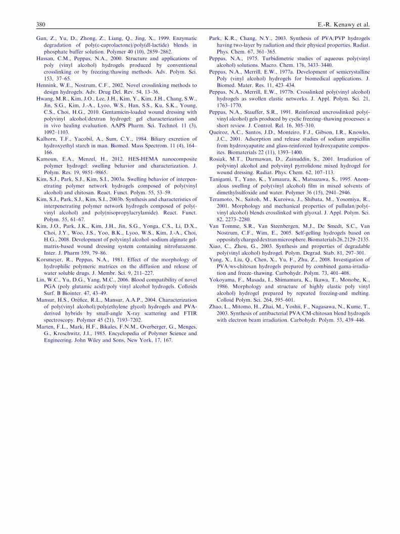
Figure 9 In vitro cumulative release profile of released ampicillin
percentage from PVA–HES hydrogel membranes in phosphate
buffer solution (at 37 �C in pH 7.5–8.0), as a function of different
HES contents in membranes.
Physically crosslinked poly(vinyl alcohol)-hydroxyethyl starch blend hydrogel membranes: Synthesis and characterization 379
Fig. 9 shows the cumulative percentage of ampicillin release
profile from PVA–HES hydrogel membranes with differentHES contents in phosphate buffer (pH 7.5, at 37 �C). The ini-tial release rate of ampicillin from PVA–HES hydrogels was
rapid, particularly after the first 15 min of release profile, butit increases slightly after several hours. In addition, the cumu-lative percentage of ampicillin was notably increased withincreasing HES contents in PVA–HES hydrogel membranes.
This initial burst release may be attributed to the rapid diffu-sion of ampicillin that was loaded close to the surface of thehydrogel membrane due to the de-swelling of the ampicillin-
loaded hydrogel in the buffer solution. Later on, ampicillinwas released more slowly from the hydrogel as compared tothe initial release to the total ampicillin release after 6 h. The
increase of ampicillin release profile as HES content increasescan be ascribed to the formation of spongy and porous shape-structure for PVA–HES hydrogels containing high HES con-tents, as was confirmed in SEM investigations (Fig. 5). More-
over, high HES contents in hydrogels facilitate the diffusion ofloaded ampicillin due to high water uptake probability as dis-cussed in Fig. 3. This means the release of ampicillin from
PVA–HES hydrogel membranes is considered to be mostlycontrolled by diffusion during high water uptake and porousstructure of carrier hydrogels.
4. Conclusion
In summary, the PVA–HES-ampicillin blend hydrogel mem-
branes have been developed using freezing–thawing techniqueas a physical crosslinking method. The PVA–HES hydrogelshave been characterized on many levels, examined for numer-
ous purposes and incredible potential applications in medicineand pharmacy. FTIR results indicate that absorption peak isrelated to hydrogen bonding between –OH groups of HESand PVA. Addition of HES in the physically crosslinked
PVA network significantly influenced its molecular structure,thermal, mechanical, and morphological properties. SEM re-sults showed that morphology structures of PVA hydrogels
were strongly dependent on HES contents, where pore sizeand pore area distribution obviously related to the introduc-tion of high HES contents. Furthermore, physically cross-
linked PVA–HES hydrogel gave more swellable, flexible,elastic, and higher protein adsorbent compared to that withonly PVA. Additionally, HES incorporation to PVA hydrogel
improved the thermal stability. The pure PVA xerogels exhib-ited lower Tg values in comparison to virgin PVA or blendedPVA with HES up to certain content. Moreover, the overall
thermal stability was notably improved by introduction ofHES as blend materials. However, the crystallization degreedisplayed a significant reduction with HES incorporation.
Both hydrolytic degradation of PVA–HES hydrogel mem-branes and release profile of loaded-ampicillin, have appar-ently increased with increasing HES contents in hydrogels.Finally, it was concluded from our results that the physico-
chemical, morphological, mechanical, thermal properties, deg-radation, and release profile study showed that the addition ofHES–PVA hydrogels is expected to improve utility as hydrogel
membrane for biomedical applications, specifically for wounddressing application mildly.
Acknowledgments
Authors gratefully acknowledge Prof. Dr. Henning Menzel,Institute for Technical Chemistry, Braunschweig Universityof Technology, Braunschweig, Germany; for conductingFTIR, and thermal characterizations by Dr. E.A. Kamoun.
Also, E.A. Kamoun acknowledges DAAD Cairo-Officethrough (WAP-2012) program for financial support and grant.
References
Ahmad, A.S., Rahman, N., Islam, F., 2004. Spectrophotometric
determination of ampicillin, amoxycillin, and carbenicillin using
Folin-Ciocalteu phenol reagent. J. Anal. Chem. 59 (2), 119–123.
Alencar, D.Q.H., Humberto, G.F., Gustavo, A.A., Maria, M.F.,
Antonio, L.B., Julio, S.R., 2003. Development of new hydroactive
dressings based on chitosan membranes: characterizations and
in vivo behavior. J. Biomed. Mater. Res. 64A, 147–154.
Balakrishnan, B., Mohanty, M., Umashankar, P.R., Jayakrishnan, A.,
2005. Evaluation of an in situ forming hydrogel wound dressing
based on oxidized alginate and gelatin. Biomaterials 26, 6335–6342.
Cascone, M.G., Maltinti, S., Barbani, N., 1999. Effect of chitosan and
dextran on the properties of poly(vinyl alcohol) hydrogels. J.
Mater. Sci. Mater. Med. 10, 431–435.
Chan, L.W., Hao, J.S., Heng, P.W.S., 1999. Evaluation of permeabil-
ity and mechanical properties of composite polyvinyl alcohol films.
Chem. Pharm. Bull. 47 (10), 1412–1416.
Choi, Y.S., Hong, S.R., Lee, Y.M., Song, K.W., Park, M.H., Nam,
Y.S., 1999. Study on gelatin-containing artificial skin. Part I.
preparation and characteristics of novel gelatin–alginate sponge.
Biomaterials 20, 409–417.
Dai, S., Barbari, T.A., 1999. Hydrogel membranes with mesh size
asymmetry based on the gradient crosslinking of poly(vinyl
alcohol). J. Membr. Sci. 156, 67–79.
Deitrich, H.J., 2001. Kolloide in der Intensivmedizin Anaesthesist. Der
Anaesthesist 15, 54–68.
Dorothee, H., Werner-Michael, K., Peter, K., Heiko, T., 1998.
Characterization of acetyl starch by means of NMR spectroscopy
and SEC/MALLS in comparison with hydroxyethyl starch. Starch
– Starke 50 (10), 431–437.
Fathi, E., Atyabi, N., Imani, M., Alinejad, Z., 2011. Physically
crosslinked polyvinyl alcohol–dextran blend xerogels: morphology
and thermal behavior. Carbohydr. Polym. 84, 145–152.
380 E.-R. Kenawy et al.
Gan, Z., Yu, D., Zhong, Z., Liang, Q., Jing, X., 1999. Enzymatic
degradation of poly(e-caprolactone)/poly(dl-lactide) blends in
phosphate buffer solution. Polymer 40 (10), 2859–2862.
Hassan, C.M., Peppas, N.A., 2000. Structure and applications of
poly (vinyl alcohol) hydrogels produced by conventional
crosslinking or by freezing/thawing methods. Adv. Polym. Sci.
153, 37–65.
Hennink, W.E., Nostrum, C.F., 2002. Novel crosslinking methods to
design hydrogels. Adv. Drug Del. Rev. 54, 13–36.
Hwang, M.R., Kim, J.O., Lee, J.H., Kim, Y., Kim, J.H., Chang, S.W.,
Jin, S.G., Kim, J.-A., Lyoo, W.S., Han, S.S., Ku, S.K., Young,
C.S., Choi, H.G., 2010. Gentamicin-loaded wound dressing with
polyvinyl alcohol/dextran hydrogel: gel characterization and
in vivo healing evaluation. AAPS Pharm. Sci. Technol. 11 (3),
1092–1103.
Kalhorn, T.F., Yacobil, A., Sum, C.Y., 1984. Biliary excretion of
hydroxyethyl starch in man. Biomed. Mass Spectrom. 11 (4), 164–
166.
Kamoun, E.A., Menzel, H., 2012. HES-HEMA nanocomposite
polymer hydrogel: swelling behavior and characterization. J.
Polym. Res. 19, 9851–9865.
Kim, S.J., Park, S.J., Kim, S.I., 2003a. Swelling behavior of interpen-
etrating polymer network hydrogels composed of poly(vinyl
alcohol) and chitosan. React. Funct. Polym. 55, 53–59.
Kim, S.J., Park, S.J., Kim, S.I., 2003b. Synthesis and characteristics of
interpenetrating polymer network hydrogels composed of poly(-
vinyl alcohol) and poly(nisopropylacrylamide). React. Funct.
Polym. 55, 61–67.
Kim, J.O., Park, J.K., Kim, J.H., Jin, S.G., Yonga, C.S., Li, D.X.,
Choi, J.Y., Woo, J.S., Yoo, B.K., Lyoo, W.S., Kim, J.-A., Choi,
H.G., 2008. Development of polyvinyl alcohol–sodium alginate gel-
matrix-based wound dressing system containing nitrofurazone.
Inter. J. Pharm 359, 79–86.
Korsmeyer, R., Peppas, N.A., 1981. Effect of the morphology of
hydrophilic polymeric matrices on the diffusion and release of
water soluble drugs. J. Membr. Sci. 9, 211–227.
Lin, W.C., Yu, D.G., Yang, M.C., 2006. Blood compatibility of novel
PGA (poly glutamic acid)/poly vinyl alcohol hydrogels. Colloids
Surf. B Biointer. 47, 43–49.
Mansur, H.S., Orefice, R.L., Mansur, A.A.P., 2004. Characterization
of poly(vinyl alcohol)/poly(ethylene glycol) hydrogels and PVA-
derived hybrids by small-angle X-ray scattering and FTIR
spectroscopy. Polymer 45 (21), 7193–7202.
Marten, F.L., Mark, H.F., Bikales, F.N.M., Overberger, G., Menges,
G., Kroschwitz, J.I., 1985. Encyclopedia of Polymer Science and
Engineering. John Wiley and Sons, New York, 17, 167.
Park, K.R., Chang, N.Y., 2003. Synthesis of PVA/PVP hydrogels
having two-layer by radiation and their physical properties. Radiat.
Phys. Chem. 67, 361–365.
Peppas, N.A., 1975. Turbidimetric studies of aqueous poly(vinyl
alcohol) solutions. Macro. Chem. 176, 3433–3440.
Peppas, N.A., Merrill, E.W., 1977a. Development of semicrystalline
Poly (vinyl alcohol) hydrogels for biomedical applications. J.
Biomed. Mater. Res. 11, 423–434.
Peppas, N.A., Merrill, E.W., 1977b. Crosslinked poly(vinyl alcohol)
hydrogels as swollen elastic networks. J. Appl. Polym. Sci. 21,
1763–1770.
Peppas, N.A., Stauffer, S.R., 1991. Reinforced uncrosslinked poly(-
vinyl alcohol) gels produced by cyclic freezing–thawing processes: a
short review. J. Control. Rel. 16, 305–310.
Queiroz, A.C., Santos, J.D., Monteiro, F.J., Gibson, I.R., Knowles,
J.C., 2001. Adsorption and release studies of sodium ampicillin
from hydroxyapatite and glass-reinforced hydroxyapatite compos-
ites. Biomaterials 22 (11), 1393–1400.
Rosiak, M.T., Darmawan, D., Zainuddin, S., 2001. Irradiation of
polyvinyl alcohol and polyvinyl pyrrolidone mixed hydrogel for
wound dressing. Radiat. Phys. Chem. 62, 107–113.
Tanigami, T., Yano, K., Yamaura, K., Matsuzawa, S., 1995. Anom-
alous swelling of poly(vinyl alcohol) film in mixed solvents of
dimethylsulfoxide and water. Polymer 36 (15), 2941–2946.
Teramoto, N., Saitoh, M., Kuroiwa, J., Shibata, M., Yosomiya, R.,
2001. Morphology and mechanical properties of pullulan/poly(-
vinyl alcohol) blends crosslinked with glyoxal. J. Appl. Polym. Sci.
82, 2273–2280.
Van Tomme, S.R., Van Steenbergen, M.J., De Smedt, S.C., Van
Nostrum, C.F., Wim, E., 2005. Self-gelling hydrogels based on
oppositelychargeddextranmicrosphere.Biomaterials26,2129–2135.
Xiao, C., Zhou, G., 2003. Synthesis and properties of degradable
poly(vinyl alcohol) hydrogel. Polym. Degrad. Stab. 81, 297–301.
Yang, X., Liu, Q., Chen, X., Yu, F., Zhu, Z., 2008. Investigation of
PVA/ws-chitosan hydrogels prepared by combined gama-irradia-
tion and freeze–thawing. Carbohydr. Polym. 73, 401–408.
Yokoyama, F., Masada, I., Shimamura, K., Ikawa, T., Monobe, K.,
1986. Morphology and structure of highly elastic poly vinyl
alcohol) hydrogel prepared by repeated freezing-and melting.
Colloid Polym. Sci. 264, 595–601.
Zhao, L., Mitomo, H., Zhai, M., Yoshii, F., Nagasawa, N., Kume, T.,
2003. Synthesis of antibacterial PVA/CM-chitosan blend hydrogels
with electron beam irradiation. Carbohydr. Polym. 53, 439–446.