Embed Size (px)
Citation preview
THE JOURNAL OF BKXOGICAL CHEMISTRY Vol. El. No. 21. Issue of November 10, pp. 67li-6766. 1976
Printed in b’.S.A.
Physical Properties of Membranes Isolated from Tissue Culture Cells with Altered Phospholipid Composition*
(Received for publication, February 5, 1976)
FRIEDHELM SCHROEDER* ,$ JOHN F. HOLLAND,% AND P. ROY VAGELOS~, 11
From the $Department of Biological Chemistry, Division of Biology and Biomedical Sciences, Washington Uniuersity, St. Louis, Missouri 63110, and the TDepartment of Biochemistry, Michigan State University, East Lansing, Michigan 48823
A choline-requiring strain of mouse fibroblast cells (LM cells) was cultured in suspension with choline, N,N’-dimethylethanolamine, N-monomethylethanolamine, or ethanolamine. These choline analogues were incorporated into membrane phospholipids as phosphatidyl-N,N’-dimethylethanolamine, phos- phatidyl-N-monomethylethanolamine, and phosphatidylethanolamine. Plasma membranes, microsomes, mitochondria, and their respective lipids were isolated and the characteristic temperatures were determined by using two types of fluorescent probes: (a) /3-parinaric acid, a naturally occurring molecule, and (b) &anilino-1-naphthalene sulfonic acid, a synthetic organic fluorophore. A computer- centered spectrofluorimeter capable of simultaneous measurement of absorbance, absorbance-corrected
fluorescence, and relative fluorescence efficiency was utilized for on-line measurement of all fluorescence parameters. Plots of absorbance corrected fluorescence or of relative fluorescence efficiency versus temperature revealed the same five characteristic temperatures with both types of probe. These characteristic temperatures were independent of the phospholipid composition of the LM suspension cell membranes or their extracted lipids. Plasma membranes, microsomes, and mitochondria containing analogue phospholipids had similar (&lo) characteristic temperatures.
The presence of analogue phopholipids altered the binding characteristics of P-parinaric acid with plasma membranes and plasma membrane lipids of LM susfiension cells. The equilibrium dissociation constant of plasma membranes and plasma membrane lipids was decreased 2- and 5-fold, respectively, when the cells had been supplemented with ethanolamine. The minimum number of phospholipid molecules per probe binding site was approximately constant in the intact plasma membrane but increased (2-fold) in the isolated plasma membrane lipids. The presence of analogue phospholipids also altered the interaction of &anilino-l-naphthalene sulfonic acid with LM cell membranes. The equilibrium dissociation constant of this probe interacting with mitochondria was decreased about 10% when LM cells were supplemented with ethanolamine. Similarly, the equilibrium dissociation constant of &anilino-1-naphthalene sulfonate interacting with mitochondrial lipid was decreased 40% by ethanolamine supplementation. The fluorescent properties of both probes were sensitive to the degree of
methylation of the polar head group. The absolute values of absorbance-corrected fluorescence and relative fluorescence efficiency were different for each type of membrane from LM cells even with the same analogue supplement. Thus, it appears that LM cells maintain the characteristic temperatures which are a measure of the physical properties of their membranes, despite large alterations of the phospholipid polar head group composition.
Membranes of cells are in a fluid state and recently direct
evidence for membrane fluidity has been presented (1). The physical properties of pure lipids have been studied extensively in order to explain the transitions in physical properties
*This investigation was supported in part by National Science Foundation Grant GB-38676X and National Institutes of Health Grant HL-10406.
I American Cancer Society Postdoctoral Fellow (PF-963). Present address, Department of Pharmacology, University of Missouri Medical School, Columbia, MO. 65201.
11 Present address, Merck Sharp & Dohme Research Laboratories, Division of Merck & Co., Inc., Rahway, N. J. 07065.
observed with probe molecules in naturally occurring mem- branes or their isolated heterogeneous lipids (2-12). It was expected that individual transitions might be ascribed to particular lipid classes. Instead a vast polymorphism has been indicated for phospholipids of mitochondria (13), brain (14,
15), egg yolk (15, 16), and neutral lipids (17). The exact transition temperature from the gel to liquid crystalline phase of a phospholipid appears to be dependent on as many as nine different variables (15). Even binary mixtures of pure phospho- lipid species give two or sometimes three characteristic tem- peratures. Such data have been interpreted as indicating the
6747
by guest on September 9, 2018
http://ww
w.jbc.org/
Dow
nloaded from

6748 Physical Properties of Membranes with Altered Phospholipids
existence of more than one fluid or solid phase at a single temperature (4). Egg yolk lecithin alone may have as many as three transitions at approximately 20, 30, and 42-45” as
determined by differential scanning calorimetry and refrac- tometry (R-20). In the presence of different salt concentra- tions dipalmityl phosphatidylcholine, which normally displays only one transition temperature, revealed two characteristic transitions (21). Similarly, a pure triglyceride such as triolein
may have two or four transition temperatures (17). Therefore, precise interpretation of characteristic temperatures of mem-
branes or lipids containing heterogeneous mixtures has not been possible. Despite these findings, it has been shown that microbial systems generally reveal only one or two characteris- tic temperatures (22, 23). However, recently the physical properties and characteristic temperatures of membranes and their extracted lipids from LM cells’ grown in suspension* and monolayer (24, 25) have been determined fluorimetrical!y and by ESR (electron spin resonance) techniques, respectively. Both methods indicated the existence of at least five character- istic temperatures. Such a multiplicity of characteristic tem- peratures in mammalian cell membranes had hitherto not been
demonstrated. But if the microviscosity data of Shinitzky and Inbar (26) are replotted, at least three characteristic tempera- tures can be demonstrated at 5, 16, and 27” in lymphocyte membranes. It has been shown that a large variety of physio- logical parameters can be correlated with the mammalian characteristic temperatures (27; for a review see Ref. 24) and that manipulation of the fatty acid composition of LM cells, for example, leads to alterations in the characteristic tempera- tures of the membranes and their lipids as well as in the physiological parameters (28, 29). Therefore, lipids may regu- late physiological parameters not only by specific interaction with membrane proteins, for example, but also by altering the
physical state of the bulk membrane lipids.
suspension culture.‘, a The results of our investigations detail the isolation of subcellular fractions and the lipid composition thereof and compare the activities of membrane-bound
enzymes as a function of polar head group composition of the phospholipids. It was shown that the activities of seven membrane-bound enzymes from LM cell membranes were independent of polar head group composition and that several compensatory mechanisms may exist for maintaining the
physical characteristics of the membranes. We have previously shown that five characteristic temperatures exist in plasma membrane, microsomes, mitochondria, and their lipids from LM cells grown in suspension culture. Herein are presented the results of investigations indicating that the characteristic temperatures of the LM cell membranes and isolated mem- brane lipids are independent of the phospholipid composition. Several interpretations of these data are presented.
The effect of polar head group modulation on transition temperatures has been studied in model systems by differential scanning calorimetry (7, 8, 30), fluorescence probes3-6 (11, 31), electron spin resonance (32), and nuclear magnetic reso- nance (33-35). In contrast to manipulation of the fatty acid composition, studies of the effect of altered polar head group composition of phospholipids on physcial properties have not yet been extended to mammalian membranes. The phospho- lipid composition of mammalian tissues has been studied extensively and analogues of choline have been incorporated into mammalian tissue lipids since 1936 (36, 37) and also into insect lipids (38, 39). Recently, these techniques were first
applied to a single cell type, LM cells, grown in monolayer in a completely defined medium (40-42). Subsequently, these stud- ies were extended to LM cells grown in large quantities in
‘The abbreviations used are: LM cells, a choline-requiring strain of mouse fibroblast cells; ANS, 8aniline-1-naphthalene sulfonic acid; CO, absorption-corrected fluorescence; RFE, relative fluorescence efficiency.
‘Schroeder, F., Holland, J. F., and Vagelos, P. R. (1976) J. Viol. Chem. 251, 6739-6746. This paper immediately precedes the present one and is part of a series.
3 Sklar, L. A. Hudson, B. S., and Simoni, R. D. (1976) J. Supramol. 4,449-465.
‘E. V. Tecoma, L. A. Sklar, R. D. Simoni, and B. S. Hudson, manuscript submitted to Biochemistry.
“L. A. Sklar, B. S. Hudson, and R. D. Simoni, manuscript submitted to Biochemistry.
‘L. A. Sklar, B. S. Hudson, M. Peterson, and J. Diamond, manuscript submitted to Biochemistry.
MATERIALS AND METHODS
Cell Culture-LM cells, a choline-requiring strain of mouse fibro- blasts, were grown in suspension culture on the chemically defined, lipid-free, serum-free medium of Higuchi (43) modified as described.7
Choline analogue supplementation was carried out by washing log phase LM cells two times with choline-deficient medium. The cells were pelleted in an International PR-1 centrifuge for 5 min at 1000 rpm after each resuspension. Aliquots of the washed cells were then resuspended at 1 x 10’ cells/ml in fresh medium and one of the following was added at 40 @g/ml: choline, N,N’-dimethylethanola- mine, N-monomethylethanolamine, or ethanolamine. After 2 days growth at 37” the cells were diluted with fresh medium containing the appropriate analogue in order to maintain the cells growing in log phase. Then after 1 additional day of growth at 37” (3 days total) the cells were harvested and plasma membranes, microsomes, mitochon- dria, and their respective extracted lipids were obtained as described previously.’ Choline, N,N’-dimethylethanolamine, N-monomethyl- ethanolamine, and ethanolamine were obtained from Eastman Kodak Co., Rochester, N. Y.
Fluorescence Spectroscopy and Sample Preparations-The com- puter-centered spectrofluorimeter described by Holland and co- workers (44, 45) was used for all measurement. 8.Parinaric acid was in- corporated into LM suspension cell membranes or lipids as described previously’ ANS (S-aniline-1-naphthalene sulfonic acid) was obtained ‘from Pierce Chemical Co., Rockford, Ill. as the ammonium salt. Stock solutions of ANS were prepared fresh daily at 1 to 5 x 10m3 M in distilled H,O. Aliquots of this stock solution were added directly to membranes (50 ag of protein/ml of phosphate-buffered saline (PBS), pH 7.4) or to lipid kxtracts. The samples-were then blended for 3 min-on a Vortex Genie mixer at maximum speed, followed by a 30.min incubation at 45” (46). The samples were then blended on a Vortex mixer for 1 min and placed in the fluorimeter sample cuvette and spectral measure- ments were determined as described.
Plots of CO,,, of ANS versus temperature in solvents such as ethanol indicated an exponential decay with increasing temperature. No discontinuities or characteristic temperatures were formed. Thus the characteristic temperatures determined under “Results” with mem- branes or lipids do not appear to be systematic instrumental artifacts.
RESULTS
Effect of Choline Analogue Supplementation on Characteris- tic Temperatures Monitored with P-Parinaric Acid-The char- acteristic temperatures of LM membranes or lipids containing
large amounts of phosphatidyl-NJ’-dimethylethanolamine, phosphatidyl-N-monomethylethanolamine, or phosphatidyl- ethanolamine may be different from those containing primarily phosphatidylcholine if no other changes in lipid composition
‘Schroeder, F., Perlmutter, J. F., Glaser, M. and Vagelos, P. R. (1976) J. Biol. Chem. 251, 5015-5026.
*Schroeder, F., and Vagelos, P. R. (1976) Biochim. Biophys. Acta 441, 239-254.
by guest on September 9, 2018
http://ww
w.jbc.org/
Dow
nloaded from
Physical Properties of Membranes with Altered Phospholipids 6749
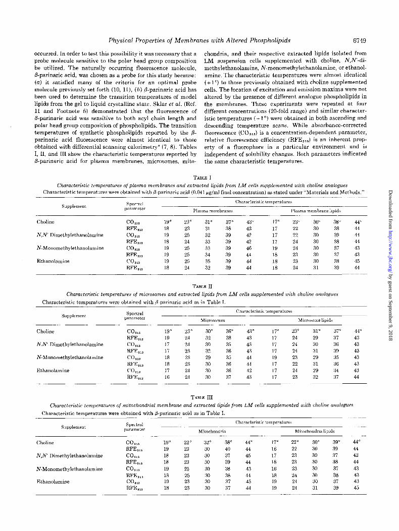
occurred. In order to test this possibility it was necessary that a probe molecule sensitive to the polar head group composition be utilized. The naturally occurring fluorescence molecule, Bparinaric acid, was chosen as a probe for this study because: (a) it satisfied many of the criteria for an optimal probe molecule previously set forth (10, ll), (b) /3-parinaric acid has been used to determine the transition temperatures of model lipids from the gel to liquid crystalline state. Sklar et al. (Ref. 11 and Footnote 5) demonstrated that the fluorescence of P-parinaric acid was sensitive to both acyl chain length and polar head group composition of phospholipids. The transition temperatures of synthetic phospholipids reported by the fl- parinaric acid fluorescence were almost identical to those obtained with differential scanning calorimetry5 (7, 8). Tables I, II, and III show the characteristic temperatures reported by /3-parinaric acid for plasma membranes, microsomes, mito-
chondria, and their respective extracted lipids isolated from LM suspension cells supplemented with choline, N,N’-di- methylethanolamine, N-monomethylethanolamine, or ethanol- amine. The characteristic temperatures were almost identical (f 1”) to those previously obt,ained with choline-supplemented cells. The location of excitation and emission maxima were not altered by the presence of different analogue phospholipids in the membranes. These experiments were repeated at four different concentrations (20-fold range) and similar character- istic temperatures (i 1”) were obtained in both ascending and descending temperature scans. While absorbance-corrected fluorescence (CO,,,) is a concentration-dependent parameter, relative fluorescence efficiency (RFE,,,) is an inherent prop- erty of a fluorophore in a particular environment and is independent of solubility changes. Both parameters indicated the same characteristic temperatures.
TABLE I
Characteristic temperatures of plasma membranes and extracted lipids from LM cells supplemented with choline analogues Characteristic temperatures were obtained with P-parka& acid (0.041 #g/ml final concentration) as stated under “Materials and Methods.”
Supplement Spectral parameter
Characteristic temperatures
Plasma membranes Plasma membrane lipids
Choline
NJ?‘-Dimethylethanolamine
N-Monomethylethanolamine
Ethanolamine
co,,, 19” 23” 31” 37” 43” 17” 23” 30” 38” 44” RFJL,, 18 23 31 38 42 17 22 30 38 44 CO8lS 19 25 32 39 42 17 22 30 39 44 RFE,,, 18 24 33 39 42 17 24 30 38 44 co*,, 19 25 33 39 46 19 24 30 37 43
RF&,, 19 25 34 39 44 18 23 30 37 43 co,,, 19 25 35 39 44 18 23 30 38 45
RF&,, 18 24 32 39 44 18 24 31 39 44
TABLE II
Characteristic temperatures of microsones and extracted lipids from LM cells supplemented with choline analogues
Characteristic temperatures were obtained with fi-parinaric acid as in Table I.
Supplement Spectral parameter
Characteristic temperatures
Microsames Microsome lipids
Choline
N,N’-Dimethylethanolamine
N-Monomethylethanolamine
Ethanolamine
co,,, 19” 23” 30” 36” 43” 17” 23” 31” 37” 44” RFE,,, 19 24 32 38 43 17 24 29 37 43
co,u 17 24 30 35 43 17 24 30 36 43
RF%,, 17 23 32 36 43 17 24 31 39 43
co,,, 18 23 29 35 44 19 23 29 35 43
RF%,, 18 23 30 36 44 17 22 31 36 43
co,,, 17 24 30 36 42 17 24 29 34 43 RF%,, 16 24 30 37 43 17 23 32 37 44
TABLE III
Characteristic temperatures of mitochondrial membrane and extracted lipids from LM cells supplemented with choline analogues
Characteristic temperatures were obtained with fi-parinaric acid as in Table I.
Supplement
Choline
N,N’-Dimethylethanolamine
N-Monomethylethanolamine
Ethanolamine
Spectral parameter
co,,, RF%,, co,,, RF%,, co,,, RF%,, co,,, RF%,,
19” 19 18 18 19 18 19 18
Mitochondria
22” 32” 38” 23 30 40 23 30 37 23 30 39 25 30 38 25 30 38 23 30 37 23 30 37
Characteristic temperatures
Mitochondria lipids
44” 17” 22” 30” 39” 44 16 22 30 39 45 17 23 30 37 44 18 23 30 38 43 16 23 30 37 44 18 24 30 38 43 19 24 30 37 44 19 24 31 39
44” 44 43 44 43 43 43 45
by guest on September 9, 2018
http://ww
w.jbc.org/
Dow
nloaded from
6750 Physical Properties of Membranes with Altered Phospholipids
It is possible that at least some of these alterations in fluorescence parameters of P-parinaric acid were due to changes in binding ability of the probe to various phospho- lipids (46, 47). It has been shown that the dissociation constant, KD, and the minimum number of phospholipid molecules providing one binding site for /3-parinaric acid in LM suspension cultured cell plasma membranes or plasma mem- brane lipids varied only slightly with temperature.’ As indi- cated in Table IV, the Ko and the size of the phospholipid binding site for P-parinaric acid were affected by the polar head group composition of the plasma membrane phospho- lipid. Plasma membrane lipids from LM suspension cells
supplemented with NJ’-dimethylethanolamine and plasma membranes and lipids from cells supplemented with N- monomethylethanolamine or ethanolamine had smaller K, values and therefore higher affinities for P-parinaric acid. The plasma membrane lipids of choline supplemented cells had a slightly higher K, for /3-parinaric acid than did the intact membrane (1.04 versus 0.93 fiM). Supplementation with ana-
logues such as N,N’-dimethylethanolamine, N-monomethyl- ethanolamine, or ethanolamine reversed this trend since the plasma membrane lipids had 2- to 3-fold smaller K, values for 8-parinaric acid than the intact plasma membranes. However, the minimum number of phospholipid molecules per binding site increased slightly in the plasma membranes (with the exception of ethanolamine supplementation), while it in- creased 2- to 3-fold in the plasma membrane lipids, depending
on the analogue supplemented. Thus, increased affinity for the probe was accompanied by increased numbers of phospholipid molecules per binding site.
Further, it appears that the absolute values of CO,,, may depend on the type of analogue present in the membrane. The presence of analogue phospholipids may result in differences in the amount of probe that can be bound to the membrane or its isolated lipids. These differences in the level of probe satura- tion may be responsible for the observed differences for P-parinaric acid in membranes of different phospholipid com- position. However, as stated earlier, nonsaturating concentra- tions of P-parinaric acid were used for all membranes and isolated membrane lipids studied here. Such criticisms would not be valid for relative fluorescence efficiency (RFE) which does not depend on concentration of the probe. This parame-
ter, RFJL, also indicated the same characteristic tempera- tures as CO,,,.
Determination of Characteristic Temperatures with Other
Fluorescence Probes-An objection to the results obtained with P-parinaric acid might be that we have measured some inherent properties of the probe, /3-parinaric acid, that do not reflect transitions or membrane properties in our system.
Therefore, the above experiments were repeated with the fluorescence probe ANS. ANS is an organic molecule not occurring in nature; it is water-soluble and fluoresces weakly in aqueous environments (31, 46). The fluorescence characteris- tics of ANS in a variety of solvents and in LM cells membranes or lipids are shown in Table V. The spectral characteristics (emission maximum and excitation maxima) of ANS in the plasma membranes, mitochondria, or their respective lipids were similar to those when the probe was present in increas- ingly hydrophobic solvents. ANS will bind to both proteins as well as lipids (10, 23, 31, 46) and the membrane data presented here represent average values possibly reflecting contributions
from bot,h of these membrane components. The increases in the relative fluorescence efficiency (RFE,,,) of ANS with different solvents (methanol and ethanol) were similar to published increases of absolute quantum efficiency using other instru-
ments and different salts of the probe (23, 31, 48). The fluorescence efficiency of ANS in plasma membrane lipids decreased significantly when compared to the intact mem- branes. This behavior is opposite to that of /3-parinaric acid which showed a slight (10%) increase in RFE,,, in the isolated lipid.’ ANS relative fluorescence efficiency increased 118-fold and 24-fold in the presence of plasma membranes and mito- chondria, respectively, the ratio between the two being approx- imately 4. The increase in relative fluorescence efficiency with
the respective isolated lipids was much smaller (71- and 16-fold) but the ratio between the two was also close to 4. Similar ratios of RFE in the plasma membrane to the RFE in the mitochondria were also noted with &parinaric acid in both
the membrane and in the isolated lipids.’ As with P-parinaric acid, the emission and excitation maxima of ANS did not change with membranes or lipids containing different phos- pholipids (plasma membranes uersus mitochondria’. “). These spectral alterations are characteristic for the behavior of ANS when present in solvents of low polarity as well as on binding to lipids, proteins, or cellular membranes (10). Since 8-anilino-l- napthalene sulfonate can interact with both proteins and
lipids, the portion of plasma membrane lipids apparently accessible to ANS can be determined as shown in Table VI by a previously published method (23). As indicated near two possible transitions, the fraction of plasma membrane lipids
TABLE IV
Binding characteristics of P-parinaric acid with plasma membrane and plasma membrane lipids of LM cells supplemented with choline analogues
Fraction Supplement T KD” Minimum number of
lipid molecules/ binding site’
Plasma membranes
Plasma membrane lipids
Choline NJ’-Dimethylethanolamine N-Monomethylethanolamine Ethanolamine Choline NJ’-Dimethylethanolamine N-Monomethylethanolamine Ethanolamine
IrM
24” 0.93 24 0.94 24 0.78 24 0.50 24 1.04 24 0.35 24 0.39 24 0.22
“K,, the equilibrium dissociation constant was determined as previously described (23). b The minimum number of phospholipid molecules providing one binding site was estimated as previously described (23).
17 22 19 16 15 33 38 40
by guest on September 9, 2018
http://ww
w.jbc.org/
Dow
nloaded from
Physical Properties of Membranes with Altered Phospholipids
TABLE V
Spectral characteristics of ANS LIZ different environments
6751
Environment ANS Emission Excitation maximum maxima RFE”
Increase in RFE
relative to PBS”
Absolute quantum efficiency’
Increase in absolute quantum efficiency relative
to PBS
PBS
Methanol Ethanol
Plasma membrane Plasma membrane lipids Mitochondria Mitochondria lipids
PM nm
3.33 555 3.33 480 3.33 465 3.12 480 4.18 480 4.16 480 4.16 480
nm
262,344 0.99 264,364 69.1
265, 364 112 265,365 117 265,365 70 265,365 24 265,365 16
-fold
70 113 118
71 24 16
0.004 0.22 0.37
-fold
55 93
“The RFE was determined at the excitation maximum with longest wavelength. b PBS, phosphate-buffered saline. c These values were taken from previously published data of Stryer et al. (50).
TABLE VI
Fraction of plasma membrane lipids accessible to ANS
ACO’!‘?,” near oossible ““_ . Cell fraction transition:
Plasma membrane Plasma membrane lipids Accessible fraction
24-30" 37-43"
3.57 1.75 7.70 3.70 0.46 0.47
“The limiting corrected fluorescence at 364 nm (ACORNS) was determined near two possible transitions for plasma membranes and plasma membrane lipids as previously described (23). The accessible fraction is defined (23) as the value of AC0 ‘&2 in the plasma membrane divided by the corresponding value in the plasma membrane lipids.
accessible to ANS appears to be approximately 46%. A similar figure for @parinaric acid was approximately 92%.3 Thus, if it is assumed that P-parinaric acid can interact with the maximal amount of lipid present, then ANS is capable of interaction with about 50% of available plasma membrane lipid. The remainder of the probe fluorescence may then be due to interaction with other molecules such as proteins or possibly to some particular interaction that requires a particular microen- vironment present only in the intact membranes. Thus, there are some fundamental differences as vie11 as some similarities in spectral behavior of ANS uersus /3-parinaric acid when each was used as a membrane probe.
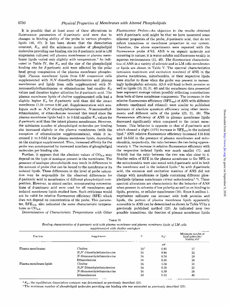
The characteristic temperatures of plasma membranes, mitochondria, and their isolated lipids were determined with &anilino-1-naphthalene sulfonate. Absorbance-corrected fluo- rescence (CO,,J and relative fluorescence efficiency (RFE,,,) were plotted uersus temperature as shown in Figs. 1 and 2. Five characteristic temperatures very similar to those noted with P-parinaric acid’ were indicated by both spectral parameters in membranes as well as lipids obtained from LM cells grown in the presence of choline. As previously indicated with P-pari- naric acid,2 the alterations in the absorbance-corrected fluores- cence (CO,,,) could be due to changes in binding ability of the probe. Such alterations have been shown to be a function of temperature with ANS (47). Therefore, the equilibrium dis-
. sociation constants, K,, were determined as previously de- scribed (23). The K, of ANS in LM suspension cell plasma membranes at 20” was 23 PM, while at 40” it was 12 pM as compared lo the relatively constant K, of P-parinaric acid over this temperature range. Similar alterations were observed with
the isolated plasma membrane lipids. Lowered CO,,, could therefore be due to binding affinity differences. However, the relative fluorescence efficiency, RFE,,,, a concentration- independent parameter, indicated the same characteristic temperatures as COa,, for ANS in LM cell membranes and lipids.
The effect of base analogue supplementation and thereby altered phospholipid composition of LM cell membranes on characteristic temperatures was investigated with ANS. As shown in Tables VII and VIII, the characteristic temperatures were almost identical (+ 1”) for plasma membranes, mitochon- dria, or their isolated lipids. Both CO,,, and RFE,,, indicated that altered phospholipid composition had little if any effect on the characteristic temperatures of either membranes or lipids. It is also possible that differences in phospholipid compositions could result in changes in ANS binding affinity to the LM cell membranes and cause decreases in COssr as shown in Figs. 1 and 2. Again equilibrium dissociation constants were deter- mined to test this possibility. In the case of mitochondria the K, (PM) values at 24” were 30, 30, 27, and 27 for choline-, N,N’-dimethylethanolamine-, N-monomethylethanolamine-, and ethanolamine-supplemented cells, respectively. The iso- lated mitochondrial lipids had lower K, values at 24” (11, 8,9, and 7 fiM, respectively). Therefore the analogue phospholipids increased binding affinity for ANS. ‘These results indicated that drastically lowered values of CO,,, with different base analogues were not primarily due to greatly decreased binding affinities of ANS for the membranes or lipids.
The results reported by ANS with membranes from LM cells supplemented with different choline analogues (Tables VII and VIII) illustrated that the characteristic temperatures indicated by ANS were not altered by the presence of analogue phospho- lipids in either the membranes or their extracted lipids. Thus the results obtained with ANS agree with those obtained by P-parinaric acid despite considerable differences in the nature of the probes, their affinity for lipid, and their location in the membranes.
Effect of Lipid Environment on Fluorescence Properties of Probes in LM Cell Membranes and Membrane Lipids-The affinity of the LM cell lipids and membranes for the probes p-parinaric acid and ANS has been documented. Hence an investigation concerning the absolute values of CO and RFE as a function of polar head group methylation appeared desirable. The results of these studies are shown with @-parinaric acid in plasma membranes, microsomes, mitochondria, and their
by guest on September 9, 2018
http://ww
w.jbc.org/
Dow
nloaded from
6752 Physical Properties of Membranes with Altered Phospholipids
6
4
piziG$q60
20”
-\ 123” -120
01 IO 10 20 30 40 50 10 20 30 40 50
TEMPERATURE (“C)
FIG. 1. Effect of temperature on spectral parameters of ANS in plasma membranes and extracted plasma membrane lipids. As de- scribed under “Materials and Methods,” ANS (final concentration, 4.16 PM) was interacted with LM cell plasma membranes and lipids, CO,,, and RFE,,, were determined, emission was monitored at 480 nm, and-temperature was varied at Z”/min and monitored.
0’ L----lo 10 20 30 40 50 10 20 30 40 50
TEMPERATURE (“C)
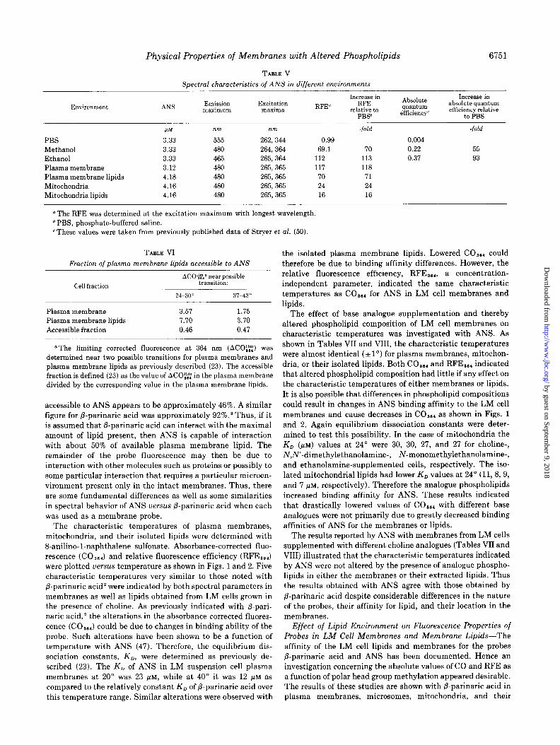
FIG. 2. Effect of temperature on spectral parameters on ANS in mitochondria and extracted mitochondrial lipids. All methods as described in legend of Fig. 1.
isolated lipids at three different temperatures (see Figs. 3 to 5). Several major trends were evident. (a) Decreases or increases in CO,,, and RFE,,, of P-parinaric acid as a function of degree
of polar head group methylation were in the same direction in both membranes and lipids. (b) The CO,,, and RFE,,, indicated similar trends at three different temperatures. (c) Lastly, the probe behaved differently in each particular
membrane or membrane lipid as a function of the number of CH, groups on the polar head group nitrogen of the phospho- lipid analogue. The absolute values of CO,,, and RFE,,,
decreased in plasma membranes and increased in microsomes
with decreasing polar head group methylation. The value for mitochondria did not illustrate any consistent trend. The lipid composition of each of the above membranes isolated from LM suspension cells was quite different,‘, ’ and the distribution of analogue phospholipids also varied between the three mem- branes.’ Similar results were obtained when these experiments were repeated with ANS as shown in Figs. 6 and 7.
DISCUSSION
Previous results (11, 15) indicate that the characteristic temperatures obtained with fluorescent probes are sensitive to the acyl as well as polar head group composition of phospho- lipids. The polar head groups of membrane lipids of LM cells grown in suspension culture with choline analogues were altered such that up to 50% of membrane phospholipids were analogue phospholipid. The data presented here indicate that the characteristic temperatures of such membranes or their lipids were unaltered. Characteristic temperatures, or transi- tions, are believed to reflect changes in the physical states of lipids in membranes (l-4, 6-12, 23-29). If it is assumed that only changes in polar head groups occur on supplementing LM
cells with base analogues, our results would be contrary to expectations of increased transition temperatures with decreasing numbers of methyl groups on the nitrogen of phospholipids as has been shown in model membranes (7, 8,
11). It was previously shown by differential scanning calorime- try as well as by fluorescence probe analysis with P-parinaric acid that the transition temperatures of a series of phospho- lipids with identical acyl substituents increased linearly with decreasing numbers of methyl groups on the nitrogen atom of the phospholipid base (7, 8, 11). The difference in transition temperatures between diacylphosphatidylcholine and an iden- tical diacylphosphatidylethanolamine can be as much as 30” with acyl groups 18 carbons long (8, 11) or 60” with acyl groups 12 or 14 carbons long (15). Therefore, the results obtained with
LM cell membranes’ and whole LM cells (40-42) in which up to 50% of the phospholipids were analogue phospholipids that were incorporated in uivo were indeed surprising. We postulate that the LM cells must be compensating for changes in polar head groups of lipids in order to maintain a constancy of the lipid physical properties as indicated by the characteristic temperatures.
A variety of mechanisms for maintaining the physicochemi- cal properties of membranes may be available to eukaryotes. Several of these mechanisms have been discussed in detail
previously.‘, a One additional compensating mechanism may
be important for LM cell response to choline analogue supple- mentation. In LM suspension cell membranes the percentage of both phosphatidylcholine and phosphatidylethanolamine decrease in the presence of choline analogues.’ These altered ratios themselves may help to compensate for the introduction of new phospholipids with different phase transition tempera- tures and theoretical considerations of this point may be presented. Table IX indicates the theoretical transition tem- peratures, TT, of phospholipid from LM suspension cells
calculated from a series of dipalmitylphosphatides. The actual values of each diacylphosphatide were taken from data of others (7, 8, 11) and were multiplied by the mole fraction of that phosphatide in the LM phospholipids. The sum of such partial contribution to the transition temperature was taken for the four major phosphatides indicated in the table and presented in the last column. The major assumptions in these
by guest on September 9, 2018
http://ww
w.jbc.org/
Dow
nloaded from
Physical Properties of Membranes with Altered Phospholipids
TABLE VII
6753
Characteristic temperatures of plasma membranes and extracted lipids from LM cells supplemented with choline analogues
Characteristic temperatures were obtained with ANS (4.16 PM) as stated under “Materials and Methods.”
Supplement Spectral parameter
Characteristic temperatures
Plasma membranes Plasma membrane lipids
Choline
NJ?-Dimethylethanolamine
N-Monomethylethanolamine
Ethanolamine
Supplement Spectral Characteristic temperatures
parameter Mitochondria Mitochondria lipids
co,,, 18” 23” 33" 37" 45" 19" 25" 30" 37" 45"
RF%,, 20 23 33 36 45 19 25 30 37 44
co,,, 19 23 33 37 42 18 22 30 37 41
RF&,, 19 23 33 37 43 20 24 30 36 42
co,,, 18 23 32 37 42 18 25 31 36 43
RF%,, 19 22 33 37 41 17 22 29 36 44
co,,, 18 23 30 36 42 16 23 31 37 43
RFE 36, 18 22 32 36 42 16 24 30 35 41 -
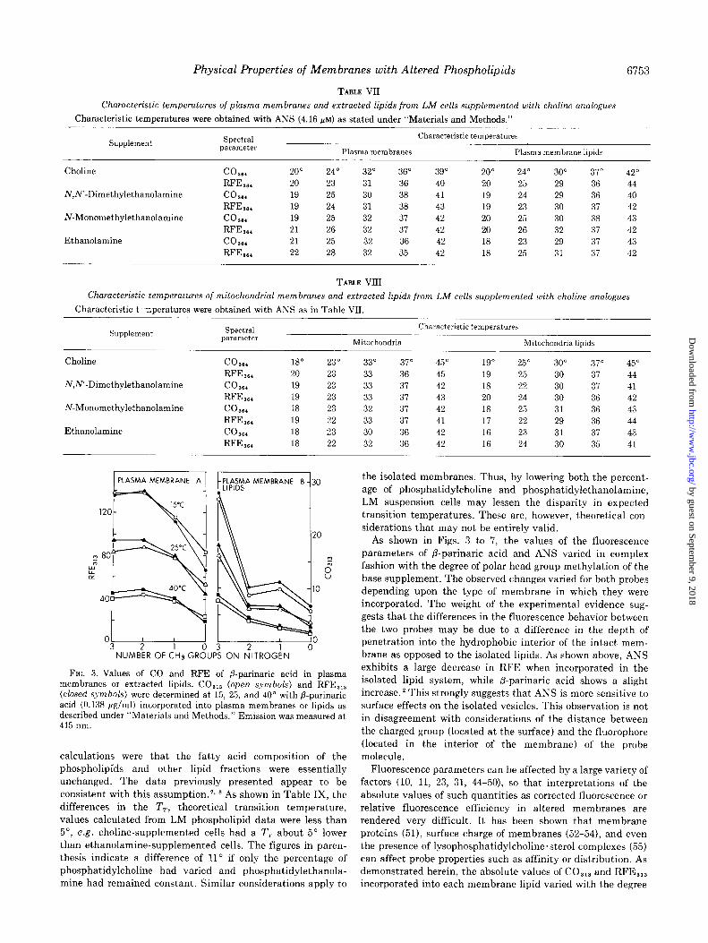
1 PLASMA MEMBRANE A 1 t lfSDh;lA MEMBRANE B 30 the isolated membranes. Thus, by lowering both the percent-
L
3 . age of phosphatidylcholine and phosphatidylethanolamine,
/
WC LM suspension cells may lessen the disparity in expected 120 transition temperatures. These are, however, theoretical con-
siderations that may not be entirely valid.
25°C 2 80
7 i
1
i
g-y
4O'C
40
I
1
NUMBER OFCHsGROUPS ON NITROGEN
As shown in Figs. 3 to 7, the values of the fluorescence parameters of P-parinaric acid and ANS varied in complex fashion with the degree of polar head group methylation of the base supplement. The observed changes varied for both probes depending upon the type of membrane in which they were incorporated. The weight of the experimental evidence sug- gests that the differences in the fluorescence behavior between the two probes may be due to a difference in the depth of penetration into the hydrophobic interior of the intact mem- brane as opposed to the isolated lipids. As shown above, ANS exhibits a large decrease in RFE when incorporated in the isolated lipid system, while fi-parinaric acid shows a slight increase. * This strongly suggests that ANS is more sensitive to
surface effects on the isolated vesicles. This observation is not in disagreement with considerations of the distance between the charged group (located at the surface) and the fluorophore (located in the interior of the membrane) of the probe molecule.
FIG. 3. Values of CO and RFE of P-parinaric acid in plasma membranes or extracted lipids. CO,,, (open symbols) and RFE,,, (closed symbols) were determined at 15, 25, and 40” with fl-parinaric acid (0.138 pg/ml) incorporated into plasma membranes or lipids as described under “Materials and Methods.” Emission was measured at 415 nm.
calculations were that the fatty acid composition of the phospholipids and other lipid fractions were essentially unchanged. The data previously presented appear to be consistent with this assumption.‘, * As shown in Table IX, the differences m the TT, theoretical transition temperature, values calculated from LM phospholipid data were less than 5”, e.g. choline-supplemented cells had a TT about 5” lower than ethanolamine-supplemented cells. The figures in paren- thesis indicate a difference of 11’ if only the percentage of phosphatidylcholine had varied and phosphatidylethanola- mine had remained constant. Similar considerations apply to incorporated into each membrane lipid varied with the degree
Fluorescence parameters can be affected by a large variety of factors (10, 11, 23, 31, 44-50) so that interpretations of the absolute values of such quantities as corrected fluorescence or
relative fluorescence efficiency in altered membranes are rendered very difficult. It has been shown that membrane proteins (51), surface charge of membranes (52-54), and even the presence of lysophosphatidylcholine.sterol complexes (55) can affect probe properties such as affinity or distribution. As demonstrated herein, the absolute values of CO,,, and RFE,,,
Choline
iV,N’-Dimethylethanolamine
N-Monomethylethanolamine
Ethanolamine
CO864 20" 24" 32" 36" 39" 20" 24" 30" 37" 42"
RF%,. 20 23 31 36 40 20 25 29 36 44
co,,, 19 25 30 38 41 19 24 29 36 40
RFE,,, 19 24 31 38 43 19 23 30 37 42
co,,, 19 25 32 37 42 20 25 30 38 43
RF&,, 21 26 32 37 42 20 26 32 37 42
cow.4 21 25 32 36 42 18 23 29 37 43
RF&,, 22 28 32 35 42 18 25 31 37 42
TABLE VIII
Characteristic temperatures of mitochondrial membranes and extracted lipids from LM cells supplemented with choline analogues
Characteristic t -nperatures were obtained with ANS as in Table VII.
by guest on September 9, 2018
http://ww
w.jbc.org/
Dow
nloaded from
6754 Physical Properties of Membranes with Altered Phospholipids
I O ; 3 1 ‘W” 0 3 0
NUMBER OF CH3 GROUPS ON NITROGEN
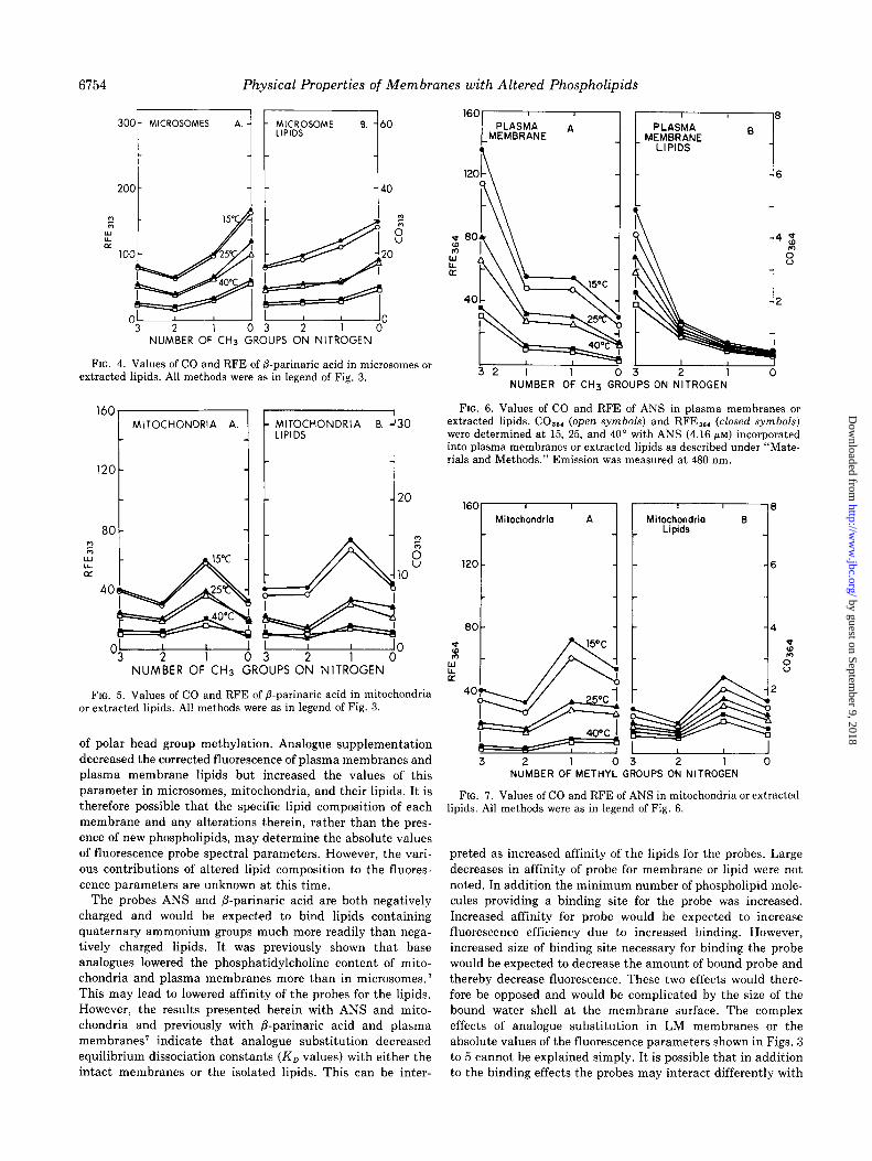
FIG. 4. Values of CO and RFE of P-parinaric acid in microsomes or extracted lipids. All methods were as in legend of Fig. 3.
NUMBER OF CH3 GROUPS ON NITROGEN
FIG. 5. Values of CO and RFE of 8.parinaric acid in mitochondria or extracted lipids. All methods were as in legend of Fig. 3.
of polar head group methylation. Analogue supplementation decreased the corrected fluorescence of plasma membranes and plasma membrane lipids but increased the values of this parameter in microsomes, mitochondria, and their lipids. It is therefore possible that the specific lipid composition of each membrane and any alterations therein, rather than the pres- ence of new phospholipids, may determine the absolute values
of fluorescence probe spectral parameters. However, the vari- ous contributions of altered lipid composition to the fluores- cence parameters are unknown at this time.
The probes ANS and P-parinaric acid are both negatively charged and would be expected to bind lipids containing quaternary ammonium groups much more readily than nega- tively charged lipids. It was previously shown that base analogues lowered the phosphatidylcholine content of mito- chondria and plasma membranes more than in microsomes.’ This may lead to lowered affinity of the probes for the lipids. However, the results presented herein with ANS and mito- chondria and previously with /3-parinaric acid and plasma membranes’ indicate that analogue substitution decreased equilibrium dissociation constants (K, values) with either the intact membranes or the isolated lipids. This can be inter-
6
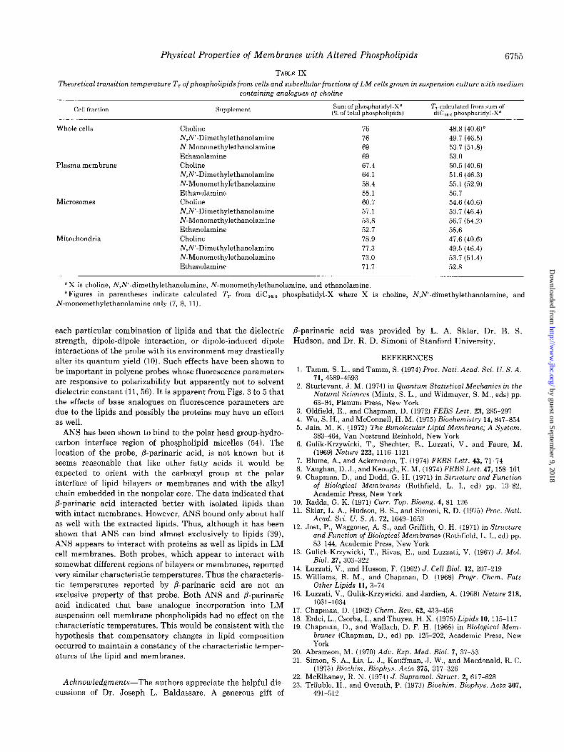
NUMBER OF CH3 GROUPS ON NITROGEN
FIG. 6. Values of CO and RFE of ANS in plasma membranes or extracted lipids. CO,,, (open symbols) and RFE,,, (closed symbols) were determined at 15, 25, and 40” with ANS (4.16 pM) incorporated into plasma membranes or extracted lipids as described under “Mate- rials and Methods.” Emission was measured at 480 nm.
160 Mitochondria A
3 2 1 0 3 2 1 0 NUMBER OF METHYL GROUPS ON NITROGEN
FIG. 7. Values of CO and RFE of ANS in mitochondria or extracted lipids. All methods were as in legend of Fig. 6.
preted as increased affinity of the lipids for the probes. Large decreases in affinity of probe for membrane or lipid were not noted. In addition the minimum number of phospholipid mole- cules providing a binding site for the probe was increased. Increased affinity for probe would be expected to increase fluorescence efficiency due to increased binding. However, increased size of binding site necessary for binding the probe would be expected to decrease the amount of bound probe and thereby decrease fluorescence. These two effects would there- fore be opposed and would be complicated by the size of the bound water shell at the membrane surface. The complex effects of analogue substitution in LM membranes or the absolute values of the fluorescence parameters shown in Figs. 3
to 5 cannot be explained simply. It is possible that in addition to the binding effects the probes may interact differently with
by guest on September 9, 2018
http://ww
w.jbc.org/
Dow
nloaded from
Physical Properties of Membranes with Altered Phospholipids
TABLE IX
6755
Theoretical transition temperature TT of phospholipids from cells and subcellular fractions of LM cells grown in suspension culture with medium
containing analogues of choline
Cell fraction Supplement Sum of phosphatidyl-X” TT calculated from sum of (% of total phospholipids) diC,. D phosphatidyl-X’
Whole cells
Plasma membrane
Microsomes
Mitocbondria
Choline 76 48.8 (40.6)* N,N’-Dimethylethanolamine 76 49.7 (46.5) N-Monomethylethanolamine 69 53.7 (51.8)
Ethanolamine 69 53.0 Choline 67.4 50.5 (40.6) N,N’-Dimethylethanolamine 64.1 51.6 (46.3)
N-Monomethylethanolamine 58.4 55.1 (52.9)
Ethanolamine 55.1 56.7 Choline 60.7 54.6 (40.6) N,N’-Dimethylethanolamine 57.1 53.7 (46.4) N-Monomethylethanolamine 53.8 56.7 (54.2)
Ethanolamine 52.7 58.6 Choline 78.9 47.6 (40.6) N,N’-Dimethylethanolamine 77.3 49.5 (46.4) N-Monomethylethanolamine 73.0 53.7 (51.4) Ethanolamine 71.7 52.8
“X is choline, N,N’-dimethylethanolamine, N-monomethylethanolamine, and ethanolamine. *Figures in parentheses indicate calculated T, from diC,.., nhosphatidyl-X where X is choline, N,N’-dimethylethanolamine, and
N-monomethylethanolamine only (7, 8, 11).
each particular combination of lipids and that the dielectric strength, dipole-dipole interaction, or dipole-induced dipole interactions of the probe with its environment may drastically alter its quantum yield (10). Such effects have been shown to
be important in polyene probes whose fluorescence parameters are responsive to polarizability but apparently not to solvent dielectric constant (11, 56). It is apparent from Figs. 3 to 5 that the effects of base analogues on fluorescence parameters are due to the lipids and possibly the proteins may have an effect as well.
ANS has been shown to bind to the polar head group-hydro- carbon interface region of phospholipid micelles (54). The location of the probe, P-parinaric acid, is not known but it seems reasonable that like other fatty acids it would be expected to orient with the carboxyl group at the polar
interface of lipid bilayers or membranes and with the alkyl chain embedded in the nonpolar core. The data indicated that /3-parinaric acid interacted better with isolated lipids than with intact membranes. However, ANS bound only about half as well with the extracted lipids. Thus, although it has been shown that ANS can bind almost exclusively to lipids (39), ANS appears to interact with proteins as well as lipids in LM cell membranes. Both probes, which appear to interact with somewhat different regions of bilayers or membranes, reported very similar characteristic temperatures. Thus the characteris-
tic temperatures reported by P-parinaric acid are not an exclusive property of that probe. Both ANS and /3-parinaric
acid indicated that base analogue incorporation into LM suspension cell membrane phospholipids had no effect on the characteristic temperatures. This would be consistent with the hypothesis that compensatory changes in lipid composition occurred to maintain a constancy of the characteristic temper- atures of the lipid and membranes.
AcknoLu2edgments-The authors appreciate the helpful dis- cussions of Dr. Joseph L. Baldassare. A generous gift of
/3-parinaric acid was provided by L. A. Sklar, Dr. B. S. Hudson, and Dr. R. D. Simoni of Stanford University.
1.
2.
3. 4. 5.
6.
7. 8. 9.
10. 11.
12.
13.
14. 15.
16.
17. 18. 19.
20. 21.
22. 23.
REFERENCES
Tamm, S. L., and Tamm, S. (1974) Proc. Natl. Acad. Sci. U. S. A. 71, 4589-4593
Sturtevant, J. M. (1974) in Quantum Statistical Mechanics in the iVatura1 Sciences (Mintz, S. L., and Widmayer, S. M., eds) pp. 63-84, Plenum Press, New York
Oldfield, E., and Chapman, D. (1972) FEBS Lett. 23, 285-297 Wu, S. H., and McConnell, H. M. (1975) Biochemistry 14,847-854 Jain, M. K. (1972) The Bimolecular Lipid Membrane; A System,
383-464, Van Nostrand Reinhold, New York Gulik-Krzywicki, T., Shechter, E., Luzzati, V., and Faure, M.
(1969) Nature 223, 1116-1121 Blume, A., and Ackermann, T. (1974) FEBS Lett. 43, 71-74 Vaughan, D. J., and Keough, K. M. (1974) FEBS Lett. 47,158-161 Chapman, D., and Dodd, G. H. (1971) in Structure and Function
of Biological Membranes (Rothfield, L. I., ed) pp. 13-82, Academic Press, New York
Radda, G. K. (1971) Curr. Top. Bioeng. 4, 81-126 Sklar, L. A., Hudson, B. S., and Simoni, R. D. (1975) Proc. Natl.
Acad. Sci. U. S. A. 72, 1649-1653 Jost, P., Waggoner, A. S., and Griffith, 0. H. (1971) in Structure
and Function of Biological Membranes (Rothfield, L. I., ed) pp. 83-144, Academic Press, New York
Gulick-Krzywicki, T., Rivas, E., and Luzzati, V. (1967) J. Mol. Biol. 27, 303-322
Luzzati, V., and Husson, F. (1962) J. Cell Biol. 12, 207-219 Williams, R. M., and Chapman, D. (1968) Progr. Chem. Fats
Other Lipids 11, 3-74 Luzzati, V., Gulik-Krzywicki, and Jardien, A. (1968) Nature 218,
1031-1034 Chapman, D. (1962) Chem. Reu. 62, 433-456 Erdei, L., Csorba, I., and Thuyen, H. X. (1975) Lipids 10,115-117 Chapman, D., and Wallach, D. F. H. (1968) in Biological Mem-
branes (Chapman, D., ed) pp. 125-202, Academic Press, New York
Abramson, M. (1970) Adu. Ezp. Med. Biol. 7, 37-53 Simon, S. A., Lis, L. J., Kauffman, J. W., and Macdonald, R. C.
(1975) Biochim. Biophys. Acta 375, 317-326 McElhaney, R. N. (1974) J. Supramol. Struct. 2, 617-628 TrZuble, H., and Overath, P. (1973) Biochim. Biophys. Acta 307,
491-512
by guest on September 9, 2018
http://ww
w.jbc.org/
Dow
nloaded from
6756 Physical Properties of Men branes with Altered Phospholipids
24.
25.
26. 21. 28.
29.
30.
31.
32. 33.
34.
35.
36.
37. 38.
39.
40.
Wisnieski, B. J., Parkes, J. G., Huang, Y. O., and Fox, C. F. (1974) Proc. Natl. Acad. Sci. U. S. A. 71, 4381-4385
Wisnieski, B. J., Huang, Y. O., and Fox, C. F. (1974) J. Supramol. Strut. 2, 593-608
Shinitzky, M., and Inbar, M. (1974) J. Mol. Biol. 85, 603-615 Grinna, L. S. (1975) Biochim. Biophys. Acta 403, 388-392 Horwitz, A. F., Hatten, M. E., and Burger, M. M. (1974) Proc.
Natl. Acad. Sci. Il. S. A. 71, 3115-3119 R,ittenhouse, H. G., Williams, R.E., and Fox, C. F. (1974) J.
Supramol. Strut. 2, 629-645 Chapman, D., Urbina, J., and Keough, K. M. (1974) J. Biol.
Chem. 249, 2512-2521 Trauble, H., and Eibl, H. (1974) Proc. Natl. Acad. Sci. U. S. A. 71,
214-219 Ohnishi, S., and Ito, T. (1974) Biochemistry 13, 881-887 Michaelson, D. M., Horwitz, A. F., and Klein, M. P. (1974)
Biochemistn, 13, 2605-2612 Henderson, T.- Glonek, T., and Myers, T. C. (1974) Biochemis-
try 13, 623-r Gally, H.-U., Niederherger, W., and Seelig, J. (1975) Biochemistry
14, 3647-3652 Lucas, C. C., and Ridout, J. H. (1967) Progr. Chem. Fats Other
Lipids 10, l-150 Chojnacki, T., and Ansell, G. B. (1967) J. Neurochem. 14,413-420 Bieber, L. L., Seller, L. G., and Kumar, S. S. (1969) J. Biol.
Chem. 244, 630-636 Bridges, R. G., and Ricketts, V. (1967) J. Insect Physiol. 13,
835-850 Glaser, M., Ferguson, K. A., and Vagelos, P. R. (1974) l’roc. Natl.
Acad. Sci. U. S. A. 71, 4072-4076
41.
42.
43. 44.
45.
46.
47.
48. 49.
50.
51.
52.
53.
54.
55.
56.
Ferguson, K. A., Glaser, M., Bayer, W. H., and Vagelos, P. R. (1975) Biochemistry 14, 146-151
Blank, M. L.. Piantadosi. C., Ishaq. K. S., and Snyder, F. (1975) Biochem. Biophys. Res. Commu~. 62, 983-988
Higuchi, K. (1970) J. Cell Physiol. 75, 65-72 Holland, J. F., Teets, R. E., and Timnick, A. (1973) Anal. Chem.
45, 145-153 Holland, J. F., Teets, R. E., Sindmack, G., and Timnick, A. (1976)
Anal. Chem., in press Eling, T. E., and DiAugustine, R. P. (1971) Biochem. J. 123,
539-549 Jacobson, K., and Papahadjopoulis, D. (1975) Biophysical J. 15,
16a Stryer, L. (1965) J. Mol. Biol. 13, 482-495 Schroeder, F., Holland, J. F., and Bieber, L. L. (1972) Biochemis-
try 11, 3105-3111 Ruwart, M. J., Holland, J. F., and Haug, A. (1974) Cryobiology 12,
26-33 Rottem, S., and Samuni, A. (1973) Biochim. Biophys. Acta 298,
32-38 Hackenbrock, C. R., and Miller, K. J. (1975) J. Cell Biol. 65,
615-630 Feinstein, M. B., and Felsenfeld, H. (1975) Biochemistry 14,
3041-3048 Lesslauer, W., Cain, J. E., and Blasie, J. K. (1972) Proc. Natl.
Acad. Sci. U. S. A. 69, 1499-1503 Rand, R. P., Pangborn, W. A., Purdon, A. D., and Tinker, D. 0.
(1975) Can. J. Biochem. 53, 189-195 Hudson, B. S., and Kohler, B. E. (1974) Annu. Reu. Phys. Chem.
25, 437-460
by guest on September 9, 2018
http://ww
w.jbc.org/
Dow
nloaded from
F Schroeder, J F Holland and P R Vagelosphospholipid composition.
Physical properties of membranes isolated from tissue culture cells with altered
1976, 251:6747-6756.J. Biol. Chem.
http://www.jbc.org/content/251/21/6747Access the most updated version of this article at
Alerts:
When a correction for this article is posted•
When this article is cited•
to choose from all of JBC's e-mail alertsClick here
http://www.jbc.org/content/251/21/6747.full.html#ref-list-1
This article cites 0 references, 0 of which can be accessed free at
by guest on September 9, 2018
http://ww
w.jbc.org/
Dow
nloaded from