Embed Size (px)
Citation preview
Physical fitness and exercise in adults with congenital heart
disease
Linda Ashman Kröönström
Department of Health and Rehabilitation/Physiotherapy
Institute of Neuroscience and Physiology
Sahlgrenska Academy, University of Gothenburg
Gothenburg 2020

Cover illustration: Unanswered questions in adults with congenital heart disease by Jenny Wallhult.
Physical fitness and exercise in adults with congenital heart disease © Linda Ashman Kröönström 2020 [email protected] ISBN 978-91-7833-884-9 (PRINT) http://hdl.handle.net/2077/63271 ISBN 978-91-7833-885-6 (PDF) Printed in Borås, Sweden 2020 Printed by Stema Specialtryck AB
SVANENMÄRKET
Trycksak3041 0234

”Learn from the past, observe the present and create the future.”
Author unknown
To my family with love,

Physical fitness and exercise in adults with congenital heart disease
Linda Ashman Kröönström
Department of Health and Rehabilitation/Physiotherapy, Institute of Neuroscience and Physiology
Sahlgrenska Academy, University of Gothenburg Gothenburg, Sweden
ABSTRACT
Background: The population of patients with adult congenital heart disease (ACHD) is growing, and the estimated life expectancy has increased. Therefore, factors related to lifestyle, such as physical activity (PA), exercise, and the risk of lifestyle-related diseases, to which these patients may be more susceptible, are becoming increasingly important to study.
Aim: The aim of this thesis was to increase understanding of physical fitness (muscle function, cardiorespiratory endurance), PA, and health-related quality of life (HRQoL) in patients with ACHD. Another aim was to study patients who have undergone heart surgery using existing arteries that may impact arterial blood supply to the affected arm, with special attention on the arm and spine, and determine whether exercise may improve physical fitness in patients with complex ACHD.
Methods: Studies I and II were register-based cross-sectional studies including patients with different types of ACHD that aimed to assess muscle function and cardiorespiratory fitness, PA level and HRQoL. Study III was also a cross-sectional study and aimed to assess patients with coarctation of the aorta (CoA), a narrowing of the descending aorta, regarding muscle function, arm length and circumference, and spinal mobility. Study IV aimed to assess exercise training with or without supplemental oxygen in patients with complex ACHD with primary outcome measures VO2peak and muscle function.
Results: Studies I and II found that patients with ACHD, even patients with less complicated ACHD, have lower isoinertial muscle function and impaired cardiorespiratory endurance compared to healthy reference values. Study III
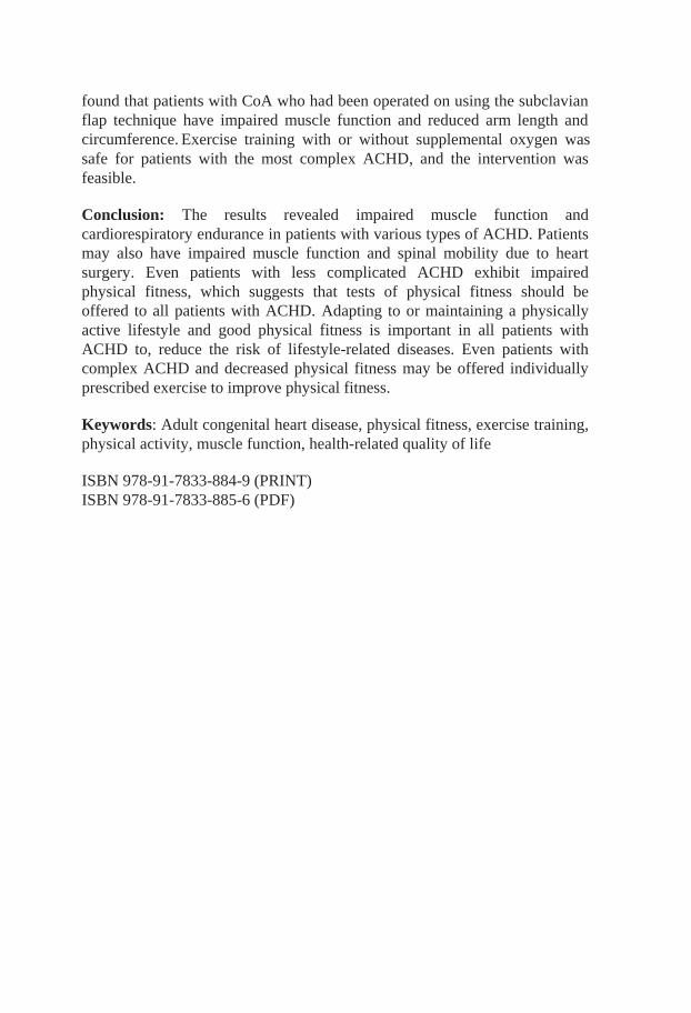
found that patients with CoA who had been operated on using the subclavian flap technique have impaired muscle function and reduced arm length and circumference. Exercise training with or without supplemental oxygen was safe for patients with the most complex ACHD, and the intervention was feasible.
Conclusion: The results revealed impaired muscle function and cardiorespiratory endurance in patients with various types of ACHD. Patients may also have impaired muscle function and spinal mobility due to heart surgery. Even patients with less complicated ACHD exhibit impaired physical fitness, which suggests that tests of physical fitness should be offered to all patients with ACHD. Adapting to or maintaining a physically active lifestyle and good physical fitness is important in all patients with ACHD to, reduce the risk of lifestyle-related diseases. Even patients with complex ACHD and decreased physical fitness may be offered individually prescribed exercise to improve physical fitness.
Keywords: Adult congenital heart disease, physical fitness, exercise training, physical activity, muscle function, health-related quality of life
ISBN 978-91-7833-884-9 (PRINT) ISBN 978-91-7833-885-6 (PDF)

SAMMANFATTNING PÅ SVENSKA Bakgrund: Populationen av vuxna med medfödda hjärtfel (GUCH) ökar och den förväntade livslängden likaså. Livsstilsrelaterade faktorer så som fysisk aktivitet, fysisk kapacitet och risken av livsstilsrelaterade sjukdomar, vilket dessa patienter kan vara extra benägna att få, är därför angelägna att studera. Syfte: Syftet med avhandlingen var att öka kunskapen om fysisk kapacitet för patienter med GUCH samt studera fysisk aktivitet och hälso-relaterad livskvalitet. Syftet var också att studera patienter som har genomgått hjärtkirurgi där man använt en befintlig artär vilket kan påverka arteriell blodförsörjning, med specifikt fokus på arm och rygg, samt studera om fysisk träning kan påverka fysisk kapacitet hos patienter med komplexa hjärtfel. Metod: Studie I och II är registerbaserade tvärsnittsstudier vilka inkluderar patienter med olika typer av GUCH och syftar till att kartlägga muskelfunktion och kardio-respiratorisk kapacitet, fysisk aktivitetsnivå och hälso-relaterad livskvalitet. Studie III är en tvärsnittsstudie som syftar till att kartlägga patienter med Coarctatio aorta (CoA, försnävning av aortas nedåtgående del) avseende muskelfunktion, armlängd och omfång samt ryggrörlighet. I studie IV studeras träning med eller utan syrgas hos patienter med komplexa medfödda hjärtfel utifrån de primära utfallsmåtten maximalt syrgasupptag (VO2peak) och muskelfunktion. Resultat: Resultatet av studie I och II visar nedsatt dynamisk muskelfunktion och kardio-respiratorisk kapacitet hos patienter med GUCH, även de med mindre komplicerade fel. Studie III visar att patienter med CoA som opererats på ett sätt där man påverkat armartären uppvisar en svagare, kortare och mindre omfångsrik arm. Träning, med eller utan syrgas, för patienter med de mest komplexa medfödda hjärtfelen var säker och interventionen var genomförbar. Sammanfattning: Resultatet visar nedsatt muskelfunktion och kardio-respiratorisk kapacitet hos patienter med olika typer av GUCH. Patienter kan också ha en påverkan på muskelfunktion och ryggrörlighet till följd av hjärtoperationer. Även patienter med mindre komplicerade medfödda hjärtfel kan ha nedsatt fysisk kapacitet vilket innebär att tester av fysisk kapacitet bör erbjudas alla patienter. Att påbörja eller bibehålla fysisk aktivitet och en god fysisk kapacitet är av vikt för alla patienter med GUCH, detta för att motverka risken av livsstilsrelaterade sjukdomar. Även patienter med de mest komplexa medfödda hjärtfelen bör erbjudas individanpassad fysisk träning.
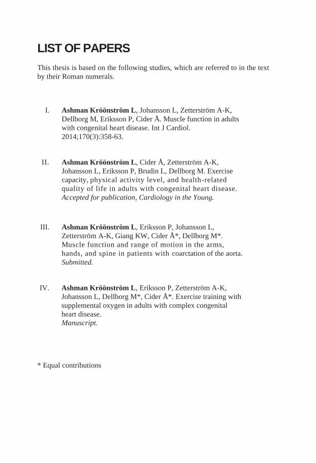
LIST OF PAPERS This thesis is based on the following studies, which are referred to in the text by their Roman numerals.
I. Ashman Kröönström L, Johansson L, Zetterström A-K, Dellborg M, Eriksson P, Cider Å. Muscle function in adults with congenital heart disease. Int J Cardiol. 2014;170(3):358-63.
II. Ashman Kröönström L, Cider Å, Zetterström A-K, Johansson L, Eriksson P, Brudin L, Dellborg M. Exercise capacity, physical activity level, and health-related quality of life in adults with congenital heart disease. Accepted for publication, Cardiology in the Young.
III. Ashman Kröönström L, Eriksson P, Johansson L, Zetterström A-K, Giang KW, Cider Å*, Dellborg M*. Muscle function and range of motion in the arms, hands, and spine in patients with coarctation of the aorta. Submitted.
IV. Ashman Kröönström L, Eriksson P, Zetterström A-K, Johansson L, Dellborg M*, Cider Å*. Exercise training with supplemental oxygen in adults with complex congenital heart disease. Manuscript.
* Equal contributions
IV
V
VII
1
2
8
8
9
9
12
12
13
15
16
17
18
18
19
20
21
23
24
26
26
28
30
30
32
ABBREVIATIONS ............................................................................................DEFINITIONS IN SHORT ..................................................................................PROLOGUE .....................................................................................................1 INTRODUCTION .........................................................................................
Congenital heart disease ......................................................................ACHD unit ..........................................................................................Physical fitness ....................................................................................
1.3.1 Cardiorespiratory endurance .......................................................1.3.2 VO2max .........................................................................................1.3.3 Submaximal exercise capacity ....................................................1.3.4 Skeletal muscles ..........................................................................1.3.5 Anthropometry ............................................................................
Physical activity ..................................................................................Exercise ...............................................................................................
1.5.1 Exercise prescription ...................................................................1.5.2 Resistance exercise ......................................................................1.5.3 Peripheral muscle training ...........................................................1.5.4 Supplemental oxygen during exercise .........................................
Health-related quality of life ...............................................................Physiotherapist a member of the team .............................................
2 RATIONALE FOR THESIS ...........................................................................3 AIMS AND HYPOTHESIS ............................................................................4 METHODS ..................................................................................................
Study population .................................................................................Measurements .....................................................................................
4.2.1 Cardiorespiratory endurance .......................................................4.2.2 Muscle function ...........................................................................4.2.3 Anthropometry ............................................................................
33
34
35
35
36
37
40
40
42
42
44
46
46
51
52
54
54
55
57
58
59
60
63
81
LINICAL IMPLICATIONS AND FUTURE RESEARCH ..................................9 EPILOGUE ................................................................................................ACKNOWLEDGEMENTS .................................................................................REFERENCES .............................................................................................APPENDIX .....................................................................................................

ABBREVIATIONS ACHD Adult congenital heart disease
a-vO2 diff Arteriovenous oxygen difference
CHD Congenital heart disease
CHF Chronic heart failure
CoA Coarctation of the aorta
GUCH Grown-up congenital heart disease
HRQoL Health-related quality of life
IPAQ International Physical Activity Questionnaire
MET Metabolic equivalent of task
NYHA
PA
New York Heart Association
Physical activity
PAH Pulmonary arterial hypertension
PSFS Patient-specific functional scale
RPE Ratings of perceived exertion
SF-36 Short form 36
TCPC Total cavo-pulmonary connection
VO2max Maximal oxygen consumption
VO2peak Peak oxygen consumption
IV
DEFINITIONS IN SHORT Coarctation of the aorta A narrowing of the descending aorta, often
situated below the left subclavian artery and above the ductus arteriosus.
Eisenmenger syndrome Shunt lesion causing pulmonary arterial hypertension and high pulmonary vascular resistance, a severe and irreversible condition (1).
Exercise A subcategory of physical activity that is “planned, structured, and repetitive” and aims at maintenance or improvement of physical fitness (2).
Fontan circulation Palliative heart surgery to convert different congenital heart diseases to a functional single ventricle (3).
Health-related fitness Part of physical fitness and includes the following: cardiorespiratory endurance, muscular endurance, muscular strength, body composition, and flexibility (2).
Isoinertial muscle action Derives from iso (same) and inertial (resistance), and refers to the same resistance in the concentric and eccentric phases of muscle contraction (4).
Isometric muscle action Muscle action without a noticeable change in muscle length (static) (5).
MET 1 MET is the resting metabolic rate or the energy cost of a person at rest (1 kcal × kg-1 × h-1) (6).
Muscle endurance The ability of muscle groups to exert external force for many repetitions, successive exertions (2) or isometric endurance.
V
Muscle function Muscle function includes strength, power, speed, and fatigability (7).
Muscle power (Force × distance)/time (8).
Muscle strength The amount of external force that a muscle can exert (2). Strength can be measured, for example, with one repetition maximum.
NYHA Functional classification system according to symptoms, ranges from I (no symptoms) to IV (symptoms at rest) (9).
Pulmonary arterial hypertension
Associated with ACHD, defined as a mean
rest.
Physical activity Any bodily movement produced by skeletal muscles that results in energy expenditure (2).
Physical fitness A set of attributes that people have or achieve and consists of skill-related and health-related fitness (2).
Scoliosis Angle of trunk rotation defined as a curve 7 degrees (10).
Skill-related fitness Part of physical fitness and includes the following: agility, balance, coordination, speed, power, and reaction time (2).
VO2max Maximal oxygen consumption despite increase in load or intensity (5).
VO2peak Peak oxygen consumption achieved (5).
VI

PROLOGUE In 2007, I started working with cardiac rehabilitation at Sahlgrenska University Hospital, Östra Hospital. During the following years I occasionally meet a few adults with congenital heart disease. However, these patients were few in numbers and mainly participated in groups with other cardiac patients. In 2008, national Swedish guidelines regarding cardiac care advocated that all patients with a congenital heart disease should be evaluated at least once by a physiotherapist and invited to exercise as appropriate and if desired by the patients. During 2009 to 2010, a clinical physiotherapy project took place in the Region Västra Götaland. My colleague, Linda Johansson and I started working on the two positions at Östra Hospital, where the unit for adults with congenital heart disease also is situated. It soon became evident that there were so many interesting queries regarding these patients, and that came to be the starting point of my thesis.
VII

14
1 INTRODUCTION Adult congenital heart disease (ACHD) is one of the most common malformations, with an incidence of slightly less than 1% in living born children. The population of patients with ACHD is growing, as the estimated life expectancy has increased. The risk of being born with congenital heart disease (CHD) has been relatively consistent, and the growing number of patients is a result of improved prognosis and increased survival due to better treatment (11, 12). In Sweden, approximately 30,000-40,000 patients have ACHD of varying degrees of severity (12) and, as in many other countries, the population of patients with ACHD is now most likely larger than the population of children with CHD (13).
Longevity and prognosis have improved in patients with ACHD, and with 95% of patients surviving into adulthood (12). Many of these patients require life-long follow-up (14). Therefore, it is becoming increasingly important to assess factors accompanying longevity and factors associated with physical fitness, health, lifestyle and lifestyle-related diseases. Especially as these patients have been shown to be more susceptible to cardiovascular disease than controls (15). The risk of being diagnosed with ischemic heart disease has also been reported to be 16.5-times greater in patients with ACHD than in healthy controls (16).
The growing population of patients with ACHD means that physiotherapists will come into contact with more of these patients. Therefore, knowledge regarding these patient’s physical fitness and health needs to improve as to provide these patients with the best possible care.
1

CONGENITAL HEART DISEASE CHD consists of approximately 200 different types of diagnoses (13). The variation in clinical presentation is broad, with a few malformations presenting within a few hours or days after birth, and others later in life (14). Patients with ACHD may suffer from the symptoms and complications of CHD, such as arrhythmias and heart failure (17). As a wide variety of diagnoses and complexities occur in ACHD, there is great heterogeneity regarding severity and physical fitness, often decreasing with the complexity of the CHD. The vast amount of CHD diagnoses can be divided into different subgroups according to complexity (Table 1). The functional limitations and symptom severity in these patients are often classified according to the New York Heart Association (NYHA) classification, which was originally designed in 1921 (9) for patients with chronic heart failure (CHF). The NYHA classification ranges from I (no limitations in physical activity) to IV (an inability to carry out physical activity without discomfort) (18).
Table 1. Classification of adult congenital heart disease according to complexity
Diagnoses group Included diagnoses Less complicated Simple shunts
Aortic valve malformations Aortic anomalies Mitral valve lesions Pulmonary valve lesions Tricuspid valve lesions
Corrected Right ventricular/tetralogy of Fallot Transposition of the great arteries
Complex Truncus arteriosus, univentricular repair Others
Coarctation of the aorta (CoA) is one type of CHD and consists of a narrowing of the descending aorta, often below the left subclavian artery and above the ductus arteriosus (17) (Figure 1). CoA constitutes approximately 5-10% of all ACHD cases, with a male predominance. CoA can be an isolated CHD or occur in combination with other CHDs, commonly a bicuspid aortic valve or mitral valve abnormalities (17).
2
16
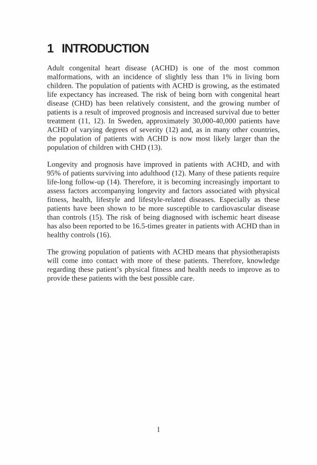
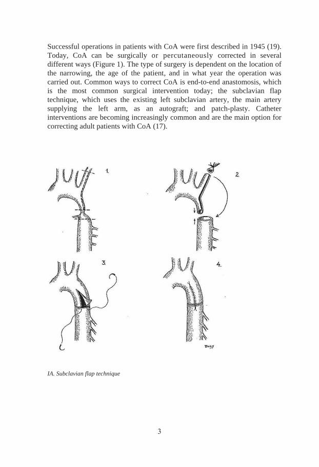
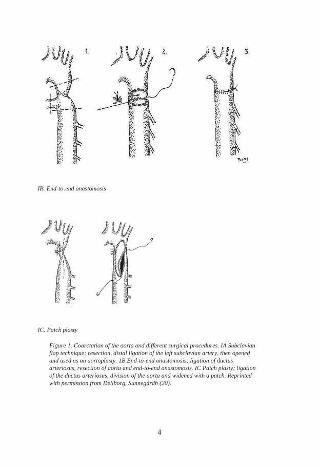
Successful operations in patients with CoA were first described in 1945 (19). Today, CoA can be surgically or percutaneously corrected in several different ways (Figure 1). The type of surgery is dependent on the location of the narrowing, the age of the patient, and in what year the operation was carried out. Common ways to correct CoA is end-to-end anastomosis, which is the most common surgical intervention today; the subclavian flap technique, which uses the existing left subclavian artery, the main artery supplying the left arm, as an autograft; and patch-plasty. Catheter interventions are becoming increasingly common and are the main option for correcting adult patients with CoA (17).
IA. Subclavian flap technique
3
IB. End-to-end anastomosis
IC. Patch plasty
Figure 1. Coarctation of the aorta and different surgical procedures. IA Subclavian flap technique; resection, distal ligation of the left subclavian artery, then opened and used as an aortoplasty. 1B End-to-end anastomosis; ligation of ductus arteriosus, resection of aorta and end-to-end anastomosis. IC Patch plasty; ligation of the ductus arteriosus, division of the aorta and widened with a patch. Reprinted with permission from Dellborg, Sunnegårdh (20).
4
18
Tetralogy of Fallot is a combination of four different CHD; ventricular septal defect, an aortic override, pulmonary stenosis and right chamber hypertrophy as a result of the pulmonary stenosis. Tetralogy of Fallot is the most common cyanotic CHD and has a slight male dominance (17). Heart surgery was first successfully described in these patients in the 1940s through a Blalock-Taussig shunt. A Blalock-Taussig shunt creates a small passage from the systemic circulation to the pulmonary circulation to secure blood flow to the lungs which is a life-saving intervention for patients in need of increased pulmonary blood flow (21). The Blalock-Taussig shunt appears in two different executions, either a classic shunt (using the existing right or left subclavian artery) or a modified shunt (using an external Gore-Tex graft) (22).
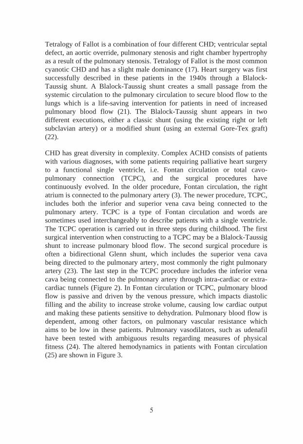
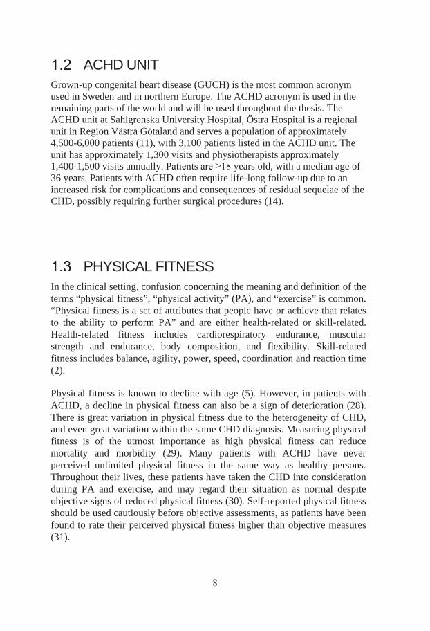
CHD has great diversity in complexity. Complex ACHD consists of patients with various diagnoses, with some patients requiring palliative heart surgery to a functional single ventricle, i.e. Fontan circulation or total cavo-pulmonary connection (TCPC), and the surgical procedures have continuously evolved. In the older procedure, Fontan circulation, the right atrium is connected to the pulmonary artery (3). The newer procedure, TCPC, includes both the inferior and superior vena cava being connected to the pulmonary artery. TCPC is a type of Fontan circulation and words are sometimes used interchangeably to describe patients with a single ventricle. The TCPC operation is carried out in three steps during childhood. The first surgical intervention when constructing to a TCPC may be a Blalock-Taussig shunt to increase pulmonary blood flow. The second surgical procedure is often a bidirectional Glenn shunt, which includes the superior vena cava being directed to the pulmonary artery, most commonly the right pulmonary artery (23). The last step in the TCPC procedure includes the inferior vena cava being connected to the pulmonary artery through intra-cardiac or extra-cardiac tunnels (Figure 2). In Fontan circulation or TCPC, pulmonary blood flow is passive and driven by the venous pressure, which impacts diastolic filling and the ability to increase stroke volume, causing low cardiac output and making these patients sensitive to dehydration. Pulmonary blood flow is dependent, among other factors, on pulmonary vascular resistance which aims to be low in these patients. Pulmonary vasodilators, such as udenafil have been tested with ambiguous results regarding measures of physical fitness (24). The altered hemodynamics in patients with Fontan circulation (25) are shown in Figure 3.
5
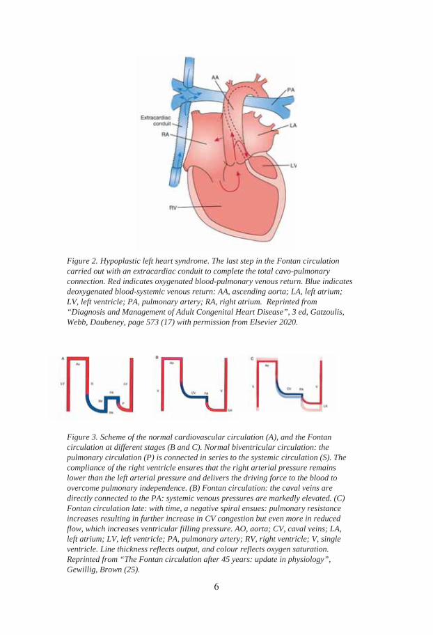
Figure 3. Scheme of the normal cardiovascular circulation (A), and the Fontan circulation at different stages (B and C). Normal biventricular circulation: the pulmonary circulation (P) is connected in series to the systemic circulation (S). The compliance of the right ventricle ensures that the right arterial pressure remains lower than the left arterial pressure and delivers the driving force to the blood to overcome pulmonary independence. (B) Fontan circulation: the caval veins are directly connected to the PA: systemic venous pressures are markedly elevated. (C) Fontan circulation late: with time, a negative spiral ensues: pulmonary resistance increases resulting in further increase in CV congestion but even more in reduced flow, which increases ventricular filling pressure. AO, aorta; CV, caval veins; LA, left atrium; LV, left ventricle; PA, pulmonary artery; RV, right ventricle; V, single ventricle. Line thickness reflects output, and colour reflects oxygen saturation. Reprinted from “The Fontan circulation after 45 years: update in physiology”, Gewillig, Brown (25).
Figure 2. Hypoplastic left heart syndrome. The last step in the Fontan circulation carried out with an extracardiac conduit to complete the total cavo-pulmonary connection. Red indicates oxygenated blood-pulmonary venous return. Blue indicates deoxygenated blood-systemic venous return: AA, ascending aorta; LA, left atrium; LV, left ventricle; PA, pulmonary artery; RA, right atrium. Reprinted from “Diagnosis and Management of Adult Congenital Heart Disease”, 3 ed, Gatzoulis, Webb, Daubeney, page 573 (17) with permission from Elsevier 2020.
6

20
Eisenmenger syndrome, one of the most complex forms of CHD, is characterized by a shunt lesion at any level, which causes secondary increased pulmonary arterial hypertension (PAH) and pulmonary vascular resistance, which may cause right ventricular dysfunction. Due to the pressure of the systemic circulation being higher than that of the pulmonary circulation, the shunting of blood is initially from left to right; however, due to equalized pressures, the shunt will turn and become a right to left shunt (Figure 4). Eisenmenger syndrome is a severe and irreversible condition (1), and mortality rates have improved, but remain high (26). Patients with Eisenmenger syndrome are often treated with endothelin receptor antagonists, such as bosentan, to alleviate the effect of the PAH. Endothelin is considered to play an important role in fibrosis and vasoconstriction (27).
Figure 4. Eisenmenger syndrome. Used with permission of Mayo Foundation for Medical Education and Research, all rights reserved.
Patients with complex ACHD or Eisenmenger syndrome may also have reduced oxygen saturation of the blood (hypoxia), leading to a bluish coloration of the skin and mucous membranes, which is cyanosis. Cyanosis is evident in approximately 10% of patients with ACHD and is not a diagnosis, but a sign and may be present in various ACHD diagnoses, such as shunt lesions (17). Exercise cyanosis is a common feature in these patients. In patients with Eisenmenger syndrome, cyanosis is present during rest, and during exercise an increased shunting of blood from right to left leads to a further decrease in oxygen saturation.
7
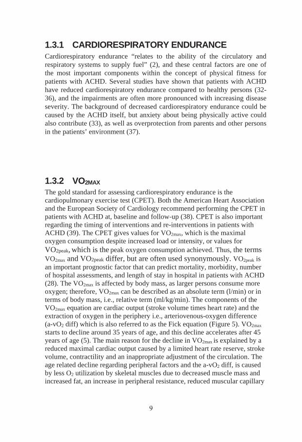
ACHD UNIT Grown-up congenital heart disease (GUCH) is the most common acronym used in Sweden and in northern Europe. The ACHD acronym is used in the remaining parts of the world and will be used throughout the thesis. The ACHD unit at Sahlgrenska University Hospital, Östra Hospital is a regional unit in Region Västra Götaland and serves a population of approximately 4,500-6,000 patients (11), with 3,100 patients listed in the ACHD unit. The unit has approximately 1,300 visits and physiotherapists approximately 1,400-1,500 visits annually. P years old, with a median age of 36 years. Patients with ACHD often require life-long follow-up due to an increased risk for complications and consequences of residual sequelae of the CHD, possibly requiring further surgical procedures (14).
PHYSICAL FITNESS In the clinical setting, confusion concerning the meaning and definition of the terms “physical fitness”, “physical activity” (PA), and “exercise” is common. “Physical fitness is a set of attributes that people have or achieve that relates to the ability to perform PA” and are either health-related or skill-related. Health-related fitness includes cardiorespiratory endurance, muscular strength and endurance, body composition, and flexibility. Skill-related fitness includes balance, agility, power, speed, coordination and reaction time (2).
Physical fitness is known to decline with age (5). However, in patients with ACHD, a decline in physical fitness can also be a sign of deterioration (28). There is great variation in physical fitness due to the heterogeneity of CHD, and even great variation within the same CHD diagnosis. Measuring physical fitness is of the utmost importance as high physical fitness can reduce mortality and morbidity (29). Many patients with ACHD have never perceived unlimited physical fitness in the same way as healthy persons. Throughout their lives, these patients have taken the CHD into consideration during PA and exercise, and may regard their situation as normal despite objective signs of reduced physical fitness (30). Self-reported physical fitness should be used cautiously before objective assessments, as patients have been found to rate their perceived physical fitness higher than objective measures (31).
8
22
1.3.1 CARDIORESPIRATORY ENDURANCE Cardiorespiratory endurance “relates to the ability of the circulatory and respiratory systems to supply fuel” (2), and these central factors are one of the most important components within the concept of physical fitness for patients with ACHD. Several studies have shown that patients with ACHD have reduced cardiorespiratory endurance compared to healthy persons (32-36), and the impairments are often more pronounced with increasing disease severity. The background of decreased cardiorespiratory endurance could be caused by the ACHD itself, but anxiety about being physically active could also contribute (33), as well as overprotection from parents and other persons in the patients’ environment (37).
1.3.2 VO2MAX The gold standard for assessing cardiorespiratory endurance is the cardiopulmonary exercise test (CPET). Both the American Heart Association and the European Society of Cardiology recommend performing the CPET in patients with ACHD at, baseline and follow-up (38). CPET is also important regarding the timing of interventions and re-interventions in patients with ACHD (39). The CPET gives values for VO2max, which is the maximal oxygen consumption despite increased load or intensity, or values for VO2peak, which is the peak oxygen consumption achieved. Thus, the terms VO2max and VO2peak differ, but are often used synonymously. VO2peak is an important prognostic factor that can predict mortality, morbidity, number of hospital assessments, and length of stay in hospital in patients with ACHD (28). The VO2max is affected by body mass, as larger persons consume more oxygen; therefore, VO2max can be described as an absolute term (l/min) or in terms of body mass, i.e., relative term (ml/kg/min). The components of the VO2max equation are cardiac output (stroke volume times heart rate) and the extraction of oxygen in the periphery i.e., arteriovenous-oxygen difference (a-vO2 diff) which is also referred to as the Fick equation (Figure 5). VO2max starts to decline around 35 years of age, and this decline accelerates after 45 years of age (5). The main reason for the decline in VO2max is explained by a reduced maximal cardiac output caused by a limited heart rate reserve, stroke volume, contractility and an inappropriate adjustment of the circulation. The age related decline regarding peripheral factors and the a-vO2 diff, is caused by less O2 utilization by skeletal muscles due to decreased muscle mass and increased fat, an increase in peripheral resistance, reduced muscular capillary
9

density, endothelial dysfunction and reduced oxidative capacity in the muscle (40).
Figure 5. The components of VO2max.
Various factors may impact VO2max including age, gender, PA level, and genetics (5). Numerous other factors in patients with ACHD may also impact VO2max. Stroke volume is one of these factors, as it may be affected in patients with ACHD who may have a right chamber as the systemic chamber or in patients with Fontan circulation with lack of a sub-pulmonary ventricle (cardiac output during rest and exercise in patients with Fontan circulation are illustrated in Figure 6). Chronotropic incompetence (i.e., inability of the heart to regulate heart rate as needed according to physiological stress) (5) may be due to different factors related to surgery and type of CHD (41) or cardiovascular medications known to affect exercise tolerance, heart rate, or blood pressure, such as beta-blockers, ACE-inhibitors, and anti-hypersensitive drugs (42). Beta-blockers blunt beta adrenergic receptors, causing a decreased heart rate. Patients with Fontan circulation with small margins to increase stroke volume, depend on the increase in heart rate, and beta-blockers may negatively impact their exercise capacity. Chronotropic incompetence is present in 84% of patients with Fontan circulation and 90% of patients with Eisenmenger syndrome (43).
Heart rate reserve is the maximal heart rate during exercise minus the heart rate during rest, and may be obtained during a CPET or other maximal exercise tests. A low value is associated with a greater risk of death in patients with complex ACHD, such as Fontan circulation (43). The respiratory exchange ratio (RER) is the ratio of the amount of inspired oxygen and the expired amount of carbon dioxide (VCO2/VO2) measured during CPET. RER also shows the oxidative capacity in the tested person´s body, and furthermore it is possible to study the amount of carbohydrates and lipids expended during the test (44). RER can be obtained during a CPET and
VO2max = cardiac output max × a-vO2 diff max
10
24
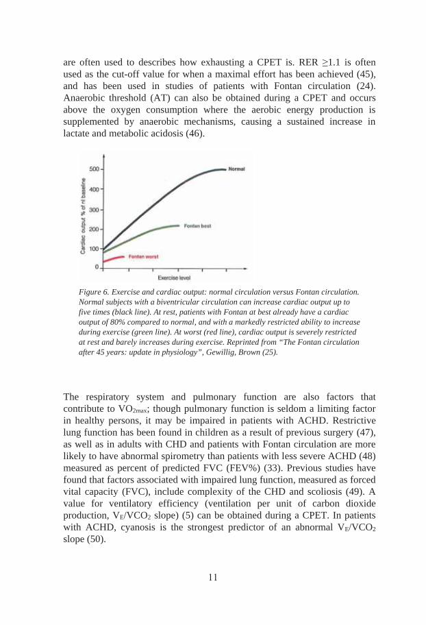
are often used to describes how exhausting a CPET is. RER 1.1 is often used as the cut-off value for when a maximal effort has been achieved (45), and has been used in studies of patients with Fontan circulation (24). Anaerobic threshold (AT) can also be obtained during a CPET and occurs above the oxygen consumption where the aerobic energy production is supplemented by anaerobic mechanisms, causing a sustained increase in lactate and metabolic acidosis (46).
Figure 6. Exercise and cardiac output: normal circulation versus Fontan circulation. Normal subjects with a biventricular circulation can increase cardiac output up to five times (black line). At rest, patients with Fontan at best already have a cardiac output of 80% compared to normal, and with a markedly restricted ability to increase during exercise (green line). At worst (red line), cardiac output is severely restricted at rest and barely increases during exercise. Reprinted from “The Fontan circulation after 45 years: update in physiology”, Gewillig, Brown (25).
The respiratory system and pulmonary function are also factors that contribute to VO2max; though pulmonary function is seldom a limiting factor in healthy persons, it may be impaired in patients with ACHD. Restrictive lung function has been found in children as a result of previous surgery (47), as well as in adults with CHD and patients with Fontan circulation are more likely to have abnormal spirometry than patients with less severe ACHD (48) measured as percent of predicted FVC (FEV%) (33). Previous studies have found that factors associated with impaired lung function, measured as forced vital capacity (FVC), include complexity of the CHD and scoliosis (49). A value for ventilatory efficiency (ventilation per unit of carbon dioxide production, VE/VCO2 slope) (5) can be obtained during a CPET. In patients with ACHD, cyanosis is the strongest predictor of an abnormal VE/VCO2 slope (50).
11
1.3.3 SUBMAXIMAL EXERCISE CAPACITY The CPET is not always performed due to, for example, cost and the availability of the measuring device in the clinical setting. In Sweden, a test with submaximal work levels, such as the symptom-limited ergometer cycle test, is often used in cardiac rehabilitation to evaluate cardiorespiratory endurance. Symptom-limited submaximal tests may be used as an alternative to the CPET when prescribing exercise.
In patients with severely diminished cardiorespiratory endurance, musculoskeletal disorders, or an inability to cycle, an alternative to a bicycle test may be a six-minute walk test (6MWT). The 6MWT may provide valuable information about the submaximal exercise capacity and is an inexpensive test that is simple to perform but less discriminative than the CPET (51).
1.3.4 SKELETAL MUSCLES Peripheral factors such as skeletal muscles (capillarization, the cross-sectional area, muscle fibers, myoglobin content as well as the number of mitochondrion) are key components of physical fitness and determine the a-vO2 diff in the VO2max equation (Figure 5). Muscle fibers (myofibers or muscle cells) are divided into “slow-twitch” type I, and “fast-twitch” type II fibers. The oxidative or “slow-twitch” fibers have slow contraction rates, a high mitochondrial content with an increased reliance on oxidative phosphorylation, are resistant to fatigue and are common in postural muscles. The “fast-twitch” type II fibers are divided into type IIA, IIX and IIB and have rapid contractions, fewer mitochondrion, decreased reliance on oxidative phosphorylation, are easily fatigued and have a high representation in muscle groups used for directional movement (52). Muscle actions require energy which is produced in the mitochondria in the form of adenosine triphosphate, however which type of energy pathway used is dependent on the activity and it´s duration and intensity. Adenosine triphosphate and creatine phosphate (high intensity activities with short duration), anaerobic glycolysis (activity during a couple of minutes) and oxidative phosphorylation (activities with lower intensities for minutes to hours) (53). The mitochondrial rate in “slow-twitch” fibers are two- to threefold higher in density compared to that of “fast-twitch” (54). A review of patients with CHF showed exercise training could increase the mitochondria volume density (55). The type of muscle fibers varies from person to person and
12

26
muscle to muscle. The cross-sectional area of the muscle fiber may increase with resistance exercise (5). Neural and muscular factors together determine muscle function (5). Age related declines in skeletal muscle function (40) are described in section 1.3.2, VO2max. There are also gender differences regarding muscle function, and men present a larger muscle mass than women (5).
Although the levels of cardiorespiratory endurance have been reported in patients with ACHD, very little is known about these patients’ muscle function. In 2006, Brassard (56) stated that the periphery was a forgotten player in patients with Fontan circulation. Since that time, an increasing number of studies have assessed muscle function in patients with ACHD. Reduced maximal hand grip has also been reported (57, 58).
1.3.5 ANTHROPOMETRY Anthropometry describes measurements of the human body in order to assess the composition of the body (59). Common measures of anthropometry are weight, height, body mass index, skinfold thickness, and body circumference which include measures of waist, hips and limbs. In this thesis, spinal and thoracic mobility, as well as the possible impact on the left arm, following surgical interventions were assessed, i.e., body composition.
The human body consists of the following four different types of tissue: epithelial, connective, muscle, and nervous (60). Heart surgery impacts bones (connective tissue), skeletal muscles (muscle tissue), and nerves (nervous tissue). The most common types of corrective heart surgery are sternotomy (incision through the sternum) (61), thoracotomy (incision through the ribcage, often between the third and fourth intercostal muscles) (62) (Figure 7), and percutaneous interventions with a catheter through the femoral artery, which are increasingly more common. A thoracotomy may impact the latissimus dorsi and serratus anterior muscles. The serratus anterior muscle is innervated by the long thoracic nerve and, if damaged, causes a winging of the scapula (63).
The type of incision depends on the localization of the heart anomaly. In patients with CoA as an isolated defect, surgery is often done via a thoracotomy. When a CoA is more complex and associated with other CHD malformations, surgery may have to be done via a sternotomy. In patients in need of a Blalock-Taussig shunt (21), surgery was originally performed
13

through a thoracotomy, left- or right-sided depending on whether the Blalock-Taussig shunt is right- or left-sided. However, surgical procedures evolved and approximately around year 1995, modified Blalock-Taussig shunts became available and can be performed through a sternotomy.
Figure 7. Place of incision for sternotomy and thoracotomy.
Patients with ACHD often go through corrective heart surgery, often multiple times. How these surgeries impact, often newborn, patients with ACHD is unclear. In the clinical setting, we occasionally observed asymmetries in the chest of patients, an area for which research is scarce. A previous study reported that the number of thoracotomies predicts impairments in lung function and physical fitness (64). An increased risk of scoliosis was first reported in patients with ACHD in 1956 (65). An increased risk of scoliosis has also been reported in patients with cardiomegaly (66). Scoliosis may impair both the heart and lungs due to decreased thoracic space, which restricts lung function and may be important to examine in the clinical setting. Other reported spinal impairments include increased kyphosis, which is present in 38% of patients who have undergone both a sternotomy and a thoracotomy (67).
In addition, in the clinical setting, patients with CoA who undergo the subclavian flap technique present signs of asymmetries in muscle function and with a shorter and less muscular arm.
14
28
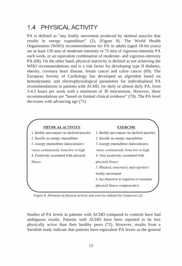
PHYSICAL ACTIVITY PA is defined as “any bodily movement produced by skeletal muscles that results in energy expenditure” (2), (Figure 8). The World Health Organization (WHO) recommendations for PA in adults (aged 18-64 years) are at least 150 min of moderate-intensity or 75 min of vigorous-intensity PA each week, or an equivalent combination of moderate- and vigorous-intensity PA (68). On the other hand, physical inactivity is defined as not achieving the WHO recommendations and is a risk factor for developing type II diabetes, obesity, coronary heart disease, breast cancer and colon cancer (69). The European Society of Cardiology has developed an algorithm based on hemodynamic and electrophysiological parameters for individualized PA recommendations in patients with ACHD, for daily or almost daily PA, from 3-4.5 hours per week with a minimum of 30 min/session. However, these recommendations are “based on limited clinical evidence” (70). The PA level decreases with advancing age (71).
Figure 8. Elements of physical activity and exercise defined by Caspersen (2).
Studies of PA levels in patients with ACHD compared to controls have had ambiguous results. Patients with ACHD have been reported to be less physically active than their healthy peers (72). However, results from a Swedish study indicate that patients have equivalent PA levels as the general
15
population (73), which is also in accordance with the results of PA levels in patients with TCPC (74). Different factors could cause a physically inactive lifestyle, such as underlying CHD. However, PA is primarily associated with factors unrelated to cardiac status in patients with Fontan circulation (75).
When measuring PA, the following four dimensions may be important to capture: mode/type of activity, frequency, duration, and intensity (76). There are several ways to objectively or subjectively assess PA, but the gold standard is the doubly labeled water technique, though it is expensive and requires sophisticated measuring devices (5). The doubly labeled water technique is often used to validate other measures of PA. Objective methods to measuring PA include the use of accelerometers, pedometers, or heart rate monitors. Accelerometers can measure intensity, frequency, and duration whereas pedometers have the disadvantage of not being able to measure intensity. Subjective methods of measuring PA include questionnaires and diaries. The strengths of using questionnaires include low cost and convenience, as well as being applicable to a large number of individuals (76). However, a disadvantage is the risk of recall bias. In this thesis, PA was measured using objective measures (study IV) and subjective measures (studies II and IV) calculated according to the short-form International Physical Activity Questionnaire (IPAQ), which categorizes the PA level as low, moderate, or high (77). PA increase energy expenditure above resting levels, and the rate of energy expenditure is related to the intensity of the PA (76). The IPAQ also estimates each patient’s energy expenditure via metabolic equivalent of task (MET) (78), which describe the energy expenditure for different activities.
Patients with ACHD may overestimate their PA levels compared to objective assessed assessment of PA (79). How PA and physical fitness interact with one another is also of interest; uncertainty exists regarding whether a higher degree of physical fitness leads to an increased level of PA and vice versa.
EXERCISE Exercise is a subset of PA that is planned, structured, and repetitive and has the improvement or maintenance of physical fitness as a final or intermediate objective (Figure 8) (2). There has been a shift in the paradigm regarding exercise in patients with ACHD. In the past, patients were given prohibitive
16

30
advice regarding exercise (80), and recommendations regarding exercise focused on restriction rather than promotion (81).
Studies of exercise in patients with ACHD have been rare, which may be an effect of previously young mortality rates. Exercise is a simple and inexpensive method that can be used to improve physical fitness in patients with ACHD (72, 82). The results of a previous systematic review in children and young adults with ACHD showed that exercise improves VO2peak in patients with ACHD, with a mean VO2peak of 2.6 ml/kg/min. A review including three articles on symptomatic II) reported overall positive effects of exercise regarding quality of life (QoL) and physical fitness (83). The American Heart Association published guidelines in 2019 asserting that cardiac rehabilitation can increase physical fitness in patients with ACHD (38), and the European Society of Cardiology has published recommended levels of intensity during exercise for individual evaluation (70). However, recommendations regarding the frequency, intensity, time and type of exercise (aerobic exercise and muscle function) for patients with ACHD are lacking. As longevity has increased and the population of patients with ACHD is growing, more exercise studies in patients with ACHD will be required to determine these factors. Recommendations for healthy persons, i.e., primary prevention, may not be optimal in patients with ACHD, as these patients may be more susceptible to diabetes (84), hypertension, obesity, and coronary artery disease (85), and in need of secondary prevention.
1.5.1 EXERCISE PRESCRIPTION Exercise aims to improve VO2peak and is often divided into aerobic and resistance exercise. Aerobic exercise is not specifically studied in this thesis and, therefore, is not described in detail, even though resistance exercise contains aerobic components. Prior tests of physical fitness are essential when prescribing exercise, as opposed to giving advice based on described levels of PA or self-perceived symptoms or without evaluating physical fitness. A period of exercise should also be initiated and ended with the same tests in order to evaluate the effects of the exercise program (8). The following five principles may be considered when planning exercise: progressive overload (increased overload), specificity (adaptations specific to the type of activity and intensity), individuality (different response due to
17
genetic factors), reversibility (use it or lose it), and variation/periodization (changes to keep exercise challenging) (8).
Furthermore, exercise needs to be individually prescribed in patients with ACHD (70). When actually prescribing the individual exercise program it is important to consider the following principles (referred to as the FITT principle): Frequency, Intensity, Time (duration), and Type of specific exercise (78). Borg´s Ratings of perceived exertion (RPE) scale (scale 6-20) can be used to determine the right intensity during different types of exercise (86).
1.5.2 RESISTANCE EXERCISE Resistance is an external force used during exercise as for example weights, elastic bands or dumbbells (4). Resistance exercise may affect peripheral factors and lead to an increase in the a-vO2 diff due to an improved capacity of muscle fibers to metabolize oxygen (5). This type of training enhances muscle strength and endurance, functional capacity, independence, and QoL, reduces disability in persons with and without cardiovascular disease, and is to be seen as a complement to aerobic exercise (87). The effects on the skeletal muscle are both neural and muscular, but the first 6-7 weeks are dominated by neural adaptations (recruitment of motor units, increased firing frequency, synchronization between motor units, and increased coordination between muscles and muscle groups). Following the initial period, are muscular adaptations (hypertrophy of existing muscle fibers and possibly an increased number of muscle fibers) (88).
Resistance exercise can be performed in many different ways, and both isometric and isoinertial dynamic tests of muscle function were used in this thesis. However, exercise was dynamic. Dynamic aerobic exercise primarily causes a volume load on the cardiovascular system (87).
1.5.3 PERIPHERAL MUSCLE TRAINING Peripheral dynamic muscle function exercise, i.e., a high relative load on individual muscle groups while maintaining low central circulatory stress (89), has been evaluated in patients with congestive heart failure (CHF) (90-
18
32
92). This type of exercise is useful for debilitated patients with CHF at the beginning of an exercise period, prior to aerobic exercise. Peripheral dynamic muscle function exercises have been efficient in the clinical setting, especially when exercise is initiated or in patients with complex ACHD.
Very few studies have evaluated the effects of muscle function and no reviews or meta-analyses are available in patients with ACHD. However, high intensity resistance exercise in patients with Fontan circulation improved muscle strength 43±7%, with an increase in muscle mass of 1.94 kg. Interestingly, this study showed improvements in not only peripheral factors (i.e., the specific muscle) but also central factors, such as measures of VO2peak (cardiac output, stroke volume) (93).
1.5.4 SUPPLEMENTAL OXYGEN DURING EXERCISE Patients with complex ACHD pose an extra challenge when prescribing exercise, as physical fitness is often severely impaired. Exercise with supplemental oxygen in patients with ACHD has not been explored. A previous study examined the effect of oxygen supplementation in patients with complex ACHD and cyanosis at rest and found an increase in oxygen saturation, probably caused by pulmonary arterial vasodilation (94). We hypothesized that supplemental oxygen may help these patients tolerate exercise at higher intensities.
Fontan circulation and Eisenmenger syndrome are both complex ACHDs, but the pathophysiological backgrounds are different. In patients with Eisenmenger syndrome a main diagnostic criterium is high pulmonary vascular resistance (1). In patients with Fontan circulation the pulmonary vascular resistance is in contrast often normal which these patients benefit from, as a low pulmonary vascular resistance increases pulmonary blood flow. Pulmonary vascular resistance may be affected when adding supplemental oxygen; therefore, patients with a high, respectively low pulmonary vascular resistance may respond differently to supplemental oxygen.
19

HEALTH-RELATED QUALITY OF LIFE QoL is defined by the WHO as: “an individual's perception of their position in life in the context of the culture and value systems in which they live and in relation to their goals, expectations, standards and concerns” (95). Health is also defined by the WHO as: “a complete physical, mental and social well-being and not merely the absence of disease or infirmity” (96). QoL and health-related QoL (HRQoL) are often used interchangeably (97), but HRQoL refers to the patient´s function and well-being when sick and during treatment (98), and is often used to assess an individual’s self-perceived health status (97). Men report a slightly higher HRQoL than women, and HRQoL decreases with age (98).
QoL measures in patients with ACHD are important factors in evaluating the impact of the CHD, as well as care and treatment, from the patient´s perspective (99). The results concerning QoL in patients with ACHD have been ambiguous, which may be partly explained by methodological limitations, such as heterogenous study populations, study designs, and different QoL instruments in a systematic review. Results have shown compromised physical aspects of QoL in these patients (100). A more recent review confirmed discrepancies regarding methodological problems (101). A large international consortium, Assessment of Patterns of Patient-Reported Outcome Measures in Adults with Congenital Heart Disease - International Study (APPROACH-IS), was conducted to assess QoL in patients with ACHD and showed overall good QoL. Characteristics associated with a worse QoL was older age, no marriage history, unemployment, and a more severe NYHA class (102). Results regarding older age are in line with a previous study showing a small decline in HRQoL, measured using the Short Form-36 (SF-36), in patients with ACHD and advancing age, even though the oldest patient was only 54 years old. The decline was most prominent in the Mental component score (103). A previous study found reduced Physical function (SF-36) in patients with moderate and complex CHD (104).
The relationship between physical fitness, PA and HRQoL in patients with ACHD is complex. APPROACH-IS specifically studied PA and QoL in patients with Fontan circulation and showed that higher levels of PA were associated with greater QoL (105). Another study correlated VO2peak with HRQoL as measured with the SF-36 and showed a poor correlation (31). Regarding the impact on HRQoL after exercise interventions, a systematic review of children with CHD found no clear relationship between these factors (106).
20
34
PHYSIOTHERAPIST – A MEMBER OF THE TEAM
The population of patients with ACHD is increasing, presumably leading to physiotherapists coming into contact with this group of patients more frequently, not only in specialized ACHD units, but also in different parts of the health care system. Thus, increased knowledge regarding patients with ACHD is important. Previous studies have indicated a gap in advice concerning PA and physical fitness in patients with ACHD. A study from 2000 (80) stated that adequate discussion on the importance of fitness and patient-centred exercise prescription is rare. In 71% of individuals, the topic of exercise had not been spontaneously raised by their pediatrician, general practitioner, or cardiologist at the adult clinic (80). In Sweden, patients with ACHD did not routinely have the possibility to see a physiotherapist. In 2008, national Swedish guidelines regarding cardiac care advocated that all patients with ACHD should be evaluated at least once by a physiotherapist and invited to exercise as appropriate and if desired by the patients (107).
Physiotherapists are not common members of the team in an ACHD unit. However, since 2009, physiotherapists have been members of the team at the ACHD unit at Sahlgrenska University Hospital, Östra Hospital, working together in the same location with assistant nurses, nurses, cardiologists/medical doctors, social workers and psychologist among others. The ACHD unit uses a method called “one stop shop”, i.e., a patient is offered to see all members of the team at a single appointment. Thus, facilitating for patients that are in the middle of life, occupied with work and family, and improving care for each patient. All members of the team contribute their knowledge and expertise. The part of physiotherapists in the team is among others, to evaluate each patient’s level of physical fitness and assess musculoskeletal problems. Furthermore, a key component is to work together with the members of the team when prescribing individualized exercise. The team approach is valuable from the safety aspect, as patients coming to take part in tests of physical fitness have had a medical examination including, amongst others, anamnesis, measures of blood pressure, and ultrasound. Close collaboration is a key component and a necessity when working with patients with ACHD.
The Swedish Association of Registered Physiotherapists (108) has stated that physiotherapy aims at promoting health, to decrease illness and suffering and furthermore, has defined four concepts important for physiotherapists: body, movement, function, and interaction. The World Confederation for Physical Therapy (WCPT) (109) also recognizes movement as one of the essential
21
definitions of physiotherapists. The WCPT also recognizes the importance of physiotherapists in developing, maintaining and restoring movement and functional ability throughout an individual´s life span. As physiotherapists are specially trained to study the body during movement (110), the aim should be to play an important role in the rehabilitation, care taking and future research of these patients. In customary cardiac rehabilitation for patients with coronary artery disease, physiotherapists play a central role in evaluating and helping patients maintain and/or increase levels of PA and/or physical fitness. Therefore, physiotherapists should aim to play an equally central role in the care of patients with ACHD.
22

36
2 RATIONALE FOR THESIS The number of patients with ACHD is increasing and longevity has improved; therefore, the knowledge about these patients needs to increase. There are gaps in knowledge regarding the components of physical fitness and exercise in patients with ACHD that needs to be addressed. Muscle function is one of these areas, in patients with ACHD. Knowledge regarding possible musculoskeletal impairments in patients with ACHD that may result from the actual CHD or the heart surgery is also lacking.
A paradigm shift has occurred regarding exercise in patients with ACHD. Previously, guidelines and advice regarding exercise were restrictive (38). Today, all patients are encouraged to be active at the individual level (70). The previous restrictive attitude towards exercise, among other factors, has contributed to a lack of knowledge regarding exercise. In particular, patients with complex ACHD and low physical fitness pose an extra difficulty when prescribing exercise. Therefore, studies of the frequency, intensity, time, and type of exercise in patients with ACHD are needed.
23

3 AIMS AND HYPOTHESIS General aim
The project aimed, using different methods, to increase understanding of physical fitness, PA, and HRQoL in a broad and unselected group of patients with ACHD. Another aim was to study patients who have undergone heart surgery using existing arteries that may impact arterial blood supply to the affected arm, with special attention on the arm and spine, and determine whether exercise may improve physical fitness in patients with complex ACHD.
The specific aim of each study is given below.
Study I
To assess muscle function in a sample of adult Swedish men and women with ACHD and to compare results with published reference values from healthy persons.
Hypothesis: Muscle function may be impaired in patients with ACHD, and may be affected by the severity of the CHD.
Study II
To evaluate aerobic capacity, PA level, and HRQoL in patients with ACHD.
Hypothesis: Aerobic capacity measured using a symptom-limited cycle test is impaired in patients with ACHD, and may correlate with the PA levels, which are unknown. HRQoL may be increased in patients with ACHD and correlate with aerobic capacity and PA levels.
Study III
To study muscle function, arm length and circumference, and spinal and thoracic mobility in patients with CoA, and compare these results to a control group.
24

38
Hypothesis: Surgical interventions that affect the circulation in the left arm impacts muscle function, arm length, and arm circumference. In addition, surgical interventions in the chest (thoracotomy or sternotomy) affect spinal and thoracic mobility.
Study IV
To evaluate the effects of exercise training with or without supplemental oxygen in complex ACHD.
Hypothesis: Exercise training with or without supplemental oxygen during exercise impacts VO2peak, muscle function, walking distance, HRQoL, PA level, self-efficacy for exercise, and self-assessed limitations in physical function and blood samples. Furthermore, we hypothesized that exercise training with or without supplemental oxygen would be most important at the start of an exercise period when physical fitness is lowest, and that supplemental oxygen may help patients tolerate exercise at a higher intensity.
25

4 METHODS
STUDY POPULATION Patients in all four studies were registered for their usual care at the ACHD unit, Sahlgrenska University Hospital, Östra Hospital. The requirement for all four studies was The study design is shown in Table 2.
Table 2. An overview of study design, CHD diagnoses, and included patients
Study I Study II Study III Study IV
Study design Cross sectional Cross sectional Cross sectional Randomized cross over intervention
Diagnoses Various CHD Various CHD
Coarctation of the aorta and Tetralogy of Fallot (control group)
Complex ACHD, cyanosis and/or single ventricle
Included patients, no 315 747 75 + 24 8
Study I Inclusion criteria: Out-patients seen at the ACHD unit between April 2009 and December 2010.
Exclusion criteria: Severe arrhythmias, due to undergo surgery, advanced heart failure, severe cerebral lesions, or intellectual disabilities that made it difficult to perform tests of muscle function.
A total of 762 patients were identified for potential inclusion: 235 patients were excluded, 212 patients declined to participate, and 315 (41.3%) patients were included.
Study II Inclusion criteria: Out-patients seen at the ACHD unit between April 2009 and February 2014.
Table 2. An overview of study design, CHD diagnoses, and included patients
CHD=Congenital heart disease.
Study I Study II Study III Study IV Study design Cross sectional Cross sectional Cross sectional Randomized
cross over intervention
Diagnoses Various CHD Various CHD Coarctation of the aorta and Tetralogy of
Fallot (control group)
Complex CHD, cyanosis and/or single ventricle
Included patients, no
315 747 75 + 24 8
26
40
Exclusion criteria: See study I.
A total of 1310 patients were identified for potential inclusion: 223 patients were excluded, 340 patients declined to participate, and 747 patients were included (57%).
Study III Inclusion criteria: Patients with CoA and patients with tetralogy of Fallot (control group) were screened between October 2017 and February 2019.
Exclusion criteria: Factors affecting the mobility or strength of the spine or upper extremities, such as acute back pain, stroke with residual strength impairment in the upper extremities, advanced pregnancy, pre-surgery phase and/or instable cardiorespiratory function, or intellectual disabilities resulting in difficulties performing the required tests. The following exclusion criteria were applied to the control group: no Blalock-Taussig shunt, more than one sternotomy, and no thoracotomy.
A total of 150 patients were identified for potential inclusion: 17 patients (11.3%) were excluded, 34 declined to participate (22.7%), and 99 patients were included (75 patients with CoA and 24 controls).
Study IV Inclusion criteria: Complex ACHD including
Exclusion criteria: Pregnancy, pre-surgery phase/infection/unstable cardiorespiratory function, or cognitive impairment (difficult to answer questionnaires, participate in tests or exercise).
A total of 35 patients were identified for potential inclusion: 3 patients were excluded, 24 patients declined to participate, and 8 patients were included in the study.
27

MEASUREMENTS An overview of measurements used in each study is given in Table 3. Reliability (i.e., the extent of consistency, freedom of random error, and the repeatably over time) and validity (i.e., the extent to which an instrument measures what it is intended to measure) are shown in Table 4.
Table 3. Overview of outcome measures
Measurements Measuring device Study I
Study II
Study III
Study IV
Cardiorespiratory CPET X endurance Symptom-
limited cycle test
X
6MWT X Muscle function Shoulder
flexion Dumbbell X X X
Heel lift X X Handgrip
strength Jamar®/Baseline® X X X
Knee extension
Isobex® X
Shoulder abduction
Isobex® X X X
Elbow flexion Dumbbell X Spinal
stabilization Stopwatch X
Anthropometry Arm length and circumference
Measuring tape X
Structural scoliosis
Scoliometer® X
Thoracic and lumbar mobility, chest expansion.
Measuring tape X
Patient reported IPAQ X X X outcomes SF-36 X X PSFS X SEE X VAS X Other Accelerometer Actigraph®GT3x X Blood samples X
CPET=Cardiopulmonary exercise test, 6MWT=Six-minute walk test, IPAQ=International Physical Activity Questionnaire, SF-36=Short-form36,
28

42
PSFS=Patient Specific Functional Scale, SEE=Self-Efficacy for Exercise, VAS=Visual Analogue Scale.
Table 4. Overview of included measurements and studies of reliability and validity
Measurements Reliability Validity
Cardiorespiratory CPET - -
endurance Symptom limited ergometer cycle test - -
6MWT X (111)
-
Muscle function Shoulder flexion
X (112) -
Heel lift X (112)
-
Handgrip strength, Jamar® X (113) -
Knee extension, Isobex® - -
Shoulder abduction, Isobex® X (114)
-
Elbow flexion - -
Spinal stabilization X (115)
-
Anthropometry Arm length and circumference - -
Structural scoliosis, Scoliometer® X (116)
X (116)
Thoracic and lumbar mobility X (117) -
Chest expansion X (117) -
Patient reported IPAQ X (77) X (77)
outcomes SF-36 X (118) X (118)
Table 4. Overview of included measurements and studies of reliability and validity
Measurements Reliability Validity
Cardiorespiratory CPET - - endurance Symptom limited ergometer cycle test - - 6MWT X (111)
-
Muscle function Shoulder flexion
X (112) - Heel lift X (112)
-
Handgrip strength, Jamar® X (113) - Knee extension, Isobex® - - Shoulder abduction, Isobex® X (114)
-
Elbow flexion - - Spinal stabilization X (115)
-
Anthropometry Arm length and circumference - - Structural scoliosis, Scoliometer® X (116)
X (116)
Thoracic and lumbar mobility X (117) - Chest expansion X (117) - Patient reported IPAQ X (77) X (77) outcomes SF-36 X (118) X (118) PSFS - X (119,
120) SEE - - VAS - - Other Accelerometer, Actigraph®GT3x X (121) X (122)
CPET=Cardiopulmonary exercise test, 6MWT=Six-minute walk test, IPAQ=International Physical Activity Questionnaire, SF-36=Short-form36, PSFS=Patient Specific Functional Scale, SEE=Self-Efficacy for Exercise, VAS=Visual Analogue Scale.
29
31
4.2.1 CARDIORESPIRATORY ENDURANCE CPETs were carried out at the ACHD unit. The starting load was 0 watt, and the protocol was step-wise with increasing loads of 20 watts/minute. Patients wore a mask, and the gas analysis was performed and other variables analyzed as follows. A 10-lead ECG (Schiller, Doral, United States) was continuously monitored, as well as oxygen saturation (SaO2) (Massimo Rad-5v, Irvine, California, USA) using a forehead probe and headband, and continuous blood pressure and heart rate measures. The recorded variables were VO2peak
(absolute and relative), maximal watt level, heart rate, ventilatory equivalent for carbon dioxide (VE/VCO2) slope, and RER.
The submaximal exercise capacity was measured by a symptom-limited ergometer bicycle test (Monark 828E, Varberg, Sweden). The protocol was designed according to the WHO (123) as a step-wise test with increasing loads and an almost steady circulatory state at each level (124-126). The initial load was 25 watts, or 50 watts in a few patients with increasing loads of 25 watts every 4.5 or 5 min. The speed was set to 60 revolutions per minute. Blood pressure was recorded manually with a sphygmomanometer every 2 min (H.E AB, Bandhagen, Sweden). Heart rate was measured via a pulse strap sending impulses to the bicycle (Polar T31, Polar, Bromma, Sweden). The test was ended when patients reached 15-17 on Borg’s RPE scale (127). The test was omitted in the case of decreasing blood pressure, chest pain, or if the patient’s cardiologist had indicated that the patient should only be permitted to reach a lower level of exertion. Due to some patients ending the submaximal ergometer bicycle test before the watt level was finished (i.e., 4.5 or 5 min), the results of the submaximal bicycle test were calculated according to Strandell (128).
Walking distance was assessed by a 6MWT according to the American Thoracic Society (129) with the addition of the assessor walking with the patient carrying the SaO2 device.
4.2.2 MUSCLE FUNCTION Tests of muscle function were performed to evaluate muscle function in the upper and lower extremities and in an isoinertial and isometric manner.
Isoinertial tests
30
32
Shoulder flexion was measured with the patient sitting on a stool, back touching the wall, and a dumbbell in the hand of the arm to be tested (3 kg for men, 2 kg for women). The patient elevated the testing arm, from 0–90 degrees flexion, a maximal number of times at a speed of 20 contractions per min using a metronome (Wittner Taktell Piccolo, Germany).
with the contralateral foot held slightly above the floor. One heel lift was performed every other second using a metronome (Wittner Taktell Piccolo, Germany). The maximal number of heel lifts was registered for each leg (study I) or the dominant leg (study IV).
Elbow flexion was measured with the patient seated on a stool and a dumbbell in the hand of the arm to be tested (4 kg for men, 3 kg for women). The patient elevated the testing arm from 0-145 degrees elbow flexion a maximal number of times at a speed of 20 contractions per min using a metronome (Wittner Taktell Piccolo, Germany).
Isometric tests
Handgrip strength was measured with a Jamar® (Sammons Preston Rolyan, Chicago, USA) or a Baseline® (Fabrication Enterprises, Inc., New York, USA). Both instruments are hydraulic dynamometers measuring maximum handgrip strength in the cylindrical grip. The patient was seated on a chair without armrests, shoulder adducted, and the elbow of the hand being tested held at 90
Shoulder abduction was measured with a portable isometric dynamometer, IsoForce Control® (Medical Device Solutions, Oberburg, Switzerland). The patient sat on a stool, back touching the wall, legs stretched forward and crossed with one heel touching the floor, and the dominanin the scapula direction with a strap placed around the wrist (styloid process).
Knee extension was also measured with IsoForce Control®. The patient sat on a stool, back touching the wall, with the testing leg and, a strap around the dominant leg just over the malleolus. The other leg was stretched forward with the heel touching the floor.
Spinal stabilization was measured by a side bridge test with the patient lying on one side and pelvis raised above the floor. The maximal time in the correct position was noted (130).
31

33
4.2.3 ANTHROPOMETRY Regarding measures of spinal and thoracic mobility, an inspection was performed prior to measuring structural scoliosis in order to took place to assess whether a functional scoliosis (i.e., a functional response to non-spinal conditions, such as a short leg or hip causing pelvic tilt, or poor posture or pain) was present (131). Structural scoliosis was assessed in the thoracic and lumbar region using a Scoliometer® (Mizuho Osi, Union City, California, US), which measures trunk asymmetry and the angle of trunk rotation. The patient was placed in a forward bent position with their arms hanging down according to the manual of the Scoliometer®. Scoliosis was present if measures were (132).
Spinal mobility was measured in the sagittal plane using a measuring tape in the thoracic region (cervical vertebra 7 and 30 cm caudally), in the lumbar region (sacral vertebra 1 and 10 cm cranially), and in the thoracic and lumbar region together (cervical vertebra 7 and sacral vertebra 1) in both flexion and extension (133). Chest expansion was measured using a measuring tape with the patients standing and hands on their head. The delta value between maximal inspiration and maximal expiration was recorded (134). The chest was also assessed in a yes/no manner regarding chest asymmetries (pectus excavatum, pectus carinatum and winged scapula).
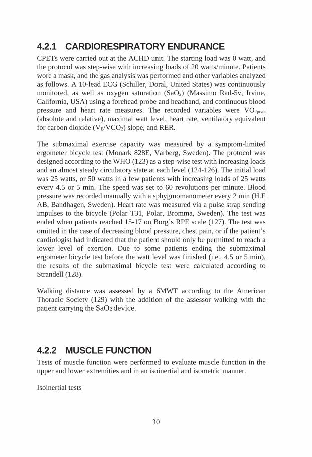
Regarding arm length and circumference, a measuring tape was used to assess the length of the upper arm (acromion/medial epicondyle), forearm (medial epicondyle/styloid process of the radius), and the arm and hand (acromion/fingertip of digitus medius). In addition, the circumference was measured in the middle of the upper arm the middle of the forearm, and the wrist (Figure 9).
32
34
A B
Figure 9. Anatomical disposition regarding measurements of arm length: A1, acromion/ fingertip of digitus medius; A2, acromion/medial epicondyle; A3, medial epicondyle/styloid process of the radius. Anatomical disposition regarding measurements of arm circumference: B1, middle of the upper arm; B2, middle of the forearm; B3, wrist.
4.2.4 PATIENT REPORTED OUTCOMES Patient reported outcomes (PRO) are measurements “of any aspect of a patient´s health status that comes directly from the patient”, and “can be used to measure the impact of an intervention on one or more aspects of patient´s health status” (135). This thesis used the following five questionnaires to measure different outcomes.
The IPAQ - Short form was used to assess self-reported PA level. The IPAQ consists of nine items, and each patient’s PA is counted in METs and divided into one of three subgroups according to the IPAQ manual: low, moderate, or high (136). The IPAQ uses the following values for the three different types of PA intensity: 8 METs, vigorous activity; 4.4 METs, moderate activity; and 3 METs, walking.
33

The Patient Specific Functional Scale (PSFS) is a patient-specific outcome that assesses patients’ self-reported functional status limitations that are most relevant to the individual patient (119).
Self-reported HRQoL was assessed with the SF-36, consisting of the following eight scales: physical function (PF), physical role function (RP), general health (GH), bodily pain (BP), role limitations caused by emotional problems (RE), social functioning (SF), vitality (VT), and mental health (MH). The PF, RP, BP, and GH scales add up to summarize the Physical component scale (PCS), and VT, SF, RE, and MH scales summarize the Mental component scale (MCS) (137).
Self-efficacy for exercise training – Swedish version (SEE) examines the estimated degree of self-efficacy in relation to adherence to exercise, which is an individual's perception of how confident they feel in being able to perform exercise despite various perceived barriers (138). The result of each of the nine questions is marked on a Likert scale (scale 0-10), where zero represents "not safe/confident at all" and 10 represents "absolutely safe/confident" (139, 140).
The visual analog scale (VAS) was originally designed to evaluate mood (141). The VAS in this study consisted of a 10 cm line (assessed in millimeters the scale consists of a 100-point scale) (142) with the wording “best imaginable” and “worst imaginable” at either end. The VAS was used to assess patients’ experiences in exercise training with or without supplemental oxygen.
4.2.5 PHYSICAL ACTIVITY An accelerometer (ActiGraph® GT3x+, ActiGraph, Pensacola, Florida, USA) was used to objectively assess levels of PA during 7 days at baseline and after intervention. Information was retrieved regarding the total amount of PA (counts per minute), the time that the patient was inactive, and the amount of time at different intensities (low, moderate, and high).
34
35
4.2.6 BLOOD SAMPLES The following blood samples were included: hemoglobin, serum ferritin, erythrocyte volume fraction (platelets), high-density lipoprotein (HDL), low-density lipoprotein (LDL), triglycerides, N-terminal prohormone brain natriuretic peptide (NT-proBNP), high-sensitivity troponin T (hsTnT), glycated hemoglobin (HbA1c), and venous blood gas. The aforementioned blood samples were chosen to assess the composition of the blood, as well as possible effects of exercise. The total amount of blood required was approximately 45 ml. The blood samples were analyzed according to the European accreditation system at Sahlgrenska University Hospital, Östra Hospital laboratory.
INTERVENTION Study IV
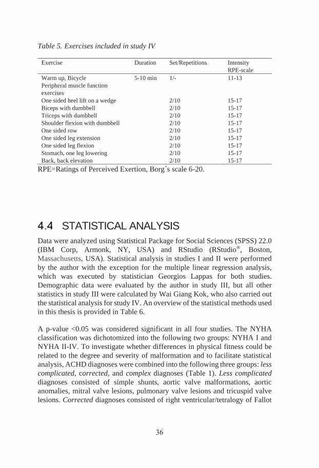
The intervention used in study IV was based on peripheral muscle function exercises and prescribed according to the FITT-principle (section 1.5.1, Exercise prescription) (78). The frequency of the exercise program was twice weekly for a total of 12 weeks. Each session constituted approximately 60 min of exercise. The type of exercise training consisted of a warm-up on a bicycle and nine peripheral muscle function exercises for both the upper and lower extremities performed in two sets of 10 repetitions (Table 5). The intensity was set to an RPE of 15-17 (hard/very hard) (86) in the exercising muscle, and resistance was individually prescribed to achieve the necessary intensity.
35
37
Table 5. Exercises included in study IV
Exercise Duration Set/Repetitions Intensity RPE-scale
Warm up, Bicycle 5-10 min 1/- 11-13 Peripheral muscle function exercises
One sided heel lift on a wedge 2/10 15-17 Biceps with dumbbell 2/10 15-17 Triceps with dumbbell 2/10 15-17 Shoulder flexion with dumbbell 2/10 15-17 One sided row 2/10 15-17 One sided leg extension 2/10 15-17 One sided leg flexion 2/10 15-17 Stomach, one leg lowering 2/10 15-17 Back, back elevation 2/10 15-17
RPE=Ratings of Perceived Exertion, Borg´s scale 6-20.
STATISTICAL ANALYSIS Data were analyzed using Statistical Package for Social Sciences (SPSS) 22.0 (IBM Corp, Armonk, NY, USA) and RStudio (RStudio®, Boston, Massachusetts, USA). Statistical analysis in studies I and II were performed by the author with the exception for the multiple linear regression analysis, which was executed by statistician Georgios Lappas for both studies. Demographic data were evaluated by the author in study III, but all other statistics in study III were calculated by Wai Giang Kok, who also carried out the statistical analysis for study IV. An overview of the statistical methods used in this thesis is provided in Table 6.
A p-value <0.05 was considered significant in all four studies. The NYHA classification was dichotomized into the following two groups: NYHA I and NYHA II-IV. To investigate whether differences in physical fitness could be related to the degree and severity of malformation and to facilitate statistical analysis, ACHD diagnoses were combined into the following three groups: less complicated, corrected, and complex diagnoses (Table 1). Less complicated diagnoses consisted of simple shunts, aortic valve malformations, aortic anomalies, mitral valve lesions, pulmonary valve lesions and tricuspid valve lesions. Corrected diagnoses consisted of right ventricular/tetralogy of Fallot
36
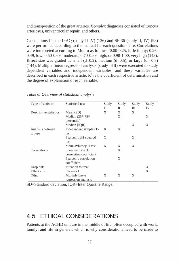
38
and transposition of the great arteries. Complex diagnoses consisted of truncus arteriosus, univentricular repair, and others.
Calculations for the IPAQ (study II-IV) (136) and SF-36 (study II, IV) (98) were performed according to the manual for each questionnaire. Correlations were interpreted according to Munro as follows: 0.00-0.25, little if any; 0.26-0.49, low; 0.50-0.69, moderate; 0.70-0.89, high; or 0.90-1.00, very high (143). Effect size was graded as small (d=0.2), medium (d=0.5), or large (d= 0.8) (144). Multiple linear regression analysis (study I-III) were executed to study dependent variables and independent variables, and these variables are described in each respective article. R2 is the coefficient of determination and the degree of explanation of each variable.
Table 6. Overview of statistical analysis
Type of statistics Statistical test Study I
Study II
Study III
Study IV
Descriptive statistics Mean (SD) X X X Median (25th-75th
percentile) X X
Median [IQR] X X Analysis between groups
Independent samples T-test
X X
Pearson´s chi-squared test
X X
Mann-Whitney U test X X X Correlations Spearman’s rank
correlation coefficient X
Pearson’s correlation coefficient
X
Drop outs Intention to treat X Effect size Cohen’s D X Other Multiple linear
regression analysis X X X
SD=Standard deviation, IQR=Inter Quartile Range.
ETHICAL CONSIDERATIONS Patients at the ACHD unit are in the middle of life, often occupied with work, family, and life in general, which is why considerations need to be made to
37
39
limit the time spent in the physical examination or participation in various studies. These patients have often been through several medical examinations and it is important to inform them that not participating in studies does not in any way impact their ordinary care at the ACHD unit. Written informed consent was obtained in all four studies. The regional ethical review board in Gothenburg, Sweden, approved all four studies (study I and II, DNR: 226-13; study III, DNR: 157-17; and study IV, DNR: 349-17 with the addition of DNR: 2019-04610). The studies complied with the Declaration of Helsinki (145).
Study IV
Exercise training with or without supplemental oxygen in adults with complex CHD has not been evaluated previously. The study included patients with complex ACHD such as Fontan circulation or Eisenmenger syndrome, i.e., patients with the most complex ACHD and impaired physical fitness. Patients performed peripheral dynamic muscle exercises. In patients with CHF, resistance training was previously restricted due to possible negative effects on left ventricular function, potential negative remodeling, and increased afterload (87). A few studies have studied aerobic exercise and the potential negative impact on the heart in patients with ACHD. Moderate to high intensity exercise was studied in patients with transposition of the great arteries with regard to the heart failure markers hsTnT and NT-proBNP. No negative impact on the heart was present to indicate more severe heart failure (146, 147). Furthermore, no negative impact on the heart was found in patients with tetralogy of Fallot (148) or after 12 weeks of aerobic exercise in patients with a single ventricle (149). A study in patients with ACHD and systemic right ventricle showed that high intensity exercise is safe and could be performed without incident (146). Based on both research (i.e., no negative remodeling of the heart) and clinical experience showing that patients benefit from exercise, this study assessed peripheral muscle exercises.
Patients included in the study were severely ill; therefore, the intervention required close monitoring. An external medical doctor, Kristofer Skoglund, Sahlgrenska University Hospital, with deep knowledge of both ACHD and research, was asked to assess patients’ medical journals for potential side effects during the study. Heart failure markers (hsTnT, NT-proBNP) and platelets (bleeding) were assessed at baseline, cross over, and after intervention. The exercise intervention was performed at the physical therapy department, and the physiotherapists in charge of the exercise had good knowledge of ACHD. The patients were closely monitored regarding SaO2
38

40
levels during exercise. An emergency plan, as well as heart- and lung resuscitation equipment, was available and staff educated on a regular basis. If necessary, medical doctors and nurses were available at the ACHD unit and, in the case of serious cardiac events, patients could be transported to the emergency rescue.
39

41
5 RESULTS The results of each of the included studies are attached in the Appendix, and a summary of the results from studies I-IV are presented below. Study II, III and IV are not yet published, and as such the results are brief.
STUDY I Muscle function in adults with CHD
The patients performed five tests of muscle function: two isoinertial tests and three isometric tests. Seven hundred and sixty-two patients were assessed, 315 (41.3%) of which performed the tests. The results indicated that patients with ACHD have lower isoinertial muscle function than healthy reference values regarding the heel lift test (Figure 10) and the shoulder flexion test (Figure 11). Regarding the heel lift test, men with ACHD performed at 63% and women at 58% of the healthy reference values, and the shoulder flexion tests (men with ACHD performed at 60% and women 85% of healthy reference values). Furthermore, NYHA class II-IV was a significant predictor of lower isoinertial muscle function regarding the following tests: heel lift in both men (p=0.05) and women (p<0.001), and shoulder flexion (p<0.001), isometric knee extension (p=0.04) and isometric shoulder abduction (p<0.001) in women (150).
40

54
Figure 10. Isoinertial muscle function test, heel lift in men and women with ACHD compared to reference values. Reprinted from publication (147) with permission from International Journal of Cardiology.
Figure 11. Isoinertial muscle function test, shoulder flexion in men and women with ACHD compared to reference values. Reprinted from publication (147) with permission from International Journal of Cardiology.
10
15
25
35
45
Num
bero
fhee
llift
s
0
10
20
30
40
50
60
70
80
Num
bero
fsho
ulde
rfle
xion
s
41
43
STUDY II Exercise capacity, PA level, and HRQoL in ACHD
In this study, the exercise capacity of men was 58.7% and of women 66.3% compared to reference values. Approximately one fourth of the patients did not achieve the recommended amount of PA. Regarding HRQoL, men scored significantly fewer points on 7 out of 10 scales on the SF-36, and women in 6 out of 10 scales compared to reference values. The strongest correlation was between exercise capacity and the Physical Function scale of the SF-36.
STUDY III Muscle function and range of motion in the arms, hands, and spine in patients with CoA
Significant delta values were measured between the right and left arm in patients with CoA, subclavian flap compared to controls in four out of five included tests of muscle function (Figure 12). The results also indicated significant differences regarding delta values for arm length as well as differences in regards to measures of arm circumference (Figure 13). Measurements of spinal and thoracic mobility showed that scoliosis was more frequent in patients who had undergone thoracotomy than patients who only underwent sternotomy. Scoliosis was often located in the thoracic part of the spine.
42

44
Figure 12. Results of muscle function in patients with: CoA, subclavian flap; compared to CoA, other (included end to end, patch plasty, balloon angioplasty and non-operated patients); and control group.
CoA, Subclavian flap CoA, Other Control group
CoA, subclavian flap compared to CoA, other; significant differences in 2 of 5 tests.
CoA, subclavian flap compared to control group; significant differences in 4 of 5 tests.
Figure 12. Results of muscle function in patients with: CoA, subclavian flap;compared to CoA, other (included end to end, patch plasty, balloon angioplastyand non-operated patients); and control group.
43

45
Figure 13. Results of arm length and circumference in patients with: CoA, subclavian flap; compared to patients with CoA, other (included end to end, patch plasty, balloon angioplasty and non-operated patients); and control group.
STUDY IV Exercise training with or without supplemental oxygen in adults with complex CHD
Patients were randomized to start exercising with supplemental oxygen (OxyStart) or without (OxyEnd). Four patients were randomized to the OxyStart group and four to the OxyEnd group. There were no drop outs in the OxyStart group, but two patients in the OxyEnd group dropped out. MET values at baseline were 5 (IQR, 2.3). RER-values regarding all CPET were 1.08 (IQR, 0.12). No adverse events occurred during exercise.
CoA, Subclavian flap CoA, Other Control group
CoA, subclavian flap compared to CoA, other; significant differences in 5 of 5 tests.
CoA, subclavian flap compared to control group; significant differences in 6 of 6 tests.
Figure 13. Results of arm length and circumference in patients with: CoA, subclavian flap;compared to CoA, other (included end to end, patch plasty, balloon angioplasty andnon-operated patients); and control group.
44
46
The patient’s own perceptions regarding exercise training, and exercise training with or without supplemental oxygen are described in the following quotations.
Exercise training;
- “Not as much anxiety over physical barriers as before… a freedom in that.”
- “I don´t think of the planning of physical barriers as before.”
Exercise training with or without supplemental oxygen;
- “I did not get an exercise-related head-ache when exercising with oxygen.”
- “More strain with oxygen.”
- “The recovery was best with oxygen.”
45

47
6 DISCUSSION
MAIN FINDINGS Study I
The results demonstrated lower isoinertial muscle function in patients with ACHD, even in patients with less severe ACHD, though the decreased muscle function was more pronounced in patients with more complex ACHD. Our results imply that lower muscle function in these patients may contribute to the impaired physical fitness (VO2peak) well known in these patients (36).
Our results of impaired isoinertial muscle function are in line with others. In patients with complex ACHD, the dynamic muscle function, measured as shoulder flexion and heel lifts, was reduced (151). However, two previous studies found a decreased isometric muscle function measured as handgrip-strength (57, 58) which was not in accordance with the results of our study which showed no significant difference regarding isometric muscle function (150). The reasons for the declined muscle function have been sparsely studied. The decreased isometric muscle function was not found to be explained by a decreased cross-sectional area (57). Furthermore, normal muscle and bone development (in proportion to reduced body height) was found in adolescents and young adults with CHD (152).
Blood flow to skeletal/peripheral muscles is a key component of efficient muscle function (5). The peripheral muscle pump aids venous filling during exercise in patients with Fontan circulation (153). The altered hemodynamics in patents with Fontan circulation may impact muscle function in ways that differ from other patients with ACHD. A blunted peripheral blood supply has been found in patients with Fontan circulation (154), and altered hemodynamics and endothelial dysfunction has been shown in the vastus lateralis muscle, which has a negative impact on muscle function (155). Furthermore, altered muscle oxygen kinetics has been found during rest, exercise, and recovery in the deltoid muscle of patients with Fontan circulation compared to controls (156). A small study of patients with Eisenmenger syndrome showed that these patients maintain the same oxidative mitochondrial capacity in skeletal muscles (measured in the gastrocnemius) as healthy controls (157). Patients with PAH exhibit structural muscle differences, with a lower proportion of “slow-twitch” type I fibers, which is explained by a conversion to “fast-twitch” type II muscle fibers (i.e., a shift
46
48
from aerobic metabolism to more anaerobic metabolism) and a decreased cross-sectional area regarding “slow-twitch” type I muscle fibers (158).
The reasons for the decreased muscle function are still unclear. The future will determine whether patients with ACHD have structural muscle differences of decreased muscle function or if the decreased muscle function has other multifactorial explanations, such as severity of CHD and physical inactivity. In healthy young men in Sweden, a good muscle strength has been found to have a good cardio-protective effect against arrhythmias and vascular disease, such as heart failure, ischemic heart disease, and stroke (159). Therefore, patients with ACHD may be likewise in need of good muscle function to decrease the aforementioned risks. Furthermore, the response to resistance exercise and its potential impact on muscle function requires further investigation.
Study II
The results showed that physical fitness is impaired in patients with ACHD, even in patients with less severe CHD diagnoses. Patients included in the study had a mean age of 35 years and performed at 48% of age-adjusted physical fitness values. Longevity has improved in the population of patients with ACHD, and the patients will get older (12). Physical fitness is known to decline with age (5) and needs to be regularly assessed. In patients with ACHD, a decline in physical fitness may also be a sign of deterioration, adding to the impact of these tests. Individually prescribed exercise may be tailored to impact declining physical fitness as, with advancing age, diminished physical fitness may impact these patients’ activities of daily living and prognosis (28).
The PA level contributes to the level of physical fitness. One fourth of the patients in the present study did not achieve the recommended amount of PA; however, three-quarters of the patients did actually achieve the recommended amount of PA. A previous study reported that patients with ACHD are equally as active as controls (73). This may indicate that the level of PA contributes to the level of physical fitness, but other factors may contribute to the greater decline in physical fitness. A low level of PA is a risk factor for developing type II diabetes, obesity, coronary heart disease, breast, and colon cancers (69), cardiovascular disease (160), and ischemic heart disease (16). Moreover, there may be patients with certain ACHD diagnoses who are more susceptible to acquired cardiovascular disease. In patients with CoA, cardiovascular risk factors (diabetes, hypertension, hyperlipidemia, smoking, obesity, and/or
47
49
sedentary life-style), are prevalent, and 9 out of 10 patients had at least one risk factor, even though patients had a median age of 43.5 years (161). All but one of these cardiovascular risk factors may be modifiable with exercise, which may imply that certain CHD diagnoses may benefit from increased levels of PA and exercise. Specific exercise and PA recommendations for adults with various ACHDs may be desirable.
Men and women with ACHD exhibit decreased HRQoL compared to reference values in 7 out of 10 and 6 out of 10 scales, respectively. The complexity of ACHD did not have a major impact on patients’ HRQoL in our study. The correlations between physical fitness, PA, and HRQoL were assessed. A posed correlation between a physically inactive lifestyle may contribute to reduced physical fitness, which may correlate with reduced HRQoL. This may imply that HRQoL can be influenced by adopting a physically active lifestyle or exercise training. Therefore, exercise training leading to increased physical fitness may be a valuable factor that impacts HRQoL positively in these patients.
Study III
The results showed that heart surgery creates a disparity between arms and impairs muscle function, arm length, and arm circumference in the left arm in patients with CoA who have undergone the subclavian flap technique. These results were in line with our stated hypothesis. One feared complication of the subclavian flap technique is gangrene in the affected arm (162). Previous articles have also assessed possible negative impacts on the arm, and a small study of nine children found normal vascular function in the affected arm. However, minor reductions in forearm growth (circumference and length) were also reported (163). A more recent study in patients with ACHD found lower muscle mass and reduced cross-sectional area in the affected left arm (164).
Both isoinertial and isometric muscle function tests were performed. Isometric tests (handgrip strength, shoulder abduction, spinal stabilization) required work for a maximum of 5 seconds. Isoinertial tests (shoulder flexion and elbow flexion) required work until fatigue (fatigue progresses until an ability to maintain required force or power, and progresses more rapidly at higher intensities (165)). Thus, the tests demanded different types of muscle activity. However, both isoinertial and isometric muscle function were impaired in the affected left arm. The results showed no significant difference in spinal
48
50
stabilization, which may indicate that this test has less clinical value when aiming to detect differences between the left and right side. Speculatively, this test may not have been as specific as the other tests and may have involved more muscles than the other tests, which aimed to assess one specific muscle.
Regarding thoracic and spinal mobility, we found an increased risk of scoliosis in patients who had undergone a thoracotomy. Many previous studies have investigated scoliosis and reported an incidence of 6% to 42.4% (66, 166-169). However, there is heterogeneity regarding the etiology and incidence of scoliosis, the position of the scoliosis, and the side of the curve. Heterogeneity may be explained by different methodology, the use of different definitions of scoliosis, the assessment of different CHD diagnoses, and different combinations of thoracotomy/sternotomy. We also found impaired mobility in the chest which may be a result of previous thoracotomies/sternotomies and may also be a contributing factor to previous studies showing restrictive lung function (48) and, thus important to evaluate.
We included a screening for other asymmetries in the chest that may be related to thoracotomy or sternotomy, such as winged scapula, and found it to be present in 9.4% of patients who underwent a thoracotomy and none of the patients who underwent sternotomy. Presumably, patients in the thoracotomy group may have had impairments in the serratus anterior muscle or the innervating nerve during heart surgery. However, the number of winged scapula were much lower than those observed in children, the prevalence of which was reported to be 70% (170). Surgical precautions are currently done to spare the serratus anterior muscle. A few patients who have expressed back trouble, or in whom tests have shown asymmetries in the spine, received an individualized exercise training program to perform at home. A few patients living in geographic proximity to the hospital came to the physical therapy department to participate in individualized training programs which mostly consisted of stabilizing trunk exercises.
Patients who had gone through radical operations for tetralogy of Fallot without a Blalock-Taussig shunt, with a maximum of one sternotomy and no thoracotomy were included as a control group, as these patients had not had surgical procedures affecting any artery to the arm or thoracotomies, making comparisons between thoracotomies and sternotomies possible.
Clinical assessment is an important component of care, which is often life-long in patients with ACHD. Patients with ACHD have gone through multiple roentgenograms and been exposed to radiation; therefore, a need exists for clinical tools to assess scoliosis. Measurements regarding muscle function, arm
49
51
length, and arm circumference, as well as measurements regarding spinal and thoracic mobility used in the present study, may be useful for detecting asymmetries in the upper extremities and when assessing patients with ACHD. Potential asymmetries in the chest, such as scoliosis, may be painful and cause potential restrictive lung function; thus, the patient would benefit from detection. Patients with scoliosis may also benefit from individually prescribed trunk stabilizing exercises. A general decline in physical fitness is to be expected as patients’ age (40, 171, 172). Whether impairments and asymmetries in the upper extremities result in pain and/or inability to work is yet to be discovered. Furthermore, the impact of exercise on muscle function impaired by surgical procedures needs to be addressed.
Study IV
Results of the present study showed that patients with Eisenmenger syndrome and complex ACHD with cyanosis exhibited diminished physical fitness, which is expected and in agreement with other studies (35, 36). We assessed the impacts of exercise training (primary aim) as well as the impacts of exercise training with or without supplemental oxygen (secondary aim).
None of the included patients reached VO2max but VO2peak. Results obtained from the CPET regarding showed 5 METs at baseline, a result that implies that physical fitness is decreased to the level that it impairs activities of daily living in these patients. Cleaning, kitchen activities such as cooking and washing dishes, and vacuuming require a MET value of 3.3 (6).
RER increases during exercise and physically active and trained persons have a lower RER than untrained persons (44). RER 1.1 is often considered to be the cut-off value for obtaining maximal exhaustion (45, 173). The median RER obtained during the study was 1.08 (0.12). However, previous studies in patients with ACHD have reported difficulties in obtaining RERs 1.1. Only 50% of patients with Eisenmenger syndrome achieved RER , despite exercising until exhaustion (157). Cyanotic patients are less likely to reach an RER of 1.0 than non-cyanotic patients (28). Pulmonary limitations have been reported in patients with ACHD (49) and may be one reason for not being able to achieve an RER 1.1 (45).
The secondary aim was to assess exercise training with or without supplemental oxygen. As this study was the first to examine exercise training with supplemental oxygen, the patient’s own perception of how they were
50
52
affected and responded to the exercise training was therefore noted and showed ambiguous results. Exercise with supplemental oxygen may not affect patients with different ACHD etiology uniformly, future studies are needed to investigate this.
Patients with complex ACHD with a very low physical fitness pose a particular challenge in prescribing individualized exercise, and little is known about which type of exercise is beneficial. In the clinical setting, it is common that patients who previously had restrictions regarding exercise are unaccustomed to exercise and how the body reacts to exercise. These patients require a lot of advice and help when starting exercise and may benefit from a longer period of exercise. Patients with complex ACHD have a low central oxygen uptake and difficulties in improving cardiac output, therefore the periphery needs to be seen as an important component. Peripheral muscle function exercises are perhaps therefore, to be emphasized and individual exercise prescription contain these types of exercise. Patients with the most complex ACHD and the most impaired physical fitness is patients whom will benefit most from exercise training and notice a difference in activities in daily living.
METHODOLOGICAL CONSIDERATIONS In Sweden, the care of patients with ACHD is centralized to seven ACHD units, including two tertiary units (includes heart surgery and other medical care related to the CHD). The ACHD unit at Sahlgrenska University Hospital is the only tertiary center within the region of Västra Götaland. Therefore, patients at the unit may be more severely ill than ACHD patients in general, which may have had an impact on the patients included in the studies. Physiotherapists are part of the ordinary care for patients with ACHD and situated in the same location, and patients are accustomed to being offered tests of physical fitness as well as individualized exercise programs as part of their routine visit at the ACHD unit.
Due to the large number of different ACHD diagnoses, divisions can be made to create larger groups and improve readability. Studies I and II used the following division to form larger diagnosis groups based on the complexity of the CHD: less complicated, corrected, and complex (Table 1). However, other divisions are nowadays widely accepted (11, 174, 175) and have increased comparability between studies. In the clinical setting, as well as in research, the NYHA classification is commonly used in patients with ACHD. The
51
53
validity of NYHA has been evaluated against objective measures obtained during a CPET (VO2peak and VE/VCO2 slope) in patients with ACHD and found to be a valid method according to functional limitations. However, the classification was found to underestimate limitations in less symptomatic patients (176). Therefore, tests of physical fitness need to be assessed as opposed to giving advice based solely on self-perceived symptoms and described PA levels.
Power calculations in studies III and IV were discussed when designing the study. The main outcome measures in study III were disparity between arms regarding muscle function and arm length and circumference. However, at the time of the study, no previous studies of these measurements were available and, therefore calculations were not performed. Regarding study IV, power calculations could have been performed according to previous intervention studies of VO2peak in patients with ACHD, but the number of eligible patients at the ACHD unit enabled these calculations, and calculations of sample size would have shown an underpowered study (Type II error).
6.2.1 PHYSICAL FITNESS The verbal communication between patient and assessor was standardized during all tests of physical fitness included in this thesis, as patient motivation is an important factor when conducting tests of physical fitness; some patients will push themselves more than others. To increase reliability between assessors (inter-rater reliability), test manuals were produced for each study. To further increase reliability, instruments were calibrated on a regular basis.
The protocol used in the CPET was a ramp protocol with increases in load with 20 watt every minute. Smaller increases in load may have increased the possibility of detecting minor changes in VO2peak.
The VO2peak and maximal heart rate is not known in all patients. Therefore, exercise training are often prescribed based on submaximal tests and ratings of perceived exertion (127). The advantages of using the submaximal symptom-limited cycle test is that patients reach a hemodynamically steady state, defined as an increase in heart rate of less than 10 beats/min from the second to the last minute of that particular load (124). Another advantage of the steady state test is the similarities between the test and the exercise often utilized in cardiac rehabilitation. In this thesis, we used a submaximal symptom-limited bicycle
52

54
test and present an algorithm to compare submaximal tests and maximal tests. One advantage of using the reference values we used (42) was that patients had cycled to the same level of exertion on Borg´s RPE scale (86) as patients with ACHD. No severe cardiac problems occurred when performing the present test therefore, a symptom- limited exercise test appears to be safe in patients with ACHD. In addition, our results imply that a submaximal test may be useful in the clinical setting when prescribing exercise and no CPET is carried out, and is simple to perform and requires less preparation. Other measurements of submaximal exercise capacity may be beneficial in patients with ACHD. In patients with coronary artery disease, a systematic review found strong evidence that the 6MWT is responsive to changes in physical fitness (111). The diagnostic value of the 6MWT has been evaluated in children with PAH, showing a close correlation between walking distance 300 m and relative VO2peak (177). In patients with ACHD, a correlation was found between relative VO2peak ml/kg/min which could excellently be identified with the 6MWT (178). However, the 6MWT is not easy to use when prescribing exercise. Furthermore, the 6MWT does provide less information of the mechanisms of exercise restrictions and a decreased sensitivity has been shown in patients with milder PAH symptoms and longer walking distance (179).
There are different ways to assess muscle function and no gold standard is available. Different aspects of muscle function, such as strength, power, speed, and endurance can be measured. In this thesis, the term muscle function was used as an umbrella term to cover all of these aspects. Muscle function may also be assessed in both the upper and lower limbs, in different muscles, and by isoinertial and isometric tests. In study I, we used the term isotonic (dynamic), but a more accurate term is isoinertial muscle action, which describes the same level of exertion in both the whole range of motion i.e. both the concentric and eccentric phase (4). Isoinertial tests often require exertion for a longer duration and, thus, require a greater amount of aerobic capacity and other effects on the muscle and its components to provide adenosine triphosphate. However, isometric tests, only require work for a short period of time, perhaps requiring different things in the muscle than longer periods of duration. The known gender differences regarding muscle function were compensated for in all isoinertial tests and women had lighter weights than men.
Handgrip strength has been found to be useful, as it can represent muscle function in the upper extremities (180). This thesis assessed handgrip strength using two different devices, Jamar® and Baseline®, but using the same execution. Inter-instrument reliability between these 2 instruments was 0.90 and the authors conclude that instruments measures equivalently and can be
53
55
used interchangeably (181). Handgrip strength was recently assessed as a diagnostic marker regarding survival but was not found to be suitable for survival assessment; the authors concluded that handgrip strength is useful for identifying and following up patients with increased cardiovascular risk (182). Handgrip strength has also been found to correlate well with better lung function, which may indicate that resistance exercise affects, and even improves, lung function (183).
6.2.2 EXERCISE In study IV, the type of exercises could have been prescribed individually, but the same exercises were chosen in order to standardize the exercise training protocol. However, individual exceptions were made if certain exercises were not suitable; in these cases, a similar exercise was chosen to exercise the same muscle but with a different execution. Exercises and tests of muscle function were created to resemble one another as much as possible, as similarities between them increase the chance of revealing an effect (8).
6.2.3 PATIENT REPORTED OUTCOMES Regarding PROs, questionnaires could not be filled out for some patients due to intellectual disabilities or problems regarding the Swedish language, which is not unexpected when assessing large numbers of patients. This thesis assessed HRQoL using the SF-36, a generic questionnaire consisting of 36 questions and covering eight scales and scores ranging from 0-100, with higher results indicative of better HRQoL (137). This generic instrument was used to assess HRQoL, as no diagnoses-specific questionnaire was found. SF-36 also had the advantage of published Swedish reference values (98). Euro-Qual 5 dimensions (EQ-5D) (184) is a generic QoL instrument consisting of a descriptive part and EQ-VAS. EQ-5D is sometimes used to assess HRQoL in patients with ACHD, but the SF-36 contains more questions than EQ-5D and can be assumed to give a greater understanding of HRQoL. In addition, the physical components can be addressed specifically. Questionnaires including more questions generally have higher validity and reliability (98). The VAS has become widely adopted due to its simplicity, as well as the possibility of
54
56
adapting the scale (142); however, the questions included in study IV have not been validated.
In this thesis, both objective (accelerometer, ActiGraph®) and subjective measurements (questionnaire, IPAQ) were used to assess PA levels. The IPAQ has been tested for validity and reliability, with ambiguous results, and the correlations between the IPAQ and objective measures has been reported to be small to medium (0.09 to 0.39) (185). Self-assessed levels of PA during the last week may be subject to recall bias, which may contribute to the low correlation. However, no other known questionnaire is preferred due to the advantages of the IPAQ having been used in many previous studies and the availability regarding published Swedish reference values (186, 187). Self-reported levels of PA assessed with questionnaires are known to be overestimated (185). When calculating the results of the IPAQ, if the patient had written an estimation of the time or day spent on PA, the lowest number was used in order to adjust for the known overestimation that occurs when using self-reported levels of PA.
6.2.4 STRENGTHS AND LIMITATIONS Reference values obtained from other published studies always infer questions about comparability. In studies I and II, a group of healthy persons who had performed all of the included tests would have been preferred but was not possible to obtain in the clinical setting. However, the strengths of studies I-II were the large number of unselected patients with ACHD that were included.
The scope of the article in study III regarding disparities between arms has not been previously described in a large number of patients with CoA. Studies regarding exercise training in patients with complex ACHD, are scarce and exercise training with supplemental oxygen is not evaluated. A randomized controlled study has the highest evidence value when it comes to clinical studies, but due to the number of eligible patients in study IV no such study could be performed. Study IV showed that the intervention was safe and feasible and therefore contributed with knowledge regarding exercise training. Even patients with complex ACHD and decreased physical fitness may be offered individually prescribed exercise training to improve physical fitness.
55
57
7 CONCLUSION The results of this thesis show that patients with ACHD have impaired physical fitness, measured as isoinertial muscle function and submaximal exercise capacity. Even patients with less complicated ACHD exhibit impaired physical fitness, which may imply that tests of physical fitness should be offered to all patients with ACHD to detect symptoms of deterioration and the possible need for interventions. Tests of muscle function can be used in addition to tests of cardiorespiratory endurance when assessing physical fitness in patients with ACHD in order to evaluate peripheral factors. Patients with ACHD can have decreased HRQOL, especially the physical function component and the correlation between the two may suggest that HRQoL can be improved with exercise.
Patients with ACHD who have undergone heart surgeries using existing arteries that impact the arterial blood supply to the affected arm can exhibit decreased muscle function, as well as reduced arm length and circumference. Surgical interventions through the chest may also impact the incidence of scoliosis and chest asymmetries.
This thesis has shed light on exercise and the impact it may have on different aspects of physical fitness for patients with complex ACHD. Exercise training with or without supplemental oxygen for patients with the most complex ACHDs was safe and the intervention feasible. In patients with complex ACHD, low central oxygen uptake, and difficulties in improving cardiac output, the periphery needs to be seen as an important component. Therefore, peripheral muscle function exercises should be emphasized and individual exercise prescriptions should contain these types of exercises. Patients with the most complex ACHDs and the most impaired physical fitness will benefit from exercise training and notice a difference in activities in daily living.
Adapting to or maintaining a physically active lifestyle and good physical fitness is important in all patients with ACHD to reduce the risk of lifestyle- related diseases. Physiotherapists, with knowledge within the specific pathophysiology of CHD and knowledge of body and movement, play an important role in assessing physical fitness and exercise in these patients. Increased knowledge regarding these patients´ physical fitness and HRQoL is important to optimizing care for these patients.
56

58
8 CLINICAL IMPLICATIONS AND FUTURE RESEARCH
Team work is essential when working with patients with ACHD and should be the gold standard when assessing these patients. As patients with ACHD age, questions will continue to arise regarding physical fitness. Therefore, physical fitness needs to be regularly in order to provide these patients optimal care.
Individually prescribed exercise should be based on measurements of physical fitness rather than advice based solely on symptoms and assumptions. Physical fitness impacts morbidity and mortality. Therefore, exercise is of uttermost importance to increase physical fitness. Exercise needs to be emphasized in patients with ACHD, especially in patients with complex ACHD with the most pronounced decrease in physical fitness. Future large randomized controlled trials should focus on the frequency, intensity, time, and type of exercise in patients with various ACHD diagnoses.
57
59
9 EPILOGUE I have worked with patients with ACHD at the unit in Gothenburg since 2009. In these years so much has happened. We have learnt from the past and found a shift in paradigm. Patients with ACHD have previously been restricted and given prohibitive advice regarding physical activity and exercise, and recommendations regarding exercise focused on restriction rather than promotion. We have observed the present in the included studies. However, the future is here now, and lies in front of us and includes encouragement to physical activity and exercise, even for patients with complex ACHD. To finalize, to refer to one of the patients describing the essence of how exercise may impact these patients lives - a feel of freedom in not having to worry over physical barriers as before.
Thank you for reading! Linda
58

60
ACKNOWLEDGEMENTS I am very grateful to all who, in many different ways, have contributed to this thesis. This thesis has taught me much and, apart from new knowledge, has given me the opportunity to meet a lot of interesting people.
All of the patients at the ACHD unit who generously participated in this thesis, thank you for all that you have taught me about your experiences living with CHD, and for teaching me everything you know about, for example, criminology, E-sports, dinosaurs, and Japanese tv-games. I hope that this thesis has contributed to a better understanding of ACHD and improved and made a difference in the everyday clinical setting.
My SUPERvisors! Åsa Cider supervisor, you encouraged me to start my PhD-studies, thank you for your support! I am fascinated with the great amount of exercise physiology you know. You are always striving to improve care for patients and are a true source of knowledge in the clinic. Mikael Dellborg co-supervisor, you have sped up this thesis up with your fast replies to an endless number of e-mails and drafts. I am very impressed with your research knowledge, thank you for sharing it with me! Peter Eriksson co-supervisor, the starting point of this thesis was the clinical project in physiotherapy for patients with ACHD in 2010 in which your positive attitude and grit was priceless. Your ability to enthuse the people you work with is impressive. All three of you have always been encouraging and willing to share your knowledge with me, I am very grateful for that! Your humorous emails have made me laugh out loud many times!
My friends and colleagues, Linda Johansson, for starting the clinical project together with me at the Physical Therapy Department over 10 years ago. So much work has been put in, but it has also been so rewarding - it has been great sharing this with you. Anna-Klara Zetterström, I just love your frankness and humor. Linda Bäckdahl for your encouragement. I am so grateful to all three of you for your never-ending support, for making me smile every day at work, and for your friendship (even though I seldom reply to your texts).
Gunilla Kjellby-Wendt, head of the Occupational and Physical Therapy Department, Sahlgrenska University Hospital for your support. Sara Lundqvist, head of the Physical Therapy Department, Östra Hospital for your encouragement. Klara Emanuelsson Hummel and other colleagues who worked on the clinical project for shorter periods of time. Lisbeth Radås for your help with practical issues, telephone calls, letters and bookings. All my
59
61
colleagues at the Physical Therapy Department, for your encouragement and interest in this thesis through these years.
The fantastic team at the ACHD unit – Karin Olander, Kerstin Lundblad, Marie Jussila, Gunilla Andulv, Ingvor Mårtensson, Caritha Tell, Eva Furenäs, Anders Ahnfält, Thomas Gilljam, Zacharias Mandalenakis and Camilla Barth. I am very grateful for your willingness to share your knowledge about these patients, your friendly manner, and the positive atmosphere at the ACHD unit. I am also very grateful for your invaluable help with assessing patients for potential enrolment in the different studies. The teamwork in the ACHD unit is great and has been a true source of inspiration. A special thank you to Zacharias Mandalenakis for help assessing the CPET in study IV and Karin Olander and Kerstin Lundblad for helping me out many times with the same device.
Kristofer Skoglund for knowledgeable help with study IV.
Georgios Lappas and Wai Giang Kok for explaining statistics in a simple way.
Lars Brudin, co-author of study II, for help with the calculations regarding the submaximal bicycle tests.
Helena Dellborg and Görel Hultsberg-Olsson for your help with blood samples in study IV.
To the colleagues in the Optimal exercise and fitness group (Opti-Ex-Fit) under Åsa Ciders supervision. Maria Borland for lending us ActiGraph, as well as showing me all the pros and cons regarding the thesis template. Other doctoral students and lecturers at the Institute of Neuroscience and Physiology, Physiotherapy for taking time to read drafts, for asking difficult questions, and discussions.
All my friends and family, for all your support during this journey and all of your interested questions about this thesis.
My parents in law, Harry and Lotta for looking out for our family and for your friendly ways.
My mother Monika Wallqvist, my father David Ashman, my sisters Jenny and Helen, thank you for everything, your support has been invaluable! Nan, your eagerness to learn new things, you inspire me!
60

62
My family, Christofer, my husband, what can I say – thank you for your endless support and patience throughout this journey. Thelma and Frank, you light up our lives and help me keep my priorities right. I am so proud of you!
This thesis was supported by the following grants:
The Research and Development of Region Västra Götaland, Swedish Heart-Lung Foundation (grant number 20180644), The Swedish Fund for Congenital Heart Defects, the Local Research and Development Council Gothenburg and southern Bohuslän, the Swedish state under the agreement between the Swedish government and the County Council under the ALF agreement (grant number 720191), Renée Eander’s Foundation, and kungliga and Hvitfeldtska Foundation for the grant and for the time at Villa Martinsson.
61

62
REFERENCES 1. Wood P. The Eisenmenger syndrome or pulmonary hypertension
with reversed central shunt. Br Med J. 1958;2(5099):755-62. 2. Caspersen CJ, Powell KE, Christenson GM. Physical activity,
exercise, and physical fitness: definitions and distinctions for health-related research. Public Health Rep. 1985;100(2):126-31.
3. Fontan F, Baudet E. Surgical repair of tricuspid atresia. Thorax. 1971;26(3):240-8.
4. Kraemer W, Fleck S. Optimizing strength training: Designing nonlinear periodization workouts. United States of America: Human Kinetics; 2007.
5. Katch VL, McArdle WD, Katch FI. Essentials of exercise physiology. 4. ed. International ed. Philadelphia: Wolters Kluwer/Lippincott Williams & Wilkins Health; 2011.
6. Ainsworth BE, Haskell WL, Herrmann SD, Meckes N, Bassett DR, Jr., Tudor-Locke C, et al. 2011 Compendium of Physical Activities: a second update of codes and MET values. Med Sci Sports Exerc. 2011;43(8):1575-81.
7. Zoladz JA. Muscle and Exercise Physiology 1. ed.: Academic Press; 2018.
8. Wilmore JH, Costill D, Kenney WL. Physiology of sport and exercise. 4. ed. Champaign, IL: Human Kinetics; 2008.
9. White P, Myers M. The classification of cardiac diagnosis. J Am Med Assoc. 1921;77(18):1313–415.
10. Bunnell WP. An objective criterion for scoliosis screening. J Bone Joint Surg Am. 1984;66(9):1381-7.
11. Marelli AJ, Mackie AS, Ionescu-Ittu R, Rahme E, Pilote L. Congenital heart disease in the general population: changing prevalence and age distribution. Circulation. 2007;115(2):163-72.
12. Mandalenakis Z, Rosengren A, Skoglund K, Lappas G, Eriksson P, Dellborg M. Survivorship in Children and Young Adults With Congenital Heart Disease in Sweden. JAMA Intern Med. 2017;177(2):224-30.
13. SWEDCON [Internet]. Uppsala, Sweden: [cited 2016 Oct 25]. Available from: http://www.ucr.uu.se/swedcon/.
63

cited]; Available from: https://www.socialstyrelsen.se/globalassets/sharepoint-dokument/artikelkatalog/nationella-riktlinjer/2018-6-28.pdf.
15. Wang T, Chen L, Yang T, Huang P, Wang L, Zhao L, et al. Congenital Heart Disease and Risk of Cardiovascular Disease: A Meta-Analysis of Cohort Studies. J Am Heart Assoc. 2019;8(10):e012030.
16. Fedchenko M, Mandalenakis Z, Rosengren A, Lappas G, Eriksson P, Skoglund K, et al. Ischemic heart disease in children and young adults with congenital heart disease in Sweden. Int J Cardiol. 2017;248:143-8.
17. Gatzoulis MA, Webb GD, Daubeney PEF. Diagnosis and Management of Adult Congenital Heart Disease 3. ed. Great Britain: Elsevier; 2018.
18. The Criteria Committee of the New York Heart Association. Nomenclature and Criteria for Diagnosis of Diseases of the Heart and Great Vessels. 9. ed. Boston: Little, Brown & Co; 1994.
19. Crafoord C, Nylin G. Congenital coarctation of the aorta and its surgical treatment. J Thorac Surg. 1945;14:347.
20. Dellborg M, Sunnegårdh J. Medfödda hjärtfel hos vuxna - en samlingsvolym. Malmö: Astra-Zeneca AB; 2005.
21. Taussig HB, Blalock A. The tetralogy of Fallot; diagnosis and indications for operation; the surgical treatment of the tetralogy of Fallot. Surgery. 1947;21(1):145.
22. Blalock A, Taussig HB. Landmark article May 19, 1945: The surgical treatment of malformations of the heart in which there is pulmonary stenosis or pulmonary atresia. By Alfred Blalock and Helen B. Taussig. JAMA. 1984;251(16):2123-38.
23. Glenn WW. Circulatory bypass of the right side of the heart. IV. Shunt between superior vena cava and distal right pulmonary artery; report of clinical application. N Engl J Med. 1958;259(3):117-20.
24. Goldberg DJ, Zak V, Goldstein BH, Schumacher KR, Rhodes J, Penny DJ, et al. Results of the Fontan Udenafil Exercise Longitudinal (FUEL) Trial. Circulation. 2019.
25. Gewillig M, Brown SC. The Fontan circulation after 45 years: update in physiology. Heart. 2016;102(14):1081-6.
26. Hjortshoj CMS, Kempny A, Jensen AS, Sorensen K, Nagy E, Dellborg M, et al. Past and current cause-specific mortality in Eisenmenger syndrome. Eur Heart J. 2017.
76
14. Socialstyrelsen. Nationella riktlinjer för hjärtsjukvård. [Internet]. Stöd för styrning och ledning. Socialstyrelsen; 2018 [updated 2018;
64

78
27. Kuang HY, Wu YH, Yi QJ, Tian J, Wu C, Shou WN, et al. The efficiency of endothelin receptor antagonist bosentan for pulmonary arterial hypertension associated with congenital heart disease: A systematic review and meta-analysis. Medicine (Baltimore). 2018;97(10):e0075.
28. Diller GP, Dimopoulos K, Okonko D, Li W, Babu-Narayan SV, Broberg CS, et al. Exercise intolerance in adult congenital heart disease: comparative severity, correlates, and prognostic implication. Circulation. 2005;112(6):828-35.
29. Balady GJ, Williams MA, Ades PA, Bittner V, Comoss P, Foody JM, et al. Core components of cardiac rehabilitation/secondary prevention programs: 2007 update: a scientific statement from the American Heart Association Exercise, Cardiac Rehabilitation, and Prevention Committee, the Council on Clinical Cardiology; the Councils on Cardiovascular Nursing, Epidemiology and Prevention, and Nutrition, Physical Activity, and Metabolism; and the American Association of Cardiovascular and Pulmonary Rehabilitation. Circulation. 2007;115(20):2675-82.
30. Hager A, Hess J. Comparison of health related quality of life with cardiopulmonary exercise testing in adolescents and adults with congenital heart disease. Heart. 2005;91(4):517-20.
31. Gratz A, Hess J, Hager A. Self-estimated physical functioning poorly predicts actual exercise capacity in adolescents and adults with congenital heart disease. Eur Heart J. 2009;30(4):497-504.
32. Fredriksen PM, Veldtman G, Hechter S, Therrien J, Chen A, Ali Warsi M, et al. Exercise capacity in adult patients with congenitally corrected transposition of the great arteries. Heart. 2001;85(2):191-5.
33. Fredriksen PM, Veldtman G, Hechter S, Therrien J, Chen A, Warsi MA, et al. Aerobic capacity in adults with various congenital heart diseases. Am J Cardiol. 2001;87(3):310-4.
34. Fredriksen PM, Therrien J, Veldtman G, Ali Warsi M, Liu P, Thaulow E, et al. Aerobic capacity in adults with tetralogy of Fallot. Cardiol Young. 2002;12(6):554-9.
35. Muller J, Hess J, Hager A. Exercise performance and quality of life is more impaired in Eisenmenger syndrome than in complex cyanotic congenital heart disease with pulmonary stenosis. Int J Cardiol. 2010.
36. Kempny A, Dimopoulos K, Uebing A, Moceri P, Swan L, Gatzoulis MA, et al. Reference values for exercise limitations among adults with congenital heart disease. Relation to activities of daily life-single centre experience and review of published data. Eur Heart J. 2012;33(11):1386-96.
65

37. Reybrouck T, Mertens L. Physical performance and physical activity in grown-up congenital heart disease. Eur J Cardiovasc Prev Rehabil. 2005;12(5):498-502.
38. Stout KK, Daniels CJ, Aboulhosn JA, Bozkurt B, Broberg CS, Colman JM, et al. 2018 AHA/ACC Guideline for the Management of Adults With Congenital Heart Disease: A Report of the American College of Cardiology/American Heart Association Task Force on Clinical Practice Guidelines. J Am Coll Cardiol. 2019;73(12):e81-e192.
39. Baumgartner H, Bonhoeffer P, De Groot NM, de Haan F, Deanfield JE, Galie N, et al. ESC Guidelines for the management of grown-up congenital heart disease (new version 2010). Eur Heart J. 2010;31(23):2915-57.
40. Garatachea N, Lucia A. Genes, physical fitness and ageing. Ageing Res Rev. 2013;12(1):90-102.
41. Norozi K, Wessel A, Alpers V, Arnhold JO, Binder L, Geyer S, et al. Chronotropic incompetence in adolescents and adults with congenital heart disease after cardiac surgery. J Card Fail. 2007;13(4):263-8.
42. Brudin L, Jorfeldt L, Pahlm O. Comparison of two commonly used reference materials for exercise bicycle tests with a Swedish clinical database of patients with normal outcome. Clin Physiol Funct Imaging. 2014;34(4):297-307.
43. Diller GP, Dimopoulos K, Okonko D, Uebing A, Broberg CS, Babu-Narayan S, et al. Heart rate response during exercise predicts survival in adults with congenital heart disease. J Am Coll Cardiol. 2006;48(6):1250-6.
44. Ramos-Jimenez A, Hernandez-Torres RP, Torres-Duran PV, Romero-Gonzalez J, Mascher D, Posadas-Romero C, et al. The Respiratory Exchange Ratio is Associated with Fitness Indicators Both in Trained and Untrained Men: A Possible Application for People with Reduced Exercise Tolerance. Clin Med Circ Respirat Pulm Med. 2008;2:1-9.
45. Balady GJ, Arena R, Sietsema K, Myers J, Coke L, Fletcher GF, et al. Clinician's Guide to cardiopulmonary exercise testing in adults: a scientific statement from the American Heart Association. Circulation. 2010;122(2):191-225.
46. Wasserman K. The anaerobic threshold: definition, physiological significance and identification. Adv Cardiol. 1986;35:1-23.
66

47. Hawkins SM, Taylor AL, Sillau SH, Mitchell MB, Rausch CM. Restrictive lung function in pediatric patients with structural
80
congenital heart disease. J Thorac Cardiovasc Surg. 2014;148(1):207-11.
48. Ginde S, Bartz PJ, Hill GD, Danduran MJ, Biller J, Sowinski J, et al. Restrictive lung disease is an independent predictor of exercise intolerance in the adult with congenital heart disease. Congenit Heart Dis. 2013;8(3):246-54.
49. Alonso-Gonzalez R, Borgia F, Diller GP, Inuzuka R, Kempny A, Martinez-Naharro A, et al. Abnormal lung function in adults with congenital heart disease: prevalence, relation to cardiac anatomy, and association with survival. Circulation. 2013;127(8):882-90.
50. Dimopoulos K, Okonko DO, Diller GP, Broberg CS, Salukhe TV, Babu-Narayan SV, et al. Abnormal ventilatory response to exercise in adults with congenital heart disease relates to cyanosis and predicts survival. Circulation. 2006;113(24):2796-802.
51. Lipkin DP, Scriven AJ, Crake T, Poole-Wilsson PA. Six minute walk test for assessing exercise capacity in chronic heart failure patients. Br Med J. 1986;292(6521):653-5.
52. Mishra P, Varuzhanyan G, Pham AH, Chan DC. Mitochondrial Dynamics is a Distinguishing Feature of Skeletal Muscle Fiber Types and Regulates Organellar Compartmentalization. Cell Metab. 2015;22(6):1033-44.
53. Frontera WR, Ochala J. Skeletal muscle: a brief review of structure and function. Calcif Tissue Int. 2015;96(3):183-95.
54. Picard M, Hepple RT, Burelle Y. Mitochondrial functional specialization in glycolytic and oxidative muscle fibers: tailoring the organelle for optimal function. Am J Physiol Cell Physiol. 2012;302(4):C629-41.
55. Tucker WJ, Lijauco CC, Hearon CM, Jr., Angadi SS, Nelson MD, Sarma S, et al. Mechanisms of the Improvement in Peak VO2 With Exercise Training in Heart Failure With Reduced or Preserved Ejection Fraction. Heart Lung Circ. 2018;27(1):9-21.
56. Brassard P, Bedard E, Jobin J, Rodes-Cabau J, Poirier P. Exercise capacity and impact of exercise training in patients after a Fontan procedure: a review. Can J Cardiol. 2006;22(6):489-95.
57. Fricke O, Witzel C, Schickendantz S, Sreeram N, Brockmeier K, Schoenau E. Mechanographic characteristics of adolescents and young adults with congenital heart disease. Eur J Pediatr. 2008;167(3):331-6.
67

58. Greutmann M, Le TL, Tobler D, Biaggi P, Oechslin EN, Silversides CK, et al. Generalised muscle weakness in young adults with congenital heart disease. Heart. 2011;97(14):1164-8.
59. Guidelines For Using Anthropometric Data In Product Design: HFES 300 Committee. Human Factors & Ergonomics Society; 2004.
60. Widmaier E, Raff H, Strang K. Vander's human physiology: the mechanisms of body function. 15. ed. New York: McGraw-Hill Education; 2018.
61. Reser D, Caliskan E, Tolboom H, Guidotti A, Maisano F. Median sternotomy. Multimed Man Cardiothorac Surg. 2015;2015.
62. Bethencourt DM, Holmes EC. Muscle-sparing posterolateral thoracotomy. Ann Thorac Surg. 1988;45(3):337-9.
63. Martin JT. Postoperative isolated dysfunction of the long thoracic nerve: a rare entity of uncertain etiology. Anesth Analg. 1989;69(5):614-9.
64. Muller J, Ewert P, Hager A. Number of thoracotomies predicts impairment in lung function and exercise capacity in patients with congenital heart disease. J Cardiol. 2018;71(1):88-92.
65. Niebauer JJ, Wright WD. Congenital heart disease and scoliosis. J Bone Joint Surg Am. 1956;38-a(5):1131-6.
66. Kaito T, Shimada M, Ichikawa H, Makino T, Takenaka S, Sakai Y, et al. Prevalence of and Predictive Factors for Scoliosis After Surgery for Congenital Heart Disease in the First Year of Life. JB JS Open Access. 2018;3(1):e0045.
67. Herrera-Soto JA, Vander Have KL, Barry-Lane P, Woo A. Spinal deformity after combined thoracotomy and sternotomy for congenital heart disease. J Pediatr Orthop. 2006;26(2):211-5.
68. World Health Organization. Recommendations on physical activity for adults aged 18-64 [Internet] [cited 2019 Dec 19]; Available from: https://www.who.int/dietphysicalactivity/factsheet_recommendations/en/.
69. Lee IM, Shiroma EJ, Lobelo F, Puska P, Blair SN, Katzmarzyk PT. Effect of physical inactivity on major non-communicable diseases worldwide: an analysis of burden of disease and life expectancy. Lancet. 2012;380(9838):219-29.
70. Budts W, Borjesson M, Chessa M, van Buuren F, Trigo Trindade P, Corrado D, et al. Physical activity in adolescents and adults with congenital heart defects: individualized exercise prescription. Eur Heart J. 2013;34(47):3669-74.
68

82
71. Westerterp KR. Daily physical activity and ageing. Curr Opin Clin Nutr Metab Care. 2000;3(6):485-8.
72. Dua JS, Cooper AR, Fox KR, Graham Stuart A. Exercise training in adults with congenital heart disease: feasibility and benefits. Int J Cardiol. 2010;138(2):196-205.
73. Sandberg C, Pomeroy J, Thilen U, Gradmark A, Wadell K, Johansson B. Habitual Physical Activity in Adults With Congenital Heart Disease Compared With Age- and Sex-Matched Controls. Can J Cardiol. 2016;32(4):547-53.
74. Muller J, Christov F, Schreiber C, Hess J, Hager A. Exercise capacity, quality of life, and daily activity in the long-term follow-up of patients with univentricular heart and total cavopulmonary connection. Eur Heart J. 2009;30(23):2915-20.
75. Longmuir P, Russell J, Corey M, Faulkner G, McCrindle B. Factors associated with the physical activity level of children who have the Fontan procedure. Am Heart J. 2011;161:411-7.
76. Strath SJ, Kaminsky LA, Ainsworth BE, Ekelund U, Freedson PS, Gary RA, et al. Guide to the assessment of physical activity: Clinical and research applications: a scientific statement from the American Heart Association. Circulation. 2013;128(20):2259-79.
77. Craig CL, Marshall AL, Sjostrom M, Bauman AE, Booth ML, Ainsworth BE, et al. International physical activity questionnaire: 12-country reliability and validity. Med Sci Sports Exerc. 2003;35(8):1381-95.
78. Garber CE, Blissmer B, Deschenes MR, Franklin BA, Lamonte MJ, Lee IM, et al. American College of Sports Medicine position stand. Quantity and quality of exercise for developing and maintaining cardiorespiratory, musculoskeletal, and neuromotor fitness in apparently healthy adults: guidance for prescribing exercise. Med Sci Sports Exerc. 2011;43(7):1334-59.
79. Larsson L, Johansson B, Wadell K, Thilen U, Sandberg C. Adults with congenital heart disease overestimate their physical activity level. Int J Cardiol Heart Vasc. 2019;22:13-7.
80. Swan L, Hillis WS. Exercise prescription in adults with congenital heart disease: a long way to go. Heart. 2000;83(6):685-7.
81. Stout KK, Daniels CJ, Aboulhosn JA, Bozkurt B, Broberg CS, Colman JM, et al. 2018 AHA/ACC Guideline for the Management of Adults With Congenital Heart Disease: Executive Summary: A Report of the American College of Cardiology/American Heart Association Task Force on Clinical Practice Guidelines. Circulation. 2019;139(14):e637-e97.
69
82. Giannakoulas G, Dimopoulos K. Exercise training in congenital heart disease: should we follow the heart failure paradigm? Int J Cardiol. 2010;138(2):109-11.
83. Hooglugt JQ, van Dissel AC, Blok IM, de Haan FH, Jorstad HT, Bouma BJ, et al. The effect of exercise training in symptomatic patients with grown-up congenital heart disease: a review. Expert Rev Cardiovasc Ther. 2018;16(6):379-86.
84. Dellborg M, Bjork A, Pirouzi Fard MN, Ambring A, Eriksson P, Svensson AM, et al. High mortality and morbidity among adults with congenital heart disease and type 2 diabetes. Scand Cardiovasc J. 2015;49(6):344-50.
85. Roche SL, Silversides CK. Hypertension, obesity, and coronary artery disease in the survivors of congenital heart disease. Can J Cardiol. 2013;29(7):841-8.
86. Borg G. Borg's Perceived exertion and pain scales. Champaign, Ill: Human Kinetics; 1998.
87. Williams MA, Haskell WL, Ades PA, Amsterdam EA, Bittner V, Franklin BA, et al. Resistance exercise in individuals with and without cardiovascular disease: 2007 update: a scientific statement from the American Heart Association Council on Clinical Cardiology and Council on Nutrition, Physical Activity, and Metabolism. Circulation. 2007;116(5):572-84.
88. Kraemer W, Fleck S, Deschenes M. Exercise physiology: integrating theory and practice. 2. ed. Philadelphia: Lippincott Williams & Wilkins; 2015.
89. Gaffney FA, Grimby G, Danneskiold-Samsoe B, Halskov O. Adaptation to peripheral muscle training. Scand J Rehabil Med. 1981;13(1):11-6.
90. Minotti JR, Johnson EC, Hudson TL, Zuroske G, Murata G, Fukushima E, et al. Skeletal muscle response to exercise training in congestive heart failure. J Clin Invest. 1990;86(3):751-8.
91. Cider A, Tygesson H, Hedberg M, Seligman L, Wennerblom B, Sunnerhagen KS. Peripheral muscle training in patients with clinical signs of heart failure. Scand J Rehabil Med. 1997;29(2):121-7.
92. McKelvie R. Exercise Training in Heart Failure: How? Heart Failiure Reviews. 1999;3:263-71.
70

84
93. Cordina RL, O'Meagher S, Karmali A, Rae CL, Liess C, Kemp GJ, et al. Resistance training improves cardiac output, exercise capacity and tolerance to positive airway pressure in Fontan physiology. Int J Cardiol. 2013;168(2):780-8.
94. Walker F, Mullen MJ, Woods SJ, Webb GD. Acute effects of 40% oxygen supplementation in adults with cyanotic congenital heart disease. Heart. 2004;90(9):1073-4.
95. World Health Organization. Measuring Quality of Life [Internet] [updated 1995, cited 2020 Jan 10]; Available from: https://www.who.int/healthinfo/survey/whoqol-qualityoflife/en/.
96. World Health Organization. Constitution of the World Health Organization [Internet] 2006 [cited 2020 Feb 10] Available from: https://www.who.int/governance/eb/who_constitution_en.pdf.
97. Karimi M, Brazier J. Health, Health-Related Quality of Life, and Quality of Life: What is the Difference? Pharmacoeconomics. 2016;34(7):645-9.
98. Sullivan M, Karlsson J, Thaft C. SF-36 Hälsoenkät: Swedish manual and interpretation guide. 2. ed. Gothenburg: Sahlgrenska University Hospital; 2002.
99. Apers S, Luyckx K, Moons P. Quality of life in adult congenital heart disease: what do we already know and what do we still need to know? Curr Cardiol Rep. 2013;15(10):407.
100. Fteropoulli T, Stygall J, Cullen S, Deanfield J, Newman SP. Quality of life of adult congenital heart disease patients: a systematic review of the literature. Cardiol Young. 2013;23(4):473-85.
101. Bratt EL, Moons P. Forty years of quality-of-life research in congenital heart disease: Temporal trends in conceptual and methodological rigor. Int J Cardiol. 2015;195:1-6.
102. Apers S, Kovacs AH, Luyckx K, Thomet C, Budts W, Enomoto J, et al. Quality of Life of Adults With Congenital Heart Disease in 15 Countries: Evaluating Country-Specific Characteristics. J Am Coll Cardiol. 2016;67(19):2237-45.
103. Muller J, Berner A, Ewert P, Hager A. Reduced health-related quality of life in older patients with congenital heart disease: a cross sectional study in 2360 patients. Int J Cardiol. 2014;175(2):358-62.
104. Kahr PC, Radke RM, Orwat S, Baumgartner H, Diller GP. Analysis of associations between congenital heart defect complexity and health-related quality of life using a meta-analytic strategy. Int J Cardiol. 2015;199:197-203.
71

105. Holbein CE, Veldtman GR, Moons P, Kovacs AH, Luyckx K, Apers S, et al. Perceived Health Mediates Effects of Physical Activity on Quality of Life in Patients With a Fontan Circulation. Am J Cardiol. 2019;124(1):144-50.
106. Duppen N, Takken T, Hopman MT, ten Harkel AD, Dulfer K, Utens EM, et al. Systematic review of the effects of physical exercise training programmes in children and young adults with congenital heart disease. Int J Cardiol. 2013;168(3):1779-87.
107. Socialstyrelsen. Nationella riktlinjer för hjärtsjukvård. Socialstyrelsen; 2008.
108. Fysioterapi: Profession och vetenskap [database on the Internet]. Fysioterapeuterna. 2016 [cited 2020 March 20]. Available from: https://www.fysioterapeuterna.se/globalassets/professionsutveckling/om-professionen/fysioterapi-webb-navigering-20190220.pdf.
109. World Confederation for Physical Therapy. [Internet]. [cited 2011 Mar 25]: Available from: http://www.wcpt.org/.
110. Cott C, Finch E, Gasner D, Yoshida K, Verrier M. The movement continuum theory of physical therapy. Physiother Can. 1995;47:87-95.
111. Bellet RN, Adams L, Morris NR. The 6-minute walk test in outpatient cardiac rehabilitation: validity, reliability and responsiveness--a systematic review. Physiotherapy. 2012;98(4):277-86.
112. Cider A, Carlsson S, Arvidsson C, Andersson B, Sunnerhagen KS. Reliability of clinical muscular endurance tests in patients with chronic heart failure. Eur J Cardiovasc Nurs. 2006;5(2):122-6.
113. Mathiowetz V, Weber K, Volland G, Kashman N. Reliability and validity of grip and pinch strength evaluations. The Journal of Hand Surgery. 1984;9A:22-6.
114. Leggin B, Neuman R, Iannotti J, Williams G, Thompson E. Intrarater and interrater reliability of three isometric dynamometers in assessing shoulder strength. J Shoulder Elbow Surg. 1996;5(1):18-24.
115. McGill SM, Childs A, Liebenson C. Endurance times for low back stabilization exercises: clinical targets for testing and training from a normal database. Arch Phys Med Rehabil. 1999;80(8):941-4.
116. Amendt LE, Ause-Ellias KL, Eybers JL, Wadsworth CT, Nielsen DH, Weinstein SL. Validity and reliability testing of the Scoliometer. Phys Ther. 1990;70(2):108-17.
72

86
117. Finsbäck C, Mannerkorpi K. Spinal and thoracic mobility - age-related reference values for healthy men and women. Nordisk fysioterapi. 2005;9:136-43.
118. Sullivan M, Karlsson J, Ware JE, Jr. The Swedish SF-36 Health Survey-I. Evaluation of data quality, scaling assumptions, reliability and construct validity across general populations in Sweden. Soc Sci Med. 1995;41(10):1349-58.
119. Chatman AB, Hyams SP, Neel JM, Binkley JM, Stratford PW, Schomberg A, et al. The Patient-Specific Functional Scale: measurement properties in patients with knee dysfunction. Phys Ther. 1997;77(8):820-9.
120. Westaway MD, Stratford PW, Binkley JM. The patient-specific functional scale: validation of its use in persons with neck dysfunction. J Orthop Sports Phys Ther. 1998;27(5):331-8.
121. Aadland E, Ylvisaker E. Reliability of the Actigraph GT3X+ Accelerometer in Adults under Free-Living Conditions. PLoS One. 2015;10(8):e0134606.
122. Van Remoortel H, Giavedoni S, Raste Y, Burtin C, Louvaris Z, Gimeno-Santos E, et al. Validity of activity monitors in health and chronic disease: a systematic review. Int J Behav Nutr Phys Act. 2012;9:84.
123. Exercise tests in relation to cardiovascular function. Report of a WHO meeting. World Health Organ Tech Rep Ser. 1968;388:1-30.
124. Sjöstrand T. Changes in the respiratory organs of workmen at an ore smelting works. Acta Med Scand Suppl. 1947;196:687-99.
125. Wahlund H. Determination of the physical working capacity. Acta Med Scand Suppl. 1948;215.
126. Arena R, Myers J, Williams MA, Gulati M, Kligfield P, Balady GJ, et al. Assessment of functional capacity in clinical and research settings: a scientific statement from the American Heart Association Committee on Exercise, Rehabilitation, and Prevention of the Council on Clinical Cardiology and the Council on Cardiovascular Nursing. Circulation. 2007;116(3):329-43.
127. Borg G. Ratings of perceived exertion and heart rates during short-term cycle exercise and their use in a new cycling strength test. Int J Sports Med. 1982;3(3):153-8.
128. Strandell T. Circulatory studies on healthy old men. With special reference to the limitation of the maximal physical working capacity. Acta Med Scand Suppl. 1964;414:SUPPL 414:1-44.
73

129. American Thoracic Society. ATS statement: guidelines for the six-minute walk test. Am J Respir Crit Care Med. 2002;166(1):111-7.
130. Liebenson C. Spinal stabilization - an update. Part 2 - Functional assessment. J Bodyw Mov Ther. 2004;8:199-210.
131. Cobb J. Outline for the study of scoliosis. Instructional Course Lectures Ann Arbour, MI: American Academy of orthopedic surgeons. 1948;5:261-75.
132. Scoliosis Research Society. Guidelines for use in spinal screening programs. In: Society SR, editor. p. 1-3.
133. Clarkson H. Musculoskeletal assessment joint motion and muscle testing. Johanneshov: TPB; 2012.
134. Moll JM, Wright V. An objective clinical study of chest expansion. Ann Rheum Dis. 1972;31(1):1-8.
135. Guidance for industry: patient-reported outcome measures: use in medical product development to support labeling claims: draft guidance. Health Qual Life Outcomes. 2006;4:79.
136. Hagströmer M. Guidelines for Data Processing and Analysis of the International Physical Activity Questionnaire (IPAQ) – Short and Long Forms Google sites; 2010 [updated 2010 Nov 2005; cited]; Available from: https://sites.google.com/site/theipaq/scoring-protocol.
137. Ware JE, Jr., Sherbourne CD. The MOS 36-item short-form health survey (SF-36). I. Conceptual framework and item selection. Med Care. 1992;30(6):473-83.
138. Resnick B, Jenkins LS. Testing the reliability and validity of the Self-Efficacy for Exercise scale. Nurs Res. 2000;49(3):154-9.
139. Likert R. A technique for the measurement of attitudes. Archives of psychology. New York; 1932.
140. Jamieson S. Likert scales: how to (ab)use them. Medical Education. Blackwell Publishing Ltd; 2004.
141. Zealley AK, Aitken RCB. A Growing Edge of Measurement of Feelings [Abridged]:Measurement of Mood. Proc R Soc Med. 1969;62(10):993-6.
142. McCormack HM, Horne DJ, Sheather S. Clinical applications of visual analogue scales: a critical review. Psychol Med. 1988;18(4):1007-19.
143. Munro BH. Statistical Methods for health care research. Philadelphia: Lippincott Williams and Wilkins; 2005.
74
88
144. Cohen J. Statistical power analysis for the behavioral sciences. 2. ed. Hillsdale: L. Erlbaum Associates; 1988.
145. World Medical Association. WMA Declaration of Helsinki - Ethical principles for emdical research involving human subjects [Internet]. [updated 2018 Jul 9; cited 2020 Jan 5] Available from: https://www.wma.net/policies-post/wma-declaration-of-helsinki-ethical-principles-for-medical-research-involving-human-subjects/.
146. Winter MM, van der Bom T, de Vries LC, Balducci A, Bouma BJ, Pieper PG, et al. Exercise training improves exercise capacity in adult patients with a systemic right ventricle: a randomized clinical trial. Eur Heart J. 2012;33(11):1378-85.
147. Shafer KM, Janssen L, Carrick-Ranson G, Rahmani S, Palmer D, Fujimoto N, et al. Cardiovascular response to exercise training in the systemic right ventricle of adults with transposition of the great arteries. J Physiol. 2015;593(11):2447-58.
148. Duppen N, Geerdink LM, Kuipers IM, Bossers SS, Koopman LP, van Dijk AP, et al. Regional ventricular performance and exercise training in children and young adults after repair of tetralogy of Fallot: randomized controlled pilot study. Circ Cardiovasc Imaging. 2015;8(4).
149. Duppen N, Kapusta L, de Rijke YB, Snoeren M, Kuipers IM, Koopman LP, et al. The effect of exercise training on cardiac remodelling in children and young adults with corrected tetralogy of Fallot or Fontan circulation: a randomized controlled trial. Int J Cardiol. 2015;179:97-104.
150. Kroonstrom LA, Johansson L, Zetterstrom AK, Dellborg M, Eriksson P, Cider A. Muscle function in adults with congenital heart disease. Int J Cardiol. 2014;170(3):358-63.
151. Sandberg C, Thilen U, Wadell K, Johansson B. Adults with complex congenital heart disease have impaired skeletal muscle function and reduced confidence in performing exercise training. Eur J Prev Cardiol. 2015;22(12):1523-30.
152. Witzel C, Sreeram N, Coburger S, Schickendantz S, Brockmeier K, Schoenau E. Outcome of muscle and bone development in congenital heart disease. Eur J Pediatr. 2006;165(3):168-74.
153. Hjortdal VE, Emmertsen K, Stenbog E, Frund T, Schmidt MR, Kromann O, et al. Effects of exercise and respiration on blood flow in total cavopulmonary connection: a real-time magnetic resonance flow study. Circulation. 2003;108(10):1227-31.
75

154. Turquetto ALR, Dos Santos MR, Sayegh ALC, de Souza FR, Agostinho DR, de Oliveira PA, et al. Blunted peripheral blood supply and underdeveloped skeletal muscle in Fontan patients: The impact on functional capacity. Int J Cardiol. 2018;271:54-9.
155. Inai K, Saita Y, Takeda S, Nakazawa M, Kimura H. Skeletal muscle hemodynamics and endothelial function in patients after Fontan operation. Am J Cardiol. 2004;93(6):792-7.
156. Sandberg C, Crenshaw AG, Elcadi GH, Christersson C, Hlebowicz J, Thilen U, et al. Slower Skeletal Muscle Oxygenation Kinetics in Adults With Complex Congenital Heart Disease. Can J Cardiol. 2019;35(12):1815-23.
157. Bowater SE, Weaver RA, Beadle RM, Frenneaux MP, Marshall JM, Clift PF. Assessment of the Physiological Adaptations to Chronic Hypoxemia in Eisenmenger Syndrome. Congenit Heart Dis. 2016;11(4):341-7.
158. Batt J, Ahmed SS, Correa J, Bain A, Granton J. Skeletal muscle dysfunction in idiopathic pulmonary arterial hypertension. Am J Respir Cell Mol Biol. 2014;50(1):74-86.
159. Andersen K, Rasmussen F, Held C, Neovius M, Tynelius P, Sundstrom J. Exercise capacity and muscle strength and risk of vascular disease and arrhythmia in 1.1 million young Swedish men: cohort study. BMJ. 2015;351:h4543.
160. Fletcher GF, Landolfo C, Niebauer J, Ozemek C, Arena R, Lavie CJ. Promoting Physical Activity and Exercise: JACC Health Promotion Series. J Am Coll Cardiol. 2018;72(14):1622-39.
161. Fedchenko M, Mandalenakis Z, Dellborg H, Hultsberg-Olsson G, Bjork A, Eriksson P, et al. Cardiovascular risk factors in adults with coarctation of the aorta. Congenit Heart Dis. 2019;14(4):549-58.
162. Mellgren G, Friberg LG, Eriksson BO, Sabel KG, Mellander M. Neonatal surgery for coarctation of the aorta. The Gothenburg experience. Scand J Thorac Cardiovasc Surg. 1987;21(3):193-7.
163. Shenberger JS, Prophet SA, Waldhausen JA, Davidson WR, Jr., Sinoway LI. Left subclavian flap aortoplasty for coarctation of the aorta: effects on forearm vascular function and growth. J Am Coll Cardiol. 1989;14(4):953-9.
164. Dennis MR, Cusick A, Borilovic J, Nicholson C, Derwin T, Puranik R, et al. Left arm structure and function late after subclavian flap repair of aortic coarctation in childhood. Cardiol Young. 2019;29(7):856-61.
76

90
165. Burnley M. Estimation of critical torque using intermittent isometric maximal voluntary contractions of the quadriceps in humans. J Appl Physiol (1985). 2009;106(3):975-83.
166. Luke MJ, McDonnell EJ. Congenital heart disease and scoliosis. J Pediatr. 1968;73(5):725-33.
167. Reckles LN, Peterson HA, Weidman WH, Bianco AJ, Jr. The association of scoliosis and congenital heart defects. J Bone Joint Surg Am. 1975;57(4):449-55.
168. Van Biezen FC, Bakx PA, De Villeneuve VH, Hop WC. Scoliosis in children after thoracotomy for aortic coarctation. J Bone Joint Surg Am. 1993;75(4):514-8.
169. Kawakami N, Mimatsu K, Deguchi M, Kato F, Maki S. Scoliosis and congenital heart disease. Spine (Phila Pa 1976). 1995;20(11):1252-5; discussion 6.
170. Bal S, Elshershari H, Celiker R, Celiker A. Thoracic sequels after thoracotomies in children with congenital cardiac disease. Cardiol Young. 2003;13(3):264-7.
171. Hall KS, Cohen HJ, Pieper CF, Fillenbaum GG, Kraus WE, Huffman KM, et al. Physical Performance Across the Adult Life Span: Correlates With Age and Physical Activity. J Gerontol A Biol Sci Med Sci. 2017;72(4):572-8.
172. Marck A, Antero J, Berthelot G, Johnson S, Sedeaud A, Leroy A, et al. Age-Related Upper Limits in Physical Performances. J Gerontol A Biol Sci Med Sci. 2019;74(5):591-9.
173. Fletcher GF, Ades PA, Kligfield P, Arena R, Balady GJ, Bittner VA, et al. Exercise standards for testing and training: a scientific statement from the American Heart Association. Circulation. 2013;128(8):873-934.
174. Webb GD, Williams RG. 32nd Bethesda Conference: “care of the adult with congenital heart disease”. J Am Coll Cardiol. 2001;37(5):1162-5.
175. Botto LD, Lin AE, Riehle-Colarusso T, Malik S, Correa A. Seeking causes: Classifying and evaluating congenital heart defects in etiologic studies. Birth Defects Res A Clin Mol Teratol. 2007;79(10):714-27.
176. Bredy C, Ministeri M, Kempny A, Alonso-Gonzalez R, Swan L, Uebing A, et al. New York Heart Association (NYHA) classification in adults with congenital heart disease: relation to objective measures of exercise and outcome. Eur Heart J Qual Care Clin Outcomes. 2018;4(1):51-8.
77

177. Lammers AE, Diller GP, Odendaal D, Tailor S, Derrick G, Haworth SG. Comparison of 6-min walk test distance and cardiopulmonary exercise test performance in children with pulmonary hypertension. Arch Dis Child. 2011;96(2):141-7.
178. Kehmeier ES, Sommer MH, Galonska A, Zeus T, Verde P, Kelm M. Diagnostic value of the six-minute walk test (6MWT) in grown-up congenital heart disease (GUCH): Comparison with clinical status and functional exercise capacity. Int J Cardiol. 2016;203:90-7.
179. Demir R, Kucukoglu MS. Six-minute walk test in pulmonary arterial hypertension. Anatol J Cardiol. 2015;15(3):249-54.
180. Bohannon RW, Magasi SR, Bubela DJ, Wang YC, Gershon RC. Grip and knee extension muscle strength reflect a common construct among adults. Muscle Nerve. 2012;46(4):555-8.
181. Mathiowetz V, Vizenor L, Melander D. Comparison of Baseline Instruments to the Jamar Dynamometer and the B&L Engineering Pinch Gauge. 2000;20(3):147-62.
182. Neidenbach RC, Oberhoffer R, Pieper L, Freilinger S, Ewert P, Kaemmerer H, et al. The value of hand grip strength (HGS) as a diagnostic and prognostic biomarker in congenital heart disease. Cardiovasc Diagn Ther. 2019;9(Suppl 2):S187-s97.
183. Smith MP, Muller J, Neidenbach R, Ewert P, Hager A. Better lung function with increased handgrip strength, as well as maximum oxygen uptake, in congenital heart disease across the lifespan. Eur J Prev Cardiol. 2019;26(5):492-501.
184. EuroQol--a new facility for the measurement of health-related quality of life. Health Policy. 1990;16(3):199-208.
185. Lee PH, Macfarlane DJ, Lam TH, Stewart SM. Validity of the International Physical Activity Questionnaire Short Form (IPAQ-SF): a systematic review. Int J Behav Nutr Phys Act. 2011;8:115.
186. Hagströmer M, Bergman P, Bauman A, Sjöström M. The international prevalence study (IPS): health-enhancing physical activity in Sweden. Journal of Public Health. 2006;14(5):301-8.
187. Bergman P, Grjibovski AM, Hagstromer M, Bauman A, Sjostrom M. Adherence to physical activity recommendations and the influence of socio-demographic correlates - a population-based cross-sectional study. BMC Public Health. 2008;8:367.
78