Embed Size (px)
Citation preview
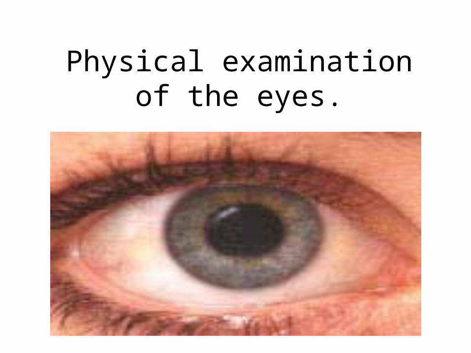
Physical examination of the eyes.
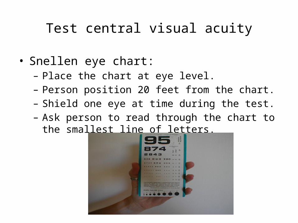
Test central visual acuity
• Snellen eye chart: – Place the chart at eye level.– Person position 20 feet from the chart.– Shield one eye at time during the test.– Ask person to read through the chart to the smallest line of
letters.
• Normal visual acuity is 20/20.• 20/20 means you can read at 20 feet what the normal
eye could have read at 20 feet.• 20/40 means you can read at 20 feet what normal eye
could read at 40 feet.
Test visual fields
• Confrontation test:– Measure of peripheral vision.– It compares the person’s peripheral vision with your own,
assuming yours is normal.– Position your self at eye level with the person, at 2 feet
away.– Direct the person to cover one eye, and with other eye to
look straight at you.– Cover your own eye opposite to the person’s covered one.
– Hold a pencil as a target midline between you and the other person, slowly advance it in from the periphery in several directions.
– Ask the person to say ‘now’ as the target is first seen; this should be just as you see the object also.
• Normal result are:– 50 degree upward.– 90 degree temporal.– 70 degree down.
– 60 degree nasal.

• If the person is unable to see the object as examiner does, this suggests peripheral field loss.
Inspect Extraocular muscle function
• Corneal light reflex:– Assess the parallel alignment of the eye axes by shining a
light toward the person eye.– Direct the person to stare straight ahead as you hold a light
about 30 cm away. – Note the reflection of the light in the corneas.– The same bright dots for each eye.
• Asymmetry of the light reflex indicates weakness or paralysis of eye muscle.
• Cover test:– Detect small degrees of deviated alignment that normally
keeps the two eyes parallel.– Ask the person to stare straight ahead at your nose.– Cover one eye, note the uncovered eye.– Normal response is a steady fixed gaze (covered eye).– Abnormal response covered eye will drift into a relaxed
position.(muscle weakness exist).
• Diagnostic position test:– Leading the eye through the six cardinal positions of gaze
will elicit any muscle any muscle weakness during movement.
– Ask the person to hold the head steady and follow the movement of your finger, only with the eyes
– Move it to each of the six positions, then back to center.– Normal response is parallel tracking of the object with both
eyes. – If not parallel weakness of Extraocular muscle or
dysfunction of cranial nerve.
Inspect external ocular structures
• Eyebrows.• Eyelashes and eyelids:– Upper lids normally overlap the superior part of the iris,
and approximate completely with the lower lids when closed.
– If you see white rim of sclera between the lid and iris is lid lag.
– Ptosis drooping of upper lid.
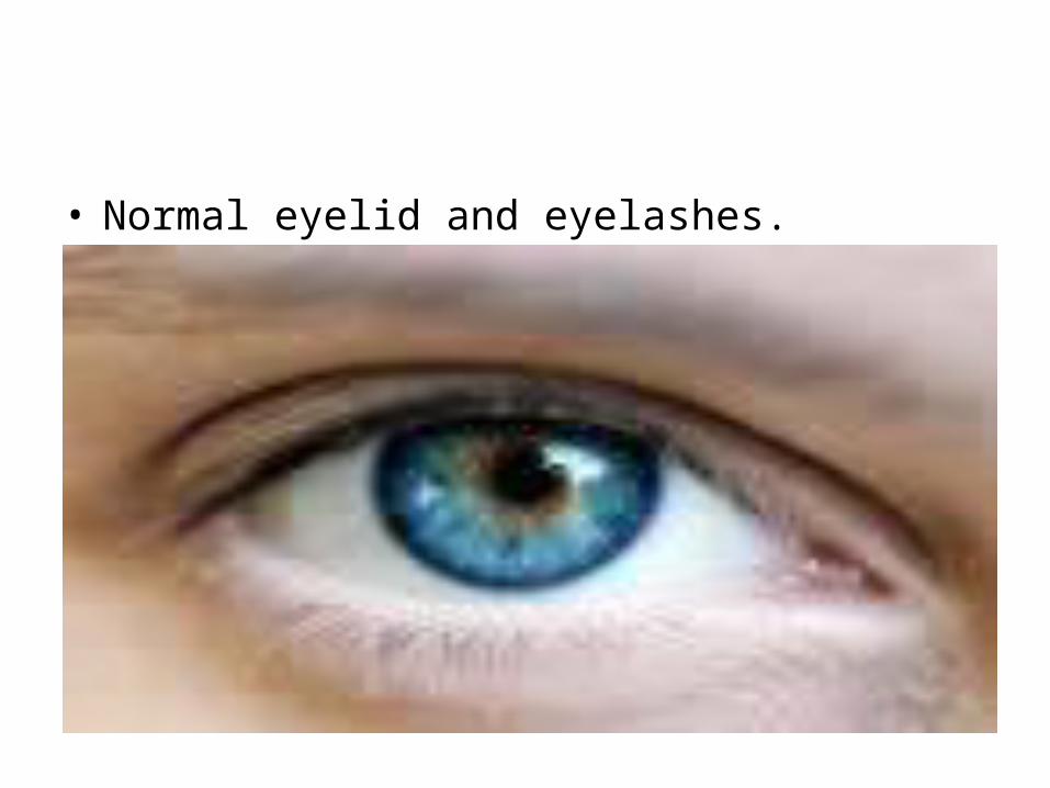
• Normal eyelid and eyelashes.
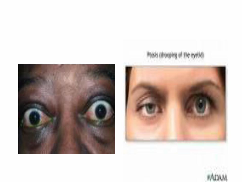
• Eyeballs: – aligned normally in their sockets with no protrusion or
sunken.
• Conjunctiva and sclera:– Conjunctiva pink over the lower lids and white over the
sclera.– Sclera is white.
• Eversion of the upper lid.


Inspect anterior eyeball structures
• Iris and pupil:– Iris normally flat, round, even coloration.– Pupils round, regular and equal size in both eyes 3-5 mm.
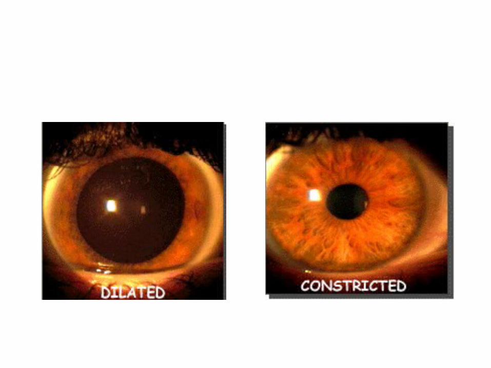
• Pupillary light reflex:– Darken the room and ask the person to gaze into distance
this dilates the pupils.– Advance a light in from the side and note the response.– Normally you will see: constriction of the same sided
pupil(direct light reflex).and simultaneous constriction of the other pupil (consensual light reflex).
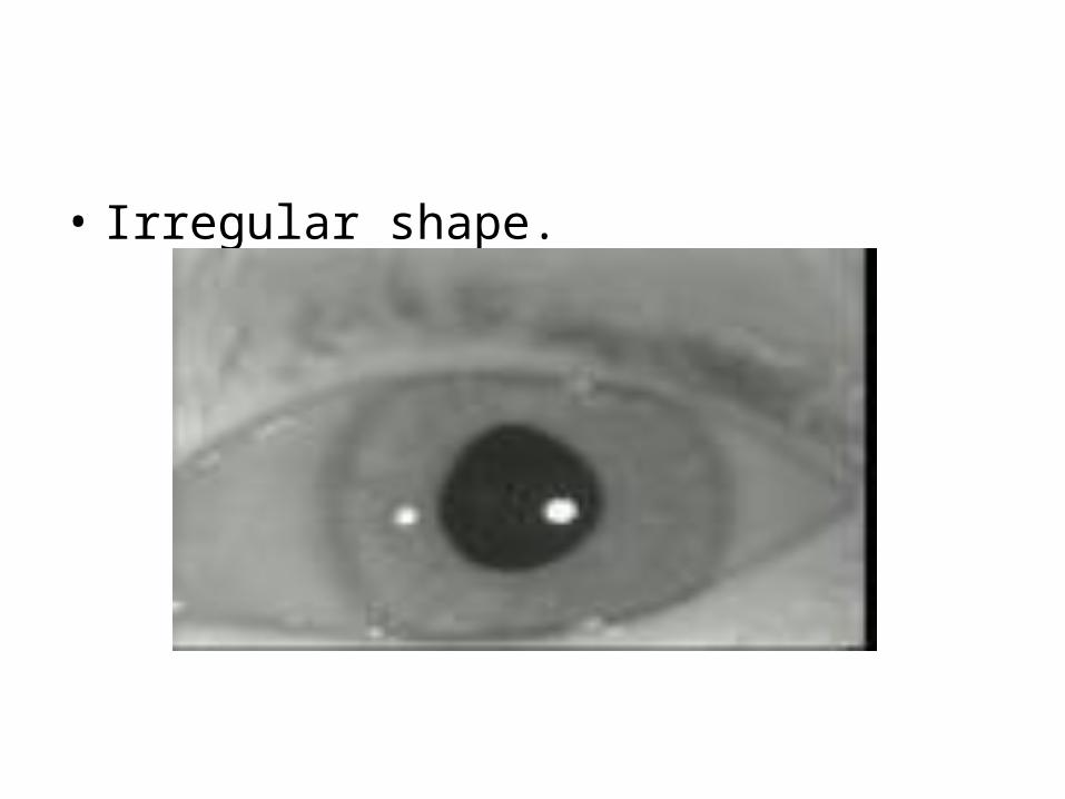
• Irregular shape.
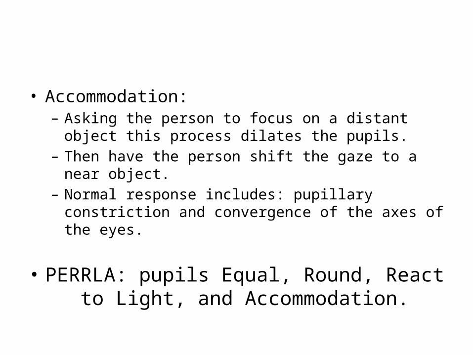
• Accommodation:– Asking the person to focus on a distant object this process
dilates the pupils.– Then have the person shift the gaze to a near object.– Normal response includes: pupillary constriction and
convergence of the axes of the eyes.
• PERRLA: pupils Equal, Round, React to Light, and Accommodation.
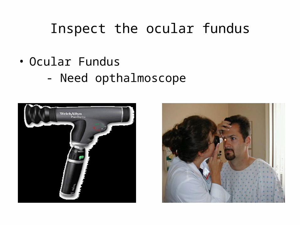
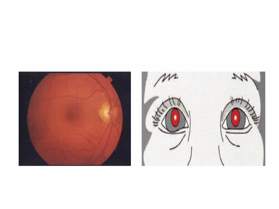
Inspect the ocular fundus
• Ocular Fundus
- Need opthalmoscope