Embed Size (px)
Citation preview
Physical Chemistry Chemical Physics
This paper is published as part of a PCCP Themed Issue on:
Water at interfaces
Guest Editor: Martin McCoustra
Editorial
Water at interfaces Phys. Chem. Chem. Phys., 2008, 10, 4676 DOI: 10.1039/b812223g
Communications
Spectroscopic and computational evidence for SO2 ionization on 128 K ice surface B. Jagoda-Cwiklik, J. P. Devlin and V. Buch, Phys. Chem. Chem. Phys., 2008, 10, 4678 DOI: 10.1039/b809839p
On the complete basis set limit and plane-wave methods in first-principles simulations of water Susan B. Rempe, Thomas R. Mattsson and K. Leung, Phys. Chem. Chem. Phys., 2008, 10, 4685 DOI: 10.1039/b810017a
Papers
Lattice match in density functional calculations: ice Ih vs. -AgI Peter J. Feibelman, Phys. Chem. Chem. Phys., 2008, 10
DOI:
, 4688
10.1039/b808482n
A proton between two waters: insight from full-dimensional quantum-dynamics simulations of the [H2O–H–OH2]+ cluster Oriol Vendrell and Hans-Dieter Meyer, Phys. Chem. Chem. Phys., 2008, 10, 4692 DOI: 10.1039/b807317a
Molecular dynamics investigation of the intrinsic structure of water–fluid interfaces via the intrinsic sampling method Fernando Bresme, Enrique Chacón and Pedro Tarazona, Phys. Chem. Chem. Phys., 2008, 10, 4704 DOI: 10.1039/b807437m
An accurate analytic representation of the water pair potential Wojciech Cencek, Krzysztof Szalewicz, Claude Leforestier, Rob van Harrevelt and Ad van der Avoird, Phys. Chem. Chem. Phys., 2008, 10, 4716 DOI: 10.1039/b809435g
Characterization of interfacial water in MOF-5 (Zn4(O)(BDC)3)—a combined spectroscopic and theoretical study K. Schröck, F. Schröder, M. Heyden, R. A. Fischer and M. Havenith, Phys. Chem. Chem. Phys., 2008, 10, 4732 DOI: 10.1039/b807458p
Water confined in reverse micelles–probe tool in biomedical informatics Florin Despa, Phys. Chem. Chem. Phys., 2008, 10, 4740 DOI: 10.1039/b805699b

Raman spectra of complexes of HNO3 and NO3- with NO2
at surfaces and with N2O4 in solution Michael A. Kamboures, Wytze van der Veer, R. Benny Gerber and Leon F. Phillips, Phys. Chem. Chem. Phys., 2008, 10, 4748 DOI: 10.1039/b810081k
Molecular level structure of the liquid/liquid interface. Molecular dynamics simulation and ITIM analysis of the water-CCl4 system Lívia B. Pártay, George Horvai and Pál Jedlovszky, Phys. Chem. Chem. Phys., 2008, 10, 4754 DOI: 10.1039/b807299j
Solvent structures of mixed water/acetonitrile mixtures at chromatographic interfaces from computer simulations Jörg Braun, Antony Fouqueau, Raymond J. Bemish and Markus Meuwly, Phys. Chem. Chem. Phys., 2008, 10, 4765 DOI: 10.1039/b807492e
Ion spatial distributions at the liquid–vapor interface of aqueous potassium fluoride solutions Matthew A. Brown, Raffaella D Auria, I.-F. William Kuo, Maria J. Krisch, David E. Starr, Hendrik Bluhm, Douglas J. Tobias and John C. Hemminger, Phys. Chem. Chem. Phys., 2008, 10, 4778 DOI: 10.1039/b807041e
Trapping proton transfer intermediates in the disordered hydrogen-bonded network of cryogenic hydrofluoric acid solutions Patrick Ayotte, Sylvain Plessis and Patrick Marchand, Phys. Chem. Chem. Phys., 2008, 10, 4785 DOI: 10.1039/b806654j
Aqueous divalent metal–nitrate interactions: hydration versus ion pairing Man Xu, James P. Larentzos, Mazen Roshdy, Louise J. Criscenti and Heather C. Allen, Phys. Chem. Chem. Phys., 2008, 10, 4793 DOI: 10.1039/b807090n
Structure and dynamics of water at a clay surface from molecular dynamics simulation Virginie Marry, Benjamin Rotenberg and Pierre Turq, Phys. Chem. Chem. Phys., 2008, 10, 4802 DOI: 10.1039/b807288d
Proton mobility in thin ice films: a revisit Eui-Seong Moon, Chang-Woo Lee and Heon Kang, Phys. Chem. Chem. Phys., 2008, 10, 4814 DOI: 10.1039/b807730b
Thermodynamics of water intrusion in nanoporous hydrophobic solids Fabien Cailliez, Mickael Trzpit, Michel Soulard, Isabelle Demachy, Anne Boutin, Joël Patarin and Alain H. Fuchs, Phys. Chem. Chem. Phys., 2008, 10, 4817 DOI: 10.1039/b807471b
Gas phase hydration of organic ions Paul O. Momoh and M. Samy El-Shall, Phys. Chem. Chem. Phys., 2008, 10, 4827 DOI: 10.1039/b809440n
Water photodissociation in free ice nanoparticles at 243 nm and 193 nm Viktoriya Poterya, Michal Fárník, Milan On ák and Petr Slavíek, Phys. Chem. Chem. Phys., 2008, 10, 4835 DOI: 10.1039/b806865h
Electroacoustic and ultrasonic attenuation measurements of droplet size and -potential of alkane-in-water emulsions: effects of oil solubility and composition Alex M. Djerdjev and James K. Beattie, Phys. Chem. Chem. Phys., 2008, 10, 4843 DOI: 10.1039/b807623e
Gas hydrate nucleation and cage formation at a water/methane interface Robert W. Hawtin, David Quigley and P. Mark Rodger, Phys. Chem. Chem. Phys., 2008, 10, 4853 DOI: 10.1039/b807455k
Hydration water rotational motion as a source of configurational entropy driving protein dynamics. Crossovers at 150 and 220 K J.-M. Zanotti, G. Gibrat and M.-C. Bellissent-Funel, Phys. Chem. Chem. Phys., 2008, 10, 4865 DOI: 10.1039/b808217k
Influence of wettability and surface charge on the interaction between an aqueous electrolyte solution and a solid surface Svetlana Guriyanova and Elmar Bonaccurso, Phys. Chem. Chem. Phys., 2008, 10, 4871 DOI: 10.1039/b806236f
Molecular dynamics study of hydrated imogolite 2. Structure and dynamics of confined water Benoît Creton, Daniel Bougeard, Konstantin S. Smirnov, Jean Guilment and Olivier Poncelet, Phys. Chem. Chem. Phys., 2008, 10, 4879 DOI: 10.1039/b803479f
Assessing the performance of implicit solvation models at a nucleic acid surface Feng Dong, Jason A. Wagoner and Nathan A. Baker, Phys. Chem. Chem. Phys., 2008, 10, 4889 DOI: 10.1039/b807384h

Aqueous peptides as experimental models for hydration water dynamics near protein surfaces Cecile Malardier-Jugroot, Margaret E. Johnson, Rajesh K. Murarka and Teresa Head-Gordon, Phys. Chem. Chem. Phys., 2008, 10, 4903 DOI: 10.1039/b806995f
Melting behavior of water in cylindrical pores: carbon nanotubes and silica glasses M. Sliwinska-Bartkowiak, M. Jazdzewska, L. L. Huang and K. E. Gubbins, Phys. Chem. Chem. Phys., 2008, 10, 4909 DOI: 10.1039/b808246d
Increased interfacial thickness of the NaF, NaCl and NaBr salt aqueous solutions probed with non-resonant surface second harmonic generation (SHG) Hong-tao Bian, Ran-ran Feng, Yan-yan Xu, Yuan Guo and Hong-fei Wang, Phys. Chem. Chem. Phys., 2008, 10, 4920 DOI: 10.1039/b806362a
Determination of the electron s solvation site on D2O/Cu(111) using Xe overlayers and femtosecond photoelectron spectroscopy Michael Meyer, Julia Stähler, Daniela O. Kusmierek, Martin Wolf and Uwe Bovensiepen, Phys. Chem. Chem. Phys., 2008, 10, 4932 DOI: 10.1039/b807314g
Breakdown of hydration repulsion between charged surfaces in aqueous Cs+ solutions Ronit Goldberg, Liraz Chai, Susan Perkin, Nir Kampf and Jacob Klein, Phys. Chem. Chem. Phys., 2008, 10, 4939 DOI: 10.1039/b807459n
A macroscopic water structure based model for describing charging phenomena at inert hydrophobic surfaces in aqueous electrolyte solutions Johannes Lützenkirchen, Tajana Preo anin and Nikola Kallay, Phys. Chem. Chem. Phys., 2008, 10, 4946 DOI: 10.1039/b807395c
Thermally induced mixing of water dominated interstellar ices Daren J. Burke, Angela J. Wolff, John L. Edridge and Wendy A. Brown, Phys. Chem. Chem. Phys., 2008, 10, 4956 DOI: 10.1039/b807220e
Water hydrogen bond analysis on hydrophilic and hydrophobic biomolecule sites Daniela Russo, Jacques Ollivier and José Teixeira, Phys. Chem. Chem. Phys., 2008, 10, 4968 DOI: 10.1039/b807551b
Hydronium and hydroxide at the interface between water and hydrophobic media Robert Vácha, Dominik Horinek, Max L. Berkowitz and Pavel Jungwirth, Phys. Chem. Chem. Phys., 2008, 10, 4975 DOI: 10.1039/b806432f
Average molecular orientations in the adsorbed water layers on silicon oxide in ambient conditions Anna L. Barnette, David B. Asay and Seong H. Kim, Phys. Chem. Chem. Phys., 2008, 10, 4981 DOI: 10.1039/b810309g
Interfacial water structure at polymer gel/quartz interfaces investigated by sum frequency generation spectroscopy Hidenori Noguchi, Minowa Hiroshi, Taiki Tominaga, Jian Ping Gong, Yoshihito Osada and Kohei Uosaki, Phys. Chem. Chem. Phys., 2008, 10, 4987 DOI: 10.1039/b807297n
Co-adsorption of water and hydrogen on Ni(111) Junjun Shan, Jacques F. M. Aarts, Aart W. Kleyn and Ludo B. F. Juurlink, Phys. Chem. Chem. Phys., 2008, 10, 4994 DOI: 10.1039/b808219g
Water–methanol mixtures: topology of hydrogen bonded network Imre Bakó, Tünde Megyes, Szabolcs Bálint, Tamás Grósz and Viorel Chihaia, Phys. Chem. Chem. Phys., 2008, 10, 5004 DOI: 10.1039/b808326f
Photochemistry of ethyl chloride caged in amorphous solid waterw
Yousif Ayoub and Micha Asscher*
Received 7th May 2008, Accepted 21st July 2008
First published as an Advance Article on the web 23rd September 2008
DOI: 10.1039/b807803n
Caging and photo-induced decomposition of ethyl chloride molecules (EC) within a layer of
amorphous solid water (ASW) on top of clean and oxygen-covered Ru(001) under ultra-high
vacuum (UHV) conditions are presented. The caged molecules were estimated to reside
1.5 � 0.2 nm above the solid surface, based on parent molecule thermal decomposition on the
clean ruthenium. Dissociative electron attachment (DEA) of the caged molecules following
193 nm laser irradiation, result in initial fragmentation to ethyl radical and chloride anion. It was
found that photoreactivity on top of the clean ruthenium surface (Ru) is twenty times faster than
on the oxygen-covered surface (O/Ru), with DEA cross sections: sRu = (3.8 � 1) � 10�19 cm2
and sO/Ru = (2.1 � 0.3) � 10�20 cm2. This difference is attributed to the higher work function of
oxygen-covered ruthenium, leading to smaller electron attachment probability due to mismatch
of the ruthenium photo-electron energy with the adsorbed EC excited electron affinity levels. EC
molecules fragmented within the cage, result in post-irradiation TPD spectra that reveal primarily
C4H8, C3H5 and C3H3, without any oxygen-containing molecules. Unique stabilization of the
photoproducts has been observed with the first layer of water molecules in direct contact with the
substrate, desorbing near 180 K, a significantly higher temperature than the desorption of fully
caged molecules. This study may contribute for understanding stratospheric photochemistry and
processes in interstellar space.
1. Introduction
Photo-induced chemistry of molecules interacting with solid
interfaces has been the focus of interest and intensive research
in recent years.1–3. Early3 and more recent studies4 have
discussed photodesorption mechanisms from oxide surfaces
and ices respectively. A stabilizing effect of the ice environment
was reported for the case of trapped polycyclic aromatic
hydrocarbon (PAHs) ions, formed by ionizing irradiation,
attempting to mimic conditions at interstellar space, based
on IR laboratory studies.5,6a The same detection technique has
also been employed to monitor long-chain hydrocarbons,
formed following UV irradiation of ice analogs made of
formaldehyde and methanol.6b Other mechanisms to form
interstellar hydrocarbons were reviewed in ref. 6c.
Photo-induced reactivity on top of solid surfaces has
become increasingly important in recent years within the
context of photocatalysis, mostly with oxides (e.g. TiO2) as
catalysts at ambient conditions.7 Under these conditions,
molecules often interact with the solid surfaces in the presence
of a few water layers. Understanding the role of neighbor
water molecules that are not directly involved in the primary
photo-event is therefore important.
A central photodissociation pathway of adsorbed molecules
on metallic and oxide surfaces involves dissociative (photo)-
electron attachment mechanism.1,2
Caging of adsorbed molecules within water ice layers on
well-defined metallic substrates has been observed already
more than a decade ago. The first experimental demonstration
of a molecular cage in amorphous solid water (ASW) was that
of N2,8 while other studies have focused mostly on haloge-
nated molecules, such as CCl4,9 CD3Cl,
10 CD3Br, but also
other molecules were shown to be trapped by ASW, e.g.
CO2.11 Subsequent desorption of the trapped molecules
proceeds via an explosive, ‘‘volcano’’ mechanism.8,9
Irradiation of molecular traps of this kind by UV light has
been suggested as a possible origin, via photodesorption12 and
fragmentation of the trapped molecular species13,14 of organic
molecules in interstellar space.
In this report we describe the caging and subsequent photo-
chemistry of ethyl chloride (EC) molecules, used as model for the
study of halogenated hydrocarbon molecules in the strato-
sphere.15 Quantifying the reactivity of caged molecules may
potentially assist studies of photo-induced processes at interstel-
lar space as well. The EC molecules were caged within layers of
ASW on well-characterized Ru(001) and O/Ru(001) substrates
under ultra-high vacuum (UHV) conditions. Subsequently the
system was irradiated by 6.4 eV photons from an excimer laser.
The clean and oxygen-covered ruthenium substrates were chosen
to demonstrate substrate effects on photoreactivity over solid
surfaces, often a neglected subject.
Several stable gaseous molecules are obtained following
reactivity among the nascent photoproducts. Cross sections
for these processes are reported.
Institute of Chemistry, The Hebrew University of Jerusalem,Jerusalem, Israel. E-mail: [email protected];Fax: +972 (0)2 6525037; Tel: +972 (0)2 6585742w This article was submitted as part of a Themed Issue on water atinterfaces. Other papers on this topic can be found in issue 32 ofvol. 10 (2008). This issue can be found from the PCCP homepage[http://www.rsc.org/pccp].
6486 | Phys. Chem. Chem. Phys., 2008, 10, 6486–6491 This journal is �c the Owner Societies 2008
PAPER www.rsc.org/pccp | Physical Chemistry Chemical Physics
2. Experimental
The experiments described in this report were performed in an
ultra-high vacuum (UHV) apparatus, typical base pressure of
2� 10�10 Torr, described elsewhere in detail.13,16 The chamber
is equipped with a quadrupole mass-spectrometer (QMS) that
is covered by a glass tube with a 3 mm aperture in front, thus
avoiding a record of desorption from surfaces other than the
sample while improving sensitivity and selectivity. In addition
to standard temperature-programmed desorption DP-TPD(P for pressure), average adsorbates dipole moment could be
determined by means of a Kelvin probe, operated in a
Dj-TPD (j for work function) mode as explained elsewhere.16
In addition, a mini excimer laser (PSX-100) provides 2.5 ns
long pulses at 193 nm, 3 mJ per pulse, and variable repetition
rate up to 100 Hz for the photo-excitation studies. The
analysis of the photoproducts was based on post irradiation
DP-TPD, simultaneously scanning a full range of masses
(see Fig. 2 below) at a heating rate of up to 2 K s�1. The
Ru(001) sample, oriented to within 0.5 degrees of the (001)
plane, could be cooled down to 82 K by pumping over a liquid
nitrogen reservoir. Temperature was determined by a C-type
thermocouple (W5%Re/W26%Re) and controlled to within
0.5 degrees using an ac resistive heating LabView routine.
3. Results and discussion
We have studied the photochemistry of EC molecules caged
inside 25 bilayers (BL) ASW following 193 nm laser
irradiation on clean Ru(001). An important element in this
study was the potential effect of the underlying substrate.
Therefore, we have compared results obtained from clean
Ru(001) to the photo-activity on top of ordered oxygen-
covered ruthenium, (2�1)–O/Ru(001). These substrates were
chosen to focus on the substrate effect on the outcome of
photo-induced processes rather than attempting to mimic
actual surfaces in the stratosphere or in interstellar space.
Before discussing the photochemistry within the cage it is
important to briefly introduce the procedure used to form a
molecular cage under ASW.
3.1 Cage formation: EC@ASW
The cage formation mechanism for EC is practically identical
to the first molecular cage observed with N2 (see ref. 8) and
other molecules in subsequent studies.9–11
Water layers were grown by back-filling the vacuum chamber,
determining the actual exposure and layer thickness based on
post-exposure-uptake TPD measurements. Such deposition
conditions form compact amorphous solid water, as was shown
before.17 The co-adsorption of EC molecules with a gradually
denser coverage of water leads to compression of the EC
molecules on the surface.10 At thick ASW layers [more than
20 BL, where 1 BL = (1 � 0.1) � 1015 molecules cm�2 on
Ru(001)]18,19 a cage has fully been developed, as evidenced by an
extremely narrow, explosive TPD peak with a typical width of
2–3 degrees at half maximum, near the onset desorption of ASW
at 165 K.
A fixed EC coverage of 0.3 ML was deposited first at 82 K,
followed by gradually thicker layers of ASW. The EC TPD
signal (mass 64) reveals three important stages of the cage
formation. Desorption of EC without water co-adsorption
(Fig. 1A) is characterized by a broad desorption peak,
centered at 175 K. At ASW converge of 6 BL one can
distinguish two different populations. The peak at 125 K
originates from a fraction of EC molecules that were
‘‘floating’’ on top of the ASW layer, as understood from the
low temperature desorption, before any water desorption
takes place. These molecules have no direct contact with the
substrate. In order to verify that these molecules are on top of
the water layer, 0.3 ML EC were deposited directly on top of a
6 BL thick ASW layer. The subsequent TPD is shown as a
dashed line in Fig. 1B, revealing identical behavior with the
floating molecules. The binding energy of EC to water was
estimated based on standard Redhead TPD lineshape analysis
of Fig. 1B, assuming a pre-exponential factor of 1013 s�1 for
the first order desorption rate. Activation energy for
desorption and its uncertainty, as derived from the TPD
analysis, is 8.4 � 1 kcal mol�1. This is a rather similar result
to the strength of interaction among EC molecules, derived
similarly from the TPD of multilayer of EC molecules (not
shown), which is 8.1 � 1 kcal mol�1.
A new and gradually narrowing peak emerges near 165 K,
at the onset of ASW desorption. This peak of caged EC
molecules contains most of the molecules that were previously
residing on the metallic substrate. There are parent molecules
and photochemically formed molecules that tend to be
associated with and stabilized by water molecules of the first
layer, desorbing at 178 K.
An important question that has not yet been addressed
regarding the caging process of molecules under layers of
ASW, is the location or distance of the trapped molecules
from the substrate, after the caging process has been
completed, as shown via TPD in Fig. 1C.
We have previously demonstrated, in the case of CD3Cl
trapped in ASW,10 based on work function change measure-
ments, that the caged molecules were lifted upwards, away
from the surface by the nature of interaction with the
post-adsorbed water molecules on a clean Ru(001) surface.
In order to better define the position of the EC molecules
above the substrate while caged within ASW, we took advan-
tage of the fact that a fraction of the first monolayer of parent
EC molecules (about 0.2) undergoes dissociation upon thermal
heating during TPD on the clean Ru(001). This dissociation is
manifested by the uptake of hydrogen molecules desorbing at
350 K, as a result of further dissociation and dehydrogenation
of the ethyl fragment.
No hydrogen signal could be detected from a full cage as
seen in Fig. 1C on top of clean Ru(001). This observation
indicates, we predict, that the caged molecules are too far from
the surface, therefore do not reach it upon cage explosive
desorption.
In order to clarify this assumption, we have defined a
maximum distance for EC molecules to reach the metallic
surface during desorption and leave its hydrogen desorption
signature. The calibration approach has been to mimic the
cage by constructing a sandwich of EC molecules between two
films of ASW. A variable thickness first layer attached to the
ruthenium substrate, then a fixed 0.3 ML of EC on top and
This journal is �c the Owner Societies 2008 Phys. Chem. Chem. Phys., 2008, 10, 6486–6491 | 6487

then a second, constant thickness (10 BL) layer of ASW. The
details of this study will appear elsewhere, but the summary of
this test has been that at layer thickness of 9 � 1 BL and
above, the EC molecules do not reach the bottom ruthenium
substrate during the desorption of this sandwich composition.
We conclude that the caged molecules must reside within a
distance of at least 1.5 � 0.2 nm from the surface.
3.2 Photochemistry of caged EC
Once the EC@ASW cage has been established, it was
irradiated by UV light at 193 nm, photon energy of 6.4 eV,
at a fixed pulse energy of 3 � 0.5 mJ per pulse. At this pulse
energy, the estimated thermal heating of the ruthenium
substrate was less than 8 K above the substrate temperature
of 82 K,20 which eliminates any thermal effects due to the UV
laser irradiation.
Parent EC molecules caged in a matrix of ASW cannot
undergo photodesorption. On the other hand, photofragments
formed within the cage may react with each other but
potentially also with the host water molecules surrounding
the cage. An additional experimental benefit is gained from the
fact that the parent molecules as well as the various photo-
products are all explosively desorbing at the same temperature
range near 165 K, the onset of ASW desorption (evidenced by
the dark blue–green stripe at that temperature scale in Fig. 2).
We have developed a way to simultaneously scan a complete
mass range during a single TPD run. This multimass-scan is an
efficient mode of operation, particularly for an initial screening
of new products that have not yet been identified.
A scan of this kind is demonstrated in a 3D form, where
QMS signal is displayed vs.mass and temperature, as shown in
Fig. 2. Fig. 2B was obtained following EC cage irradiation for
90 sec (equivalent to photons dose of 5.2 � 1018 at 193 nm),
while in Fig. 2A the same plot is a thermal scan of caged EC
molecules, without irradiation.
Post-irradiation stable molecular products can be observed
in the TPD scan at the mass range of 54–57 a.u. and 39–43 a.u.,
shown in the blow-up in Fig. 2B.
There is significant background signal at masses 35 and 37
due to chlorine, a dissociation product inside the QMS that
masks other possible stable molecular products at the same
mass range. It is evident from analysis of Fig. 2 that most of
the cage molecular content, including the parent molecules
and the photoproducts, explosively desorb simultaneously at
165 K upon the onset of ice desorption. Yet, a significant
fraction of the molecules are stabilized by water molecules in
the first layer, therefore desorbing only near 178 K.
Post-irradiation TPD spectra in Fig. 3 reveal the EC parent
molecule (mass 64) uptake as a function of irradiation time on
clean Ru (Fig. 3A) and O/Ru (Fig. 3B) substrates. The total
EC uptake signal decreases monotonically at a first order-like
kinetics with photons exposure.
The comparison between photo-induced decomposition on
clean vs. oxygen-covered ruthenium surfaces originates from
the attempt to modify the strength of interaction between
photo-excited, negatively charged EC (dissociative electron
attachment process—DEA1,2) with the underlying substrate.
Generally one expects that the oxygen-covered surface, on top
Fig. 1 DP-TPD at mass 64 (parent EC molecule) following water
post-deposition on top of a fixed 0.3 ML EC on clean Ru(001). The
TPD spectra demonstrate the gradual (from A to B) compression and
eventual full caging (C) of EC under the indicated ASW layers. Note
the substantially higher but narrower peak recorded following com-
plete caging under 20 BL. The dashed line in (B) was obtained from
0.3 ML EC directly deposited on top of a 6 BL thick water layer.
Fig. 2 (A) A multimass-TPD taken simultaneously at all masses
between 20–95 a.u. in a 3D representation without irradiation and (B)
following 90 s of irradiation time (equivalent to 5.2 � 1018 193 nm
photons). The blow-up reveals the signal of various photoproducts of
the EC@ice system.
6488 | Phys. Chem. Chem. Phys., 2008, 10, 6486–6491 This journal is �c the Owner Societies 2008
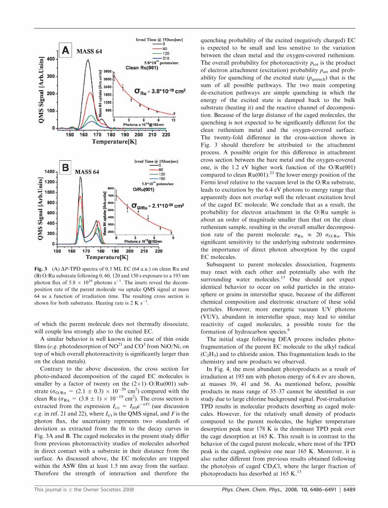
of which the parent molecule does not thermally dissociate,
will couple less strongly also to the excited EC.
A similar behavior is well known in the case of thin oxide
films (e.g. photodesorption of NO21 and CO3 from NiO/Ni, on
top of which overall photoreactivity is significantly larger than
on the clean metals).
Contrary to the above discussion, the cross section for
photo-induced decomposition of the caged EC molecules is
smaller by a factor of twenty on the (2�1)–O/Ru(001) sub-
strate (sO/Ru = (2.1 � 0.3) � 10�20 cm2) compared with the
clean Ru (sRu = (3.8 � 1) � 10�19 cm2). The cross section is
extracted from the expression I(t) = I(0)e�sFt (see discussion
e.g. in ref. 21 and 22), where I(t) is the QMS signal, and F is the
photon flux, the uncertainty represents two standards of
deviation as extracted from the fit to the decay curves in
Fig. 3A and B. The caged molecules in the present study differ
from previous photoreactivity studies of molecules adsorbed
in direct contact with a substrate in their distance from the
surface. As discussed above, the EC molecules are trapped
within the ASW film at least 1.5 nm away from the surface.
Therefore the strength of interaction and therefore the
quenching probability of the excited (negatively charged) EC
is expected to be small and less sensitive to the variation
between the clean metal and the oxygen-covered ruthenium.
The overall probability for photoreactivity ptot is the product
of electron attachment (excitation) probability patt and prob-
ability for quenching of the excited state (pquench) that is the
sum of all possible pathways. The two main competing
de-excitation pathways are simple quenching in which the
energy of the excited state is damped back to the bulk
substrate (heating it) and the reactive channel of decomposi-
tion. Because of the large distance of the caged molecules, the
quenching is not expected to be significantly different for the
clean ruthenium metal and the oxygen-covered surface.
The twenty-fold difference in the cross-section shown in
Fig. 3 should therefore be attributed to the attachment
process. A possible origin for this difference in attachment
cross section between the bare metal and the oxygen-covered
one, is the 1.2 eV higher work function of the O/Ru(001)
compared to clean Ru(001).23 The lower energy position of the
Fermi level relative to the vacuum level in the O/Ru substrate,
leads to excitation by the 6.4 eV photons to energy range that
apparently does not overlap well the relevant excitation level
of the caged EC molecule. We conclude that as a result, the
probability for electron attachment in the O/Ru sample is
about an order of magnitude smaller than that on the clean
ruthenium sample, resulting in the overall smaller decomposi-
tion rate of the parent molecule: sRu E 20 sO/Ru. This
significant sensitivity to the underlying substrate undermines
the importance of direct photon absorption by the caged
EC molecules.
Subsequent to parent molecules dissociation, fragments
may react with each other and potentially also with the
surrounding water molecules.13 One should not expect
identical behavior to occur on solid particles in the strato-
sphere or grains in interstellar space, because of the different
chemical composition and electronic structure of these solid
particles. However, more energetic vacuum UV photons
(VUV), abundant in interstellar space, may lead to similar
reactivity of caged molecules, a possible route for the
formation of hydrocarbon species.6
The initial stage following DEA process includes photo-
fragmentation of the parent EC molecule to the alkyl radical
(C2H5) and to chloride anion. This fragmentation leads to the
chemistry and new products we observed.
In Fig. 4, the most abundant photoproducts as a result of
irradiation at 193 nm with photon energy of 6.4 ev are shown,
at masses 39, 41 and 56. As mentioned before, possible
products in mass range of 35–37 cannot be identified in our
study due to large chlorine background signal. Post-irradiation
TPD results in molecular products desorbing as caged mole-
cules. However, for the relatively small density of products
compared to the parent molecules, the higher temperature
desorption peak near 178 K is the dominant TPD peak over
the cage desorption at 165 K. This result is in contrast to the
behavior of the caged parent molecule, where most of the TPD
peak is the caged, explosive one near 165 K. Moreover, it is
also rather different from previous results obtained following
the photolysis of caged CD3Cl, where the larger fraction of
photoproducts has desorbed at 165 K.13
Fig. 3 (A) DP-TPD spectra of 0.3 ML EC (64 a.u.) on clean Ru and
(B) O/Ru substrate following 0, 60, 120 and 150 s exposure to a 193 nm
photon flux of 5.8 � 1016 photons s�1. The insets reveal the decom-
position rate of the parent molecule via uptake QMS signal at mass
64 as a function of irradiation time. The resulting cross section is
shown for both substrates. Heating rate is 2 K s�1.
This journal is �c the Owner Societies 2008 Phys. Chem. Chem. Phys., 2008, 10, 6486–6491 | 6489
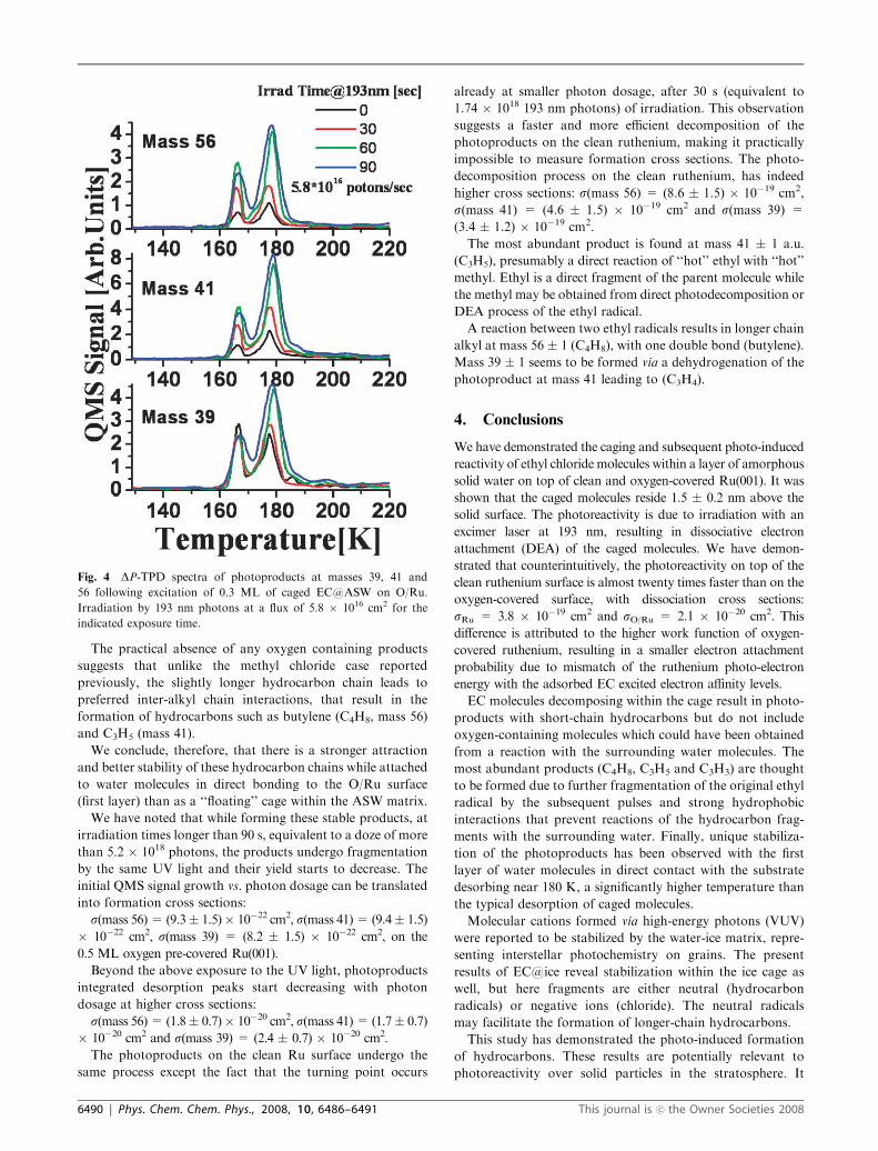
The practical absence of any oxygen containing products
suggests that unlike the methyl chloride case reported
previously, the slightly longer hydrocarbon chain leads to
preferred inter-alkyl chain interactions, that result in the
formation of hydrocarbons such as butylene (C4H8, mass 56)
and C3H5 (mass 41).
We conclude, therefore, that there is a stronger attraction
and better stability of these hydrocarbon chains while attached
to water molecules in direct bonding to the O/Ru surface
(first layer) than as a ‘‘floating’’ cage within the ASW matrix.
We have noted that while forming these stable products, at
irradiation times longer than 90 s, equivalent to a doze of more
than 5.2 � 1018 photons, the products undergo fragmentation
by the same UV light and their yield starts to decrease. The
initial QMS signal growth vs. photon dosage can be translated
into formation cross sections:
s(mass 56)= (9.3� 1.5)� 10�22 cm2, s(mass 41)= (9.4� 1.5)
� 10�22 cm2, s(mass 39) = (8.2 � 1.5) � 10�22 cm2, on the
0.5 ML oxygen pre-covered Ru(001).
Beyond the above exposure to the UV light, photoproducts
integrated desorption peaks start decreasing with photon
dosage at higher cross sections:
s(mass 56)= (1.8� 0.7)� 10�20 cm2, s(mass 41)= (1.7� 0.7)
� 10�20 cm2 and s(mass 39) = (2.4 � 0.7) � 10�20 cm2.
The photoproducts on the clean Ru surface undergo the
same process except the fact that the turning point occurs
already at smaller photon dosage, after 30 s (equivalent to
1.74 � 1018 193 nm photons) of irradiation. This observation
suggests a faster and more efficient decomposition of the
photoproducts on the clean ruthenium, making it practically
impossible to measure formation cross sections. The photo-
decomposition process on the clean ruthenium, has indeed
higher cross sections: s(mass 56) = (8.6 � 1.5) � 10�19 cm2,
s(mass 41) = (4.6 � 1.5) � 10�19 cm2 and s(mass 39) =
(3.4 � 1.2) � 10�19 cm2.
The most abundant product is found at mass 41 � 1 a.u.
(C3H5), presumably a direct reaction of ‘‘hot’’ ethyl with ‘‘hot’’
methyl. Ethyl is a direct fragment of the parent molecule while
the methyl may be obtained from direct photodecomposition or
DEA process of the ethyl radical.
A reaction between two ethyl radicals results in longer chain
alkyl at mass 56 � 1 (C4H8), with one double bond (butylene).
Mass 39 � 1 seems to be formed via a dehydrogenation of the
photoproduct at mass 41 leading to (C3H4).
4. Conclusions
We have demonstrated the caging and subsequent photo-induced
reactivity of ethyl chloride molecules within a layer of amorphous
solid water on top of clean and oxygen-covered Ru(001). It was
shown that the caged molecules reside 1.5 � 0.2 nm above the
solid surface. The photoreactivity is due to irradiation with an
excimer laser at 193 nm, resulting in dissociative electron
attachment (DEA) of the caged molecules. We have demon-
strated that counterintuitively, the photoreactivity on top of the
clean ruthenium surface is almost twenty times faster than on the
oxygen-covered surface, with dissociation cross sections:
sRu = 3.8 � 10�19 cm2 and sO/Ru = 2.1 � 10�20 cm2. This
difference is attributed to the higher work function of oxygen-
covered ruthenium, resulting in a smaller electron attachment
probability due to mismatch of the ruthenium photo-electron
energy with the adsorbed EC excited electron affinity levels.
EC molecules decomposing within the cage result in photo-
products with short-chain hydrocarbons but do not include
oxygen-containing molecules which could have been obtained
from a reaction with the surrounding water molecules. The
most abundant products (C4H8, C3H5 and C3H3) are thought
to be formed due to further fragmentation of the original ethyl
radical by the subsequent pulses and strong hydrophobic
interactions that prevent reactions of the hydrocarbon frag-
ments with the surrounding water. Finally, unique stabiliza-
tion of the photoproducts has been observed with the first
layer of water molecules in direct contact with the substrate
desorbing near 180 K, a significantly higher temperature than
the typical desorption of caged molecules.
Molecular cations formed via high-energy photons (VUV)
were reported to be stabilized by the water-ice matrix, repre-
senting interstellar photochemistry on grains. The present
results of EC@ice reveal stabilization within the ice cage as
well, but here fragments are either neutral (hydrocarbon
radicals) or negative ions (chloride). The neutral radicals
may facilitate the formation of longer-chain hydrocarbons.
This study has demonstrated the photo-induced formation
of hydrocarbons. These results are potentially relevant to
photoreactivity over solid particles in the stratosphere. It
Fig. 4 DP-TPD spectra of photoproducts at masses 39, 41 and
56 following excitation of 0.3 ML of caged EC@ASW on O/Ru.
Irradiation by 193 nm photons at a flux of 5.8 � 1016 cm2 for the
indicated exposure time.
6490 | Phys. Chem. Chem. Phys., 2008, 10, 6486–6491 This journal is �c the Owner Societies 2008
may open a new channel of understanding of photoreactivity
on grains in interstellar space as well.
Acknowledgements
We wish to thank Yigal Lilach for his valuable help in perform-
ing this study. This research was partially supported by a grant
from the German–Israel Foundation, US–Israel Binational
Science Foundation and the Israel Science Foundation. The
Farkas Center is supported by the Bundesministerium fur
Forschung und Technologie and the Minerva Gesellschaft fur
die Forschung mbh.
References
1 X. L. Zhou, X. Y. Zhu and M. J. White, Surf. Sci. Rep., 1991, 13, 73.2 Laser Spectroscopy and Photochemistry on Metal Surfaces, Part Iand II, ed. H.-L. Dai andW. Ho,World Scientific, New York, 1995.
3 M. Asscher, F. Zimmermann, L. Springsteen, P. L. Houston andW. Ho, J. Chem. Phys., 1992, 96(5), 4808–11.
4 J. D. Thrower, D. J. Burke, M. P. Collins, A. Dawes, P. D.Holtom, F. Jamme, P. Kendall, W. A. Brown, I. P. Clark, H. J.Fraser, M. R. S. McCoustra, N. J. Mason and A. W. Parker,Astrophys. J., 2008, 673, 1233.
5 M. P. Bernstein, S. A. Sandford, A. L. Mattioda and L. J.Allamandola, Astrophys. J., 2007, 664, 1264.
6 (a) M. S. Gudipati and L. J. Allamandola, Astrophys. J., 2003, 596,L198; (b) P. A. Gerakines, W. A. Schutte and P. Ehrenfreund,Astron. Astrophys., 1996, 312, 289 and references therein; (c) A. G.G. M. Tielens and L. J. Allamandola, in Interstellar Processes, ed.D. J. Hollenbach and H. A. Thronton, Riedel, Dordrecht, 1987.
7 T. Tachikawa, M. Fujitsuka and T. Majima, J. Phys. Chem., 2007,111, 5259.
8 T. Livneh, L. Romm and M. Asscher, Surf. Sci., 1996, 315, 250.9 R. S. Smith, C. Huang, E. K. L. Wong and B. D. Kay, Phys. Rev.Lett., 1997, 79, 909.
10 Y. Lilach and M. Asscher, J. Chem. Phys., 2002, 117, 6730.11 R. Berger, Y. Lilach, Y. Ayoub and M. Asscher, Isr. J. Chem.,
2005, 45, 97.12 J. D. Thrower, D. J. Burke, M. P. Collins, A. Dawes, P. D.
Holtom, F. Jamme, P. Kendall, W. A. Brown, I. P. Clark, H. J.Fraser, M. R. S. McCourstra, N. J. Mason and A. W. Parker,Astrophys. J., 2008, 673, 1233.
13 Y. Lilach and M. Asscher, J. Chem. Phys., 2003, 119, 407.14 T. Livneh and M. Asscher, J. Phys. Chem. B, 2003, 107, 11382.15 John R. Barker, Progress And Problems In Atmospheric Chemistry,
World Scientific, Singapore, 1996.16 T. Livneh, Y. Lilach and M. Asscher, J. Chem. Phys., 1999,
111(24), 11138.17 G. A. Kimmel, N. G. Petrik, Z. Dohnalek and B. D. Kay, J. Chem.
Phys., 2006, 125, 044713.18 R. S. Smith, C. Huang, E. K. L. Wong and B. Kay, Surf. Sci.,
1996, 376, L13.19 G. Held and D. Menzel, Surf. Sci., 1994, 316, 92.20 The surface temperature jump due to pulsed laser heating is
difficult to measure directly. However, indirect measurementsand calculations based on absorption at the laser wavelength, heatconductivity and diffusivity of the substrate enable such estimates.See for example: Z. Rosenzweig and M. Asscher, J. Chem. Phys.,1992, 96(5), 4040–43 and references therein.
21 M. Menges, B. Baumeister, K. Al-Shamery, H.-J. Freund, C.Fischer and P. Andresen, Surf. Sci., 1994, 316, 103.
22 R. Zehr, A. Solodukhin, B. C. Haynie, C. French and I. Harrison,J. Phys. Chem. B, 2000, 104, 3094.
23 Y. Lilach and M. Asscher, J. Phys. Chem. B, 2004, 108, 4358–61and references therein.
This journal is �c the Owner Societies 2008 Phys. Chem. Chem. Phys., 2008, 10, 6486–6491 | 6491