Embed Size (px)
Citation preview

Physical Characteristics and Volume
Blood is a sticky, opaque fluid with a metallic taste Color varies from scarlet (oxygen-rich) to dark red
(oxygen-poor) The pH of blood is 7.35–7.45 (slightly basic) Temperature is 38C (100.4 F), slightly higher than
“normal” body temperature Blood accounts for approximately 8% of body weight Average volume of blood is 5–6 L for males, and 4–5
L for females

1. Substance Distribution– Oxygen from the lungs and nutrients from the
digestive tract– Metabolic wastes from cells to the lungs and
kidneys for elimination– Hormones from endocrine glands to target organs
Functions of Blood

Functions of Blood
2. Regulation of Blood Levels – Appropriate body temperature by absorbing and
distributing heat– Normal pH in body tissues using buffer systems– Adequate fluid volume in the circulatory system

3. Body Protection– Blood prevents blood loss by:
Activating plasma proteins and platelets Initiating clot formation when a vessel is broken
– Blood prevents infection by: Synthesizing and utilizing antibodies Activating complement proteins Activating WBCs to defend the body against foreign
invaders
Functions of Blood cont.

Composition of Blood
Blood is the body’s only fluid tissue It is composed of liquid plasma and formed
elements– Formed elements include:
Erythrocytes, or red blood cells (RBCs) Buffy Coat
– Leukocytes, or white blood cells (WBCs)– Platelets

Composition of Blood Cont.

Blood Plasma
Blood plasma is approximately 90% water Blood plasma contains over 100 solutes, including:
– Proteins – albumin, globulins, clotting proteins, and others– Nonprotein nitrogenous substances – lactic acid, urea,
creatinine– Organic nutrients – glucose, carbohydrates, amino acids– Electrolytes – sodium, potassium, calcium, chloride,
bicarbonate – Respiratory gases – oxygen and carbon dioxide

Formed Elements
Erythrocytes, leukocytes, and platelets make up the formed elements– Only WBCs are complete cells– RBCs have no nuclei or organelles, and platelets
are just cell fragments
Most formed elements survive in the bloodstream for only a few days
Most blood cells do not divide but are renewed by cells in bone marrow


Production of Blood Cells
Hematopoiesis – blood cell formation
Hematopoiesis occurs in the red bone marrow of the:– Axial skeleton and girdles– Ends of the humerus and femur
Hemocytoblasts stem cells that give rise to all formed elements

Red Blood Cells

Erythropoiesis

Erythrocytes (RBCs)
Biconcave discs, anucleate, essentially no organelles
Filled with hemoglobin (Hb), a protein that functions in gas transport
Erythrocytes are an example of the complementarily of structure and function– Biconcave shape (huge SA relative to volume)– A dehydrated RBC is 97% Hb – ATP is generated anaerobically (does not use the
O2 it transports)
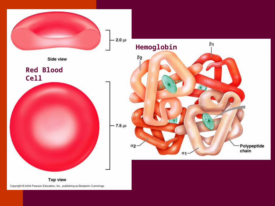
Red Blood Cell
Hemoglobin

Fate and Destruction of Erythrocytes
The life span of an erythrocyte is 100–120 days
Old erythrocytes become rigid and fragile, and their hemoglobin begins to degenerate
Dying erythrocytes are engulfed by macrophages
Heme and globin are separated and the iron is salvaged for reuse


Decreases O2 carrying capacity– It is a symptom rather than a disease– Blood O2 levels cannot support normal metabolism
– Signs/symptoms include fatigue, paleness, shortness of breath, and chills
– Three Causes Decreased RBCs Decreased Hemoglobin Abnormal Hemoglobin
RBC Disorder: Anemia

White Blood Cells
Leukocytes (WBCs)
Leukocytes, the only blood components that are complete cells:– Are less numerous than RBCs– Make up 1% of the total blood volume– Can leave capillaries via diapedesis– Move through tissue spaces
Leukocytosis – WBC count over 11,000 per cubic millimeter – Normal response to bacterial or viral invasion
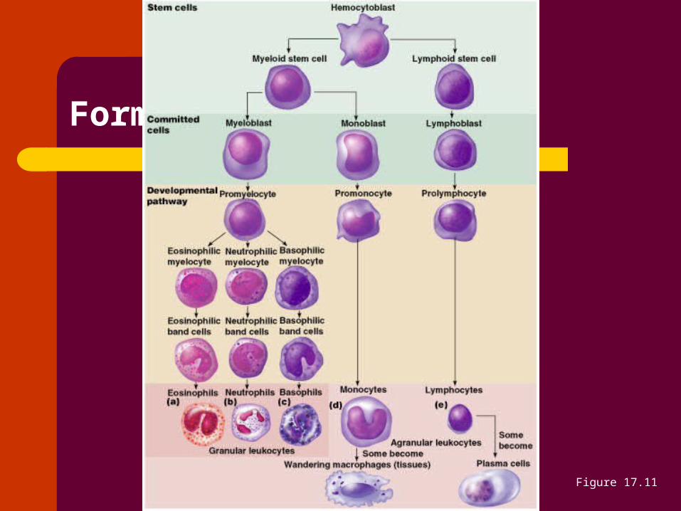
Formation of Leukocytes
Figure 17.11

Leukocytes (WBCs) cont.

Granulocytes
Granulocytes – neutrophils, eosinophils, and basophils– Contain cytoplasmic granules that stain
specifically (acidic, basic, or both) with Wright’s stain
– Are larger and usually shorter-lived than RBCs– Have lobed nuclei– Are all phagocytic cells

Neutrophils have 2 types of granules that:– Take up both acidic and basic dyes– Give the cytoplasm a lilac color– Neutrophils are our body’s bacteria slayers
Types of Granulocytes

Types of Granulocytes
Eosinophils account for 1–4% of WBCs – Have red-staining, bilobed nuclei– Lead the body’s counterattack against parasitic
worms– Lessen the severity of allergies by phagocytizing
immune complexes

Types of Granulocytes
Basophils: Account for 0.5% of WBCs and:– Have U- or S-shaped nuclei – Have large, purplish-black (basophilic) granules
that contain histamine Histamine – inflammatory chemical that acts as a
vasodilator and attracts other WBCs
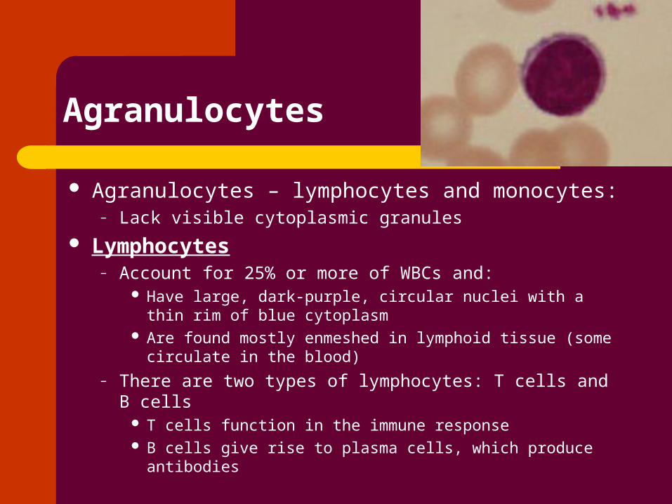
Agranulocytes – lymphocytes and monocytes:– Lack visible cytoplasmic granules
Lymphocytes– Account for 25% or more of WBCs and:
Have large, dark-purple, circular nuclei with a thin rim of blue cytoplasm
Are found mostly enmeshed in lymphoid tissue (some circulate in the blood)
– There are two types of lymphocytes: T cells and B cells T cells function in the immune response B cells give rise to plasma cells, which produce antibodies
Agranulocytes
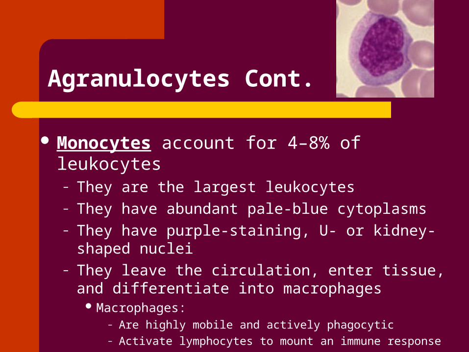
Monocytes account for 4–8% of leukocytes – They are the largest leukocytes– They have abundant pale-blue cytoplasms– They have purple-staining, U- or kidney-shaped nuclei– They leave the circulation, enter tissue, and
differentiate into macrophages Macrophages:
– Are highly mobile and actively phagocytic– Activate lymphocytes to mount an immune response
Agranulocytes Cont.

Immature WBCs are found in the bloodstream in all leukemias
Bone marrow becomes totally occupied with cancerous leukocytes
The WBCs produced are numerous but not functional Death is caused by internal hemorrhage and
overwhelming infections Treatments include irradiation, antileukemic drugs, and
bone marrow transplants
Leukemia

Platelets

Platelets are fragments of megakaryocytes with a blue-staining outer region and a purple granular center
Platelets function in the clotting mechanism by forming a temporary plug that helps seal breaks in blood vessels
Platelets

Genesis of Platelets
The stem cell for platelets is the hemocytoblast
Figure 17.12

A series of reactions designed for stoppage of bleeding
During hemostasis, three phases occur in rapid sequence– Vascular spasms – immediate vasoconstriction in
response to injury– Platelet plug formation– Coagulation (blood clotting)
Hemostasis

A set of reactions in which blood is transformed from a liquid to a gel
Coagulation

Unnecessary clotting is prevented by the structural and molecular characteristics of endothelial cells lining the blood vessels– The smooth endothelial lining of blood vessels– Heparin and PGI2 secreted by endothelial cells
– Vitamin E quinone, a potent anticoagulant
Factors Preventing Undesirable Clotting

Substances used to prevent undesirable clots include:– Aspirin – an antiprostaglandin that inhibits
thromboxane A2
– Heparin – an anticoagulant used clinically for pre- and postoperative cardiac care
– Warfarin – used for those prone to atrial fibrillation
Prevention of Undesirable Clots

RBC membranes have glycoprotein antigens on their external surfaces
These antigens are:– Unique to the individual – Recognized as foreign if transfused into another
individual– Promoters of agglutination and are referred to as
agglutinogens Presence or absence of these antigens is
used to classify blood groups
Human Blood Groups

Humans have 30 varieties of naturally occurring RBC antigens
The antigens of the ABO and Rh blood groups cause vigorous transfusion reactions when they are improperly transfused
Other blood groups (M, N, Dufy, Kell, and Lewis) are mainly used for legalities
Blood Groups

The ABO blood groups consists of:– Two antigens (A and B) on the surface of the RBCs – Two antibodies in the plasma (anti-A and anti-B)
An individual with ABO blood may have various types of antigens and spontaneously preformed antibodies
Agglutinogens and their corresponding antibodies cannot be mixed without serious hemolytic reactions
ABO Blood Groups

ABO Blood Groups
Table 17.4

There are eight different Rh agglutinogens, three of which (C, D, and E) are common
Presence of the Rh agglutinogens on RBCs is indicated as Rh+
Anti-Rh antibodies are not spontaneously formed in Rh– individuals
However, if an Rh– individual receives Rh+ blood, anti-Rh antibodies form
A second exposure to Rh+ blood will result in a typical transfusion reaction
Rh Blood Groups
Blood Typing
When serum containing anti-A or anti-B agglutinins is added to blood, agglutination will occur between the agglutinin and the corresponding agglutinogens
Positive reactions indicate agglutination

Blood Typing
Blood type being tested
RBC agglutinogens Serum Reaction
Anti-A Anti-B
AB A and B + +
B B – +
A A + –
O None – –

Whole blood transfusions are used: – When blood loss is substantial – In treating thrombocytopenia
Packed red cells (cells with plasma removed) are used to treat anemia
Blood Transfusions

Transfusion reactions occur when mismatched blood is infused
Donor’s cells are attacked by the recipient’s plasma agglutinins causing:– Diminished oxygen-carrying capacity– Clumped cells that impede blood flow– Ruptured RBCs that release free hemoglobin into the
bloodstream Circulating hemoglobin precipitates in the
kidneys and causes renal failure
Transfusion Reactions

Blood Disorders

Decreased RBC Number
Hemorrhagic anemia – result of acute or chronic loss of blood
Hemolytic anemia – prematurely ruptured erythrocytes
Aplastic anemia – destruction or inhibition of red bone marrow

Iron-deficiency anemia results from:– A secondary result of hemorrhagic anemia– Inadequate intake of iron-containing foods– Impaired iron absorption
Pernicious anemia results from:– Deficiency of vitamin B12
– Lack of intrinsic factor needed for absorption of B12
– Treatment is intramuscular injection of B12
Decreased Hemoglobin

Abnormal Hemoglobin
Thalassemias – absent or faulty globin chain in hemoglobin – Erythrocytes are thin, delicate, and deficient in
hemoglobin Sickle-cell anemia – results from a defective
gene coding for an abnormal hemoglobin called hemoglobin S (HbS)– HbS has a single amino acid substitution – This defect causes RBCs to become sickle-
shaped in low oxygen situations

Sickle Cell Anemia

Polycythemia
Polycythemia – excess RBCs that increase blood viscosity (impairs circulation)– Polycythemia vera: bone cancer– Secondary polycythemia: high altitude

Thrombus – a clot that develops and persists in an unbroken blood vessel– Thrombi can block circulation, resulting in tissue
death– Coronary thrombosis – thrombus in blood vessel
of the heart
Hemostasis Disorders:Thromboembolytic Conditions

Embolus – a thrombus freely floating in the blood stream– Pulmonary emboli can impair the ability of the
body to obtain oxygen– Cerebral emboli can cause strokes
Hemostasis Disorders:Thromboembolytic Conditions

Thrombocytopenia – condition where the number of circulating platelets is deficient– petechiae – Caused by suppression or destruction of bone
marrow (e.g., malignancy, radiation)– Platelet counts less than 50,000/mm3 – Treated with whole blood transfusions
Bleeding Disorders

Inability to synthesize procoagulants by the liver results in severe bleeding disorders
Causes can range from vitamin K deficiency to hepatitis and cirrhosis
Inability to absorb fat can lead to vitamin K deficiencies as it is a fat-soluble substance and is absorbed along with fat
Liver disease can also prevent the liver from producing bile, which is required for fat and vitamin K absorption
Bleeding Disorders Cont.

Hemophilias – hereditary bleeding disorders caused by lack of clotting factors
– Hemophilia A – most common type (83% of all cases) due to a deficiency of factor VIII
– Hemophilia B – results from a deficiency of factor IX– Hemophilia C – mild type, caused by a deficiency of factor
XI Symptoms include prolonged bleeding and painful
and disabled joints Treatment is with blood transfusions and the
injection of missing factors
Bleeding Disorders

Hemolytic disease of the newborn – Rh+ antibodies of a sensitized Rh– mother cross the placenta and attack and destroy the RBCs of an Rh+ baby
Rh– mother becomes sensitized when Rh+ blood (from a previous pregnancy of an Rh+ baby or a Rh+ transfusion) causes her body to synthesis Rh+ antibodies
The drug RhoGAM can prevent the Rh– mother from becoming sensitized
Treatment of hemolytic disease of the newborn involves pre-birth transfusions and exchange transfusions after birth
Hemolytic Disease of the Newborn