Embed Size (px)
Citation preview
Article ID: WMC002530 ISSN 2046-1690
Phylogeny of Age-Related Fitness Decline in theWild and of Related PhenomenaCorresponding Author:Dr. Giacinto Libertini,Cardiology, ASL NA2 Nord - Naples, via Cavour 13, 80023 - Italy
Submitting Author:Dr. Giacinto Libertini,Cardiology, ASL NA2 Nord - Naples, via Cavour 13, 80023 - Italy
Article ID: WMC002530
Article Type: Review articles
Submitted on:28-Nov-2011, 11:59:33 AM GMT Published on: 28-Nov-2011, 05:22:53 PM GMT
Article URL: http://www.webmedcentral.com/article_view/2530
Subject Categories:AGING
Keywords:IMICAW, IMICAC, Aging, Telomere, Telomerase, Apoptosis, Proapoptosis
How to cite the article:Libertini G . Phylogeny of Age-Related Fitness Decline in the Wild and of RelatedPhenomena . WebmedCentral AGING 2011;2(11):WMC002530
Source(s) of Funding:
None
Competing Interests:
None
WebmedCentral > Review articles Page 1 of 23
WMC002530 Downloaded from http://www.webmedcentral.com on 28-Nov-2011, 05:22:54 PM
Phylogeny of Age-Related Fitness Decline in theWild and of Related PhenomenaAuthor(s): Libertini G
Abstract
Apoptosis, the telomere-telomerase system, cellsenescence and replicative senescence, acharacteristic of cell senescence, are ubiquitous ineukaryotic species. Moreover, in some eubacterialspecies, “proapoptosis”, a type of cell suicide, isdetermined by molecules homologous to apoptoticproteins, suggesting a common phylogenetic origin.The sophisticated mechanisms and regulatorsunderlying these phenomena are geneticallydetermined.A common feature is that they are always harmful forthe individual cell or for the multicellular organism orfor the single cell in a multicellular organism in whichthey act. However, they are probably advantageousfor kin cells or individuals.In particular, in some eukaryotic species, a significanteffect is that they may cause, in natural conditions, anage-related fitness decline, which is also referred to as“aging”, an imprecise term.Here I suggest that their evolutionary meanings lie inkin selection, and the analogies between their action inmonocellular and multicellular eukaryotes areunderlined.A phylogenetic reconstruction based on their adaptivemeanings is proposed.
Introduction
Preliminary remarksSome preliminary consideration are indispensable toavoid misunderstandings.The phenomenon of an “increasing mortality withincreasing chronological age in populations in the wild”("IMICAW" [Libertini, 1988]), alias “actuarialsenescence in the wild” [Holmes and Austad, 1995],alias "age-related fitness decline in the wild", is a realand well documented phenomenon [Deevey, 1947;Laws and Parker, 1968; Spinage, 1970, 1972; Finch,1990; Holmes and Austad, 1995; Ricklefs, 1998].By definition, according to its presence in wildconditions, IMICAW phenomenon is subject to naturalselection and should not be mixed up with the"increasing mortality with increasing chronological agein captivity" ("IMICAC" [Libertini 1988]), which is found
in laboratory conditions at ages not existing in the wildfor species that in natural conditions do not showIMICAW phenomenon. By definition, according to itsabsence in wild conditions, IMICAC is not subject tonatural selection. In particular, the "fitness" in"age-related fitness decline in the wild" definition isunsuitable to the artificial conditions defined in IMICACconcept.This paper regards only IMICAW, alias aged-relatedfitness decline, and related phenomena and notIMICAC phenomenon. This remark is important as incurrent scientific literature and in the prevailing ideasabout age-related fitness decline both phenomena areconfused in a single imprecise term, namely "aging"(or "senescence").The concepts and the results referred to "aging" in itsimprecise meaning but, in fact, to IMICACphenomenon (e.g., the numberless papers regardingthe survival in laboratory conditions and at ages notexisting in the wild of C. elegans and D. melanogaster)will not be considered in this paper, not for inaccuracyor for the sake of brevity but as not regarding the topic.Moreover, in this paper, the term "aging" will be usedonly making reference to current ideas where aprecise meaning is not defined.IntroductionIf species separated by different evolutionary historiesof hundreds of millions of years show equal or similarfeatures that are clearly of common phylogeneticorigin, it is necessary to inquire about an equality oranalogy of functions explaining their evolutionarypersistence and similarity.P h e n o m e n a s u c h a s a p o p t o s i s , t h etelomere-telomerase system, cell senescence andreplicative senescence (that is, in relation to thenumber of cell replications, in a single cell: abruptdecline of cell functions and block of duplicationcapacities; in a cell culture: overall progressive declineof cell functions and of duplication capacities), whichwill be discussed in the next section, exist in yeast, amonocellular eukaryote, and in multicellular eukaryoticspecies with a divergent evolutionary history at leastfrom the beginning of Cambrian period, about 600millions of years ago [Minkoff, 1983].Moreover, “proapoptosis” [Hochman, 1997], a form ofeubacterial cell suicide with mechanisms clearlyrelated to eukaryotic apoptosis [Koonin and Aravind,2002], indicates a much older evolutionary persistence
WebmedCentral > Review articles Page 2 of 23
WMC002530 Downloaded from http://www.webmedcentral.com on 28-Nov-2011, 05:22:54 PM
and similarity.The present paper expounds the main commonfeatures of these phenomena, trying to explain theirgeneral evolutionary meanings and phylogeneticrelations.
Review
EMPIRICAL EVIDENCEA) LIMITS IN DUPLICATION CAPACITIESA-1) In multicellular eukaryotesNormal eukaryotic non-germ cells of multicellularorganisms with limited lifespan can, in general,duplicate themselves only a limited number of timesboth in vitro [Hayflick, 1965; Hayflick and Moorhead,1961] and in vivo [Schneider and Mitsui, 1976]. Thisphenomenon (Hayflick limit), well documented formany types of cells [Rheinwald and Green, 1975;Bierman, 1978; Tassin et al., 1979], shows an inverserelation with the ages of donors of origin [Martin et al.,1970] and, with exceptions that will be discussed later,a rough direct correlation with the life span of thespecies from which cells are derived [Röhme, 1981].The main cause of the phenomenon, for many yearsknown to be caused by something acting in thenucleus [Wright and Hayflick, 1975], was suggested tobe a result of the incomplete action of DNApolymerase, which at each duplication leaves out apart of the terminal portion of DNA, the telomere[Watson, 1972]. This incomplete replication leads to aprogressive shortening of the DNA molecule with arelated increase in duplication impairment [Olovnikov,1973].Telomeres are highly conserved repetitive sequencesof DNA (e.g., TTGGGG in a protozoan [Blackburn andGall, 1978], TTAGGG in mammals [Moyzis et al.,1988] and many other species [Blackburn, 1991]).Telomeres shorten with every duplication event[Harley et al., 1990], but an enzyme, telomerase, canelongate telomeres at each replication, therebycompensating for the incomplete action of DNApolymerase. The action of telomerase explains whysome cells, such as those of germ line, have unlimitedduplication capacities [Greider and Blackburn, 1985].With telomerase deactivation, telomeres shorten ateach duplication and, in a cell culture or in a tissue,overall duplication capacity is reduced [Yu et al., 1990].On the other hand, telomerase activation elongatestelomeres and cells become capable of numberlessduplications [Bodnar et al., 1998; Counter et al., 1998;de Lange and Jacks, 1999; Vaziri, 1998; Vaziri andBenchimol, 1998]. Moreover, active telomerase wasdemonstrated in immortal human cell lines [Morin,
1989], while in other cells it was proven to berepressed by regulatory proteins [van Steensel and deLange, 1997].In a cell culture, the final incapability of a cell toduplicate (replicative senescence) was shown not tobe an abrupt event for all the cells at the same time,but a progressive reduction of cell culture growthpotential that depended on the reduction of telomerelength [Jones et al., 1985; Pontèn et al., 1983].According to Blackburn’s model [Blackburn, 2000],particular protective nucleoproteins cap telomeres,which oscillate between capped and uncappedconditions: the duration of the first state directlycorrelates with telomere length while the other state isvulnerable to the passage to “noncycling state” or finalstage of replicative senescence (fig. 1).A population of cells with telomeres at their maximumlength, but inactivated telomerase, shows aprogressive decline in replication capacities. Evencells with telomerase activated and so telomeresconstantly at maximum length, should show a smallpercentage of cells passing to noncycling state at eachdivision. Moreover, it has been proposed that stemcells, unlike germ cells, have levels of telomeraseactivity that are only partially able to stabilise telomerelength [Holt et al., 1996] and for this reason theycannot indefinitely replace the apoptotic elements forcell populations in renewal [Fossel, 2004].The absolute length of telomeres does not enable oneto predict a species life span. Species, such as themouse and the hamster have long telomeres[Slijepcevic and Hande, 1999], yet they age moreprecociously than species such as man, which haveshorter telomeres. Moreover, in rodents, telomeraseactivity is not related to maximum lifespan [Gorbunovaet al., 2008]. However, Blackburn’s hypothesis doesnot postulate for different species a fixed ratio betweentelomere length or telomerase activity and the stabilityof telomere-capping nucleoproteins complex: it is easyto suppose that the stability of the complex and, ingeneral, the modulation of telomere-telomerasesystem is different from species to species. What islikely important is the species-specific critical telomerelength and the relative rather than absolute telomereshortening [Fossel, 2004].In relation to the mean number of duplications in cellculture or in a tissue, there is an increasing probabilityof cell senescence, a "fundamental cellular program"[Ben-Porath and Weinberg, 2005], which ischaracterized by an altered expression of many genesusually active in the cell, compromising cell overallfunctionality, and by replicative senescence. Asenescent cell has deleterious consequences on theextracellular matrix as well as other cells that are
WebmedCentral > Review articles Page 3 of 23
WMC002530 Downloaded from http://www.webmedcentral.com on 28-Nov-2011, 05:22:54 PM
physically near or physiologically interdependent. Cellsenescence, and replicative senescence that is one ofits characteristic, certainly derive somehow from therelative shortening of telomere (Fossel’s “cellsenescence limited model”) [Fossel, 2004].About the mechanism underlying cell senescence:“One model of telomere-gene expression linkage is analtered chromosomal structure (Ferguson et al., 1991),such as a heterochromatin ‘hood’ that covers thetelomere and a variable length of the subtelomericchromosome (Fossel, 1996; Villeponteau, 1997;Wright et al., 1999). As the telomere shortens, thehood slides further down the chromosome (theheterochromatin hood remains invariant in size andsimply moves with the shortening terminus) or thehood shortens (as the telomere is less capable ofretaining heterochromatin). In either case, the result isan alteration of transcription from portions of thechromosome immediately adjacent to the telomericcomplex, usually causing transcriptional silencing,although the control is doubtless more complex thanmerely telomere effect through propinquity (Aparicioand Gottschling, 1994; Singer et al., 1998; Stevensonand Gottschling, 1999). These silenced genes may inturn modulate other, more distant genes (or set ofgenes). There is some direct evidence for suchmodulation in the subtelomere ...” [Fossel, 2004].These statements are largely based on experiments inyeast, but possible deductions for monocellulareukaryotes must consider the invariability of telomerelength with duplications in these organisms (see nextparagraph).On the other hand, the likelihood that a mechanism ofthis type is true for multicellular eukaryotes is widelydiscussed by Fossel (see pages 45-56 in Fossel, 2004;a plausible scheme is illustrated in fig. 2).Heterochromatin ‘hood’ [Fossel, 2004] and cappingnucleoproteins [Blackburn, 2000] are most likely thesame thing because: 1) they are supposed in thesame part of the chromosome; 2) telomeraseactivation and the consequent telomere lengtheningcause the reversal both of manifestations of cellsenescence and of replicative senescence [Bodnar etal., 1998; Counter et al., 1998; de Lange and Jacks,1999].For germ line cells and for donor somatic cells thatoriginate a cloned animal, the resetting of telomereclock is indispensable [Fossel, 2004]. The startinglength of telomere must be established as with eachsubsequent shortening of the telomere, the probabilityof cell senescence and replicative senescence willincrease. The absolute value of “telomere length isirrelevant” [Fossel, 2004]: two Mus strains withdifferent telomere length (10 and 20 kb, respectively)
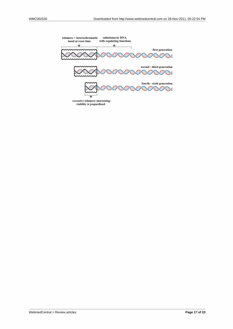
show the same life span and an equal timing of cellsenescence; analogously the same is true for donoranimals and for cloned animals derived from cells withshortened telomeres [Fossel, 2004]. An appropriateshaping of the heterochromatin hood depending ontelomere length could explain the equal timing of cellsenescence and replicative senescence in spite ofdifferent telomere lengths (fig. 3; the hypotheticalmodel exposed in the figure could explain the largeirrelevance of initial telomere length for theconsequences of its subsequent shortening [Fossel,2004]).Mice and other animals have a shorter life span,despite a baseline telomerase activity in most somaticcells [Prowse and Greider, 1995] and much longertelomeres than our species [Slijepcevic and Hande,1999]. (But, in mice microglia cells, telomeres shortenwith age and "the low levels of telomerase activitypresent may be preferentially recruited to maintain theshortest telomeres while allowing the longer ones toshorten more rapidly" [Flanary, 2003].) Moreover, inknockout (mTR-/-) mice, which have telomerasegenetically inactivated, only after four [Herrera et al.,1999] to six [Blasco et al., 1997] generations, with veryshortened telomeres, fertility and viability arejeopardized, although organs with high cell turnovershow dysfunctions in early generations [Herrera et al.,1999; Lee et al., 1998]. The model of fig. 3, asexpounded in fig. 4, could explain this apparentlyparadoxical phenomenon. The short life span of miceand other species with long telomeres could beexplained by a species-specific low degree of telomere+ heterochromatin hood complex stability.Subtelomeric DNA appears to have both a pivotalimportance for overall cell functionality and a positionvulnerable to inactivation by telomere shortening itself.Excluding the possibility of an absurd evolutionaryillogicality, this coincidence can be explained only assomething favoured by natural selection to determinecell senescence and replicative senescence. Apossible scenario for the evolution of the telomere-cellsenescence system is proposed below.A-2) In a monocellular eukaryoteYeast (Saccharomyces cerevisiae), a well studiedeukaryotic monocellular species, reproduces byasymmetric division between mother and daughtercells. The mother lineage can reproduce a limitednumber of times only, specifically between 25 and 35generations in about 3 days [Jazwinski, 1993].Both in mother and daughter yeast cells, telomerelength does not decrease with duplications [D’Melloand Jazwinski, 1991; Smeal et al., 1996]. “Budding [=daughter] yeast cells express telomerase and divideindefinitely.” [Maringele and Lydall, 2004]
WebmedCentral > Review articles Page 4 of 23
WMC002530 Downloaded from http://www.webmedcentral.com on 28-Nov-2011, 05:22:54 PM
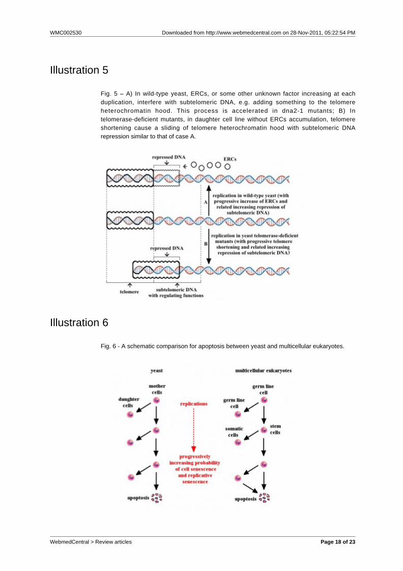
In mother cells of wild-type yeast, extrachromosomalribosomal DNA circles (ERCs) accumulate inproportion to the number of duplications [Sinclair andGuarente, 1997] and “several lines of evidencesuggest that accumulation of ERCs is one determinantof life span” [Lesur and Campbell, 2004].ERCs, or some other unknown factor, interfere withgene expression and mutants such as dna2-1, whichshow abnormalities in the replication of DNA andtherefore increased rates of ERCs accumulation,suffer by precocious alterations of gene expression.Specifically, transcriptome of older (18-generation-old)individuals of wild-type yeast are similar to those ofyoung (8-generation-old) individuals of dna2-1 mutants[Lesur and Campbell, 2004].Telomerase-deficient mutants (tlc1∆ mutants) show,both in mother and daughter cells, telomere shortening.Additionally, older individuals of daughter cell lineages,which have no ERCs accumulation, show an overallexpression of genes (transcriptome) similar to that ofolder individuals of wild-type yeast, and of youngindividuals of dna2-1 mutants [Lesur and Campbell,2004]. It is possible that in telomerase-deficient yeastmutants, as in cells of multicellular eukaryotes,telomere shortening causes the sliding of a telomereheterochromatin hood that interferes with a critical partof subtelomeric DNA, while in wild-type yeastsubtelomeric DNA is somehow repressed by ERCs, orby some other unknown factor (fig. 5).In old yeast cells, besides the replicative senescence,there are increasing metabolic alterations [Lesur andCampbell, 2004], which can be defined as cellsenescence.B) APOPTOSISIn contrast with necrosis, which is the cell deathcaused by acute cellular injury, apoptosis is anordered form of cell self-destruction, which isubiquitous in eukaryotic species [Longo et al., 2005].Apoptosis was characterised and clearly differentiatedfrom necrosis for the first time during observations ofnormal liver hepatocytes [Kerr et al., 1972]. It isdescribed as a definite series of biochemical eventsleading to specific morphological changes (blebbing,loss of membrane asymmetry and attachment, cellshrinkage, nuclear fragmentation, chromatincondensation, chromosomal DNA fragmentation, etc.).B-1) In multicellular eukaryotesProgrammed cell death by apoptosis, selectivelytriggered for some cells in specific times, is essentialfor morphogenetic mechanisms (e.g., embryo neuraldevelopment [Nijhawan et al., 2000], wound healing[Greenhalgh, 1998]), lymphocyte selection [Cohen,1993; Opferman, 2008], cell turnover in healthy adultorgans [Israels and Israels, 1999; Lynch et al., 1986;
Medh and Thompson, 2000; Wyllie et al., 1980] (asdocumented for many tissues and organs [Libertini,2006]), removal of damaged or infected cells[Tesfaigzi, 2006; White, 2006], etc.Apoptotic cellular debris does not damage other cellsbecause phagocytes remove such cell fragments in anorderly manner without eliciting an inflammatoryresponse [Erwig and Henson, 2008].Inactivated telomerase and short telomeres increasethe probability of apoptosis [Fossel, 2004; Ozen et al.,1998; Holt et al. 1999; Seimiya et al., 1999; Ren et al.,2001].B-2) In a monocellular eukaryoteIn yeast, a phenomenon closely resembling apoptosisof multicellular eukaryotes was described quiterecently [Madeo et al., 1997]. It was soon evident thatthe overexpression of human Bcl-2, an apoptosisinhibiting factor, in yeast delays processes leading tothe phenomenon [Longo et al., 1997], while theoverexpression of an apoptosis inducing factor inmammalians (BAX) could elicit it [Ligr et al., 1998].A growing body of evidence has documentedsimilarities between this phenomenon in yeast andapoptosis in multicellular eukaryotes, to the extent thatboth deserve the same name. This data thus suggestthat the two phenomena share a commonphylogenetic origin [Kaeberlein et al., 2007; Longo etal., 2005; Madeo et al., 1999]: “... since the firstdescription of apoptosis in a yeast (Saccharomycescerevisiae) strain carrying a CDC48 mutation (Madeoet al., 1997), several yeast orthologues of crucialmammalian apoptotic proteins have been discovered(Madeo et al., 2002; Fahrenkrog et al., 2004; Wissinget al., 2004; Qiu et al., 2005; Li et al., 2006; Walter etal., 2006), and conserved proteasomal, mitochondrial,and histone-regulated apoptotic pathways have beendelineated (Fig. 1; Manon et al., 1997; Ligr et al., 2001;Ludovico et al., 2002; Fannjiang et al., 2004; Ahn etal . , 2005a; Gourlay and Ayscough, 2005;Pozniakovsky et al., 2005).” [Büttner et al., 2006]In yeast, there is an increasing vulnerability toapoptosis and replicative senescence, when thenumber of duplications increases, together with themetabolic alterations of cell senescence [Büttner et al.,2006; Fabrizio and Longo, 2008; Herker et al., 2004;Laun et al., 2001]. The age-related death rateincrements in yeast follow exponential dynamics [Launet al., 2007], as they also do for multicellular organism[Ricklefs, 1998].Apoptosis is also triggered or favoured by: a)unsuccessful mating [Büttner et al., 2006]; b) dwindlingnutrients [Granot et al., 2003]; c) chemical alterations[Madeo et al., 1999]; and d) killer toxins secreted bycompeting yeast tribes [Büttner et al., 2006].
WebmedCentral > Review articles Page 5 of 23
WMC002530 Downloaded from http://www.webmedcentral.com on 28-Nov-2011, 05:22:54 PM
When a yeast individual dies by apoptosis, cellularfragments do not damage other cells and are usefullyphagocytised by other cells, which, consequently, “areable to survive longer with substances released bydying cells” [Herker et al., 2004].A schematic comparison between apoptosis in yeastand multicellular eukaryotes is illustrated in fig. 6.EVOLUTIONARY INTERPRETATIONSC) IN MONOCELLULAR EUKARYOTESC-1) ApoptosisApoptotic patterns in yeast have been interpreted asadaptive because they are useful to the survival of theclone, which is likely made up of kin individuals[Fabrizio et al., 2004; Herker et al., 2004; Longo et al.,2005; Mitteldorf, 2006; Skulachev, 1999, 2002, 2003;Skulachev and Longo, 2005]. An exception isapoptosis triggered by toxin secreted by competingyeast tribes, where apoptotic mechanisms areexploited by competitors for increasing their fitness[Büttner et al., 2006].The adaptive hypothesis appears plausible when aspecies is divided in many small demes, each of whichis made up of one or a few clones, previously derivedfrom as many individuals, and in conditions ofK-selection, that is with population size “at or near [orover] carrying capacity of the environment” [Pianka,1970]. In fact, in such conditions, the sacrifice of partof the population increases the survival probabilities ofthe remaining individuals, which are kin individuals(coefficient of relationship, r, equal to 1 in the case of ademe made up of a single clone, and greater thanzero in the case of a deme made up of few clones). Interms of inclusive fitness [Hamilton, 1964, 1970;Trivers, 1971; Trivers and Hare, 1976], suicideindividuals - by action of a hypothetical gene C -reduce their individual fitness but increases it forsurviving kin individuals, in which there is a probabilityr of the existence of a copy of C. The inclusive fitnessof C (FC) is given by the sum of individual fitnessreduction of suicide individuals plus the sum ofindividual fitness increase of survivors each multipliedby the probability that C is present in them:
n1 n2
FC = Σ (rx Sx) + Σ (-S’x) (1)
x=1 x=1
where n1 = number of surviving individuals; Sx =advantage for a surviving individual; rx = coefficient ofrelationship between a surviving individual and suicideindividuals; n2 = number of suicide individuals; -S’x =disadvantage for each suicide individual.
If FC is positive, C is favoured by selection.The possibility that the suicide of an individual, called“phenoptosis” by analogy to the term apoptosis[Skulachev, 1999], is also favoured by naturalselection in prokaryote organisms, is necessary toexplain the existence of “programmed death inbacteria” [Lewis, 2000; Skulachev, 2003]: e.g.,bacterial phytoplankton mass suicide as defenceagainst viruses [Lane, 2008], bacterial suicidetriggered by phage infection “thereby curtailing viralmultiplication and protecting nearby E. coli frominfection” [Raff, 1998] and the “built-in suicide module”activated by antibiotics in E. coli [Engelberg-Kulka etal., 2004]. Interestingly, these mechanisms have beendefined as “proapoptosis” and hypothesised asphylogenetic precursors of eukaryotic apoptosis[Hochman, 1997], as they share with it variousfeatures: “Several key enzymes of the apoptoticmachinery, including the paracaspase andmetacaspase families of the caspase-like proteasesuperfamily, apoptotic ATPases and NACHT familyNTPases, and mitochondrial HtrA-like proteases, havediverse homologs in bacteria, but not in archaea.Phylogenetic analysis strongly suggests amitochondrial origin for metacaspases and theHtrA-like proteases, whereas acquisition fromActinomycetes appears to be the most likely scenariofor AP-ATPases. The homologs of apoptotic proteinsare particularly abundant and diverse in bacteria thatundergo complex development , such asA c t i n o m y c e t e s , C y a n o b a c t e r i a a n dalpha-proteobacteria, the latter being progenitors ofthe mitochondria.” [Koonin and Aravind, 2002].C-2) Cell senescence and replicative senescenceIn yeast, increasing vulnerability to apoptosis inrelation to the number of duplications, a feature of cellsenescence [Fabrizio and Longo, 2008; Herker et al.,2004], determines, or contributes to determining,which cells will die in conditions in which the sacrificeof part of the population may allow the survival of theothers.Cell senescence and replicative senescence may beexplained by a mechanism similar to that justifyingapoptosis but with a different evolutionary advantage.In fact, Büttner et al. suggested that “apoptosiscoupled to chronological and replicative aging limitslongevity that would maintain ancient genetic variantswithin the population and, therefore, favor geneticconservatism.” [Büttner et al., 2006]This is not a new argument. Yeast ecological lifeconditions, if they are of the K-selection type, allowone to hypothesise that cell senescence andreplicative senescence are adaptive and explainablewith the same evolutionary mechanism proposed for
WebmedCentral > Review articles Page 6 of 23
WMC002530 Downloaded from http://www.webmedcentral.com on 28-Nov-2011, 05:22:54 PM
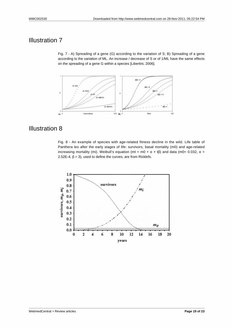
age-related fitness decline in multicellular speciessubject to K-selection [Libertini, 1988, 2006]. This isthe same above-mentioned suggestion of Büttner etal., but formulated in terms of individual selection.In short, the diffusion of a gene G is dependent bothon its advantage S over a neutral allele and on theinverse of the mean duration of life (ML), or generationtime (fig. 7).A gene C that causes the premature death of anindividual I, where C is present, and so reduces its MLand causes a disadvantage S’, accelerates thespreading of any favourable gene in the individual (I’)that takes the place of I. If I’ is kin to I, the inclusivefitness (FC) will be positive and C will be favoured byselection, if:
n
FC = r × Σ (Sx) × (1/MLC - 1/MLC’) - S’ > 0 (2)
x=1
where: MLC and MLC’are the ML of individuals withthe gene C and the neutral allele C’, respectively; Σ(Sx)is the summation notation of the advantages of the nfavourable genes spreading within the species; -S’ isthe disadvantage of a smaller ML; r is the meancoefficient of relationship between I and I’. (The use ofkin selection to explain the age-related fitness declineshould not be confused with the use of the same typeof select ion to explain the survival in thepost-reproductive period, as suggested in other papers[Lee, 2008].)This hypothesis was formulated for multicellularorganisms, but there is no theoretical argumentagainst its application to monocellular eukaryotes.Büttner et al. do not express alternative evolutionaryexplanations for cell senescence and replicativesenescence besides the above-mentioned suggestion[Büttner et al., 2006], which is a short reformulation ofthe theory described.In contrast with this hypothesis, Lewis argues againstthe “suggestion that yeast cells provide a precedentfor programmed death” [Lewis, 2000], proposed byothers AA. [Sinclair et al., 1998], with the followingobservation: if a yeast cell of the mother lineage diesafter n duplication (n = 25-35 in laboratory conditions[Jazwinski, 1993]), the death of a single individualamong 2n descendants (= 107-1010 individuals)appears insignificant for any theory of programmeddeath that is somehow favoured by natural selection.In fact, in natural conditions the probability that anindividual of the mother lineage dies by apoptosis aftern duplications is practically zero and the phenomenon,
being observable in laboratory conditions only, cannothave selective value. However, this argument missesa pivotal point: it is important not the death after nduplications of a single individual among innumerabledescendants, but the exponentially progressive - inrelation to the number of duplications - increasingprobability of apoptosis, coupled with a difference inmortality rates and capability of having offspringbetween “younger” and “older” individuals (“in apopulation of [yeast] cells the lifespan distributionfollows the Gompertz law” [Laun et al., 2007], that isan age-related exponential increase of mortality; “Theprobability that an individual yeast cell will producedaughters declines exponentially as a function of itsage in cell divisions or generations (Jazwinski et al.,1998).” [Lesur and Campbell, 2004]) and, therefore, afaster generation turnover caused by the preferentialdeath of “older” individuals. If cell senescence andreplicative senescence manifest themselves in naturalconditions and reduce significantly wild yeast ML,Lewis’ objection does not invalidate the hypothesisthat yeast fitness decline related to duplication numbermay have a selective value and may be favoured bynatural selection. However, Lewis’ objection is veryinteresting because it echoes a similar argumentagainst programmed aging theories for multicellularorganisms that will be discussed in the next section.D) IN MULTICELLULAR EUKARYOTESD-1) Apoptosis
In multicellular organisms, apoptosis is essential formany physiological functions as outlined above. Theevolutionary justification for these phenomena isevident and will not be discussed.D-2) Cell senescence and replicative senescenceAs underlined in the preliminary remark, anage-related increasing mortality, or fitness decline, isdocumented for many species in wild conditions (fig.8).A plausible mechanism for this fitness decline is theprogressive slowdown of cell turnover, that is aprogressive prevalence of programmed cell death(PCD), by apoptosis or other forms of PCD, on cellsubstitution by duplication of stem cells (Fossel’s “cellsenescence general model of aging” [Fossel, 2004;Libertini, 2006]). A hypothesis of this type wassuggested for the first time by Weissmann [Kirkwoodand Cremer, 1982] while the concept of senescenceas a result of decrease in cellularity of organs wasdiscussed by Szilard [Szilard, 1959], although in thecontext of a theory that attributed the cell loss to theaccumulation of somatic mutations. In support of thisthesis, for some species, as Rockfish and lobsters,both telomere length and mortality rate are unvariedwith the age [Klapper, Heidorn et al., 1998; Klapper,
WebmedCentral > Review articles Page 7 of 23
WMC002530 Downloaded from http://www.webmedcentral.com on 28-Nov-2011, 05:22:54 PM
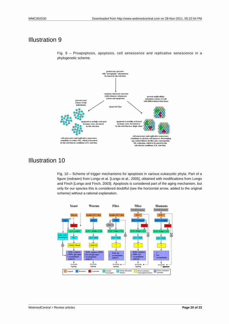
Kühne et al., 1998].There is empirical evidence for an adaptive meaningof the age-related fitness decline phenomenon[Libertini, 2008], which in its more advancedexpression, common in protected conditions, is usuallycalled ‘aging’, an imprecise term [Libertini, 2006]. Atheory, the same as the above-mentioned to elucidatecell senescence and replicative senescence in yeast,explains this fitness decline as evolutionarilyadvantageous by a mechanism of kin selection that, inconsequence of a quicker generation turnover, allowsa faster spreading of any advantageous mutations.According to this theory, the advantage exists inconditions of K-selection (species divided in demes,populated by kin individuals, and with saturatedhabitats in which only the death of an individual givesspace to a new individual) [Libertini, 1988, 2006].The main objection against this theory, analogous toLewis’ argument above-mentioned, is that “As a rule,wild animals simply do not live long enough to growold. Therefore, natural selection has limitedopportunity to exert a direct influence over the processof senescence.” [Kirkwood and Austad, 2000]. Thisobjection, analogous to Lewis’ argument, misses apivotal point: the existence or absence in the wild of“old” individuals (e.g., individuals of P. leo older than15 years) is not important. Individuals of P. leoyounger than 15 years are “not old” individuals, yetthey show an increasing fitness reduction at agespresent in the wild: this significantly reduces ML with aconsequent faster generation turnover and a possibleselective advantage.“Senescence reduces average life span ... by almost80% when m0 = 0.01 yr-1” [Ricklefs, 1998]. For thefraction of a population that survived the high mortalityrisk of the early stages of life, the ratio between theresidual MLs without and with age-related increasingmortality has been estimated to be in the range 2.5-5for eight mammal species in wild conditions. Withoutthe subtraction of the early stages of life, the ratio hasbeen estimated in the range 1.55-3.21 [Libertini, 1988].In short, in wild conditions, ML reduction caused byage-related increasing mortality is not irrelevant,although the equivalents of septuagenarian or oldermen for animal species are likely inexistent in the wild.PHYLOGENETIC CORRELATIONSThe empirical evidence and the above-mentionedarguments suggest a phylogenetic correlation betweenphenomena observed in colonies of kin yeast cells andanalogous phenomena in multicellular organisms.These phenomena require the formulation of a generalphy logene t i c hypo thes is o f apop tos is ,telomere-telomerase system, cell senescence,replicative senescence and the age-related fitness
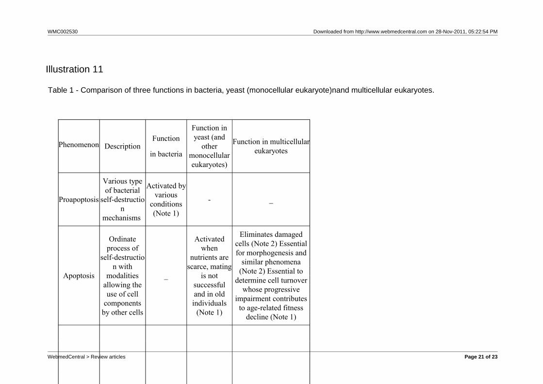
decline, which is commonly but imprecisely called“aging”. Correlated phenomena in bacteria must bealso considered in the phylogenetic model.In particular (see Table 1 and fig. 9):a) Phenomena described in eubacteria as“proapoptosis”, activated in particular conditions andprobably favoured by the mechanism of kin selection(e.g., for bacterial phytoplankton: “As most plankton ina bloom are near identical genetically, from theperspective of their genes, a die-off that createsenough scorched earth to stop the viral advance canmake sense” [Lane, 2008]), have been interpreted asplausible phylogenetic precursors of eukaryoticapoptosis [Hochman, 1997]. Proapoptosis, a form of“suicide useful in critical conditions”, is necessarilyderived from a previous condition in which this patternwas inexistent.b) Eubacteria evolved in monocellular eukaryotes.Apoptosis of monocellular eukaryotes, likely derivedfrom a form of eubacterial proapoptosis, is in yeasttriggered by starvation, damaged cell conditions,unsuccessful mating, etc. In these cases, it is favouredby kin selection because cell suicide increasessurvival probability of kin cells [Herker et al., 2004](“suicide useful in critical conditions”).c) Both for proapoptosis and for apoptosis, amechanism that triggers the suicide pattern, but killsonly a part of the population - proportional to theseverity of stress condition - is indispensable. Yeastevolved an efficient mechanism based on the numbero f p r e v i o u s d u p l i c a t i o n s a n d atelomere-telomerase-ERCs clock [Büttner et al., 2006;Fabrizio and Longo, 2008; Herker et al., 2004; Laun etal., 2007].d) Apoptosis of multicellular eukaryotic species has aclear phylogenetic relationship with monocellulareukaryotic apoptosis [Longo and Finch, 2003]. In mostspecies, the evolved clock does not use ERCs [Fossel,2004]. Considering each multicellular individual as aclone having all cells with the same genes (coefficientof relationship, r, equal to 1) but with differentiatedfunctions, apoptosis of less fit cells may be consideredas favoured by analogous mechanisms of kin selection.e) In multicellular organisms, apoptosis as part ofmorphogenetic mechanisms (e.g., embryogenesis,tissue development or reshaping, tissue turnover) andof lymphocyte selection is clearly a derived function,being impossible in monocellular organisms;f) In yeast, apoptosis, cell senescence and replicativesenescence, genetically determined by mechanismsbased on telomere-telomerase system, appear tocontrast “genetic conservatism” [Büttner et al., 2006].Furthermore, these phenomena might be explained asfavoured by kin selection, as for multicellular
WebmedCentral > Review articles Page 8 of 23
WMC002530 Downloaded from http://www.webmedcentral.com on 28-Nov-2011, 05:22:54 PM
o r g a n i s m s [ L i b e r t i n i , 1 9 8 8 , 2 0 0 6 ] .Suicide-predisposition passes from a pattern usefulonly in emergency conditions to a pattern useful innon-stress conditions too (“suicide useful innon-critical conditions”).g) In multicellular organisms, apoptosis, cellsenescence and replicative senescence, causeage-related limits in cell turnover with consequentage-related fitness decline [Fossel, 2004; Libertini,2006] (“senile state” in its more advanced expressions[Libertini, 2006]), and this has been explained by kinselection in conditions of K-selection [Libertini, 1988,2006].In short, “aging” mechanisms in yeast, a monocellulareukaryote, and in multicellular eukaryotic species,separated by about 600 millions of distinct evolution,are incredibly similar in their basic physiologicalcomponents and selective explanations. Moreover,apoptosis, the core of these mechanisms, has itsphylogenetic roots in eubacterial proapoptoticphenomena.
Conclusion(s)
Aging in yeast is considered adaptive while, formulticellular eukaryotes, this idea is excluded by thecurrent gerontological paradigm [Kirkwood and Austad,2000], which is contrasted both by theoreticalarguments and empirical evidence [Goldsmith, 2003;Libertini, 1988, 2006, 2008; Longo et al., 2005;Mitteldorf, 2006; Skulachev, 1997]. Figure 10 showsthat even authoritative Authors, not restrained bycurrent paradigm, do not state openly that apoptosis ispart of aging mechanisms in our species, while forother species this is maintained [Longo and Finch,2003; Longo et al., 2005].Apoptosis and the telomere-telomerase system aresophisticated mechanisms, necessarily determinedand highly regulated by genes forged by naturalselection. They are ubiquitous in the eukaryotic worldand the many variations among the different phyla donot obscure their single origin [Longo et al., 2005].This strongly suggests that they have significantevolutionary meanings that are related to cellsenescence (Fossel’s “cell senescence limited model”[Fossel, 2004]) and, likely, to the age-related fitnessdecline of the whole organism (Fossel’s “cellsenescence general model of aging” [Fossel, 2004;Libertini, 2006]). In contrast with this evidence, currentgerontological theories state that age-related fitnessdecline, a phenomenon certainly observable at agesexistent in the wild [Libertini, 2008], is determined byrandom factors (harmful mutations, unpredictable
effects of pleiotropic genes or of conflictingevolutionary exigencies [Edney and Gill, 1968;Hamilton, 1966; Kirkwood, 1977; Kirkwood andHolliday, 1979; Medawar, 1952; Mueller, 1987;Partridge and Barton, 1993; Rose, 1991; Williams,1957]). This excludes the aforementionedmechanisms, which are sophisticated and highlyregulated, as causes of the phenomenon.It is important to underline that the life-limiting effectsof telomere-telomerase system are currently explainedas a general defence against cancer [Campisi, 1997,2003; Troen, 2003; Wright and Shay, 2005] but thereare strong arguments and evidence against thishypothesis [Fossel, 2004; Libertini, 2008; Milewski,2010] (e.g., senescent cells secrete substances thatincrease mutation rates and the risk of oncogenesis[Parrinello et al., 2005; Coppé et al., 2008]). Thesteady affection to defence-against-cancer hypothesisby the supporters of non-adaptive aging theories maybe explained by the fact that there is no otherproposed explanation compatible with non-adaptivehypotheses and using philosophical and historicalknowledge [Milewski 2010].Current gerontological theories contrast strongly withthe functions of apoptosis, the telomere-telomerasesystem, cell senescence and replicative senescence,in their phylogenetic schematisation outlined in thispaper, which is based on the concept that all thesephenomena are certainly adaptive. This contrastshould be solved by current gerontological theories or,on the other hand, these theories should be droppedand substituted by the alternative paradigm that theage-related fitness decline is a function with anevolutionary advantage and its physiologicalmechanisms.Moreover, the thesis maintained in this paper, namelythat genetically regulated active mechanisms, basedon telomere-telomerase system and determining thedeath of an organism, have a very ancientphylogenetic history should not be considered asurprise if we consider the numberless well-knowncases of phenoptosis through rapid senescence andsudden death widely described elsewhere [Finch,1990].
References
1. Ben-Porath, I. and Weinberg, R., 2005. The signalsand pathways activating cellular senescence. Int. J.Biochem. Cell Biol. 37, 961-76.2. Bierman, E.L., 1978. The effect of donor age on thein vitro l ife span of cultured human arterialsmooth-muscle cells. In Vitro 14, 951-5.
WebmedCentral > Review articles Page 9 of 23
WMC002530 Downloaded from http://www.webmedcentral.com on 28-Nov-2011, 05:22:54 PM
3. Blackburn, E.H., 1991. Structure and function oftelomeres. Nature 350, 569-73.4. Blackburn, E.H., 2000. Telomere states and cellfates. Nature 408, 53-6.5. Blackburn, E.H. and Gall, J.G., 1978. A tandemlyrepeated sequence at the termin i o f theextrachromosomal ribosomal RNA genes inTetrahymena. J. Mol. Biol. 120, 33-53.6. Blasco, M.A., Lee, H.W., Hande, M.P., Samper, E.,Lansdorp P.M., DePinho, R.A. and Greider, C.W.,1997. Telomere shortening and tumor formation bymouse cells lacking telomerase RNA. Cell 91, 25-34.7. Bodnar, A.G., Ouellette, M., Frolkis, M., Holt, S.E.,Chiu, C., Morin, G.B., Harley, C.B., Shay, J.W.,Lichsteiner, S. and Wright, W.E., 1998. Extension oflife-span by introduction of telomerase into normalhuman cells. Science 279, 349-52.8. Büttner, S., Eisenberg, T., Herker, E.,Carmona-Gutierrez, D., Kroemer, G. and Madeo, F.,2006. Why yeast cells can undergo apoptosis: death intimes of peace, love, and war. J. Cell Biol. 175, 521–5.9. Campisi, J., 1997. The biology of replicativesenescence. Eur. J. Cancer 33, 703–9.10. Campisi, J., 2003. Cancer and ageing: rivaldemons? Nat. Rev. Cancer 3, 339-49.11. Cohen, J.J., 1993. Programmed cell death andapoptosis in lymphocyte development and function.Chest 103, S99-101.12. Coppé, J.-P., Patil, C.K., Rodier, F., Sun, Y.,Muñoz, D.P., Goldstein, J., Nelson, P.S., Desprez,P.-Y. and Campisi, J., 2008. Senescence-associatedsecretory phenotypes reveal cell-nonautonomousfunctions of oncogenic RAS and the p53 tumorsuppressor. PLoS Biol. 6, 2853-68.13. Counter, C.M., Hahn, W.C., Wei, W., Caddle, S.D.,Beijersbergen, R.L., Lansdorp, P.M., Sedivy, J.M. andWeinberg, R.A., 1998. Dissociation among in vitrotelomerase activity, telomere maintenance, andcellular immortalization. Proc. Natl. Acad. Sci. USA 95,14723-8.14. Deevey, E.S.Jr., 1947. Life tables for naturalpopulations of animals. Quart. Rev. Biol. 22, 283–314.15. de Lange, T. and Jacks, T., 1999. For better orworse? Telomerase inhibition and cancer. Cell 98,273-5.16. D'Mello, N.P. and Jazwinski, S.M., 1991. Telomerelength constancy during aging of Saccharomycescerevisiae. J. Bacteriol. 173, 6709-13.17. Edney, E.B. and Gill, R.W., 1968. Evolution ofsenescence and specific longevity. Nature 220, 281-2.18. Engelberg-Kulka, H., Sat, B., Reches, M., Amitai,S. and Hazan, R., 2004. Bacterial programmed celldeath systems as targets for antibiotics. TrendsMicrobiol. 12, 66-71.
19. Erwig, L.-P. and Henson, P.M., 2008. Clearance ofapoptotic cells by phagocytes. Cell Death Differ. 15,243-50.20. Fabrizio, P., Battistella, L., Vardavas, R., Gattazzo,C., Liou, L.L., Diaspro, A., Dossen, J.W., Gralla, E.B.and Longo, V.D., 2004. Superoxide is a mediator of analtruistic aging program in Saccharomyces cerevisiae.J. Cell Biol. 166, 1055–67.21. Fabrizio, P. and Longo, V.D., 2008. Chronologicalaging-induced apoptosis in yeast. Biochim. Biophys.Acta 1783, 1280-5.22. Finch, C.E., 1990. Longevity, Senescence, and theGenome. The University of Chicago Press, Chicago(USA).23. Flanary, B.E., 2003. Telomeres shorten with age inrat cerebellum and cortex in vivo. J. Anti-AgingMedicine 6, 299-308.24. Fossel, M.B., 2004. Cells, Aging and HumanDisease. Oxford University Press, New York (USA).25. Goldsmith, T.C., 2003. The Evolution of Aging:How Darwin's Dilemma is Affecting Your Chance for aLonger and Healthier Life. iUniverse, Lincoln,Nebraska (USA).26. Gorbunova, V., Bozzella, M.J. and Seluanov, A.,2008. Rodents for comparative aging studies: frommice to beavers. Age 30, 111-9.27. Granot, D., Levine, A. and Dor-Hefetz, E., 2003.Sugar-induced apoptosis in yeast cells. FEMS YeastRes. 4, 7–13.28. Greenhalgh, D.G., 1998. The role of apoptosis inwound healing. Int. J. Biochem. Cell Biol. 30, 1019-30.29. Greider, C.W. and Blackburn, E.H., 1985.Identification of a specific telomere terminaltransferase activity in Tetrahymena extracts. Cell 51,405-13.30. Hamilton, W.D., 1964. The genetical evolution ofsocial behaviour, I, II. J. Theor. Biol. 7, 1-52.31. Hamilton, W.D., 1966. The moulding ofsenescence by natural selection. J. Theor. Biol. 12,12-45.32. Hamilton, W.D., 1970. Selfish and spitefulbehaviour in an evolutionary model. Nature 228,1218-20.33. Harley, C.B., Futcher, A.B. and Greider, C.W.,1990. Telomeres shorten during ageing of humanfibroblasts. Nature 345, 458-60.34. Hayflick, L., 1965. The limited in vitro lifetime ofhuman diploid cell strains. Exp. Cell Res. 37, 614-36.35. Hayflick, L. and Moorhead, P.S., 1961. The serialcultivation of human diploid cell strains. Exp. Cell Res.25, 585-621.36. Herker, E., Jungwirth, H., Lehmann, K.A.,Maldener, C., Fröhlich, K.U., Wissing, S., Büttner, S.,Fehr, M., Sigrist, S. and Madeo, F., 2004.
WebmedCentral > Review articles Page 10 of 23

WMC002530 Downloaded from http://www.webmedcentral.com on 28-Nov-2011, 05:22:54 PM
Chronological aging leads to apoptosis in yeast. J. CellBiol. 164, 501-7.37. Herrera, E., Samper, E., Martín-Caballero, J.,Flores, J.M., Lee, H.W. and Blasco, M.A., 1999.Disease states associated with telomerase deficiencyappear earlier in mice with short telomeres. EMBO J.18, 2950-60.38. Hochman, A., 1997. Programmed cell death inprokaryotes. Crit. Rev. Microbiol. 23, 207-14.39. Holmes, D.J. and Austad, S.N., 1995. Birds asanimal models for the comparative biology of aging: aprospectus. J. Gerontol. A Biol. Sci. 50, B59–66.40. Holt, S.E., Glinsky, V.V., Ivanova, A.B. and Glinsky,G.V., 1999. Resistance to apoptosis in human cellsconferred by telomerase function and telomerestability. Mol. Carcinog. 25, 241-8.41. Holt, S.E., Shay, J.W. and Wright, W.E., 1996.Refining the telomere-telomerase hypothesis of agingand cancer. Nature Biotechnol. 14, 836-9.42. Israels, L.G. and Israels, E.D., 1999. Apoptosis.Stem Cells 17, 306-13.43. Jazwinski, S.M., 1993. The genetics of aging in theyeast Saccharomyces cerevisiae. Genetica 91, 35-51.44. Jones, R.B., Whitney, R.G. and Smith, J.R., 1985.Intramitotic variation in proliferative potential:stochastic events in cellular aging. Mech. Ageing Dev.29, 143-9.45. Kaeberlein, M., Burtner, C.R. and Kennedy, B.K.,2007. Recent developments in yeast aging. PLoSGenetics 3(5): e84.46. Kerr, J.F.R., Wyllie, A.H. and Currie, A.R., 1972.Apoptosis: a basic biological phenomenon withwide-ranging implications in tissue kinetics. Br. J.Cancer 26, 239-57.47. Kirkwood, T.B.L., 1977. Evolution of ageing.Nature 270, 301-4.48. Kirkwood, T.B.L. and Austad, S.N., 2000. Why dowe age? Nature 408, 233-8.49. Kirkwood, T.B.L. and Cremer, T., 1982.Cytogerontology since 1881: a reappraisal of AugustWeissmann and a review of modern progress. Hum.Genet. 60, 101-21.50. Kirkwood, T.B.L. and Holliday, R., 1979. Theevolution of ageing and longevity. Proc. R. Soc. Lond.B 205, 531-46.51. Klapper, W., Heidorn, H., Kühne, K., Parwaresch,R. and Krupp, G., 1998. Telomerase activity in'immortal’ fish. FEBS Letters 434, 409-12.52. Klapper, W., Kühne, K., Singh, K.K., Heidorn, K.,Parwaresch, R. and Krupp, G., 1998. Longevity oflobsters is linked to ubiquitous telomerase expression.FEBS Letters 439, 143-6.53. Koonin, E.V. and Aravind, L., 2002. Origin andevolution of eukaryotic apoptosis: the bacterial
connection. Cell Death Differ. 9, 394-404.54. Lane, N., 2008. Marine microbiology: origins ofdeath. Nature 453, 583-5.55. Laun, P., Pichova, A., Madeo, F., Fuchs, J.,Ellinger, A., Kohlwein, S., Dawes, I., Fröhlich, K.-U.and Breitenbach, M., 2001. Aged mother cells ofSaccharomyces cerevisiae show markers of oxidativestress and apoptosis. Mol. Microbiol. 39, 1166-73.56. Laun, P., Bruschi, C.V., Dickinson, J.R.,Rinnerthaler, M., Heeren, G., Schwimbersky, R., Rid,R. and Breitenbach, M., 2007. Yeast mothercell-specific ageing, genetic (in)stability, and thesomatic mutation theory of ageing. Nucleic Acids Res.35, 7514-26.57. Laws, R.M. and Parker, I.S.C., 1968. Recentstudies on elephant populations in East Africa. Symp.Zool. Soc. Lond. 21, 319–59.58. Lee, R., 2008. Sociality, selection, and survival:simulated evolution of mortality with intergenerationaltransfers and food sharing. Proc. Natl. Acad. Sci. USA105, 7124-8.59. Lee, H.W., Blasco, M.A., Gottlieb, G.J., Horner,J.W. 2nd, Greider, C.W. and DePinho, R.A., 1998.Essential role of mouse telomerase in highlyproliferative organs. Nature 392, 569-574.60. Lesur, I. and Campbell, J.L., 2004. Thetranscriptome of prematurely aging yeast cells issimilar to that of telomerase-deficient cells. MBCOnline 15, 1297-312.61. Lewis, K., 2000. Programmed death in bacteria.Microbiol. Mol. Biol. Rev. 64, 503-14.62. Libertini, G., 1988. An adaptive theory of theincreasing mortality with increasing chronological agein populations in the wild. J. Theor. Biol. 132, 145-62.63. Libertini, G., 2006. Evolutionary explanations ofthe “actuarial senescence in the wild” and of the “stateof senility”. TheScientificWorldJOURNAL 6, 1086-108DOI 10.1100/tsw.2006.209.64. Libertini, G., 2008. Empirical evidence for variousevolutionary hypotheses on species demonstratingincreasing mortality with increasing chronological agein the wild. TheScientificWorldJOURNAL 8, 182-93DOI 10.1100/tsw.2008.36.65. Ligr, M., Madeo, F., Fröhlich, E., Hilt, W., Fröhlich,K.U. and Wolf, D.H., 1998. Mammalian Bax triggersapoptotic changes in yeast. FEBS Lett. 438, 61–5.66. Longo, V.D. and Finch, C.E., 2003. Evolutionarymedicine: from dwarf model systems to healthycentenarians? Science 299, 1342–6.67. Longo, V.D., Ellerby, L.M., Bredesen, D.E.,Valentine, J.S. and Gralla, E.B., 1997. Human Bcl-2reverses survival defects in yeast lacking superoxidedismutase and delays death of wild-type yeast. J. CellBiol. 137, 1581–8.
WebmedCentral > Review articles Page 11 of 23
WMC002530 Downloaded from http://www.webmedcentral.com on 28-Nov-2011, 05:22:54 PM
68. Longo, V.D., Mitteldorf, J. and Skulachev V.P.,2005. Programmed and altruistic ageing. Nat. Rev.Genet. 6, 866-72.69. Lynch, M.P., Nawaz S. and Gerschenson, L.E.,1986. Evidence for soluble factors regulating celldeath and cell proliferation in primary cultures of rabbitendometrial cells grown on collagen. Proc. Natl. Acad.Sci. USA 83, 4784-8.70. Madeo, F., Fröhlich, E. and Fröhlich, K.U., 1997. Ayeast mutant showing diagnostic markers of early andlate apoptosis. J. Cell Biol. 139, 729–34.71. Madeo, F., Fröhlich, E., Ligr, M., Grey, M., Sigrist,S.J., Wolf, D.H. and Fröhlich, K.U., 1999. Oxygenstress: a regulator of apoptosis in yeast. J. Cell Biol.145, 757–67.72. Maringele, L. and Lydall, D., 2004. Telomerase-and recombination-independent immortalization ofbudding yeast. Genes Dev. 18, 2663–75.73. Martin, G.M., Sprague, C.A. and Epstein, C.J.,1970. Replicative life-span of cultivated human cells.Effects of donor's age, tissue, and genotype. Lab.Invest. 23, 86-92.74. Medawar, P.B., 1952. An Unsolved Problem inBiology. Lewis, H.K., London. Reprinted in: Medawar,P.B., 1957. The Uniqueness of the Individual. Methuen,London (UK).75. Medh, R.D. and Thompson, E.B., 2000. Hormonalregulation of physiological cell turnover and apoptosis.Cell Tissue Res. 301, 101-24.76. Milewski, L.A.K., 2010. The evolution of ageing.Bioscience Horizons 3, 77-84.77. Minkoff, E.C., 1983. Evolutionary biology.Addison-Wesley, Reading, Massachusetts (USA).78. Mitteldorf, J., 2006. How evolutionary thinkingaffects people's ideas about aging interventions.Rejuvenation Res. 9, 346-50.79. Morin, G.B., 1989. The human telomere terminaltransferase enzyme is a ribonucleoprotein thatsynthesizes TTAGGG repeats. Cell 59, 521-9.80. Moyzis, R.K., Buckingham, J.M., Cram, L.S., Dani,M., Deaven, L.L., Jones, M.D., Meyne, J., Ratliff, R.L.and Wu, J.R., 1988. A highly conserved repetitiveDNA sequence, (TTAGGG)n, present at the telomeresof human chromosomes. Proc. Natl. Acad. Sci. USA85, 6622-6.81. Mueller, L.D., 1987. Evolution of acceleratedsenescence in laboratory populations of Drosophila.Proc. Natl. Acad. Sci. USA 84, 1974-7.82. Nijhawan, D., Honarpour, N. and Wang, X., 2000.Apoptosis in neural development and disease. Annu.Rev. Neurosci. 23, 73-87.83. Olovnikov, A.M., 1973. A theory of marginotomy:The incomplete copying of template margin in enzymesynthesis of polynucleotides and biological
significance of the problem. J. Theor. Biol. 41, 181-90.84. Opferman, J.T., 2008. Apoptosis in thedevelopment of the immune system. Cell Death Differ.15, 234-42.85. Ozen, M., Imam, S.A., Datar, R.H., Multani, A.S.,Narayanan, R., Chung, L.W., von Eschenbach, A.C.and Pathak, S., 1998. Telomeric DNA: marker forhuman prostate cancer development? Prostate 36,264-71.86. Parrinello, S., Coppé, J.-P., Krtolica, A. andCampisi, J., 2005. Stromal-epithelial interactions inaging and cancer: senescent fibroblasts alter epithelialcell differentiation. J. Cell Biol. 118, 485-96.87. Partridge, L. and Barton, N.H., 1993. Optimality,mutation and the evolution of ageing. Nature 362,305-11.88. Pianka, E.R., 1970. On r- and K-selection. Am. Nat.104, 592-7.89. Pontèn, J., Stein, W.D. and Shall, S., 1983. Aquantitative analysis of the aging of human glial cellsin culture. J. Cell Phys. 117, 342-52.90. Prowse, K.R. and Greider, C.W., 1995.Developmental and tissue-specific regulation of mousetelomerase and telomere length. Proc. Natl. Acad. Sci.USA 92, 4818-22.91. Raff, M.C., 1998. Cell suicide for beginners.Nature 396, 119-22.92. Ren, J.G., Xia, H.L., Tian, Y.M., Just, T., Cai, G.P.and Dai, Y.R., 2001. Expression of telomerase inhibitshydroxyl radical-induced apoptosis in normaltelomerase negative human lung fibroblasts. FEBSLett. 488, 133-8.93. Rheinwald, J.G. and Green, H., 1975. Serialcultivation of strains of human epidermal keratinocytes:the formation of keratinizing colonies from single cells.Cell 6, 331-44.94. Ricklefs, R.E., 1998. Evolutionary theories of aging:confirmation of a fundamental prediction, withimplications for the genetic basis and evolution of lifespan. Am. Nat. 152, 24-44.95. Röhme, D., 1981. Evidence for a relationshipbetween longevity of mammalian species and lifespans of normal fibroblasts in vitro and erythrocytes invivo. Proc. Natl. Acad. Sci. USA 78, 5009-13.96. Rose, M.R., 1991. Evolutionary biology of aging.Oxford University Press, New York (USA).97. Schneider, E.L. and Mitsui, Y., 1976. Therelationship between in vitro cellular aging and in vivohuman age. Proc. Natl. Acad. Sci. USA 73, 3584-8.98. Seimiya, H., Tanji, M., Oh-hara, T., Tomida, A.,Naasani, I . and Tsuruo, T., 1999. Hypoxiaup-regulates telomerase activity via mitogen-activatedprotein kinase signaling in human solid tumor cells.Biochem. Biophys. Res. Commun. 260, 365-70.
WebmedCentral > Review articles Page 12 of 23
WMC002530 Downloaded from http://www.webmedcentral.com on 28-Nov-2011, 05:22:54 PM
99. Sinclair, D.A. and Guarente, L., 1997.Extrachromosomal rDNA circles - a cause of aging inyeast. Cell 91, 1033–42.100. Sinclair, D., Mills, K. and Guarente, L., 1998.Aging in Saccharomyces cerevisiae. Annu. Rev.Microbiol. 52, 533-60.101. Skulachev, V.P., 1997. Aging is a specificbiological function rather than the result of a disorderin complex living systems: biochemical evidence insupport of Weismann's hypothesis. Biochemistry(Mosc.) 62, 1191-5.102. Skulachev, V.P., 1999. Phenoptosis:programmed death of an organism. Biochemistry(Mosc.) 64, 1418-26.103. Skulachev, V.P., 2002. Programmed death inyeast as adaptation? FEBS Lett. 528, 23-6.104. Skulachev, V.P., 2003. Aging and theprogrammed death phenomena. In: Topics in CurrentGenetics, Vol. 3, Nyström, T. and Osiewacz, H.D.(eds), Model Systems in Aging. Springer-Verlag, BerlinHeidelberg (Germany).105. Skulachev, V.P. and Longo, V.D., 2005. Aging asa mitochondria-mediated atavistic program: can agingbe switched off? Ann. N. Y. Acad. Sci. 1057, 145-64.106. Slijepcevic, P. and Hande, M.P., 1999. Chinesehamster telomeres are comparable in size to mousetelomeres. Cytogenet. Cell Genet. 85, 196-9.107. Smeal, T., Claus, J., Kennedy, B., Cole, F. andGuarente, L., 1996. Loss of transcriptional silencingcauses sterility in old mother cells of Saccharomycescerevisiae. Cell 84, 633-42.108. Spinage, C.A., 1970. Population dynamics of theUganda Defassa Waterbuck (Kobus defassa UgandaeNeumann) in the Queen Elizabeth park, Uganda. J.Anim. Ecol. 39, 51–78.109. Spinage, C.A., 1972. African ungulate life tables.Ecology 53, 645–52.110. Szilard, L., 1959. On the nature of the agingprocess. Proc. Natl. Acad. Sci. USA 45, 30-45.111. Tassin, J., Malaise, E. and Courtois, Y., 1979.Human lens cells have an in vitro proliferative capacityinversely proportional to the donor age. Exp. Cell Res.123, 388-92.112. Tesfaigzi, Y., 2006. Roles of apoptosis in airwayepithelia. Am. J. Respir. Cell Mol. Biol. 34, 537-47.113. Trivers, R.L., 1971. The evolution of reciprocalaltruism. Quart. Rev. Biol. 46, 35-57.114. Trivers, R.L. and Hare, H., 1976. Haploidiploidyand the evolution of the social insect. Science 191,249-63.115. Troen, B. 2003. The biology of aging. Mt. Sinai J.Med. 30, 3-22.116. van Steensel, B. and de Lange, T., 1997. Controlof telomere length by the human telomeric protein
TRF1. Nature 385, 740-3.117. Vaziri, H., 1998. Extension of life span in normalhuman cells by telomerase activation: a revolution incultural senescence. J. Anti-Aging Med. 1, 125-30.118. Vazir i , H. and Benchimol, S. , 1998.Reconstitution of telomerase activity in normal cellsleads to elongation of telomeres and extendedreplicative life span. Curr. Biol. 8, 279-82.119. Watson, J.D., 1972. Origin of concatemeric T7DNA. Nature New Biol. 239, 197-201.120. White, E., 2006. Mechanisms of apoptosisregulation by viral oncogenes in infection andtumorigenesis. Cell Death Differ. 13, 1371-7.121. Williams, G.C., 1957. Pleiotropy, natural selectionand the evolution of senescence. Evolution 11,398-411.122. Wright, W.E. and Hayflick, L., 1975. Nuclearcontrol of cellular ageing demonstrated byhybridization of anucleate and whole cultured normalhuman fibroblasts. Exp. Cell. Res. 96, 113-21.123. Wright, W.E. and Shay, J.W., 2005. Telomerebiology in aging and cancer. J. Am. Geriatr. Soc. 53,S292–4.124. Wyllie, A.H., Kerr, J.F.R. and Currie, A.R., 1980.Cell death: the significance of apoptosis. Int. Rev.Cytol. 68, 251-306.125. Yu, G.L., Bradley, J.D., Attardi, L.D. andBlackburn, E.H., 1990. In vivo alteration of telomeresequences and senescence caused by mutatedTetrahymena telomerase RNAs. Nature 344, 126-2.
WebmedCentral > Review articles Page 13 of 23
WMC002530 Downloaded from http://www.webmedcentral.com on 28-Nov-2011, 05:22:54 PM
Illustrations
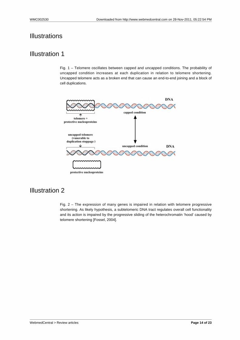
Illustration 1
Fig. 1 – Telomere oscillates between capped and uncapped conditions. The probability ofuncapped condition increases at each duplication in relation to telomere shortening.Uncapped telomere acts as a broken end that can cause an end-to-end joining and a block ofcell duplications.
Illustration 2
Fig. 2 – The expression of many genes is impaired in relation with telomere progressiveshortening. As likely hypothesis, a subtelomeric DNA tract regulates overall cell functionalityand its action is impaired by the progressive sliding of the heterochromatin ‘hood’ caused bytelomere shortening [Fossel, 2004].
WebmedCentral > Review articles Page 14 of 23
WMC002530 Downloaded from http://www.webmedcentral.com on 28-Nov-2011, 05:22:54 PM
WebmedCentral > Review articles Page 15 of 23
WMC002530 Downloaded from http://www.webmedcentral.com on 28-Nov-2011, 05:22:54 PM
Illustration 3
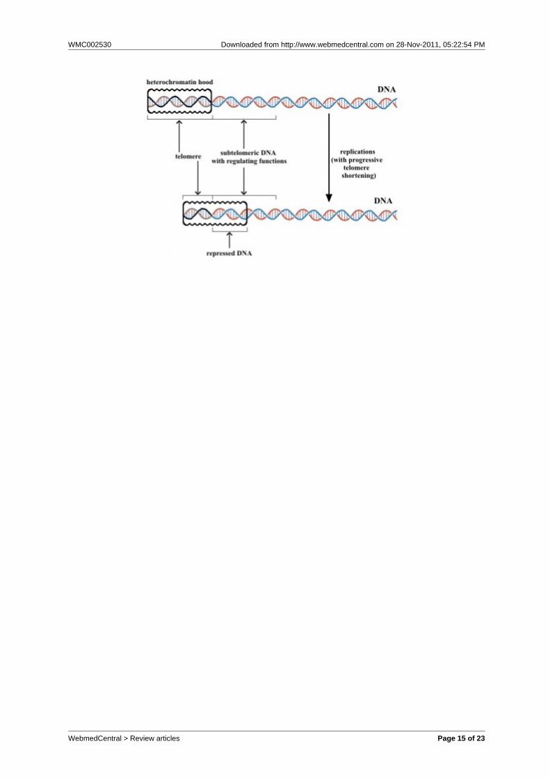
Fig. 3 – In the reset of telomere clock, the hood is shaped proportionally to telomere lengthand does not vary for all the cell life. Telomere shortening in relation to the number ofduplications causes the sliding of hood over subtelomeric DNA that regulates both overall cellfunctionality and telomere capped / uncapped condition equilibrium. The progressiverepression of subtelomeric DNA increases the degree of cell senescence and the probabilityof replicative senescence.
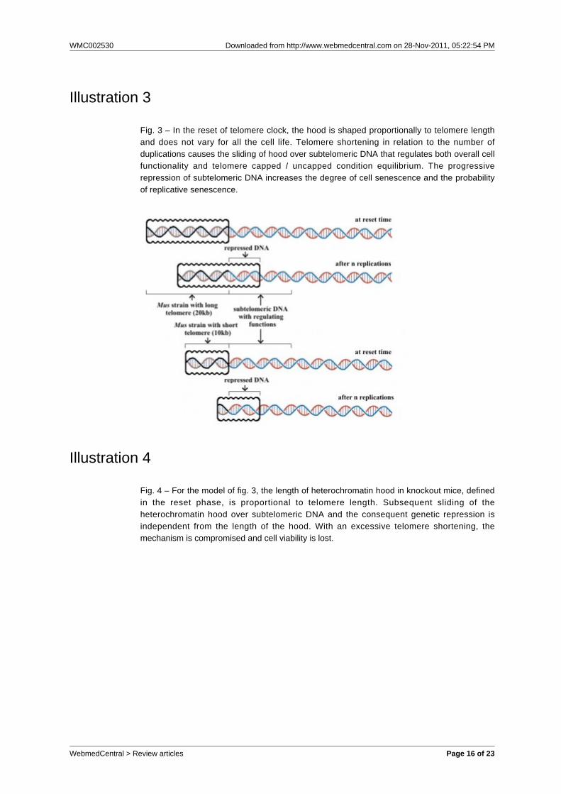
Illustration 4
Fig. 4 – For the model of fig. 3, the length of heterochromatin hood in knockout mice, definedin the reset phase, is proportional to telomere length. Subsequent sliding of theheterochromatin hood over subtelomeric DNA and the consequent genetic repression isindependent from the length of the hood. With an excessive telomere shortening, themechanism is compromised and cell viability is lost.
WebmedCentral > Review articles Page 16 of 23
WMC002530 Downloaded from http://www.webmedcentral.com on 28-Nov-2011, 05:22:54 PM
WebmedCentral > Review articles Page 17 of 23
WMC002530 Downloaded from http://www.webmedcentral.com on 28-Nov-2011, 05:22:54 PM
Illustration 5
Fig. 5 – A) In wild-type yeast, ERCs, or some other unknown factor increasing at eachduplication, interfere with subtelomeric DNA, e.g. adding something to the telomereheterochromatin hood. This process is accelerated in dna2-1 mutants; B) Intelomerase-deficient mutants, in daughter cell line without ERCs accumulation, telomereshortening cause a sliding of telomere heterochromatin hood with subtelomeric DNArepression similar to that of case A.
Illustration 6
Fig. 6 - A schematic comparison for apoptosis between yeast and multicellular eukaryotes.
WebmedCentral > Review articles Page 18 of 23
WMC002530 Downloaded from http://www.webmedcentral.com on 28-Nov-2011, 05:22:54 PM
Illustration 7
Fig. 7 - A) Spreading of a gene (G) according to the variation of S; B) Spreading of a geneaccording to the variation of ML. An increase / decrease of S or of 1/ML have the same effectson the spreading of a gene G within a species [Libertini, 2006].
Illustration 8
Fig. 8 - An example of species with age-related fitness decline in the wild. Life table ofPanthera leo after the early stages of life: survivors, basal mortality (m0) and age-relatedincreasing mortality (mi). Weibull’s equation (mt = m0 + α × tβ) and data (m0= 0.032, α =2.52E-4, β = 3), used to define the curves, are from Ricklefs.
WebmedCentral > Review articles Page 19 of 23
WMC002530 Downloaded from http://www.webmedcentral.com on 28-Nov-2011, 05:22:54 PM
Illustration 9
Fig. 9 – Proapoptosis, apoptosis, cell senescence and replicative senescence in aphylogenetic scheme.
Illustration 10
Fig. 10 – Scheme of trigger mechanisms for apoptosis in various eukaryotic phyla. Part of afigure (redrawn) from Longo et al. [Longo et al., 2005], obtained with modifications from Longoand Finch [Longo and Finch, 2003]. Apoptosis is considered part of the aging mechanism, butonly for our species this is considered doubtful (see the horizontal arrow, added to the originalscheme) without a rational explanation.
WebmedCentral > Review articles Page 20 of 23
WMC002530 Downloaded from http://www.webmedcentral.com on 28-Nov-2011, 05:22:54 PM
Phenomenon DescriptionFunction
in bacteria
Function inyeast (and
othermonocellulareukaryotes)
Function in multicellulareukaryotes
Proapoptosis
Various typeof bacterial
self-destruction
mechanisms
Activated byvarious
conditions(Note 1)
- _
Apoptosis
Ordinateprocess of
self-destruction with
modalitiesallowing theuse of cell
componentsby other cells
_
Activatedwhen
nutrients arescarce, mating
is notsuccessfuland in oldindividuals
(Note 1)
Eliminates damagedcells (Note 2) Essentialfor morphogenesis and
similar phenomena(Note 2) Essential to
determine cell turnoverwhose progressive
impairment contributesto age-related fitness
decline (Note 1)
Illustration 11
Table 1 - Comparison of three functions in bacteria, yeast (monocellular eukaryote)nand multicellular eukaryotes.
WebmedCentral > Review articles Page 21 of 23
WMC002530 Downloaded from http://www.webmedcentral.com on 28-Nov-2011, 05:22:54 PM
Cellsenescence
andreplicativesenescence
In relation tothe number ofreplications,
in a cellculture,
progressiveimpairment ofcell functions,
increasingprobability ofapoptosis and
of losingduplicationcapacity,
determined bythe repression
ofsubtelomeric
DNA
_ ReduceML,causing a
fastergenerationturnover(Note 1)
Contribute toprogressively slacken
cell turnover,determining age-relatedfitness decline (defined
“senile state” in its moreadvanced expressions)
and, therefore, MLreduction and fastergeneration turnover
(Note 1)
Note 1 = altruistic behaviour(s) favoured by kin selection in conditions ofK-selection
Note 2 = altruistic behaviour considering the multicellular individual as a clone
WebmedCentral > Review articles Page 22 of 23
WMC002530 Downloaded from http://www.webmedcentral.com on 28-Nov-2011, 05:22:54 PM
DisclaimerThis article has been downloaded from WebmedCentral. With our unique author driven post publication peerreview, contents posted on this web portal do not undergo any prepublication peer or editorial review. It iscompletely the responsibility of the authors to ensure not only scientific and ethical standards of the manuscriptbut also its grammatical accuracy. Authors must ensure that they obtain all the necessary permissions beforesubmitting any information that requires obtaining a consent or approval from a third party. Authors should alsoensure not to submit any information which they do not have the copyright of or of which they have transferredthe copyrights to a third party.
Contents on WebmedCentral are purely for biomedical researchers and scientists. They are not meant to cater tothe needs of an individual patient. The web portal or any content(s) therein is neither designed to support, norreplace, the relationship that exists between a patient/site visitor and his/her physician. Your use of theWebmedCentral site and its contents is entirely at your own risk. We do not take any responsibility for any harmthat you may suffer or inflict on a third person by following the contents of this website.
WebmedCentral > Review articles Page 23 of 23