Embed Size (px)
Citation preview

648 PHYTOPATHOLOGY
Ecology and Population Biology
Phylogenetic Analysis of Cercospora and Mycosphaerella Based on the Internal Transcribed Spacer Region of Ribosomal DNA
Stephen B. Goodwin, Larry D. Dunkle, and Victoria L. Zismann
Crop Production and Pest Control Research, U.S. Department of Agriculture-Agricultural Research Service, Department of Botany and Plant Pathology, 1155 Lilly Hall, Purdue University, West Lafayette, IN 47907.
Current address of V. L. Zismann: The Institute for Genomic Research, 9712 Medical Center Drive, Rockville, MD 20850. Accepted for publication 26 March 2001.
ABSTRACT
Goodwin, S. B., Dunkle, L. D., and Zismann, V. L. 2001. Phylogenetic analysis of Cercospora and Mycosphaerella based on the internal transcribed spacer region of ribosomal DNA. Phytopathology 91:648-658.
Most of the 3,000 named species in the genus Cercospora have no known sexual stage, although a Mycosphaerella teleomorph has been identified for a few. Mycosphaerella is an extremely large and important genus of plant pathogens, with more than 1,800 named species and at least 43 associated anamorph genera. The goal of this research was to perform a large-scale phylogenetic analysis to test hypotheses about the past evolutionary history of Cercospora and Mycosphaerella. Based on the phylogenetic analysis of internal transcribed spacer (ITS) sequence data (ITS1, 5.8S rRNA gene, ITS2), the genus Mycosphaerella is mono-phyletic. In contrast, many anamorph genera within Mycosphaerella were polyphyletic and were not useful for grouping species. One exception was Cercospora, which formed a highly supported monophyletic group. Most Cercospora species from cereal crops formed a subgroup within the
main Cercospora cluster. Only species within the Cercospora cluster produced the toxin cercosporin, suggesting that the ability to produce this compound had a single evolutionary origin. Intraspecific variation for 25 taxa in the Mycosphaerella clade averaged 1.7 nucleotides (nts) in the ITS region. Thus, isolates with ITS sequences that differ by two or more nucleotides may be distinct species. ITS sequences of groups I and II of the gray leaf spot pathogen Cercospora zeae-maydis differed by 7 nts and clearly represent different species. There were 6.5 nt differences on average between the ITS sequences of the sorghum pathogen Cercospora sorghi and the maize pathogen Cercospora sorghi var. maydis, indicating that the latter is a separate species and not simply a variety of Cerco-spora sorghi. The large monophyletic Mycosphaerella cluster contained a number of anamorph genera with no known teleomorph associations. Therefore, the number of anamorph genera related to Mycosphaerella may be much larger than suspected previously.
Additional keywords: Dothistroma, Lecanosticta, mating type, Mycocen-trospora.
Fungi in the genus Cercospora are among the most prevalent
and destructive plant pathogens. As a group, they are nearly uni-versally pathogenic, occurring on a wide range of hosts in almost all major families of dicots, most monocot families, and even some gymnosperms and ferns (26). Chupp (6) listed over 1,800 species names in his monograph of the genus in 1954, and the list had grown to over 3,000 by 1987 (26). In a major effort to clarify the taxonomy of the genus, Deighton (12–15) segregated and reclassified many Cercospora species into other genera, including Cercosporella, Cercosporidium, Paracercospora, Pseudocerco-spora, Pseudocercosporella, and Pseudocercosporidium, among others. This broad assemblage is referred to as the Cercospora complex, with members of Cercospora proper having conidia that are acicular, hyaline, and septate with a conspicuous hilum produced on pigmented, unbranched, septate, smooth conidio-phores (17,27).
Many species of Cercospora are characterized by the produc-tion of a phytotoxic metabolite of polyketide origin called cerco-sporin (3). Although this compound may enhance virulence (33),
it is not a universal pathogenicity factor because it is not produced by all species (3,16,18,21). Fajola (18) concluded that cercosporin production is associated with “true” Cercospora species and suggested that those species that do not produce cercosporin may belong to other, related genera. However, the ability to produce cercosporin is often specific to strains or isolates (16,21,35), and is influenced by various environmental and nutritional conditions (21). These inconsistencies preclude definitive application of cercosporin production to taxonomy.
Due to the paucity of useful morphological and physiological characters, taxonomy of the Cercospora complex remains confusing and depends heavily on the host. This is further compli-cated because most species have no known sexual stage. For those few species in which a sexual stage has been identified, the teleomorph is in the genus Mycosphaerella (5–7,29,34). Examples include the banana pathogens Mycosphaerella fijiensis (Cerco-spora fijiensis = Paracercospora fijiensis) and Mycosphaerella musicola (Cercospora musae = Pseudocercospora musae) and the peanut pathogen Mycosphaerella arachidis (Cercospora arachi-dicola) (7). Many other associations between Cercospora species and Mycosphaerella teleomorphs have been reported but not confirmed.
Mycosphaerella also is a very large genus, with over 1,800 names and at least 500 species associated with more than 40 ana-morph genera (7). Similar to Cercospora, the taxonomy of Myco-sphaerella is complicated, and several competing classification systems have been proposed (5,29,34). Due to the large number of associated anamorphs, Crous and Wingfield (10) concluded that Mycosphaerella was a polyphyletic assemblage of presumably monophyletic anamorph genera. Barr (5) agreed, and separated
Corresponding author: S. B. Goodwin; E-mail address: [email protected]
Names are necessary to report factually on available data. However, the USDA neither guarantees nor warrants the standard of the product, and the use of the name implies no approval of the product to the exclusion of others that also may be suitable.
Publication no. P-2001-0511-01R This article is in the public domain and not copyrightable. It may be freely re-printed with customary crediting of the source. The American PhytopathologicalSociety, 2001.

Vol. 91, No. 7, 2001 649
species with Dothistroma and Lecanosticta anamorphs into a new genus, Eruptio. There clearly is a great need for increased understanding of the phylogenetic relationships within Myco-sphaerella.
Recent molecular analyses have begun to clarify the taxonomic confusion surrounding Mycosphaerella and a few of its associated anamorph genera. Stewart et al. (30) used ribosomal DNA sequence analyses to divide species with cercosporoid anamorphs into three clusters. One group corresponded to the genus Cerco-spora sensu stricto, the second included Paracercospora and Pseudocercospora, and the third was composed of species of Passalora. Because no other species with Mycosphaerella teleo-morphs were included, it was not possible to determine the phylo-genetic relationships of the cercosporoid species to other anamorph genera. Goodwin and Zismann (20) identified a monophyletic group that included six out of seven species of Mycosphaerella tested. The Mycosphaerella cluster included seven anamorph genera, two of which had no known teleomorph associations. The only exception was Mycosphaerella pini (anamorph Dothistroma septospora), which clustered outside the main Mycosphaerella group. No species of Cercospora were included in that study. Therefore, the relationships between Cercospora and the other anamorphs tested could not be determined.
Neither of the previously described studies included species of Cercospora infecting cereal crops. One Cercospora of recent importance to grain production is the gray leaf spot pathogen of maize, Cercospora zeae-maydis (23). Although epidemics of gray leaf spot have caused substantial economic losses in the mid-western and eastern U.S. corn belts during the past several years, nothing is known about the phylogenetic relationships of the causal organisms. Analyses of amplified fragment length polymor-phisms (AFLPs) and internal transcribed spacer (ITS) sequence data revealed that gray leaf spot is caused by two sibling species of Cercospora, designated group I and group II (16,35). ITS sequences of the two groups differ by 7 nucleotides (nts); based on AFLP data, the groups are as different from each other as they are from the sorghum pathogen Cercospora sorghi or the soybean pathogen Cercospora kikuchii (35). The gray leaf spot sibling species also differed in their production of cercosporin; isolates of Cercospora zeae-maydis group I produce cercosporin, whereas those of group II do not (16,35). Cercospora may contain other
cryptic species in addition to those within Cercospora zeae-maydis. For example, due to their different host specificities, Chupp (6) suggested that the sorghum pathogen Cercospora sorghi and the corn pathogen Cercospora sorghi var. maydis might be different species, even though they are identical morphologically. However, the evolutionary relationships of these two taxa to each other, to the gray leaf spot pathogens, and to other species of Cercospora are not known.
The purpose of this research was to perform a large-scale phylogenetic analysis of the genus Mycosphaerella and associated anamorphs to test hypotheses about the evolutionary history of the genus Cercospora. The first goal was to test the hypothesis that the Cercospora species from cereal crops form a monophyletic group with the true Cercospora clade as defined by Stewart et al. (30). The second goal was to develop empirical data on the number of nucleotide differences within and between species to determine whether groups I and II of Cercospora zeae-maydis represent different species. The third goal was to test Chupp’s (6) hypothesis that Cercospora sorghi and Cercospora sorghi var. maydis are different species. The fourth goal was to test the hypothesis that Cercospora species that produce cercosporin form a monophyletic group. Within Mycosphaerella, the primary goal was to test the hypothesis of Crous and Wingfield (10) and Barr (5) that the genus Mycosphaerella is polyphyletic. A secondary goal within Mycosphaerella was to determine whether anamorph genera are monophyletic.
MATERIALS AND METHODS
Sources of isolates and culture methods. The ITS region (ITS1, 5.8S rRNA gene, ITS2) was sequenced from 15 isolates representing five species each of Cercospora and Mycosphaerella (Table 1). Most isolates were received as axenic cultures from collaborators or were purchased from the American Type Culture Collection (Mycosphaerella citrullina and Mycosphaerella fra-gariae). Cultures of Cercospora kalmiae and Mycosphaerella macrospora were isolated from infected leaves of mountain laurel (Kalmia latifolia) and iris (Iris germanica), respectively, showing symptoms of leaf spot disease. ITS sequences of Mycosphaerella brassicicola were obtained from DNA samples provided by G. Kema (Wageningen, the Netherlands). For DNA extraction, cul-
TABLE 1. Summary information for isolates of six anamorph genera analyzed for the internal transcribed spacer sequence database
Anamorph Teleomorph Isolate Host Location GenBank no.
Ascochyta cucumis Mycosphaerella citrullinaa ATCC 16241b Cucumis melo Florida AF297228 Asteromella brassicae Mycosphaerella brassicicola IPO99156 Brassica oleraceac France AF297227 Asteromella brassicae Mycosphaerella brassicicola IPO99157 Brassica oleraceac France AF297236 Asteromella brassicae Mycosphaerella brassicicola IPO99510 Brassica oleracead The Netherlands AF297223 Cercospora arachidicola Mycosphaerella arachidis –e Arachis hypogaea – AF297224 Cercospora asparagi – – Asparagus officinalis – AF297229 Cercospora beticola – – Beta vulgaris – AF297222 Cercospora kalmiae – Ceka 1 Kalmia latifolia Virginia AF297226 Cercospora kikuchii – C4RI99 Glycine max Indiana AF291708 Cercospora nicotianae – ATCC 18366 Nicotiana tabacum Tennessee AF297230 Cercospora sorghif – TX3 Sorghum bicolor Texas AF291707 Cercospora sorghi var. maydis – NC Zea mays North Carolina AF297233 Cercospora sorghi var. maydis – Kenya 1 Zea mays Kenya AF297232 Cercospora zeae-maydis group If – GBIN11 Zea mays Indiana AF291709 Cercospora zeae-maydis group IIf – LSNCX1 Zea mays North Carolina AF291710 Cladosporium iridis Mycosphaerella macrospora Myma 1 Iris germanica Indiana AF297231 Paracercospora fijiensis Mycosphaerella fijiensis rCRB2 Musa sp. – AF297234 Paracercospora fijiensis Mycosphaerella fijiensis 8837 Musa sp. – AF297225 Ramularia brunnea Mycosphaerella fragariae ATCC 24113 Fragaria Illinois AF297235
a This culture was listed as Mycosphaerella citrullina by ATCC, but is considered Didymella bryoniae by Corlett (7). Cluster analysis confirmed that it is not related to Mycosphaerella.
b Accession number, American Type Culture Collection. c Cauliflower. d Brussels sprouts. e Not known. f From Wang et al. (35).

650 PHYTOPATHOLOGY
tures of Cercospora arachidicola, Cercospora kalmiae, and Myco-sphaerella macrospora were grown in complete medium (CM) (10 ml of solution A [10 g of Ca(NO3)2·4H2O in 100 ml of H2O], 10 ml of solution B [2 g of KH2PO4, 2.5 g of MgSO4·7H2O, and 1.5 g of NaCl in 100 ml of H2O, pH 5.3], 10 g of glucose, 1 g of yeast extract, and 1 g of casein hydrolysate in 1 liter of total volume), that of Mycosphaerella fragariae in malt medium (15 g of malt extract, 3 g of peptone, and 30 g of glucose per liter), and those of Mycosphaerella fijiensis in potato dextrose broth (Difco Laboratories, Detroit). The isolate of Mycosphaerella citrullina was grown in rabbit-food medium (25 g of commercial rabbit-food pellets per liter) and those of Cercospora sorghi var. maydis in V8 medium as described in Wang et al. (35). The remaining isolates were grown in both CM and malt media. Cultures were grown at room temperature on a shaking platform at 150 rpm, harvested by vacuum filtration, lyophilized overnight, and stored at –80°C. All isolates were maintained on solid media (the same as described previously for each species but with 1.5% agar) at room temperature. Long-term storage of cultures was on lyo-philized filter paper disks at –80°C or as agar disks containing mycelia in water at 4°C. Kanamycin (50 µg/ml) was added to all media to prevent bacterial contamination.
DNA extraction, polymerase chain reaction amplification, and sequencing. DNA was extracted according to the method of Ossanna and Mischke (25) with minor modifications (20) and was quantified with a fluorometer (DyNAQuant 2000; Hoefer Scientific Instruments, San Francisco). The complete ITS region of each species was amplified with primers ITS4 and ITS5 of White et al. (36). Amplification was completed in a thermalcycler (9600; Perkin-Elmer, Foster City, CA) as described by Nakasone (24) with the following cycling parameters: 94°C for 2 min, 30 cycles of 93°C for 30 s, 53°C for 2 min, 72°C for 2 min, and a final extension of 10 min at 72°C. Amplification of products of the correct size was verified on 1% agarose gels. The remaining amplified product was purified with a polymerase chain reaction (PCR) prep kit (Wizard; Promega Corp., Madison, WI) according to the manufacturer’s instructions, except the DNA was eluted in sterile water rather than Tris-EDTA. Purified products were cloned with the TA cloning kit (Invitrogen Corp., Carlsbad, CA), and the presence of inserts was confirmed by digestion with EcoRI and agarose electrophoresis. Plasmid DNA was prepared with a miniprep kit (Promega), as described previously, and the DNA samples were quantified with a fluorometer. DNA samples were sequenced with the ThermoSequenase fluorescent labeled primer cycle sequencing kit (Amersham Pharmacia Biotech, Piscataway, NJ) by mixing 8 pmol of CY-5-labeled primer with approximately 500 ng of plasmid DNA in a total volume of 26 µl. Six microliters of the DNA solution was added to each of four tubes containing 2 µl of A, C, G, or T termination mix and mixed by pipetting up and down. DNA was amplified in a thermalcycler at 94°C for 3 min, followed by 25 cycles of 55°C for 30 s, 72°C for 2 min, and 94°C for 30 s. After adding 6 µl of stop dye, 6 µl of each reaction was analyzed on an ALFexpress automated DNA sequencer (Amersham Pharmacia Biotech). Each clone was sequenced in both directions with the M13 reverse and M13-40 primers. Three to six clones per fungal isolate were sequenced to minimize the impact of errors caused by PCR amplification.
Assembling the ITS database. To identify additional species closely related to Cercospora, a BLAST (1) search was performed on the ITS sequence of the Cercospora sorghi var. maydis isolate from North Carolina. Sequences of 52 species with high similarity to the ITS sequence of Cercospora sorghi var. maydis were downloaded from GenBank and added to the database (Table 2). The ITS sequences for Cercospora sorghi and groups I and II of Cercospora zeae-maydis were taken from Wang et al. (35) (Table 1). In addition, the entire data set for a recent paper by Stewart et al. (30) was downloaded from TreeBASE (available on-line from the Harvard University Herbaria), converted into FASTA format,
and added to the database. The ITS sequence for Phaeosphaeria nodorum, shown to cluster outside the Mycosphaerella group in a previous analysis (20), was included as an outgroup. Multiple sequences of the same species were retained if they differed or were listed originally as separate species in the database. The final database contained sequences of 94 isolates representing 77 species and varieties in 24 anamorph and eight teleomorph genera (Tables 1 and 2). The anamorphs of species of Mycosphaerella listed in Tables 1 and 2 are as indicated in Corlett (7).
DNA sequence alignment and analysis. All sequences were trimmed to include the complete ITS1, 5.8S ribosomal RNA gene, and ITS2 sequences. Seven bases, each of the 18S and 26S gene sequences, were included at the beginning and end of most se-quences, respectively, to aid alignment. The aligned region corre-sponds to bases 48 to 508 of the Cercospora sorghi var. maydis North Carolina isolate. The DNA sequences were aligned by a three-step process with the profile mode of ClustalX (32). First, a simultaneous multiple alignment of all sequences was performed to identify groups of closely related taxa. Then a separate align-ment was performed for each group and saved as a different profile. Finally, the profiles were aligned to each other using the original dendrogram as a guide. Sequences that did not cluster with any of the others in the initial step were aligned as separate profiles. Each profile was checked by eye and edited manually if necessary before proceeding to the next step. Following align-ment, genetic distances among all isolates were calculated, and a neighbor-joining tree was prepared with the Draw N-J Tree option of ClustalX. This option uses Kimura’s two-parameter method for estimating evolutionary distances (22) and implements the neigh-bor-joining algorithm of Saitou and Nei (28). Bootstrap analysis (1,000 replications) was performed on the resulting tree with the Bootstrap N-J Tree option of ClustalX, and the final tree was visualized and printed with Njplot.
Analysis of cercosporin production. Cultures of Cercospora species were grown for 5 to 10 days on dilute (0.2×) potato dextrose agar at 25°C under a 12-h photoperiod provided by two fluorescent bulbs (Phillips, Somerset, NJ). Cylinders of agar medium with mycelium were removed, and the reddish-purple pigment was extracted into 5 N KOH as described by Jenns et al. (21). Compounds showing a green color in alkali and having a characteristic absorption spectrum with Amax at 480, 595, and 640 nm were assumed to represent cercosporin (4).
Intraspecific sequence differences. For 25 taxa, multiple ITS sequences were available in GenBank or in our database. For each species with two or more sequences, a separate alignment was made with ClustalX, and the number of differences among iso-lates within species was tabulated. To determine which type of mutation occurred most commonly, a separate count was made for transitions, transversions, and insertions/deletions (indels) within the entire ITS region. To test the hypothesis that ITS1 is more variable than ITS2, counts were made for each region separately. For many taxa, this analysis used sequences in addition to those that were included in the phylogenetic trees.
PCR amplification with mating-type primers. In an attempt to determine the mating type of Cercospora and Mycosphaerella isolates, Loculoascomycete primers ChHMG1 and ChHMG2 of Arie et al. (2) were synthesized commercially (Operon Technolo-gies Inc., Alameda, CA) and used in PCR analysis. These primers amplify the high mobility group (HMG) mating-type gene (MAT-2) in Cochliobolus and Mycosphaerella zeae-maydis. DNA of Cercospora sorghi, Cercospora zeae-maydis groups I and II, Mycosphaerella citri, and Mycosphaerella graminicola was ex-tracted as described previously. DNA of Cochliobolus hetero-strophus (MAT-1 and MAT-2) and Bipolaris sorghicola (MAT-2) were included as positive and negative controls. PCR conditions were as described in Arie et al. (2). Amplification products were separated on agarose gels, stained with ethidium bromide, and photographed under ultraviolet illumination.

Vol. 91, No. 7, 2001 651
TABLE 2. Additional DNA sequences for the internal transcribed spacer database that were obtained from GenBank, TreeBASE, or other published sourcesa
Anamorph Teleomorph Isolate GenBank no.
– Dothidea hippophaesb CBS 186.58 AF027763 – Dothiora cannabinae CBS 737.71 AJ244243 – Dothiora rhamni-alpinae CBS 745.71 AJ244245 – Elsinoë banksiae – AF097572 – Elsinoë proteae – AF097578 – Mycosphaerella africana STE-U 794 AF173314 – Mycosphaerella keniensis STE-U 1084 AF173300 – Mycosphaerella marksii STE-U 935 AF173316 Asteromellopsis insculpta Dothidea insculpta CBS 189.58 AF027764 Capnobotryella renispora – CBS 214.90 AJ244238 Cercospora apii – CA29 TreeBASE Cercospora beticola – CB4 TreeBASE Cercospora canescens – CCA19 TreeBASE Cercospora hayi – CH5 TreeBASE Cercospora hayi – CH6 TreeBASE Cercospora kikuchii – CK35 TreeBASE Cercospora kikuchii – CK39 TreeBASE Cercospora nicotianae – CN17 TreeBASE Cercospora sojina – CS43 TreeBASE Cladosporium allii-cepae Mycosphaerella allii-cepae 96-1 AB026160 Cladosporium cladosporioides – CBS 170.54 AJ244241 Cladosporium fulvum – – L25430 Cladosporium herbarum Mycosphaerella tassiana CBS 111.82 AJ238469 Cladosporium herbarum Mycosphaerella tassiana CBS 399.80 AJ244227 Cladosporium macrocarpum – CBS 175.62 AJ244229 Cladosporium oxysporum – – L25432 Cladosporium sphaerospermum – CBS 122.47 AJ244228 Cladosporium tenuissium – P196 AF132797 Colletogloeopsis molleriana Mycosphaerella molleriana STE-U 1214 AF173301 Dothistroma septospora Mycosphaerella pini – AF013227 Dothistroma septospora Mycosphaerella pini MP002 AF211197 Hormonema dematioides Sydowia polyspora – AF013228 Hormonema dematioides Sydowia polyspora CBS 128.64 AJ244262 Hormonema macrosporum – CBS 536.94 AJ244247 Hortaea werneckii – CBS 359.66 AJ244249 Hortaea werneckii – CBS 373.92 AJ238474 Lacazia loboi – – AF035674 Lecanosticta acicola Mycosphaerella dearnessii – AF260818 Lecanosticta acicola Mycosphaerella dearnessii MDUS1 AF211196 Mycocentrospora acerina – MA12 TreeBASE Mycovellosiella tasmaniensis Mycosphaerella tasmaniensis STE-U 1457 AF173307 Paracercospora fijiensis Mycosphaerella fijiensis ATCC 22116 AF181705 Paracercospora fijiensis Mycosphaerella fijiensis PF7 TreeBASE Paracercospora fijiensis Mycosphaerella fijiensis PF8 TreeBASE Paracercospora fijiensis var. difformis Mycosphaerella fijiensis var. difformis PFD9 TreeBASE Passalora arachidicola – PA16 TreeBASE Passalora personatac Mycosphaerella berkeleyi PP15 TreeBASE Phaeotheca triangularis – CBS 471.90 AJ244256 Pseudocercospora cruenta Mycosphaerella cruenta PCR18 TreeBASE Pseudocercospora musae Mycosphaerella musicola ATCC 22115 AF181706 Pseudocercospora musae Mycosphaerella musicola PM10 TreeBASE Pseudocercospora musae Mycosphaerella musicola PM11 TreeBASE Ramichloridium cerophilum – CBS 103.59 AF050286 Ramularia brunnea Mycosphaerella fragariae STE-U 656 AF173312 Ramularia collo-cygni – STE-U 2045 AF173310 Ramulispora acuformis Tapesia acuformis RAC44 TreeBASE Ramulispora aestiva – RAE22 TreeBASE Ramulispora anguioides – RAN45 TreeBASE Ramulispora herpotrichoides Tapesia yallundae RH26 TreeBASE Septoria passerinii – ATCC 26515 AF181696 Septoria passerinii – ATCC 26516 AF181697 Septoria tritici Mycosphaerella graminicola T48 AF181694 Sphaceloma australis Elsinoë australis Val-2, Bat0 U28057 Sphaceloma fawcettii Elsinoë fawcettii S36954, Marc3 U28058 Sphaceloma sp. Elsinoë leucospermi STE-U 2042 AF131089 Stagonospora nodorum Phaeosphaeria nodorum N2 AF181710 Stenella araguata – CBS 486.80 AJ244261 Stenella citri-grisea Mycosphaerella citri Fellsmere AF181703 Stenella parkii Mycosphaerella parkii STE-U 353 AF173311 Trimmatostroma abietina – CBS 290.90 AJ244267 Trimmatostroma abietina – CBS 618.84 AJ244266 Trimmatostroma salicis – CBS 300.81 AJ244264 Trimmatostroma salinum – MZKI B-962 AJ238676 Uwebraunia ellipsoidea Mycosphaerella ellipsoidea STE-U 1224 AF173302 Uwebraunia juvenis Mycosphaerella juvenis STE-U 1005 AF173299 a – Indicates not known. CBS = Centraalbureau voor Schimmelcultures accession number; ATCC = American Type Culture Collection accession number. b Listed as Dothidea berberidis at CBS. c The anamorph for this species is listed as Phaeoisariopsis personata by Corlett (7), but was named a Passalora by Stewart el al. (30).

652 PHYTOPATHOLOGY
Fig. 1. Unrooted neighbor-joining tree of 94 sequences of the internal transcribed spacer (ITS) region of ribosomal DNA from species of Mycosphaerella and related anamorphs and teleomorphs. All bootstrap values of 70 or greater (percentage of 1,000 replications) are indicated, rounded to the nearest integer. The ITS sequence of Phaeosphaeria nodorum was used as an outgroup. All species are indicated by anamorph name, if known, otherwise by teleomorph. If more than one isolate of a species was analyzed, isolate designations are provided after the species name. The probable teleomorph genus for each major group, if known, is indicated by brackets. Branch lengths are proportional to genetic distance, which is indicated by a bar at the upper left.

Vol. 91, No. 7, 2001 653
RESULTS
ITS sequencing and alignment. The length of the ITS region, including the primer region, for the 15 isolates sequenced ranged from 548 nts for Mycosphaerella fijiensis isolate 8837 to 574 for Mycosphaerella macrospora. The extensive length variation com-monly detected among fungi (20) was not found in the species sequenced in this study. GenBank accession numbers for the 15 sequences are indicated in Table 1.
A BLAST (1) search of the Cercospora sorghi var. maydis North Carolina isolate on the GenBank database identified strong matches with many species of Mycosphaerella, as well as the anamorph genera Trimmatostroma, Ramularia, and Clado-sporium. The highest BLAST score was obtained to an isolate of Guignardia bidwellii, followed by Mycosphaerella tasmaniensis and Mycosphaerella africana. All sequences downloaded from GenBank had expected values of 4 × 10–89 or lower in the BLAST results.
Alignment of the 94 sequences required 45 profile steps, with the original simultaneous multiple alignment as a guide. Minor manual editing was required on approximately half of the profiles. Use of the Profile mode of ClustalX to build the alignment en-sured that accurate relationships among species within each group were maintained at each step. This yielded a better result with generally higher bootstrap support compared with the original simultaneous multiple alignment (data not shown).
For most of the profile alignments, the gap opening and extension penalties were left at the default values of 15.00 and 6.66, respectively. A few of the sequences downloaded from GenBank contained large insertions or deletions. These usually occurred at or near the 5′ end of ITS1. Aligning these sequences was more difficult and required lowering the gap opening and extension penalties until an accurate alignment could be obtained.
Cercosporin production. For each species that produced a reddish-purple pigment in the agar medium, cercosporin was confirmed by spectrophotometric analysis. Confirmed cercosporin producers were Cercospora asparagi, Cercospora beticola, Cercospora nicotianae, and Cercospora sorghi var. maydis. The isolates of Cercospora kikuchii, Cercospora sorghi, and Cerco-spora zeae-maydis group I produced cercosporin in a previous study (35). Isolates of Cercospora arachidicola, Cercospora sojina, Cercospora zeae-maydis group II, Mycosphaerella bras-sicicola, Mycosphaerella macrospora, Mycosphaerella fijiensis, and Mycosphaerella fragariae were tested and did not produce cercosporin.
Phylogenetic analyses. Most of the Cercospora species tested formed a single, monophyletic group with high (97%) bootstrap support (Fig. 1). The only exceptions were Cercospora kalmiae and Cercospora arachidicola, which clustered with species of Pseudocercospora and Passalora, respectively. Interestingly, Asteromella brassicae (teleomorph Mycosphaerella brassicicola) was a sister group to the main Cercospora cluster and separated most of the Cercospora species from a Paracercospora/Pseudo-cercospora/Cercospora kalmiae cluster. Two species of Myco-sphaerella with no known anamorphs, Mycosphaerella africana and Mycosphaerella keniensis, clustered with Cladosporium fulvum and Dothistroma septospora (teleomorph Mycosphaerella pini), which was a sister group to a Passalora/Cercospora arachi-dicola cluster (Fig. 1).
In addition to Cercospora, three other anamorph genera clearly were polyphyletic. Species of Stenella were in three widely separated clusters (Fig. 1). The two species with Uwebraunia anamorphs were phylogenetically unrelated, and Trimmatostroma salinum was in a different cluster from T. abietina and T. salicis.
Anamorph genera that formed monophyletic clusters included Ramularia and Septoria (Fig. 1). All species of Cladosporium except Cladosporium fulvum formed a monophyletic group that included Lacazia loboi, a fungus with previously unknown phylo-
genetic affinities that was isolated from the skin of bottlenose dolphin (31). The Cladosporium, Ramularia, and Septoria clusters each had 100% bootstrap support (Fig. 1).
Most species with Mycosphaerella teleomorphs formed a monophyletic group with high (89%) bootstrap support. The only exception was one isolate of Mycosphaerella pini (anamorph Dothistroma septospora), which did not cluster with any other species (Fig. 1). A second isolate of Mycosphaerella pini clustered within the large Mycosphaerella group together with Myco-sphaerella africana, Mycosphaerella keniensis, and Cladosporium fulvum. The only species within the Mycosphaerella cluster with a different teleomorph was one isolate of Guignardia bidwellii (GenBank Accession No. AF216533), which clustered as a sister taxon to Mycosphaerella brassicicola near the large Cercospora cluster (data not shown). However, because this sequence appeared unrelated to those from other species in the genus Botryosphaeria (sometimes considered a synonym for Guignardia [19]), it was assumed to have been identified incorrectly and was excluded from further analysis.
Two species in this analysis clustered with the outgroup taxon Phaeosphaeria nodorum. One of these was labeled as Myco-sphaerella citrullina when it was received from the ATCC. However, the correct name for this species is Didymella bryoniae (7) (anamorph Ascochyta cucumis). The other species was Mycocentrospora acerina, which has no known teleomorph (19).
In addition to the species tested in this study, reports of cerco-sporin production or nonproduction for other species were taken from the literature (3,18) and added on to a second analysis of a reduced data set with the Septoria cluster as an outgroup (Fig. 2). All of the cercosporin-producing species were within the mono-phyletic Cercospora cluster that had 97% bootstrap support. The only taxa within this cluster that did not produce cercosporin were Cercospora sojina and Cercospora zeae-maydis group II. All species outside this cluster for which data were available did not produce cercosporin, including Mycosphaerella brassicicola (ana-morph Asteromella brassicae), the most closely related species with a confirmed Mycosphaerella teleomorph (Fig. 2).
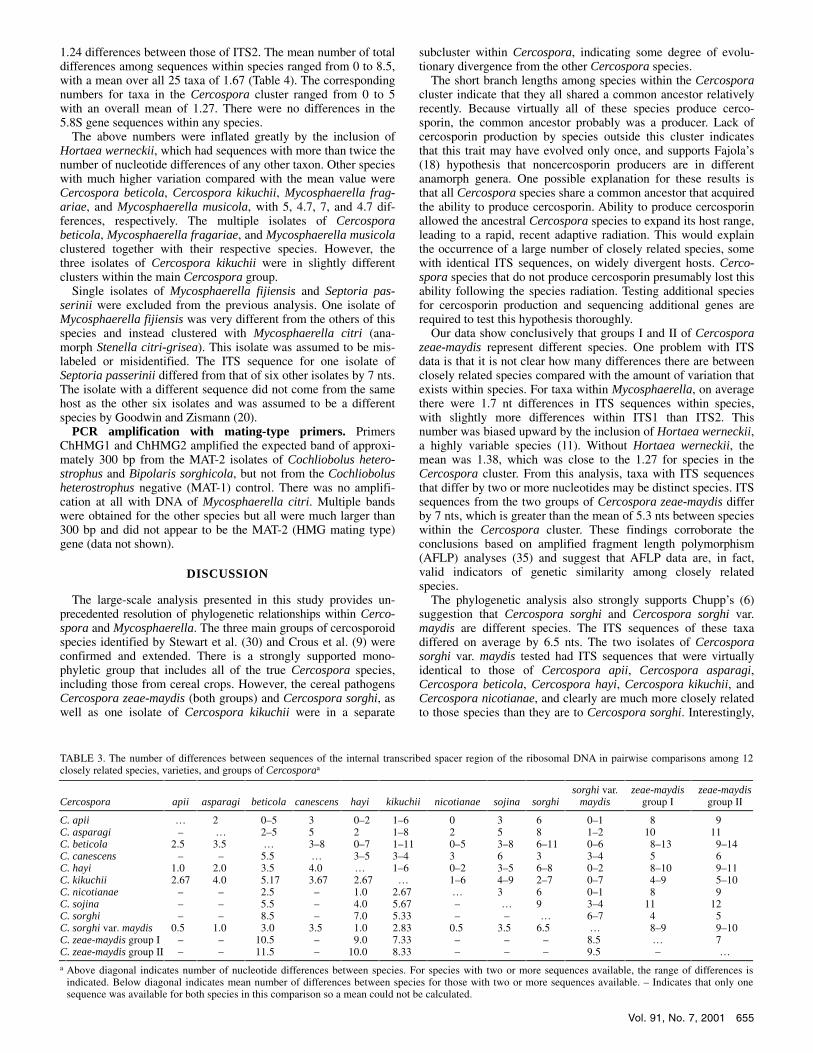
Nucleotide differences between and within species. The num-ber of nucleotide differences between species for the 12 taxa in the monophyletic Cercospora cluster ranged from 0 to 14 (Table 3). Five taxa (Cercospora apii, Cercospora beticola, Cercospora hayi, Cercospora nicotianae, and Cercospora sorghi var. maydis) had isolates with identical ITS sequences, although single isolates of Cercospora beticola, Cercospora hayi, and Cercospora sorghi var. maydis differed from the others by 5, 2, and 1 nts, respec-tively. The largest nucleotide difference was between Cercospora zeae-maydis group II and one isolate of Cercospora beticola (Table 3). There were 7 nt differences between the sequences of Cercospora zeae-maydis groups I and II, and an average of 6.5 nts between Cercospora sorghi and the two isolates of Cercospora sorghi var. maydis. The overall mean number of differences between taxa within the main Cercospora cluster was 5.28 nts over all 66 pairwise comparisons.
Within the large, monophyletic Mycosphaerella cluster, 25 taxa were represented by two or more sequences in the databases. The number of sequences available per species ranged from 2 to 8 with a mean of 3.28 (Table 4). The numbers of transitions, trans-versions, and insertions/deletions (indels) within species ranged from 0 to 6, 0 to 7, and 0 to 9, respectively. Over all 25 taxa, transitions and indels occurred at approximately the same fre-quency, with means of 1.08 and 0.96 of each per taxon, respec-tively. Transversions only occurred about one half as often, with a mean of 0.56 transversions per taxon.
There was little difference in the number of changes between ITS1 and ITS2. The total number of differences between se-quences within species ranged from 0 to 12 for ITS1 compared with 0 to 10 for ITS2 (Table 4). Intraspecific variation among all 25 taxa averaged 1.36 differences between ITS1 sequences and

654 PHYTOPATHOLOGY
Fig. 2. Relationship between cercosporin production and phylogeny in the genera Cercospora and Mycosphaerella. Unrooted neighbor-joining tree from a reduced data set of 44 internal transcribed spacer sequences. All bootstrap values above 70 (percentage of 1,000 replications) are indicated and rounded to the nearest integer. The Septoria cluster was used as an outgroup. If more than one isolate of a species was analyzed, isolate designations are provided after the species name. Species that produce cercosporin are indicated by +, those that do not produce cercosporin are indicated by –, and those that were not tested are indicated by nt. All species are listed by anamorph name, if known, otherwise by teleomorph. For those with a known teleomorph, the species is indicated to the right. Branch lengths are proportional to genetic distance, which is indicated by a bar at the upper left.
Vol. 91, No. 7, 2001 655
1.24 differences between those of ITS2. The mean number of total differences among sequences within species ranged from 0 to 8.5, with a mean over all 25 taxa of 1.67 (Table 4). The corresponding numbers for taxa in the Cercospora cluster ranged from 0 to 5 with an overall mean of 1.27. There were no differences in the 5.8S gene sequences within any species.
The above numbers were inflated greatly by the inclusion of Hortaea werneckii, which had sequences with more than twice the number of nucleotide differences of any other taxon. Other species with much higher variation compared with the mean value were Cercospora beticola, Cercospora kikuchii, Mycosphaerella frag-ariae, and Mycosphaerella musicola, with 5, 4.7, 7, and 4.7 dif-ferences, respectively. The multiple isolates of Cercospora beticola, Mycosphaerella fragariae, and Mycosphaerella musicola clustered together with their respective species. However, the three isolates of Cercospora kikuchii were in slightly different clusters within the main Cercospora group.
Single isolates of Mycosphaerella fijiensis and Septoria pas-serinii were excluded from the previous analysis. One isolate of Mycosphaerella fijiensis was very different from the others of this species and instead clustered with Mycosphaerella citri (ana-morph Stenella citri-grisea). This isolate was assumed to be mis-labeled or misidentified. The ITS sequence for one isolate of Septoria passerinii differed from that of six other isolates by 7 nts. The isolate with a different sequence did not come from the same host as the other six isolates and was assumed to be a different species by Goodwin and Zismann (20).
PCR amplification with mating-type primers. Primers ChHMG1 and ChHMG2 amplified the expected band of approxi-mately 300 bp from the MAT-2 isolates of Cochliobolus hetero-strophus and Bipolaris sorghicola, but not from the Cochliobolus heterostrophus negative (MAT-1) control. There was no amplifi-cation at all with DNA of Mycosphaerella citri. Multiple bands were obtained for the other species but all were much larger than 300 bp and did not appear to be the MAT-2 (HMG mating type) gene (data not shown).
DISCUSSION
The large-scale analysis presented in this study provides un-precedented resolution of phylogenetic relationships within Cerco-spora and Mycosphaerella. The three main groups of cercosporoid species identified by Stewart et al. (30) and Crous et al. (9) were confirmed and extended. There is a strongly supported mono-phyletic group that includes all of the true Cercospora species, including those from cereal crops. However, the cereal pathogens Cercospora zeae-maydis (both groups) and Cercospora sorghi, as well as one isolate of Cercospora kikuchii were in a separate
subcluster within Cercospora, indicating some degree of evolu-tionary divergence from the other Cercospora species.
The short branch lengths among species within the Cercospora cluster indicate that they all shared a common ancestor relatively recently. Because virtually all of these species produce cerco-sporin, the common ancestor probably was a producer. Lack of cercosporin production by species outside this cluster indicates that this trait may have evolved only once, and supports Fajola’s (18) hypothesis that noncercosporin producers are in different anamorph genera. One possible explanation for these results is that all Cercospora species share a common ancestor that acquired the ability to produce cercosporin. Ability to produce cercosporin allowed the ancestral Cercospora species to expand its host range, leading to a rapid, recent adaptive radiation. This would explain the occurrence of a large number of closely related species, some with identical ITS sequences, on widely divergent hosts. Cerco-spora species that do not produce cercosporin presumably lost this ability following the species radiation. Testing additional species for cercosporin production and sequencing additional genes are required to test this hypothesis thoroughly.
Our data show conclusively that groups I and II of Cercospora zeae-maydis represent different species. One problem with ITS data is that it is not clear how many differences there are between closely related species compared with the amount of variation that exists within species. For taxa within Mycosphaerella, on average there were 1.7 nt differences in ITS sequences within species, with slightly more differences within ITS1 than ITS2. This number was biased upward by the inclusion of Hortaea werneckii, a highly variable species (11). Without Hortaea werneckii, the mean was 1.38, which was close to the 1.27 for species in the Cercospora cluster. From this analysis, taxa with ITS sequences that differ by two or more nucleotides may be distinct species. ITS sequences from the two groups of Cercospora zeae-maydis differ by 7 nts, which is greater than the mean of 5.3 nts between species within the Cercospora cluster. These findings corroborate the conclusions based on amplified fragment length polymorphism (AFLP) analyses (35) and suggest that AFLP data are, in fact, valid indicators of genetic similarity among closely related species.
The phylogenetic analysis also strongly supports Chupp’s (6) suggestion that Cercospora sorghi and Cercospora sorghi var. maydis are different species. The ITS sequences of these taxa differed on average by 6.5 nts. The two isolates of Cercospora sorghi var. maydis tested had ITS sequences that were virtually identical to those of Cercospora apii, Cercospora asparagi, Cercospora beticola, Cercospora hayi, Cercospora kikuchii, and Cercospora nicotianae, and clearly are much more closely related to those species than they are to Cercospora sorghi. Interestingly,
TABLE 3. The number of differences between sequences of the internal transcribed spacer region of the ribosomal DNA in pairwise comparisons among 12 closely related species, varieties, and groups of Cercosporaa
Cercospora
apii
asparagi
beticola
canescens
hayi
kikuchii
nicotianae
sojina
sorghi
sorghi var. maydis
zeae-maydis group I
zeae-maydis group II
C. apii … 2 0–5 3 0–2 1–6 0 3 6 0–1 8 9 C. asparagi – … 2–5 5 2 1–8 2 5 8 1–2 10 11 C. beticola 2.5 3.5 … 3–8 0–7 1–11 0–5 3–8 6–11 0–6 8–13 9–14 C. canescens – – 5.5 … 3–5 3–4 3 6 3 3–4 5 6 C. hayi 1.0 2.0 3.5 4.0 … 1–6 0–2 3–5 6–8 0–2 8–10 9–11 C. kikuchii 2.67 4.0 5.17 3.67 2.67 … 1–6 4–9 2–7 0–7 4–9 5–10 C. nicotianae – – 2.5 – 1.0 2.67 … 3 6 0–1 8 9 C. sojina – – 5.5 – 4.0 5.67 – … 9 3–4 11 12 C. sorghi – – 8.5 – 7.0 5.33 – – … 6–7 4 5 C. sorghi var. maydis 0.5 1.0 3.0 3.5 1.0 2.83 0.5 3.5 6.5 … 8–9 9–10 C. zeae-maydis group I – – 10.5 – 9.0 7.33 – – – 8.5 … 7 C. zeae-maydis group II – – 11.5 – 10.0 8.33 – – – 9.5 – …
a Above diagonal indicates number of nucleotide differences between species. For species with two or more sequences available, the range of differences is indicated. Below diagonal indicates mean number of differences between species for those with two or more sequences available. – Indicates that only one sequence was available for both species in this comparison so a mean could not be calculated.

656 PHYTOPATHOLOGY
no other species from cereal hosts were in the Cercospora sub-group that contained Cercospora sorghi var. maydis.
All of the Cercospora species tested grouped within a much larger cluster of species that have Mycosphaerella teleomorphs. Thus, the genus Cercospora must have evolved within the Myco-sphaerella lineage. The teleomorphs for these Cercospora species, if they exist, most likely will be in Mycosphaerella. This agrees with the unconfirmed report of a Mycosphaerella teleomorph for Cercospora zeae-maydis (23).
The large-scale phylogenetic analysis provided evidence that Mycosphaerella is monophyletic and contains numerous polyphy-letic anamorph genera. This is in contrast to the hypothesis of Crous and Wingfield (10) who suggested that Mycosphaerella is a polyphyletic assemblage of monophyletic anamorphs. The only ITS sequence of a Mycosphaerella species that did not cluster with this genus in our analysis was one isolate of Mycosphaerella pini. However, the sequence of this isolate was very different from that of a second isolate of this species that clustered well within Mycosphaerella. Therefore, the aberrant isolate most likely was misidentified. Within the Mycosphaerella cluster, the only isolate with a different teleomorph was one of Guignardia bidwellii. The sequence for this isolate was very different from those for Botryosphaeria species that also were present in GenBank (data not shown). Because Guignardia is considered a synonym of Botryosphaeria (19), the GenBank sequence for Guignardia bidwellii probably came from an isolate that was misidentified or mislabeled. Overall, the data provide very strong support for the hypothesis that the genus Mycosphaerella is monophyletic, which confirms the results of Crous et al. (9) and Goodwin and Zismann (20) from analyses of much smaller data sets.
Although Mycosphaerella clearly appears to be monophyletic, branch lengths among groups within Mycosphaerella are quite long. Genetic distances between some clusters within Myco-sphaerella are larger than those between the teleomorph genera Dothiora, Dothidea, and Sydowia. Therefore, the Mycosphaerella teleomorph probably is of ancient origin and has been maintained
through a long period of evolutionary history by selection. The long branch lengths lead others to conclude incorrectly that the genus is polyphyletic. This issue could only be resolved by a large-scale phylogenetic analysis.
In contrast to the teleomorph, certain anamorph genera associ-ated with Mycosphaerella clearly are polyphyletic. This was particularly evident for Stenella and the new genus Uwebraunia, which had representatives in very different clusters. A mono-phyletic origin for Uwebraunia was already in question by mor-phological analysis of the teleomorphs. Crous (8) noted that the two species of Uwebraunia included in the phylogenetic analysis have teleomorphs with different shaped ascospores. Therefore, it is not surprising that Uwebraunia is polyphyletic. Evidently, many anamorph characters are highly mutable; the same anamorph probably arose multiple times by convergent evolution. Thus, anamorphs in Mycosphaerella in general may not be useful for resolution of phylogenetic relationships. This supports the conclu-sion of von Arx (34) that anamorphs should not be used to sepa-rate groups within Mycosphaerella.
Based on these results, some recent changes in the taxonomy of Mycosphaerella should be revisited. For example, Barr (5) erected the new teleomorph genus Eruptio to include species with ana-morphs in Dothistroma and Lecanosticta on the assumptions that: (i) these anamorphs are closely related; and (ii) they are different from other species within Mycosphaerella. Our large-scale phylogenetic analysis contradicted both of these assumptions. These two anamorphs are not particularly closely related and both are located well within the Mycosphaerella cluster. Therefore, the teleomorph names for Dothistroma septospora and Lecanosticta acicola should remain within Mycosphaerella.
Not all anamorphs were polyphyletic. Anamorphs that were clearly monophyletic included Cercospora sensu Stewart et al. (30), Ramularia, Septoria, and all of the Cladosporium species except Cladosporium fulvum. For Ramularia and Septoria, the number of species tested was too small for firm conclusions. However, Cercospora and Cladosporium formed well-supported
TABLE 4. Number of nucleotide differences in the internal transcribed spacer (ITS) region among isolates within species for taxa in the Mycosphaerella cluster
Type of change No. of differences
Species No. of
sequences Transitions Transversions Indelsa ITS1 ITS2 Total Rangeb Mean
Cercospora apii 3 0 0 0 0 0 0 0 0.0 Cercospora beticola 2 0 2 3 4 1 5 – 5 Cercospora hayi 2 2 0 0 0 2 2 – 2 Cercospora kikuchii 3 5 1 1 1 6 7 2–7 4.7 Cercospora nicotianae 2 0 0 0 0 0 0 – 0 Cercospora sojina 3 0 0 0 0 0 0 0 0.0 Cercospora sorghi 4 0 0 0 0 0 0 0 0.0 Cercospora sorghi var. maydis 2 1 0 0 0 1 1 – 1 Cercospora zeae-maydis group I 4 0 0 0 0 0 0 0 0.0 Cercospora zeae-maydis group II 4 0 0 0 0 0 0 0 0.0 Cladosporium allii-cepae 3 0 0 0 0 0 0 0 0.0 Cladosporium herbarum 3 0 0 0 0 0 0 0 0.0 Cladosporium sphaerospermum 2 0 0 0 0 0 0 – 0 Hortaea werneckii 8 6 7 9 12 10 22 1–16 8.5 Mycosphaerella brassicicola 3 0 0 1 1 0 1 0–1 0.7 Mycosphaerella citri 2 0 0 1 0 1 1 – 1 Mycosphaerella dearnessii 5 0 0 2 2 0 2 0–2 1.0 Mycosphaerella ellipsoidea 2 1 1 0 1 1 2 – 2 Mycosphaerella fijiensis 5c 3 1 2 2 4 6 0–5 2.8 Mycosphaerella fragariae 2 2 2 3 7 0 7 – 7 Mycosphaerella graminicola 4 0 0 0 0 0 0 0 0.0 Mycosphaerella musicola 3 6 0 1 3 4 7 1–7 4.7 Septoria passerinii 6d 0 0 0 0 0 0 0 0.0 Trimmatostroma abietina 2 0 0 0 0 0 0 – 0 Trimmatostroma salinum 3 1 0 1 1 1 2 1–2 1.3
Overall mean 3.28 1.08 0.56 1.96 1.36 1.24 2.60 – 1.67
a Insertions, deletions, or both. b The range was only calculated when three or more sequences were available. c One isolate of Mycosphaerella fijiensis that probably was misidentified was excluded. d Isolate P26515 of Septoria passerinii was considered a separate species by Goodwin and Zismann (20) so was excluded from this analysis.

Vol. 91, No. 7, 2001 657
monophyletic groups. Interestingly, the Cladosporium cluster included Lacazia loboi, the cause of lobomycosis in humans and bottlenose dolphins (31).
In addition to addressing phylogenetic questions, the large-scale analysis identified a number of sequences from isolates that probably were misidentified, mislabeled, or misclassified. Two of these were Mycosphaerella pini and Guignardia bidwellii as discussed previously. The others were Mycosphaerella fijiensis and Mycosphaerella citrullina. Five isolates of Mycosphaerella fijiensis clustered together, but the sixth isolate had an ITS sequence that was almost identical to that of Stenella citri-grisea (Mycosphaerella citri). The most likely explanations for this are that the isolate was misidentified or mislabeled, or that there was contamination during PCR amplification. The isolate received as Mycosphaerella citrullina from the ATCC clustered with Phaeo-sphaeria nodorum in the Pleosporales, not within Myco-sphaerella. This isolate was simply misclassified; the correct name for Mycosphaerella citrullina is Didymella bryoniae (7), which is supported by the phylogenetic analysis.
The Mycosphaerella cluster included a number of species with no known teleomorphs. These included Capnobotryella, Hortaea, Lacazia, Phaeotheca, and Trimmatostroma. Many of these are black yeasts that are found on a variety of substrates and on humans (11), but are evolutionarily related to the large group of plant pathogens within Mycosphaerella. Because these anamorphs have not been associated with Mycosphaerella previously, the true number of anamorphs within Mycosphaerella may be much larger than the 43 listed by Corlett (7).
In addition to Cercospora and Mycosphaerella, phylogenetic analysis indicated the probable teleomorph association for Mycocentrospora acerina. This species was used as an outgroup by Stewart et al. (30), but did not cluster with any other species. Our analysis revealed that it clustered with Phaeosphaeria nodorum and Didymella bryoniae in the Pleosporales. An ex-panded analysis (data not shown) confirmed that it clustered with-in the Phaeosphaeria/Leptosphaeria clade identified by Goodwin and Zismann (20). Thus, Mycocentrospora acerina probably has a teleomorph related to those genera.
The Loculoascomycete HMG mating-type primers described by Arie et al. (2) may not be useful for species of Mycosphaerella. In our preliminary analyses, we were unable to amplify a MAT-2 HMG homologue from species of Cercospora and Mycosphae-rella. The only species of Mycosphaerella tested by Arie et al. (2) was Mycosphaerella zeae-maydis, which did contain a homo-logous MAT-2 idiomorph. However, Mycosphaerella zeae-maydis is a synonym for Didymella zeae-maydis (7) and, therefore, it is not a species of Mycosphaerella. The species of Didymella tested in our analysis (Didymella bryoniae, listed as Mycosphaerella citrullina in the collection of the ATCC) clustered with Stagono-spora nodorum, the anamorph of Phaeosphaeria nodorum. The ChHMG1 and ChHMG2 primers (2) did amplify the HMG se-quence from isolates of Phaeosphaeria nodorum (S. B. Goodwin and V. L. Zismann, unpublished data). The most likely expla-nation for lack of amplification with species of Cercospora and Mycosphaerella is that Mycosphaerella zeae-maydis is classified incorrectly and is not really a Mycosphaerella. Therefore, the primers developed by Arie et al. (2) may be useful for some Loculoascomycetes, but not Mycosphaerella species. Inclusion of Mycosphaerella zeae-maydis in a phylogenetic analysis and cloning of the mating-type genes from a Mycosphaerella species are needed to test this hypothesis thoroughly.
ACKNOWLEDGMENTS
This work was supported by USDA CRIS project 3602-22000-009-00D. Published as paper 16389, Purdue University Agricultural Experi-ment Station. We thank M. Daub for providing cultures of several Cerco-spora species, G. Kema for providing DNA and cultures for isolates of
Mycosphaerella brassicicola, J. Cavaletto and B. Roberts for generating some of the sequence data and submitting the sequences to GenBank, respectively, M. McClenning for providing general technical support, and M. Scholler for providing helpful comments on a previous draft of the manuscript.
LITERATURE CITED
1. Altschul, S. F., Madden, T. L., Schäffer, A. A., Zhang, J., Zhang, Z., Miller, W., and Lipman, D. J. 1997. Gapped BLAST and PSI-BLAST: A new generation of protein database search programs. Nucleic Acids Res. 25:3389-3402.
2. Arie, T., Christiansen, S. K., Yoder, O. C., and Turgeon, B. G. 1997. Efficient cloning of ascomycete mating type genes by PCR amplification of the conserved MAT HMG box. Fungal Genet. Biol. 21:118-130.
3. Assante, G., Locci, R., Camarda, L., Merlini, L., and Nasini, G. 1977. Screening of the genus Cercospora for secondary metabolites. Phyto-chemistry 16:243-247.
4. Balis, C., and Payne, M. G. 1971. Triglycerides and cercosporin from Cercospora beticola: Fungal growth and cercosporin production. Phyto-pathology 61:1477-1484.
5. Barr, M. E. 1996. Planistromellaceae, a new family in the Dothideales. Mycotaxon 60:433-442.
6. Chupp, C. 1954. A Monograph of the Fungus Genus Cercospora. Ronald Press Co., New York.
7. Corlett, M. 1991. An annotated list of the published names in Myco-sphaerella and Sphaerella. Mycol. Mem. 18:1-328.
8. Crous, P. W. 1998. Mycosphaerella spp. and Their Anamorphs Associ-ated with Leaf Spot Diseases of Eucalyptus. The American Phytopatho-logical Society, St. Paul, MN.
9. Crous, P. W., Aptroot, A., Kang, J.-C., Braun, U., and Wingfield, M. J. 2000. The genus Mycosphaerella and its anamorphs. Stud. Mycol. 45: 107-121.
10. Crous, P. W., and Wingfield, M. J. 1996. Species of Mycosphaerella and their anamorphs associated with leaf blotch disease of Eucalyptus in South Africa. Mycologia 88:441-458.
11. de Hoog, G. S., Zalar, P., Urzì, C., de Leo, F., Yurlova, N. A., and Sterflinger, K. 1999. Relationships of dothideaceous black yeasts and meristematic fungi based on 5.8S and ITS2 rDNA sequence comparison. Stud. Mycol. 43:31-37.
12. Deighton, F. C. 1967. Studies on Cercospora and allied genera. II. Passalora, Cercosporidium, and some species of Fusicladium on Euphorbia. Mycol. Pap. 112:1-80.
13. Deighton, F. C. 1973. Studies on Cercospora and allied genera. IV. Cercosporella Sacc., Pseudocercosporella gen. nov. and Pseudocerco-sporidium gen. nov. Mycol. Pap. 133:1-62.
14. Deighton, F. C. 1976. Studies on Cercospora and allied genera. VI. Pseudocercospora Speg., Pantospora Cif. and Cercoseptoria. Mycol. Pap. 140:1-168.
15. Deighton, F. C. 1979. Studies on Cercospora and allied genera. VII. New species and redispositions. Mycol. Pap. 144:1-56.
16. Dunkle, L. D., and Levy, M. 2000. Genetic relatedness of African and United States populations of Cercospora zeae-maydis. Phytopathology 90:486-490.
17. Fajola, A. O. 1978. Cultural studies in Cercospora taxonomy: I. Interrelationships between some species from Nigeria. Nova Hedwigia 29:912-921.
18. Fajola, A. O. 1978. Cercosporin, a phytotoxin from Cercospora spp. Physiol. Plant Pathol. 13:157-164.
19. Farr, D. F., Bills, G. F., Chamuris, G. P., and Rossman, A. Y. 1989. Fungi on Plants and Plant Products in the United States. The American Phyto-pathological Society, St. Paul, MN.
20. Goodwin, S. B., and Zismann, V. L. 2001. Phylogenetic analyses of the ITS region of ribosomal DNA reveal that Septoria passerinii from barley is closely related to the wheat pathogen Mycosphaerella graminicola. Mycologia (In press.)
21. Jenns, A. E., Daub, M. E., and Upchurch, R. G. 1989. Regulation of cercosporin accumulation in culture by medium and temperature manipu-lation. Phytopathology 79:213-219.
22. Kimura, M. 1980. A simple method for estimating evolutionary rates of base substitutions through comparative studies of nucleotide sequences. J. Mol. Evol. 16:111-120.
23. Latterell, F. M., and Rossi, A. E. 1983. Gray leaf spot of corn: A disease on the move. Plant Dis. 67:842-847.
24. Nakasone, K. K. 1996. Morphological and molecular studies on Auricu-lariopsis albomellea and Phlebia albida and a reassessment of A. ampla. Mycologia 88:762-775.
25. Ossanna, N., and Mischke, S. 1990. Genetic transformation of the bio-control fungus Gliocladium virens to benomyl resistance. Appl. Environ.

658 PHYTOPATHOLOGY
Microbiol. 56:3052-3056. 26. Pollack, F. G. 1987. An annotated compilation of Cercospora names.
Mycol. Mem. 12:1-212. 27. Pons, N., and Sutton, B. C. 1988. Cercospora and similar fungi on yams
(Dioscorea species). Mycol. Pap. 160:1-78. 28. Saitou, N., and Nei, M. 1987. The neighbor-joining method: A new
method for reconstructing phylogenetic trees. Mol. Biol. Evol. 4:406-425. 29. Sivanesan, A. 1984. The Bitunicate Ascomycetes and their Anamorphs.
J. Cramer, Vaduz, Liechtenstein. 30. Stewart, E. L., Liu, Z., Crous, P. W., and Szabo, L. J. 1999. Phylogenetic
relationships among some cercosporoid anamorphs of Mycosphaerella based on rDNA sequence analysis. Mycol. Res. 103:1491-1499.
31. Taborda, P. R., Taborda, V. A., and McGinnis, M. R. 1999. Lacazia loboi gen. nov., comb. nov., the etiologic agent of lobomycosis. J. Clin. Micro-biol. 37:2031-2033.
32. Thompson, J. D., Gibson, T. J., Plewniak, F., Jeanmougin, F., and
Higgins, D. G. 1997. The CLUSTAL-X windows interface: Flexible strategies for multiple sequence alignment aided by quality analysis tools. Nucleic Acids Res. 25:4876-4882.
33. Upchurch, R. G., Walker, D. C., Rollins, J. A., Ehrenshaft, M., and Daub, M. E. 1991. Mutants of Cercospora kikuchii altered in cercosporin synthesis and pathogenicity. Appl. Environ. Microbiol. 57:2940-2945.
34. von Arx, J. A. 1983. Mycosphaerella and it anamorphs. Proc. K. Ned. Akad. Wet., Series C 86:15-54.
35. Wang, J., Levy, M., and Dunkle, L. D. 1998. Sibling species of Cercospora associated with gray leaf spot of maize. Phytopathology 88:1269-1275.
36. White, T. J., Bruns, T., Lee, S., and Taylor, J. 1990. Amplification and direct sequencing of fungal ribosomal RNA genes for phylogenetics. Pages 315-322 in: PCR Protocols, A Guide to Methods and Applications. M. A. Innis, D. H. Gelfand, J. J. Sninsky, and T. J. White, eds. Academic Press, San Diego, CA.