Embed Size (px)
Citation preview
Photoreceptor Cilia and Retinal Ciliopathies
Kinga M. Bujakowska, Qin Liu, and Eric A. Pierce
Ocular Genomics Institute, Massachusetts Eye and Ear Infirmary, Department of Ophthalmology, HarvardMedical School, Boston, Massachusetts 02114
Correspondence: [email protected]
Photoreceptors are sensory neurons designed to convert light stimuli into neurological re-sponses. This process, called phototransduction, takes place in the outer segments (OS) of rodand cone photoreceptors. OS are specialized sensory cilia, with analogous structures tothose present in other nonmotile cilia. Deficient morphogenesis and/or dysfunction of pho-toreceptor sensory cilia (PSC) caused by mutations in a variety of photoreceptor-specific andcommon cilia genes can lead to inherited retinal degenerations (IRDs). IRDs can manifest asisolated retinal diseases or syndromic diseases. In this review, we describe the structure andcomposition of PSC and different forms of ciliopathies with retinal involvement. We reviewthe genetics of the IRDs, which are monogenic disorders but genetically diverse with regardto causality.
Photoreceptors are sensory neurons designedto convert light stimuli into electrical re-
sponses, a process called phototransduction.Phototransduction takes place in the highly spe-cialized compartment of photoreceptors, theouter segment (OS) (Pearring et al. 2013; Mol-day and Moritz 2015). The OS of the rod andcone photoreceptors differ in structure and pro-tein composition, related to their functional ad-aptation, in which rods have high sensitivitynecessary in dim light and cones are responsiblefor the high-resolution color vision working inbright light (Lamb and Pugh 2006; Lamb et al.2007). Research over the past decade on thegenetic and molecular components of photore-ceptors in vertebrate retinae has led to the clearrecognition that photoreceptor OS are special-ized sensory cilia (Liu et al. 2007a; Ramamurthyand Cayouette 2009; Khanna 2015). Deficient
morphogenesis and/or dysfunction of photore-ceptor sensory cilia (PSC) caused by mutationsin a variety of photoreceptor-specific and com-mon cilia genes can lead to a group of clinicalmanifestations, called inherited retinal degener-ations (IRDs). In this review, we will discuss thestructure and composition of PSC and differentforms of ciliopathies with retinal involvement.
SPECIALIZED PHOTORECEPTORSENSORY CILIA
In vertebrate retina, the visual function dependson the formation of complex sensory cilia of rodand cone photoreceptors. Photoreceptors arehighly polarized neurons, composed of fourdistinct compartments: the OS, the inner seg-ment (IS), the nucleus and a short axon extend-ing to second order neurons (bipolar and hor-
Editors: Wallace Marshall and Renata Basto
Additional Perspectives on Cilia available at www.cshperspectives.org
Copyright # 2017 Cold Spring Harbor Laboratory Press; all rights reserved
Advanced Online Article. Cite this article as Cold Spring Harb Perspect Biol doi: 10.1101/cshperspect.a028274
1
on January 1, 2020 - Published by Cold Spring Harbor Laboratory Press http://cshperspectives.cshlp.org/Downloaded from
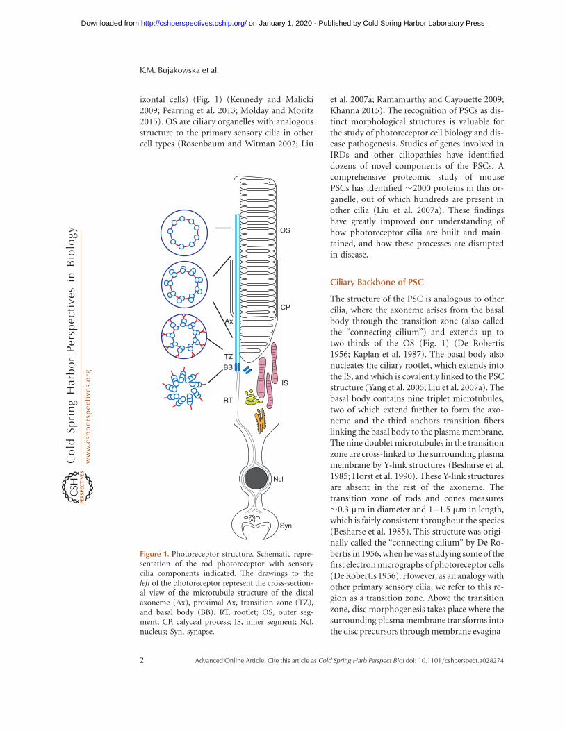
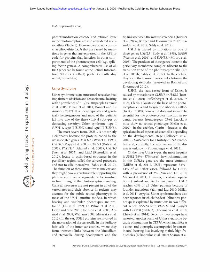
izontal cells) (Fig. 1) (Kennedy and Malicki2009; Pearring et al. 2013; Molday and Moritz2015). OS are ciliary organelles with analogousstructure to the primary sensory cilia in othercell types (Rosenbaum and Witman 2002; Liu
et al. 2007a; Ramamurthy and Cayouette 2009;Khanna 2015). The recognition of PSCs as dis-tinct morphological structures is valuable forthe study of photoreceptor cell biology and dis-ease pathogenesis. Studies of genes involved inIRDs and other ciliopathies have identifieddozens of novel components of the PSCs. Acomprehensive proteomic study of mousePSCs has identified �2000 proteins in this or-ganelle, out of which hundreds are present inother cilia (Liu et al. 2007a). These findingshave greatly improved our understanding ofhow photoreceptor cilia are built and main-tained, and how these processes are disruptedin disease.
Ciliary Backbone of PSC
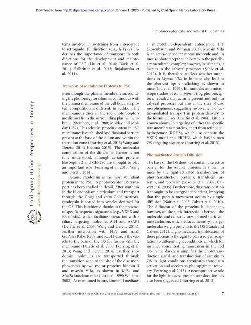
The structure of the PSC is analogous to othercilia, where the axoneme arises from the basalbody through the transition zone (also calledthe “connecting cilium”) and extends up totwo-thirds of the OS (Fig. 1) (De Robertis1956; Kaplan et al. 1987). The basal body alsonucleates the ciliary rootlet, which extends intothe IS, and which is covalently linked to the PSCstructure (Yang et al. 2005; Liu et al. 2007a). Thebasal body contains nine triplet microtubules,two of which extend further to form the axo-neme and the third anchors transition fiberslinking the basal body to the plasma membrane.The nine doublet microtubules in the transitionzone are cross-linked to the surrounding plasmamembrane by Y-link structures (Besharse et al.1985; Horst et al. 1990). These Y-link structuresare absent in the rest of the axoneme. Thetransition zone of rods and cones measures�0.3 mm in diameter and 1–1.5 mm in length,which is fairly consistent throughout the species(Besharse et al. 1985). This structure was origi-nally called the “connecting cilium” by De Ro-bertis in 1956, when he was studying some of thefirst electron micrographs of photoreceptor cells(De Robertis 1956). However, as an analogy withother primary sensory cilia, we refer to this re-gion as a transition zone. Above the transitionzone, disc morphogenesis takes place where thesurrounding plasma membrane transforms intothe disc precursors through membrane evagina-
OS
CP
IS
RT
Ncl
Syn
Ax
BB
TZ
Figure 1. Photoreceptor structure. Schematic repre-sentation of the rod photoreceptor with sensorycilia components indicated. The drawings to theleft of the photoreceptor represent the cross-section-al view of the microtubule structure of the distalaxoneme (Ax), proximal Ax, transition zone (TZ),and basal body (BB). RT, rootlet; OS, outer seg-ment; CP, calyceal process; IS, inner segment; Ncl,nucleus; Syn, synapse.
K.M. Bujakowska et al.
2 Advanced Online Article. Cite this article as Cold Spring Harb Perspect Biol doi: 10.1101/cshperspect.a028274
on January 1, 2020 - Published by Cold Spring Harbor Laboratory Press http://cshperspectives.cshlp.org/Downloaded from
tion (Steinberg et al. 1980; Ding et al. 2015; Pugh2015). At the distal part of the PSC axoneme, thedouble microtubules are reduced to singlets(Fig. 1) (Rosenbaum and Witman 2002; Pear-ring et al. 2013).
Other Structural Components of OuterSegments
PSCs are highly specialized sensory cilia, adapt-ed for light detection by the presence of tightlypacked membranous discs containing visualpigments and other phototransduction pro-teins (Sjostrand 1953; Nickell et al. 2007; Gil-liam et al. 2012). PSCs are among the largestof mammalian cilia (Pan et al. 2005) and, likeother cilia, they are comprised of a cytoskeletonbackbone and a membrane domain, which isdistinct from the surrounding plasma mem-brane (Steinberg et al. 1980; Molday and Mol-day 1987). In murine rods, the numerous mem-branous discs in the PSC compartment arestacked at a density of �30 discs per microme-ter, which is thought to be constant throughoutspecies (Nickell et al. 2007; Gilliam et al. 2012).Such OS organization provides a large surfacearea for optimized photon capture and rapidsignal transduction reactions to occur. Rhodop-sin is the most abundant disc membrane pro-tein, organized as rows of dimers with a densityof �48,000 monomers per mm2 (Fotiadis et al.2003). With this high density in the disc mem-branes, rhodopsin plays an important structuralrole apart from being the main visual pigmentin the retina (Wang and Deretic 2014). The rimof the photoreceptor discs contains two tetra-spanins: Rds/peripherin-2 (PRPH2) and reti-nal OS membrane protein 1 (ROM1), whichfacilitate the folding of the OS discs and arecrucial for rim formation and sorting of theOS proteins during the OS biogenesis (Moldayet al. 1987; Goldberg and Molday 1996; Arikawaet al. 2011). PRPH2 and its homolog ROM1both form homodimers and then associate to-gether to form tetrameric complexes, exclusive-ly present at the disc rims (Molday et al. 1987;Goldberg and Molday 1996; Arikawa et al.2011). Two other membrane proteins prominin1 (PROM1) and cadherin-related family mem-
ber 1 (CDHR1) were associated with the openlamellar evaginations in rod and cone discs inXenopus laevis and mice, respectively (Rattneret al. 2001; Han et al. 2012). PSC are responsiblefor mediating the sensory transduction of thevisual system with a number of proteins in-volved in this process, including the above-mentioned rhodopsin. Most of these proteinsare expressed specifically in PSC and, whenmutated, cause nonsyndromic IRDs (Table 1)(Dryja et al. 1990; Farrar et al. 1990; Kajiwaraet al. 1991, 1994; Travis et al. 1991; Bascom et al.1992; Rosenfeld et al. 1992; Dryja et al. 1993;Maw et al. 2000; Yang et al. 2008). Studies ofmutant animals have shown that the above-mentioned proteins are essential for OS discmorphogenesis and maintenance (Sanyal et al.1980; Clarke et al. 2000; Rattner et al. 2001;Dellett et al. 2015).
PROTEIN TRANSPORT TO PSC
A unique feature of the photoreceptor OS is thehigh level of its renewal. Each day �10% of theOS is shed from the distal tip, which is replacedby new disc formation at the base of the PSC(Young 1967). This necessitates a robust systemof protein synthesis in the IS and efficient traf-ficking of selected proteins to the photorecep-tor OS.
Intraflagellar Transport in PSC
The axoneme, initiated at the mother centriole,is built and maintained by extending its distal(þ) end (Pedersen and Rosenbaum 2008). Be-cause protein synthesis occurs in the IS, the ax-oneme building blocks need to be transportedto the distal end via intraflagellar transport(IFT) (Rosenbaum and Witman 2002; Pedersenand Rosenbaum 2008; Taschner et al. 2012). Theanterograde transport from the base to the tip ofthe axoneme is mediated by IFT complex B(IFT-B), where kinesin-2 is the motor protein(Rosenbaum and Witman 2002). Kinesin-2 is aheterotrimeric protein composed of Kif3A,Kif3B, and KAP, which is further associatedwith 14 other IFT proteins that bind cargo mol-ecules (Taschner et al. 2012). Once the axoneme
Photoreceptor Cilia and Retinal Ciliopathies
Advanced Online Article. Cite this article as Cold Spring Harb Perspect Biol doi: 10.1101/cshperspect.a028274 3
on January 1, 2020 - Published by Cold Spring Harbor Laboratory Press http://cshperspectives.cshlp.org/Downloaded from
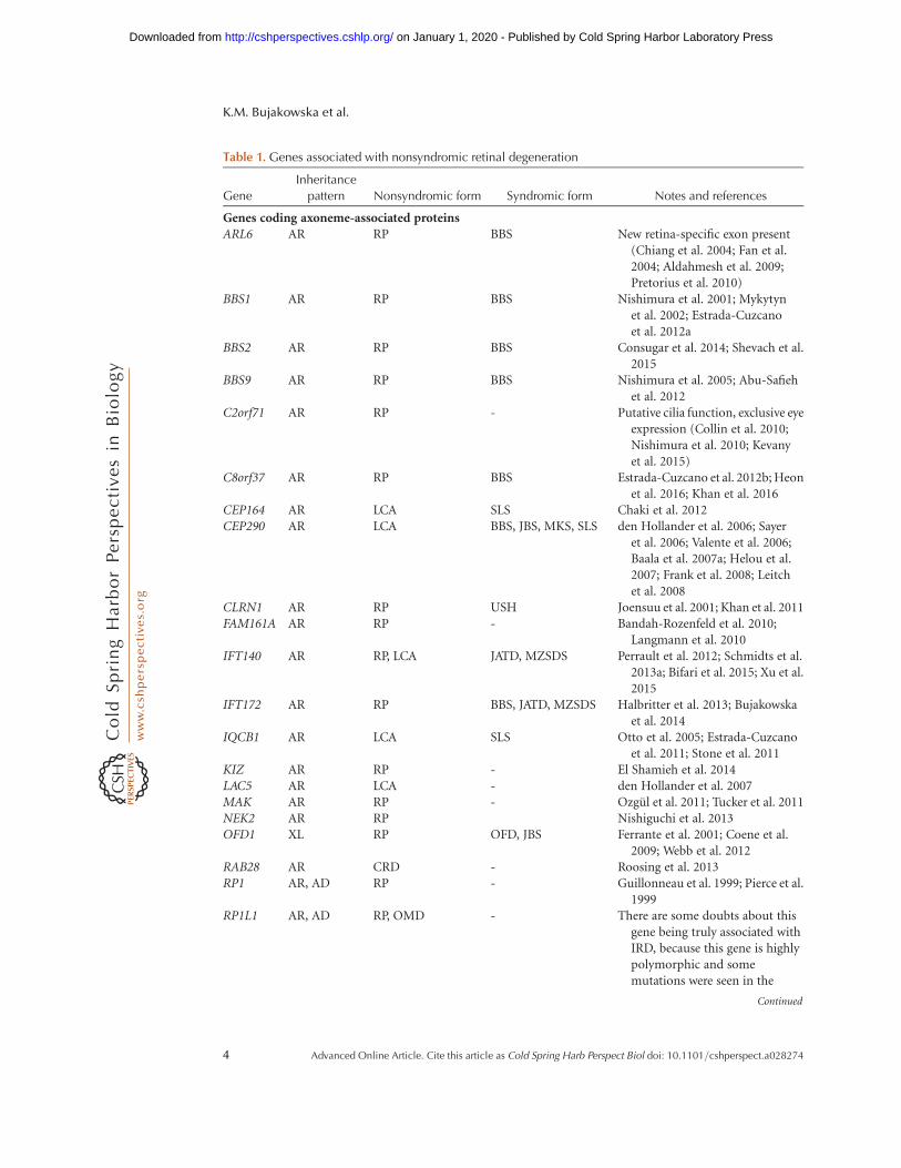
Table 1. Genes associated with nonsyndromic retinal degeneration
Gene
Inheritance
pattern Nonsyndromic form Syndromic form Notes and references
Genes coding axoneme-associated proteinsARL6 AR RP BBS New retina-specific exon present
(Chiang et al. 2004; Fan et al.2004; Aldahmesh et al. 2009;Pretorius et al. 2010)
BBS1 AR RP BBS Nishimura et al. 2001; Mykytynet al. 2002; Estrada-Cuzcanoet al. 2012a
BBS2 AR RP BBS Consugar et al. 2014; Shevach et al.2015
BBS9 AR RP BBS Nishimura et al. 2005; Abu-Safiehet al. 2012
C2orf71 AR RP - Putative cilia function, exclusive eyeexpression (Collin et al. 2010;Nishimura et al. 2010; Kevanyet al. 2015)
C8orf37 AR RP BBS Estrada-Cuzcano et al. 2012b; Heonet al. 2016; Khan et al. 2016
CEP164 AR LCA SLS Chaki et al. 2012CEP290 AR LCA BBS, JBS, MKS, SLS den Hollander et al. 2006; Sayer
et al. 2006; Valente et al. 2006;Baala et al. 2007a; Helou et al.2007; Frank et al. 2008; Leitchet al. 2008
CLRN1 AR RP USH Joensuu et al. 2001; Khan et al. 2011FAM161A AR RP - Bandah-Rozenfeld et al. 2010;
Langmann et al. 2010IFT140 AR RP, LCA JATD, MZSDS Perrault et al. 2012; Schmidts et al.
2013a; Bifari et al. 2015; Xu et al.2015
IFT172 AR RP BBS, JATD, MZSDS Halbritter et al. 2013; Bujakowskaet al. 2014
IQCB1 AR LCA SLS Otto et al. 2005; Estrada-Cuzcanoet al. 2011; Stone et al. 2011
KIZ AR RP - El Shamieh et al. 2014LAC5 AR LCA - den Hollander et al. 2007MAK AR RP - Ozgul et al. 2011; Tucker et al. 2011NEK2 AR RP Nishiguchi et al. 2013OFD1 XL RP OFD, JBS Ferrante et al. 2001; Coene et al.
2009; Webb et al. 2012RAB28 AR CRD - Roosing et al. 2013RP1 AR, AD RP - Guillonneau et al. 1999; Pierce et al.
1999RP1L1 AR, AD RP, OMD - There are some doubts about this
gene being truly associated withIRD, because this gene is highlypolymorphic and somemutations were seen in the
Continued
K.M. Bujakowska et al.
4 Advanced Online Article. Cite this article as Cold Spring Harb Perspect Biol doi: 10.1101/cshperspect.a028274
on January 1, 2020 - Published by Cold Spring Harbor Laboratory Press http://cshperspectives.cshlp.org/Downloaded from
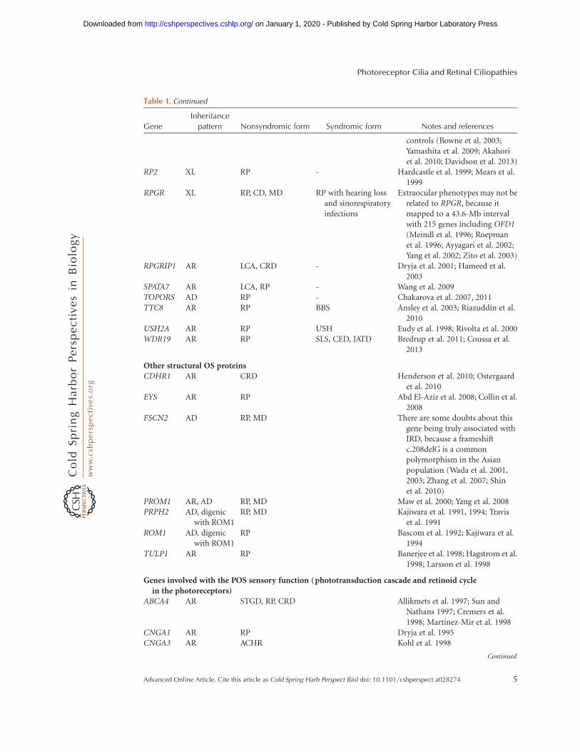
Table 1. Continued
Gene
Inheritance
pattern Nonsyndromic form Syndromic form Notes and references
controls (Bowne et al. 2003;Yamashita et al. 2009; Akahoriet al. 2010; Davidson et al. 2013)
RP2 XL RP - Hardcastle et al. 1999; Mears et al.1999
RPGR XL RP, CD, MD RP with hearing lossand sinorespiratoryinfections
Extraocular phenotypes may not berelated to RPGR, because itmapped to a 43.6-Mb intervalwith 215 genes including OFD1(Meindl et al. 1996; Roepmanet al. 1996; Ayyagari et al. 2002;Yang et al. 2002; Zito et al. 2003)
RPGRIP1 AR LCA, CRD - Dryja et al. 2001; Hameed et al.2003
SPATA7 AR LCA, RP - Wang et al. 2009TOPORS AD RP - Chakarova et al. 2007, 2011TTC8 AR RP BBS Ansley et al. 2003; Riazuddin et al.
2010USH2A AR RP USH Eudy et al. 1998; Rivolta et al. 2000WDR19 AR RP SLS, CED, JATD Bredrup et al. 2011; Coussa et al.
2013
Other structural OS proteinsCDHR1 AR CRD Henderson et al. 2010; Ostergaard
et al. 2010EYS AR RP Abd El-Aziz et al. 2008; Collin et al.
2008FSCN2 AD RP, MD There are some doubts about this
gene being truly associated withIRD, because a frameshiftc.208delG is a commonpolymorphism in the Asianpopulation (Wada et al. 2001,2003; Zhang et al. 2007; Shinet al. 2010)
PROM1 AR, AD RP, MD Maw et al. 2000; Yang et al. 2008PRPH2 AD, digenic
with ROM1RP, MD Kajiwara et al. 1991, 1994; Travis
et al. 1991ROM1 AD, digenic
with ROM1RP Bascom et al. 1992; Kajiwara et al.
1994TULP1 AR RP Banerjee et al. 1998; Hagstrom et al.
1998; Larsson et al. 1998
Genes involved with the POS sensory function ( phototransduction cascade and retinoid cyclein the photoreceptors)
ABCA4 AR STGD, RP, CRD Allikmets et al. 1997; Sun andNathans 1997; Cremers et al.1998; Martınez-Mir et al. 1998
CNGA1 AR RP Dryja et al. 1995CNGA3 AR ACHR Kohl et al. 1998
Continued
Photoreceptor Cilia and Retinal Ciliopathies
Advanced Online Article. Cite this article as Cold Spring Harb Perspect Biol doi: 10.1101/cshperspect.a028274 5
on January 1, 2020 - Published by Cold Spring Harbor Laboratory Press http://cshperspectives.cshlp.org/Downloaded from
and other PSC components have been deliveredto the tip of the cilium, the IFT-B componentsare recycled back to the base of the cilium byretrograde transport mediated by IFT complexA (IFT-A) (Rosenbaum and Witman 2002). Dy-nein-2 is the motor protein of IFT-A and it isassociated with six other IFT proteins (Taschneret al. 2012). Apart from the IFT complexes, Bar-det–Biedl syndrome proteins (BBSome) are
also involved in the transport of membrane pro-teins to the cilium (Taschner et al. 2012; Wil-liams et al. 2014).
Because 10% of the PSC is shed and re-newed every day, the necessity for the retro-grade transport in this cell type was not clear.However, identification of IRD patients withmutations in genes coding for retrogradetransport proteins (e.g., TTC21B) and pro-
Table 1. Continued
Gene
Inheritance
pattern Nonsyndromic form Syndromic form Notes and references
CNGB1 AR RP Bareil et al. 2001CNGB3 AR ACHM, CD Kohl et al. 2000GNAT1 AD, AR CSNB Dryja et al. 1996GNAT2 AR ACHM Aligianis et al. 2002; Kohl et al. 2002GRK AR CSNB Yamamoto et al. 1997GUCA1A AD CD, CRD Payne et al. 1998; Sokal et al. 1998GUCA1B AD RP, MD Sato et al. 2005GUCY2D AR, AD LCA, CRD Perrault et al. 1996; Kelsell et al.
1998OPN1LW XL Deuteranopia, blue
conemonochromacy
Nathans et al. 1986; Winderickxet al. 1992; Ayyagari et al. 1999
OPN1MW XL Protanopia, blueconemonochromacy
Nathans et al. 1986; Ayyagari et al.1999
OPN1SW AD Tritanopia Nathans et al. 1992; Weitz et al.1992a,b
PDE6A AR RP Huang et al. 1995PDE6B AR, AD RP, CSNB McLaughlin et al. 1993; Gal et al.
1994PDE6C AR CD, ACHM Thiadens et al. 2009PDE6G AR RP Dvir et al. 2010RDH12 AR, AD LCA, RP Janecke et al. 2004; Perrault et al.
2004; Fingert et al. 2008RGS9 AR Delayed cone
adaptationNishiguchi et al. 2004
RGS9BP AR Delayed coneadaptation
Nishiguchi et al. 2004
RHO AD, AR RP, CSNB Dryja et al. 1990; Farrar et al. 1990;Rosenfeld et al. 1992; Dryja et al.1993
SAG AR RP, CSNB Fuchs et al. 1995; Nakazawa et al.1998
ACHM, Achromatopsia; BBS, Bardet–Biedl syndrome; CD, cone dystrophy; CED, cranioectodermal dysplasia, also known
as Sensenbrenner syndrome; CRD, cone–rod dystrophy; CSNB, congenital stationary night blindness; JBS, Joubert syndrome;
JATD, Jeune asphyxiating thoracic dystrophy; LCA, Leber congenital amaurosis; MKS, Meckel–Gruber syndrome; MZSDS,
Mainzer–Saldino syndrome; OFD, oral-facial-digital syndrome; OMD, occult macular dystrophy; RP, retinitis pigmentosa;
SLS, Senior–Løken syndrome; STGD, Stargardt disease; USH, Usher syndrome.
K.M. Bujakowska et al.
6 Advanced Online Article. Cite this article as Cold Spring Harb Perspect Biol doi: 10.1101/cshperspect.a028274
on January 1, 2020 - Published by Cold Spring Harbor Laboratory Press http://cshperspectives.cshlp.org/Downloaded from
teins involved in switching from anterogradeto retrograde IFT direction (e.g., IFT172) un-derlines the importance of transport in bothdirections for the development and mainte-nance of PSC (Liu et al. 2010; Davis et al.2011; Halbritter et al. 2013; Bujakowska etal. 2014).
Transport of Membrane Proteins to PSC
Even though the plasma membrane surround-ing the photoreceptor cilium is continuous withthe plasma membrane of the cell body, its pro-tein composition is different. In addition, themembranous discs in the rod photoreceptorsare distinct from the surrounding plasma mem-brane (Steinberg et al. 1980; Molday and Mol-day 1987). This selective protein content in PSCmembranes is established by diffusional barrierspresent at the base of the cilium and within thetransition zone (Pearring et al. 2013; Wang andDeretic 2014; Khanna 2015). The molecularcomposition of the diffusional barrier is notfully understood, although certain proteinslike Septin 2 and CEP290 are thought to playan important role (Pearring et al. 2013; Wangand Deretic 2014).
Because rhodopsin is the most abundantprotein in the PSC, its photoreceptor OS trans-port has been studied in detail. After synthesisin the IS endoplasmic reticulum and transportthrough the Golgi and trans-Golgi network,rhodopsin is sorted into vesicles destined forthe OS. This is achieved thanks to the presenceof specific sequence signatures (e.g., VXPX andFR motifs), which facilitate interaction with aciliary targeting molecules Arf4 and ASAP1(Deretic et al. 2005; Wang and Deretic 2014).Further interaction with FIP3 and smallGTPases Rab6, Rab8, and Rab11 directs the ves-icle to the base of the OS for fusion with themembrane (Deretic et al. 2005; Pearring et al.2013; Wang and Deretic 2014). Further, rho-dopsin molecules are transported throughthe transition zone to the site of the disc mor-phogenesis by two motor proteins, kinesin IIand myosin VIIa, as shown in Kif3a andMyo7a knockout mice (Liu et al. 1999; Williams2002). As mentioned before, kinesin II mediates
a microtubule-dependent anterograde IFT(Rosenbaum and Witman 2002). Myosin VIIais an actin-dependent motor molecule and, inmouse photoreceptors, it locates to the pericili-ary membrane complex; however, in primates, itlocates to the calyceal processes (Sahly et al.2012). It is, therefore, unclear whether muta-tions in Myosin VIIa in humans also lead tothe aberrant opsin trafficking as shown inmice (Liu et al. 1999). Immunoelectron micro-scope studies of Rana pipiens frog photorecep-tors, revealed that actin is present not only incalyceal processes but also at the sites of discmorphogenesis, suggesting involvement of ac-tin-mediated transport in protein delivery tothe forming discs (Chaitin et al. 1984). Little isknown about OS targeting of other OS-specifictransmembrane proteins, apart from retinol de-hydrogenase (RDH8), which also contains theVXPX motif and PRPH2, which has its ownOS-targeting sequence (Pearring et al. 2013).
Photoactivated Protein Diffusion
The base of the OS does not contain a selectivebarrier for the soluble proteins as shown inmice by the light-activated translocation ofphototransduction proteins: transducin, ar-restin, and recoverin (Sokolov et al. 2002; Cal-vert et al. 2006). Furthermore, this translocationis thought to be energy-independent, implyingthat the protein movement occurs by simplediffusion (Nair et al. 2005; Calvert et al. 2010).The diffusion of the proteins is dependent,however, on the steric interactions between themolecules and cell structures, termed steric vol-ume exclusion, which reduces the entry of largermolecular weight proteins to the OS (Najafi andCalvert 2012). Light-mediated translocation ofthese proteins is thought to play a role in adap-tation to different light conditions, in which forinstance concentrating transducin in the rodOS in the darkness amplifies the phototrans-duction signal, and translocation of arrestin toOS in light conditions terminates transducinactivation and accelerates photopigment recov-ery (Pearring et al. 2013). A neuroprotective rolefor the light-induced protein translocation hasalso been suggested (Pearring et al. 2013).
Photoreceptor Cilia and Retinal Ciliopathies
Advanced Online Article. Cite this article as Cold Spring Harb Perspect Biol doi: 10.1101/cshperspect.a028274 7
on January 1, 2020 - Published by Cold Spring Harbor Laboratory Press http://cshperspectives.cshlp.org/Downloaded from
RETINAL CILIOPATHIES
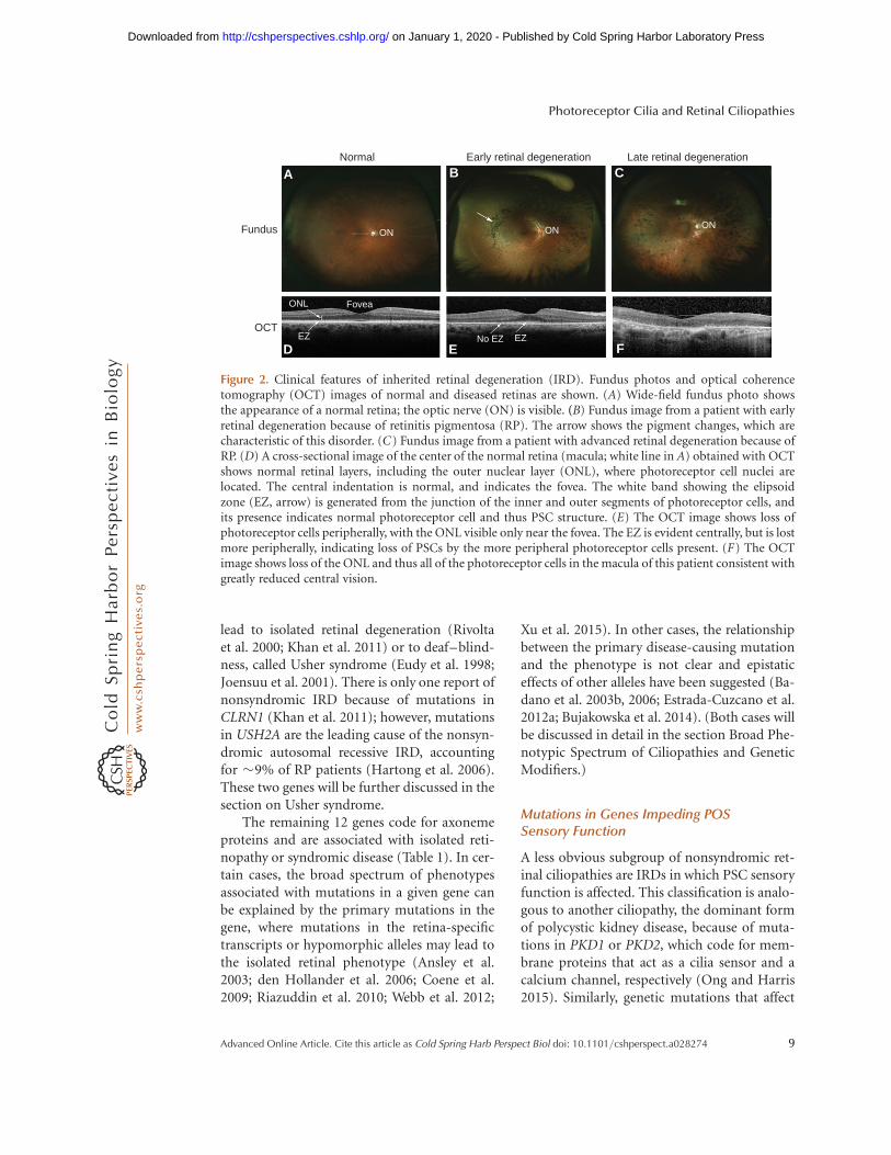
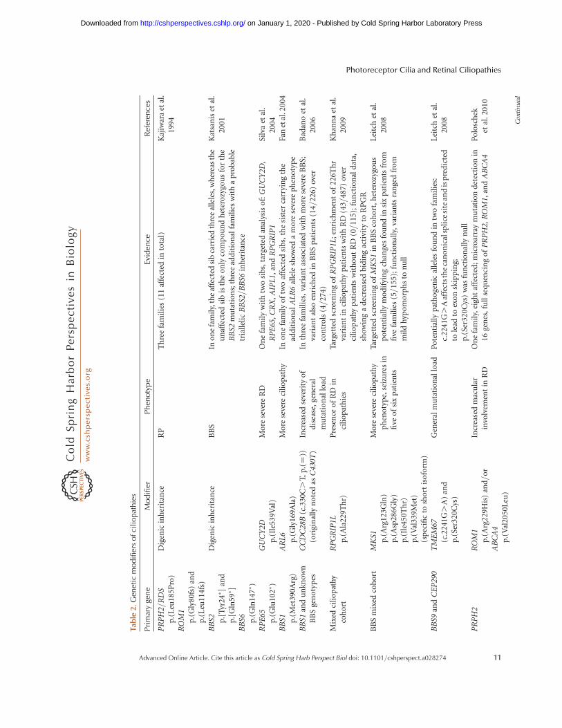
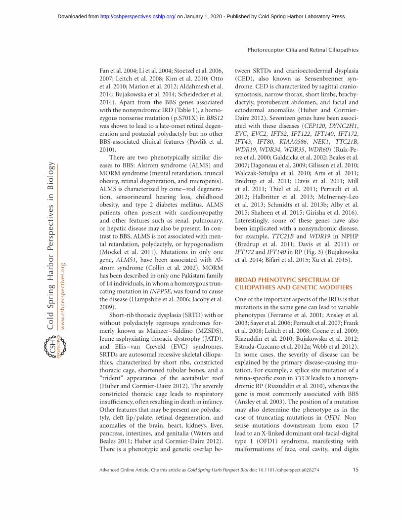
Mutations in genes coding for ciliary proteinslead to ciliopathies, rare genetic disorders thatmay affect one or more organs, including theretina, central nervous system, olfactory epithe-lium, cardiovascular system, liver, kidney, skel-etal system, gonads, and adipose tissue (Goetzand Anderson 2010; Patel and Honore 2010;Mockel et al. 2011; Waters and Beales 2011).In this review, we will focus on ciliopathiesthat involve the retina, manifesting most com-monly as retinitis pigmentosa (RP) (Hamel2006; Hartong et al. 2006; Berger et al. 2010)or Leber congenital amaurosis (LCA) (Weleber2002; Chung and Traboulsi 2009). RP is a con-dition that primarily affects rod photoreceptorsand retinal pigment epithelium. It is the mostfrequent cause of the IRDs, with a prevalence of�1/3500 and accounting for roughly 25% ofvision loss in adults (Hamel 2006; Hartonget al. 2006; Berger et al. 2010). It may start inthe first or second decade of life, often withnyctalopia and peripheral vision loss as earlysymptoms, because of the dysfunction of PSCsand photoreceptor cell death in the peripheralretina. In many cases, the disease progresses toinclude central vision loss as well, because ofeventual dysfunction of PSCs and death of pho-toreceptor cells in the macula (central retina)(Fig. 2) (Hamel 2006; Hartong et al. 2006;Berger et al. 2010). LCA affects rods and conesand leads to vision loss in infancy or early child-hood (Weleber 2002; den Hollander et al. 2008;Chung and Traboulsi 2009). LCA is rare, with apopulation frequency of �1/50,000, yet affect-ing �20% of children attending schools for theblind (Weleber 2002; Koenekoop 2004; Bergeret al. 2010). Other subtypes of IRD are presentin ciliopathy patients and often involve conephotoreceptors and the macula (Michaelideset al. 2006; Estrada-Cuzcano et al. 2012c).
Nonsyndromic Retinal Ciliopathies
As mentioned above, photoreceptor OS can beregarded as specialized cilia designed to detectlight and to convert this information into a bio-chemical signal. Therefore, we consider that all
proteins that participate in this sensory func-tion, as well as proteins that build the PSC struc-ture, are in effect cilia proteins. Consequently,we can distinguish two groups of retinal cilio-pathies: (1) affecting the structure, and (2) thesensory function of photoreceptor OS.
Mutations in Genes Disrupting POS Structure
There are currently 36 known genes that havebeen identified to harbor mutations that dis-rupt PSC structure and can lead to an isolatedor syndromic retinal degeneration (Table 1).Thirteen genes that encode axoneme or bas-al body-associated proteins (C2ORF71,FAM161A, KIZ, LCA5, MAK, NEK2, RAB28,RPGRIP1, RP1, RP1L1, RP2, SPATA7, TOPORS)and seven genes coding for other structural PSCcomponents (CDHR1, EYS, FSCN2, PROM1,PRPH2, ROM1, TULP1) have been exclusivelyassociated with nonsyndromic retinal degener-ation (Kajiwara et al. 1991, 1994; Bascom et al.1992; Banerjee et al. 1998; Hagstrom et al. 1998;Guillonneau et al. 1999; Hardcastle et al. 1999;Mears et al. 1999; Pierce et al. 1999; Maw et al.2000; Dryja et al. 2001; Wada et al. 2001, 2003;Chakarova et al. 2007; den Hollander et al. 2007;Abd El-Aziz et al. 2008; Collin et al. 2008; Wanget al. 2009; Akahori et al. 2010; Bandah-Rozen-feld et al. 2010; Collin et al. 2010; Hendersonet al. 2010; Langmann et al. 2010; Nishimuraet al. 2010; Ostergaard et al. 2010; Ozgul et al.2011; Tucker et al. 2011; Estrada-Cuzcano etal. 2012b; Davidson et al. 2013; Nishiguchiet al. 2013; Roosing et al. 2013; El Shamiehet al. 2014). A query of the human proteomemap (Kim et al. 2014) shows that nine of thesegenes (CDHR1, FSCN2, MAK, PROM1,PRPH2, ROM1, RP1, RP1L1, TULP1) are pre-dominantly expressed in the human retina,which corroborates with the retina-specificphenotype. With the exception of LCA5, whichshows no significant expression in any of theassayed tissues, the remaining genes are also sig-nificantly expressed in other human tissues, andit remains unclear why mutations in these genesaffect specifically the retina.
Two genes stand apart in IRD ciliopathies,USH2A and CLRN1. Mutations in these genes
K.M. Bujakowska et al.
8 Advanced Online Article. Cite this article as Cold Spring Harb Perspect Biol doi: 10.1101/cshperspect.a028274
on January 1, 2020 - Published by Cold Spring Harbor Laboratory Press http://cshperspectives.cshlp.org/Downloaded from
lead to isolated retinal degeneration (Rivoltaet al. 2000; Khan et al. 2011) or to deaf–blind-ness, called Usher syndrome (Eudy et al. 1998;Joensuu et al. 2001). There is only one report ofnonsyndromic IRD because of mutations inCLRN1 (Khan et al. 2011); however, mutationsin USH2A are the leading cause of the nonsyn-dromic autosomal recessive IRD, accountingfor �9% of RP patients (Hartong et al. 2006).These two genes will be further discussed in thesection on Usher syndrome.
The remaining 12 genes code for axonemeproteins and are associated with isolated reti-nopathy or syndromic disease (Table 1). In cer-tain cases, the broad spectrum of phenotypesassociated with mutations in a given gene canbe explained by the primary mutations in thegene, where mutations in the retina-specifictranscripts or hypomorphic alleles may lead tothe isolated retinal phenotype (Ansley et al.2003; den Hollander et al. 2006; Coene et al.2009; Riazuddin et al. 2010; Webb et al. 2012;
Xu et al. 2015). In other cases, the relationshipbetween the primary disease-causing mutationand the phenotype is not clear and epistaticeffects of other alleles have been suggested (Ba-dano et al. 2003b, 2006; Estrada-Cuzcano et al.2012a; Bujakowska et al. 2014). (Both cases willbe discussed in detail in the section Broad Phe-notypic Spectrum of Ciliopathies and GeneticModifiers.)
Mutations in Genes Impeding POSSensory Function
A less obvious subgroup of nonsyndromic ret-inal ciliopathies are IRDs in which PSC sensoryfunction is affected. This classification is analo-gous to another ciliopathy, the dominant formof polycystic kidney disease, because of muta-tions in PKD1 or PKD2, which code for mem-brane proteins that act as a cilia sensor and acalcium channel, respectively (Ong and Harris2015). Similarly, genetic mutations that affect
Normal
Fundus
A B C
D E F
OCT
ON
ONL
EZ
Fovea
No EZ EZ
ON ON
Early retinal degeneration Late retinal degeneration
Figure 2. Clinical features of inherited retinal degeneration (IRD). Fundus photos and optical coherencetomography (OCT) images of normal and diseased retinas are shown. (A) Wide-field fundus photo showsthe appearance of a normal retina; the optic nerve (ON) is visible. (B) Fundus image from a patient with earlyretinal degeneration because of retinitis pigmentosa (RP). The arrow shows the pigment changes, which arecharacteristic of this disorder. (C) Fundus image from a patient with advanced retinal degeneration because ofRP. (D) A cross-sectional image of the center of the normal retina (macula; white line in A) obtained with OCTshows normal retinal layers, including the outer nuclear layer (ONL), where photoreceptor cell nuclei arelocated. The central indentation is normal, and indicates the fovea. The white band showing the elipsoidzone (EZ, arrow) is generated from the junction of the inner and outer segments of photoreceptor cells, andits presence indicates normal photoreceptor cell and thus PSC structure. (E) The OCT image shows loss ofphotoreceptor cells peripherally, with the ONL visible only near the fovea. The EZ is evident centrally, but is lostmore peripherally, indicating loss of PSCs by the more peripheral photoreceptor cells present. (F) The OCTimage shows loss of the ONL and thus all of the photoreceptor cells in the macula of this patient consistent withgreatly reduced central vision.
Photoreceptor Cilia and Retinal Ciliopathies
Advanced Online Article. Cite this article as Cold Spring Harb Perspect Biol doi: 10.1101/cshperspect.a028274 9
on January 1, 2020 - Published by Cold Spring Harbor Laboratory Press http://cshperspectives.cshlp.org/Downloaded from
phototransduction cascade and retinoid cyclein the photoreceptors are also considered as cil-iopathies (Table 1). However, we do not consid-er as ciliopathies IRDs that are caused by muta-tions in genes that are expressed in the RPE orcode for proteins that function in other com-partments of the photoreceptor cell (e.g., splic-ing factor genes). A comprehensive list of allIRD genes can be found at the Retinal Informa-tion Network (RetNet) portal (sph.uth.edu/retnet/home.htm).
Usher Syndrome
Usher syndrome is an autosomal recessive dualimpairment of vision and sensorineural hearingwith a prevalence of �1/25,000 people (Kremeret al. 2006; Millan et al. 2011; Bonnet and El-Amraoui 2012). It is phenotypically and genet-ically heterogeneous and most of the patientsfall into one of the three clinical subtypes ofdecreasing severity: Usher syndrome type I(USH1), type II (USH2), and type III (USH3).
The most severe form, USH1, is not strictlya ciliopathy because the proteins coded by thesix associated genes: MYO7A (Weil et al. 1995),USH1C (Verpy et al. 2000), CDH23 (Bolz et al.2001), PCDH15 (Ahmed et al. 2001), USH1G(Weil et al. 2003), and CIB2 (Riazuddin et al.2012), locate to actin-based structures in thepericiliary region, called the calyceal processes,and not to cilia themselves (Sahly et al. 2012).The function of these structures is unclear andthey might have a structural role supporting thephotoreceptor outer segments or be involvedin fine tuning of the photoreceptor signaling.Calyceal processes are not present in all of thevertebrates and their absence in rodents mayaccount for the subtle retinal phenotypes inmost of the USH1 murine models, in whichhearing and vestibular phenotypes are pro-found (Liu et al. 1999; Di Palma et al. 2001;Libby and Steel 2001; Johnson et al. 2003; Ah-med et al. 2008; Williams 2008; Miyasaka et al.2013). In the ear, USH1 proteins are involved inthe maturation of the stereocilia in the auditoryhair cells of the inner-ear cochlea, where theyform transient links between the kinociliumand stereocilia during development and the
tip links between the mature stereocilia (Kremeret al. 2006; Bonnet and El-Amraoui 2012; Ria-zuddin et al. 2012; Sahly et al. 2012).
USH2 is caused by mutations in one ofthree genes: USH2A (Eudy et al. 1998), GPR98(Weston et al. 2004), and DFNB31 (Mburu et al.2003). The products of these genes locate to thepericiliary membrane complex adjacent to thetransition zone of the photoreceptor cilia (Liuet al. 2007b; Sahly et al. 2012). In the cochlea,they form the transient ankle links between thedeveloping sterocilia (reviewed in Bonnet andEl-Amraoui 2012).
USH3, the least severe form of Usher, iscaused by mutations in CLRN1 or HARS (Joen-suu et al. 2001; Puffenberger et al. 2012). Inmice, Clarin-1 locates to the base of the photo-receptors cilia and to synaptic ribbons (Zalloc-chi et al. 2009); however, it does not seem to beessential for the photoreceptor function in ro-dents, because homozygous Clrn1 knockoutmice show no retinal phenotype (Geller et al.2009). In the cochlea, Clarin-1 locates to theapical and basal aspects of stereocilia dependingon the developmental stage (Zallocchi et al.2009). HARS codes for a histidyl-tRNA synthe-tase and, currently, the mechanism of the dis-ease is unknown (Puffenberger et al. 2012).
Of the three Usher types, the most frequentis USH2 (56%–57% cases), in which mutationsin the USH2A gene are the most common(Millan et al. 2011). USH1 represents 33%–44% of all Usher cases, followed by USH3,with a prevalence of 2% (Yan and Liu 2010;Millan et al. 2011). However, in certain popula-tions (Finland and Ashkenazi Jewish), USH3reaches 40% of all Usher patients because offounder mutations (Yan and Liu 2010; Millanet al. 2011). Atypical Usher syndromes have alsobeen reported in which the deaf–blindness phe-notype is explained by mutations in two differ-ent genes: USH2A with PDZD7 and C2orf71with CEP250 (Table 2) (Ebermann et al. 2010;Khateb et al. 2014). Recently, two groups havereported another form of Usher syndrome be-cause of mutations in CEP78, which manifest asa cone–rod dystrophy accompanied by sensor-ineural hearing loss involving mainly high fre-quencies (Nikopoulos et al. 2016; Sharon et al.
K.M. Bujakowska et al.
10 Advanced Online Article. Cite this article as Cold Spring Harb Perspect Biol doi: 10.1101/cshperspect.a028274
on January 1, 2020 - Published by Cold Spring Harbor Laboratory Press http://cshperspectives.cshlp.org/Downloaded from
Tabl
e2.
Gen
etic
modifi
ers
ofci
liopat
hie
s
Pri
mar
yge
ne
Modifi
erPhen
oty
pe
Evid
ence
Ref
eren
ces
PR
PH
2/R
DS
p.(
Leu
185P
ro)
RO
M1
p.(
Gly
80fs
)an
dp
.(L
eu11
4fs)
Dig
enic
inh
erit
ance
RP
Th
ree
fam
ilie
s(1
1af
fect
edin
tota
l)K
ajiw
ara
etal
.19
94
BB
S2 p.[
Tyr
24� ]
and
p.[
Gln
59� ]
BB
S6 p.(
Gln
147�
)
Dig
enic
inh
erit
ance
BB
SIn
on
efa
mil
y,th
eaf
fect
edsi
bca
rrie
dth
ree
alle
les,
wh
erea
sth
eu
naf
fect
edsi
bis
the
on
lyco
mp
ou
nd
het
ero
zygo
us
for
the
BB
S2m
uta
tio
ns;
thre
ead
dit
ion
alfa
mil
ies
wit
ha
pro
bab
letr
iall
elic
BB
S2/B
BS6
inh
erit
ance
Kat
san
iset
al.
2001
RP
E65
p.(
Glu
102�
)G
UC
Y2D
p.(
Ile5
39V
al)
Mo
rese
vere
RD
On
efa
mil
yw
ith
two
sib
s,ta
rget
edan
alys
iso
f:G
UC
Y2D
,R
PE
65,
CR
X,
AIP
L1,
and
RP
GR
IP1
Silv
aet
al.
2004
BB
S1 p.(
Met
390A
rg)
AR
L6
p.(
Gly
169A
la)
Mo
rese
vere
cili
op
ath
yIn
on
efa
mil
yo
ftw
oaf
fect
edsi
bs,
the
sist
erca
rryi
ng
the
add
itio
nal
AL
R6
alle
lesh
owed
am
ore
seve
rep
hen
oty
pe
Fan
etal
.200
4
BB
S1an
du
nkn
own
BB
Sge
no
typ
esC
CD
C28
B(c
.330
C.
T,p
.(¼
))(o
rigi
nal
lyn
ote
das
C43
0T)
Incr
ease
dse
veri
tyo
fd
isea
se,
gen
eral
mu
tati
on
allo
ad
Inth
ree
fam
ilie
s,va
rian
tas
soci
ated
wit
hm
ore
seve
reB
BS;
vari
ant
also
enri
ched
inB
BS
pat
ien
ts(1
4/22
6)ov
erco
ntr
ols
(4/2
74)
Bad
ano
etal
.20
06
Mix
edci
lio
pat
hy
coh
ort
RP
GR
IP1L
p.(
Ala
229T
hr)
Pre
sen
ceo
fR
Din
cili
op
ath
ies
Targ
ette
dsc
reen
ing
of
RP
GR
IP1L
;en
rich
men
to
f22
6Th
rva
rian
tin
cili
op
ath
yp
atie
nts
wit
hR
D(4
3/48
7)ov
erci
lio
pat
hy
pat
ien
tsw
ith
ou
tR
D(0
/115
);fu
nct
ion
ald
ata,
show
ing
ad
ecre
ased
bid
ing
acti
vity
toR
PG
R
Kh
ann
aet
al.
2009
BB
Sm
ixed
coh
ort
MK
S1p
.(A
rg12
3Gln
)p
.(A
sp28
6Gly
)p
.(Il
e450
Th
r)p
.(V
al33
9Met
)(s
pec
ific
tosh
ort
iso
form
)
Mo
rese
vere
cili
op
ath
yp
hen
oty
pe,
seiz
ure
sin
five
of
six
pat
ien
ts
Targ
ette
dsc
reen
ing
of
MK
S1in
BB
Sco
ho
rt,h
eter
ozy
gou
sp
ote
nti
ally
mo
dif
yin
gch
ange
sfo
un
din
six
pat
ien
tsfr
om
five
fam
ilie
s(5
/15
5);f
un
ctio
nal
ly,
vari
ants
ran
ged
fro
mm
ild
hyp
om
orp
hs
ton
ull
Lei
tch
etal
.20
08
BB
S9an
dC
EP
290
TM
EM
67(c
.224
1G.
A)
and
p.(
Ser3
20C
ys)
Gen
eral
mu
tati
on
allo
adP
ote
nti
ally
pat
ho
gen
ical
lele
sfo
un
din
two
fam
ilie
s:c.
2241
G.
Aaf
fect
sth
eca
no
nic
alsp
lice
site
and
isp
red
icte
dto
lead
toex
on
skip
pin
g;p
.(Se
r320
Cys
)w
asfu
nct
ion
ally
nu
ll
Lei
tch
etal
.20
08
PR
PH
2R
OM
1p
.(A
rg22
9His
)an
d/
or
AB
CA
4p
.(V
al20
50L
eu)
Incr
ease
dm
acu
lar
invo
lvem
ent
inR
DO
ne
fam
ily,
eigh
taf
fect
ed;
mic
roar
ray
mu
tati
on
det
ecti
on
in16
gen
es,
full
seq
uen
cin
go
fP
RP
H2,
RO
M1,
and
AB
CA
4P
olo
sch
eket
al.
2010
Con
tin
ued
Photoreceptor Cilia and Retinal Ciliopathies
Advanced Online Article. Cite this article as Cold Spring Harb Perspect Biol doi: 10.1101/cshperspect.a028274 11
on January 1, 2020 - Published by Cold Spring Harbor Laboratory Press http://cshperspectives.cshlp.org/Downloaded from
Tabl
e2.
Continued
Pri
mar
yge
ne
Modifi
erPhen
oty
pe
Evid
ence
Ref
eren
ces
USH
2AP
DZ
D7
p.(
Arg
56fs
)M
ore
seve
reR
DO
ne
fam
ily
wit
htw
osi
bs;
targ
eted
anal
ysis
of
PD
ZD
7an
do
ther
Ush
erge
nes
;d
igen
icin
her
itan
cest
ated
bu
tin
suffi
cien
tge
net
icd
ata
top
rove
it
Eb
erm
ann
etal
.20
10
CE
P29
0A
HI1 p.(
Asn
811L
ys)
and
p.(
His
758P
ro)
Mo
rese
vere
neu
rolo
gica
lp
hen
oty
pe
Targ
eted
scre
enin
go
fA
HI1
inei
ght
pat
ien
ts;
vari
ants
det
ecte
din
two
pat
ien
tsC
op
pie
ters
etal
.20
10
NP
HP
1an
du
nkn
own
NP
HP
gen
oty
pes
AH
I1 p.(
Arg
830T
rp)
Pre
sen
ceo
fR
DTa
rget
edsc
reen
ing
of
AH
I1in
153
NP
HP+
RD
pat
ien
tsL
ou
ieet
al.
2010
Mix
edM
KS
and
BB
Sco
ho
rtC
2OR
F86
(var
iou
sch
ange
s)G
ener
alm
uta
tio
nal
load
Targ
eted
scre
enin
go
fC
2OR
F86
;en
rich
men
to
fn
on
syn
on
ymo
us
cod
ing
chan
ges
inp
atie
nts
(6/1
92)
vers
us
con
tro
ls(0
/384
)
Kim
etal
.20
10
RP
GR
RP
GR
IP1L
p.(
Arg
744G
ln)
and
IQC
B1
p.(
Ile3
93A
sn)
Seve
rity
of
RD
Targ
eted
scre
enin
go
fR
PG
RIP
1,R
PG
RIP
1L,
CE
P29
0,an
dIQ
CB
1in
98m
ale
pat
ien
ts;
the
resu
lts
wer
em
argi
nal
lysi
gnifi
can
t
Fah
imet
al.
2011
Mix
edci
lio
pat
hy
coh
ort
TT
C21
B(v
ario
us
chan
ges)
Gen
eral
mu
tati
on
allo
adTa
rget
edsc
reen
ing
of
TT
C21
B;
enri
chm
ent
of
pat
ho
gen
icch
ange
sin
cili
op
ath
yp
atie
nts
(28/
555)
over
con
tro
ls(4
/30
5)
Dav
iset
al.
2011
C2o
rf71
p.(
Gln
1097� )
CE
P25
0p
.(A
rg11
55� )
Mo
rese
vere
reti
nal
deg
ener
atio
nþ
hea
rin
glo
ss
Ho
mo
zygo
sity
map
pin
gin
fam
ily
wit
hse
ven
affe
cted
,su
bse
qu
ent
WE
Sin
two
affe
cted
;h
om
ozy
gou
sC
EP
250
mu
tati
on
lead
sto
anea
rly-
on
set
seve
reh
eari
ng
loss
wit
ha
mil
dre
tin
ald
egen
erat
ion
and
anad
dit
ion
alh
om
ozy
gou
sst
op
mu
tati
on
inC
2orf
71ex
acer
bat
edth
ere
tin
alp
hen
oty
pe
inth
ree
ind
ivid
ual
s
Kh
ateb
etal
.20
14
Mix
edB
BS
coh
ort
NP
HP
1w
ho
lege
ne
del
etio
nan
dp
.(A
rg5L
eu)
Gen
eral
mu
tati
on
allo
adin
cili
op
ath
ies
Targ
eted
anal
ysis
of
NP
HP
1in
aB
BS
coh
ort
of
200
fam
ilie
s,m
uta
tio
ns
enri
ched
inth
ep
atie
nt
po
pu
lati
on
com
par
edw
ith
con
tro
l(i
nci
den
ceo
f1.
5%(d
elet
ion
)an
d2.
5%(m
isse
nse
);fu
nct
ion
ald
ata
for
NP
HP
1-B
BS
gen
esin
tera
ctio
n
Lin
dst
ran
det
al.
2014
PR
PH
2(c
.828þ
3A.
T)
PR
PH
2p
.[(G
lu30
4;Ly
s310
;Gly
338)
]h
aplo
typ
ein
tran
sw
ith
the
cau
sal
mu
tati
on
Mo
rese
vere
reti
nal
ph
eno
typ
ep
.[(G
lu30
4;Ly
s310
;Gly
338)
]h
aplo
typ
ein
tran
sw
ith
the
spli
cesi
tem
uta
tio
nw
asas
soci
ated
wit
ha
mo
rese
vere
ph
eno
typ
eas
inve
stig
ated
in62
pat
ien
ts
Shan
kar
etal
.20
16
BB
S,B
ard
et–
Bie
dl
syn
dro
me;
RD
,re
tin
ald
egen
erat
ion
;R
P,re
tin
itis
pig
men
tosa
.
K.M. Bujakowska et al.
12 Advanced Online Article. Cite this article as Cold Spring Harb Perspect Biol doi: 10.1101/cshperspect.a028274
on January 1, 2020 - Published by Cold Spring Harbor Laboratory Press http://cshperspectives.cshlp.org/Downloaded from
2016). Hearing loss can also be part of othersyndromes, such as Altrom, as will be discussedlater (Mockel et al. 2011).
Other Syndromic IRDs
Based on the presence of particular symptoms,ciliopathies are subdivided into different sub-types, traditionally named after clinicians whofirst described them. Here, we will review themajor syndromes, which involve the retina.Even though these conditions are consideredas distinct clinical entities, it is being increasing-ly recognized that there is a large phenotypicoverlap between these diseases, in which char-acteristic features of two different syndromes arepresent in the same patient or distinct ciliopa-thies co-occur in single families (Lehman et al.2010; Zaki et al. 2011; Valente et al. 2014). Tra-ditional naming of these conditions is thereforeoften inaccurate and depends on the clinicians’training and their specialty. To overcome thisbias, we believe that it is crucial to include inthe syndrome naming the underlying molecu-lar cause of the disease (e.g., AHI1-associatedciliopathy). However, for the purpose of thisreview, we describe each syndrome as tradition-ally called and present the genes associatedwith them.
Senior–Løken syndrome (SLS) is an auto-somal recessive disease characterized by juvenilenephronophtitis (NPHP) and early-onset reti-nal degeneration (Løken et al. 1961; Senior et al.1961). NPHP is a medullary cystic kidney dis-ease leading to the end-stage renal failure laterin childhood or in adolescence (Ronquillo et al.2012). About 10% of NPHP patients also haveretinal degeneration (Otto et al. 2005; Mockelet al. 2011). So far, eight genes are associatedwith SLS (CEP164, CEP290, IQCB1, NPHP1,NPHP4, SDCCAG8, TRAF3IP1, WDR19), al-though other NPHP-associated genes are mu-tated in different syndromes involving the retina(Caridi et al. 1998; Otto et al. 2002, 2005, 2010;Sayer et al. 2006; Chaki et al. 2012; Coussa et al.2013; Bizet et al. 2015). There is a considerableoverlap between SLS and other ciliopathies,where almost all SLS genes are associated withother diseases (Fig. 3).
Joubert syndrome (JBS) is a neurologicalcondition characterized by a distinctive abnor-mality of the midbrain–hindbrain junctionand cerebellar vermis hypoplasia, presentingas the molar tooth sign (MTS) on brain imaging(Maria et al. 1997; Valente et al. 2013). Theseneurological defects correlate with the clinicalpresentation of hypotonia, ataxia, abnormalbreathing, developmental delay, and abnormalocular movements. JBS patients may also pre-sent with retinal degeneration, renal or hepaticdefects, polydactyly, and orofacial dysmor-phism (Mockel et al. 2011; Valente et al. 2013).The prevalence of JBS is estimated to be between1/80,000 and 1/100,000 of live births (Valenteet al. 2014). Mutations in 26 genes have beenreported to cause JBS (AHI1, ARL13B, B9D1,C5orf42, CC2D2A, CEP104, CEP290, CEP41,CSPP1, INPP5E, KIAA0556, KIAA0586, KIF7,NPHP1, OFD1, RPGRIP1L, SRTD1, TCTN2,TCTN3, TECT1, TMEM67, TMEM138,TMEM216, TMEM231, TMEM237, ZNF423)(Dixon-Salazar et al. 2004; Ferland et al. 2004;Parisi et al. 2004; Sayer et al. 2006; Baala et al.2007b; Delous et al. 2007; Cantagrel et al. 2008;Gorden et al. 2008; Noor et al. 2008; Bielaset al. 2009; Coene et al. 2009; Edvardson et al.2010; Dafinger et al. 2011; Garcia-Gonzalo et al.2011; Huang et al. 2011; Chaki et al. 2012;Lee et al. 2012a,b; Srour et al. 2012a,b; Thomaset al. 2012; Romani et al. 2014; Shaheen et al.2014; Thomas et al. 2014; Tuz et al. 2014;Sanders et al. 2015). All except one of the abovegenes cause autosomal-recessive or X-linkeddisease; ZNF423 has been associated with adominant JBS form, although this associationshowed limited genetic evidence (Chaki et al.2012). ZNF423 is also the only JBS gene, whichis not associated with the cilium but with theDNA damage response pathway (Chaki et al.2012).
Meckel–Gruber syndrome, also known asMeckel syndrome (MKS) is a neonatal lethalautosomal recessive disorder defined by themalformation of the central nervous system(occipital encephalocele), cystic kidneys, andliver fibrosis (Wright et al. 1994; Logan et al.2011). Other features that may be present arepostaxial or preaxial polydactyly, skeletal dys-
Photoreceptor Cilia and Retinal Ciliopathies
Advanced Online Article. Cite this article as Cold Spring Harb Perspect Biol doi: 10.1101/cshperspect.a028274 13
on January 1, 2020 - Published by Cold Spring Harbor Laboratory Press http://cshperspectives.cshlp.org/Downloaded from
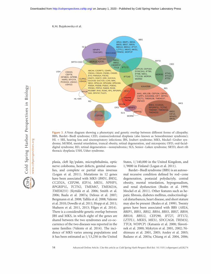
plasia, cleft lip/palate, microphthalmia, opticnerve coloboma, heart defects, genital anoma-lies, and complete or partial situs inversus(Logan et al. 2011). Mutations in 12 geneshave been associated with MKS (B9D1, B9D2,CC2D2A, CEP290, KIF14, MKS1, NPHP3,RPGRIP1L, TCTN2, TMEM67, TMEM216,TMEM231) (Kyttala et al. 2006; Smith et al.2006; Baala et al. 2007a; Delous et al. 2007;Bergmann et al. 2008; Tallila et al. 2008; Valenteet al. 2010; Dowdle et al. 2011; Hopp et al. 2011;Shaheen et al. 2011, 2013; Filges et al. 2014).There is a considerable genetic overlap betweenJBS and MKS, in which eight of the genes areshared between the two syndromes and co-oc-currence of the two diseases was reported in thesame families (Valente et al. 2014). The inci-dence of MKS varies among populations andit has been estimated as 1/13,250 in the United
States, 1/140,000 in the United Kingdom, and1/9000 in Finland (Logan et al. 2011).
Bardet–Biedl syndrome (BBS) is an autoso-mal recessive condition defined by rod–conedegeneration, postaxial polydactyly, centralobesity, mental retardation, hypogonadism,and renal dysfunction (Beales et al. 1999;Mockel et al. 2011). Other features such as he-patic fibrosis, diabetes mellitus, endocrinologi-cal disturbances, heart disease, and short staturemay also be present (Beales et al. 1999). Twentygenes have been associated with BBS (ARL6,BBIP1, BBS1, BBS2, BBS4, BBS5, BBS7, BBS9,BBS10, BBS12, CEP290, IFT27, IFT172,LZTFL1, MKKS, MKS1, SDCCAG8, TRIM32,TTC8, WDPCP) (Katsanis et al. 2000; Slavoti-nek et al. 2000; Mykytyn et al. 2001, 2002; Ni-shimura et al. 2001, 2005; Ansley et al. 2003;Badano et al. 2003a; Chiang et al. 2004, 2006;
NPHP4TRAF3IP1
CEP164,IQCB1
CLRN1,USH2A
CEP120, CEP164,DYNC2H1, IFT80,IFT88, NEK1,TTC21B, WDR34,WDR60
IFT43,IFT122,IFT52
AHI1, ARL13B, CEP104, CEP41,CSPP1, KIAA0556, KIF7, SRTD1,TECT1, TMEM138, TMEM237,ZNF423
B9D1, CC2D2A,RPGRIP1L, TCTN2,TMEM67, TMEM216,TMEM231 B9D2,
KIF14,NPHP3,MKS1
BBS1,BBS2,
C8ORF37,TTC8
CDH23,CEP78,
DFNB31, GPR98,HARS, MYO7A,
PCDH15, USH1C,USH1G
KIAA0586
IFT172
WDR35IFT140
OFD1
C5orf42,TNTN3
DDX59
NPHP1
INPP5E
WDR19
RPGR
SDCCAG8
SLS
SRTD+CED
USH
HL+SRIRD
OFD
SLS
MORM
MKS
JBS
CED
SRTD
BBSARL6, BBIP1, BBS4,BBS5, BBS7, BBS9,BBS10, BBS12, IFT27,LZTFL1, MKKS, MKS1,TRIM32, WDPCP
ABCA4, C2ORF71, CDHR1,CNGA1, CNGA3, CNGB1, CNGB3,EYS, FAM161A, FSCN2,GNAT1, GNAT2, GRK, GUCA1A,GUCA1B, GUCY2D, LCA5, MAK,NEK2, OPN1LW, OPN1MW, OPN1SW,PDE6A, PDE6B, PDE6C, PDE6G,PROM1, PRPH2, RAB28, RGS9,RGS9BP, RHO, ROM1, RP1, RPGRIP1,SAG, SPATA7,TULP1
CEP290
DDX5959
C5orf42,CTNTN3
59
C
5959
Figure 3. A Venn diagram showing a phenotypic and genetic overlap between different forms of ciliopathy.BBS, Bardet–Biedl syndrome; CED, cranioectodermal dysplasia (also known as Sensenbrenner syndrome);HL þ SRI, hearing loss and sinorespiratory infections; JBS, Joubert syndrome; MKS, Meckel–Gruber syn-drome; MORM, mental retardation, truncal obesity, retinal degeneration, and micropenis; OFD, oral-facial-digital syndrome; RD, retinal degeneration—nonsyndromic; SLS, Senior–Løken syndrome; SRTD, short-ribthoracic dysplasia; USH, Usher syndrome.
K.M. Bujakowska et al.
14 Advanced Online Article. Cite this article as Cold Spring Harb Perspect Biol doi: 10.1101/cshperspect.a028274
on January 1, 2020 - Published by Cold Spring Harbor Laboratory Press http://cshperspectives.cshlp.org/Downloaded from
Fan et al. 2004; Li et al. 2004; Stoetzel et al. 2006,2007; Leitch et al. 2008; Kim et al. 2010; Ottoet al. 2010; Marion et al. 2012; Aldahmesh et al.2014; Bujakowska et al. 2014; Scheidecker et al.2014). Apart from the BBS genes associatedwith the nonsyndromic IRD (Table 1), a homo-zygous nonsense mutation (p.S701X) in BBS12was shown to lead to a late-onset retinal degen-eration and postaxial polydactyly but no otherBBS-associated clinical features (Pawlik et al.2010).
There are two phenotypically similar dis-eases to BBS: Alstrom syndrome (ALMS) andMORM syndrome (mental retardation, truncalobesity, retinal degeneration, and micropenis).ALMS is characterized by cone–rod degenera-tion, sensorineural hearing loss, childhoodobesity, and type 2 diabetes mellitus. ALMSpatients often present with cardiomyopathyand other features such as renal, pulmonary,or hepatic disease may also be present. In con-trast to BBS, ALMS is not associated with men-tal retardation, polydactyly, or hypogonadism(Mockel et al. 2011). Mutations in only onegene, ALMS1, have been associated with Al-strom syndrome (Collin et al. 2002). MORMhas been described in only one Pakistani familyof 14 individuals, in whom a homozygous trun-cating mutation in INPP5E, was found to causethe disease (Hampshire et al. 2006; Jacoby et al.2009).
Short-rib thoracic dysplasia (SRTD) with orwithout polydactyly regroups syndromes for-merly known as Mainzer–Saldino (MZSDS),Jeune asphyxiating thoracic dystrophy (JATD),and Ellis–van Creveld (EVC) syndromes.SRTDs are autosomal recessive skeletal ciliopa-thies, characterized by short ribs, constrictedthoracic cage, shortened tubular bones, and a“trident” appearance of the acetabular roof(Huber and Cormier-Daire 2012). The severelyconstricted thoracic cage leads to respiratoryinsufficiency, often resulting in death in infancy.Other features that may be present are polydac-tyly, cleft lip/palate, retinal degeneration, andanomalies of the brain, heart, kidneys, liver,pancreas, intestines, and genitalia (Waters andBeales 2011; Huber and Cormier-Daire 2012).There is a phenotypic and genetic overlap be-
tween SRTDs and cranioectodermal dysplasia(CED), also known as Sensenbrenner syn-drome. CED is characterized by sagittal cranio-synostosis, narrow thorax, short limbs, brachy-dactyly, protuberant abdomen, and facial andectodermal anomalies (Huber and Cormier-Daire 2012). Seventeen genes have been associ-ated with these diseases (CEP120, DYNC2H1,EVC, EVC2, IFT52, IFT122, IFT140, IFT172,IFT43, IFT80, KIAA0586, NEK1, TTC21B,WDR19, WDR34, WDR35, WDR60) (Ruiz-Pe-rez et al. 2000; Galdzicka et al. 2002; Beales et al.2007; Dagoneau et al. 2009; Gilissen et al. 2010;Walczak-Sztulpa et al. 2010; Arts et al. 2011;Bredrup et al. 2011; Davis et al. 2011; Millet al. 2011; Thiel et al. 2011; Perrault et al.2012; Halbritter et al. 2013; McInerney-Leoet al. 2013; Schmidts et al. 2013b; Alby et al.2015; Shaheen et al. 2015; Girisha et al. 2016).Interestingly, some of these genes have alsobeen implicated with a nonsyndromic disease,for example, TTC21B and WDR19 in NPHP(Bredrup et al. 2011; Davis et al. 2011) orIFT172 and IFT140 in RP (Fig. 3) (Bujakowskaet al. 2014; Bifari et al. 2015; Xu et al. 2015).
BROAD PHENOTYPIC SPECTRUM OFCILIOPATHIES AND GENETIC MODIFIERS
One of the important aspects of the IRDs is thatmutations in the same gene can lead to variablephenotypes (Ferrante et al. 2001; Ansley et al.2003; Sayer et al. 2006; Perrault et al. 2007; Franket al. 2008; Leitch et al. 2008; Coene et al. 2009;Riazuddin et al. 2010; Bujakowska et al. 2012;Estrada-Cuzcano et al. 2012a; Webb et al. 2012).In some cases, the severity of disease can beexplained by the primary disease-causing mu-tation. For example, a splice site mutation of aretina-specific exon in TTC8 leads to a nonsyn-dromic RP (Riazuddin et al. 2010), whereas thegene is most commonly associated with BBS(Ansley et al. 2003). The position of a mutationmay also determine the phenotype as in thecase of truncating mutations in OFD1. Non-sense mutations downstream from exon 17lead to an X-linked dominant oral-facial-digitaltype 1 (OFD1) syndrome, manifesting withmalformations of face, oral cavity, and digits
Photoreceptor Cilia and Retinal Ciliopathies
Advanced Online Article. Cite this article as Cold Spring Harb Perspect Biol doi: 10.1101/cshperspect.a028274 15
on January 1, 2020 - Published by Cold Spring Harbor Laboratory Press http://cshperspectives.cshlp.org/Downloaded from
in affected females and lethal in males (Ferranteet al. 2001). However, truncating mutations up-stream of exon 17 lead to an X-linked recessiveJBS (Coene et al. 2009). In addition, hypomor-phic alleles can arise by mutations activatingcryptic splice sites, which leads to severely re-duced levels of wild-type transcripts as in thecase of CEP290 (den Hollander et al. 2006) andOFD1 (Webb et al. 2012).
In many cases, however, even a precise ge-netic diagnosis does not yield a clear genotype–phenotype correlation and the severity of dis-ease can vary greatly even between patients withthe same genetic cause of disease. Examples ofthis include family members that share the same3bp deletion (c.461_463del) in the PRPH2 genebut show phenotypes varying from RP involv-ing the peripheral retina to macular disease in-volving only the central retina (Weleber et al.1993). Similarly, individuals with mutations inthe RP1 gene show variable phenotypes, rang-ing from near normal to profoundly affected byretinal degeneration (Jacobson et al. 2000; Ber-son et al. 2001). Several genetic modifiers havealready been identified in IRD disease (Table 2),in which extreme examples are cases of digenicinheritance of nonsyndromic IRD (Kajiwaraet al. 1994) and BBS (Katsanis et al. 2001) orthe rescuing effect of the wild-type PRPF31 al-lele in the dominant PRPF31-associated disease(McGee et al. 1997; Vithana et al. 2003; Roseet al. 2016). Even though more than a dozenof genetic modifiers of IRD disease severityhave been reported, our knowledge about thesevariants is still limited because the studies wereconducted on a limited number of patients(sometimes single families) targeting a smallnumber of genes and functional validationwas not always performed (Table 2). In addi-tion, no study has yet shown the validity ofthe previously reported modifiers and thereforethey remain to be scrutinized by future research.
CONCLUSIONS
In summary, mutations in many different genescan cause retinal ciliopathies, reflecting the di-versity of protein functions required for normalPSC function. As indicated, it is increasingly
clear that the phenotypes ascribed to specificgenetic forms of disease overlap, and thus a re-vised system of disease definitions that includesthe genetic etiology in the disease name wouldimprove our understanding of these disorders,and their description for patients and clinicians.Further, as we have attempted to illustrate, stud-ies of retinal ciliopathies have provided insightsinto syndromic disorders, and cilia function ingeneral. Given the ubiquitous presence of ciliaon mammalian cells, we anticipate that furtherstudy of these disorders and their pathogenesiswill continue to inform us about cilia functionbroadly, and to be informed by the results ofcilia in other contexts.
REFERENCES
Abd El-Aziz MM, Barragan I, O’Driscoll CA, Goodstadt L,Prigmore E, Borrego S, Mena M, Pieras JI, El-Ashry MF,Safieh LA, et al. 2008. EYS, encoding an ortholog of Dro-sophila spacemaker, is mutated in autosomal recessiveretinitis pigmentosa. Nat Genet 40: 1285–1287.
Abu-Safieh L, Al-Anazi S, Al-Abdi L, Hashem M, Alku-raya H, Alamr M, Sirelkhatim MO, Al-Hassnan Z, Al-kuraya B, Mohamed JY, et al. 2012. In search of trial-lelism in Bardet–Biedl syndrome. Eur J Hum Genet 20:420–427.
Ahmed ZM, Riazuddin S, Bernstein SL, Ahmed Z, Khan S,Griffith AJ, Morell RJ, Friedman TB, Wilcox ER. 2001.Mutations of the protocadherin gene PCDH15 causeUsher syndrome type 1F. Am J Hum Genet 69: 25–34.
Ahmed ZM, Kjellstrom S, Haywood-Watson RJL, Bush RA,Hampton LL, Battey JF, Riazuddin S, Frolenkov G, Siev-ing PA, Friedman TB. 2008. Double homozygous waltzerand Ames waltzer mice provide no evidence of retinaldegeneration. Mol Vis 14: 2227–2236.
Akahori M, Tsunoda K, Miyake Y, Fukuda Y, Ishiura H,Tsuji S, Usui T, Hatase T, Nakamura M, Ohde H, et al.2010. Dominant mutations in RP1L1 are responsible foroccult macular dystrophy. Am J Hum Genet 87: 424–429.
Alby C, Piquand K, Huber C, Megarbane A, Ichkou A, Le-gendre M, Pelluard F, Encha-Ravazi F, Abi-Tayeh G, Bes-sieres B, et al. 2015. Mutations in KIAA0586 cause lethalciliopathies ranging from a hydrolethalus phenotype toshort-rib polydactyly syndrome. Am J Hum Genet 97:311–318.
Aldahmesh MA, Safieh LA, Alkuraya H, Al-Rajhi A, Sham-seldin H, Hashem M, Alzahrani F, Khan AO, Alqahtani F,Rahbeeni Z, et al. 2009. Molecular characterization ofretinitis pigmentosa in Saudi Arabia. Mol Vis 15: 2464–2469.
Aldahmesh MA, Li Y, Alhashem A, Anazi S, Alkuraya H,Hashem M, Awaji AA, Sogaty S, Alkharashi A, AlzahraniS, et al. 2014. IFT27, encoding a small GTPase compo-nent of IFT particles, is mutated in a consanguineous
K.M. Bujakowska et al.
16 Advanced Online Article. Cite this article as Cold Spring Harb Perspect Biol doi: 10.1101/cshperspect.a028274
on January 1, 2020 - Published by Cold Spring Harbor Laboratory Press http://cshperspectives.cshlp.org/Downloaded from
family with Bardet–Biedl syndrome. Hum Mol Genet 23:3307–3315.
Aligianis IA, Forshew T, Johnson S, Michaelides M, JohnsonCA, Trembath RC, Hunt DM, Moore AT, Maher ER. 2002.Mapping of a novel locus for achromatopsia (ACHM4) to1p and identification of a germline mutation in the alphasubunit of cone transducin (GNAT2). J Med Genet 39:656–660.
Allikmets R, Singh N, Sun H, Shroyer NF, Hutchinson A,Chidambaram A, Gerrard B, Baird L, Stauffer D, PeifferA, et al. 1997. A photoreceptor cell-specific ATP-bindingtransporter gene (ABCR) is mutated in recessive Star-gardt macular dystrophy. Nat Genet 15: 236–246.
Ansley SJ, Badano JL, Blacque OE, Hill J, Hoskins BE, LeitchCC, Kim JC, Ross AJ, Eichers ER, Teslovich TM, et al.2003. Basal body dysfunction is a likely cause of pleiotro-pic Bardet–Biedl syndrome. Nature 425: 628–633.
Arikawa K, Molday LL, Molday RS, Williams DS. 2011.Localization of peripherin/RDS in the disk membranesof cone and rod photoreceptors: Relationship to diskmembrane morphogenesis and retinal degeneration.J Cell Biol 116: 659–667.
Arts HH, Bongers EMHF, Mans DA, van Beersum SEC, OudMM, Bolat E, Spruijt L, Cornelissen EAM, Schuurs-Hoeijmakers JHM, de Leeuw N, et al. 2011.C14ORF179 encoding IFT43 is mutated in Sensenbren-ner syndrome. J Med Genet 48: 390–395.
Ayyagari R, Kakuk LE, Coats CL, Bingham EL, Toda Y, FeliusJ, Sieving P. 1999. Bilateral macular atrophy in blue conemonochromacy (BCM) with loss of the locus controlregion (LCR) and part of the red pigment gene. Mol Vis5: 13.
Ayyagari R, Demirci FY, Liu J, Bingham EL, Stringham H,Kakuk LE, Boehnke M, Gorin MB, Richards JE, SievingPA. 2002. X-linked recessive atrophic macular degenera-tion from RPGR mutation. Genomics 80: 166–171.
Baala L, Audollent S, Martinovic J, Ozilou C, Babron M-C,Sivanandamoorthy S, Saunier S, Salomon R, GonzalesM, Rattenberry E, et al. 2007a. Pleiotropic effects ofCEP290 (NPHP6) mutations extend to Meckel syn-drome. Am J Hum Genet 81: 170–179.
Baala L, Romano S, Khaddour R, Saunier S, Smith UM,Audollent S, Ozilou C, Faivre L, Laurent N, Foliguet B,et al. 2007b. The Meckel–Gruber syndrome gene, MKS3,is mutated in Joubert syndrome. Am J Hum Genet 80:186–194.
Badano JL, Ansley SJ, Leitch CC, Lewis RA, Lupski JR, Kat-sanis N. 2003a. Identification of a novel Bardet–Biedlsyndrome protein, BBS7, that shares structural featureswith BBS1 and BBS2. Am J Hum Genet 72: 650–658.
Badano JL, Kim JC, Hoskins BE, Lewis RA, Ansley SJ, CutlerDJ, Castellan C, Beales PL, Leroux MR, Katsanis N.2003b. Heterozygous mutations in BBS1, BBS2 andBBS6 have a potential epistatic effect on Bardet–Biedlpatients with two mutations at a second BBS locus.Hum Mol Genet 12: 1651–1659.
Badano JL, Leitch CC, Ansley SJ, May-Simera H, Lawson S,Lewis RA, Beales PL, Dietz HC, Fisher S, Katsanis N.2006. Dissection of epistasis in oligogenic Bardet–Biedlsyndrome. Nature 439: 326–330.
Bandah-Rozenfeld D, Mizrahi-Meissonnier L, Farhy C,Obolensky A, Chowers I, Pe’er J, Merin S, Ben-Yosef T,
Ashery-Padan R, Banin E, et al. 2010. Homozygositymapping reveals null mutations in FAM161A as a causeof autosomal-recessive retinitis pigmentosa. Am J HumGenet 87: 382–391.
Banerjee P, Kleyn PW, Knowles JA, Lewis CA, Ross BM,Parano E, Kovats SG, Lee JJ, Penchaszadeh GK, Ott J, etal. 1998. TULP1 mutation in two extended Dominicankindreds with autosomal recessive retinitis pigmentosa.Nat Genet 18: 177–179.
Bareil C, Hamel CP, Delague V, Arnaud B, Demaille J,Claustres M. 2001. Segregation of a mutation inCNGB1 encoding the b-subunit of the rod cGMP-gatedchannel in a family with autosomal recessive retinitispigmentosa. Hum Genet 108: 328–334.
Bascom RA, Manara S, Collins L, Molday RS, Kalnins VI,McInnes RR. 1992. Cloning of the cDNA for a novelphotoreceptor membrane protein (rom-1) identifies adisk rim protein family implicated in human retinopa-thies. Neuron 8: 1171–1184.
Beales PL, Elcioglu N, Woolf AS, Parker D, Flinter FA. 1999.New criteria for improved diagnosis of Bardet–Biedl syn-drome: Results of a population survey. J Med Genet 36:437–446.
Beales PL, Bland E, Tobin JL, Bacchelli C, Tuysuz B, Hill J,Rix S, Pearson CG, Kai M, Hartley J, et al. 2007. IFT80,which encodes a conserved intraflagellar transport pro-tein, is mutated in Jeune asphyxiating thoracic dystrophy.Nat Genet 39: 727–729.
Berger W, Kloeckener-Gruissem B, Neidhardt J. 2010. Themolecular basis of human retinal and vitreoretinal dis-eases. Prog Retin Eye Res 29: 335–375.
Bergmann C, Fliegauf M, Bruchle NO, Frank V, Olbrich H,Kirschner J, Schermer B, Schmedding I, Kispert A, Kran-zlin B, et al. 2008. Loss of nephrocystin-3 function cancause embryonic lethality, Meckel–Gruber-like syn-drome, situs inversus, and renal-hepatic-pancreatic dys-plasia. Am J Hum Genet 82: 959–970.
Berson EL, Grimsby JL, Adams SM, McGee TL, Sweklo E,Pierce EA, Sandberg MA, Dryja TP. 2001. Clinical fea-tures and mutations in patients with dominant retinitispigmentosa-1 (RP1). Invest Ophthalmol Vis Sci 42: 2217–2224.
Besharse JC, Forestner DM, Defoe DM. 1985. Membraneassembly in retinal photoreceptors. III: Distinct mem-brane domains of the connecting cilium of developingrods. J Neurosci 5: 1035–1048.
Bielas SL, Silhavy JL, Brancati F, Kisseleva MV, Al-Gazali L,Sztriha L, Bayoumi RA, Zaki MS, Abdel-Aleem A, RostiRO, et al. 2009. Mutations in INPP5E, encoding inositolpolyphosphate-5-phosphatase E, link phosphatidyl ino-sitol signaling to the ciliopathies. Nat Genet 41: 1032–1036.
Bifari IN, Elkhamary SM, Bolz HJ, Khan AO. 2015. Theophthalmic phenotype of IFT140-related ciliopathyranges from isolated to syndromic congenital retinal dys-trophy. Br J Ophthalmol 6: 829–833.
Bizet AA, Becker-Heck A, Ryan R, Weber K, Filhol E, Krug P,Halbritter J, Delous M, Lasbennes M-C, Linghu B, et al.2015. Mutations in TRAF3IP1/IFT54 reveal a new rolefor IFT proteins in microtubule stabilization. Nat Com-mun 6: 8666.
Photoreceptor Cilia and Retinal Ciliopathies
Advanced Online Article. Cite this article as Cold Spring Harb Perspect Biol doi: 10.1101/cshperspect.a028274 17
on January 1, 2020 - Published by Cold Spring Harbor Laboratory Press http://cshperspectives.cshlp.org/Downloaded from
Bolz H, von Brederlow B, Ramırez A, Bryda EC, Kutsche K,Nothwang HG, Seeliger M, del C-Salcedo Cabrera M,Vila MC, Molina OP, et al. 2001. Mutation of CDH23,encoding a new member of the cadherin gene family,causes Usher syndrome type 1D. Nat Genet 27: 108–112.
Bonnet C, El-Amraoui A. 2012. Usher syndrome (sensori-neural deafness and retinitis pigmentosa): Pathogenesis,molecular diagnosis and therapeutic approaches. CurrOpin Neurol 25: 42–49.
Bowne SJ, Daiger SP, Malone KA, Heckenlively JR, KennanA, Humphries P, Hughbanks-Wheaton D, Birch DG, LiuQ, Pierce EA, et al. 2003. Characterization of RP1L1, ahighly polymorphic paralog of the retinitis pigmentosa 1(RP1) gene. Mol Vis 9: 129–137.
Bredrup C, Saunier S, Oud MM, Fiskerstrand T, Hoischen A,Brackman D, Leh SM, Midtbø M, Filhol E, Bole-Feysot C,et al. 2011. Ciliopathies with skeletal anomalies and renalinsufficiency due to mutations in the IFT-A geneWDR19. Am J Hum Genet 89: 634–643.
Bujakowska K, Audo I, Mohand-Saıd S, Lancelot ME, An-tonio A, Germain A, Leveillard T, Letexier M, Saraiva JP,Lonjou C, et al. 2012. CRB1 mutations in inherited ret-inal dystrophies. Hum Mutat 33: 306–315.
Bujakowska KM, Zhang Q, Siemiatkowska AM, Liu Q, PlaceE, Falk MJ, Consugar M, Lancelot ME, Antonio A, Lon-jou C, et al. 2014. Mutations in IFT172 cause isolatedretinal degeneration and Bardet–Biedl syndrome. HumMol Genet 24: 230–242.
Calvert PD, Strissel KJ, Schiesser WE, Pugh EN, ArshavskyVY. 2006. Light-driven translocation of signaling proteinsin vertebrate photoreceptors. Trends Cell Biol 16: 560–568.
Calvert PD, Schiesser WE, Pugh EN. 2010. Diffusion of asoluble protein, photoactivatable GFP, through a sensorycilium. J Gen Physiol 135: 173–196.
Cantagrel V, Silhavy JL, Bielas SL, Swistun D, Marsh SE,Bertrand JY, Audollent S, Attie-Bitach T, Holden KR,Dobyns WB, et al. 2008. Mutations in the cilia geneARL13B lead to the classical form of Joubert syndrome.Am J Hum Genet 83: 170–179.
Caridi G, Murer L, Bellantuono R, Sorino P, Caringella DA,Gusmano R, Ghiggeri GM. 1998. Renal–retinal syn-dromes: Association of retinal anomalies and recessivenephronophthisis in patients with homozygous deletionof the NPH1 locus. Am J Kidney Dis 32: 1059–1062.
Chaitin MH, Schneider BG, Hall MO, Papermaster DS.1984. Actin in the photoreceptor connecting cilium: Im-munocytochemical localization to the site of outer seg-ment disk formation. J Cell Biol 99: 239–247.
Chakarova CF, Papaioannou MG, Khanna H, Lopez I, Wa-seem N, Shah A, Theis T, Friedman J, Maubaret C, Buja-kowska K, et al. 2007. Mutations in TOPORS cause au-tosomal dominant retinitis pigmentosa with perivascularretinal pigment epithelium atrophy. Am J Hum Genet 81:1098–1103.
Chakarova CF, Khanna H, Shah AZ, Patil SB, Sedmak T,Murga-Zamalloa CA, Papaioannou MG, Nagel-WolfrumK, Lopez I, Munro P, et al. 2011. TOPORS, implicated inretinal degeneration, is a cilia-centrosomal protein. HumMol Genet 20: 975–987.
Chaki M, Airik R, Ghosh AK, Giles RH, Chen R, Slaats GG,Wang H, Hurd TW, Zhou W, Cluckey A, et al. 2012.
Exome capture reveals ZNF423 and CEP164 mutations,linking renal ciliopathies to DNA damage response sig-naling. Cell 150: 533–548.
Chiang AP, Nishimura D, Searby C, Elbedour K, Carmi R,Ferguson AL, Secrist J, Braun T, Casavant T, Stone EM, etal. 2004. Comparative genomic analysis identifies anADP-ribosylation factor-like gene as the cause of Bar-det–Biedl syndrome (BBS3). Am J Hum Genet 75:475–484.
Chiang AP, Beck JS, Yen HJ, Tayeh MK, Scheetz TE, Swider-ski RE, Nishimura DY, Braun T, Kim KY, Huang J, et al.2006. Homozygosity mapping with SNP arrays identifiesTRIM32, an E3 ubiquitin ligase, as a Bardet–Biedl syn-drome gene (BBS11). Proc Natl Acad Sci 103: 6287–6292.
Chung DC, Traboulsi EI. 2009. Leber congenital amaurosis:Clinical correlations with genotypes, gene therapy trialsupdate, and future directions. J AAPOS 13: 587–592.
Clarke G, Goldberg AF, Vidgen D, Collins L, Ploder L,Schwarz L, Molday LL, Rossant J, Szel A, Molday RS, etal. 2000. Rom-1 is required for rod photoreceptor viabil-ity and the regulation of disk morphogenesis. Nat Genet25: 67–73.
Coene KLM, Roepman R, Doherty D, Afroze B, Kroes HY,Letteboer SJF, Ngu LH, Budny B, van Wijk E, Gorden NT,et al. 2009. OFD1 is mutated in X-linked Joubert syn-drome and interacts with LCA5-encoded lebercilin. Am JHum Genet 85: 465–481.
Collin GB, Marshall JD, Ikeda A, So WV, Russell-Eggitt I,Maffei P, Beck S, Boerkoel CF, Sicolo N, Martin M, et al.2002. Mutations in ALMS1 cause obesity, type 2 diabetesand neurosensory degeneration in Alstrom syndrome.Nat Genet 31: 74–78.
Collin RWJ, Littink KW, Klevering BJ, van den Born LI,Koenekoop RK, Zonneveld MN, Blokland EAW, StromTM, Hoyng CB, den Hollander AI, et al. 2008. Identifi-cation of a 2 Mb human ortholog of Drosophila eyesshut/spacemaker that is mutated in patients with retinitispigmentosa. Am J Hum Genet 83: 594–603.
Collin RWJ, Safieh C, Littink KW, Shalev SA, Garzozi HJ,Rizel L, Abbasi AH, Cremers FPM, den Hollander AI,Klevering BJ, et al. 2010. Mutations in C2ORF71 causeautosomal-recessive retinitis pigmentosa. Am J Hum Ge-net 86: 783–788.
Consugar MB, Navarro-Gomez D, Place EM, BujakowskaKM, Sousa ME, Fonseca-Kelly ZD, Taub DG, JanessianM, Wang DY, Au ED, et al. 2014. Panel-based geneticdiagnostic testing for inherited eye diseases is highly ac-curate and reproducible, and more sensitive for variantdetection, than exome sequencing. Genet Med 17: 253–261.
Coppieters F, Casteels I, Meire F, De Jaegere S, Hooghe S, vanRegemorter N, Van Esch H, Matuleviciene A, Nunes L,Meersschaut V, et al. 2010. Genetic screening of LCA inBelgium: Predominance of CEP290 and identification ofpotential modifier alleles in AHI1 of CEP290-related phe-notypes. Hum Mutat 31: E1709–E1766.
Coussa RG, Otto EA, Gee HY, Arthurs P, Ren H, Lopez I,Keser V, Fu Q, Faingold R, Khan A, et al. 2013. WDR19:An ancient, retrograde, intraflagellar ciliary protein ismutated in autosomal recessive retinitis pigmentosaand in Senior–Løken syndrome. Clin Genet 84: 150–159.
K.M. Bujakowska et al.
18 Advanced Online Article. Cite this article as Cold Spring Harb Perspect Biol doi: 10.1101/cshperspect.a028274
on January 1, 2020 - Published by Cold Spring Harbor Laboratory Press http://cshperspectives.cshlp.org/Downloaded from
Cremers FP, van de Pol DJ, van Driel M, den Hollander AI,van Haren FJ, Knoers NV, Tijmes N, Bergen AA, Rohr-schneider K, Blankenagel A, et al. 1998. Autosomal reces-sive retinitis pigmentosa and cone–rod dystrophy causedby splice site mutations in the Stargardt’s disease geneABCR. Hum Mol Genet 7: 355–362.
Dafinger C, Liebau MC, Elsayed SM, Hellenbroich Y, Bolt-shauser E, Korenke GC, Fabretti F, Janecke AR, EbermannI, Nurnberg G, et al. 2011. Mutations in KIF7 link Joubertsyndrome with Sonic Hedgehog signaling and microtu-bule dynamics. J Clin Invest 121: 2662–2667.
Dagoneau N, Goulet M, Genevieve D, Sznajer Y, MartinovicJ, Smithson S, Huber C, Baujat G, Flori E, Tecco L, et al.2009. DYNC2H1 mutations cause asphyxiating thoracicdystrophy and short rib-polydactyly syndrome, type III.Am J Hum Genet 84: 706–711.
Davidson AE, Sergouniotis PI, Mackay DS, Wright GA, Wa-seem NH, Michaelides M, Holder GE, Robson AG,Moore AT, Plagnol V, et al. 2013. RP1L1 variants are as-sociated with a spectrum of inherited retinal diseasesincluding retinitis pigmentosa and occult macular dys-trophy. Hum Mutat 34: 506–514.
Davis EE, Zhang Q, Liu Q, Diplas BH, Davey LM, Hartley J,Stoetzel C, Szymanska K, Ramaswami G, Logan CV, et al.2011. TTC21B contributes both causal and modifyingalleles across the ciliopathy spectrum. Nat Genet 43:189–196.
Dellett M, Sasai N, Nishide K, Becker S, Papadaki V, AstridLimb G, Moore AT, Kondo T, Ohnuma SI. 2015. Geneticbackground and light-dependent progression of photo-receptor cell degeneration in Prominin-1 knockout mice.Investig Ophthalmol Vis Sci 56: 164–176.
Delous M, Baala L, Salomon R, Laclef C, Vierkotten J, ToryK, Golzio C, Lacoste T, Besse L, Ozilou C, et al. 2007. Theciliary gene RPGRIP1L is mutated in cerebello-oculo-re-nal syndrome (Joubert syndrome type B) and Meckelsyndrome. Nat Genet 39: 875–881.
den Hollander AI, Koenekoop RK, Yzer S, Lopez I, ArendsML, Voesenek KEJ, Zonneveld MN, Strom TM, Mei-tinger T, Brunner HG, et al. 2006. Mutations in theCEP290 (NPHP6) gene are a frequent cause of Lebercongenital amaurosis. Am J Hum Genet 79: 556–561.
den Hollander AI, Koenekoop RK, Mohamed MD, Arts HH,Boldt K, Towns KV, Sedmak T, Beer M, Nagel-Wolfrum K,McKibbin M, et al. 2007. Mutations in LCA5, encodingthe ciliary protein lebercilin, cause Leber congenital am-aurosis. Nat Genet 39: 889–895.
den Hollander AI, Roepman R, Koenekoop RK, CremersFPM. 2008. Leber congenital amaurosis: Genes, proteinsand disease mechanisms. Prog Retin Eye Res 27: 391–419.
Deretic D, Williams AH, Ransom N, Morel V, Hargrave PA,Arendt A. 2005. Rhodopsin C terminus, the site of mu-tations causing retinal disease, regulates trafficking bybinding to ADP-ribosylation factor 4 (ARF4). Proc NatlAcad Sci 102: 3301–3306.
De Robertis E. 1956. Electron microscope observations onthe submicroscopic organization of the retinal rods. JBiophys Biochem Cytol 2: 319–330.
Ding JD, Salinas RY, Arshavsky VY. 2015. Discs of mamma-lian rod photoreceptors form through the membraneevagination mechanism. J Cell Biol 211: 495–502.
Di Palma F, Holme RH, Bryda EC, Belyantseva IA, Pelle-grino R, Kachar B, Steel KP, Noben-Trauth K. 2001. Mu-tations in Cdh23, encoding a new type of cadherin, causestereocilia disorganization in waltzer, the mouse modelfor Usher syndrome type 1D. Nat Genet 27: 103–107.
Dixon-Salazar T, Silhavy JL, Marsh SE, Louie CM, ScottLC, Gururaj A, Al-Gazali L, Al-Tawari AA, Kayserili H,Sztriha L, et al. 2004. Mutations in the AHI1 gene, en-coding jouberin, cause Joubert syndrome with corticalpolymicrogyria. Am J Hum Genet 75: 979–987.
Dowdle WE, Robinson JF, Kneist A, Sirerol-Piquer MS,Frints SGM, Corbit KC, Zaghloul NA, Zaghloul NA,van Lijnschoten G, Mulders L, et al. 2011. Disruptionof a ciliary B9 protein complex causes Meckel syndrome.Am J Hum Genet 89: 94–110.
Dryja TP, McGee TL, Reichel E, Hahn LB, Cowley GS, Yan-dell DW, Sandberg MA, Berson EL. 1990. A point muta-tion of the rhodopsin gene in one form of retinitis pig-mentosa. Nature 343: 364–366.
Dryja TP, Berson EL, Rao VR, Oprian DD. 1993. Heterozy-gous missense mutation in the rhodopsin gene as a causeof congenital stationary night blindness. Nat Genet 4:280–283.
Dryja TP, Finn JT, Peng YW, McGee TL, Berson EL, Yau KW.1995. Mutations in the gene encoding the a subunit ofthe rod cGMP-gated channel in autosomal recessive ret-initis pigmentosa. Proc Natl Acad Sci 92: 10177–10181.
Dryja TP, Hahn LB, Reboul T, Arnaud B. 1996. Missensemutation in the gene encoding thea subunit of rod trans-ducin in the Nougaret form of congenital stationarynight blindness. Nat Genet 13: 358–360.
Dryja TP, Adams SM, Grimsby JL, McGee TL, Hong DH, LiT, Andreasson S, Berson EL. 2001. Null RPGRIP1 allelesin patients with Leber congenital amaurosis. Am J HumGenet 68: 1295–1298.
Dvir L, Srour G, Abu-Ras R, Miller B, Shalev S, Ben-Yosef T.2010. Autosomal-recessive early-onset retinitis pigmen-tosa caused by a mutation in PDE6G, the gene encodingthe gamma subunit of rod cGMP phosphodiesterase. AmJ Hum Genet 87: 258–264.
Ebermann I, Phillips JB, Liebau MC, Koenekoop RK,Schermer B, Lopez I, Schafer E, Roux AF, Dafinger C,Bernd A, et al. 2010. PDZD7 is a modifier of retinal dis-ease and a contributor to digenic Usher syndrome. J ClinInvest 120: 1812–1823.
Edvardson S, Shaag A, Zenvirt S, Erlich Y, Hannon GJ,Shanske AL, Gomori JM, Ekstein J, Elpeleg O. 2010. Jou-bert syndrome 2 (JBTS2) in Ashkenazi Jews is associatedwith a TMEM216 mutation. Am J Hum Genet 86: 93–97.
El Shamieh S, Neuille M, Terray A, Orhan E, Condroyer C,Demontant V, Michiels C, Antonio A, Boyard F, LancelotME, et al. 2014. Whole-exome sequencing identifies KIZas a ciliary gene associated with autosomal-recessiverod–cone dystrophy. Am J Hum Genet 94: 625–633.
Estrada-Cuzcano A, Koenekoop RK, Coppieters F, Kohl S,Lopez I, Collin RW, De Baere E, Roeleveld D, Marek J,Bernd A, et al. 2011. IQCB1 mutations in patientswith Leber congenital amaurosis. Invest Ophthalmol VisSci 52: 834–839.
Estrada-Cuzcano A, Koenekoop RK, Senechal A, De BaereEBW, de Ravel T, Banfi S, Kohl S, Ayuso C, Sharon D,Hoyng CB, et al. 2012a. BBS1 mutations in a wide spec-
Photoreceptor Cilia and Retinal Ciliopathies
Advanced Online Article. Cite this article as Cold Spring Harb Perspect Biol doi: 10.1101/cshperspect.a028274 19
on January 1, 2020 - Published by Cold Spring Harbor Laboratory Press http://cshperspectives.cshlp.org/Downloaded from
trum of phenotypes ranging from nonsyndromic retinitispigmentosa to Bardet–Biedl syndrome. Arch Ophthalmol130: 1425–1432.
Estrada-Cuzcano A, Neveling K, Kohl S, Banin E, Roten-streich Y, Sharon D, Falik-Zaccai TC, Hipp S, RoepmanR, Wissinger B, et al. 2012b. Mutations in C8orf37, en-coding a ciliary protein, are associated with autosomal-recessive retinal dystrophies with early macular involve-ment. Am J Hum Genet 90: 102–109.
Estrada-Cuzcano A, Roepman R, Cremers FPM, den Hol-lander AI, Mans D. 2012c. Non-syndromic retinal cilio-pathies: Translating gene discovery into therapy. HumMol Genet 21: R111–R124.
Eudy JD, Weston MD, Yao S, Hoover DM, Rehm HL, Ma-Edmonds M, Yan D, Ahmad I, Cheng JJ, Ayuso C, et al.1998. Mutation of a gene encoding a protein with extra-cellular matrix motifs in Usher syndrome type IIa. Science280: 1753–1757.
Fahim AT, Bowne SJ, Sullivan LS, Webb KD, Williams JT,Wheaton DK, Birch DG, Daiger SP. 2011. Allelic hetero-geneity and genetic modifier loci contribute to clinicalvariation in males with X-linked retinitis pigmentosa dueto RPGR mutations. PLoS ONE 6: e23021.
Fan Y, Esmail MA, Ansley SJ, Blacque OE, Boroevich K, RossAJ, Moore SJ, Badano JL, May-Simera H, Compton DS, etal. 2004. Mutations in a member of the Ras superfamily ofsmall GTP-binding proteins causes Bardet–Biedl syn-drome. Nat Genet 36: 989–993.
Farrar GJ, McWilliam P, Bradley DG, Kenna P, Lawler M,Sharp EM, Humphries MM, Eiberg H, Conneally PM,Trofatter JA. 1990. Autosomal dominant retinitis pig-mentosa: Linkage to rhodopsin and evidence for geneticheterogeneity. Genomics 8: 35–40.
Ferland RJ, Eyaid W, Collura RV, Tully LD, Hill RS, Al-NouriD, Al-Rumayyan A, Topcu M, Gascon G, Bodell A, et al.2004. Abnormal cerebellar development and axonal de-cussation due to mutations in AHI1 in Joubert syndrome.Nat Genet 36: 1008–1013.
Ferrante MI, Giorgio G, Feather SA, Bulfone A, Wright V,Ghiani M, Selicorni A, Gammaro L, Scolari F, Woolf AS,et al. 2001. Identification of the gene for oral-facial-dig-ital type I syndrome. Am J Hum Genet 68: 569–576.
Filges I, Nosova E, Bruder E, Tercanli S, Townsend K, GibsonWT, Rothlisberger B, Heinimann K, Hall JG, Gregory-Evans CY, et al. 2014. Exome sequencing identifies mu-tations in KIF14 as a novel cause of an autosomal reces-sive lethal fetal ciliopathy phenotype. Clin Genet 86:220–228.
Fingert JH, Oh K, Chung M, Scheetz TE, Andorf JL, John-son RM, Sheffield VC, Stone EM. 2008. Association of anovel mutation in the retinol dehydrogenase 12 (RDH12)gene with autosomal dominant retinitis pigmentosa.Arch Ophthalmol 126: 1301–1307.
Fotiadis D, Liang Y, Filipek S, Saperstein DA, Engel A, Pal-czewski K. 2003. Rhodopsin dimers in native disc mem-branes. Nature 421: 127–128.
Frank V, den Hollander AI, Bruchle NO, Zonneveld MN,Nurnberg G, Becker C, Du Bois G, Kendziorra H, Roo-sing S, Senderek J, et al. 2008. Mutations of the CEP290gene encoding a centrosomal protein cause Meckel–Gruber syndrome. Hum Mutat 29: 45–52.
Fuchs S, Nakazawa M, Maw M, Tamai M, Oguchi Y, Gal A.1995. A homozygous 1-base pair deletion in the arrestingene is a frequent cause of Oguchi disease in Japanese.Nat Genet 10: 360–362.
Gal A, Orth U, Baehr W, Schwinger E, Rosenberg T. 1994.Heterozygous missense mutation in the rod cGMP phos-phodiesterase beta-subunit gene in autosomal dominantstationary night blindness. Nat Genet 7: 64–68.
Galdzicka M, Patnala S, Hirshman MG, Cai JF, Nitowsky H,Egeland JA, Ginns EI. 2002. A new gene, EVC2, is mutat-ed in Ellis–van Creveld syndrome. Mol Genet Metab 77:291–295.
Garcia-Gonzalo FR, Corbit KC, Sirerol-Piquer MS, Rama-swami G, Otto EA, Noriega TR, Seol AD, Robinson JF,Bennett CL, Josifova DJ, et al. 2011. A transition zonecomplex regulates mammalian ciliogenesis and ciliarymembrane composition. Nat Genet 43: 776–784.
Geller SF, Guerin KI, Visel M, Pham A, Lee ES, Dror AA,Avraham KB, Hayashi T, Ray CA, Reh TA, et al. 2009.CLRN1 is nonessential in the mouse retina but is requiredfor cochlear hair cell development. PLoS Genet 5: 17–19.
Gilissen C, Arts HH, Hoischen A, Spruijt L, Mans DA, ArtsP, van Lier B, Steehouwer M, van Reeuwijk J, Kant SG, etal. 2010. Exome sequencing identifies WDR35 variantsinvolved in Sensenbrenner syndrome. Am J Hum Genet87: 418–423.
Gilliam JC, Chang JT, Sandoval IM, Zhang Y, Li T, Pittler SJ,Chiu W, Wensel TG. 2012. Three-dimensional architec-ture of the rod sensory cilium and its disruption in retinalneurodegeneration. Cell 151: 1029–1041.
Girisha KM, Shukla A, Trujillano D, Bhavani GS, Hebbar M,Kadavigere R, Rolfs A. 2016. A homozygous nonsensevariant in IFT52 is associated with a human skeletal cili-opathy. Clin Genet doi: 10.1111/cge.12762.
Goetz SC, Anderson KV. 2010. The primary cilium: A sig-nalling centre during vertebrate development. Nat RevGenet 11: 331–344.
Goldberg AFX, Molday RS. 1996. Subunit composition ofthe peripherin/RDS–rom-1 disk rim complex from rodphotoreceptors: Hydrodynamic evidence for a tetramericquaternary structure. Biochemistry 35: 6144–6149.
Gorden NT, Arts HH, Parisi MA, Coene KLM, Letteboer SJF,van Beersum SEC, Mans DA, Hikida A, Eckert M, Knut-zen D, et al. 2008. CC2D2A is mutated in Joubert syn-drome and interacts with the ciliopathy-associated basalbody protein CEP290. Am J Hum Genet 83: 559–571.
Guillonneau X, Piriev NI, Danciger M, Kozak CA, CideciyanAV, Jacobson SG, Farber DB. 1999. A nonsense mutationin a novel gene is associated with retinitis pigmentosa in afamily linked to the RP1 locus. Hum Mol Genet 8: 1541–1546.
Hagstrom SA, North MA, Nishina PL, Berson EL, Dryja TP.1998. Recessive mutations in the gene encoding the tub-by-like protein TULP1 in patients with retinitis pigmen-tosa. Nat Genet 18: 174–176.
Halbritter J, Bizet AA, Schmidts M, Porath JD, Braun DA,Gee HY, McInerney-Leo AM, Krug P, Filhol E, Davis EE,et al. 2013. Defects in the IFT-B component IFT172 causeJeune and Mainzer–Saldino syndromes in humans. Am JHum Genet 93: 1–11.
Hameed A, Abid A, Aziz A, Ismail M, Mehdi SQ, Khaliq S.2003. Evidence of RPGRIP1 gene mutations associated
K.M. Bujakowska et al.
20 Advanced Online Article. Cite this article as Cold Spring Harb Perspect Biol doi: 10.1101/cshperspect.a028274
on January 1, 2020 - Published by Cold Spring Harbor Laboratory Press http://cshperspectives.cshlp.org/Downloaded from
with recessive cone-rod dystrophy. J Med Genet 40: 616–619.
Hamel C. 2006. Retinitis pigmentosa. Orphanet J Rare Dis 1:40.
Hampshire DJ, Ayub M, Springell K, Roberts E, Jafri H,Rashid Y, Bond J, Riley JH, Woods CG. 2006. MORMsyndrome (mental retardation, truncal obesity, retinaldystrophy and micropenis), a new autosomal recessivedisorder, links to 9q34. Eur J Hum Genet 14: 543–548.
Han Z, Anderson DW, Papermaster DS. 2012. Prominin-1localizes to the open rims of outer segment lamellae inXenopus laevis rod and cone photoreceptors. InvestigOphthalmol Vis Sci 53: 361–373.
Hardcastle AJ, Thiselton DL, Van Maldergem L, Saha BK,Jay M, Plant C, Taylor R, Bird AC, Bhattacharya S. 1999.Mutations in the RP2 gene cause disease in 10% of fam-ilies with familial X-linked retinitis pigmentosa assessedin this study. Am J Hum Genet 64: 1210–1215.
Hartong DT, Berson EL, Dryja TP. 2006. Retinitis pigmen-tosa. Lancet 368: 1795–1809.
Helou J, Otto EA, Attanasio M, Allen SJ, Parisi MA, Glass I,Utsch B, Hashmi S, Fazzi E, Omran H, et al. 2007. Mu-tation analysis of NPHP6/CEP290 in patients with Jou-bert syndrome and Senior–Løken syndrome. J Med Ge-net 44: 657–663.
Henderson RH, Li Z, Abd El Aziz MM, Mackay DS, EljininiMA, Zeidan M, Moore AT, Bhattacharya SS, Webster AR.2010. Biallelic mutation of protocadherin-21 (PCDH21)causes retinal degeneration in humans. Mol Vis 16: 46–52.
Heon E, Kim G, Qin S, Garrison JE, Tavares E, Vincent A,Nuangchamnong N, Scott CA, Slusarski DC, SheffieldVC. 2016. Mutations in C8ORF37 cause Bardet–Biedlsyndrome (BBS21). Hum Mol Genet doi: 10.1093/hmg/ddw096.
Hopp K, Heyer CM, Hommerding CJ, Henke SA, SundsbakJL, Patel S, Patel P, Consugar MB, Czarnecki PG, GliemTJ, et al. 2011. B9D1 is revealed as a novel Meckel syn-drome (MKS) gene by targeted exon-enriched next-gen-eration sequencing and deletion analysis. Hum Mol Genet20: 2524–2534.
Horst CJ, Johnson LV, Besharse JC. 1990. Transmembraneassemblage of the photoreceptor connecting cilium andmotile cilium transition zone contain a common immu-nologic epitope. Cell Motil Cytoskeleton 17: 329–344.
Huang SHH, Pittler SJJ, Huang X, Oliveira L, Berson ELL,Dryja TPP. 1995. Autosomal recessive retinitis pigmen-tosa caused by mutations in the a subunit of rod cGMPphosphodiesterase. Nat Genet 11: 468–471.
Huang L, Szymanska K, Jensen VL, Janecke AR, Innes AM,Davis EE, Frosk P, Li C, Willer JR, Chodirker BN, et al.2011. TMEM237 is mutated in individuals with a Joubertsyndrome related disorder and expands the role of theTMEM family at the ciliary transition zone. Am J HumGenet 89: 713–730.
Huber C, Cormier-Daire V. 2012. Ciliary disorder of theskeleton. Am J Med Genet Semin Med Genet 160C:165–174.
Jacobson SG, Cideciyan AV, Iannaccone A, Weleber RG,Fishman GA, Maguire AM, Affatigato LM, Bennett J,Pierce EA, Danciger M, et al. 2000. Disease expression
of RP1 mutations causing autosomal dominant retinitispigmentosa. Invest Ophthalmol Vis Sci 41: 1898–1908.
Jacoby M, Cox JJ, Gayral S, Hampshire DJ, Ayub M, Block-mans M, Pernot E, Kisseleva MV, Compere P, SchiffmannSN, et al. 2009. INPP5E mutations cause primary ciliumsignaling defects, ciliary instability and ciliopathies inhuman and mouse. Nat Genet 41: 1027–1031.
Janecke AR, Thompson DA, Utermann G, Becker C, HubnerCA, Schmid E, McHenry CL, Nair AR, Ruschendorf F,Heckenlively J, et al. 2004. Mutations in RDH12 encodinga photoreceptor cell retinol dehydrogenase cause child-hood-onset severe retinal dystrophy. Nat Genet 36: 850–854.
Joensuu T, Hamalainen R, Yuan B, Johnson C, Tegelberg S,Gasparini P, Zelante L, Pirvola U, Pakarinen L, LehesjokiAE, et al. 2001. Mutations in a novel gene with transmem-brane domains underlie Usher syndrome type 3. Am JHum Genet 69: 673–684.
Johnson KR, Gagnon LH, Webb LS, Peters LL, Hawes NL,Zheng QY, Chang B, Zheng QY. 2003. Mouse models ofUSH1C and DFNB18: Phenotypic and molecular analy-ses of two new spontaneous mutations of the Ush1c gene.Hum Mol Genet 12: 3075–3086.
Kajiwara K, Hahn LB, Mukai S, Travis GH, Berson EL, DryjaTP. 1991. Mutations in the human retinal degenerationslow gene in autosomal dominant retinitis pigmentosa.Nature 354: 480–483.
Kajiwara K, Berson EL, Dryja TP. 1994. Digenic retinitispigmentosa due to mutations at the unlinked periph-erin/RDS and ROM1 loci. Science 264: 1604–1608.
Kaplan MW, Iwata RT, Sears RC. 1987. Lengths of immu-nolabeled ciliary microtubules in frog photoreceptorouter segments. Exp Eye Res 44: 623–632.
Katsanis N, Beales PL, Woods MO, Lewis RA, Green JS,Parfrey PS, Ansley SJ, Davidson WS, Lupski JR. 2000.Mutations in MKKS cause obesity, retinal dystrophyand renal malformations associated with Bardet–Biedlsyndrome. Nat Genet 26: 67–70.
Katsanis N, Ansley SJ, Badano JL, Eichers ER, Lewis RA,Hoskins BE, Scambler PJ, Davidson WS, Beales PL, Lup-ski JR. 2001. Triallelic inheritance in Bardet–Biedl syn-drome, a Mendelian recessive disorder. Science 293:2256–2259.
Kelsell RE, Gregory-Evans K, Payne AM, Perrault I, KaplanJ, Yang RB, Garbers DL, Bird AC, Moore AT, HuntDM. 1998. Mutations in the retinal guanylate cyclase(RETGC-1) gene in dominant cone–rod dystrophy.Hum Mol Genet 7: 1179–1184.
Kennedy B, Malicki J. 2009. What drives cell morphogene-sis—A look inside the vertebrate photoreceptor. Dev Dyn238: 2115–2138.
Kevany BM, Zhang N, Jastrzebska B, Palczewski K. 2015.Animals deficient in C2Orf71, an autosomal recessiveretinitis pigmentosa-associated locus, develop severe ear-ly-onset retinal degeneration. Hum Mol Genet 24: 2627–2640.
Khan MI, Kersten FFJ, Azam M, Collin RWJ, Hussain A,Shah STA, Keunen JEE, Kremer H, Cremers FPM, QamarR, et al. 2011. CLRN1 mutations cause nonsyndromicretinitis pigmentosa. Ophthalmology 118: 1444–1448.
Khan AO, Decker E, Bachmann N, Bolz HJ, Bergmann C.2016. C8orf37 is mutated in Bardet–Biedl syndrome and
Photoreceptor Cilia and Retinal Ciliopathies
Advanced Online Article. Cite this article as Cold Spring Harb Perspect Biol doi: 10.1101/cshperspect.a028274 21
on January 1, 2020 - Published by Cold Spring Harbor Laboratory Press http://cshperspectives.cshlp.org/Downloaded from
constitutes a locus allelic to non-syndromic retinal dys-trophies. Ophthalmic Genet 8: 1–4.
Khanna H. 2015. Photoreceptor sensory cilium: Traversingthe ciliary gate. Cells 4: 674–686.
Khanna H, Davis EE, Murga-Zamalloa CA, Estrada-Cuz-cano A, Lopez I, den Hollander AI, Zonneveld MN,Othman MI, Waseem N, Chakarova CF, et al. 2009. Acommon allele in RPGRIP1L is a modifier of retinaldegeneration in ciliopathies. Nat Genet 41: 739–745.
Khateb S, Zelinger L, Mizrahi-Meissonnier L, Ayuso C, Koe-nekoop RK, Laxer U, Gross M, Banin E, Sharon D. 2014.A homozygous nonsense CEP250 mutation combinedwith a heterozygous nonsense C2orf71 mutation is asso-ciated with atypical Usher syndrome. J Med Genet 51:460–469.
Kim SK, Shindo A, Park TJ, Oh EC, Ghosh S, Gray RS, LewisRA, Johnson CA, Attie-Bittach T, Katsanis N, et al. 2010.Planar cell polarity acts through septins to control collec-tive cell movement and ciliogenesis. Science 329: 1337–40.
Kim MS, Pinto SM, Getnet D, Nirujogi RS, Manda SS,Chaerkady R, Madugundu AK, Kelkar DS, Isserlin R,Jain S, et al. 2014. A draft map of the human proteome.Nature 509: 575–581.
Koenekoop RK. 2004. An overview of leber congenital am-aurosis: A model to understand human retinal develop-ment. Surv Ophthalmol 49: 379–398.
Kohl S, Marx T, Giddings I, Jagle H, Jacobson SG, Apfel-stedt-Sylla E, Zrenner E, Sharpe LT, Wissinger B. 1998.Total colourblindness is caused by mutations in the geneencoding the a-subunit of the cone photoreceptorcGMP-gated cation channel. Nat Genet 19: 257–259.
Kohl S, Baumann B, Broghammer M, Jagle H, Sieving P,Kellner U, Spegal R, Anastasi M, Zrenner E, Sharpe LT,et al. 2000. Mutations in the CNGB3 gene encoding theb-subunit of the cone photoreceptor cGMP-gated chan-nel are responsible for achromatopsia (ACHM3) linkedto chromosome 8q21. Hum Mol Genet 9: 2107–2116.
Kohl S, Baumann B, Rosenberg T, Kellner U, Lorenz B, Va-dala M, Jacobson SG, Wissinger B. 2002. Mutations in thecone photoreceptor G-protein a-subunit gene GNAT2 inpatients with achromatopsia. Am J Hum Genet 71: 422–425.
Kremer H, van Wijk E, Marker T, Wolfrum U, Roepman R.2006. Usher syndrome: Molecular links of pathogenesis,proteins and pathways. Hum Mol Genet 15: R262–R270.
Kyttala M, Tallila J, Salonen R, Kopra O, Kohlschmidt N,Paavola-Sakki P, Peltonen L, Kestila M. 2006. MKS1, en-coding a component of the flagellar apparatus basal bodyproteome, is mutated in Meckel syndrome. Nat Genet 38:155–157.
Lamb TD, Pugh EN. 2006. Phototransduction, dark adap-tation, and rhodopsin regeneration the proctor lecture.Invest Ophthalmol Vis Sci 47: 5137–5152.
Lamb TD, Collin SP, Pugh EN. 2007. Evolution of the ver-tebrate eye: Opsins, photoreceptors, retina and eye cup.Nat Rev Neurosci 8: 960–976.
Langmann T, Di Gioia SA, Rau I, Stohr H, MaksimovicNS, Corbo JC, Renner AB, Zrenner E, KumaramanickavelG, Karlstetter M, et al. 2010. Nonsense mutations inFAM161A cause RP28-associated recessive retinitis pig-mentosa. Am J Hum Genet 87: 376–381.
Larsson NG, Wang J, Wilhelmsson H, Oldfors A, Rustin P,Lewandoski M, Barsh GS, Clayton DA. 1998. Recessivemutations in TULP1 in RP. Nat Genet 18: 231–236.
Lee JE, Silhavy JL, Zaki MS, Schroth J, Bielas SL, Marsh SE,Olvera J, Brancati F, Iannicelli M, Ikegami K, et al. 2012a.CEP41 is mutated in Joubert syndrome and is requiredfor tubulin glutamylation at the cilium. Nat Genet 44:193–199.
Lee JH, Silhavy JL, Lee JE, Al-Gazali L, Thomas S, Davis EE,Bielas SL, Hill KJ, Iannicelli M, Brancati F, et al. 2012b.Evolutionarily assembled cis-regulatory module at a hu-man ciliopathy locus. Science 335: 966–969.
Lehman AM, Eydoux P, Doherty D, Glass IA, Chitayat D,Chung BYH, Langlois S, Yong SL, Lowry RB, HildebrandtF, et al. 2010. Co-occurrence of Joubert syndrome andJeune asphyxiating thoracic dystrophy. Am J Med GenetPart A 152: 1411–1419.
Leitch CC, Zaghloul NA, Davis EE, Stoetzel C, Diaz-Font A,Rix S, Alfadhel M, Al-Fadhel M, Lewis RA, Eyaid W, et al.2008. Hypomorphic mutations in syndromic encephalo-cele genes are associated with Bardet–Biedl syndrome.Nat Genet 40: 443–448.
Li JB, Gerdes JM, Haycraft CJ, Fan Y, Teslovich TM, May-Simera H, Li H, Blacque OE, Li L, Leitch CC, et al. 2004.Comparative genomics identifies a flagellar and basalbody proteome that includes the BBS5 human diseasegene. Cell 117: 541–552.
Libby RT, Steel KP. 2001. Electroretinographic anomalies inmice with mutations in Myo7a, the gene involved in hu-man Usher syndrome type 1B. Invest Ophthalmol Vis Sci42: 770–778.
Lindstrand A, Davis EE, Carvalho CMB, Pehlivan D, WillerJR, Tsai IC, Ramanathan S, Zuppan C, Sabo A, Muzny D,et al. 2014. Recurrent CNVs and SNVs at the NPHP1locus contribute pathogenic alleles to Bardet–Biedl syn-drome. Am J Hum Genet 94: 745–754.
Liu X, Udovichenko IP, Brown SD, Steel KP, Williams DS.1999. Myosin VIIa participates in opsin transportthrough the photoreceptor cilium. J Neurosci 19: 6267–6274.
Liu Q, Tan G, Levenkova N, Li T, Pugh EN, Rux JJ, SpeicherDW, Pierce EA. 2007a. The proteome of the mouse pho-toreceptor sensory cilium complex. Mol Cell Proteomics6: 1299–1317.
Liu X, Bulgakov OV, Darrow KN, Pawlyk B, Adamian M,Liberman MC, Li T. 2007b. Usherin is required for main-tenance of retinal photoreceptors and normal develop-ment of cochlear hair cells. Proc Natl Acad Sci 104: 4413–4418.
Liu Q, Zhang Q, Pierce EA. 2010. Photoreceptor sensorycilia and inherited retinal degeneration. Adv Exp MedBiol 664: 223–232.
Logan CV, Abdel-Hamed Z, Johnson CA. 2011. Moleculargenetics and pathogenic mechanisms for the severe cil-iopathies: Insights into neurodevelopment and patho-genesis of neural tube defects. Mol Neurobiol 43: 12–26.
Løken AC, Hanssen O, Halvorsen S, Jolster NJ. 1961. Hered-itary renal dysplasia and blindness. Acta Paediatr 50:177–184.
Louie CM, Caridi G, Lopes VS, Brancati F, Kispert A, Lan-caster MA, Schlossman AM, Otto EA, Leitges M, GroneHJ, et al. 2010. AHI1 is required for photoreceptor outer
K.M. Bujakowska et al.
22 Advanced Online Article. Cite this article as Cold Spring Harb Perspect Biol doi: 10.1101/cshperspect.a028274
on January 1, 2020 - Published by Cold Spring Harbor Laboratory Press http://cshperspectives.cshlp.org/Downloaded from
segment development and is a modifier for retinal degen-eration in nephronophthisis. Nat Genet 42: 175–180.
Maria BL, Hoang KB, Tusa RJ, Mancuso AA, Hamed LM,Quisling RG, Hove MT, Fennell EB, Booth-Jones M,Ringdahl DM, et al. 1997. “Joubert syndrome” revisited:Key ocular motor signs with magnetic resonance imagingcorrelation. J Child Neurol 12: 423–430.
Marion V, Stutzmann F, Gerard M, De Melo C, Schaefer E,Claussmann A, Helle S, Delague V, Souied E, Barrey C, etal. 2012. Exome sequencing identifies mutations inLZTFL1, a BBSome and smoothened trafficking regula-tor, in a family with Bardet–Biedl syndrome with situsinversus and insertional polydactyly. J Med Genet 49:317–321.
Martınez-Mir A, Paloma E, Allikmets R, Ayuso C, del Rio T,Dean M, Vilageliu L, Gonzalez-Duarte R, Balcells S. 1998.Retinitis pigmentosa caused by a homozygous mutationin the Stargardt disease gene ABCR. Nat Genet 18: 11–12.
Maw MA, Corbeil D, Koch J, Hellwig A, Wilson-Wheeler JC,Bridges RJ, Kumaramanickavel G, John S, Nancarrow D,Roper K, et al. 2000. A frameshift mutation in prominin(mouse)-like 1 causes human retinal degeneration. HumMol Genet 9: 27–34.
Mburu P, Mustapha M, Varela A, Weil D, El-Amraoui A,Holme RH, Rump A, Hardisty RE, Blanchard S, CoimbraRS, et al. 2003. Defects in whirlin, a PDZ domain mole-cule involved in stereocilia elongation, cause deafness inthe whirler mouse and families with DFNB31. Nat Genet34: 421–428.
McGee TL, Devoto M, Ott J, Berson EL, Dryja TP. 1997.Evidence that the penetrance of mutations at the RP11locus causing dominant retinitis pigmentosa is influ-enced by a gene linked to the homologous RP11 allele.Am J Hum Genet 61: 1059–1066.
McInerney-Leo AM, Schmidts M, Cortes CR, Leo PJ, GenerB, Courtney AD, Gardiner B, Harris JA, Lu Y, Marshall M,et al. 2013. Short-rib polydactyly and Jeune syndromesare caused by mutations in WDR60. Am J Hum Genet 93:515–523.
McLaughlin MEE, Sandberg MAA, Berson ELL, Dryja TPP.1993. Recessive mutations in the gene encoding the b-subunit of rod phosphodiesterase in patients with retini-tis pigmentosa. Nat Genet 4: 130–134.
Mears AJ, Gieser L, Yan D, Chen C, Fahrner S, Hiriyanna S,Fujita R, Jacobson SG, Sieving PA, Swaroop A. 1999.Protein-truncation mutations in the RP2 gene in a NorthAmerican cohort of families with X-linked retinitis pig-mentosa. Am J Hum Genet 64: 897–900.
Meindl A, Dry K, Herrmann K, Manson F, Ciccodicola A,Edgar A, Carvalho MR, Achatz H, Hellebrand H, LennonA, et al. 1996. A gene (RPGR) with homology to theRCC1 guanine nucleotide exchange factor is mutated inX-linked retinitis pigmentosa (RP3). Nat Genet 13: 35–42.
Michaelides M, Hardcastle AJ, Hunt DM, Moore AT. 2006.Progressive cone and cone–rod dystrophies: Phenotypesand underlying molecular genetic basis. Surv Ophthalmol51: 232–258.
Mill P, Lockhart PJ, Fitzpatrick E, Mountford HS, Hall EA,Reijns MAM, Keighren M, Bahlo M, Bromhead CJ, BuddP, et al. 2011. Human and mouse mutations in WDR35
cause short-rib polydactyly syndromes due to abnormalciliogenesis. Am J Hum Genet 88: 508–515.
Millan JM, Aller E, Jaijo T, Blanco-Kelly F, Gimenez-PardoA, Ayuso C. 2011. An update on the genetics of Ushersyndrome. J Ophthalmol 2011: 417217.
Miyasaka Y, Suzuki S, Ohshiba Y, Watanabe K, Sagara Y,Yasuda SP, Matsuoka K, Shitara H, Yonekawa H, Komi-nami R, et al. 2013. Compound heterozygosity of thefunctionally null Cdh23v-ngt and hypomorphic Cdh23ahl
alleles leads to early-onset progressive hearing loss inmice. Exp Anim 62: 333–346.
Mockel A, Perdomo Y, Stutzmann F, Letsch J, Marion V,Dollfus H. 2011. Retinal dystrophy in Bardet–Biedl syn-drome and related syndromic ciliopathies. Prog Retin EyeRes 30: 258–274.
Molday RS, Molday LL. 1987. Differences in the proteincomposition of bovine retinal rod outer segment diskand plasma membranes isolated by a ricin–gold–dex-tran density perturbation method. J Cell Biol 105:2589–2601.
Molday RS, Moritz OL. 2015. Photoreceptors at a glance. JCell Sci 128: 4039–4045.
Molday RS, Hicks D, Molday L. 1987. Peripherin: A rimspecific membrane protein of rod outer segment discs.Investig Opthalmology Vis Sci 28: 50–61.
Mykytyn K, Braun T, Carmi R, Haider NB, Searby CC, Shas-tri M, Beck G, Wright AF, Iannaccone A, Elbedour K, et al.2001. Identification of the gene that, when mutated,causes the human obesity syndrome BBS4. Nat Genet28: 188–191.
Mykytyn K, Nishimura DY, Searby CC, Shastri M, Yen H,Beck JS, Braun T, Streb LM, Cornier AS, Cox GF, et al.2002. Identification of the gene (BBS1) most commonlyinvolved in Bardet–Biedl syndrome, a complex humanobesity syndrome. Nat Genet 31: 435–438.
Nair KS, Hanson SM, Mendez A, Gurevich EV, Kennedy MJ,Shestopalov VI, Vishnivetskiy SA, Chen J, Hurley JB,Gurevich VV, et al. 2005. Light-dependent redistributionof arrestin in vertebrate rods is an energy-independentprocess governed by protein–protein interactions. Neu-ron 46: 555–567.
Najafi M, Calvert PD. 2012. Transport and localization ofsignaling proteins in ciliated cells. Vision Res 75: 11–18.
Nakazawa M, Wada Y, Tamai M. 1998. Arrestin gene muta-tions in autosomal recessive retinitis pigmentosa. ArchOphthalmol 116: 498–501.
Nathans J, Thomas D, Hogness DS. 1986. Molecular genet-ics of human color vision: The genes encoding blue,green, and red pigments. Science 232: 193–202.
Nathans J, Merbs S, Sung CH, Weitz C, Wang Y. 1992. Mo-lecular genetics of human visual pigments. Annu RevGenet 26: 403–424.
Nickell S, Park PSH, Baumeister W, Palczewski K. 2007.Three-dimensional architecture of murine rod outer seg-ments determined by cryoelectron tomography. J CellBiol 177: 917–925.
Nikopoulos K, Farinelli P, Royer-Bertrand B, Bedoni N,Kjellstrom U, Andreasson S, Tsilimbaris MK, Tsika C,Blazaki S, Rivolta C. 2016. Whole exome sequencing re-veals CEP78 as a novel disease gene for cone-rod dys-
Photoreceptor Cilia and Retinal Ciliopathies
Advanced Online Article. Cite this article as Cold Spring Harb Perspect Biol doi: 10.1101/cshperspect.a028274 23
on January 1, 2020 - Published by Cold Spring Harbor Laboratory Press http://cshperspectives.cshlp.org/Downloaded from
trophy. ARVO Annual Meeting, Abstract 1423. Seattle,WA, May 1–6.
Nishiguchi KM, Sandberg MA, Kooijman AC, Martemya-nov KA, Pott JWR, Hagstrom SA, Arshavsky VY, BersonEL, Dryja TP. 2004. Defects in RGS9 or its anchor proteinR9AP in patients with slow photoreceptor deactivation.Nature 427: 75–78.
Nishiguchi KM, Tearle RG, Liu YP, Oh EC, Miyake N, Be-naglio P, Harper S, Koskiniemi-Kuendig H, Venturini G,Sharon D, et al. 2013. Whole genome sequencing in pa-tients with retinitis pigmentosa reveals pathogenic DNAstructural changes and NEK2 as a new disease gene. ProcNatl Acad Sci 110: 16139–16144.
Nishimura DY, Searby CC, Carmi R, Elbedour K, Van Mal-dergem L, Fulton AB, Lam BL, Powell BR, Swiderski RE,Bugge KE, et al. 2001. Positional cloning of a novel geneon chromosome 16q causing Bardet–Biedl syndrome(BBS2). Hum Mol Genet 10: 865–874.
Nishimura DY, Swiderski RE, Searby CC, Berg EM, Fergu-son AL, Hennekam R, Merin S, Weleber RG, BieseckerLG, Stone EM, et al. 2005. Comparative genomics andgene expression analysis identifies BBS9, a new Bardet–Biedl syndrome gene. Am J Hum Genet 77: 1021–1033.
Nishimura DY, Baye LM, Perveen R, Searby CC, Avila-Fer-nandez A, Pereiro I, Ayuso C, Valverde D, Bishop PN,Manson FDC, et al. 2010. Discovery and functional anal-ysis of a retinitis pigmentosa gene, C2ORF71. Am J HumGenet 86: 686–695.
Noor A, Windpassinger C, Patel M, Stachowiak B, Mikhai-lov A, Azam M, Irfan M, Siddiqui ZK, Naeem F, PatersonAD, et al. 2008. CC2D2A, encoding a coiled-coil and C2domain protein, causes autosomal-recessive mental re-tardation with retinitis pigmentosa. Am J Hum Genet 82:1011–1018.
Ong ACM, Harris PC. 2015. A polycystin-centric view ofcyst formation and disease: The polycystins revisited.Kidney Int 88: 699–710.
Ostergaard E, Batbayli M, Duno M, Vilhelmsen K, Rosen-berg T. 2010. Mutations in PCDH21 cause autosomalrecessive cone–rod dystrophy. J Med Genet 47: 665–669.
Otto E, Hoefele J, Ruf R, Mueller AM, Hiller KS, Wolf MTF,Schuermann MJ, Becker A, Birkenhager R, Sudbrak R, etal. 2002. A gene mutated in nephronophthisis and reti-nitis pigmentosa encodes a novel protein, nephroretinin,conserved in evolution. Am J Hum Genet 71: 1161–1117.
Otto EA, Loeys B, Khanna H, Hellemans J, Sudbrak R, Fan S,Muerb U, O’Toole JF, Helou J, Attanasio M, et al. 2005.Nephrocystin-5, a ciliary IQ domain protein, is mutatedin Senior–Løken syndrome and interacts with RPGR andcalmodulin. Nat Genet 37: 282–288.
Otto EA, Hurd TW, Airik R, Chaki M, Zhou W, Stoetzel C,Patil SB, Levy S, Ghosh AK, Murga-Zamalloa CA, et al.2010. Candidate exome capture identifies mutation ofSDCCAG8 as the cause of a retinal–renal ciliopathy.Nat Genet 42: 840–850.
Ozgul RK, Siemiatkowska AM, Yucel D, Myers CA, CollinRWJ, Zonneveld MN, Beryozkin A, Banin E, Hoyng CB,van den Born LI, et al. 2011. Exome sequencing and cis-regulatory mapping identify mutations in MAK, a geneencoding a regulator of ciliary length, as a cause of reti-nitis pigmentosa. Am J Hum Genet 89: 253–264.
Pan J, Wang Q, Snell WJ. 2005. Cilium-generated signalingand cilia-related disorders. Lab Invest 85: 452–463.
Parisi MA, Bennett CL, Eckert ML, Dobyns WB, Gleeson JG,Shaw DWW, McDonald R, Eddy A, Chance PF, Glass IA.2004. The NPHP1 gene deletion associated with juvenilenephronophthisis is present in a subset of individualswith Joubert syndrome. Am J Hum Genet 75: 82–91.
Patel A, Honore E. 2010. Polycystins and renovascular me-chanosensory transduction. Nat Rev Nephrol 6: 530–538.
Pawlik B, Mir A, Iqbal H, Li Y, Nurnberg G, Becker C, QamarR, Nurnberg P, Wollnik B. 2010. A novel familial BBS12mutation associated with a mild phenotype: Implicationsfor clinical and molecular diagnostic strategies. Mol Syn-dromol 1: 27–34.
Payne AM, Downes SM, Bessant DA, Taylor R, Holder GE,Warren MJ, Bird AC, Bhattacharya SS. 1998. A mutationin guanylate cyclase activator 1A (GUCA1A) in an auto-somal dominant cone dystrophy pedigree mapping to anew locus on chromosome 6p21.1. Hum Mol Genet 7:273–277.
Pearring JN, Salinas RY, Baker SA, Arshavsky VY. 2013. Pro-tein sorting, targeting and trafficking in photoreceptorcells. Prog Retin Eye Res 36: 24–51.
Pedersen LB, Rosenbaum JL. 2008. Intraflagellar transport(IFT) role in ciliary assembly, resorption and signalling.Curr Top Dev Biol 85: 23–61.
Perrault I, Rozet JM, Calvas P, Gerber S, Camuzat A, DollfusH, Chatelin S, Souied E, Ghazi I, Leowski C, et al. 1996.Retinal-specific guanylate cyclase gene mutations in Leb-er’s congenital amaurosis. Nat Genet 14: 461–464.
Perrault I, Hanein S, Gerber S, Barbet F, Ducroq D, DollfusH, Hamel C, Dufier JL, Munnich A, Kaplan J, et al. 2004.Retinal dehydrogenase 12 (RDH12) mutations in lebercongenital amaurosis. Am J Hum Genet 75: 639–646.
Perrault I, Delphin N, Hanein S, Gerber S, Dufier JL, RocheO, Defoort-Dhellemmes S, Dollfus H, Fazzi E, MunnichA, et al. 2007. Spectrum of NPHP6/CEP290 mutations inLeber congenital amaurosis and delineation of the asso-ciated phenotype. Hum Mutat 28: 416.
Perrault I, Saunier S, Hanein S, Filhol E, Bizet AA, Collins F,Salih MAM, Gerber S, Delphin N, Bigot K, et al. 2012.Mainzer–Saldino syndrome is a ciliopathy caused byIFT140 mutations. Am J Hum Genet 90: 864–870.
Pierce EA, Quinn T, Meehan T, McGee TL, Berson EL, DryjaTP. 1999. Mutations in a gene encoding a new oxygen-regulated photoreceptor protein cause dominant retinitispigmentosa. Nat Genet 22: 248–254.
Poloschek CM, Bach M, Lagreze WA, Glaus E, Lemke JR,Berger W, Neidhardt J, Lagreze WA, Glaus E, Lemke JR, etal. 2010. ABCA4 and ROM1: Implications for modifica-tion of the PRPH2-associated macular dystrophy pheno-type. Invest Ophthalmol Vis Sci 51: 4253–4265.
Pretorius PR, Baye LM, Nishimura DY, Searby CC, Bugge K,Yang B, Mullins RF, Stone EM, Sheffield VC, SlusarskiDC. 2010. Identification and functional analysis of thevision-specific BBS3 (ARL6) long isoform. PLoS Genet 6:e1000884.
Puffenberger EG, Jinks RN, Sougnez C, Cibulskis K, WillertRA, Achilly NP, Cassidy RP, Fiorentini CJ, Heiken KF,Lawrence JJ, et al. 2012. Genetic mapping and exomesequencing identify variants associated with five noveldiseases. PLoS ONE 7: e28936.
K.M. Bujakowska et al.
24 Advanced Online Article. Cite this article as Cold Spring Harb Perspect Biol doi: 10.1101/cshperspect.a028274
on January 1, 2020 - Published by Cold Spring Harbor Laboratory Press http://cshperspectives.cshlp.org/Downloaded from
Pugh EN. 2015. Photoreceptor disc morphogenesis: Theclassical evagination model prevails. J Cell Biol 211: 2–4.
Ramamurthy V, Cayouette M. 2009. Development and dis-ease of the photoreceptor cilium. Clin Genet 76: 137–145.
Rattner A, Smallwood PM, Williams J, Cooke C, SavchenkoA, Lyubarsky A, Pugh EN, Nathans J. 2001. A photore-ceptor-specific cadherin is essential for the structural in-tegrity of the outer segment and for photoreceptor sur-vival. Neuron 32: 775–786.
Riazuddin SA, Iqbal M, Wang Y, Masuda T, Chen Y, Bowne S,Sullivan LS, Waseem NH, Bhattacharya S, Daiger SP, et al.2010. A splice-site mutation in a retina-specific exon ofBBS8 causes nonsyndromic retinitis pigmentosa. Am JHum Genet 86: 805–812.
Riazuddin S, Belyantseva IA, Giese APJ, Lee K, IndzhykulianAA, Nandamuri SP, Yousaf R, Sinha GP, Lee S, Terrell D, etal. 2012. Alterations of the CIB2 calcium- and integrin-binding protein cause Usher syndrome type 1J and non-syndromic deafness DFNB48. Nat Genet 44: 1265–1271.
Rivolta C, Sweklo EA, Berson EL, Dryja TP. 2000. Missensemutation in the USH2A gene: Association with recessiveretinitis pigmentosa without hearing loss. Am J HumGenet 66: 1975–1978.
Roepman R, van Duijnhoven G, Rosenberg T, Pinckers AJ,Bleeker-Wagemakers LM, Bergen AA, Post J, Beck A,Reinhardt R, Ropers HH, et al. 1996. Positional cloningof the gene for X-linked retinitis pigmentosa 3: Homol-ogy with the guanine-nucleotide-exchange factor RCC1.Hum Mol Genet 5: 1035–1041.
Romani M, Micalizzi A, Kraoua I, Dotti MT, Cavallin M,Sztriha L, Ruta R, Mancini F, Mazza T, Castellana S, et al.2014. Mutations in B9D1 and MKS1 cause mild Joubertsyndrome: Expanding the genetic overlap with the lethalciliopathy Meckel syndrome. Orphanet J Rare Dis 9: 72.
Ronquillo CC, Bernstein PS, Baehr W. 2012. Senior-Løkensyndrome: A syndromic form of retinal dystrophy as-sociated with nephronophthisis. Vision Res 75: 88–97.
Roosing S, Rohrschneider K, Beryozkin A, Sharon D, Weis-schuh N, Staller J, Kohl S, Zelinger L, Peters TA, NevelingK, et al. 2013. Mutations in RAB28, encoding a farnesy-lated small GTPase, are associated with autosomal-reces-sive cone–rod dystrophy. Am J Hum Genet 93: 110–117.
Rose AM, Shah AZ, Venturini G, Krishna A, Chakravarti A,Rivolta C, Bhattacharya SS. 2016. Transcriptional regula-tion of PRPF31 gene expression by MSR1 repeat elementscauses incomplete penetrance in retinitis pigmentosa. SciRep 6: 19450.
Rosenbaum JL, Witman GB. 2002. Intraflagellar transport.Nat Rev Mol Cell Biol 3: 813–825.
Rosenfeld PJ, Cowley GS, McGee TL, Sandberg MA, BersonEL, Dryja TP. 1992. A null mutation in the rhodopsingene causes rod photoreceptor dysfunction and autoso-mal recessive retinitis pigmentosa. Nat Genet 1: 209–213.
Ruiz-Perez VL, Ide SE, Strom TM, Lorenz B, Wilson D,Woods K, King L, Francomano C, Freisinger P, SprangerS, et al. 2000. Mutations in a new gene in Ellis–van Cre-veld syndrome and Weyers acrodental dysostosis. Nat Ge-net 24: 283–286.
Sahly I, Dufour E, Schietroma C, Michel V, Bahloul A, Per-fettini I, Pepermans E, Estivalet A, Carette D, Aghaie A, etal. 2012. Localization of Usher 1 proteins to the photo-
receptor calyceal processes, which are absent from mice. JCell Biol 199: 381–399.
Sanders AAWM, de Vrieze E, Alazami AM, Alzahrani F,Malarkey EB, Sorusch N, Tebbe L, Kuhns S, van DamTJP, Alhashem A, et al. 2015. KIAA0556 is a novel ciliarybasal body component mutated in Joubert syndrome.Genome Biol 16: 293.
Sanyal S, De Ruiter A, Hawkins R. 1980. Development anddegeneration of retina in rds mutant mice: Light micros-copy. J Comp Neurol 194: 193–207.
Sato M, Nakazawa M, Usui T, Tanimoto N, Abe H, OhguroH. 2005. Mutations in the gene coding for guanylatecyclase-activating protein 2 (GUCA1B gene) in patientswith autosomal dominant retinal dystrophies. GraefesArch Clin Exp Ophthalmol 243: 235–242.
Sayer JA, Otto EA, O’Toole JF, Nurnberg G, Kennedy MA,Becker C, Hennies HC, Helou J, Attanasio M, Fausett BV,et al. 2006. The centrosomal protein nephrocystin-6 ismutated in Joubert syndrome and activates transcriptionfactor ATF4. Nat Genet 38: 674–681.
Scheidecker S, Etard C, Pierce NW, Geoffroy V, Schaefer E,Muller J, Chennen K, Flori E, Pelletier V, Poch O, et al.2014. Exome sequencing of Bardet–Biedl syndrome pa-tient identifies a null mutation in the BBSome subunitBBIP1 (BBS18). J Med Genet 51: 132–136.
Schmidts M, Frank V, Eisenberger T, Al Turki S, Bizet AA,Antony D, Rix S, Decker C, Bachmann N, Bald M, et al.2013a. Combined NGS approaches identify mutations inthe intraflagellar transport gene IFT140 in skeletal cili-opathies with early progressive kidney disease. Hum Mu-tat 34: 714–724.
Schmidts M, Vodopiutz J, Christou-Savina S, Cortes CR,McInerney-Leo AM, Emes RD, Arts HH, Tuysuz B, D’Sil-va J, Leo PJ, et al. 2013b. Mutations in the gene encodingIFT dynein complex component WDR34 cause Jeuneasphyxiating thoracic dystrophy. Am J Hum Genet 93:932–944.
Senior B, Friedmann AI, Braudo JL. 1961. Juvenile familialnephropathy with tapetoretinal degeneration. A newoculorenal dystrophy. Am J Ophthalmol 52: 625–633.
Shaheen R, Faqeih E, Seidahmed MZ, Sunker A, Alali FE,AlQahtani K, Alkuraya FS. 2011. A TCTN2 mutationdefines a novel Meckel–Gruber syndrome locus. HumMutat 32: 573–578.
Shaheen R, Ansari S, Mardawi EAl, Alshammari MJ, Alku-raya FS. 2013. Mutations in TMEM231 cause Meckel–Gruber syndrome. J Med Genet 50: 160–162.
Shaheen R, Shamseldin HE, Loucks CM, Seidahmed MZ,Ansari S, Ibrahim Khalil M, Al-Yacoub N, Davis EE, MolaNA, Szymanska K, et al. 2014. Mutations in CSPP1,encoding a core centrosomal protein, cause a range ofciliopathy phenotypes in humans. Am J Hum Genet 94:73–79.
Shaheen R, Schmidts M, Faqeih E, Hashem A, Lausch E,Holder I, Superti-Furga A, Mitchison HM, AlmoisheerA, Alamro R, et al. 2015. A founder CEP120 mutation inJeune asphyxiating thoracic dystrophy expands the roleof centriolar proteins in skeletal ciliopathies. Hum MolGenet 24: 1410–1419.
Shankar SP, Hughbanks-Wheaton DK, Birch DG, SullivanLS, Conneely KN, Bowne SJ, Stone EM, Daiger SP. 2016.Autosomal dominant retinal dystrophies caused by a
Photoreceptor Cilia and Retinal Ciliopathies
Advanced Online Article. Cite this article as Cold Spring Harb Perspect Biol doi: 10.1101/cshperspect.a028274 25
on January 1, 2020 - Published by Cold Spring Harbor Laboratory Press http://cshperspectives.cshlp.org/Downloaded from
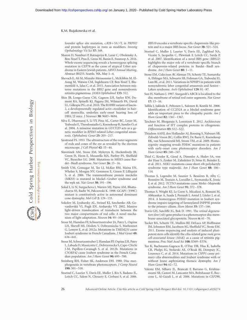
founder splice site mutation, c.828þ3A.T, in PRPH2and protein haplotypes in trans as modifiers. InvestigOpthalmology Vis Sci 57: 349.
Sharon D, Namburi P, Ratnapriya R, Lazar C, Obolensky A,Ben-Yosef T, Pras E, Gross M, Banin E, Swaroop A. 2016.Whole exome sequencing reveals a homozygous splicingmutation in CEP78 as the cause of atypical Usher syn-drome in Eastern Jewish patients. ARVO Annual Meeting,Abstract B0253. Seattle, WA, May 1–6.
Shevach E, Ali M, Mizrahi-Meissonnier L, McKibbin M, El-Asrag M, Watson CM, Inglehearn CF, Ben-Yosef T, Blu-menfeld A, Jalas C, et al. 2015. Association between mis-sense mutations in the BBS2 gene and nonsyndromicretinitis pigmentosa. JAMA Ophthalmol 133: 312.
Shin JB, Longo-Guess CM, Gagnon LH, Saylor KW, Du-mont RA, Spinelli KJ, Pagana JM, Wilmarth PA, DavidLL, Gillespie PG, et al. 2010. The R109H variant of fascin-2, a developmentally regulated actin crosslinker in hair-cell stereocilia, underlies early-onset hearing loss ofDBA/2J mice. J Neurosci 30: 9683–9694.
Silva E, Dharmaraj S, Li YY, Pina AL, Carter RC, Loyer M,Traboulsi E, Theodossiadis G, Koenekoop R, Sundin O, etal. 2004. A missense mutation in GUCY2D acts as a ge-netic modifier in RPE65-related Leber congenital amau-rosis. Ophthalmic Genet 25: 205–217.
Sjostrand FS. 1953. The ultrastructure of the outer segmentsof rods and cones of the eye as revealed by the electronmicroscope. J Cell Physiol 42: 15–44.
Slavotinek AM, Stone EM, Mykytyn K, Heckenlively JR,Green JS, Heon E, Musarella MA, Parfrey PS, SheffieldVC, Biesecker LG. 2000. Mutations in MKKS cause Bar-det–Biedl syndrome. Nat Genet 26: 15–16.
Smith UM, Consugar M, Tee LJ, McKee BM, Maina EN,Whelan S, Morgan NV, Goranson E, Gissen P, LilliquistS, et al. 2006. The transmembrane protein meckelin(MKS3) is mutated in Meckel–Gruber syndrome andthe wpk rat. Nat Genet 38: 191–196.
Sokal I, Li N, Surgucheva I, Warren MJ, Payne AM, Bhatta-charya SS, Baehr W, Palczewski K. 1998. GCAP1 (Y99C)mutant is constitutively active in autosomal dominantcone dystrophy. Mol Cell 2: 129–133.
Sokolov M, Lyubarsky AL, Strissel KJ, Savchenko AB, Go-vardovskii VI, Pugh EN, Arshavsky VY. 2002. Massivelight-driven translocation of transducin between thetwo major compartments of rod cells: A novel mecha-nism of light adaptation. Neuron 34: 95–106.
Srour M, Hamdan FF, Schwartzentruber JA, Patry L, OspinaLH, Shevell MI, Desilets V, Dobrzeniecka S, MathonnetG, Lemyre E, et al. 2012a. Mutations in TMEM231 causeJoubert syndrome in French Canadians. J Med Genet 49:636–641.
Srour M, Schwartzentruber J, Hamdan FF, Ospina LH, PatryL, Labuda D, Massicotte C, Dobrzeniecka S, Capo-ChichiJ-M, Papillon-Cavanagh S, et al. 2012b. Mutations inC5ORF42 cause Joubert syndrome in the French Cana-dian population. Am J Hum Genet 90: 693–700.
Steinberg RH, Fisher SK, Anderson DH. 1980. Disc mor-phogenesis in vertebrate photoreceptors. J Comp Neurol190: 501–518.
Stoetzel C, Laurier V, Davis EE, Muller J, Rix S, Badano JL,Leitch CC, Salem N, Chouery E, Corbani S, et al. 2006.
BBS10 encodes a vertebrate-specific chaperonin-like pro-tein and is a major BBS locus. Nat Genet 38: 521–524.
Stoetzel C, Muller J, Laurier V, Davis EE, Zaghloul NA,Vicaire S, Jacquelin C, Plewniak F, Leitch CC, Sarda P,et al. 2007. Identification of a novel BBS gene (BBS12)highlights the major role of a vertebrate-specific branchof chaperonin-related proteins in Bardet–Biedl syn-drome. Am J Hum Genet 80: 1–11.
Stone EM, Cideciyan AV, Aleman TS, Scheetz TE, SumarokaA, Ehlinger MA, Schwartz SB, Fishman GA, Traboulsi EI,Lam BL, et al. 2011. Variations in NPHP5 in patients withnonsyndromic leber congenital amaurosis and Senior–Løken syndrome. Arch Ophthalmol 129: 81–87.
Sun H, Nathans J. 1997. Stargardt’s ABCR is localized to thedisc membrane of retinal rod outer segments. Nat Genet17: 15–16.
Tallila J, Jakkula E, Peltonen L, Salonen R, Kestila M. 2008.Identification of CC2D2A as a Meckel syndrome geneadds an important piece to the ciliopathy puzzle. Am JHum Genet 82: 1361–1367.
Taschner M, Bhogaraju S, Lorentzen E. 2012. Architectureand function of IFT complex proteins in ciliogenesis.Differentiation 83: S12–S22.
Thiadens AAHJ, den Hollander AI, Roosing S, Nabuurs SB,Zekveld-Vroon RC, Collin RWJ, De Baere E, KoenekoopRK, van Schooneveld MJ, Strom TM, et al. 2009. Homo-zygosity mapping reveals PDE6C mutations in patientswith early-onset cone photoreceptor disorders. Am JHum Genet 85: 240–247.
Thiel C, Kessler K, Giessl A, Dimmler A, Shalev SA, vonder Haar S, Zenker M, Zahnleiter D, Stoss H, Beinder E,et al. 2011. NEK1 mutations cause short-rib polydactylysyndrome type majewski. Am J Hum Genet 88: 106–114.
Thomas S, Legendre M, Saunier S, Bessieres B, Alby C,Bonniere M, Toutain A, Loeuillet L, Szymanska K, JossicF, et al. 2012. TCTN3 mutations cause Mohr–Majewskisyndrome. Am J Hum Genet 91: 372–378.
Thomas S, Wright KJ, Le Corre S, Micalizzi A, Romani M,Abhyankar A, Saada J, Perrault I, Amiel J, Litzler J, et al.2014. A homozygous PDE6D mutation in Joubert syn-drome impairs targeting of farnesylated INPP5E proteinto the primary cilium. Hum Mutat 35: 137–146.
Travis GH, Sutcliffe JG, Bok D. 1991. The retinal degenera-tion slow (rds) gene product is a photoreceptor disc mem-brane-associated glycoprotein. Neuron 6: 61–70.
Tucker BA, Scheetz TE, Mullins RF, DeLuca AP, HoffmannJM, Johnston RM, Jacobson SG, Sheffield VC, Stone EM.2011. Exome sequencing and analysis of induced pluri-potent stem cells identify the cilia-related gene male germcell-associated kinase (MAK) as a cause of retinitis pig-mentosa. Proc Natl Acad Sci 108: E569–E576.
Tuz K, Bachmann-Gagescu R, O’Day DR, Hua K, IsabellaCR, Phelps IG, Stolarski AE, O’Roak BJ, Dempsey JC,Lourenco C, et al. 2014. Mutations in CSPP1 cause pri-mary cilia abnormalities and Joubert syndrome with orwithout Jeune asphyxiating thoracic dystrophy. Am JHum Genet 94: 62–72.
Valente EM, Silhavy JL, Brancati F, Barrano G, Krishna-swami SR, Castori M, Lancaster MA, Boltshauser E, Boc-cone L, Al-Gazali L, et al. 2006. Mutations in CEP290,
K.M. Bujakowska et al.
26 Advanced Online Article. Cite this article as Cold Spring Harb Perspect Biol doi: 10.1101/cshperspect.a028274
on January 1, 2020 - Published by Cold Spring Harbor Laboratory Press http://cshperspectives.cshlp.org/Downloaded from
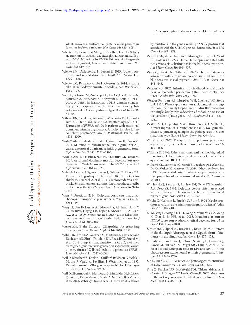
which encodes a centrosomal protein, cause pleiotropicforms of Joubert syndrome. Nat Genet 38: 623–625.
Valente EM, Logan CV, Mougou-Zerelli S, Lee JH, SilhavyJL, Brancati F, Iannicelli M, Travaglini L, Romani S, Illi B,et al. 2010. Mutations in TMEM216 perturb ciliogenesisand cause Joubert, Meckel and related syndromes. NatGenet 42: 619–625.
Valente EM, Dallapiccola B, Bertini E. 2013. Joubert syn-drome and related disorders. Handb Clin Neurol 113:1879–1888.
Valente EM, Rosti RO, Gibbs E, Gleeson JG. 2014. Primarycilia in neurodevelopmental disorders. Nat Rev Neurol10: 27–36.
Verpy E, Leibovici M, Zwaenepoel I, Liu XZ, Gal A, Salem N,Mansour A, Blanchard S, Kobayashi I, Keats BJ, et al.2000. A defect in harmonin, a PDZ domain-contain-ing protein expressed in the inner ear sensory haircells, underlies Usher syndrome type 1C. Nat Genet 26:51–55.
Vithana EN, Safieh LA, Pelosini L, Winchester E, Hornan D,Bird AC, Hunt DM, Bustin SA, Bhattacharya SS. 2003.Expression of PRPF31 mRNA in patients with autosomaldominant retinitis pigmentosa: A molecular clue for in-complete penetrance? Invest Ophthalmol Vis Sci 44:4204–4209.
Wada Y, Abe T, Takeshita T, Sato H, Yanashima K, Tamai M.2001. Mutation of human retinal fascin gene (FSCN2)causes autosomal dominant retinitis pigmentosa. InvestOphthalmol Vis Sci 42: 2395–2400.
Wada Y, Abe T, Itabashi T, Sato H, Kawamura M, Tamai M.2003. Autosomal dominant macular degeneration asso-ciated with 208delG mutation in the FSCN2 gene. ArchOphthalmol 121: 1613–1620.
Walczak-Sztulpa J, Eggenschwiler J, Osborn D, Brown DA,Emma F, Klingenberg C, Hennekam RC, Torre G, Gar-shasbi M, Tzschach A, et al. 2010. Cranioectodermal Dys-plasia, Sensenbrenner syndrome, is a ciliopathy caused bymutations in the IFT122 gene. Am J Hum Genet 86: 949–956.
Wang J, Deretic D. 2014. Molecular complexes that directrhodopsin transport to primary cilia. Prog Retin Eye Res38: 1–19.
Wang H, den Hollander AI, Moayedi Y, Abulimiti A, Li Y,Collin RWJ, Hoyng CB, Lopez I, Abboud EB, Al-RajhiAA, et al. 2009. Mutations in SPATA7 cause Leber con-genital amaurosis and juvenile retinitis pigmentosa. Am JHum Genet 84: 380–387.
Waters AM, Beales PL. 2011. Ciliopathies: An expandingdisease spectrum. Pediatr Nephrol 26: 1039–1056.
Webb TR, Parfitt DA, Gardner JC, Martinez A, Bevilacqua D,Davidson AE, Zito I, Thiselton DL, Ressa JHC, Apergi M,et al. 2012. Deep intronic mutation in OFD1, identifiedby targeted genomic next-generation sequencing, causesa severe form of X-linked retinitis pigmentosa (RP23).Hum Mol Genet 21: 3647–3654.
Weil D, Blanchard S, Kaplan J, Guilford P, Gibson G, Walsh J,Mburu P, Varela A, Levilliers J, Weston M, et al. 1995.Defective myosin VIIA gene responsible for Usher syn-drome type 1B. Nature 374: 60–61.
Weil D, El-Amraoui A, Masmoudi S, Mustapha M, KikkawaY, Laine S, Delmaghani S, Adato A, Nadifi S, Ben Zina Z,et al. 2003. Usher syndrome type I G (USH1G) is caused
by mutations in the gene encoding SANS, a protein thatassociates with the USH1C protein, harmonin. Hum MolGenet 12: 463–471.
Weitz CJ, Miyake Y, Shinzato K, Montag E, Zrenner E, WentLN, Nathans J. 1992a. Human tritanopia associated withtwo amino acid substitutions in the blue-sensitive opsin.Am J Hum Genet 50: 498–507.
Weitz CJ, Went LN, Nathans J. 1992b. Human tritanopiaassociated with a third amino acid substitution in theblue-sensitive visual pigment. Am J Hum Genet 51:444–446.
Weleber RG. 2002. Infantile and childhood retinal blind-ness: A molecular perspective (The Franceschetti Lec-ture). Ophthalmic Genet 23: 71–97.
Weleber RG, Carr RE, Murphey WH, Sheffield VC, StoneEM. 1993. Phenotypic variation including retinitis pig-mentosa, pattern dystrophy, and fundus flavimaculatusin a single family with a deletion of codon 153 or 154 ofthe peripherin/RDS gene. Arch Ophthalmol 111: 1531–1542.
Weston MD, Luijendijk MWJ, Humphrey KD, Moller C,Kimberling WJ. 2004. Mutations in the VLGR1 gene im-plicate G-protein signaling in the pathogenesis of Ushersyndrome type II. Am J Hum Genet 74: 357–366.
Williams DS. 2002. Transport to the photoreceptor outersegment by myosin VIIa and kinesin II. Vision Res 42:455–462.
Williams D. 2008. Usher syndrome: Animal models, retinalfunction of Usher proteins, and prospects for gene ther-apy. Vision Res 48: 433–441.
Williams CL, McIntyre JC, Norris SR, Jenkins PM, Zhang L,Pei Q, Verhey K, Martens JR. 2014. Direct evidence forBBSome-associated intraflagellar transport reveals dis-tinct properties of native mammalian cilia. Nat Commun5: 5813.
Winderickx J, Sanocki E, Lindsey DT, Teller DY, MotulskyAG, Deeb SS. 1992. Defective colour vision associatedwith a missense mutation in the human green visualpigment gene. Nat Genet 1: 251–256.
Wright C, Healicon R, English C, Burn J. 1994. Meckel syn-drome: What are the minimum diagnostic criteria? J MedGenet 31: 482–485.
Xu M, Yang L, Wang F, Li HH, Wang X, Wang W, Ge Z, WangK, Zhao L, Li HH, et al. 2015. Mutations in humanIFT140 cause non-syndromic retinal degeneration. HumGenet 134: 1069–1078.
Yamamoto S, Sippel KC, Berson EL, Dryja TP. 1997. Defectsin the rhodopsin kinase gene in the Oguchi form of sta-tionary night blindness. Nat Genet 15: 175–178.
Yamashita T, Liu J, Gao J, LeNoue S, Wang C, Kaminoh J,Bowne SJ, Sullivan LS, Daiger SP, Zhang K, et al. 2009.Essential and synergistic roles of RP1 and RP1L1 in rodphotoreceptor axoneme and retinitis pigmentosa. J Neu-rosci 29: 9748–9760.
Yan D, Liu XZ. 2010. Genetics and pathological mechanismsof Usher syndrome. J Hum Genet 55: 327–335.
Yang Z, Peachey NS, Moshfeghi DM, Thirumalaichary S,Chorich L, Shugart YY, Fan K, Zhang K. 2002. Mutationsin the RPGR gene cause X-linked cone dystrophy. HumMol Genet 11: 605–611.
Photoreceptor Cilia and Retinal Ciliopathies
Advanced Online Article. Cite this article as Cold Spring Harb Perspect Biol doi: 10.1101/cshperspect.a028274 27
on January 1, 2020 - Published by Cold Spring Harbor Laboratory Press http://cshperspectives.cshlp.org/Downloaded from
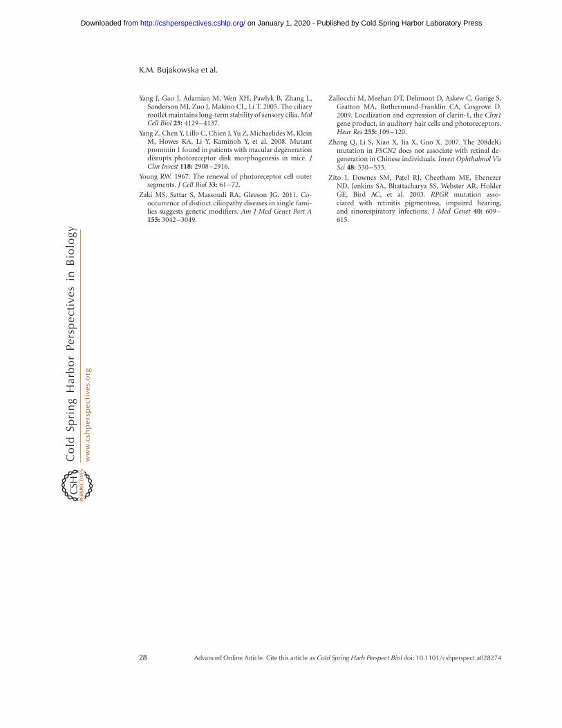
Yang J, Gao J, Adamian M, Wen XH, Pawlyk B, Zhang L,Sanderson MJ, Zuo J, Makino CL, Li T. 2005. The ciliaryrootlet maintains long-term stability of sensory cilia. MolCell Biol 25: 4129–4137.
Yang Z, Chen Y, Lillo C, Chien J, Yu Z, Michaelides M, KleinM, Howes KA, Li Y, Kaminoh Y, et al. 2008. Mutantprominin 1 found in patients with macular degenerationdisrupts photoreceptor disk morphogenesis in mice. JClin Invest 118: 2908–2916.
Young RW. 1967. The renewal of photoreceptor cell outersegments. J Cell Biol 33: 61–72.
Zaki MS, Sattar S, Massoudi RA, Gleeson JG. 2011. Co-occurrence of distinct ciliopathy diseases in single fami-lies suggests genetic modifiers. Am J Med Genet Part A155: 3042–3049.
Zallocchi M, Meehan DT, Delimont D, Askew C, Garige S,Gratton MA, Rothermund-Franklin CA, Cosgrove D.2009. Localization and expression of clarin-1, the Clrn1gene product, in auditory hair cells and photoreceptors.Hear Res 255: 109–120.
Zhang Q, Li S, Xiao X, Jia X, Guo X. 2007. The 208delGmutation in FSCN2 does not associate with retinal de-generation in Chinese individuals. Invest Ophthalmol VisSci 48: 530–533.
Zito I, Downes SM, Patel RJ, Cheetham ME, EbenezerND, Jenkins SA, Bhattacharya SS, Webster AR, HolderGE, Bird AC, et al. 2003. RPGR mutation asso-ciated with retinitis pigmentosa, impaired hearing,and sinorespiratory infections. J Med Genet 40: 609–615.
K.M. Bujakowska et al.
28 Advanced Online Article. Cite this article as Cold Spring Harb Perspect Biol doi: 10.1101/cshperspect.a028274
on January 1, 2020 - Published by Cold Spring Harbor Laboratory Press http://cshperspectives.cshlp.org/Downloaded from
published online March 13, 2017Cold Spring Harb Perspect Biol Kinga M. Bujakowska, Qin Liu and Eric A. Pierce Photoreceptor Cilia and Retinal Ciliopathies
Subject Collection Cilia
Polycystic Kidney DiseaseCiliary Mechanisms of Cyst Formation in
SomloMing Ma, Anna-Rachel Gallagher and Stefan
Right Symmetry Breaking−Cilia in LeftKyosuke Shinohara and Hiroshi Hamada
Photoreceptor Cilia and Retinal CiliopathiesKinga M. Bujakowska, Qin Liu and Eric A. Pierce Ciliopathies
Discovery, Diagnosis, and Etiology of Craniofacial
Elizabeth N. Schock and Samantha A. BrugmannG-Protein-Coupled Receptor Signaling in Cilia
Kirk Mykytyn and Candice AskwithAxoneme Structure from Motile Cilia
Takashi IshikawaEvolution of Cilia
David R. MitchellCilia and Ciliopathies in Congenital Heart Disease
Nikolai T. Klena, Brian C. Gibbs and Cecilia W. Lo
Centriole ElongationCytoplasmic Ciliogenesis and Postaxonemal Transition Zone Migration: A Mechanism for
BasiriTomer Avidor-Reiss, Andrew Ha and Marcus L.
Sperm Sensory Signaling
KauppDagmar Wachten, Jan F. Jikeli and U. Benjamin
Cilia and Obesity
BerbariChristian Vaisse, Jeremy F. Reiter and Nicolas F.
) Signalingβ (TGF-βFactor Tyrosine Kinase (RTK) and Transforming Growth Primary Cilia and Coordination of Receptor
B. Mogensen, et al.Søren T. Christensen, Stine K. Morthorst, Johanne
CiliaPosttranslational Modifications of Tubulin and
et al.Dorota Wloga, Ewa Joachimiak, Panagiota Louka,
Primary Cilia and Mammalian Hedgehog SignalingFiona Bangs and Kathryn V. Anderson
FlagellaRegulation, Assembly, and Evolution of Cilia and
A Snapshot of the Motility−−Radial Spokes
Xiaoyan Zhu, Yi Liu and Pinfen Yang
Cilia and Mucociliary Clearance
OstrowskiXimena M. Bustamante-Marin and Lawrence E.
http://cshperspectives.cshlp.org/cgi/collection/ For additional articles in this collection, see
Copyright © 2017 Cold Spring Harbor Laboratory Press; all rights reserved
on January 1, 2020 - Published by Cold Spring Harbor Laboratory Press http://cshperspectives.cshlp.org/Downloaded from