Embed Size (px)
Citation preview

www.scholarsresearchlibrary.comt Available online a
Scholars Research Library
Central European Journal of Experimental Biology, 2013, 2 (4):7-15
(http://scholarsresearchlibrary.com/archive.html)
ISSN: 2278–7364
7 Scholars Research Library
Photoperiod, melatonin and its importance in fish reproduction
Mukesh Kumar Bairwa*, Neelam Saharan, Kiran Dube Rawat, Jitender Kumar Jakhar and Aritra Bera
Aquaculture division, Central Institute of Fisheries Education (CIFE), Mumbai, Maharashtra, India _____________________________________________________________________________________________
ABSTRACT Light is a key environmental factor that synchronizes all life-stages of fish, from embryo development to sexual maturation. The present review covers information gathered in recent years to emphasize the importance of photoperiod and melatonin in fish physiology. It is well known that melatonin is a much conserved feature in vertebrates that plays a central role in the entrainment of daily and annual physiological rhythms. In this article we have reviewed about the photoperiod and its importance in fish reproduction, pineal gland structure and function, sites of melatonin production in fish, role of AANAT enzyme in melatonin production, endocrine regulation of melatonin production and its role in neuroendocrine regulations of reproduction, growth, feeding behavioural responses and its importance in aquaculture. Keywords: Photoperiod, melatonin, AANAT, Fish reproduction _____________________________________________________________________________________________
INTRODUCTION
Daily behavioural processes in fish, such as locomotor activity, sedation, skin pigmentation, oxygen consumption, thermoregulation, food intake and shoaling behavior, are under the influence of environmental factors. The alternation of light and darkness [the 24-hour light-dark (LD) cycle] has a major role in the synchronization of these daily rhythms; other factors, including temperature, salinity and food availability, might also shape the oscillations. Fish have also adapted to the annual changes of these external cues, so that physiological functions such as growth and reproduction also display annual rhythmicity.A circadian system comprises all the different components by which light enters the organism and is transformed into a timed nervous or hormonal signal. The core of the system is made of a clock machinery, the autonomous activity of which is synchronized to the prevailing 24 h LD cycle by light perceived through specific light sensors; in turn, the clocks drive the production of rhythmic output signals. Photic Cues In photic cues two importantparts are duration and quality of the photic signal [9]. If photoperiod varies along the annual cycle in a regular and predictable manner, light quality in terms of intensity and spectrum is less predictable and will depend on habitats characterized by depth, clarity and structure. Thus, in order to optimize the rearing conditions and eventually being able to manipulate fish physiology one has to consider the respective durations of day and night and, in both situations, light intensity, spectral composition and orientation [47, 51]. It is important to emphasize that fish perceive light both from above (via the pineal organ), the sides (via the eyes) and possibly through deep brain photoreceptors. All these light centres may have different sensitivities. Fish are able to visually discriminate colours although species specific sensitivities exist probably as a result to adaptation of the visual and non-visual systems to specific natural habitats [14, 56]. Light of different spectral
Mukesh Kumar Bairwa et al_____________________________________________________________________________
composition cantherefore affect fish growth and survival as shown in different species stress response [87], behaviour [44] However, some species can grow better and develop quicker at low light intensities, lx [15], juvenile halibut at 1-10 lx [36]reported to show improved growth at very intense light levels, [3,59], Atlantic cod larvae at 2400 lx light intensity on growth and survival are species Pineal Gland In most species investigated, the pineal organ appears as a vesicle attached to the roof of the diencephaloslender stalk; it is usually located below a window in the skull through which light enters. The vesicle is made of a pseudo-stratified epithelium that is opened to the cerebrospinal fluid (CSF); folliculated (as in birds) as well as compact (as in mammals) glands have also been described photoreceptor cells and of ependymal interstitial cells that contact the CSF in their most apical part photoreceptor cells establish synaptic contacts pineal organ of non-mammalian vertebrates thus resembles very much to a simplified retina, and the structural and functional analogies between the two organs have been exshare more than structural analogies with the retinal conesthey have a similar composition in lipids and proteins of the phototransduction cascade (opsins, tracyclic nucleotide gated channel). And, their electrical response to light stimuli is similar: light induces a dosedependent cell hyperpolarization that results in the inhibition of an excitatory neurotransmitter (aspartate or glutamate). In the pineal organ, the excitatory neurotransmitter reaches directly the ganglion cells, which send their axons to the brain. Thus, the signals that are conveyed to the brain reflect mainly the response of the photoreceptor cells, i.e., the pineal organ is a luminance detector that provides information on lightduration of day-length. It is interesting that the pineal and retinal ganglion cells may target similar brain areas, particularly in the thalamus and pretectumphotoreceptors both produce melatonin at night, following cell depolarization Brain Pituitary Gonadal (BPG) Axis The neuroendocrine system initiates and controls the procethrough the activation of the hypothalamuscontrolled by environmental and the hypothalamus which, in turn stimulates the release of gonadotropin releasing hormonestimulates the release of gonadotropins (GTH) from the of other vertebrates, have been shown to secrete two kinds of gonadotropins; GTHStimulating Hormone; FSH) and GTHgonadal growth and gametogenesis while GTH
Fig: 1 Pineal Gland (A) Arrow mark showing the position on pineal gland in fish brain (B) Arrow mark showing the cross sectiogland in teleost fish and (C) Showing the different parts of pineal gland
et al Cent. Euro. J. Exp. Bio.,_____________________________________________________________________________
Scholars Research Library
composition cantherefore affect fish growth and survival as shown in different species [56][44] and reproduction [52].
However, some species can grow better and develop quicker at low light intensities, such as striped bass larvae at 1 [36] and juvenile haddock at 30 lx [82]. On the other hand,
reported to show improved growth at very intense light levels, European sea bass larvae at 1400larvae at 2400 lx [63], and black porgy juveniles at 3000 lx [72]. It seems that the effect of
survival are species-specific.
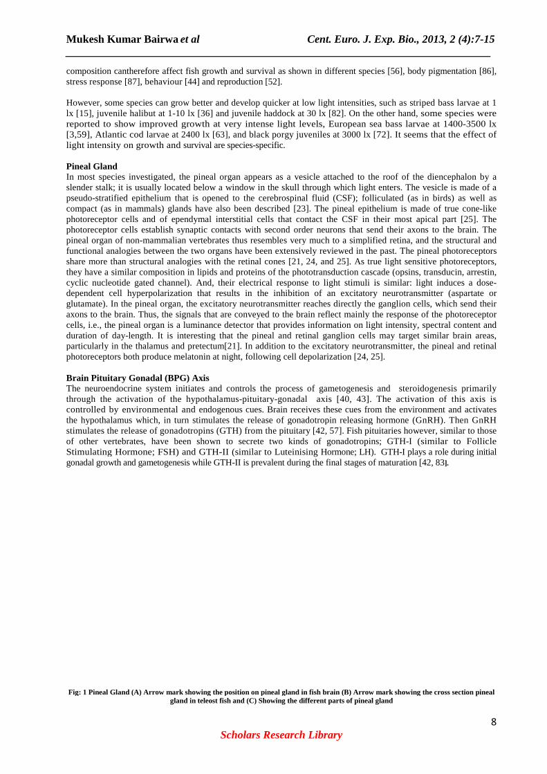
In most species investigated, the pineal organ appears as a vesicle attached to the roof of the diencephaloslender stalk; it is usually located below a window in the skull through which light enters. The vesicle is made of a
stratified epithelium that is opened to the cerebrospinal fluid (CSF); folliculated (as in birds) as well as mammals) glands have also been described [23]. The pineal epithelium is made of true cone
photoreceptor cells and of ependymal interstitial cells that contact the CSF in their most apical part photoreceptor cells establish synaptic contacts with second order neurons that send their axons to the brain. The
mammalian vertebrates thus resembles very much to a simplified retina, and the structural and functional analogies between the two organs have been extensively reviewed in the past. The pineal photoreceptors share more than structural analogies with the retinal cones [21, 24, and 25]. As true light sensitive photoreceptors, they have a similar composition in lipids and proteins of the phototransduction cascade (opsins, tracyclic nucleotide gated channel). And, their electrical response to light stimuli is similar: light induces a dosedependent cell hyperpolarization that results in the inhibition of an excitatory neurotransmitter (aspartate or
. In the pineal organ, the excitatory neurotransmitter reaches directly the ganglion cells, which send their axons to the brain. Thus, the signals that are conveyed to the brain reflect mainly the response of the photoreceptor
is a luminance detector that provides information on light intensity, spectral content and length. It is interesting that the pineal and retinal ganglion cells may target similar brain areas,
particularly in the thalamus and pretectum[21]. In addition to the excitatory neurotransmitter, the pineal and retinal photoreceptors both produce melatonin at night, following cell depolarization [24, 25].
Axis The neuroendocrine system initiates and controls the process of gametogenesis and through the activation of the hypothalamus-pituitary-gonadal axis [40, 43]. The activation of this axis is controlled by environmental and endogenous cues. Brain receives these cues from the environment and the hypothalamus which, in turn stimulates the release of gonadotropin releasing hormonestimulates the release of gonadotropins (GTH) from the pituitary [42, 57]. Fish pituitaries however, similar to those of other vertebrates, have been shown to secrete two kinds of gonadotropins; GTHStimulating Hormone; FSH) and GTH-II (similar to Luteinising Hormone; LH). GTHgonadal growth and gametogenesis while GTH-II is prevalent during the final stages of maturation [42, 83
Fig: 1 Pineal Gland (A) Arrow mark showing the position on pineal gland in fish brain (B) Arrow mark showing the cross sectio
in teleost fish and (C) Showing the different parts of pineal gland
Cent. Euro. J. Exp. Bio., 2013, 2 (4):7-15 _____________________________________________________________________________
8
[56], body pigmentation [86],
such as striped bass larvae at 1 . On the other hand, some species were
European sea bass larvae at 1400-3500 lx . It seems that the effect of
In most species investigated, the pineal organ appears as a vesicle attached to the roof of the diencephalon by a slender stalk; it is usually located below a window in the skull through which light enters. The vesicle is made of a
stratified epithelium that is opened to the cerebrospinal fluid (CSF); folliculated (as in birds) as well as . The pineal epithelium is made of true cone-like
photoreceptor cells and of ependymal interstitial cells that contact the CSF in their most apical part [25]. The with second order neurons that send their axons to the brain. The
mammalian vertebrates thus resembles very much to a simplified retina, and the structural and . The pineal photoreceptors
. As true light sensitive photoreceptors, they have a similar composition in lipids and proteins of the phototransduction cascade (opsins, transducin, arrestin, cyclic nucleotide gated channel). And, their electrical response to light stimuli is similar: light induces a dose-dependent cell hyperpolarization that results in the inhibition of an excitatory neurotransmitter (aspartate or
. In the pineal organ, the excitatory neurotransmitter reaches directly the ganglion cells, which send their axons to the brain. Thus, the signals that are conveyed to the brain reflect mainly the response of the photoreceptor
intensity, spectral content and length. It is interesting that the pineal and retinal ganglion cells may target similar brain areas,
In addition to the excitatory neurotransmitter, the pineal and retinal
steroidogenesis primarily axis [40, 43]. The activation of this axis is
endogenous cues. Brain receives these cues from the environment and activates the hypothalamus which, in turn stimulates the release of gonadotropin releasing hormone (GnRH). Then GnRH
7]. Fish pituitaries however, similar to those of other vertebrates, have been shown to secrete two kinds of gonadotropins; GTH-I (similar to Follicle
Hormone; LH). GTH-I plays a role during initial II is prevalent during the final stages of maturation [42, 83].
Fig: 1 Pineal Gland (A) Arrow mark showing the position on pineal gland in fish brain (B) Arrow mark showing the cross section pineal

Mukesh Kumar Bairwa et al Cent. Euro. J. Exp. Bio., 2013, 2 (4):7-15 _____________________________________________________________________________
9 Scholars Research Library
In ovaries, the gonadotropins stimulate the theca and granulosa cells of the ovarian follicle to secrete steroids including androgens (e.g. testosterone), oestrogens (e.g. 17 β-oestradiol) and progestagens(e.g.17α-20 β-dihydroprogesterone) [13]. During the early oocyte growth and development phases, FSH stimulates the follicular cells of the oocyte to produce testosterone (T) which is then converted into 17β-oestradiol(E2) in the granulosa cells via the activity of the aromatase enzyme [66]. Increased E2 levels in the blood stimulate the hepatic system to synthetisevitellogenin (VTG). Estrogen(E2) peaks generally, during the period of most active vitellogenesis,and returns to basal levels before ovulation. Testosterone (T) starts to increase in conjunction with E2, but peak levels are not attained until 1-2 months after E2[8, 32]. VTG is a large glycophospho-lipoprotein produced by the liver and transported via the circulatory system to the ovarian follicle, processed into yolk proteins then accumulated as yolk globules in the growing oocytes. Once vitellogenesis is complete, plasma E2 levels fall rapidly and a negative feedback on the hypothalamus and pituitary triggers the release of LH [42, 52]. LH stimulates the final oocyte maturation through the secretion of progestagens (17α-20βDHP or 17,20-20β, 21-P), acting as the maturation inducing hormone (MIH) [57].This hormone then binds to the oocyte nucleus and forms the maturation promoting factors (MPF) which stimulates the germinal vesicle migration (GVM) towards the micropyle. The germinal vesicle then breaks down (GVBD) prior to the hydration stage and subsequent ovulation [42, 74]. In testes, the two GTH hormones play different functional roles. FSH levels are elevated during spermatogenesis while LH levels peak during spermiation[66]. Both gonadotropins have been reported to have two types of receptors, onein the Leydig cells which is specific to LH, and the other one located in Leydig and Sertolicells with affinities to both FSH and LH [73]. In fish every single primary spermatogonium is enveloped by one or two Sertoli cells which support germ cells by providing the optimal microenvironment [57]. Leydig cells are steroidogenic, e.g. responsible for the production of steroids. FSH acts by regulating Sertoli cell functions to stimulate germ cells growth during spermatogenesis [42]. The LH receptor is then expressed by Leydig cells, which respond to LH and release androgens (testosterone and 11-ketotestosterone) with T stimulating pituitary LH synthesis during puberty [37, 57]. Peak levels of 11-KT coincide with spermiation[84]. In male fish, 11-ketotestosterone (11-KT) is considered to be the main androgen hormone, stimulating the development of secondary sexual characters and spermatogenesis. Seasonal cycles of gonadal activity have been described in many teleosts species. Although the BPG axis has been well described in fish over the last few decades, the cascade from environmental cue to brain gonadotropin stimulation resulting in the timing and regulation of gonadal development remained unknown. Recently, a new peptide, Kisspeptin, has been identified as a key actor in the initiation of puberty and regulation of seasonal breeding in mammals [68]. It has been proposed that Kisspeptin actions are mediated by melatonin signalling by directly regulating KiSS-1 expression as well as changing sensitivity of KiSS-1 to sex steroid feedback [35]. The study of kisspeptin in fish is still in its early stages, though it is becoming a very active research field. Photoperiod and reproduction Fish reproductive physiology shows an extraordinary close adaptation to the cyclical variations of the environment; fish synchronize their spawning to the period of the year most favourable for the survival of progeny. Accordingly, fish have developed predictive mechanisms using photoperiod as a reliable environmental cue (proximal factor) to anticipate and activate gametogenesis long before spawning [9]. The period of reproduction of most temperate commercial fish species important for aquaculture is restricted to only a few months of the year, when the most appropriate environmental conditions are found; this guarantees the best chances of progeny survival. However, the restriction of reproductive activity to a short annual window is a problem for fish farmers that rely on yearlong supply of juveniles to satisfy an increasing demand in fish. Early maturation during on growing is another major bottleneck leading to losses due to deterioration of flesh quality, external appearance and poor growth performances. Consequently, based on the basic understanding of the circadian axis and photoperiodic entrainment of reproduction, regimes have been developed with great success to manipulate the natural circannual rhythm of spawning in many temperate fish species. Importantly, photoperiod could also be used to improve reproductive performances in tropical fish species although these species do not experience significant annual photoperiodic changes in their natural habitat [12]. Compression or extension of the seasonal light cycle results in spawning advance or delay, respectively, in salmonid species [9]. Similarly, the time of first sexual maturation can also be modified by photoperiod manipulations in teleosts. In salmonids, it is now evident that the decreasing and increasing components of the seasonally changing day-length are responsible for the recruitment of fish into gonadogenesis and reproduction, respectively [23]. Melatonin, a time-keeping molecule in fish Melatonin is a common output signal of the vertebrate circadian clock which is produced primarily by the pineal

Mukesh Kumar Bairwa et al Cent. Euro. J. Exp. Bio., 2013, 2 (4):7-15 _____________________________________________________________________________
10 Scholars Research Library
organ and released into the blood stream [24]. Melatonin is also synthesized in the retina and has been detected in the gastrointestinal tissues [6]. There is evidence indicating that pineal melatonin contributes largely to the circulating plasma and cerebrospinal fluid (CSF) levels [81], whereas ocular melatonin serves local functions in an autocrine/paracrine mode. The role of melatonin is still not elucidated in fish, it can act on reproduction in seasonal breeders, be involved in the regulation of circadian rhythms (including locomotor activity, body temperature and feeding) amongst other suggested roles [60,91]. However, crucially, direct evidences of melatonin actions are still lacking in fish [24, 48] as opposed to higher vertebrates (mainly mammals) where clear links between reproduction and melatonin have been reported [1]. Retinal melatonin acts primarily within the eye, where it is involved in the control of rhythmic processes, such as retinomotor movements, dopamine synthesis, release and metabolism, rod outer segment discs shedding and phagocytosis[91]. In mammals, however, the pineal gland is the only organ capable of synthesizing melatonin, and hence its removal completely abolishes the melatonin rhythm. In fish, the retinacan also synthesize melatonin, which could explain why pinealectomy diminishes, but does not abolish, the daily rhythm of melatonin in the blood in some species, but these melatonin levels are not significant and will not effect blood stream concentration [9]. The retinal melatonin production profile of 32 teleosts has been studies and classified into three types; First is normal profiles, which is parallel with the pineal releasing the melatonin hormone during the dark phase (24 out of 32 species); Second is reversed profile with higher levels of melatonin during the light phase (Four species; common mummichog, European sea bass, Nile tilapia and torafugu); In Third type profile no significant differences in daily melatonin profile (Four species; Japanese eel, Japanese sea perch, Japanese amberjack, and Kusafugu). These differential patterns of retinal melatonin production can be explained by differences in the molecular machinery responsible within photoreceptor cells for melatonin production and regulation [24] In almost all species studied to date, pineal melatonin levels have been shown to increase during the night and remain low during the day, thus reflecting the prevailing photoperiod throughout the seasons [9, 24]).Although the pattern of production is conserved across all vertebrates (high at night/low during the day), three variants have been identified. The most common profile, C-type, is characterised by a rapid rise in melatonin immediately following the onset of the dark period (within the first hour of darkness) and is commonly found in migratory salmonids and other teleosts as well as higher vertebrates such as domestic cat, Djungarianhamster and sheep [65, 67]. A-type profiles are characterised by a delay after the start of the dark phase before melatonin rises to its peak towards the endof the dark phase. This was observed in gadoid species such as Atlantic cod and haddock as well as mouse and Syrian hamster [17, 62, 67]. Finally, B-type profiles are characterised by a discrete peak in the middle of the dark phase as observed in Mozambique tilapia and human [53 67]. The meanings of these different profile types are not understood but might be linked to an ability or inability to anticipate photic signals under the control of circadian clocks [67]. The production of melatonin by the pineal gland in a given fish species might also be correlated to the amount and spectral quality of the light reaching the pineal photoreceptors depending on the degree of absorbance of the skin and skull. The amount and quality of light that crosses the pineal window varies from one species to another [34, 51]. Differences in light penetration through the skull range from 1 to 8% of simulated daylight in Atlantic salmon and sea bass and long wavelengths (650-700 nm) are far more effective at penetrating the skull than shorter wavelengths (400-450 nm) [51]. In terms of melatonin production the threshold of light intensity above which melatonin is suppressed depends on the species, experimental conditions, light quality and duration [51, 58]. All these findings have led researchers to suggest that a threshold value of light intensity and water quality (such as salinity) must exist in order to influence physiological functions in fish. Recently it has been that the light intensity threshold for Atlantic salmon to be around 0.016 W.m-2, after allowing for the 2.4% of light lost during transmission through the cranium. Importantly, large species specific differences were observed with cod, for example, being up to 10,000 times more sensitive than salmon [51]. Melatonin Biosynthesis The biosynthesis of melatonin and other indoles occurs in photoreceptor cells. The general mechanism of biosynthesis of melatonin in fish appears to be identical to that in mammals [10]. The pineal gland takes up tryptophan selectively from the blood [25] and utilizes the amino acid in the synthesis of melatonin and other indoles. The first step is the conversion of tryptohpan to 5-hydroxytryptophan, by tryptophan hydroxylase (TPOH) enzyme. 5-hydroxytryptophan is decarboxylated by the aromatic amino acid decarboxylase to produce serotonin or 5-hydroxytryptamine, which in the pineal gland may undergo different metabolic pathways: (a) oxidative deamination by monoamine oxidase (MAO-A and MAOB) to produce 5-hydroxyindole acetaldehyde, which in turn may be converted to 5-hydroxyindole acetic acid or 5- ydroxytryptophol; and (b) N-acetylation by Nacetyltransferase (NAT) to produce N-acetylserotonin, which in turn is methylated by hydroxyindole-O-methyl

Mukesh Kumar Bairwa et al_____________________________________________________________________________
transferase (HIOMT) to produce Nbeen detected in the pineal photoreceptor cells in pike. The HIOMTcone-like photoreceptors in the fish [27]
Regulation of melatonin productionLight acts on photoreceptor cells of the pineal organ and retina, which allows synchronization of their internal molecular clocks. Light might also impact on other possible photosensitive and circadian structures in the ventral diencephalon (POA and hypothalamic area) and peripheral organs. In response to the photoperiodic information, the retina and the pineal organ elaborate two types of rhythmic information. The neural information (blue arrows) from the retina and pineal organ reach the ventral diencepharespectively. This information provides an indication of day length, as well as of subtle variations in ambient illumination. The hormonal information is season. In the retina, melatonin is an autocrine and/or paracrine factor, which is metabolized locally. Pineal melatonin is released into the cerebrospinal fluid and blood, and acts on specific targIn the hypothalamus, melatonin might contribute to synchronizing the activities of circadian oscillatory units [SCN and others and modulating the production of pituitary gland releasing factors. Melatonin receptors have been identified in areas that impact on pituitary function, including the POA, which also receives nervous input from both the pineal organ and the retina. Melatonin impacts on the pituitary gland itself to modulate the production of hormones. All teleost species have been shown to the day/night cycle [7] although photoFurthermore, both in vitro [51] and in vivo with the irradiance of the incident light. It is thought that plasma melatonin may define the response of biological functions in fish to electrophysiological studies have described the luminance and chromatic response of the pineal gland better knowledge of such light requirements for melatonin regulation is needed especially regarding the quality of the light, as the aquaticenvironment acts as a potent filter significantly modifying spectrum and intensity. Other factors may affect melatonin production, studies have shown that temperature modulate melatonin secretion, through the regulation of correlation between the peak of AANAT2 response and the fish optimal physiological temperature (trout: pike: 20°C, sea bream: 27°C, zebrafish: 30°C), (ii) the response to temperature is an itself, because the same response curves were obtained when activities were measured from cultured pineal organ homogenates or recombinant AANAT2 enzymes. In the pike, temperature had no effect on the phase and period of the circadian rhythm). In vitro, it has been melatonin rhythm was expressed at 19°C up toin the white sucker, pineal gland by
et al Cent. Euro. J. Exp. Bio.,_____________________________________________________________________________
Scholars Research Library
transferase (HIOMT) to produce N-acetyl-5- methoxytryptamine or melatonin. Monoamine oxidase activity has been detected in the pineal photoreceptor cells in pike. The HIOMT-like immunoreactivity was associated with the
the fish [27].
Fig: 2 Melatonin biosynthesis process
egulation of melatonin production Light acts on photoreceptor cells of the pineal organ and retina, which allows synchronization of their internal molecular clocks. Light might also impact on other possible photosensitive and circadian structures in the ventral
alamic area) and peripheral organs. In response to the photoperiodic information, the retina and the pineal organ elaborate two types of rhythmic information. The neural information (blue arrows) from the retina and pineal organ reach the ventral diencephalon through the retinohypothalamic and the pineal tracts, respectively. This information provides an indication of day length, as well as of subtle variations in ambient illumination. The hormonal information is relayed by melatonin the production of whichseason. In the retina, melatonin is an autocrine and/or paracrine factor, which is metabolized locally. Pineal melatonin is released into the cerebrospinal fluid and blood, and acts on specific targets through melatonin In the hypothalamus, melatonin might contribute to synchronizing the activities of circadian oscillatory units [SCN and others and modulating the production of pituitary gland releasing factors. Melatonin receptors have been
that impact on pituitary function, including the POA, which also receives nervous input from both the pineal organ and the retina. Melatonin impacts on the pituitary gland itself to modulate the production of
All teleost species have been shown to have photosensitive pineal glands which produce melatonin according although photo-responsiveness has recently been questioned in Mozambique tilapia
and in vivo [58] studies have demonstrated that melatonin synthesis varies inversely with the irradiance of the incident light. It is thought that plasma melatonin may have threshold levels, which define the response of biological functions in fish to environmental influences electrophysiological studies have described the luminance and chromatic response of the pineal gland better knowledge of such light requirements for melatonin regulation is needed especially regarding the quality of
environment acts as a potent filter significantly modifying spectrum and intensity.
Other factors may affect melatonin production, studies have shown that temperature acts directly on the pineal organ to on, through the regulation of AANAT2 activity [5, 25, 27] Interestingly, (i) there is a good
the peak of AANAT2 response and the fish optimal physiological temperature (trout: pike: 20°C, sea bream: 27°C, zebrafish: 30°C), (ii) the response to temperature is an intrinsic property of the enzyme itself, because the same response curves were obtained when activities were measured from cultured pineal organ
AANAT2 enzymes. In the pike, temperature had no effect on the phase and period of it has been demonstrated in pikepineal glands cultured at different temperatures, that a
melatonin rhythm was expressed at 19°C up to 30°C but not at 10°C or 15°C[27]. A similar effect was also reported sucker, pineal gland by [90]. Closer examination in relation to acclimation temperature prior to
Cent. Euro. J. Exp. Bio., 2013, 2 (4):7-15 _____________________________________________________________________________
11
methoxytryptamine or melatonin. Monoamine oxidase activity has like immunoreactivity was associated with the
Light acts on photoreceptor cells of the pineal organ and retina, which allows synchronization of their internal molecular clocks. Light might also impact on other possible photosensitive and circadian structures in the ventral
alamic area) and peripheral organs. In response to the photoperiodic information, the retina and the pineal organ elaborate two types of rhythmic information. The neural information (blue arrows) from
lon through the retinohypothalamic and the pineal tracts, respectively. This information provides an indication of day length, as well as of subtle variations in ambient
melatonin the production of which reflects day length and season. In the retina, melatonin is an autocrine and/or paracrine factor, which is metabolized locally. Pineal
ets through melatonin receptors. In the hypothalamus, melatonin might contribute to synchronizing the activities of circadian oscillatory units [SCN and others and modulating the production of pituitary gland releasing factors. Melatonin receptors have been
that impact on pituitary function, including the POA, which also receives nervous input from both the pineal organ and the retina. Melatonin impacts on the pituitary gland itself to modulate the production of
produce melatonin according recently been questioned in Mozambique tilapia [53].
studies have demonstrated that melatonin synthesis varies inversely have threshold levels, which nces [9, 51]. Although some
electrophysiological studies have described the luminance and chromatic response of the pineal gland [11, 21], better knowledge of such light requirements for melatonin regulation is needed especially regarding the quality of
environment acts as a potent filter significantly modifying spectrum and intensity.
acts directly on the pineal organ to Interestingly, (i) there is a good
the peak of AANAT2 response and the fish optimal physiological temperature (trout: 12°C, intrinsic property of the enzyme
itself, because the same response curves were obtained when activities were measured from cultured pineal organ AANAT2 enzymes. In the pike, temperature had no effect on the phase and period of
pineal glands cultured at different temperatures, that a . A similar effect was also reported acclimation temperature prior to

Mukesh Kumar Bairwa et al Cent. Euro. J. Exp. Bio., 2013, 2 (4):7-15 _____________________________________________________________________________
12 Scholars Research Library
experimentation showed melatonin release was lower from pineals incubated at 20°C when the fish had been previously acclimated to 10°C. Thus, the concurrent action of photoperiod, that determines the duration of the melatonin signal, and of temperature, that determines its amplitude, is thought to provide accurate definitions of both the daily and annual cycles. Any changes in temperature, related to husbandry conditions or global warming, may thus have dramatic consequences on the time-keeping system of fish. Feeding was shown to synchronize patterns of behaviour (i.e. locomotor) and physiology in different fish species [2, 72] However, importantly, in mammals, light and feed entrainable oscillators are suggested to be independent [49]. To our knowledge, in fish, no evidence of feeding entrainment affecting the melatonin synthesis rhythm system has yet been found. Arylalkylamine N-Acetyltransferase (AANAT): Melaton in Rate-Limiting Enzyme In order to better understand the melatonin synthesis pathway, activities of HIOMT and AANAT enzymes were investigated in different species. An increase in melatonin production at night reflects increased AANAT activity and termination of melatonin production during the day reflects proteasomal degradation of the enzyme [21]. Therefore the AANAT is commonly known to be the melatonin ratelimiting enzyme of the melatonin biosynthetic pathway [41, 78]. On the other hand, HIOMT enzyme synthesis does not exhibit any significant rhythmic changes throughout the 24-hr period [41, 70]suggested that HIOMT might be implicated in seasonal, rather than daily, oscillations in melatonin production. Different types of AANAT have been found in mammalian and non mammalian species. Only one type is present in mammals, bird and anurans, called AANAT, while there are at least three homologenous genes in teleosts localized between the retina (AANAT-1a and AANAT1b) and the pineal (AANAT2) [16]. Cyclic adenosine monophosphate (cAMP) is a second messenger that is important in many biological processes. cAMP is derived from adenosine triphosphate (ATP) and used for intracellular signal transduction in many different organisms. cAMP is an important signal carrier that is necessary for the proper biological response of cells to hormones and other extracellular signals [41]. It is required for cell communication in the hypothalamus/pituitary gland axis and for the feedback control of hormones. In melatonin synthesis, cAMP plays a central role in regulating through effects on AANAT activity through the pineal organs which accumulate cAMP (only in the presence of forskolin enzyme) during the dark phase [16]. Light exposure during the scotophase suppresses AANAT activity and melatonin synthesis. However, unexpected periods of darkness during the light phase do not necessarily induce a rise in AANAT activity [25]. The effects of light,however, depend on the time of application at night [29]. Light pulses given before mid-scotophase of a 24 hr LD cycle induce a rise in AANAT activity later than expected following the scotophase in rat [38]. The rise in AANAT activity is advanced when the light pulse is applied after the mid scotophase. In addition, melatonin synthesis responds differently to variations in light intensity and temperature [9, 62]. Melatonin levels are directly correlated with light intensity [34, 51] with increased light intensity reducing levels of plasma melatonin. Plasma melatonin synthesis was shown todecrease proportionally with increasing light intensities in juvenile Atlantic salmon. Even with dark phase levels as low as 20 lux, significant differences were found between light and dark phase melatonin levels [62]. On the other handtemperature was shown to directly affect melatoninsecretion in ectothermic species [27]. In juvenile Atlantic salmon, melatonin levels were higher in groups maintained at 12◦C compared with groups maintained at 4◦C in [62].Similar results were found in pike and rainbow trout with greater amplitude of secretion at increased temperatures[65]. However, the mechanism behind temperature-dependent melatonin production is thought to occur at an early stage in its biosynthetic pathway. It was shown that the forskolin enzyme, which induced cAMP formation, then regulates N-acetyltransferase(AANAT) production which in turn controls the synthesis of melatonin, is temperaturedependent [80, 89]. Melatonin and Reproduction The importance of melatonin as the most critical candidate involved in the mediation of photic effects on piscine reproduction has been emphasized in several studies. Melatonin is important in controlling the reproductive seasonality by stimulating the final stages of sexual maturation and by synchronizing the oocyte maturity with optimal timing of spawning [61]. Melatonin has also been found to affect estradiol levels in mature carp females [59] and to indirectly influence the GtH II secretion via hypothalamic stimulatory (GnRH) centers[61]. In Atlantic croaker, Micropogonias undulates, melatonin influences LH secretion directly at the pituitary level and indirectly at the brain level [61]. Melatonin has been considered as one of the candidates that mediate the transduction of photoperiodic information to the brainpituitary-gonad axis in gonadal maturation of precocious male masu salmon.. But the mechanism by which melatonin performs this function remains speculative. Many studies have studied the role of melatonin (plasma levels or receptors) in reproduction. Such studies involved

Mukesh Kumar Bairwa et al Cent. Euro. J. Exp. Bio., 2013, 2 (4):7-15 _____________________________________________________________________________
13 Scholars Research Library
the removal of the source(s) of melatonin through ophthalmectomy (EX) and/or pinealectomy (PNX) and/or administration of exogenous melatonin [48]. The pinealectomy (PNX) in male Atlantic salmon parr abolished the nocturnal rise in melatonin but failed to alter the timing of early maturation [46]. Similarly, it failed to inhibit daily gonadotropin cycling in common carp [61]. In contrast, in the female rainbow trout, PNX performed during the summer resulted in a delay in spawning relative to controls suggesting that in this case the pineal was necessary for the entrainment of the final reproductive stages which, in part, is normally brought about by the decreasing daylengths and subsequently increased daily duration of melatonin secretion [9]. Following PNX and EX, Ayu still became sexually mature under SD photoperiods while under LD did not [45]. Clearly these findings draw parallels with the avian model and suggest that, at least in these species, some form of deep brain photoreceptors are responsible for photic entrainment of reproduction which needs to be more thoroughly investigated. The response to direct melatonin administration has been variable though evidence for its involvement in reproduction as part of a complex regulatory network is becoming clearer. The stronger evidence was recently found by [75] who showed that melatonin can stimulate the dopaminergic system of the preoptic area, which is involved in the inhibitory control of gonadotropin (LH and FSH) synthesis and release. Earlier studies have shown that injection of both male and female goldfish with melatonin can reduce gonadal development induced by long-day conditions known to cause gonadal stimulation [31].Intraperitoneal injections of melatonin in mature Atlantic croaker during the late photophase elicited significant elevations in plasma GtH II (LH) levels. In contrast, injection of rainbow trout female with melatonin failed to impact on their reproductive seasonality [65] thus perhaps the effect of PNX on reproduction in this species, as described above, was via a non melatoninergic pathway. Thus it must be concluded that melatonin can influence the BPG axis, owever, its functions are apparently dose dependent and can clearly operate at different levels within the cascade.
CONCLUSION
It is widely accepted that melatonin is a much conserved feature that plays a central role in the entrainment of daily and annual physiological rhythms in vertebrates. Over the last decades, a large amount of research has been carried out in fish, particularly teleost; to unravel the puzzling roles melatonin plays and characterizes inter-species differences. As a result, the picture is becoming much clearer as regards the control of the melatonin synthesis (limiting enzyme AANAT, connection/projection, etc.) and the identification of target tissues, neurons and cells (melatonin receptor expression and localization). However, investigations on the relationships between the pineal organ and melatonin on the one hand, and the neuroendocrine system on theother hand are just at their beginnings, and only few direct evidences exists on how melatonin acts on the brain-pituitary-gonadal axis. Nevertheless, it seems more and more evident that the time-keeping hormone impacts directly or indirectly on the production of brain and pituitary hormones, thus affecting time regulated functions, including feeding, growth, reproduction and immunity.More studies are necessary to unravel the regulatory processes activated by melatonin on a daily and annual basis.
REFERENCES
[1] J. Arendt,Reviews of Reproductio,1998, 3:13-22. [2] M Azzaydi, V.C Rubio, Martinez F.J Lopez, F.J. Sanchez-Vasquez, S. Zamora, J.A. Madrid, Chronobiology International 2007, 24, 859-874. [3] M.H. Barahona-Fernandes, Aquaculture1979, 17, 311-321. [4] T.D. Beacham, C.B. Murray, Aquaculture, 1993,109 (3-4) 315-325. [5] A. Benyassi, C. Schwartz, S.L. Coon, D.C. Klein, J. Falcon, Neuroreport, 2000, 11, 255-258. [6] L. Besseau, A. Benyassi, M. Moller, S. L. Coon, J. L. Weller, G.Boeuf, D. C. Klein, J. Falcon, Experimental Eye Research,2006, 82, 620-627. [7] V. Bolliet, M.A. Ali, F.J Lapointe, J. Falcon,Journal of Comparative Physiology1996: Part B165, 677-683. [8] B. Borg, , P. Ekstrom, V. Veen, ActaZoologica(Stockholm),1983, 64, 211-218. [9] N.R Bromage, M. Porter, C. Randall, Aquaculture,2001, 197, 63- 98. [10] N.R. Bromage, J.A. Elliott, J.R.C. Springate, C. Whitehead, Aquaculture,1984, 43, 213-223. [11] G.M. Cahill, Brain Research, 1996, 708, 177-181. [12] A.Campos-Mendoza, J.A. McAndrew, K. Coward, N. Bromage, Aquaculture, 2004,231, 299-313. [13] J.E.B. Cavaco, J.G.D. Lambert, R.W. Schulz, H.J.T.hGoos, Fish Physiology and Biochemistry, 1997, 16, 129-138. [14] C.L. Cheng, I.N. Flamarique, Nature, 2004, 428, 279:291 [15] E.J. Chesney,Marine Ecology Progress Series, 1989, 3,191-200. [16] S.L. Coon, D.C. Klein,Molecular and Cellular Endocrinology, 2006, 252, 2-10. [17] A. Davie, M. Porter. N. Bromage, H. Migaud,Canadian Journal of Fisheries and Aquatic Sciences, 2007a, 64(1), 98-112.

Mukesh Kumar Bairwa et al Cent. Euro. J. Exp. Bio., 2013, 2 (4):7-15 _____________________________________________________________________________
14 Scholars Research Library
[18] A. Davie, M. Minghetti, H. Migaud, Chronobiology International, 2009, 26, 379-395. [19] J. Duston, N. BromageFish Physiology and Biochemistry, 1986, 2, 35-51. [20] J. Duston, N. Bromage,Journal of Biological Rhythms, 1991, 6: 49-53 [21] P. Ekstrom, H. Meissl,Reviews in Fish Biology and Fisheries 7,,1997, 199-284. [22] P. Ekstrom, H. Meissl,Philosophical Transactions of the Royal Society of London, 2003, 358, 1679-1700. [23] H.P. Endal, G.L. Taranger, S.O. Steffansson, T. Hansen,Aquaculture, 2000, 191, 337 -349 [24] J. Falcon, L. Besseau, S.a.Sauzet, BoeufTrends in Endocrinology and Metabolism, 2007, 18, 81-88. [25] J. Falcon, Progress in Neurobiology, 1999,58, (2) 121-162. [26] J. Falcon, P. Gaildrat, European Journal of Physiology, 1997, 433 (3), 336-342 [27] J. Falcon, V. Bolliet, J.P. Ravault, D. Chesneau, M.A. Ali, J.P. Collin,Journal of Neuroendocrinology, 1994, 60, 535-543. [28] J. Falcon, K.M. Galarneau, J.L. Weller, B. Ron, G. Chen, S.L. Coon. D.C. Klein,Endocrinology, 2001, 142, 1804-1813. [29] J. Falcon, J.F. Guerlottt, P. Voisin, J.P. Collin,Neuroendocrinology, 1987,45, 479-486. [30] J. Falcon. J.B. Marmillon, B. Claustrat, J.P. Collin,Journal of Neuroscience, 1989, 9, 1943-1950. [31] J.C. Fenwick,General and Comparative Endocrinology, 1970, 14, 86-97. [32] M. Frantzen, H.K. Johnsen, I. Mayer, Journal of Fish Biology, 1997, 51 (4), 697-709. [33] S.K. Garg,Journal of Pineal Research, 1989, 7, 91-104. [34] W.A. Gern, S.S. Greenhouse, J.M.Nervina, P.J. Gasser,In: Ali, M. A., (ed.) PlenumPublishing Corporation. New York.1992, pp. 199-218. [35] T.J. Greives, L.J. Kriegsfeld, G.E.DemasGeneral and Comparative Endocrinology, 2008, 156, 552-558. [36] G. Hole, K. Pittman,In: Pittman K., Batty R.S., Verreth, J.(Eds), Bergen, 1995, 21-23 June vol. 201pp. 197. [37] D. Holzberg, U. Albrecht,Journal of Neuroendocrinology, 2003, 15, 339-343. [38] H. Illnerova, J. Vanecek, K. Ho.mann,Journal of Biological Rhythms, 1989, 4, 187-200. [39] K.A. Imsland, M. Dragsnes, S.O. Stefanson,Aquaculture, 2003, 219, 911-919. [40] O. Kah, I. Anglade, E. Lepretre, P. Dubourg, D. Monbrison,Fish Physiology and Biochemistry, 1993, 11, 85- 98. [41] D.C. Klein, S. Ganguly, S. Coon, J.L. Weller, T. Obsil, A. Hickman, F. Dyda,Biochemical Society Transactions, 2002, 30, 365-373. [42] P.P.J. Kouril,VeterinarniMedicina, 200954 (3), 97-110 [43] J.F. Lopez-Olmeda, J.A. Madrid, F.J. Sánchez-Vázquez,Chronobiology International, 200623, 537-550. [44] M. Marchesan, M. Spoto, L. Verginella, E.A. Ferrero,Fisheries Research, 2005, 73, 171-185. [45] T. Masuda, M. Iigo, K. Aida,Comparative Biochemistry and Physiology, 2005, (A): 140, 414-422. [46] I. Mayer,Aquatic Living Resources, 2000, 13, 139-144. [47] D. Mazurais, G. Le Drean, I. Brierley, I. Anglade, N. Bromage, L.M Williams, O. Kah,Journal of Comparative Neurology, 2000, 422, 612-620. [48] I. Mayer, C. Bornestaf, B. Borg,Comparative Biochemistry and Physiology Part A: Physiology, 1997, 118: 515-531. [49] J.H. Meijer, W.J. Rietveld,Physiological Reviews, 1989, 69, 671-707. [50] M. Menaker, L.F. Moreira, G. Tosini,Brazilian Journal of Medical and Biological Research, 1997, 30, 305-313. [51] H. Migaud, J.F. Taylor, G.L. Taranger, A. Davie, J.M. Cerda-Reverter, M. Carrillo, T. Hansen, N.B. Bromage,Journal Pineal Research, 2006, 41, 42-52. [52] A. Naor, N. Segev, K. Bressler, A. Peduel, E. Hadas, B. Ron,Israeli Journal of Aquaculture, 2003, 55 (4), 230-237. [53] Y. Nikaido, S. Ueda, A. Takemura,Comparative Biochemistry and Physiology, 2009, 152, 77-82 [54] B. Norberg, C.L. Brown, O. Halldorsson, K. Stensland, B.T. Björnsson,Aquaculture, 2004, 229, 451-467. [55] B. Norberg, F. A. Weltzien, J.C. Karlsen, Holm,Comparative Biochemistry and Physiology, 2001, 129 (B), 357-365. [56] D.K. Okimoto M.H. Stetson,General and Comparative Endocrinology, 1999, 114, 304-312. [57] K. Okubo, Y. Nagahama, ActaPhysiologica,2008, 193, 3-15. [58] C. Oliveira, A. Ortega, J.F. López-Olmeda, L.M. Vera, F.J. Sánchez-Vázquez,Chronobiology International, 2007, 24, 615-627. [59] W. Popek, J. Galas P. Epler,Arch Ryb Pol,1997, 5: 259-265 [60] P. Pevet, B. Bothorel, H. Slotten, M. Saboureau,Cell and Tissue Research, 2002, 309, 183-191. [61] W. Popek, B. Breton, W. Piotrowski, P. Epler, K. Bieniarz,Neuroendocrinological Letters, 1994, 16, 183-193. [62] M.J.R. Porter, N. Duncan, S.O. Stefansson, N.R. Bromage,Journal of Biological Rhythms, 2001, 58, 431-438. [63] V. Puvanendran, J.A. Brown,Aquaculture, 2002, 214, 131-151. [64] C.F. Randall, N.R. Bromage, J. Duston, J. Symes,Journal of Reproduction and Fertility, 1998, 112, 399-405. [65] C.F. Randall, N.R. Bromage, J.E. Thorpe, M.S. Miles, J.S. Muir,General and Comparative Endocrinology,

Mukesh Kumar Bairwa et al Cent. Euro. J. Exp. Bio., 2013, 2 (4):7-15 _____________________________________________________________________________
15 Scholars Research Library
1995, 98, 73-86. [66] J.M. Redding, R. Patino,In: Evans, D.H. (Ed.), CRC Press, Boca Raton, FL, 1993, pp. 503-534. [67] R.J. Reiter, Animal and Plant Sciences,1988, 1, 111-116. [68] F.G. Revel,Reviews in Endocrine &Metabolic Disorders, 2007, 8, 57-65. [69] M.T. Ridha, E.M. Cruz,Aquaculture Research, 2000,31, 609-717. [70] C.P. Ribelayga, V. Pevet, I. Simonneaux,American Journal of Physiology, 2000, 278, 1339–1345. [71] P.M. Rosenblum, J. Pudney, I.P. Callard,Journal of Fish Biology, 1987, 31, 325-341. [72] F.J. Sanchez-Vazquez, A. Aranda, J.A. Madrid,Journal of Biological Rhythms, 2001,16, 58-65. [73] R.W. Schulz, H.J.T. Goos,Aquaculture, 1999, 177, 5-12. [74] R.W. Schulz, L. Van der Corput, C. Janssen-Dommerholt, H.J.T. Goos,Journal of Comparative Physiology, 1994, 164, 195-205. [75] M.E. Sebert, C. Legros, F.A. Weltzien, B. Malpaux, P. Chemineau, S. Dufour, Journal of Neuroendocrinology, 2008, , 20, 917-929. [76] J.T. Smith, I.J. Clarke,Reviews in Endocrine and Metabolic Disorders, 2007, 8, 1-9. [77] J.H. Stehle, C. Von Gall, H.W. Korf, Journal of Neuroendocrinology, 2003, 15, 383-389. [78] G.L. Taranger, C. Haux, T. Hansen, S.O. Stefansson, B.T. Björnsson, B.T. Walther, H. Kryvi,Aquaculture, 1999, 177, 47-60. [79] G.L. Taranger, C. Haux, S.O. Stefansson, B.Th. Björnsson, B.T.h. Walther, T. Hansen,Aquaculture, 1998, 162, 85-98. [80] C. Thibault, J. Falcon, S.S. Greenhouse, C.A. Lowery, W.A. Gern, J. Collin,Journal of Neurochemistry, 1993, 61, 332-339. [81] H. Tricoire, A. Locatelli, P. Chemineau, B. Malpaux,Endocrinology, 2002, 143, 84-90. [82] E.A. Trippel, S.R.E. Neil, Aquaculture, 2003, 217, 633-645. [83] V.L. Trudeau,Reviews Reproduction, 1997, 2, 57-68. [84] H. Tveiten, I. Mayer, H.K. Johnsen, M. Jobling,Journal of Fish Biology, 1998, 53 (4), 714-727. [85] D. Vallone, K. Lahiri, T. Dickmeis, N.S. Foulkes,Zebra fish Chronobiology, 2005, 2, 171-187. [86] A.L. Van der Salm, M. Martínez, G. Flik, S.E. BongaAquaculture, 2004, 241, 371-386. [87] G.L. Volpato, R.E. Barreto,Brazilian Journal of Medical and Biological Research, 2001, 34, 1041-1045. [88] C. Whitehead, N.R. Bromage, J.R.M. Forster, A.J. MattyAnnales de Biologie Animale Biochimie Biophysique, 1978, 18(4), 1035-1043. [89] D. Whitmore, N.S. Foulkes, P. Sassone-CorsiNature, 2000, 404, 87-91. [90] A. Zachmann, J. Falcon, S.C.M. KniJff, V. Bolliet, M.A. Ali, General and Comparative Endocrinology, 1992b, 86, 26-33. [91] J.B. Zawilska, J.Z. Nowak,Polish journal of Pharmacology, 1999, 51, 3-23.