Embed Size (px)
Citation preview
photons at workphotons at work
CellMonitor
Cell Incubator With Automated Imaging & Monitoring
CellMonitor - System Overview

CellMonitor – Automated* Microscope in Incubator
System Overview
Automation of all functions
Fast screening
High resolution fluorescence and brightfield microscopy
Image recognition software
Integrated in cell culture incubator
Enviromental conditions control
Multiphoton microscopy and laser based cell manipulation** (on demand)
*developed by Fraunhofer IPM & FIT, ** developed by ROWIAK GmbH
Robot
Microscope-table
Microscope
Incubation rack
Transfer-position
Lock
Features of CellMonitor
High resolution microscopic imaging in brightfield and fluorescence mode
Image analysis of fluorescence signals
Monitoring of confluency and cell counting
Continuous documentation of cell growth parameters
Selection of transfected cell cultures and single cell colonies
Condition dependent system control
Constant quality control

CellMonitor – Microscope Configuration
Fully automated inverse microscope
Bright field and fluorescence modes
Hardware and software autofocus
Automated phase contrast
LED illumination at 530 nm and 464nm
Standard filter set: GFP (525nm), other filters on request
Integration of multiphoton microscopy possible

Transmitted light: LED
Fluorescence: reflected light illumination with LED's at a variety of wavelengths possible (e.g. 464nm, 488nm, 530nm)
Up to threee different filter sets
Objectives (standard outfit): 125x/0.04, 5x/0.12 with phase contrast, 40x/0.6 with phase contrast
Resolution down to 0.8µm
Sample carrier for microtiter plates
Autofocus: Fast hardware autofocus by optical triangulation for scanning, software autofocus for high resolution
Imaging speed: Complete scan of a 6 well MTP at 1.25x magnification ca. 200 images in ca. 4 min
CellMonitor – Microscope Specifications
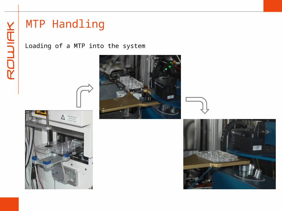
MTP Handling
Loading of a MTP into the system

CellMonitor – Software Features

Standard Software Features
Analysis of cell confluency
Assessment of number of cells or confluent colonies
Reporting on position, roundness, and diameter of cells or cell colonies in the field of view
User adjustable cell recognition parameters
Standard software has been programmed to automatically recognize cells with certain optical performance i.e. transfected HeLa cells or mouse embryonic stem cells
Implementation of automatic recognition of cell types with other optical performance possible
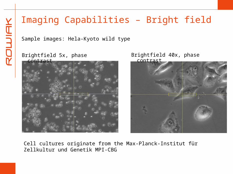
Imaging Capabilities – Bright field
Sample images: Hela-Kyoto wild type
Cell cultures originate from the Max-Planck-Institut für Zellkultur und Genetik MPI-CBG
Brightfield 5x, phase contrast Brightfield 40x, phase contrast
Sample images
Cell cultures orginiate from Max-Planck-Institut für Zellkultur und Genetik MPI-CBG
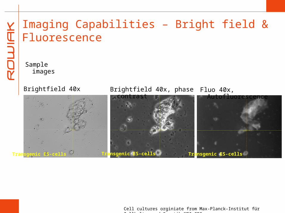
Brightfield 40x Brightfield 40x, phase contrast
Fluo 40x, Autofluorescence
Imaging Capabilities – Bright field & Fluorescence
Transgenic ES-cells Transgenic ES-cells Transgenic ES-cells

Sample images
Imaging Capabilities – Multiphoton Mode
DAPIDAPI
Multiphoton image of HeLa cells
Connexin46-GFP Connexin46-GFP
3D reconstruction of 25µm Z-stack of HeLa cells

Sample images
Mosaic image of mouse vertebra (1x1mm)
Imaging Capabilities – Multiphoton Mode
Autofluorescence/SHG

Image Recognition Software
Confluency measurement
Self learning algorithms
User defines sample patterns for
1. Foreground
2. Background
Automated confluency measurement in high-throughput
Bright field mode
Recognized patterns
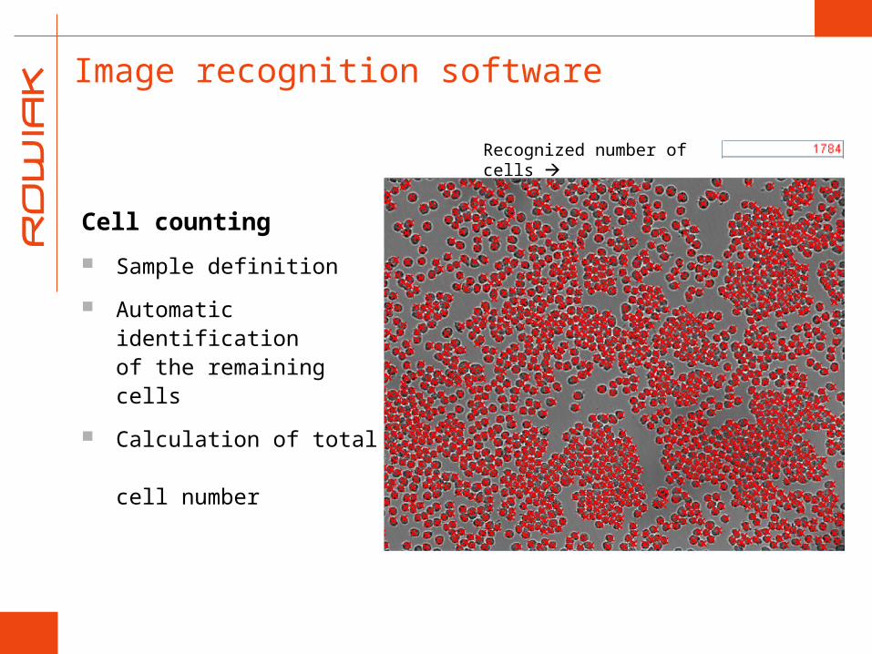
Image recognition software
Cell counting
Sample definition
Automatic identification of the remaining cells
Calculation of total cell number
Recognized number of cells

Illumination filtersTRAINED
Foreground/Background analysis
Extraction of individual regions
Selection of patterns based upon characteristics like diameter and morphology
Image Recognition – Colony Counting

Illumination filtersTRAINED
Foreground/Background analysis
Extraction of individual regions
Selection of specific
colonies
Image recognition – Fluorescence I

CellMonitor – Modular System Design

CellMonitor – Modularity
CellMonitor – Key Components
Fully automated microscope
Microscope incubator
Image recognition software
Multiphoton microscopy for imaging and manipulation (on request)
Additional modules can be added
Colony Picker

Air recirculation Laminar-Flow
Microscope incubator
Liquid-handling unit
refrigerator
Pickermodule
Storage incubator
Workbench
2D-Planar table as handling system driven by linear motors incl. runner
From CellMonitor to CellCultivator
Additional modules can be added to the Cell Monitor
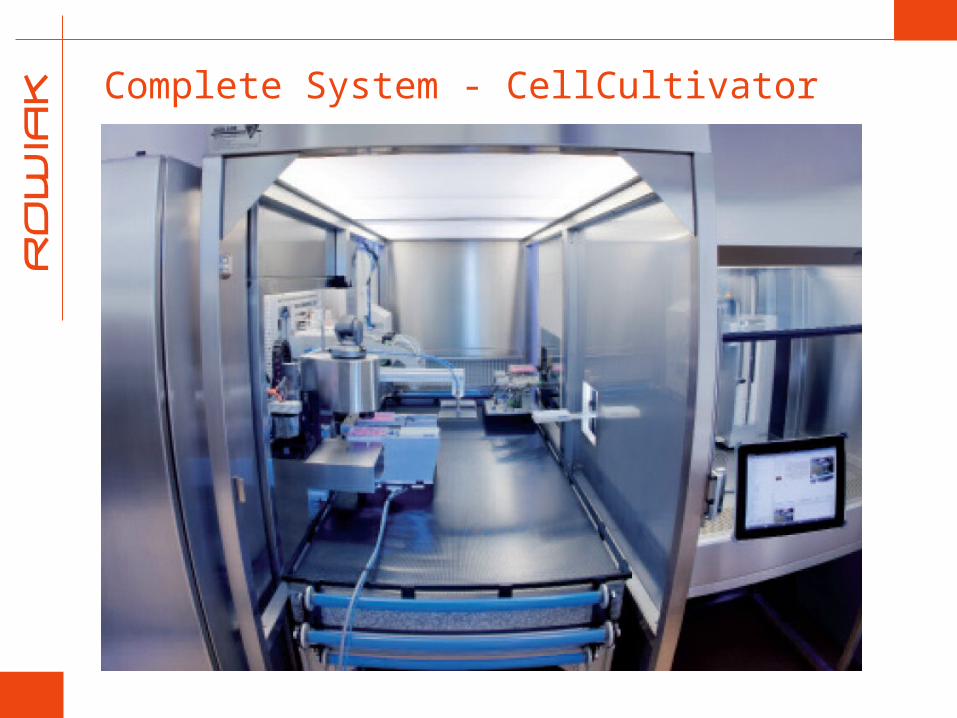
Complete System - CellCultivator

Storage Incubator Grappler for MTP supply to the
microscope table
Automated positing system
Conditions: 37°C, > 95% rel., 5% CO2; incubator sterilizable
Outer dimensions (WxDxH): 110x80x186 cm
Inner dimensions (WxDxH): 76,8 cm x 67,8 cm x 74,6 cm
Weight: approx. 550 kg
Power consumption: ca. 2 kW
Capacity of Storage Incubator: 500 plates
Gate
Plate Feeder
CellMonitor - USPs
Optimized for sensitive cell cultures
Continuous maintenance of optimal conditions
Integration of multiphoton microscopy
Optimized monitoring of tissue culture for tissue engineering
Laser cell manipulation possible
Modular setup of complete system
Individual customized solutions possible

CellMonitor – Key Applications
Cultivation of stem cells
Cultivation of primary cells
Long term monitoring
Transfection by optoporation
Tissue engineering
3D culture
3D live cell imaging and manipulation
Imaging of tissue up to a high depth
© BD Biosciences
Suitable plate format
Connexin46-GFP expressing HeLa cells
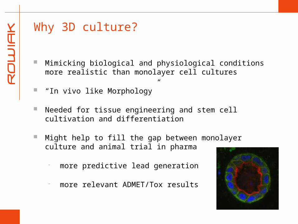
Why 3D culture?
Mimicking biological and physiological conditions more realistic than monolayer cell cultures
“In vivo like Morphology”
Needed for tissue engineering and stem cell cultivation and differentiation
Might help to fill the gap between monolayer culture and animal trial in pharma
more predictive lead generation
more relevant ADMET/Tox results

Examples of three dimensional tissue models
Francesco Pampaloni, Ernst H. K. Stelzer and Andrea Masotti: Three-Dimensional Tissue Models for Drug Discovery and Toxicology; Recent Patents on Biotechnology 2009, 3, 103-117
CellMonitor – Marketing
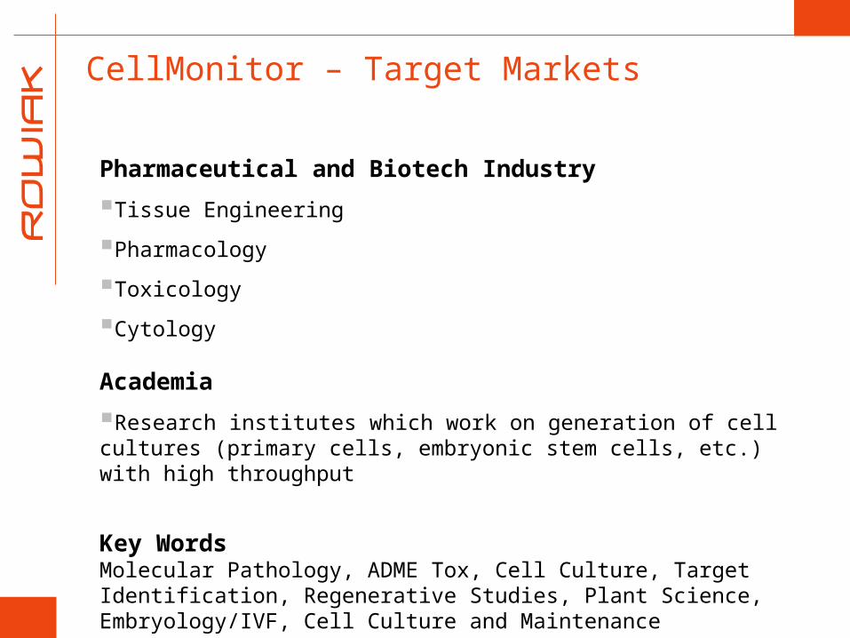
CellMonitor – Target Markets
Pharmaceutical and Biotech Industry
Tissue Engineering
Pharmacology
Toxicology
Cytology
Academia
Research institutes which work on generation of cell cultures (primary cells, embryonic stem cells, etc.) with high throughput
Key WordsMolecular Pathology, ADME Tox, Cell Culture, Target Identification, Regenerative Studies, Plant Science, Embryology/IVF, Cell Culture and Maintenance

CellMonitor – Summary
Automated functions : Microscope control, autofocus, objective changer, phase rings, condenser, light source control, etc.
Integration into automated cell culture processes: Automated transfer of MTPs from and to robot systems
Climatization: Temperature, humidity and CO2 controlled
Image analysis: Confluency, cell counting, fluorescence signal analysis
Low maintenance: High power LED light sources
Customized solutions possible: Integration of laser manipulation for laser cell transfection or organelle ablation, multiphoton microscopy