Embed Size (px)
Citation preview
CLIFFORD J. RUDDLE, DDS
Founder & Director
122 S. Patterson Avenue, Ste. 206 Santa Barbara, CA 93111 (805) 964-8838 • (800) 753-3636 • Fax (805) 965-8253 www.endoruddle.com • email: [email protected]
PHOTON-INDUCED PHOTOACOUSTIC STREAMING (PIPS) *
In the 40+ years that I have practiced and taught clinical endodontics, I have observed
an awakening with the advent of the dental operating microscope, ultrasonically-driven
instrumentation, NiTi files, and MTA. Recently, the renaissance has continued with the
emergence of CBCT, 3D disinfection methods and the promise of regenerative
endodontics. Today, Photon-Induced Photoacoustic Streaming (PIPS) represents a
leading advancement in laser-activated irrigation and disinfection. Any dentist who is
committed to exquisitely cleaning root canal systems will definitely appreciate PIPS. This
technology will not only send photoacoustic shockwaves through both minimally and
fully prepared canals, but will also propagate shockwaves through our profession by
promoting predictably successful results.
-- Clifford J. Ruddle, DDS
* Summary of PIPS references and articles attached: Organized alphabetically Post date: September 2016
SUMMARY OF PIPS REFERENCES
Organized Alphabetically Post Date: September 2016
ADVANCED ENDODONTICS • CLIFFORD J. RUDDLE DDS
122 S. Patterson Avenue, Ste. 206, Santa Barbara, CA 93111 • (800) 753-3636 • www.endoruddle.com
1. Akcay M, Arslan H, Durmus N, Mese M, Capar ID: Dentinal tubule penetration of AH Plus, iRoot SP, MTA fillapex, and guttaflow bioseal root canal sealers after different final irrigation procedures: A confocal microscopic study, Lasers Surg Med 48:1, pp. 70-76, 2016.
2. Ackay M, Arslan H, Mese M, Sahin NN: The effect of photon-initiated photoacoustic streaming, ultrasonically and sonically irrigation techniques on the push-out bond strength of a resin sealer to the root dentin, Clin Oral Investig 19:5, pp. 1055-1061, 2015.
3. Al Shahrani M, DiVito E, Hughes CV, Nathanson D, Huang GT: Enhanced removal of Enterococcus faecalis biofilms in the root canal using sodium hypochlorite plus photon-induced photoacoustic streaming: an in vitro study, Photomed Laser Surg 32:5, pp. 260-266, 2014
4. Arslan H, Akcay M, Capar ID, Ertas H, Ok E, Uysal B: Efficacy of needle irrigation, EndoActivator, and photon-initiated photoacoustic streaming technique on removal of double and triple antibiotic pastes, J Endod 40:9, pp. 1439-1442, 2014.
5. Arslan H, Akcay M, Capar ID, Saygili G, Gok T, Ertas H: An in vitro comparison of irrigation using photon-initiated photoacoustic streaming, ultrasonic, sonic and needle techniques in removing calcium hydroxide, Int Endod J 48:3, pp. 246-251, 2015.
6. Arslan H, Akcay M, Ertas H, Capar ID, Saygili G, Mese M: Effect of PIPS technique at different power settings on irrigating solution extrusion, Lasers Med Sci 30:6, pp. 1641-1645, 2015.
7. Arslan H, Akcay M, Saygili G, Keski A, Mese IT, Gok A, Dalli M: Bond strength of self-adhesive resin cement to root dentin. Comparison of photon-initiated photoacoustic streaming technique with needle and ultrasonic irrigation, Acta Odontol Scand 73:5, pp. 348-352, 2015.
8. Arslan H, Akcay M, Yasa B, Hatirli H, Saygili G: Bleaching effect of activation of hydrogen peroxide using photon-initiated photoacoustic streaming technique, Clin Oral Investig 19:2, pp. 253-259, 2015.
9. Arslan H, Capar ID, Saygili G, Gok T, Akcay M: Effect of photon-initiated photoacoustic streaming on removal of apically placed dentinal debris, Int Endod J 47:11, pp. 1072-1077, 2014.
10. DiVito E, Peters OA, Olivi G: Effectivenss of the erbium: YAG laser and new design radial and stripped tips in removing the smear layer after root canal instrumentation, Lasers Med Sci 27:2, pp. 273-280, 2012.
11. Guneser MB, Arslan D, Usumez A: Tissue dissolution ability of sodium hypochlorite activated by photon-initiated photoacoustic streaming technique, J Endod 41:5, pp. 729-732, 2015.
12. Jaramillo DE, Aprecio RM, Angelov N, DiVito E, McClammy TV: Efficacy of photon induced photoacoustic streaming (PIPS) on root canals infected with Enterococcus faecalis: A pilot study, Endodontic Practice US 5:3, pp. 28-32, 2011.
SUMMARY OF PIPS REFERENCES
Organized Alphabetically Post Date: September 2016
ADVANCED ENDODONTICS • CLIFFORD J. RUDDLE DDS
122 S. Patterson Avenue, Ste. 206, Santa Barbara, CA 93111 • (800) 753-3636 • www.endoruddle.com
13. Jiang S, Zou T, Li D, Chang JW, Huang X, Zhang C: Effectiveness of sonic, ultrasonic, and photon-induced photoacoustic streaming activation of NaOCl on filling material removal following retreatment in oval canal anatomy, Photomed Laser Surg 34:1, pp. 3-10, 2016.
14. Koch JD, Jaramillo DE, DiVito E, Peters OA: Irrigant flow during photon-induced photoacoustic streaming (PIPS) using Particle Image Velocimetry (PIV), Clin Oral Investig 20:2, pp. 381-386, 2016.
15. Lloyd A, Uhles JP, Clement DJ, Garcia-Godoy F: Elimination of intracanal tissue and debris through a novel laser-activated system assessed using high-resolution micro-computed tomography: A pilot study, J Endod 40:4, pp. 584-587, 2014.
16. Moinzadeh AT, Aznar Portoles C, Schembri Wismayer P, Camilleri J: Bioactivity potential of EndoSequence BC RRM putty, J Endod 42:4, pp. 615-621, 2016.
17. Ordinola-Zapata R, Bramante CM, Aprecio RM, Handysides R, Jaramillo DE: Biofilm removal by 6% sodium hypochlorite activated by different irrigation techniques, Int Endod J 47:7, pp. 659-666, 2014.
18. Pope O, Sathorn C, Parashos P: A comparative investigation of cone-beam computed tomography and periapical radiography in the diagnosis of a healthy periapex, J Endod 40:3, pp. 360-365, 2014.
19. Zhu X, Yin X, Chang JW, Wang Y, Cheung GS, Zhang C: Comparison of the antibacterial effect and smear layer removal using photon-initiated photoacoustic streaming aided irrigation versus a conventional irrigation in single-rooted canals: An in vitro study, Photomed Laser Surg 31:8, pp. 371-377, 2013.
Lasers in Surgery and Medicine 48:70–76 (2016)
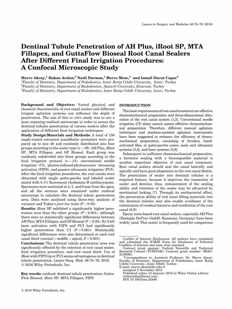
Dentinal Tubule Penetration of AH Plus, iRoot SP, MTAFillapex, and GuttaFlow Bioseal Root Canal SealersAfter Different Final Irrigation Procedures:A Confocal Microscopic Study
Merve Akcay,1 Hakan Arslan,2 Nazli Durmus,1 Merve Mese,1� and Ismail Davut Capar31Faculty of Dentistry, Department of Pedodontics, Izmir Katip Celebi University, Izmir, Turkey2Faculty of Dentistry, Department of Endodontics, Ataturk University, Erzurum, Turkey3Faculty of Dentistry, Department of Endodontics, Izmir Katip Celebi University, Izmir, Turkey
Background and Objective: Varied physical andchemical characteristic of root canal sealers and differentirrigant agitation systems can influence the depth ofpenetration. The aim of this in vitro study was to use alaser scanning confocal microscope in order to assess thedentinal tubules penetration of various sealers after theapplication of different final irrigation techniques.
Study Design/Materials and Methods: A total of 156single-rooted extracted mandibular premolars were pre-pared up to size 40 and randomly distributed into fourgroups according to the sealer type (n¼39): AHPlus, iRootSP, MTA Fillapex, and GF Bioseal. Each group wasrandomly subdivided into three groups according to thefinal irrigation protocol (n¼13): conventional needleirrigation (CI), photon-induced-photoacoustic streamingactivation (PIPS), and passive ultrasonic irrigation (PUI).After the final irrigation procedures, the root canals wereobturated with single gutta-percha and labeled sealermixed with 0.1% fluorescent rhodamine B isothiocyanate.Specimens were sectioned at 2, 5, and 8mm from the apex,and all the sections were examined under confocalmicroscope to calculate the dentinal tubule penetrationarea. Data were analyzed using three-way analysis ofvariance and Tukey’s post hoc tests (P¼ 0.05).
Results: iRoot SP exhibited a significantly higher pene-tration area than the other groups (P< 0.001), althoughthere were no statistically significant differences betweenAHPlus,MTAFillapex, andGFBioseal (P>0.05). Er:YAGlaser activation with PIPS and PUI had significantlyhigher penetration than CI (P<0.001). Statisticallysignificant differences were also determined at each rootcanal third (coronal>middle> apical; P<0.001).
Conclusions: The dentinal tubule penetration area wassignificantly affected by the selection of root canal sealer,final irrigation procedure, and root canal third. Use ofiRoot withPIPS tip or PUI seems advantageous in dentinaltubule penetration. Lasers Surg. Med. 48:70–76, 2016.� 2016 Wiley Periodicals, Inc.
Key words: confocal; dentinal tubule penetration; Gutta-Flow Bioseal; iRoot SP; MTA Fillapex; PIPS
INTRODUCTION
Thebasic requirementsof rootcanal treatmentareeffectivechemomechanical preparation and three-dimensional obtu-ration of the root canal system [1,2]. Conventional needleirrigation (CI) alone cannot assure effective chemomechan-ical preparation. Therefore, different manual agitationtechniques and machine-assisted agitation instrumentshave been suggested to enhance the efficiency of chemo-mechanical preparation, consisting of brushes, hand-activated files, or gutta-percha cones, sonic and ultrasonicsystems [3,4], and laser systems [5,6].Subsequent to sufficient chemomechanical preparation,
a hermetic sealing with a biocompatible material isanother important objective of root canal treatment.Root canal sealers should seal the canal laterally andapically and have good adaptation to the root canal dentin.The penetration of sealer into dentinal tubules is arequired feature, because it improves the connection ofsealer and dentine; thus, enhancement of the sealingability and retention of the sealer may be advanced bymechanical locking [7]. Through its antibacterial effect,the penetration ability of root canal filling materials intothe dentinal tubules may also enable avoidance of thecolonization of residual bacteria and reinfection of the rootcanal [8,9].Epoxy resin-based root canal sealers, especially AH Plus
(Dentsply DeTrey GmbH, Konstanz, Germany) have beenwidely used. This sealer is frequently used for comparison
Conflict of Interest Disclosures: All authors have completedand submitted the ICMJE Form for Disclosure of PotentialConflicts of Interest and none were reported.
Contract grant sponsor: Turkish Scientific and TechnicalResearch Council (TUBITAK); Contract grant number: SBAG-213S095.
�Correspondence to: Assistant Professor, Dr. Merve Akcay,Faculty of Dentistry, Department of Pedodontics, Izmir KatipCelebi University, Izmir 35640, Turkey.E-mail: [email protected]
Accepted 5 November 2015Published online 12 January 2016 in Wiley Online Library(wileyonlinelibrary.com).DOI 10.1002/lsm.22446
� 2016 Wiley Periodicals, Inc.
because of its good physicochemical features and adapt-ability to the root canal walls [10,11]. However, inves-tigators have demonstrated that it displays highercytotoxic effects than MTA and iRoot SP sealers [12].iRoot SP (Innovative BioCeramix Inc., Vancouver,
Canada), a new root canal sealer, is a convenient pre-mixed, aluminum-free, ready-to-use, injectable whitehydraulic cement paste developed for permanent rootcanal filling and sealing applications. iRoot SP is composedof calcium silicate, calcium phosphate, calcium hydroxide,and zirconium oxide, and it requires the presence of waterto set and harden. With a similar composition to that ofwhitemineral trioxide aggregate (MTA), iRoot SP has bothexcellent physical properties and biocompatibility [13].iRoot SP was shown to have an apical sealing abilityequivalent to that of AH Plus [13] but with lesscytotoxicity [12,14].MTA Fillapex (Angelus dental solutions, Londrina, PR,
Brazil) is another currently available calcium silicate-based root canal sealer. This sealer consists of salicylateresin, diluting resin, natural resin, bismuth oxide, nano-particulated silica, and MTA. It was developed to utilizethe good features of MTA; relatively high levels ofbiocompatibility, antimicrobial activity, and sealing abilitywere reported for this material [15].GuttaFlow 2 (Coltene Whaledent, GmBHþCo KG,
Langenau, Switzerland), a new formulation of GuttaFlow,is a silicone-based root canal sealer that combines sealerand gutta-percha in powder formwith a particle size of lessthan 30mm. It consists of a mixture of gutta-perchapowder, poly-dimethylsiloxane, platinum catalyst, zirco-nium dioxide, and micro-silver. GuttaFlow 2 has beenshown to be more biocompatible than AH Plus Jetsealer [16] and less toxic to human gingival fibroblastscells than AH Plus [17]. Recently, the same manufacturerhas launched a new bioactive sealer, GuttaFlow Bioseal(GuttaFlow 3).To our knowledge, no study has been made of the
dentinal tubule penetration of these new root canal sealerswith different final irrigation techniques application.Therefore, the aim of this in vitro study was to assessthe dentinal tubule penetration of the four various sealersafter the application of different final irrigation techni-ques, namely, CI, Er:YAG laser with photon-induced-photoacoustic streaming activation (PIPS), and passiveultrasonic activation (PUI) by using a laser scanningconfocal microscope. The null hypothesis was that therewould be no difference among the sealer penetration foreither (i) the different final irrigation techniques or (ii) theapplication of different sealers into dentinal tubules.
MATERIALS AND METHODS
A total of 156 human mandibular premolars wereselected from a collection of teeth that had been extractedfor reasons unconnected to this study. The specimens werethen immersed in thymol solution for 48hours fordisinfection and stored in 48C distilled water until used.Periapical radiographs were taken in the buccolingual andmesiodistal directions in each tooth selected with a long:
short diameter ratio of �2.5 at 5mm from the apex.Therefore, only single and round root canals wereselected [18]. The inclusion criteria were as follows: noroot canal treatment, internal or external resorption,calcification, immature root apices, caries/cracks/fractureson the root surface, and/or root canal curvature of less than10 degrees.
A size 10K-file (Dentsply Maillefer, Ballaigues,Switzerland) was inserted into the canal to confirm thepatency. The working length was determined by sub-tracting 1mm from the distance to the apical foramen.Root canals were prepared using ProTaper Universalrotary instruments (Dentsply, Maillefer) up to size F4(size 40, 0.06 taper). Specimens were irrigated with 2mlof 5% NaOCl (Werax; SDD, Izmir, Turkey) at eachinstrument change. A final irrigation was applied using5ml of 17% EDTA (Werax) for 1min and 5ml of 5%NaOCl for 1min. The specimens were randomly distrib-uted into four groups according to the sealer type (n¼39).
AH Plus Group
The specimens were randomly subdivided into threegroups according to the final irrigation protocol (n¼ 13). Inthe CI group, the blunt-tip needle was placed into the rootcanal within 2mmof theworking length and 5ml ofNaOClwas utilized for 1minute.
In the PIPS tip group, final irrigation was performed viathe laser irradiation protocol, which was done by means ofan Er:YAG laser with a wavelength of 2,940nm (FidelisAT, Fotona, Ljubljana, Slovenia). The laser activation wasperformedwith a 14-mm-long and conical 300mmdiameterPIPS tip at 0.9W, with 30mJ/pulse and at 30Hz for thevery short pulse (VSP, 100ms) mode [19,20]. The air andwater on the laser systemwere switched off. Subsequently,0.5ml NaOCl was applied into the root canal and the tipwas inserted into the coronal access opening of the pulpchamber only. When the irrigating solution in the coronalreservoir diminished, the remaining 4.5ml NaOCl wasgradually sent overall the root canal opening. The laseractivation was proceeded during the placement of irrigant.The total activation timewas 1minute, and the total size ofNaOCl was 5ml.
In the PUI group, 0.5ml of NaOCl was placed into theroot canal via a blunt-tip needle; a stainless steel file #20/25(IrriSafe tip, Satelec Acteon Group,Merignac, France) wasthen placed 2mm short of the working length with an up-and-down movement, and the ultrasonic device (P5 New-tron XS, Satelec Acteon Group, Merignac, France) wasactivated for 1minute at the power setting “blue” 11 inaccordance with manufacturer instruction.
After the final irrigation procedures, the root canal wasirrigated with 5ml of distilled water and dried using paperpoints (Dentsply Maillefer). For fluorescence under confo-cal laser scanning microscopy, all the root canal sealerswere mixed with 0.1% fluorescent rhodamine B isothiocy-anate (Bereket Chemical Industry, Istanbul, Turkey). Allthe labeled root canal sealers were placed into the canal to1mm short of the working length using a size 30 lentulospiral. A single gutta-percha cone (ProTaper Universal F4,
DENTINAL TUBULE PENETRATION OF SEALERS 71
Dentsply Maillefer) was then slightly coated with labeledepoxy resin-based sealer, AH Plus (Dentsply DeTrey,Konstanz, Germany), and placed in the root canal to theWL. After the root filling, the coronal opening was filledwith a temporary filling material (Cavit, 3M; ESPE, St.Paul, MN), and the specimens were stored at 100%humidity, 378C for 1 week to completely set.
iRoot SP Group
The specimens were randomly sub-divided into samethree groups according to the final irrigation protocol(n¼ 13) and all procedures were performed as in the AHPlus group. Differently from AH Plus group, iRoot SPsealer was used for the root canal obturation.
MTA Fillapex Group
Similarly, the specimens were randomly sub-dividedinto three groups according to the final irrigation protocol(n¼ 13), and all procedures were performed as in AH Plusgroup but with MTA Fillapex sealer.
GuttaFlow Bioseal Group
Likewise, the specimenswere randomly sub-divided intothree groups according to the final irrigation protocol(n¼ 13) and all procedures were performed as in AH Plusgroup, but with GuttaFlow Bioseal sealer.
Confocal Laser Scanning Microscope Analysis
After the root canal sealer had set, each specimen wassectioned perpendicular to its long axis using a precisionsaw (IsoMet 1000; Buehler, Lake Bluff, IL) at a slowspeed under water cooling. Three slices were obtainedfrom each tooth at depths of 2, 5, and 8mm (apical,middle, and coronal) and approximately 1� 0.1mmthickness. The sections were polished with siliconcarbide abrasive paper. The samples were then mountedonto glass slides and examined with a Leica TCS-SPEconfocal laser scanning microscope (Leica, Mannheim,Germany) at 10� with a wavelength of 560–600 nm. Incase when the entire canal could not examined in oneimage, further partial images were taken and then
assembled as a singly image using Photoshop (AdobeSystems, Inc., San Jose, CA). Digital images wereimported into the ImageJ program (ImageJ software,NIH) to measure the total dentinal tubule penetrationarea. The dentinal tubule penetration area was mea-sured as micrometers (mm) and converted to squaremillimeters (mm2) for the statistical analysis.
Statistically Analysis
The data were analyzed using the three-way analysis ofvariance (ANOVA) and Tukey’s post hoc tests to detect theeffects of the independent variables (root canal sealers,final irrigation regimens, and root canal thirds) and theirinteractions on the dentinal tubule penetration into theroot canal dentin (P¼0.05). All statistical analyses weremade using software (SigmaStat for Windows Version 3.5;Systat Software, Inc., Erkrath, Germany) at a significancelevel of 0.05 and a confidence interval of 95%.
RESULTS
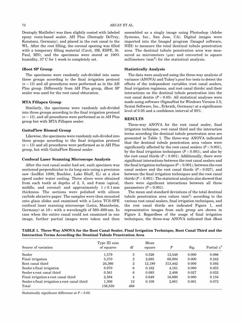
Three-way ANOVA for the root canal sealer, finalirrigation technique, root canal third and the interactionterms according the dentinal tubule penetration area arepresented in Table 1. The three-way ANOVA indicatedthat the dentinal tubule penetration area values weresignificantly affected by the root canal sealers (P< 0.001),by the final irrigation techniques (P<0.001), and also bythe root canal thirds (P< 0.001). Additionally, there weresignificant interactions between the root canal sealers andthefinal irrigation techniques (P<0.001), between the rootcanal sealers and the root canal thirds (P¼ 0.027), andbetween the final irrigation techniques and the root canalthirds (P< 0.001). The statistical analysis also showed thatthere were significant interactions between all threeparameters (P¼0.001).The mean and standard deviations of the total dentinal
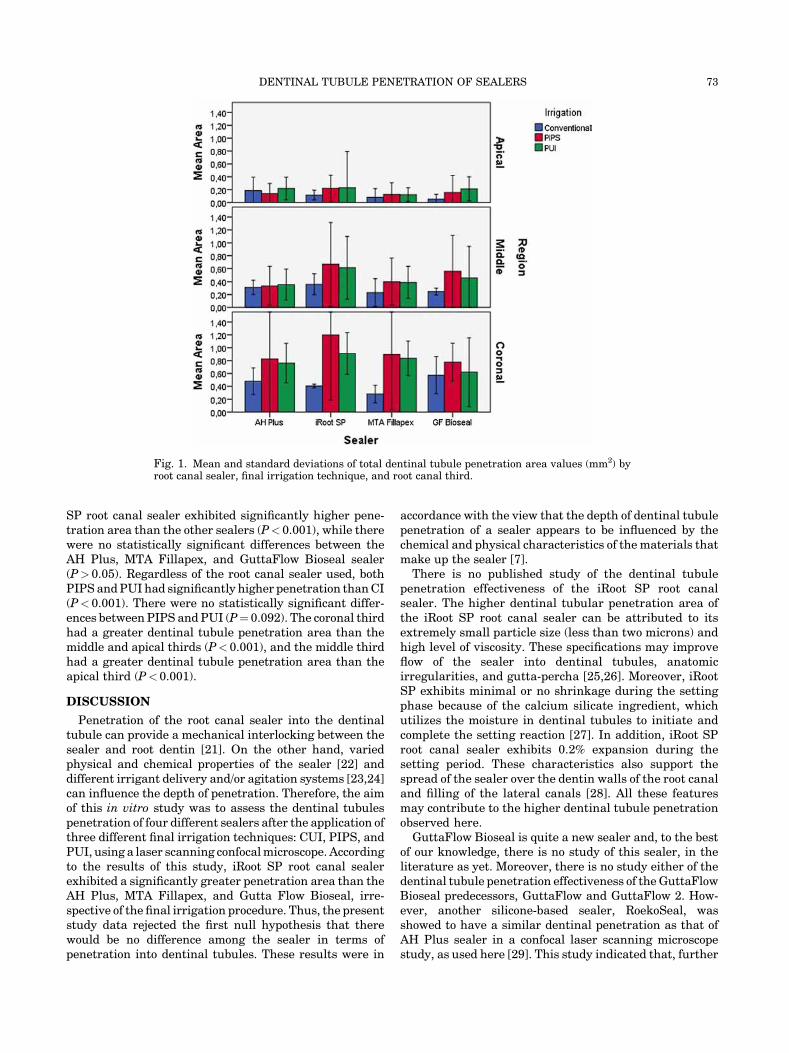
tubule penetration area values (mm2) according to thevarious root canal sealers, final irrigation techniques, andthe root canal thirds are indicated Figure 1, andrepresentative images from each group are shown inFigure 2. Regardless of the usage of final irrigationtechniques, the three-way ANOVA indicated that iRoot
TABLE 1. Three-Way ANOVA for the Root Canal Sealer, Final Irrigation Technique, Root Canal Third and the
Interaction Terms According the Dentinal Tubule Penetration Area
Source of variation
Type III sum
of squares df
Mean
square F Sig. Partial h2
Sealer 1,579 3 0.526 13,548 0.000 0.086
Final irrigation 5,370 2 2,685 69,094 0.000 0.242
Root canal third 24,360 2 12,180 313,442 0.000 0.592
Sealer x final irrigation 0.970 6 0.162 4,161 0.000 0.055
Sealer x root canal third 0.561 6 0.093 2,406 0.027 0.032
Final irrigation x root canal third 2,594 4 0.649 16,690 0.000 0.134
Sealer x final irrigation x root canal third 1,306 12 0.109 2,801 0.001 0.072
Total 138,550 468
Statistically significant difference at P<0.05.
72 AKCAY ET AL.
SP root canal sealer exhibited significantly higher pene-tration area than the other sealers (P< 0.001), while therewere no statistically significant differences between theAH Plus, MTA Fillapex, and GuttaFlow Bioseal sealer(P> 0.05). Regardless of the root canal sealer used, bothPIPS andPUI had significantly higher penetration thanCI(P< 0.001). There were no statistically significant differ-ences between PIPS and PUI (P¼ 0.092). The coronal thirdhad a greater dentinal tubule penetration area than themiddle and apical thirds (P< 0.001), and the middle thirdhad a greater dentinal tubule penetration area than theapical third (P<0.001).
DISCUSSION
Penetration of the root canal sealer into the dentinaltubule can provide a mechanical interlocking between thesealer and root dentin [21]. On the other hand, variedphysical and chemical properties of the sealer [22] anddifferent irrigant delivery and/or agitation systems [23,24]can influence the depth of penetration. Therefore, the aimof this in vitro study was to assess the dentinal tubulespenetration of four different sealers after the application ofthree different final irrigation techniques: CUI, PIPS, andPUI, using a laser scanning confocalmicroscope. Accordingto the results of this study, iRoot SP root canal sealerexhibited a significantly greater penetration area than theAH Plus, MTA Fillapex, and Gutta Flow Bioseal, irre-spective of the final irrigation procedure. Thus, the presentstudy data rejected the first null hypothesis that therewould be no difference among the sealer in terms ofpenetration into dentinal tubules. These results were in
accordance with the view that the depth of dentinal tubulepenetration of a sealer appears to be influenced by thechemical and physical characteristics of the materials thatmake up the sealer [7].
There is no published study of the dentinal tubulepenetration effectiveness of the iRoot SP root canalsealer. The higher dentinal tubular penetration area ofthe iRoot SP root canal sealer can be attributed to itsextremely small particle size (less than two microns) andhigh level of viscosity. These specifications may improveflow of the sealer into dentinal tubules, anatomicirregularities, and gutta-percha [25,26]. Moreover, iRootSP exhibits minimal or no shrinkage during the settingphase because of the calcium silicate ingredient, whichutilizes the moisture in dentinal tubules to initiate andcomplete the setting reaction [27]. In addition, iRoot SProot canal sealer exhibits 0.2% expansion during thesetting period. These characteristics also support thespread of the sealer over the dentin walls of the root canaland filling of the lateral canals [28]. All these featuresmay contribute to the higher dentinal tubule penetrationobserved here.
GuttaFlow Bioseal is quite a new sealer and, to the bestof our knowledge, there is no study of this sealer, in theliterature as yet. Moreover, there is no study either of thedentinal tubule penetration effectiveness of theGuttaFlowBioseal predecessors, GuttaFlow and GuttaFlow 2. How-ever, another silicone-based sealer, RoekoSeal, wasshowed to have a similar dentinal penetration as that ofAH Plus sealer in a confocal laser scanning microscopestudy, as used here [29]. This study indicated that, further
Fig. 1. Mean and standard deviations of total dentinal tubule penetration area values (mm2) byroot canal sealer, final irrigation technique, and root canal third.
DENTINAL TUBULE PENETRATION OF SEALERS 73
studies should investigate not only dentinal penetrationbut also physical and chemical properties in order tounderstand clinic applicability of this new bioactive sealer.
The dentinal tubule penetration of the AHPlus andMTAFillapex root canal sealers has been evaluated withAmorosa-Silva et al. [30] reporting that their penetrationinto the dentinal tubules was statistically similar. Thefinding in the present study was in agreement with thisstudy. Kuci et al. [31] evaluated the penetration intodentinal tubules of the MTA Fillapex and AH 26 root canalsealers. Differently from this study, the effects of the coldlateral compaction and warm vertical compaction techni-ques were investigated. This study found that greaterdentinal tubule penetrationwas foundwhen tMTAFillapexwasusedwith cold lateral compactionandAH26withwarmvertical compaction. These results were associated with agreater flow of MTA Fillapex under compaction pressureand decreased viscosity ofAH26at the higher temperature.In the present study, only a single gutta-percha cone wasplaced into the root canal and without any pressure. Ourdifferent results can be linked to this.
According to the results of the present study, thedentinal tubule penetration of the irrigation solution was
better in the coronal thirds of the root canals than in themiddle and apical thirds, and in the middle than apicalthirds. Other investigations into dentinal tubule penetra-tion have also reported decreasing penetration values fromthe coronal to apical parts [23,32,33]. The poorer dentinaltubule penetration in the apical thirds can be explained bythe ineffective delivery of irrigant to this region of thecanal, the smaller diameter and reduced number ofdentinal tubules in this third, and its greater more tubularsclerosis [34,35].According to the results of the present study, PIPS and
PUI both obtain a higher dentinal penetration than CI.This finding is in conformance with an earlier study, whichinvestigated various root canal irrigant agitation methodsin respect of the penetration of endodontic irrigants intodentinal tubules. The results of that study showed thatNd:YAG laser activation with either NaOCl or EDTA wasmuch better than NaOCl irrigation alone for sealerpenetration into dentinal tubules and as effective as anEDTA final flush [23]. Also, another study demonstratedthat ultrasonic agitation significantly improved the pene-tration of an endodontic irrigant when compared to sonicagitation [36].
Fig. 2. Representative confocal laser scanning microscopic images from each group at coronal andapical thirds.
74 AKCAY ET AL.
The PIPS technique is based upon photo-acoustic and-mechanical action without needing to arrive to the rootapex, which makes it dissimilar to other agitationtechniques.With this technique, each impulse reactswith the water molecules, prompting expansion and serialshock waves that cause the creation of an effectivestreaming fluid [37]. Similarly, the PUI technique is basedon the transmission of acoustic energy to an irrigant in theroot canal space through ultrasonic waves and can causeacoustic streaming of the irrigant [38]. The higher dentinaltubule penetration ratio for these types of activation canthus be attributed to the acoustic energy and high-speedfluid motion. Before the root canal obturation, thissuggests that the activation of the irrigant and creationof streaming with Er:YAG laser activation with PIPS orPUI has a positive effect on the dentinal penetration of theresin sealer to root dentin as compared to that achievedwith CI. Thereby, the present study data rejected thesecond null hypothesis that there would be no differencefor the sealer penetration after the application of differentfinal irrigation techniques into dentinal tubules.For the dentinal tubule penetration evaluation in the
confocal laser scanning microscopy two parameters havebeen measured, the maximum depth of penetration andthe percentage of sealer penetration,employing a similarmethod to that used by Gharib et al. [32]. Unique or anumber of measurements were performed to calculate thedeepest penetration and the outlined areas along the canalwalls in which sealer penetrated into dentinal tubules,with distances divided by the canal circumference tocalculate the percentage of sealer penetration. However,these methods have some limitations. Single/multiplemeasurements could have affect the overall depth andthe thicker/looser penetration also changed the percent-age. Therefore, in the present study, the ImageJ programwas used to measure the total dentinal tubule penetrationarea. This program can calculate area and pixelvalue statistics of user-defined selections and intensity-thresholded objects.Within the limitations of this study, the following
conclusions can be made:
� iRoot SP root canal sealer exhibited a significantlygreater penetration area than the AH Plus, MTAFillapex, and GF Bioseal sealers, among which therewere no statistically significant differences,
� Er:YAG laser activation with PIPS and PUI had asignificantly greater penetration than the control groupand there were no statistically significant differencesbetween PIPS and PUI,
� iRoot SP root canal sealer usage after the final irrigationprocedures with PIPS or PUI seems advantageous interms of dentinal tubule penetration. Further studiesshould be conducted to confirm these findings.
ACKNOWLEDGMENT
The authors thank Coltane Switzerland for providingGuttaFlow Bioseal in this study.
REFERENCES
1. Siqueira JF, Jr., Lima KC, Magalhaes FA, Lopes HP, deUzedaM. Mechanical reduction of the bacterial population inthe root canal by three instrumentation techniques. J Endod1999;25(5):332–335.
2. Caron G, Nham K, Bronnec F, Machtou P. Effectiveness ofdifferent final irrigant activation protocols on smear layerremoval in curved canals. J Endod 2010;36(8):1361–1366.
3. Gu LS, Kim JR, Ling J, Choi KK, Pashley DH, Tay FR.Review of contemporary irrigant agitation techniques anddevices. J Endod 2009;35(6):791–804.
4. Capar ID, Aydinbelge HA. Effectiveness of various irriga-tion activation protocols and the self-adjusting file systemon smear layer and debris removal. Scanning 2014;36(6):640–647.
5. DiVito E, Lloyd A. ER: AG laser for 3-dimensional debride-ment of canal systems: Use of photon-induced photoacousticstreaming. Dent Today 2012;31(11):122.
6. Deleu E, Meire MA, De Moor RJ. Efficacy of laser-basedirrigant activation methods in removing debris from simu-lated root canal irregularities. Lasers Med Sci 2015;30(2):831–835.
7. Mamootil K, Messer HH. Penetration of dentinal tubules byendodontic sealer cements in extracted teeth and in vivo. IntEndod J 2007;40(11):873–881.
8. Bouillaguet S, Shaw L, Barthelemy J, Krejci I, Wataha JC.Long-term sealing ability of pulp canal sealer, AH-Plus,GuttaFlow and Epiphany. Int Endod J 2008;41(3):219–226.
9. Heling I, Chandler NP. The antimicrobial effect withindentinal tubules of four root canal sealers. J Endod 1996;22(5):257–259.
10. Marin-Bauza GA, Rached-Junior FJ, Souza-Gabriel AE,Sousa-Neto MD, Miranda CE, Silva-Sousa YT. Physicochem-ical properties of methacrylate resin-based root canal sealers.J Endod 2010;36(9):1531–1536.
11. MarcianoMA, Guimaraes BM, Ordinola-Zapata R, BramanteCM, Cavenago BC, Garcia RB, Bernardineli N, Andrade FB,Moraes IG, Duarte MA. Physical properties and interfacialadaptation of three epoxy resin-based sealers. J Endod2011;37(10):1417–1421.
12. Zhang W, Li Z, Peng B. Ex vivo cytotoxicity of a new calciumsilicate-based canal filling material. Int Endod J 2010;43(9):769–774.
13. ZhangW,Li Z, PengB.Assessment of a new root canal sealer’sapical sealing ability. Oral Surg Oral Med Oral Pathol OralRadiol Endod 2009;107(6):e79–e82.
14. Zhou HM, Du TF, Shen Y,Wang ZJ, Zheng YF, HaapasaloM.In vitro cytotoxicity of calcium silicate-containing endodonticsealers. J Endod 2015;41(1):56–61.
15. Torabinejad M, Parirokh M. Mineral trioxide aggregate: Acomprehensive literature review-part II: Leakage andbiocompatibility investigations. J Endod 2010;36(2):190–202.
16. Accardo C, Himel VT, Lallier TE. A novel GuttaFlow sealersupports cell survival and attachment. J Endod 2014;40(2):231–234.
17. Mandal P, Zhao J, Sah SK, Huang Y, Liu J. In vitrocytotoxicity of guttaflow 2 on human gingival fibroblasts.J Endod 2014;40(8):1156–1159.
18. De-Deus G, Reis C, Beznos D, de Abranches AM, Coutinho-Filho T, Paciornik S. Limited ability of three commonly usedthermoplasticized gutta-percha techniques in filling oval-shaped canals. J Endod 2008;34(11):1401–1405.
19. Akcay M, Arslan H, Mese M, Sahin NN. The effect of photon-initiated photoacoustic streaming, ultrasonically and soni-cally irrigation techniques on the push-out bond strength of aresin sealer to the root dentin. Clin Oral Investig 2015;19(5):1055–1061.
20. Arslan H, Akcay M, Capar ID, Saygili G, Gok T, Ertas H. Anin vitro comparison of irrigation using photon-initiatedphotoacoustic streaming, ultrasonic, sonic and needletechniques in removing calcium hydroxide. Int Endod J2015;48(3):246–251.
21. HaragushikuGA, Sousa-NetoMD, Silva-SousaYT, AlfredoE,Silva SC, Silva RG. Adhesion of endodontic sealers to human
DENTINAL TUBULE PENETRATION OF SEALERS 75
root dentine submitted to different surface treatments.Photomed Laser Surg 2010;28(3):405–410.
22. Oksan T, Aktener BO, Sen BH, Tezel H. The penetration ofroot canal sealers into dentinal tubules. A scanning electronmicroscopic study. Int Endod J 1993;26(5):301–305.
23. MoonYM,KimHC, BaeKS, Baek SH, ShonWJ, LeeW. Effectof laser-activated irrigation of 1320-nanometer Nd:YAG laseron sealer penetration in curved root canals. J Endod2012;38(4):531–535.
24. Guimaraes BM, Amoroso-Silva PA, Alcalde MP, MarcianoMA, de Andrade FB, Duarte MA. Influence of ultrasonicactivation of 4 root canal sealers on the filling quality. J Endod2014;40(7):964–968.
25. Shokouhinejad N, Gorjestani H, Nasseh AA, Hoseini A,Mohammadi M, Shamshiri AR. Push-out bond strength ofgutta-percha with a new bioceramic sealer in the presence orabsence of smear layer. Aust Endod J 2013;39(3):102–106.
26. Ken K, Brave D, Nasseh AA. A review of bioceramictechnology in endodontics. Roots 2012;4:6–12.
27. Ersahan S, Aydin C. Dislocation resistance of iRoot SP, acalcium silicate-based sealer, from radicular dentine. J Endod2010;36(12):2000–2002.
28. Kossev D, Stefanov V. Ceramics-based sealers as newalternative to currently used endodontic sealers. Roots2009;1:42–48.
29. Chandra SS, Shankar P, Indira R. Depth of penetration offour resin sealers into radicular dentinal tubules: A confocalmicroscopic study. J Endod 2012;38(10):1412–1416.
30. Amoroso-Silva PA, Guimaraes BM, Marciano MA, DuarteMA, Cavenago BC, Ordinola-Zapata R, Almeida MM, Moraes
IG. Microscopic analysis of the quality of obturation andphysical properties of MTA Fillapex. Microsc Res Tech2014;77(12):1031–1036.
31. Kuci A, Alacam T, Yavas O, Ergul-Ulger Z, Kayaoglu G.Sealer penetration into dentinal tubules in the presence orabsence of smear layer: A confocal laser scanning microscopicstudy. J Endod 2014;40(10):1627–1631.
32. Gharib SR, Tordik PA, Imamura GM, Baginski TA, GoodellGG. A confocal laser scanning microscope investigation of theepiphany obturation system. J Endod 2007;33(8):957–961.
33. Kara Tuncer A, Tuncer S. Effect of different final irrigationsolutions on dentinal tubule penetration depth and percent-age of root canal sealer. J Endod 2012;38(6):860–863.
34. Teixeira CS, Felippe MC, Felippe WT. The effect of applica-tion time of EDTA and NaOCl on intracanal smear layerremoval: An SEM analysis. Int Endod J 2005;38(5):285–290.
35. Paque F, Luder HU, Sener B, Zehnder M. Tubular sclerosisrather than the smear layer impedes dye penetration into thedentine of endodontically instrumented root canals. IntEndod J 2006;39(1):18–25.
36. Paragliola R, FrancoV, FabianiC,MazzoniA,NatoF, TayFR,Breschi L, Grandini S. Final rinse optimization: Influence ofdifferent agitation protocols. J Endod 2010;36(2):282–285.
37. DiVitoE, PetersOA,Olivi G. Effectiveness of the erbium:YAGlaser and new design radial and stripped tips in removing thesmear layer after root canal instrumentation. Lasers Med Sci2012;27(2):273–280.
38. AhmadM, Pitt Ford TJ, Crum LA. Ultrasonic debridement ofroot canals: acoustic streaming and its possible role. J Endod1987;13(10):490–499.
76 AKCAY ET AL.
ORIGINAL ARTICLE
The effect of photon-initiated photoacoustic streaming,ultrasonically and sonically irrigation techniques on the push-outbond strength of a resin sealer to the root dentin
Merve Akcay & Hakan Arslan & Merve Mese &
N. Nur Sahin
Received: 21 May 2014 /Accepted: 3 October 2014 /Published online: 15 October 2014# Springer-Verlag Berlin Heidelberg 2014
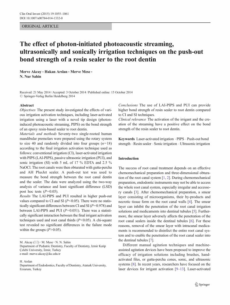
AbstractObjectives The present study investigated the effects of vari-ous irrigation activation techniques, including laser-activatedirrigation using a laser with a novel tip design (photon-induced photoacoustic streaming, PIPS) on the bond strengthof an epoxy resin-based sealer to root dentin.Materials and methods Seventy-two single-rooted humanmandibular premolars were prepared using the rotary systemto size 40 and randomly divided into four groups (n=18)according to the final irrigation activation technique used asfollows: conventional irrigation (CI), laser-activated irrigationwith PIPS (LAI-PIPS), passive ultrasonic irrigation (PUI), andsonic irrigation (SI) with 5 mL of 17 % EDTA and 2.5 %NaOCl. The root canals were then obturated with gutta-perchaand AH PlusJet sealer. A push-out test was used tomeasure the bond strength between the root canal dentinand the sealer. The data were analyzed using the two-wayanalysis of variance and least significant difference (LSD)post hoc tests (P=0.05).Results The LAI-PIPS and PUI resulted in higher push-outvalues compared to CI and SI (P<0.05). There were no statis-tically significant differences between CI and SI (P=0.978) andbetween LAI-PIPS and PUI (P=0.051). There was a statisti-cally significant interaction between the final irrigant activationtechniques used and root canal thirds (P<0.05). A chi-squaretest revealed no significant differences in the failure modewithin the groups (P>0.05).
Conclusions The use of LAI-PIPS and PUI can providehigher bond strength of resin sealer to root dentin comparedto CI and SI techniques.Clinical relevance The activation of the irrigant and the cre-ation of the streaming have a positive effect on the bondstrength of the resin sealer to root dentin.
Keywords Laser-activated irrigation . PIPS . Push-out bondstrength . Resin sealer . Sonic irrigation . Ultrasonic irrigation
Introduction
The success of root canal treatment depends on an effectivechemomechanical preparation and three-dimensional obtura-tion of the root canal system [1, 2]. During chemomechanicalpreparation, endodontic instruments may not be able to accessthe whole root canal system, especially irregular and accesso-ry canals [3]. After chemomechanical preparation, a smearlayer consisting of microorganisms, their by-products andnecrotic tissue form on the root canal walls [4]. The smearlayer can inhibit the penetration of the root canal irrigationsolutions and medicaments into dentinal tubules [5]. Further-more, the smear layer adversely affects the penetration of theroot canal sealers inside the dentinal tubules [6]. For thesereasons, removal of the smear layer with intracanal medica-ments is recommended to disinfect the entire root canal sys-tem and to enable the penetration of the root canal sealer intothe dentinal tubules [7].
Different manual agitation techniques and machine-assisted agitation devices have been proposed to improve theefficacy of irrigation solutions including brushes, hand-activated files, or gutta-percha cones, sonic, and ultrasonicsystems [8]. In recent years, researchers have focused on thelaser devices for irrigant activation [9–13]. Laser-activated
M. Akcay (*) :M. Mese :N. N. SahinDepartment of Pediatric Dentistry, Faculty of Dentistry, Izmir KatipÇelebi University, İzmir, Turkeye-mail: [email protected]
H. ArslanDepartment of Endodontics, Faculty of Dentistry, Ataturk University,Erzurum, Turkey
Clin Oral Invest (2015) 19:1055–1061DOI 10.1007/s00784-014-1332-0
irrigation produces explosive vapor bubbles with a secondarycavitation effect, providing effective removal of the debris andsmear layer from the complex root canal systems. Photon-induced photoacoustic streaming (PIPS) is a novel laser agi-tation technique, which uses an erbium:yttrium-aluminum-garnet (Er:YAG) laser. The laser has a new design for use inendodontics, with both a radial and stripped tip of noveldesign at subablative levels. This technique uses low-energylevels and short microsecond pulse rates (50 μs) to generatepeak power spikes. The profound photoacoustic shock waveallows for three-dimensional movement of the irrigation solu-tions. In contrast to other agitation techniques, the tip is onlyinserted into the canal orifice without moving into the rootapex [9]. Adhesion of the root canal sealers to the root canaldentin by close contact is crucial to resist the disruption of theestablished seal via micromechanical retention or frictionduring intraoral tooth flexure [14, 15] or preparation of coresor postspaces along the coronal and middle thirds of canalwalls [16]. Comparisons of the different agitation techniquesare needed to understand the micromechanical forces exertedby these techniques and their effect on the adhesion of the rootcanal sealers. Thus far, no studies have investigated thepush-out bond strength of the endodontic sealers to rootcanal dentin after using PIPS as the final irrigation tech-nique. Therefore, the aim of the present study was toinvestigate the effect of various irrigation techniques in-cluding laser-activated irrigation with a novel tip design(LAI-PIPS), on the bond strength of an epoxy resin-basedsealer (AH Plus Jet) to root canal dentin. The null hy-pothesis was that there would be no difference betweenthe laser-activated irrigation and the other agitation tech-niques in terms of the push-out bond strength of theendodontic sealer.
Material and methods
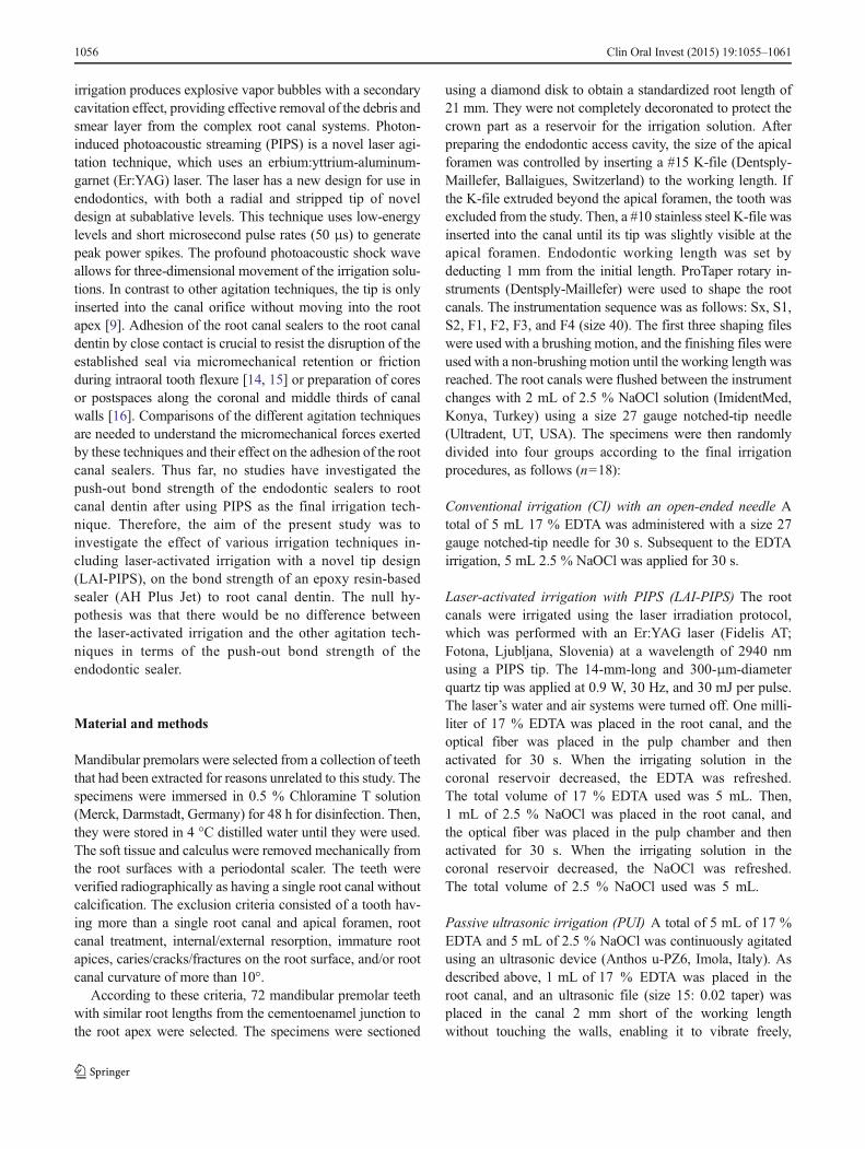
Mandibular premolars were selected from a collection of teeththat had been extracted for reasons unrelated to this study. Thespecimens were immersed in 0.5 % Chloramine T solution(Merck, Darmstadt, Germany) for 48 h for disinfection. Then,they were stored in 4 °C distilled water until they were used.The soft tissue and calculus were removed mechanically fromthe root surfaces with a periodontal scaler. The teeth wereverified radiographically as having a single root canal withoutcalcification. The exclusion criteria consisted of a tooth hav-ing more than a single root canal and apical foramen, rootcanal treatment, internal/external resorption, immature rootapices, caries/cracks/fractures on the root surface, and/or rootcanal curvature of more than 10°.
According to these criteria, 72 mandibular premolar teethwith similar root lengths from the cementoenamel junction tothe root apex were selected. The specimens were sectioned
using a diamond disk to obtain a standardized root length of21 mm. They were not completely decoronated to protect thecrown part as a reservoir for the irrigation solution. Afterpreparing the endodontic access cavity, the size of the apicalforamen was controlled by inserting a #15 K-file (Dentsply-Maillefer, Ballaigues, Switzerland) to the working length. Ifthe K-file extruded beyond the apical foramen, the tooth wasexcluded from the study. Then, a #10 stainless steel K-file wasinserted into the canal until its tip was slightly visible at theapical foramen. Endodontic working length was set bydeducting 1 mm from the initial length. ProTaper rotary in-struments (Dentsply-Maillefer) were used to shape the rootcanals. The instrumentation sequence was as follows: Sx, S1,S2, F1, F2, F3, and F4 (size 40). The first three shaping fileswere used with a brushing motion, and the finishing files wereused with a non-brushing motion until the working length wasreached. The root canals were flushed between the instrumentchanges with 2 mL of 2.5 % NaOCl solution (ImidentMed,Konya, Turkey) using a size 27 gauge notched-tip needle(Ultradent, UT, USA). The specimens were then randomlydivided into four groups according to the final irrigationprocedures, as follows (n=18):
Conventional irrigation (CI) with an open-ended needle Atotal of 5 mL 17 % EDTA was administered with a size 27gauge notched-tip needle for 30 s. Subsequent to the EDTAirrigation, 5 mL 2.5 % NaOCl was applied for 30 s.
Laser-activated irrigation with PIPS (LAI-PIPS) The rootcanals were irrigated using the laser irradiation protocol,which was performed with an Er:YAG laser (Fidelis AT;Fotona, Ljubljana, Slovenia) at a wavelength of 2940 nmusing a PIPS tip. The 14-mm-long and 300-μm-diameterquartz tip was applied at 0.9 W, 30 Hz, and 30 mJ per pulse.The laser’s water and air systems were turned off. One milli-liter of 17 % EDTA was placed in the root canal, and theoptical fiber was placed in the pulp chamber and thenactivated for 30 s. When the irrigating solution in thecoronal reservoir decreased, the EDTA was refreshed.The total volume of 17 % EDTA used was 5 mL. Then,1 mL of 2.5 % NaOCl was placed in the root canal, andthe optical fiber was placed in the pulp chamber and thenactivated for 30 s. When the irrigating solution in thecoronal reservoir decreased, the NaOCl was refreshed.The total volume of 2.5 % NaOCl used was 5 mL.
Passive ultrasonic irrigation (PUI) A total of 5 mL of 17 %EDTA and 5 mL of 2.5 % NaOCl was continuously agitatedusing an ultrasonic device (Anthos u-PZ6, Imola, Italy). Asdescribed above, 1 mL of 17 % EDTA was placed in theroot canal, and an ultrasonic file (size 15: 0.02 taper) wasplaced in the canal 2 mm short of the working lengthwithout touching the walls, enabling it to vibrate freely,
1056 Clin Oral Invest (2015) 19:1055–1061
and then activated at 25 % power for 30 s. When theirrigating solution in the coronal reservoir decreased, theEDTA was refreshed. A total of 5 mL of 2.5 % NaOClwas continuously activated in a similar manner for 30 s.
Sonic irrigation (SI) The same irrigation procedures de-scribed above were performed using the EndoActivatorhandpiece (Dentsply Tulsa Dental Specialties, Tulsa, OK,USA). A total of 5 mL of 17 % EDTA (30 s) followed by5 mL of 2.5 % NaOCl (30 s) was agitated using theEndoActivator handpiece, which was set at 10,000 cyclesper minute with a red (25/04) tip, inserted 2 mm short of theworking length.
In all the groups, the irrigation solution was transportedwith the Surgic XT Plus device (NSK, Kanuma, Japan) to fixthe flow rate of the irrigating solution constantly and equally(0.16 mL/s). After the procedures, whole root canals wereirrigated with 5 mL of distilled water and dried using paperpoints (Dentsply Maillefer). A single gutta-percha cone sur-face (F4, Dentsply Maillefer) was then slightly coated with athin layer of the freshly mixed [17] epoxy resin-based sealer,AH Plus Jet (Dentsply DeTrey, Kontanz, Germany). Thesealer was only covered around the circumference of thegutta-percha cone, and the excess sealer was slipped to theglass plate. Then, the single gutta-percha cone was placed inthe root canal to the working length. Because the root canalswere prepared using rotary instruments up to F4 files, allspecimens were obturated using the single technique withmatching taper F4 gutta-percha cones to obtain standard spec-imens for the push-out test [18]. Mesiodistal and buccolingualradiographs were taken to confirm the complete filling. Afterthe root filling, the coronal opening was filled with a tempo-rary filling material (Cavit; 3M ESPE, Seefeld, Germany) andthe specimens were stored at 100 % humidity, 37 °C for1 week to completely set.
Each specimen was sectioned perpendicular to its long axisusing a precision saw (Isomet 1000; Buehler, Lake Bluff, IL,USA) at a slow speed under water cooling. Three slices wereobtained from each tooth (n=54 for each group) at depths of 4,7, and 10mm (apical, middle, and coronal) and approximately1±0.1 mm thickness. The thickness of each slice was con-firmedwith a digital caliper (Teknikel, Istanbul, Turkey). Boththe apical and coronal aspects of the specimens were thenmicroscopically examined to confirm a circular canal shape[19]. Both diameters of each hole were measured under astereomicroscope (Zeiss Stemi 2000C; Carl Zeiss; Jena;Germany) at ×32 magnification to determine the diametersof the pluggers to be used for the push-out test. The push-out test was performed on each specimen with a universaltest machine (AGS-X, Shimadzu Corporation, Tokyo,Japan) at a crosshead speed of 1 mm per minute byapplying a continuous load to the apical side of each sliceusing 0.6-, 0.7-, and 0.8-mm-diameter cylindrical pluggers,
matching the diameter of each canal third. The diameter ofthe pluggers was approximately at least 80 % of thediameter of the canal. The maximum load applied to thefilling material before the failure was recorded in Newtons(N) and converted to megapascals (MPa) according to thefollowing formula:
push−out bond strength MPað Þ¼ maximum load Nð Þ=adhesion area of the root filling Að Þ mm2
� �:
The adhesion area of the root canal filling was calculatedusing the following equation:
A ¼ πr1þπr2ð ÞXL;
where L=ffiffiffiffiffiffiffiffiffiffiffiffiffiffiffiffiffiffiffiffiffiffiffiffiffiffiffiffiffir1−r2ð Þ2þ h2
p, where r1 is the smaller radius,
r2 is the larger radius of the canal diameter (mm), hrepresents the thickness of the root section (mm), and πis the constant 3.14 [20].
After the test procedure, each specimen was visuallyexamined under a stereomicroscope at ×32 magnificationto evaluate the failure mode. Three types of the failure wereclassified: adhesive failure (between the sealer and root dentin),cohesive failure (within the sealer or root dentin), andmixed (a combination of cohesive and adhesive) [21].
The data were analyzed using the two-way analysis ofvariance (ANOVA) and least significant difference (LSD) posthoc tests to detect the effect of the independent variables (finalirrigant activation techniques and root canal thirds) and theirinteractions on the push-out bond strength (P=0.05). Thefailure mode data were statistically analyzed using a chi-square test (P=0.05). All statistical analyses were performedusing software (SigmaStat for Windows Version 3.5; SystatSoftware, Inc., Erkrath, Germany) at a significance level of0.05 and a confidence interval of 95 %.
Results
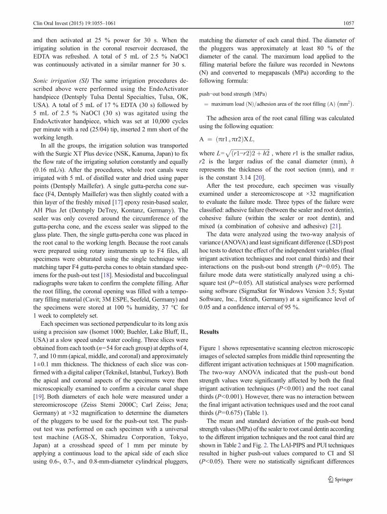
Figure 1 shows representative scanning electron microscopicimages of selected samples frommiddle third representing thedifferent irrigant activation techniques at 1500 magnification.The two-way ANOVA indicated that the push-out bondstrength values were significantly affected by both the finalirrigant activation techniques (P<0.001) and the root canalthirds (P<0.001). However, there was no interaction betweenthe final irrigant activation techniques used and the root canalthirds (P=0.675) (Table 1).
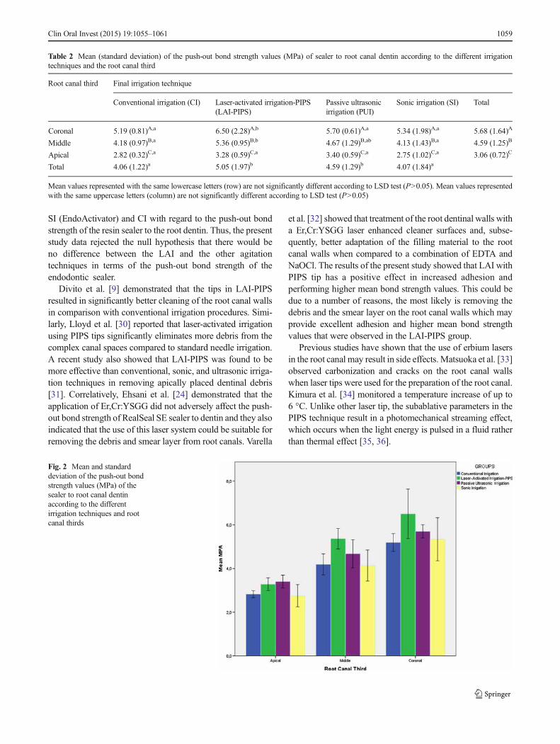
The mean and standard deviation of the push-out bondstrength values (MPa) of the sealer to root canal dentin accordingto the different irrigation techniques and the root canal third areshown in Table 2 and Fig. 2. The LAI-PIPS and PUI techniquesresulted in higher push-out values compared to CI and SI(P<0.05). There were no statistically significant differences
Clin Oral Invest (2015) 19:1055–1061 1057
between CI and SI (P=0.978) or between LAI-PIPS and PUI(P=0.051). There was a statistically significant interaction be-tween the final irrigant activation techniques and the root canalthirds (P<0.05). The coronal third had higher bond strengthvalues than the middle third and the apical third (P<0.001).The middle third had higher bond strength values than the apicalthird (P<0.001).
The chi-square test revealed no significant differences inthe failure mode within the groups (P>0.05). Adhesive failurebetween the resin sealer and dentin was the most frequent typeof failure mode in all the groups. Only one mixed failure wasobserved for the LAI-PIPS and SI groups and two mixedfailure were observed for the PUI and CI groups.
Discussion
Adhesion of the sealers to the root canal dentin by closecontact and penetration of the sealer tags into dentinal tubulesis crucial for micromechanical retention or frictional resistance[15]. Numerous investigators have evaluated the adhesion ofthe resin-based sealer to root dentin after different irrigationregimens and found that the irrigation regimens affect thebond strength of the root filling negatively or positively [22,23]. Comparisons of different agitation techniques are neededto understand the micromechanical forces exerted by thesetechniques and their effect on the adhesion of the root canalsealers. However, to the best of our knowledge, there are nodata in the literature about the push-out bond strength of anepoxy resin-based sealer to root canal dentin after LAI-PIPS
application as the final irrigation technique. Therefore, the aimof the present study was to investigate the effect of variousirrigation techniques including LAI-PIPS on the bond strengthof an epoxy resin-based sealer (AH Plus Jet) to root canaldentin.
In the present study, all controllable factors except the finalirrigation protocol were standardized to the greatest extentpossible. Specimens having similar root lengths were selectedand sectioned from the same length. Then, specimens wereinstrumented using the same technique.
In the present study, the bond strength of an epoxy resin-based sealer was evaluated using a push-out test method. It hasbeen reported that as the bond between the root canal sealerand root dentin increases, it is likely that the fracture resistanceincreases and the apical leakage reduces. Thus, the clinicallongevity of the endodontic treatment can be improved [24].Moreover, the root canal sealer should be able to remain in thesame location under dislocating forces such as mechanicalstresses caused by tooth function or operative procedures[25]. To evaluate the resistance to dislocating forces, thepush-out test has been shown to be efficient, practical, andreliable [25–28]. Although this test method was not advisablefor thermoplastic materials such as gutta-percha [29], Paneet al. [19] demonstrated that the push-out test could still besuitable for ranking the bonding of root filling materials. Inaddition, investigators indicated that a plugger size 70 to 90%of the canal diameter did not affect the bond strength [19].Therefore, different plugger size approximately at least 80 %of the diameter of the canal was used for this study. Accordingto the results of this study, LAI-PIPS and PUI were superior to
Fig. 1 a Effect of 17 % EDTA and 2.5 % NaOCl (conventional irriga-tion, CI) on middle third of the root canal wall. b Effect of Er:YAG laserirradiation using the PIPS tip (LAI-PIPS) onmiddle third of the root canal
wall. c Effect of passive ultrasonic irrigation (PUI) on middle third of theroot canal wall. d Effect of sonic irrigation (SI) on middle third of the rootcanal wall [scanning electron microscopy (SEM) ×1500]
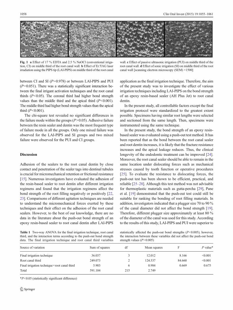
Table 1 Two-way ANOVA for the final irrigation technique, root canalthird, and the interaction terms according to the push-out bond strengthdata. The final irrigation technique and root canal third variables
statistically affected the push-out bond strengths (P<0.005); however,the interaction between these variables did not affect the push-out bondstrength values (P>0.005)
Source of variation Sum of squares df Mean squares F P value*
Final irrigation technique 36.037 3 12.012 8.166 <0.001
Root canal third 249.073 2 124.537 84.660 <0.001
Final irrigation technique×root canal third 5.903 6 0.984 0.669 0.675
Total 591.104 215 2.749
*P<0.05 (statistically significant difference)
1058 Clin Oral Invest (2015) 19:1055–1061
SI (EndoActivator) and CI with regard to the push-out bondstrength of the resin sealer to the root dentin. Thus, the presentstudy data rejected the null hypothesis that there would beno difference between the LAI and the other agitationtechniques in terms of the push-out bond strength of theendodontic sealer.
Divito et al. [9] demonstrated that the tips in LAI-PIPSresulted in significantly better cleaning of the root canal wallsin comparison with conventional irrigation procedures. Simi-larly, Lloyd et al. [30] reported that laser-activated irrigationusing PIPS tips significantly eliminates more debris from thecomplex canal spaces compared to standard needle irrigation.A recent study also showed that LAI-PIPS was found to bemore effective than conventional, sonic, and ultrasonic irriga-tion techniques in removing apically placed dentinal debris[31]. Correlatively, Ehsani et al. [24] demonstrated that theapplication of Er,Cr:YSGG did not adversely affect the push-out bond strength of RealSeal SE sealer to dentin and they alsoindicated that the use of this laser system could be suitable forremoving the debris and smear layer from root canals. Varella
et al. [32] showed that treatment of the root dentinal walls witha Er,Cr:YSGG laser enhanced cleaner surfaces and, subse-quently, better adaptation of the filling material to the rootcanal walls when compared to a combination of EDTA andNaOCl. The results of the present study showed that LAI withPIPS tip has a positive effect in increased adhesion andperforming higher mean bond strength values. This could bedue to a number of reasons, the most likely is removing thedebris and the smear layer on the root canal walls which mayprovide excellent adhesion and higher mean bond strengthvalues that were observed in the LAI-PIPS group.
Previous studies have shown that the use of erbium lasersin the root canal may result in side effects. Matsuoka et al. [33]observed carbonization and cracks on the root canal wallswhen laser tips were used for the preparation of the root canal.Kimura et al. [34] monitored a temperature increase of up to6 °C. Unlike other laser tip, the subablative parameters in thePIPS technique result in a photomechanical streaming effect,which occurs when the light energy is pulsed in a fluid ratherthan thermal effect [35, 36].
Table 2 Mean (standard deviation) of the push-out bond strength values (MPa) of sealer to root canal dentin according to the different irrigationtechniques and the root canal third
Root canal third Final irrigation technique
Conventional irrigation (CI) Laser-activated irrigation-PIPS(LAI-PIPS)
Passive ultrasonicirrigation (PUI)
Sonic irrigation (SI) Total
Coronal 5.19 (0.81)A,a 6.50 (2.28)A,b 5.70 (0.61)A,a 5.34 (1.98)A,a 5.68 (1.64)A
Middle 4.18 (0.97)B,a 5.36 (0.95)B,b 4.67 (1.29)B,ab 4.13 (1.43)B,a 4.59 (1.25)B
Apical 2.82 (0.32)C,a 3.28 (0.59)C,a 3.40 (0.59)C,a 2.75 (1.02)C,a 3.06 (0.72)C
Total 4.06 (1.22)a 5.05 (1.97)b 4.59 (1.29)b 4.07 (1.84)a
Mean values represented with the same lowercase letters (row) are not significantly different according to LSD test (P>0.05). Mean values representedwith the same uppercase letters (column) are not significantly different according to LSD test (P>0.05)
Fig. 2 Mean and standarddeviation of the push-out bondstrength values (MPa) of thesealer to root canal dentinaccording to the differentirrigation techniques and rootcanal thirds
Clin Oral Invest (2015) 19:1055–1061 1059
Er:YAG laser irradiation is highly absorbed by hydroxyap-atite and water [37, 38]. With a low power, each impulseinteracts with the water molecules, resulting in the expansionof vapor bubbles and the formation of a void in front of thelaser light. Successive shock waves lead to the formation of apowerful stream of liquid [9, 39, 40]. The photomechanical-induced streaming effect occurs when only the tip of the laseris placed in the pulp chamber of the tooth. In a previous study,the researchers observed the high-speed motion of fluid whenthe tip of an Er:YAG was placed a distance of 5 mm from theapical stop of an artificial glass root canal model. They reportedthat the bubble caused by the laser activation of the irrigatingsolution increased in size and reached up to 1800 μm in 220 μs[41]. In the present study, although the tip of the laser wasinserted only into the pulp chamber, it was superior to SI and CIwhen applied 2 mm short of the working length.
The traditional laser applications in the root canal necessi-tate conventional preparation for at least up to size 30 and thelaser tip need to reach apical third of the root. However, thePIPS tip does not need to reach the root apex, and it isonly placed into the coronal reservoir of the root canal.Therefore, this technique allows for minimally invasivepreparation of the root canal [9, 35]. The effect may beexplained by the increased NaOCl reaction kinetics withthe laser activation [42].
According to the results of the present study, LAI withPIPS may be beneficial in obtaining high bond strength be-tween resin sealer and root dentin. This finding is in agree-ment with a previous investigation that examined the effect ofdifferent root canal irrigant agitation protocols in the penetra-tion of an endodontic irrigant into dentinal tubules. The resultsof that study showed that ultrasonic agitation was significantlymore successful than sonic agitation [43]. Also, another studydemonstrated that sonic activation did not significantly im-prove the penetration of the sealer when compared to CI [44].Similarly, in the present study, SI technique resulted in similarvalues compared to CI.
As reported by Ahmad et al. [45], the PUI technique isbased upon the transmission of the acoustic energy to anirrigant in the root canal space through ultrasonic waves andcan cause acoustic streaming of the irrigant. The acousticstreaming effect of the irrigant in the ultrasonic has beenshown to bemore effective than syringe irrigation in removingartificially created dentine debris placed in simulateduninstrumented extensions and irregularities in root canals[46]. De Moor et al. [47] evaluated the efficacy of LAI witherbium lasers and PUI in removing artificially placed dentindebris in root canals. They showed that the application of theLAI technique for 20 s is as efficient as PUI for 3×20 s. In thepresent study, the acoustic energy with both PUI and LAI-PIPS resulted in similar bond strength, and the bond strengthwas greater than that achieved in the CI and SI groups. Thissuggests that the activation of the irrigant and the creation of
the streaming have a positive effect on the bond strength of theresin sealer to root dentin.
Conclusions
Within the limitations of the present study, it can be concludedthat the use of laser-activated irrigation with a novel tip design(PIPS) and PUI can provide higher bond strength of resinsealer to root dentin compared to CI and SI techniques.
Acknowledgments The authors deny any financial affiliations relatedto this study or its sponsors.
Conflict of interest The authors declare that they have no conflict ofinterest.
References
1. Siqueira JF Jr, Lima KC, Magalhaes FA, Lopes HP, de Uzeda M(1999) Mechanical reduction of the bacterial population in the rootcanal by three instrumentation techniques. J Endod 25:332–335. doi:10.1016/S0099-2399(06)81166-0
2. Caron G, Nham K, Bronnec F, Machtou P (2010) Effectiveness ofdifferent final irrigant activation protocols on smear layer removalin curved canals. J Endod 36:1361–1366. doi:10.1016/j.joen.2010.03.037
3. Ricucci D, Siqueira JF Jr (2010) Fate of the tissue in lateral canals andapical ramifications in response to pathologic conditions and treat-ment procedures. J Endod 36:1–15. doi:10.1016/j.joen.2009.09.038
4. McComb D, Smith DC (1975) A preliminary scanning electronmicroscopic study of root canals after endodontic procedures. JEndod 1:238–242. doi:10.1016/S0099-2399(75)80226-3
5. Orstavik D, HaapasaloM (1990) Disinfection by endodontic irrigantsand dressings of experimentally infected dentinal tubules. EndodDent Traumatol 6:142–149.
6. Oksan T, Aktener BO, Sen BH, Tezel H (1993) The penetration ofroot canal sealers into dentinal tubules. A scanning electron micro-scopic study. Int Endod J 26:301–305
7. Violich DR, Chandler NP (2010) The smear layer in endodontics—areview. Int Endod J 43:2–15. doi:10.1111/j.1365-2591.2009.01627.x
8. Gu LS, Kim JR, Ling J, Choi KK, Pashley DH, Tay FR (2009)Review of contemporary irrigant agitation techniques and devices. JEndod 35:791–804. doi:10.1016/j.joen.2009.03.010
9. DiVito E, Lloyd A (2012) ER:YAG laser for 3-dimensional debride-ment of canal systems: use of photon-induced photoacoustic stream-ing. Dent Today 31(122):124–127
10. Pedulla E, Genovese C, Campagna E, Tempera G, Rapisarda E(2012) Decontamination efficacy of photon-initiated photoacousticstreaming (PIPS) of irrigants using low-energy laser settings: anex vivo study. Int Endod J 45:865–870. doi:10.1111/j.1365-2591.2012.02044.x
11. Zhu X, Yin X, Chang JW, Wang Y, Cheung GS, Zhang C (2013)Comparison of the antibacterial effect and smear layer removal usingphoton-initiated photoacoustic streaming aided irrigation versus aconventional irrigation in single-rooted canals: an in vitro study.Photomed Laser Surg 31:371–377. doi:10.1089/pho.2013.3515
12. Deleu E, Meire MA, De Moor RJ (2013) Efficacy of laser-basedirrigant activation methods in removing debris from simulated rootcanal irregularities. Lasers Med Sci doi:10.1007/s10103-013-1442-y
1060 Clin Oral Invest (2015) 19:1055–1061
13. George R, Meyers IA, Walsh LJ (2008) Laser activation of endodon-tic irrigants with improved conical laser fiber tips for removing smearlayer in the apical third of the root canal. J Endod 34:1524–1527. doi:10.1016/j.joen.2008.08.029
14. Erickson RL (1992) Surface interactions of dentin adhesivematerials.Oper Dent Suppl 5:81–94
15. Panitvisai P, Messer HH (1995) Cuspal deflection in molars inrelation to endodontic and restorative procedures. J Endod 21:57–61
16. Munoz HR, Saravia-Lemus GA, Florian WE, Lainfiesta JF (2007)Microbial leakage of Enterococcus faecalis after post space prepara-tion in teeth filled in vivowith RealSeal versus Gutta-percha. J Endod33:673–675. doi:10.1016/j.joen.2007.02.007
17. Saleh IM, Ruyter IE, Haapasalo MP, Orstavik D (2003) Adhesion ofendodontic sealers: scanning electron microscopy and energy disper-sive spectroscopy. J Endod 29:595–601. doi:10.1097/00004770-200309000-00013
18. Amin SA, Seyam RS, El-Samman MA (2012) The effect of priorcalcium hydroxide intracanal placement on the bond strength of twocalcium silicate-based and an epoxy resin-based endodontic sealer. JEndod 38:696–699. doi:10.1016/j.joen.2012.02.007
19. Pane ES, Palamara JE, Messer HH (2013) Critical evaluation of thepush-out test for root canal filling materials. J Endod 39:669–673.doi:10.1016/j.joen.2012.12.032
20. Prado M, Simao RA, Gomes BP (2013) Effect of different irrigationprotocols on resin sealer bond strength to dentin. J Endod 39:689–692. doi:10.1016/j.joen.2012.12.009
21. Nagas E, Uyanik MO, Eymirli A, Cehreli ZC, Vallittu PK, LassilaLV, Durmaz V (2012) Dentin moisture conditions affect the adhesionof root canal sealers. J Endod 38:240–244. doi:10.1016/j.joen.2011.09.027
22. Vilanova WV, Carvalho-Junior JR, Alfredo E, Sousa-Neto MD,Silva-Sousa YT (2012) Effect of intracanal irrigants on the bondstrength of epoxy resin-based and methacrylate resin-based sealersto root canal walls. Int Endod J 45:42–48. doi:10.1111/j.1365-2591.2011.01945.x
23. NassarM, Awawdeh L, Jamleh A, Sadr A, Tagami J (2011) Adhesionof Epiphany self-etch sealer to dentin treated with intracanal irrigat-ing solutions. J Endod 37:228–230. doi:10.1016/j.joen.2010.11.016
24. Ehsani S, Bolhari B, Etemadi A, Ghorbanzadeh A, Sabet Y, Nosrat A(2013) The effect of Er, Cr:YSGG laser irradiation on the push-outbond strength of RealSeal self-etch sealer. Photomed Laser Surg 31:578–585. doi:10.1089/pho.2013.3569
25. Saghiri MA, Shokouhinejad N, Lotfi M, Aminsobhani M, SaghiriAM (2010) Push-out bond strength of mineral trioxide aggregate inthe presence of alkaline pH. J Endod 36:1856–1859. doi:10.1016/j.joen.2010.08.022
26. Rahimi S, Ghasemi N, Shahi S, Lotfi M, Froughreyhani M, MilaniAS, Bahari M (2013) Effect of blood contamination on the retentioncharacteristics of two endodontic biomaterials in simulated furcationperforations. J Endod 39:697–700. doi:10.1016/j.joen.2013.01.002
27. El-Ma’aita AM,QualtroughAJ,Watts DC (2013) The effect of smearlayer on the push-out bond strength of root canal calcium silicatecements. Dent Mater 29:797–803. doi:10.1016/j.dental.2013.04.020
28. Guneser MB, Akbulut MB, Eldeniz AU (2013) Effect of variousendodontic irrigants on the push-out bond strength of biodentine andconventional root perforation repair materials. J Endod 39:380–384.doi:10.1016/j.joen.2012.11.033
29. Stiegemeier D, Baumgartner JC, Ferracane J (2010) Comparison ofpush-out bond strengths of Resilon with three different sealers. JEndod 36:318–321. doi:10.1016/j.joen.2009.10.026
30. Lloyd A, Uhles JP, Clement DJ, Garcia-Godoy F (2013) Eliminationof intracanal tissue and debris through a novel laser-activated system
assessed using high-resolution micro-computed tomography: a pilotstudy. J Endod doi:10.1016/j.joen.2013.10.040
31. Arslan H, Capar I, Saygili G, Gok T, Akcay M (2014) Effect ofphoton-initiated photoacoustic streaming on removal of apicallyplaced dentinal debris. Int Endod J. doi:10.1111/iej.12251
32. Varella CH, Pileggi R (2007) Obturation of root canal system treatedby Cr, Er: YSGG laser irradiation. J Endod 33:1091–1093. doi:10.1016/j.joen.2007.05.012
33. Matsuoka E, Jayawardena JA, Matsumoto K (2005) Morphologicalstudy of the Er, Cr:YSGG laser for root canal preparation in mandib-ular incisors with curved root canals. Photomed Laser Surg 23:480–484. doi:10.1089/pho.2005.23.480
34. Kimura Y, Yonaga K, Yokoyama K, Kinoshita J, Ogata Y,Matsumoto K (2002) Root surface temperature increase during Er:YAG laser irradiation of root canals. J Endod 28:76–78
35. DiVito E, Peters OA, Olivi G (2012) Effectiveness of the erbium:YAG laser and new design radial and stripped tips in removing thesmear layer after root canal instrumentation. Lasers Med Sci 27:273–280. doi:10.1007/s10103-010-0858-x
36. Peeters HH, Suardita K (2011) Efficacy of smear layer removal at theroot tip by using ethylenediaminetetraacetic acid and erbium, chro-mium: yttrium, scandium, gallium garnet laser. J Endod 37:1585–1589. doi:10.1016/j.joen.2011.08.022
37. Paghdiwala AF (1991) Does the laser work on hard dental tissue? JAm Dent Assoc 122:79–80
38. Armengol V, Jean A, Rohanizadeh R, Hamel H (1999) Scanningelectron microscopic analysis of diseased and healthy dental hardtissues after Er:YAG laser irradiation: in vitro study. J Endod 25:543–546. doi:10.1016/S0099-2399(99)80376-8
39. Brugnera A Jr, Zanin F, Barbin EL, Spano JC, Santana R, Pecora JD(2003) Effects of Er:YAG and Nd:YAG laser irradiation on radiculardentine permeability using different irrigating solutions. Lasers SurgMed 33:256–259. doi:10.1002/lsm.10214
40. Kivanc BH, Ulusoy OI, Gorgul G (2008) Effects of Er:YAG laser andNd:YAG laser treatment on the root canal dentin of human teeth: a SEMstudy. Lasers Med Sci 23:247–252. doi:10.1007/s10103-007-0474-6
41. Matsumoto H, Yoshimine Y, Akamine A (2011) Visualization ofirrigant flow and cavitation induced by Er:YAG laser within a rootcanal model. J Endod 37:839–843. doi:10.1016/j.joen.2011.02.035
42. Macedo RG, Wesselink PR, Zaccheo F, Fanali D, Van Der Sluis LW(2010) Reaction rate of NaOCl in contact with bovine dentine: effectof activation, exposure time, concentration and pH. Int Endod J 43:1108–1115. doi:10.1111/j.1365-2591.2010.01785.x
43. Paragliola R, Franco V, Fabiani C, Mazzoni A, Nato F, Tay FR,Breschi L, Grandini S (2010) Final rinse optimization: influence ofdifferent agitation protocols. J Endod 36:282–285. doi:10.1016/j.joen.2009.10.004
44. Bolles JA, He J, Svoboda KK, Schneiderman E, Glickman GN(2013) Comparison of Vibringe, EndoActivator, and needle irrigationon sealer penetration in extracted human teeth. J Endod 39:708–711.doi:10.1016/j.joen.2013.01.006
45. Ahmad M, Pitt Ford TJ, Crum LA (1987) Ultrasonic debridementof root canals: acoustic streaming and its possible role. J Endod13:490–499
46. Lee SJ, Wu MK, Wesselink PR (2004) The effectiveness of syringeirrigation and ultrasonics to remove debris from simulated irregular-ities within prepared root canal walls. Int Endod J 37:672–678. doi:10.1111/j.1365-2591.2004.00848.x
47. De Moor RJ, Meire M, Goharkhay K, Moritz A, Vanobbergen J(2010) Efficacy of ultrasonic versus laser-activated irrigation to re-move artificially placed dentin debris plugs. J Endod 36:1580–1583.doi:10.1016/j.joen.2010.06.007
Clin Oral Invest (2015) 19:1055–1061 1061
Enhanced Removal of Enterococcus faecalis Biofilmsin the Root Canal Using Sodium Hypochlorite Plus
Photon-Induced Photoacoustic Streaming:An In Vitro Study
Mohammed Al Shahrani, BDS,1 Enrico DiVito, DDS,2,3 Christopher V. Hughes, DMD, PhD,4,5
Dan Nathanson, DMD, MSD,6 and George T.-J. Huang, DDS, MSD, DSc1,7
Abstract
Objective: The purpose of this study was to determine the effectiveness of laser-activated irrigation by photon-induced photoacoustic streaming (PIPS) using Er:YAG laser energy in decontaminating heavily colonized rootcanal systems in vitro. Materials and methods: Extracted single-rooted human teeth (n = 60) were me-chanically and chemically prepared, sterilized, inoculated with Enterococcus faecalis for 3 weeks, andrandomly assigned to four groups (n = 15): Group I (control, no decontamination), Group II (PIPS + 6%NaOCl), Group III (PIPS + saline), and Group IV (6% NaOCl). PIPS settings were all preset to 50 lsec pulse,20 mJ, 15 Hz, for an average power of 0.3 W. After decontamination, the remaining live microbes from allspecimens were collected and recovered via plate counting of the colony-forming units (CFUs). Randomizedroot canal surfaces were examined with scanning electron microscopy and confocal laser microscopy. Meanvariance and Dunnett’s t test (post-hoc test) comparisons were used to compare mean scores for the threegroups with the control group. Results: The CFU analysis showed the following measurements (mean – SE):Group I (control), 336.8 – 1.8; Group II (PIPS + NaOCl), 0.27 – 0.21; Group III (PIPS + saline), 225.0 – 21;and Group IV (NaOCl), 46.9 – 20.29. Group II had significantly lower CFUs than any other groups ( p < 0.05).Both imaging analyses confirmed levels of remaining bacteria on examined root surfaces. Conclusions: Theuse of the PIPS system along with NaOCl showed the most efficient eradication of the bacterial biofilm. Itappears that laser-activated irrigation (LAI) utilizing PIPS may enhance the disinfection of the root canalsystem.
Introduction
Effective cleaning and shaping of the root canalsystem to maximally eliminate microbes is a prerequisite
for successful endodontic treatment.1–3 One important aspectof successful treatment involves the irrigant selected as wellas how it is delivered and agitated.4 Various approaches toagitate the irrigant have been tested. Sonic and ultrasonicirrigation techniques appear to be more effective than syringeirrigation alone.4–6 Laser-activated irrigation (LAI) utilizinglaser energy has been found to enhance the irrigation efficacy
of NaOCl.7,8 This is because the Er:YAG’s wavelength isabsorbed more effectively by the water molecules within theirrigants, resulting in more aggressive irrigant agitation.9–11
A new LAI system device that has been recently intro-duced, photon-induced photoacoustic streaming (PIPS), usesa very low power source (subablative) to rapidly pulse laserlight energy, which is absorbed by the molecules within theirrigant. This transfer of energy results in a series of rapid andpowerful shockwaves, capable of forcefully propelling theirrigant throughout the entire root canal system.12,13 Thespecially designed Er:YAG laser-based PIPS tip utilizes a
1Boston University, Department of Endodontics, Henry M. Goldman School of Dental Medicine, Boston, Massachusetts.2Arizona Center for Laser Dentistry, Private Practice, Scottsdale, Arizona.3Arizona School of Dentistry and Oral Health, Mesa, Arizona.4Boston University, Department of Pediatric Dentistry, Henry M. Goldman School of Dental Medicine, Boston, Massachusetts.5Department of Pediatric Dentistry, Rutgers School of Dental Medicine, Rutgers, The State University of New Jersey, Newark, New Jersey.6Boston University, Department of Biomaterials and Prosthodontics, Henry M. Goldman School of Dental Medicine, Boston, Massachusetts.7University of Tennessee Health Science Center, Department of Bioscience Research, College of Dentistry, Memphis, Tennessee.
Photomedicine and Laser SurgeryVolume 32, Number 5, 2014ª Mary Ann Liebert, Inc.Pp. 260–266DOI: 10.1089/pho.2014.3714
260
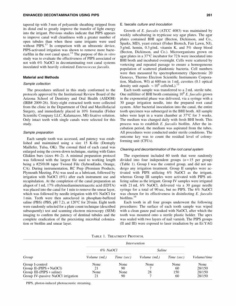
tapered tip with 3 mm of polyamide sheathing stripped fromits distal end to greatly improve the transfer of light energyinto the irrigant. Previous studies indicate that PIPS appearsto improve canal wall cleanliness with a greater number ofopen tubules than when these same irrigants were usedwithout PIPS.13 In comparison with an ultrasonic device,PIPS-activated irrigation was shown to remove more bacte-ria/film in the root canal space.14 The purpose of this in vitrostudy was to evaluate the effectiveness of PIPS associated ornot with 6% NaOCl in decontaminating root canal systemsinoculated with heavily colonized Enterococcus faecalis.
Material and Methods
Sample collection
The procedures utilized in this study conformed to theprotocols approved by the Institutional Review Board of theArizona School of Dentistry and Oral Health (ASDOH)(IRB# 2009-26). Sixty-eight extracted teeth were collectedfrom the clinic in the Department of Oral and MaxillofacialSurgery, and immediately placed in 10% formalin (FisherScientific Company LLC, Kalamazoo, MI) fixative solution.Only intact teeth with single canals were selected for thisstudy.
Sample preparation
Each sample tooth was accessed, and patency was estab-lished and maintained using a size 15 K-file (DentsplyMaillefer, Tulsa, OK). The coronal third of each canal wasenlarged using the crown-down technique, starting with GatesGlidden burs (sizes #4–2). A minimal preparation protocolwas followed with the largest file used to working lengthbeing a #25/0.08 taper Twisted File (SybronEndo, Orange,CA). During instrumentation, RC Prep (Premium Products,Plymouth Meeting, PA) was used as a lubricant, followed byirrigation with NaOCl (6%) after each instrument use andrecapitulation. At the completion of the canal preparation analiquot of 1 mL 17% ethylenediaminetetraacetic acid (EDTA)was placed into the canal for 1 min to remove the smear layer,which was followed by needle irrigation with 6% NaOCl for1 min. Teeth were then autoclaved in phosphate-bufferedsaline (PBS) (PBS, pH 7.2), at 120�C for 20 min. Eight teethwere randomly selected for a plate count technique (describedsubsequently) test and scanning electron microscopy (SEM)imaging to confirm the patency of dentinal tubules and thecomplete eradication of the preexisting microbial coloniza-tion or biofilm and smear layer.
E. faecalis culture and inoculation
Growth of E. faecalis (ATCC 4083) was maintained byweekly subculturing in trypticase soy agar plates. The agarplates contained BHI agar (Becton, Dickinson, and Co.,Sparks, MD), yeast extract (Fisher Biotech, Fair Lawn, NJ),5 g/mL hemin, 0.3 g/mL vitamin K, and 5% sheep blood(Becton, Dickinson, and Co.). Microorganisms grown onagar plates in a 37�C incubator for 72 h were inoculated intoBHI broth and incubated overnight. Cells were scattered byvortexing and repeated passage to ensure a homogeneouspopulation of scattered planktonic bacteria. Cell numberswere then measured by spectrophotometry (Spectronic 20Genesys, Thermo Electron Scientific Instruments Corpora-tion, Madison, WI) at 600 nm in 1 mL cuvettes (0.1 opticaldensity unit equals *108 cells/mL).15
Each tooth sample was transferred to a 2 mL sterile tube.One milliliter of BHI broth containing 108 E. faecalis grownin the exponential phase was delivered, via a syringe with a30 gauge irrigation needle, into the prepared root canalsystem. After bacterial inoculation into the canal, the entiretooth specimen was submerged in the BHI broth. All sampletubes were kept in a warm chamber at 37�C for 3 weeks.The medium was changed daily with fresh BHI broth. Thisprocess was to establish E. faecalis biofilm. After the in-cubation period, the medium was aspirated from the tubes.All procedures were conducted under sterile conditions. Theoutcome key was to count the residual level of colony-forming unit (CFUs).
Cleaning and decontamination of the root canal system
The experiment included 60 teeth that were randomlydivided into four independent groups (n = 15 per group)(Table 1). Group I was the control group, and did not un-dergo any irrigation treatment. Group II samples were ac-tivated with PIPS utilizing 6% NaOCl as the irrigant,whereas Group III samples were activated with PIPS uti-lizing saline as the irrigant. Group IV samples were irrigatedwith 21 mL 6% NaOCl, delivered via a 30 gauge needlesyringe for a total of 90 sec, but no PIPS. The 6% NaOClwas chosen for its effectiveness in disinfecting E. faecalisbiofilms.16
Each tooth in all four groups underwent the followingprocedures: The surface of each tooth sample was wipedwith a clean gauze pad soaked with NaOCl, after which thetooth was mounted onto a sterile plastic holder. The apexwas sealed with two layers of nail varnish. The PIPS groups(II and III) were exposed to laser irradiation by an Er:YAG
Table 1. Treatment Protocol
Intervention
6% NaOCl Saline
Group Volume (mL) Time (sec) Volume (mL) Time (sec) Volume/ time
Group I-control None None None None NoneGroup II-(PIPS + NaOCl) 21 90 7 60 28/150Group III-(PIPS + saline) None None 28 150 28/150Group IV-passive NaOCl irrigation 21 90 7 60 28/150
PIPS, photon-induced photoacoustic streaming.
ENHANCED DECONTAMINATION USING PIPS 261
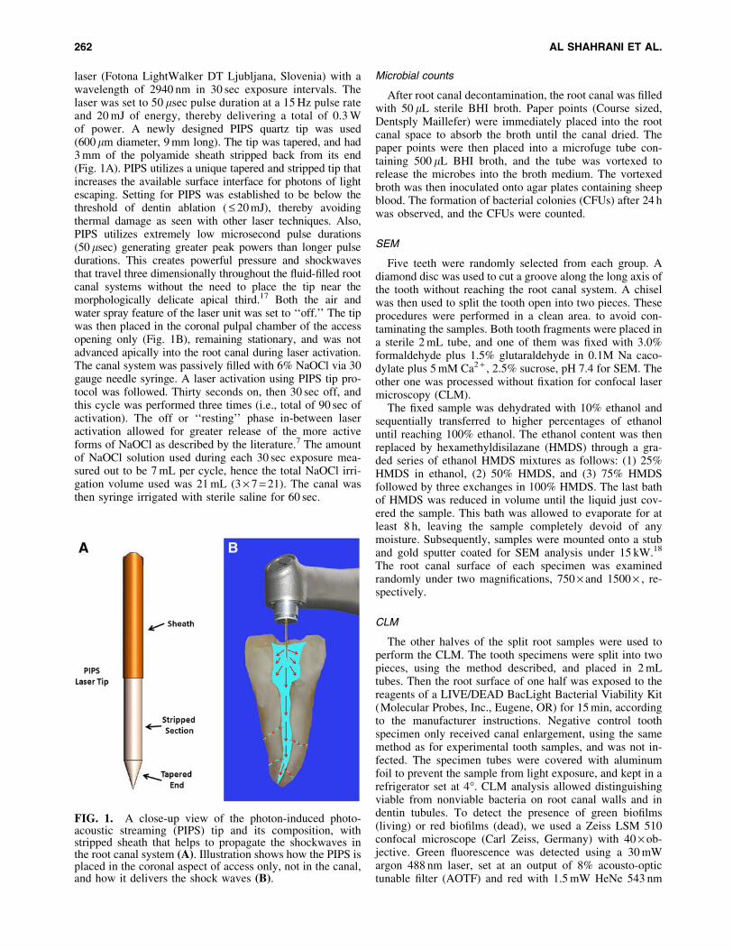
laser (Fotona LightWalker DT Ljubljana, Slovenia) with awavelength of 2940 nm in 30 sec exposure intervals. Thelaser was set to 50 lsec pulse duration at a 15 Hz pulse rateand 20 mJ of energy, thereby delivering a total of 0.3 Wof power. A newly designed PIPS quartz tip was used(600 lm diameter, 9 mm long). The tip was tapered, and had3 mm of the polyamide sheath stripped back from its end(Fig. 1A). PIPS utilizes a unique tapered and stripped tip thatincreases the available surface interface for photons of lightescaping. Setting for PIPS was established to be below thethreshold of dentin ablation ( £ 20 mJ), thereby avoidingthermal damage as seen with other laser techniques. Also,PIPS utilizes extremely low microsecond pulse durations(50 lsec) generating greater peak powers than longer pulsedurations. This creates powerful pressure and shockwavesthat travel three dimensionally throughout the fluid-filled rootcanal systems without the need to place the tip near themorphologically delicate apical third.17 Both the air andwater spray feature of the laser unit was set to ‘‘off.’’ The tipwas then placed in the coronal pulpal chamber of the accessopening only (Fig. 1B), remaining stationary, and was notadvanced apically into the root canal during laser activation.The canal system was passively filled with 6% NaOCl via 30gauge needle syringe. A laser activation using PIPS tip pro-tocol was followed. Thirty seconds on, then 30 sec off, andthis cycle was performed three times (i.e., total of 90 sec ofactivation). The off or ‘‘resting’’ phase in-between laseractivation allowed for greater release of the more activeforms of NaOCl as described by the literature.7 The amountof NaOCl solution used during each 30 sec exposure mea-sured out to be 7 mL per cycle, hence the total NaOCl irri-gation volume used was 21 mL (3 · 7 = 21). The canal wasthen syringe irrigated with sterile saline for 60 sec.
Microbial counts
After root canal decontamination, the root canal was filledwith 50 lL sterile BHI broth. Paper points (Course sized,Dentsply Maillefer) were immediately placed into the rootcanal space to absorb the broth until the canal dried. Thepaper points were then placed into a microfuge tube con-taining 500 lL BHI broth, and the tube was vortexed torelease the microbes into the broth medium. The vortexedbroth was then inoculated onto agar plates containing sheepblood. The formation of bacterial colonies (CFUs) after 24 hwas observed, and the CFUs were counted.
SEM
Five teeth were randomly selected from each group. Adiamond disc was used to cut a groove along the long axis ofthe tooth without reaching the root canal system. A chiselwas then used to split the tooth open into two pieces. Theseprocedures were performed in a clean area. to avoid con-taminating the samples. Both tooth fragments were placed ina sterile 2 mL tube, and one of them was fixed with 3.0%formaldehyde plus 1.5% glutaraldehyde in 0.1M Na caco-dylate plus 5 mM Ca2 + , 2.5% sucrose, pH 7.4 for SEM. Theother one was processed without fixation for confocal lasermicroscopy (CLM).
The fixed sample was dehydrated with 10% ethanol andsequentially transferred to higher percentages of ethanoluntil reaching 100% ethanol. The ethanol content was thenreplaced by hexamethyldisilazane (HMDS) through a gra-ded series of ethanol HMDS mixtures as follows: (1) 25%HMDS in ethanol, (2) 50% HMDS, and (3) 75% HMDSfollowed by three exchanges in 100% HMDS. The last bathof HMDS was reduced in volume until the liquid just cov-ered the sample. This bath was allowed to evaporate for atleast 8 h, leaving the sample completely devoid of anymoisture. Subsequently, samples were mounted onto a stuband gold sputter coated for SEM analysis under 15 kW.18
The root canal surface of each specimen was examinedrandomly under two magnifications, 750 · and 1500 · , re-spectively.
CLM
The other halves of the split root samples were used toperform the CLM. The tooth specimens were split into twopieces, using the method described, and placed in 2 mLtubes. Then the root surface of one half was exposed to thereagents of a LIVE/DEAD BacLight Bacterial Viability Kit(Molecular Probes, Inc., Eugene, OR) for 15 min, accordingto the manufacturer instructions. Negative control toothspecimen only received canal enlargement, using the samemethod as for experimental tooth samples, and was not in-fected. The specimen tubes were covered with aluminumfoil to prevent the sample from light exposure, and kept in arefrigerator set at 4�. CLM analysis allowed distinguishingviable from nonviable bacteria on root canal walls and indentin tubules. To detect the presence of green biofilms(living) or red biofilms (dead), we used a Zeiss LSM 510confocal microscope (Carl Zeiss, Germany) with 40 · ob-jective. Green fluorescence was detected using a 30 mWargon 488 nm laser, set at an output of 8% acousto-optictunable filter (AOTF) and red with 1.5 mW HeNe 543 nm
FIG. 1. A close-up view of the photon-induced photo-acoustic streaming (PIPS) tip and its composition, withstripped sheath that helps to propagate the shockwaves inthe root canal system (A). Illustration shows how the PIPS isplaced in the coronal aspect of access only, not in the canal,and how it delivers the shock waves (B).
262 AL SHAHRANI ET AL.
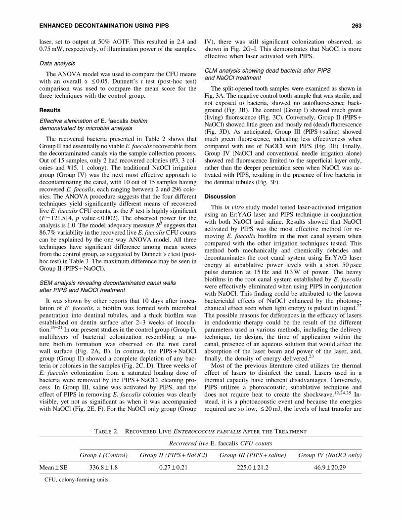
laser, set to output at 50% AOTF. This resulted in 2.4 and0.75 mW, respectively, of illumination power of the samples.
Data analysis
The ANOVA model was used to compare the CFU meanswith an overall a £ 0.05. Dunnett’s t test (post-hoc test)comparison was used to compare the mean score for thethree techniques with the control group.
Results
Effective elimination of E. faecalis biofilmdemonstrated by microbial analysis
The recovered bacteria presented in Table 2 shows thatGroup II had essentially no viable E. faecalis recoverable fromthe decontaminated canals via the sample collection process.Out of 15 samples, only 2 had recovered colonies (#3, 3 col-onies and #15, 1 colony). The traditional NaOCl irrigationgroup (Group IV) was the next most effective approach todecontaminating the canal, with 10 out of 15 samples havingrecovered E. faecalis, each ranging between 2 and 296 colo-nies. The ANOVA procedure suggests that the four differenttechniques yield significantly different means of recoveredlive E. faecalis CFU counts, as the F test is highly significant(F = 121.514, p value < 0.002). The observed power for theanalysis is 1.0. The model adequacy measure R2 suggests that86.7% variability in the recovered live E. faecalis CFU countscan be explained by the one way ANOVA model. All threetechniques have significant difference among mean scoresfrom the control group, as suggested by Dunnett’s t test (post-hoc test) in Table 3. The maximum difference may be seen inGroup II (PIPS + NaOCl).
SEM analysis revealing decontaminated canal wallsafter PIPS and NaOCl treatment
It was shown by other reports that 10 days after inocu-lation of E. faecalis, a biofilm was formed with microbialpenetration into dentinal tubules, and a thick biofilm wasestablished on dentin surface after 2–3 weeks of inocula-tion.19–21 In our present studies in the control group (Group I),multilayers of bacterial colonization resembling a ma-ture biofilm formation was observed on the root canalwall surface (Fig. 2A, B). In contrast, the PIPS + NaOClgroup (Group II) showed a complete depletion of any bac-teria or colonies in the samples (Fig. 2C, D). Three weeks ofE. faecalis colonization from a saturated loading dose ofbacteria were removed by the PIPS + NaOCl cleaning pro-cess. In Group III, saline was activated by PIPS, and theeffect of PIPS in removing E. faecalis colonies was clearlyvisible, yet not as significant as when it was accompaniedwith NaOCl (Fig. 2E, F). For the NaOCl only group (Group
IV), there was still significant colonization observed, asshown in Fig. 2G–I. This demonstrates that NaOCl is moreeffective when laser activated with PIPS.
CLM analysis showing dead bacteria after PIPSand NaOCl treatment
The split-opened tooth samples were examined as shown inFig. 3A. The negative control tooth sample that was sterile, andnot exposed to bacteria, showed no autofluorescence back-ground (Fig. 3B). The control (Group I) showed much green(living) fluorescence (Fig. 3C). Conversely, Group II (PIPS +NaOCl) showed little green and mostly red (dead) fluorescence(Fig. 3D). As anticipated, Group III (PIPS + saline) showedmuch green fluorescence, indicating less effectiveness whencompared with use of NaOCl with PIPS (Fig. 3E). Finally,Group IV (NaOCl and conventional needle irrigation alone)showed red fluorescence limited to the superficial layer only,rather than the deeper penetration seen when NaOCl was ac-tivated with PIPS, resulting in the presence of live bacteria inthe dentinal tubules (Fig. 3F).
Discussion
This in vitro study model tested laser-activated irrigationusing an Er:YAG laser and PIPS technique in conjunctionwith both NaOCl and saline. Results showed that NaOClactivated by PIPS was the most effective method for re-moving E. faecalis biofilm in the root canal system whencompared with the other irrigation techniques tested. Thismethod both mechanically and chemically debrides anddecontaminates the root canal system using Er:YAG laserenergy at subablative power levels with a short 50 lsecpulse duration at 15 Hz and 0.3 W of power. The heavybiofilms in the root canal system established by E. faecaliswere effectively eliminated when using PIPS in conjunctionwith NaOCl. This finding could be attributed to the knownbactericidal effects of NaOCl enhanced by the photome-chanical effect seen when light energy is pulsed in liquid.22
The possible reasons for differences in the efficacy of lasersin endodontic therapy could be the result of the differentparameters used in various methods, including the deliverytechnique, tip design, the time of application within thecanal, presence of an aqueous solution that would affect theabsorption of the laser beam and power of the laser, and,finally, the density of energy delivered.23
Most of the previous literature cited utilizes the thermaleffect of lasers to disinfect the canal. Lasers used in athermal capacity have inherent disadvantages. Conversely,PIPS utilizes a photoacoustic, subablative technique anddoes not require heat to create the shockwave.12,24,25 In-stead, it is a photoacoustic event and because the energiesrequired are so low, £ 20 mJ, the levels of heat transfer are
Table 2. Recovered Live Enterococcus faecalis After the Treatment
Recovered live E. faecalis CFU counts
Group I (Control) Group II (PIPS + NaOCl) Group III (PIPS + saline) Group IV (NaOCl only)
Mean – SE 336.8 – 1.8 0.27 – 0.21 225.0 – 21.2 46.9 – 20.29
CFU, colony-forming units.
ENHANCED DECONTAMINATION USING PIPS 263
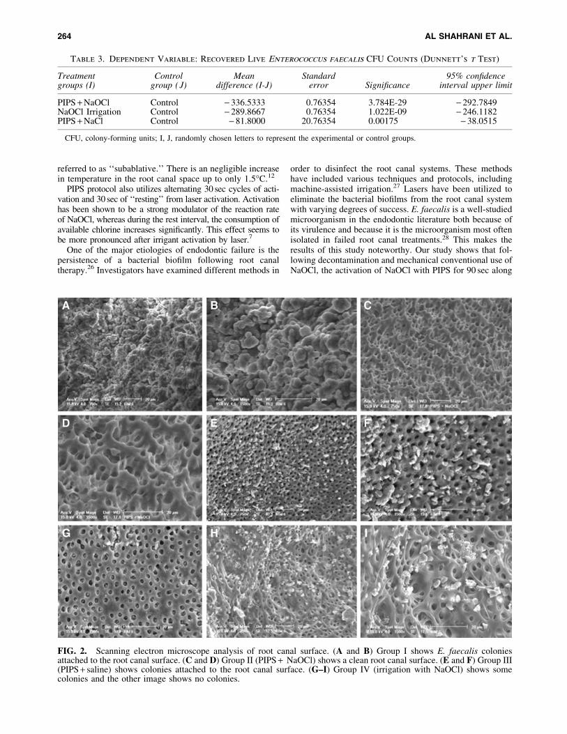
referred to as ‘‘subablative.’’ There is an negligible increasein temperature in the root canal space up to only 1.5�C.12
PIPS protocol also utilizes alternating 30 sec cycles of acti-vation and 30 sec of ‘‘resting’’ from laser activation. Activationhas been shown to be a strong modulator of the reaction rateof NaOCl, whereas during the rest interval, the consumption ofavailable chlorine increases significantly. This effect seems tobe more pronounced after irrigant activation by laser.7
One of the major etiologies of endodontic failure is thepersistence of a bacterial biofilm following root canaltherapy.26 Investigators have examined different methods in
order to disinfect the root canal systems. These methodshave included various techniques and protocols, includingmachine-assisted irrigation.27 Lasers have been utilized toeliminate the bacterial biofilms from the root canal systemwith varying degrees of success. E. faecalis is a well-studiedmicroorganism in the endodontic literature both because ofits virulence and because it is the microorganism most oftenisolated in failed root canal treatments.28 This makes theresults of this study noteworthy. Our study shows that fol-lowing decontamination and mechanical conventional use ofNaOCl, the activation of NaOCl with PIPS for 90 sec along
Table 3. Dependent Variable: Recovered Live Enterococcus faecalis CFU Counts (Dunnett’s t Test)
Treatmentgroups (I)
Controlgroup ( J)
Meandifference (I-J)
Standarderror Significance
95% confidenceinterval upper limit
PIPS + NaOCl Control - 336.5333 0.76354 3.784E-29 - 292.7849NaOCl Irrigation Control - 289.8667 0.76354 1.022E-09 - 246.1182PIPS + NaCl Control - 81.8000 20.76354 0.00175 - 38.0515
CFU, colony-forming units; I, J, randomly chosen letters to represent the experimental or control groups.
FIG. 2. Scanning electron microscope analysis of root canal surface. (A and B) Group I shows E. faecalis coloniesattached to the root canal surface. (C and D) Group II (PIPS + NaOCl) shows a clean root canal surface. (E and F) Group III(PIPS + saline) shows colonies attached to the root canal surface. (G–I) Group IV (irrigation with NaOCl) shows somecolonies and the other image shows no colonies.
264 AL SHAHRANI ET AL.
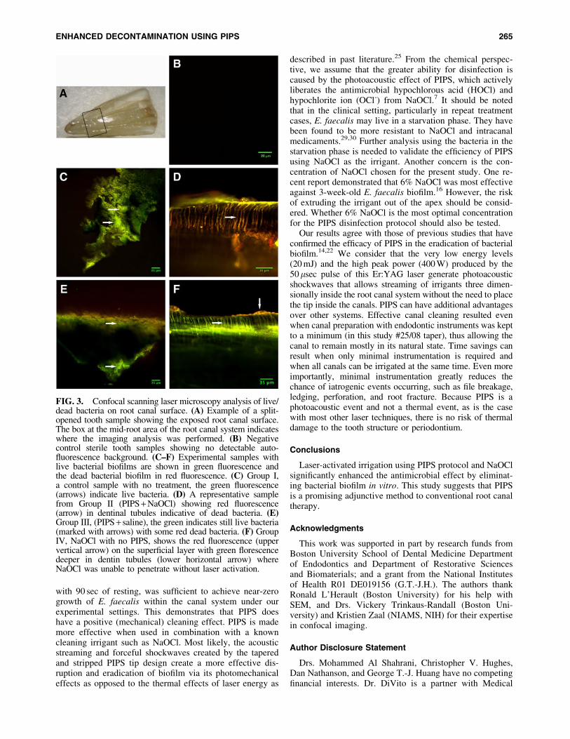
with 90 sec of resting, was sufficient to achieve near-zerogrowth of E. faecalis within the canal system under ourexperimental settings. This demonstrates that PIPS doeshave a positive (mechanical) cleaning effect. PIPS is mademore effective when used in combination with a knowncleaning irrigant such as NaOCl. Most likely, the acousticstreaming and forceful shockwaves created by the taperedand stripped PIPS tip design create a more effective dis-ruption and eradication of biofilm via its photomechanicaleffects as opposed to the thermal effects of laser energy as
described in past literature.25 From the chemical perspec-tive, we assume that the greater ability for disinfection iscaused by the photoacoustic effect of PIPS, which activelyliberates the antimicrobial hypochlorous acid (HOCl) andhypochlorite ion (OCl-) from NaOCl.7 It should be notedthat in the clinical setting, particularly in repeat treatmentcases, E. faecalis may live in a starvation phase. They havebeen found to be more resistant to NaOCl and intracanalmedicaments.29,30 Further analysis using the bacteria in thestarvation phase is needed to validate the efficiency of PIPSusing NaOCl as the irrigant. Another concern is the con-centration of NaOCl chosen for the present study. One re-cent report demonstrated that 6% NaOCl was most effectiveagainst 3-week-old E. faecalis biofilm.16 However, the riskof extruding the irrigant out of the apex should be consid-ered. Whether 6% NaOCl is the most optimal concentrationfor the PIPS disinfection protocol should also be tested.
Our results agree with those of previous studies that haveconfirmed the efficacy of PIPS in the eradication of bacterialbiofilm.14,22 We consider that the very low energy levels(20 mJ) and the high peak power (400 W) produced by the50 lsec pulse of this Er:YAG laser generate photoacousticshockwaves that allows streaming of irrigants three dimen-sionally inside the root canal system without the need to placethe tip inside the canals. PIPS can have additional advantagesover other systems. Effective canal cleaning resulted evenwhen canal preparation with endodontic instruments was keptto a minimum (in this study #25/08 taper), thus allowing thecanal to remain mostly in its natural state. Time savings canresult when only minimal instrumentation is required andwhen all canals can be irrigated at the same time. Even moreimportantly, minimal instrumentation greatly reduces thechance of iatrogenic events occurring, such as file breakage,ledging, perforation, and root fracture. Because PIPS is aphotoacoustic event and not a thermal event, as is the casewith most other laser techniques, there is no risk of thermaldamage to the tooth structure or periodontium.
Conclusions
Laser-activated irrigation using PIPS protocol and NaOClsignificantly enhanced the antimicrobial effect by eliminat-ing bacterial biofilm in vitro. This study suggests that PIPSis a promising adjunctive method to conventional root canaltherapy.
Acknowledgments
This work was supported in part by research funds fromBoston University School of Dental Medicine Departmentof Endodontics and Department of Restorative Sciencesand Biomaterials; and a grant from the National Institutesof Health R01 DE019156 (G.T.-J.H.). The authors thankRonald L’Herault (Boston University) for his help withSEM, and Drs. Vickery Trinkaus-Randall (Boston Uni-versity) and Kristien Zaal (NIAMS, NIH) for their expertisein confocal imaging.
Author Disclosure Statement
Drs. Mohammed Al Shahrani, Christopher V. Hughes,Dan Nathanson, and George T.-J. Huang have no competingfinancial interests. Dr. DiVito is a partner with Medical
FIG. 3. Confocal scanning laser microscopy analysis of live/dead bacteria on root canal surface. (A) Example of a split-opened tooth sample showing the exposed root canal surface.The box at the mid-root area of the root canal system indicateswhere the imaging analysis was performed. (B) Negativecontrol sterile tooth samples showing no detectable auto-fluorescence background. (C–F) Experimental samples withlive bacterial biofilms are shown in green fluorescence andthe dead bacterial biofilm in red fluorescence. (C) Group I,a control sample with no treatment, the green fluorescence(arrows) indicate live bacteria. (D) A representative samplefrom Group II (PIPS + NaOCl) showing red fluorescence(arrow) in dentinal tubules indicative of dead bacteria. (E)Group III, (PIPS + saline), the green indicates still live bacteria(marked with arrows) with some red dead bacteria. (F) GroupIV, NaOCl with no PIPS, shows the red fluorescence (uppervertical arrow) on the superficial layer with green florescencedeeper in dentin tubules (lower horizontal arrow) whereNaOCl was unable to penetrate without laser activation.
ENHANCED DECONTAMINATION USING PIPS 265
Dental Advance Technologies Group (MDATG) and has afinancial affiliation as a partner in the MDATG group, whichhelped support a portion of the present studies (use of thelaser machine).
References
1. Basmadjian-Charles, C.L., Farge, P., Bourgeois, D.M., andLebrun, T. (2002). Factors influencing the long-term resultsof endodontic treatment: a review of the literature. Int.Dent. J. 52, 81–86.
2. Siqueira, J.F., Jr., and Rocas, I.N. (2008). Clinical impli-cations and microbiology of bacterial persistence aftertreatment procedures. J. Endod. 34, 1291–1301.
3. Wong, R. (2004). Conventional endodontic failure and re-treatment. Dent. Clin. North Am. 48, 265–289.
4. Gu, L.S., Kim, J.R., Ling, J., Choi, K.K., Pashley, D.H., andTay, F.R. (2009). Review of contemporary irrigant agita-tion techniques and devices. J. Endod. 35, 791–804.
5. Cameron, J.A. (1987). The synergistic relationship betweenultrasound and sodium hypochlorite: a scanning electronmicroscope evaluation. J. Endod. 13, 541–545.
6. Sabins, R.A., Johnson, J.D., and Hellstein, J.W. (2003). Acomparison of the cleaning efficacy of short-term sonic andultrasonic passive irrigation after hand instrumentation inmolar root canals. J. Endod. 29, 674–678.
7. Macedo, R.G., Wesselink, P.R., Zaccheo, F., Fanali, D.,and Van Der Sluis, L.W. (2010). Reaction rate of NaOCl incontact with bovine dentine: effect of activation, exposuretime, concentration and pH. Int. Endod. J. 43, 1108–1115.
8. Yasuda, Y., Kawamorita, T., Yamaguchi, H., and Saito, T.(2010). Bactericidal effect of Nd:YAG and Er:YAG lasersin experimentally infected curved root canals. Photomed.Laser Surg. 28 Suppl 2, S75–S78.
9. Blanken, J., De Moor, R.J., Meire, M., and Verdaasdonk, R.(2009). Laser induced explosive vapor and cavitation re-sulting in effective irrigation of the root canal. Part 1: avisualization study. Lasers Surg. Med. 41, 514–519.
10. De Moor, R.J., Blanken, J., Meire, M., and Verdaasdonk, R.(2009). Laser induced explosive vapor and cavitation re-sulting in effective irrigation of the root canal. Part 2:evaluation of the efficacy. Lasers Surg. Med. 41, 520–523.
11. Matsumoto, H., Yoshimine, Y., and Akamine, A. (2011).Visualization of irrigant flow and cavitation induced byEr:YAG laser within a root canal model. J. Endod. 37, 839–843.
12. DiVito, E.E., Colonna, M.P., and Olivi, G. (2011). Thephotoacoustic efficacy of an Er: YAG laser with radial andstripped tips on root canal dentin walls: an SEM evaluation.J. Laser Dent. 19, 156–161.
13. DiVito, E., Peters, O.A., and Olivi, G. (2012). Effectivenessof the erbium:YAG laser and new design radial and strip-ped tips in removing the smear layer after root canal in-strumentation. Lasers Med. Sci. 27, 273–280.
14. Peters, O.A., Bardsley, S., Fong, J., Pandher, G., and Divito,E. (2011). Disinfection of root canals with photon-initiatedphotoacoustic streaming. J. Endod. 37, 1008–1012.
15. Fimple, J.L., Fontana, C.R., Foschi, F., Ruggiero, K., Song,X., Pagonis, T.C., Tanner, A.C., Kent, R., Doukas, A.G.,Stashenko, P.P., and Soukos, N.S. (2008). Photodynamictreatment of endodontic polymicrobial infection in vitro. J.Endod. 34, 728–734.
16. Wang, Z., Shen, Y., and Haapasalo, M. (2012). Effective-ness of endodontic disinfecting solutions against young and
old Enterococcus faecalis biofilms in dentin canals. J. En-dod. 38, 1376–1379.
17. Olivi, G. (2013). Laser use in endodontics: evolution fromdirect laser irradiation to laser-activated irrigation. J. LaserDent. 21, 58–71.
18. Braet, F., De Zanger, R., and Wisse, E. (1997). Drying cellsfor SEM, AFM and TEM by hexamethyldisilazane: a studyon hepatic endothelial cells. J. Microsc. 186, 84–87.
19. Saber Sel, D., and El-Hady, S.A. (2012). Development ofan intracanal mature Enterococcus faecalis biofilm and itssusceptibility to some antimicrobial intracanal medications;an in vitro study. Eur. J. Dent. 6, 43–50.
20. Kishen, A., George, S., and Kumar, R. (2006). Entero-coccus faecalis-mediated biomineralized biofilm formationon root canal dentine in vitro. J Biomed. Mater. Res. A 77,406–415.
21. Case, P.D., Bird, P.S., Kahler, W.A., George, R., andWalsh, L.J. (2012). Treatment of root canal biofilms ofEnterococcus faecalis with ozone gas and passive ultra-sound activation. J. Endod. 38, 523–526.
22. De Groot, S.D., Verhaagen, B., Versluis, M., Wu, M.K.,Wesselink, P.R., and Van der Sluis, L.W. (2009). Laser-activated irrigation within root canals: cleaning efficacyand flow visualization. Int. Endod. J. 42, 1077–1083.
23. Seet, A.N., Zilm, P.S., Gully, N.J., and Cathro, P.R. (2012).Qualitative comparison of sonic or laser energisation of 4%sodium hypochlorite on an Enterococcus faecalis biofilmgrown in vitro. Aust. Endod. J. 38, 100–106.
24. Doukas, A.G., Zweig, A.D., Frisoli, J.K., Birngruber, R.,and Deutsch, T.F. (1991). Non-invasive determination ofshock wave pressure generated by optical breakdown.Appl. Phys. B 53, 237–245.
25. Muller, P., Guggenheim, B., Attin, T., Marlinghaus, E., andSchmidlin, P.R. (2011). Potential of shock waves to removecalculus and biofilm. Clin. Oral Investig. 15, 959–965.
26. Baumgartner, J.C. (2004). Microbiologic aspects of end-odontic infections. J. Calif. Dent. Assoc. 32, 459–468.
27. Stuart, C.H., Schwartz, S.A., Beeson, T.J., and Owatz, C.B.(2006). Enterococcus faecalis: its role in root canal treat-ment failure and current concepts in retreatment. J. Endod.32, 93–98.
28. Dumani, A., Yoldas, O., Yilmaz, S., Koksal, F., Kayar, B.,Akcimen, B., and Seydaoglu, G. (2012). Polymerase chainreaction of enterococcus faecalis and candida albicans inapical periodontitis from Turkish patients. J. Clin. Exp.Dent. 4, e34–e39.
29. Portenier, I., Waltimo, T., Orstavik, D., and Haapasalo, M.(2005). The susceptibility of starved, stationary phase, andgrowing cells of Enterococcus faecalis to endodontic me-dicaments. J. Endod. 31, 380–386.
30. Aoki, A., Sasaki, K.M., Watanabe, H., and Ishikawa, I.(2004). Lasers in nonsurgical periodontal therapy. Period-ontol. 2000 36, 59–97.
Address correspondence to:George T.-J. Huang
Department of Bioscience ResearchCollege of Dentistry
University of Tennessee Health Science Center875 Union Avenue
Memphis, TN 38163
E-mail: [email protected]
266 AL SHAHRANI ET AL.
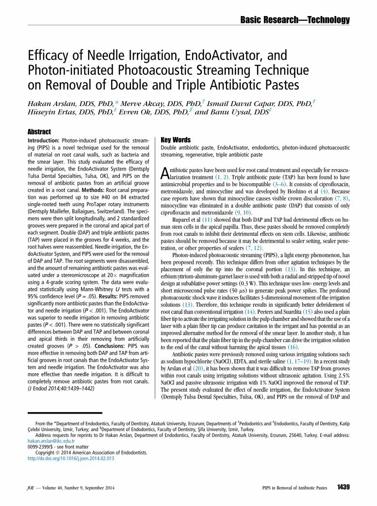
Basic Research—Technology
Efficacy of Needle Irrigation, EndoActivator, andPhoton-initiated Photoacoustic Streaming Techniqueon Removal of Double and Triple Antibiotic PastesHakan Arslan, DDS, PhD,* Merve Akcay, DDS, PhD,† Ismail Davut Capar, DDS, PhD,‡
H€useyin Ertas, DDS, PhD,‡ Evren Ok, DDS, PhD,§ and Banu Uysal, DDS‡
Abstract
Introduction: Photon-induced photoacoustic stream-ing (PIPS) is a novel technique used for the removalof material on root canal walls, such as bacteria andthe smear layer. This study evaluated the efficacy ofneedle irrigation, the EndoActivator System (DentsplyTulsa Dental Specialties, Tulsa, OK), and PIPS on theremoval of antibiotic pastes from an artificial groovecreated in a root canal. Methods: Root canal prepara-tion was performed up to size #40 on 84 extractedsingle-rooted teeth using ProTaper rotary instruments(Dentsply Maillefer, Ballaigues, Switzerland). The speci-mens were then split longitudinally, and 2 standardizedgrooves were prepared in the coronal and apical part ofeach segment. Double (DAP) and triple antibiotic pastes(TAP) were placed in the grooves for 4 weeks, and theroot halves were reassembled. Needle irrigation, the En-doActivator System, and PIPS were used for the removalof DAP and TAP. The root segments were disassembled,and the amount of remaining antibiotic pastes was eval-uated under a stereomicroscope at 20� magnificationusing a 4-grade scoring system. The data were evalu-ated statistically using Mann-Whitney U tests with a95% confidence level (P = .05). Results: PIPS removedsignificantly more antibiotic pastes than the EndoActiva-tor and needle irrigation (P < .001). The EndoActivatorwas superior to needle irrigation in removing antibioticpastes (P < .001). There were no statistically significantdifferences between DAP and TAP and between coronaland apical thirds in their removing from artificiallycreated grooves (P > .05). Conclusions: PIPS wasmore effective in removing both DAP and TAP from arti-ficial grooves in root canals than the EndoActivator Sys-tem and needle irrigation. The EndoActivator was alsomore effective than needle irrigation. It is difficult tocompletely remove antibiotic pastes from root canals.(J Endod 2014;40:1439–1442)From the *Department of Endodontics, Faculty of Dentistry, AtatCelebi University, _Izmir, Turkey; and §Department of Endodontics, F
Address requests for reprints to Dr Hakan Arslan, [email protected]/$ - see front matter
Copyright ª 2014 American Association of Endodontists.http://dx.doi.org/10.1016/j.joen.2014.02.013
JOE — Volume 40, Number 9, September 2014
Key WordsDouble antibiotic paste, EndoActivator, endodontics, photon-induced photoacousticstreaming, regenerative, triple antibiotic paste
Antibiotic pastes have been used for root canal treatment and especially for revascu-larization treatment (1, 2). Triple antibiotic paste (TAP) has been found to have
antimicrobial properties and to be biocompatible (3–6). It consists of ciprofloxacin,metronidazole, and minocycline and was developed by Hoshino et al (4). Becausecase reports have shown that minocycline causes visible crown discoloration (7, 8),minocycline was eliminated in a double antibiotic paste (DAP) that consists of onlyciprofloxacin and metronidazole (9, 10).
Ruparel et al (11) showed that both DAP and TAP had detrimental effects on hu-man stem cells in the apical papilla. Thus, these pastes should be removed completelyfrom root canals to inhibit their detrimental effects on stem cells. Likewise, antibioticpastes should be removed because it may be detrimental to sealer setting, sealer pene-tration, or other properties of sealers (7, 12).
Photon-induced photoacoustic streaming (PIPS), a light energy phenomenon, hasbeen proposed recently. This technique differs from other agitation techniques by theplacement of only the tip into the coronal portion (13). In this technique, anerbium:yttrium-aluminum-garnet laser is used with both a radial and stripped tip of noveldesign at subablative power settings (0.3 W). This technique uses low- energy levels andshort microsecond pulse rates (50 ms) to generate peak power spikes. The profoundphotoacoustic shock wave it induces facilitates 3-dimensional movement of the irrigationsolutions (13). Therefore, this technique results in significantly better debridement ofroot canal than conventional irrigation (14). Peeters and Suardita (15) also used a plainfiber tip to activate the irrigating solution in the pulp chamber and showed that the use of alaser with a plain fiber tip can produce cavitation in the irrigant and has potential as animproved alternative method for the removal of the smear layer. In another study, it hasbeen reported that the plain fiber tip in the pulp chamber can drive the irrigation solutionto the end of the canal without harming the apical tissues (16).
Antibiotic pastes were previously removed using various irrigating solutions suchas sodium hypochlorite (NaOCl), EDTA, and sterile saline (1, 17–19). In a recent studyby Arslan et al (20), it has been shown that it was difficult to remove TAP from grooveswithin root canals using irrigating solutions without ultrasonic agitation. Using 2.5%NaOCl and passive ultrasonic irrigation with 1% NaOCl improved the removal of TAP.The present study evaluated the effect of needle irrigation, the EndoActivator System(Dentsply Tulsa Dental Specialties, Tulsa, OK), and PIPS on the removal of DAP and
urk University, Erzurum; Departments of †Pedodontics and ‡Endodontics, Faculty of Dentistry, Katipaculty of Dentistry, Sifa University, _Izmir, Turkey.of Endodontics, Faculty of Dentistry, Ataturk University, Erzurum, 25640, Turkey. E-mail address:
PIPS in Removal of Antibiotic Pastes 1439
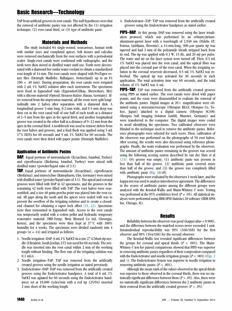
Basic Research—Technology
TAP from artificial grooves in root canals. The null hypotheses were thatthe removal of antibiotic pastes was not affected by the (1) irrigationtechnique, (2) root canal third, or (3) type of antibiotic paste.Materials and MethodsThe study included 84 single-rooted, noncarious, human teeth
with similar sizes and completed apices. Soft tissues and calculuswere removed mechanically from the root surfaces with a periodontalscaler. Single-root canals were confirmed with radiographs, and theteeth were then stored in distilled water until use. Teeth were decoro-nated with a diamond bur under water coolant to obtain a standardizedroot length of 14 mm. The root canals were shaped with ProTaper ro-tary files (Dentsply Maillefer, Ballaigues, Switzerland) up to an F4(D0 = .40 mm). During preparation, the root canals were irrigatedwith 2 mL 1% NaOCl solution after each instrument. The specimenswere fixed in Eppendorf vials (Eppendorf-Elkay, Shrewsbury, MA)with a silicone material (Optosil; Heraeus Kulzer, Hanau, Germany). Af-ter removal from the impression material, all the roots were split longi-tudinally into 2 halves after separation with a diamond disk. Alongitudinal groove 3-mm long, 0.2-mm wide, and 0.5-mm deep wasthen cut in the root canal wall of one half of each tooth at a distanceof 2–5 mm from the apex in the apical third, and another longitudinalgroove was created in the other half at a distance of 9–12 mm from theapex in the coronal third. A toothbrush was used to remove debris fromthe root halves and grooves, and a final flush was applied using 5 mL17% EDTA for 60 seconds and 5 mL 1% NaOCl for 60 seconds. Theroot canals were then dried with paper points (Dentsply Maillefer).
Application of Antibiotic PastesDAP. Equal portions of metronidazole (Eczacibasi, Istanbul, Turkey)and ciprofloxacin (Biofarma, Istanbul, Turkey) were mixed withdistilled water (powder/liquid ratio of 3:1).
TAP. Equal portions of metronidazole (Eczacibasi), ciprofloxacin(Biofarma), andminocycline (Ratiopharm, Ulm, Germany) weremixedwith distilled water (powder/liquid ratio of 3:1). The apical and coronalgrooves were filled with DAP in 42 specimens, and the grooves in theremaining 42 teeth were filled with TAP. The root halves were reas-sembled, and a size 40 gutta-percha point was placed into the root ca-nal. All gaps along the tooth and the apices were sealed with wax toprevent the overflow of the irrigating solution and to create a closed-end channel for obtaining a vapor lock effect (21, 22). Specimenswere then remounted in Eppendorf vials. Access to the root canalswas temporarily sealed with a cotton pellet and hydraulic temporaryrestorative material (MD-Temp; Meta Biomed Co Ltd, Cheongju,Korea), and the specimens were then kept at 37�C with 100%humidity for 4 weeks. The specimens were divided randomly into 4groups (n = 14) and irrigated as follows:
1. Needle irrigation–DAP: 6 mL 1% NaOCl via a size 27-G blunt-tip nee-dle (Ultradent, South Jordan, UT) was used for 60 seconds. The nee-dle was inserted into the root canal within 2 mm of the workinglength without binding. The flow rate of the irrigating solution was0.1 mL/s.
2. Needle irrigation–TAP: TAP was removed from the artificiallycreated grooves using the needle irrigation as stated previously.
3. EndoActivator–DAP: DAP was removed from the artificially createdgrooves using the EndoActivator handpiece. A total of 6 mL 1%NaOCl was agitated for 60 seconds using the EndoActivator hand-piece set at 10,000 cycles/min with a red tip (25/04) inserted2 mm short of the working length.
1440 Arslan et al.
4. EndoActivator–TAP: TAP was removed from the artificially createdgrooves using the EndoActivator handpiece as stated earlier.
PIPS–DAP. In this group, DAP was removed using the laser irradi-ation protocol, which was performed by an erbium:yttrium-aluminum-garnet laser with a wavelength of 2,940 nm (Fidelis AT;Fotona, Ljubljana, Slovenia); a 14-mm-long, 300-mm quartz tip wastapered and had 3 mm of the polyamide sheath stripped back fromits end. The tip was applied with 0.3 W, 15 Hz, and 20 mJ per pulse.The water and air on the laser system were turned off. Then, 0.5 mL1% NaOCl was placed into the root canal, and the optical fiber wasplaced into the coronal part of the root canal. When the irrigating so-lution in the coronal reservoir decreased, 0.5 mL 1% NaOCl was re-freshed. The optical tip was activated for 30 seconds in eachapplication. The total activation time was 60 seconds, and the totalvolume of 1% NaOCl was 6 mL.
PIPS–TAP. TAP was removed from the artificially created groovesusing PIPS as stated earlier. The root canals were dried with paperpoints, and the roots were disassembled to evaluate the removal ofthe antibiotic pastes. Digital images at 20� magnification were ob-tained using a stereomicroscope (Olympus BX43; Olympus Co, To-kyo, Japan) attached to a digital camera (Olympus SC100;Olympus Soft Imaging Solution GmbH, Munster, Germany) andwere transferred to the computer. The digital images were codedto avoid identifying the specimens. Two calibrated observers wereblinded to the technique used to remove the antibiotic pastes. Refer-ence photographs were selected for each score. Then, calibration ofthe observers was performed on the photographs of 50 root halves.After scoring, the results were also discussed using reference photo-graphs. Finally, the main evaluation was performed by the observers.The amount of antibiotic pastes remaining in the grooves was scoredusing the following scoring system described by van der Sluis et al(23): (0) groove was empty, (1) antibiotic paste was present inless than half of the groove, (2) antibiotic paste covered morethan half of the groove, and (3) the groove was completely filledwith antibiotic paste (Fig. 1A–H).
Photographs were evaluated by the observers 1 week later, and thekappa test was used to analyze interobserver agreement. The differencesin the scores of antibiotic pastes among the different groups wereanalyzed with the Kruskal-Wallis and Mann-Whitney U tests. Testingwas performed at the 95% confidence level (P = .05). All statistical an-alyses were performed using IBM SPSS Statistics 20 software (IBM SPSSInc, Chicago, IL).
ResultsReliability between the observers was good (kappa value= 0.908),
and the difference between the matched scores never exceeded 1 unit.Intraindividual reproducibility was 99% (166/168) for the firstobserver and 98% (164/168) for the second observer.
The Kruskal-Wallis test revealed significant differences betweenthe groups for coronal and apical thirds (P < .001). The Mann-Whitney U test for paired comparisons showed that PIPS was superiorin removing antibiotic pastes regardless of their composition comparedwith the EndoActivator and needle irrigation groups (P< .001) (Figs. 2and 3). The EndoActivator System was superior to needle irrigation inremoving antibiotic pastes (P < .001).
Although the mean rank of the values observed in the apical thirdswas superior to those observed in the coronal thirds, there was no sta-tistically significant difference between them (P> .05). Also, there wereno statistically significant differences between the 2 antibiotic pastes intheir removal from the artificially created grooves (P > .05).
JOE — Volume 40, Number 9, September 2014
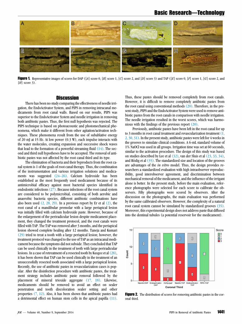
Figure 2. The distribution of scores for removing antibiotic pastes in the cor-onal third.
Figure 1. Representative images of scores for DAP ([A] score 0, [B] score 1, [C] score 2, and [D] score 3) and TAP ([E] score 0, [F] score 1, [G] score 2, and[H] score 3).
Basic Research—Technology
DiscussionThere has been no study comparing the effectiveness of needle irri-
gation, the EndoActivator System, and PIPS in removing intracanal me-dicaments from root canal walls. Based on our results, PIPS wassuperior to the EndoActivator System and needle irrigation in removingboth antibiotic pastes. Thus, the first null hypothesis was rejected. ThePIPS technique is based on photoacoustic and photomechanical phe-nomena, which make it different from other agitation/activation tech-niques. These phenomena result from the use of subablative energyof 20 mJ at 15 Hz. At low power (0.3 W), each impulse interacts withthe water molecules, creating expansion and successive shock wavesthat lead to the formation of a powerful streaming fluid (14). The sec-ond and third null hypotheses have to be accepted. The removal of anti-biotic pastes was not affected by the root canal third and its type.
The elimination of bacteria and their byproducts from the root ca-nal system is 1 of the goals of root canal therapy. Thus, the combinationof the instrumentation and various irrigation solutions and medica-ments was suggested (24–26). Calcium hydroxide has beenestablished as the most frequently used medicament because of itsantimicrobial efficacy against most bacterial species identified inendodontic infections (27). Because infections of the root canal systemare considered to be polymicrobial, consisting of both aerobic andanaerobic bacteria species, different antibiotic combinations havealso been used (2, 28, 29). In a previous report by Er et al (2), theroot canal of a mandibular premolar with a large periapical lesionwas initially filled with calcium hydroxide paste. However, because ofthe enlargement of the periradicular lesion despite medicament place-ment, they changed the treatment protocol, and the root canals werefilled with TAP. The TAP was removed after 3 months, and the periapicallesion showed complete healing after 12 months. Taneja and Kumari(29) tried to treat a tooth with a large periapical lesion; however, thetreatment protocol was changed to the use of TAP as an intracanal medi-cament because the symptoms did not subside. They concluded that TAPcan be used clinically in the treatment of teeth with large periradicularlesions. In a case of retreatment of a resected tooth by Kusgoz et al (28),it has been shown that TAP can be used clinically in the treatment of anunsuccessfully resected tooth associated with a large periapical lesion.Recently, the use of antibiotic pastes in revascularization cases is pop-ular. After the disinfection procedure with antibiotic pastes, the treat-ment strategy includes antibiotic paste removal followed by theplacement of mineral trioxide aggregate (17, 18). Likewise,medicaments should be removed to avoid an effect on sealerpenetration and tooth discoloration sealer setting and otherproperties (7, 12). Also, it has been shown that antibiotic pastes hada detrimental effect on human stem cells in the apical papilla (11).
JOE — Volume 40, Number 9, September 2014
Thus, these pastes should be removed completely from root canals.However, it is difficult to remove completely antibiotic pastes fromthe root canal using conventional methods (20). Therefore, in the pre-sent study, PIPS and the EndoActivator Systemwere used to remove anti-biotic pastes from the root canals in comparison with needle irrigation.The needle irrigation resulted in the worst scores, which was harmo-nious with the findings of the previous report (20).
Previously, antibiotic pastes have been left in the root canal for upto 3 months in root canal treatment and revascularization treatment (1,2, 30, 31). In the present study, antibiotic pastes were left for 4 weeks inthe grooves to simulate clinical conditions. A 6-mL standard volume of1% NaOCl was used in all groups. Irrigation time was set at 60 seconds,similar to the activation procedure. The design of this study was basedon studies described by Lee et al (32), van der Sluis et al (23, 33, 34),and R€odig et al (35). The standardized size and location of the groovesare advantages of the in vitro model. Thus, the design provides re-searchers a standardized evaluation with high intraobserver reproduc-ibility, good interobserver agreement, and discrimination betweenmechanical removal of the medicament, and the influence of the irrigantalone is better. In the present study, before the main evaluation, refer-ence photographs were selected for each score to calibrate the ob-servers. Fifty photographs were scored by observers. After thediscussion on the photographs, the main evaluation was performedby the same calibrated observers. However, the complexity of a naturalroot canal system cannot be simulated by standardized grooves (35).Moreover, this experimental design does not address paste that diffusedinto the dentinal tubules (a potential reservoir for the medicament).
PIPS in Removal of Antibiotic Pastes 1441
Figure 3. The distribution of scores for removing antibiotic pastes in the api-cal third.
Basic Research—Technology
The EndoActivator System has previously been compared withmanual and ultrasonic irrigation (36–38). Kanter et al (36) showedthat the EndoActivator was significantly superior to ultrasonic irrigationin removing debris. In addition, the EndoActivator was found to bemoreeffective than ultrasonic irrigation in removing the smear layer (38).However, in another study, the EndoActivator was found to be similarto ultrasonic irrigation in removing the smear layer (37). In the presentstudy, the EndoActivator was found to be superior to needle irrigation inthe removal of antibiotic pastes from artificially created grooves.
ConclusionsUnder the conditions of this study, it was difficult to completely re-
move antibiotic pastes from root canals. PIPS was more effective inremoving both DAP and TAP from artificial grooves in root canalsthan the EndoActivator System and needle irrigation. The EndoActivatorwas also more effective than needle irrigation.
AcknowledgmentsWe would like to thank Hazal Erg€un and Burak Celik for their
contributions.Supported in part by a Izmir Katip Celebi University Research
Fund (project no. 2013-2tsbp-25).The authors deny any conflicts of interest related to this study.
References1. Bezgin T, Yilmaz AD, Celik BN, et al. Concentrated platelet-rich plasma used in root
canal revascularization: 2 case reports. Int Endod J 2014;47:41–9.2. Er K, Kustarci A, Ozan U, et al. Nonsurgical endodontic treatment of dens invaginatus
in a mandibular premolar with large periradicular lesion: a case report. J Endod2007;33:322–4.
3. Sato I, Ando-Kurihara N, Kota K, et al. Sterilization of infected root-canal dentine bytopical application of a mixture of ciprofloxacin, metronidazole and minocycline insitu. Int Endod J 1996;29:118–24.
4. Hoshino E, Kurihara-Ando N, Sato I, et al. In-vitro antibacterial susceptibility ofbacteria taken from infected root dentine to a mixture of ciprofloxacin, metronida-zole and minocycline. Int Endod J 1996;29:125–30.
5. Thibodeau B, Teixeira F, Yamauchi M, et al. Pulp revascularization of immature dogteeth with apical periodontitis. J Endod 2007;33:680–9.
6. Gomes-Filho JE, Duarte PC, de Oliveira CB, et al. Tissue reaction to a triantibioticpaste used for endodontic tissue self-regeneration of nonvital immature permanentteeth. J Endod 2012;38:91–4.
7. Kim JH, Kim Y, Shin SJ, et al. Tooth discoloration of immature permanent incisorassociated with triple antibiotic therapy: a case report. J Endod 2010;36:1086–91.
8. Miller EK, Lee JY, Tawil PZ, et al. Emerging therapies for the management of trau-matized immature permanent incisors. Pediatr Dent 2012;34:66–9.
9. Iwaya SI, Ikawa M, Kubota M. Revascularization of an immature permanent toothwith apical periodontitis and sinus tract. Dent Traumatol 2001;17:185–7.
1442 Arslan et al.
10. Hargreaves KM, Diogenes A, Teixeira FB. Treatment options: biological basis ofregenerative endodontic procedures. J Endod 2013;39:S30–43.
11. Ruparel NB, Teixeira FB, Ferraz CC, et al. Direct effect of intracanal medicaments onsurvival of stem cells of the apical papilla. J Endod 2012;38:1372–5.
12. Akcay M, Arslan H, Yasa B, et al. Spectrophotometric analysis of crown discolorationinduced by various antibiotic pastes used in revascularization. J Endod 2014;40:845–8.
13. DiVito E, Lloyd A. ER: YAG laser for 3-dimensional debridement of canal systems:use of photon-induced photoacoustic streaming. Dent Today 2012;31:4–7.
14. DiVito E, Peters OA, Olivi G. Effectiveness of the erbium:YAG laser and new designradial and stripped tips in removing the smear layer after root canal instrumenta-tion. Lasers Med Sci 2012;27:273–80.
15. Peeters HH, Suardita K. Efficacy of smear layer removal at the root tip by using ethyl-enediaminetetraacetic acid and erbium, chromium: yttrium, scandium, galliumgarnet laser. J Endod 2011;37:1585–9.
16. Peeters HH, Mooduto L. Radiographic examination of apical extrusion of root canalirrigants during cavitation induced by Er,Cr:YSGG laser irradiation: an in vivo study.Clin Oral Investig 2013;17:2105–12.
17. Zhu W, Zhu X, Huang GT, et al. Regeneration of dental pulp tissue in immature teethwith apical periodontitis using platelet-rich plasma and dental pulp cells. Int EndodJ 2013;46:962–70.
18. Tawfik H, Abu-Seida AM, Hashem AA, et al. Regenerative potential following revascular-ization of immature permanent teeth with necrotic pulps. Int Endod J 2013;46:910–22.
19. Utneja S, Garg G, Arora S, et al. Nonsurgical endodontic retreatment of advancedinflammatory external root resorption using mineral trioxide aggregate obturation.Case Rep Dent 2012;2012:624–792.
20. Arslan H, Capar ID, Saygili G, et al. Efficacy of various irrigation protocols on theremoval of triple antibiotic paste. Int Endod J 2013 [Epub ahead of print].
21. Alfredo E, Souza-Gabriel AE, Silva SR, et al. Morphological alterations of radiculardentine pretreated with different irrigating solutions and irradiated with 980-nmdiode laser. Microsc Res Tech 2009;72:22–7.
22. Pedulla E, Genovese C, Campagna E, et al. Decontamination efficacy of photon-initiated photoacoustic streaming (PIPS) of irrigants using low-energy laser settings:an ex vivo study. Int Endod J 2012;45:865–70.
23. van der Sluis LW, Wu MK, Wesselink PR. The evaluation of removal of calcium hy-droxide paste from an artificial standardized groove in the apical root canal usingdifferent irrigation methodologies. Int Endod J 2007;40:52–7.
24. Estrela C, Holland R, Bernabe PF, et al. Antimicrobial potential of medicaments used inhealing process in dogs’ teeth with apical periodontitis. Braz Dent J 2004;15:181–5.
25. Nair PN. On the causes of persistent apical periodontitis: a review. Int Endod J 2006;39:249–81.
26. Athanassiadis B, Abbott PV, Walsh LJ. The use of calcium hydroxide, antibiotics andbiocides as antimicrobial medicaments in endodontics. Aust Dent J 2007;52:S64–82.
27. Bystrom A, Claesson R, Sundqvist G. The antibacterial effect of camphorated para-monochlorophenol, camphorated phenol and calcium hydroxide in the treatment ofinfected root canals. Endod Dent Traumatol 1985;1:170–5.
28. Kusgoz A, Yildirim T, Er K, et al. Retreatment of a resected tooth associated with alarge periradicular lesion by using a triple antibiotic paste and mineral trioxideaggregate: a case report with a thirty-month follow-up. J Endod 2009;35:1603–6.
29. Taneja S, Kumari M. Use of triple antibiotic paste in the treatment of large perira-dicular lesions. J Investig Clin Dent 2012;3:72–6.
30. Banchs F, Trope M. Revascularization of immature permanent teeth with apical peri-odontitis: new treatment protocol? J Endod 2004;30:196–200.
31. Petrino JA, Boda KK, Shambarger S, et al. Challenges in regenerative endodontics: acase series. J Endod 2010;36:536–41.
32. Lee SJ, Wu MK, Wesselink PR. The effectiveness of syringe irrigation and ultrasonicsto remove debris from simulated irregularities within prepared root canal walls. IntEndod J 2004;37:672–8.
33. van der Sluis LW, Wu MK, Wesselink PR. A comparison between a smooth wire and aK-file in removing artificially placed dentine debris from root canals in resin blocksduring ultrasonic irrigation. Int Endod J 2005;38:593–6.
34. van der Sluis LW, Wu MK, Wesselink PR. The efficacy of ultrasonic irrigation to re-move artificially placed dentine debris from human root canals prepared using in-struments of varying taper. Int Endod J 2005;38:764–8.
35. Rodig T, Vogel S, Zapf A, et al. Efficacy of different irrigants in the removal of calciumhydroxide from root canals. Int Endod J 2010;43:519–27.
36. Kanter V, Weldon E, Nair U, et al. A quantitative and qualitative analysis of ultrasonicversus sonic endodontic systems on canal cleanliness and obturation. Oral Surg OralMed Oral Pathol Oral Radiol Endod 2011;112:809–13.
37. Blank-Goncalves LM, Nabeshima CK, Martins GH, et al. Qualitative analysis of theremoval of the smear layer in the apical third of curved roots: conventional irrigationversus activation systems. J Endod 2011;37:1268–71.
38. Rodig T, Dollmann S, Konietschke F, et al. Effectiveness of different irrigant agitationtechniques on debris and smear layer removal in curved root canals: a scanningelectron microscopy study. J Endod 2010;36:1983–7.
JOE — Volume 40, Number 9, September 2014
An in vitro comparison of irrigation usingphoton-initiated photoacoustic streaming,ultrasonic, sonic and needle techniques inremoving calcium hydroxide
H. Arslan1, M. Akcay2, I. D. Capar3, G. Saygili3, T. Gok3 & H. Ertas3
1Department of Endodontics, Faculty of Dentistry, Ataturk University, Erzurum; 2Department of Pedodontics, Faculty of
Dentistry, Izmir Katip Celebi University, _Izmir; and 3Department of Endodontics, Faculty of Dentistry, Izmir Katip Celebi
University, _Izmir, Turkey
Abstract
Arslan H, Akcay M, Capar ID, Saygili G, Gok T,
Ertas H. An in vitro comparison of irrigation using photon-
initiated photoacoustic streaming, ultrasonic, sonic and needle
techniques in removing calcium hydroxide. International
Endodontic Journal, 48, 246–251, 2015.
Aim To evaluate the effect of various techniques
including photon-initiated photoacoustic streaming
(PIPS), ultrasonic, sonic and needle irrigation on the
removal of calcium hydroxide [Ca(OH)2] from artificial
grooves created in root canals.
Methods The root canals of 48 extracted single-
rooted teeth with straight canals were prepared using
ProTaper rotary instruments up to size 40. After the
specimens had been split longitudinally, a standard-
ized groove was prepared in the apical part of one
segment that was filled with Ca(OH)2 powder mixed
with distilled water. Each tooth was reassembled and
the apices closed with wax. The specimens were irri-
gated for 60 s with one of the following techniques:
needle irrigation using 17% EDTA, PIPS with 17%
EDTA, ultrasonic irrigation using 17% EDTA and
sonic irrigation (EndoActivator) using 17% EDTA.
The root segments were then disassembled, and the
amount of remaining Ca(OH)2 evaluated under a ste-
reomicroscope at 259 magnification. A pixel count of
Ca(OH)2 remaining on the artificially created grooves
was recorded as a percentage of the overall groove
surface. The data were evaluated statistically using
one-way analysis of variance and the least significant
difference post hoc tests at 95% confidence level
(P = 0.05).
Results Photon-initiated photoacoustic streaming
was superior in removing Ca(OH)2 as compared to
needle irrigation (P < 0.001), sonic irrigation
(P < 0.001) and ultrasonic irrigation (P = 0.046).
Conclusion Photon-initiated photoacoustic stream-
ing provided complete removal of Ca(OH)2 from artifi-
cial grooves in straight root canals. Ultrasonic
irrigation enhanced the Ca(OH)2 removal capacity of
irrigating solution but did not provide complete
removal from artificial grooves.
Keywords: calcium hydroxide, endodontics,
photoacoustic streaming, PIPS, sonic, ultrasonic.
Received 25 November 2013; accepted 26 April 2014
Introduction
Calcium hydroxide [Ca(OH)2] is used in root canal
treatment and has good antimicrobial properties
against the majority of endodontically relevant patho-
gens (Athanassiadis et al. 2007). Research has shown
that remnants of Ca(OH)2 on dentine walls can affect
Correspondence: Dr. Hakan Arslan, Department of Endodon-
tics, Faculty of Dentistry, Atat€urk University, Erzurum
25240, Turkey (Tel.: +90 442 231 2270; Fax: +90 442
231 2270; e-mail: [email protected]).
© 2014 International Endodontic Journal. Published by John Wiley & Sons LtdInternational Endodontic Journal, 48, 246–251, 2015
doi:10.1111/iej.12306
246
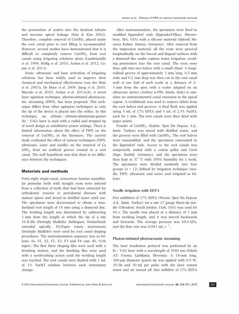
the penetration of sealers into the dentinal tubules
and increase apical leakage (Kim & Kim 2002).
Therefore, complete removal of Ca(OH)2 placed inside
the root canal prior to root filling is recommended.
However, several studies have demonstrated that it is
difficult to completely remove Ca(OH)2 from root
canals using irrigating solutions alone (Lambrianidis
et al. 1999, R€odig et al. 2010, Arslan et al. 2012, Ca-
par et al. 2013).
Sonic, ultrasonic and laser activation of irrigating
solutions has been widely used to improve their
chemical and mechanical effectiveness (van der Sluis
et al. 2007a, De Moor et al. 2009, Jiang et al. 2010,
Macedo et al. 2010, Arslan et al. 2013a,b). A novel
laser agitation technique, photon-initiated photoacou-
stic streaming (PIPS), has been proposed. This tech-
nique differs from other agitation techniques as only
the tip of the device is placed into the orifice. In this
technique, an erbium : yttrium-aluminium-garnet
(Er : YAG) laser is used with a radial and stripped tip
of novel design at subablative power settings. There is
limited information about the effect of PIPS on the
removal of Ca(OH)2 in the literature. The current
study evaluated the effect of various techniques (PIPS,
ultrasonic, sonic and needle) on the removal of Ca
(OH)2 from an artificial groove created in a root
canal. The null hypothesis was that there is no differ-
ence between the techniques.
Materials and methods
Forty-eight single-canal, noncarious human mandibu-
lar premolar teeth with straight roots were selected
from a collection of teeth that had been extracted for
orthodontic reasons or periodontal diseases with
mature apices and stored in distilled water until use.
The specimens were decoronated to obtain a stan-
dardized root length of 14 mm using a diamond disc.
The working length was determined by subtracting
1 mm from the length at which the tip of a size
10 K-file (Dentsply Maillefer, Ballaigues, Switzerland)
extruded apically. ProTaper rotary instruments
(Dentsply Maillefer) were used for root canal shaping
procedures. The instrumentation sequence was as fol-
lows: Sx, S1, S2, F1, F2, F3 and F4 (size 40, 0.06
taper). The first three shaping files were used with a
brushing motion, and the finishing files were used
with a nonbrushing action until the working length
was reached. The root canals were flushed with 1 mL
of 1% NaOCl solution between each instrument
change.
After instrumentation, the specimens were fixed in
modified Eppendorf vials (Eppendorf-Elkay, Shrews-
bury, MA, USA) with a silicone material (Optosil; He-
raeus Kulzer, Hanau, Germany). After removal from
the impression material, all the roots were grooved
longitudinally on the buccal and lingual surfaces with
a diamond disc under copious water irrigation, avoid-
ing penetration into the root canal. The roots were
then split into two halves with a small chisel. A longi-
tudinal groove of approximately 3 mm long, 0.5 mm
wide and 0.2 mm deep was then cut in the root canal
wall of one half of each tooth at a distance of 2–
5 mm from the apex with a scaler adapted on an
ultrasonic device (Anthos u-PZ6, Imola, Italy) to sim-
ulate an uninstrumented canal extension in the apical
region. A toothbrush was used to remove debris from
the root halves and grooves. A final flush was applied
using 5 mL of 17% EDTA and 5 mL of 2.5% NaOCl,
each for 1 min. The root canals were then dried with
paper points.
Powder of Ca(OH)2 (Kalsin; Spot Dis Deposu A.S�.,Izmir, Turkey) was mixed with distilled water, and
the grooves were filled with Ca(OH)2. The root halves
were reassembled, and the specimens remounted in
the Eppendorf vials. Access to the root canals was
temporarily sealed with a cotton pellet and Cavit
(Espe, Seefeld, Germany), and the specimens were
then kept at 37 °C with 100% humidity for 1 week.
The specimens were divided randomly into four
groups (n = 12) defined by irrigation technique (nee-
dle, PIPS, ultrasonic and sonic) and irrigated as fol-
lows.
Needle irrigation with EDTA
Five millilitres of 17% EDTA (Werax; Spot Dis Deposu
A.S�., Izmir, Turkey) via a size 27 gauge blunt-tip nee-
dle (Ultradent; South Jordon, Utah, USA) was used for
60 s. The needle was placed at a distance of 1 mm
from working length, and it was moved backwards
and forwards. The average pressure was 69.6 kPa,
and the flow rate was 0.083 mL s�1.
Photon-initiated photoacoustic streaming
The laser irradiation protocol was performed by an
Er : YAG laser with a wavelength of 2940 nm (Fidelis
AT; Fotona, Ljubljana, Slovenia). A 14-mm long,
300-lm diameter quartz tip was applied with 0.9 W,
30 Hz and 30 mJ per pulse with the laser system
water and air turned off. One millilitre of 17% EDTA
Arslan et al. Efficacy of PIPS on calcium hydroxide removal
© 2014 International Endodontic Journal. Published by John Wiley & Sons Ltd International Endodontic Journal, 48, 246–251, 2015 247
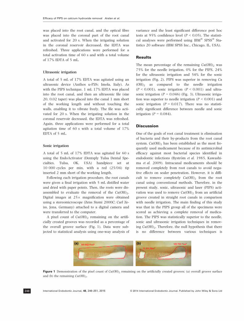
was placed into the root canal, and the optical fibre
was placed into the coronal part of the root canal
and activated for 20 s. When the irrigating solution
in the coronal reservoir decreased, the EDTA was
refreshed. Three applications were performed for a
total activation time of 60 s and with a total volume
of 17% EDTA of 5 mL.
Ultrasonic irrigation
A total of 5 mL of 17% EDTA was agitated using an
ultrasonic device (Anthos u-PZ6; Imola, Italy). As
with the PIPS technique, 1 mL 17% EDTA was placed
into the root canal, and then an ultrasonic file (size
20, 0.02 taper) was placed into the canal 1 mm short
of the working length and without touching the
walls, enabling it to vibrate freely. The file was acti-
vated for 20 s. When the irrigating solution in the
coronal reservoir decreased, the EDTA was refreshed.
Again, three applications were performed for a total
agitation time of 60 s with a total volume of 17%
EDTA of 5 mL.
Sonic irrigation
A total of 5 mL of 17% EDTA was agitated for 60 s
using the EndoActivator (Dentsply Tulsa Dental Spe-
cialties, Tulsa, OK, USA) handpiece set at
10 000 cycles per min, with a red (25/04) tip
inserted 2 mm short of the working length.
Following each irrigation procedure, the root canals
were given a final irrigation with 5 mL distilled water
and dried with paper points. Then, the roots were dis-
assembled to evaluate the removal of the Ca(OH)2.
Digital images at 259 magnification were obtained
using a stereomicroscope (Zeiss Stemi 2000C; Carl Ze-
iss, Jena, Germany) attached to a digital camera and
were transferred to the computer.
A pixel count of Ca(OH)2 remaining on the artifi-
cially created grooves was recorded as a percentage of
the overall groove surface (Fig. 1). Data were sub-
jected to statistical analysis using one-way analysis of
variance and the least significant difference post hoc
tests at 95% confidence level (P < 0.05). The statisti-
cal analyses were performed using IBM� SPSS� Sta-
tistics 20 software (IBM SPSS Inc., Chicago, IL, USA).
Results
The mean percentage of the remaining Ca(OH)2 was
75% for the needle irrigation, 0% for the PIPS, 24%
for the ultrasonic irrigation and 54% for the sonic
irrigation (Fig. 2). PIPS was superior in removing Ca
(OH)2 as compared to the needle irrigation
(P < 0.001), sonic irrigation (P < 0.001) and ultra-
sonic irrigation (P = 0.046) (Fig. 3). Ultrasonic irriga-
tion was superior to needle irrigation (P < 0.001) and
sonic irrigation (P = 0.017). There was no statisti-
cally significant difference between needle and sonic
irrigation (P = 0.084).
Discussion
One of the goals of root canal treatment is elimination
of bacteria and their by-products from the root canal
system. Ca(OH)2 has been established as the most fre-
quently used medicament because of its antimicrobial
efficacy against most bacterial species identified in
endodontic infections (Bystr€om et al. 1985, Kawashi-
ma et al. 2009). Intracanal medicaments should be
removed completely from root canals to avoid nega-
tive effects on sealer penetration. However, it is diffi-
cult to remove completely Ca(OH)2 from the root
canal using conventional methods. Therefore, in the
present study, sonic, ultrasonic and laser (PIPS) acti-
vation was used to remove Ca(OH)2 from an artificial
groove created in straight root canals in comparison
with needle irrigation. The main finding of this study
was that in the PIPS group all of the specimens were
scored as achieving a complete removal of medica-
tion. The PIPS was statistically superior to the needle,
sonic and ultrasonic irrigation techniques in remov-
ing Ca(OH)2. Therefore, the null hypothesis that there
is no difference between various techniques is
(a) (b)
Figure 1 Demonstration of the pixel count of Ca(OH)2 remaining on the artificially created grooves; (a) overall groove surface
and (b) the remaining Ca(OH)2.
Efficacy of PIPS on calcium hydroxide removal Arslan et al.
© 2014 International Endodontic Journal. Published by John Wiley & Sons LtdInternational Endodontic Journal, 48, 246–251, 2015248
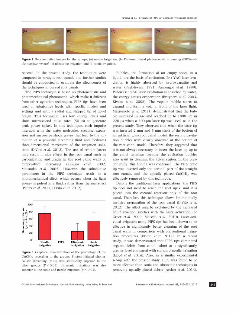
rejected. In the present study, the techniques were
compared in straight root canals and further studies
should be conducted to evaluate the effectiveness of
the techniques in curved root canals.
The PIPS technique is based on photoacoustic and
photomechanical phenomena, which make it different
from other agitation techniques. PIPS tips have been
used at subablative levels with specific models and
settings and with a radial and stripped tip of novel
design. This technique uses low energy levels and
short microsecond pulse rates (50 ls) to generate
peak power spikes. In this technique, each impulse
interacts with the water molecules, creating expan-
sion and successive shock waves that lead to the for-
mation of a powerful streaming fluid and facilitates
three-dimensional movement of the irrigation solu-
tions (DiVito et al. 2012). The use of erbium lasers
may result in side effects in the root canal such as
carbonization and cracks in the root canal walls or
temperature increasing (Kimura et al. 2002,
Matsuoka et al. 2005). However, the subablative
parameters in the PIPS technique result in a
photomechanical effect, which occurs when the light
energy is pulsed in a fluid, rather than thermal effect
(Peters et al. 2011, DiVito et al. 2012).
Bubbles, the formation of an empty space in a
liquid, are the basis of cavitation. Er : YAG laser irra-
diation is highly absorbed by hydroxyapatite and
water (Paghdiwala 1991, Armengol et al. 1999).
When Er : YAG laser irradiation is absorbed by water,
the energy causes evaporation (Brugnera et al. 2003,
Kivanc et al. 2008). The vapour bubble starts to
expand and form a void in front of the laser light.
Matsumoto et al. (2011) demonstrated that the bub-
ble increased in size and reached up to 1800 lm in
220 ls when a 300-lm laser tip was used, as in the
present study. They observed that when the laser tip
was inserted 2 mm and 5 mm short of the bottom of
an artificial glass root canal model, the second cavita-
tion bubbles were clearly observed at the bottom of
the root canal model. Therefore, they suggested that
it is not always necessary to insert the laser tip up to
the canal terminus because the cavitation bubbles
also assist in cleaning the apical region. In the pres-
ent study, this finding was confirmed. The PIPS optic
tip was inserted only the coronal part of the straight
root canals, and the apically placed Ca(OH)2 was
effectively removed by this technique.
Despite the traditional laser applications, the PIPS
tip does not need to reach the root apex, and it is
placed into the coronal reservoir only of the root
canal. Therefore, this technique allows for minimally
invasive preparation of the root canal (DiVito et al.
2012). The affect may be explained by the increased
liquid reaction kinetics with the laser activation (de
Groot et al. 2009, Macedo et al. 2010). Laser-acti-
vated irrigation using PIPS tips has been shown to be
effective in significantly better cleaning of the root
canal walls in comparison with conventional irriga-
tion procedures (DiVito et al. 2012). In a recent
study, it was demonstrated that PIPS tips eliminated
organic debris from canal isthmi at a significantly
greater level compared with standard needle irrigation
(Lloyd et al. 2014). Also, in a similar experimental
set-up with the present study, PIPS was found to be
more effective than sonic and ultrasonic techniques in
removing apically placed debris (Arslan et al. 2014).
(a) (b) (c) (d)
Figure 2 Representative images for the groups; (a) needle irrigation, (b) Photon-initiated photoacoustic streaming (PIPS)-note
the complete removal, (c) ultrasonic irrigation and (d) sonic irrigation.
Figure 3 Graphical demonstration of the percentage of the
Ca(OH)2 according to the groups. Photon-initiated photoa-
coustic streaming (PIPS) was statistically superior to the
other groups (P < 0.05). Ultrasonic irrigations was also
superior to the sonic and needle irrigation (P < 0.05).
Arslan et al. Efficacy of PIPS on calcium hydroxide removal
© 2014 International Endodontic Journal. Published by John Wiley & Sons Ltd International Endodontic Journal, 48, 246–251, 2015 249
In the present experiment, the PIPS tip had a positive
effect in removing Ca(OH)2 from an artificial groove
created in the apical third of the straight root canals,
and this result is compatible with those of aforemen-
tioned studies.
Activation of the irrigant in the ultrasonic system
has been shown to be more effective than syringe irri-
gation in removing Ca(OH)2 from the root canal walls
(Maalouf et al. 2013, Yucel et al. 2013). As men-
tioned above, in the present study, ultrasonic irriga-
tion was superior to needle irrigation. However, PIPS
and ultrasonic irrigation techniques are based upon
the transmission of acoustic energy to an irrigant in
the root canal space (Ahmad et al. 1987, DiVito et al.
2012), and the PIPS tip was more effective than the
ultrasonic system in terms of removing Ca(OH)2 from
an artificial groove created in the apical third of the
straight root canals in this study.
Jiang et al. (2010) reported that no cavitation seemed
to take place either on the sonic tip itself or on the wall
of the glass model of the root canal. They explained this
by the velocity of the sonic tip, which was below the
threshold needed for cavitation. Recently, Macedo et al.
(2014) confirmed this result. In the present study, the
cavitation effects of the techniques were not directly
evaluated. However, sonic irrigation was found to be
similar to needle irrigation in removing Ca(OH)2 from
artificially created grooves and had no perfect score for
any sample. The ineffectiveness of sonic irrigation could
result from its inability to create cavitation.
R€odig et al. (2010) compared the efficacy of different
irrigating solutions in the removal of Ca(OH)2 from
root canals. According to the their results, chelating
agents such as citric acid and EDTA showed the best
results, and the addition of NaOCl to the chelators did
not result in significant improvement of Ca(OH)2removal. Thus, in the present study, the control group
included the single use of a chelating agent (EDTA)
followed by irrigation with distilled water. EDTA irriga-
tion with needle was not able to completely remove the
Ca(OH)2 from artificially created grooves. This finding
was in accordance with those of R€odig et al. (2010).
The design of this study was based on studies
described by Lee et al. (2004), van der Sluis et al.
(2005a, b, 2007b) and R€odig et al. (2010). The stan-
dardized size and location of the grooves are advanta-
ges of the in vitro model. In addition, in the present
study, a pixel count of Ca(OH)2 remaining on the arti-
ficially created grooves was recorded as a percentage
of the overall groove surface. The design provides
researchers with a standardized evaluation that has
high reproducibility, and discrimination between
mechanical removal of the medicament and influence
of the irrigant alone is enhanced. However, the com-
plexity of a natural root canal system cannot be sim-
ulated by standardized grooves (R€odig et al. 2010).
Moreover, this experimental design does not address
medicament that diffused into the dentinal tubules.
Conclusion
Photon-initiated photoacoustic streaming provided
complete removal of Ca(OH)2 from artificial grooves
in straight root canals. This technique could be bene-
ficial in endodontics for activating irrigating solutions.
Ultrasonic irrigation enhanced the Ca(OH)2 removal
capacity of irrigating solution but did not provide
complete removal. Sonic irrigation was inefficient and
similar to needle irrigation. Further studies should be
conducted to determine apical extrusion during acti-
vation procedures.
References
Ahmad M, Pitt Ford TJ, Crum LA (1987) Ultrasonic debride-
ment of root canals: acoustic streaming and its possible
role. Journal of Endodontics 13, 490–9.
Armengol V, Jean A, Rohanizadeh R, Hamel H (1999) Scan-
ning electron microscopic analysis of diseased and healthy
dental hard tissues after Er:YAG laser irradiation: in vitro
study. Journal of Endodontics 25, 543–6.
Arslan H, Topcuoglu HS, Karatas E, Barutcigil C, Aladag H,
Topcu KM (2012) Effect of the smear layer in the removal
of calcium hydroxide from root canal walls. Journal of Con-
servative Dentistry 15, 113–7.
Arslan H, Capar ID, Saygili G et al. (2013a) Efficacy of
various irrigation protocols on the removal of triple
antibiotic paste. International Endodontic Journal 47, 594–
599.
Arslan H, Yilmaz CB, Karatas E, Barutcigil C, Topcuoglu HS,
Yeter KY (2013b) Efficacy of different treatments of root
canal walls on the pull-out bond strength of the fiber
posts. Lasers in Medical Science. doi:10.1007/
s10103-013-1457-4.
Arslan H, Capar I, Saygili G, Gok T, Akcay M (2014) Effect
of photon-initiated photoacoustic streaming on removal of
apically placed dentinal debris. International Endodontic
Journal doi:10.1111/iej.12251.
Athanassiadis B, Abbott PV, Walsh LJ (2007) The use of cal-
cium hydroxide, antibiotics and biocides as antimicrobial
medicaments in endodontics. Australian Dental Journal 52,
S64–82.
Brugnera A Jr, Zanin F, Barbin EL, Spano JC, Santana R,
Pecora JD (2003) Effects of Er:YAG and Nd:YAG laser irra-
Efficacy of PIPS on calcium hydroxide removal Arslan et al.
© 2014 International Endodontic Journal. Published by John Wiley & Sons LtdInternational Endodontic Journal, 48, 246–251, 2015250
diation on radicular dentine permeability using different
irrigating solutions. Lasers in Surgery and Medicine 33,
256–9.
Bystr€om A, Claesson R, Sundqvist G (1985) The antibacte-
rial effect of camphorated paramonochlorophenol, cam-
phorated phenol and calcium hydroxide in the treatment
of infected root canals. Endodontics & Dental Traumatology
1, 170–5.
Capar ID, Ozcan E, Arslan H, Ertas H, Aydinbelge HA
(2013) Effect of different final irrigation methods on the
removal of calcium hydroxide from an artificial standard-
ized groove in the apical third of root canals. Journal of
Endodontics 40, 451–4.
de Groot SD, Verhaagen B, Versluis M, Wu MK, Wesselink
PR, van der Sluis LW (2009) Laser-activated irrigation
within root canals: cleaning efficacy and flow visualiza-
tion. International Endodontic Journal 42, 1077–83.
De Moor RJ, Blanken J, Meire M, Verdaasdonk R (2009)
Laser induced explosive vapor and cavitation resulting in
effective irrigation of the root canal. Part 2: evaluation of
the efficacy. Lasers in Surgery and Medicine 41, 520–3.
DiVito E, Peters OA, Olivi G (2012) Effectiveness of the
erbium:YAG laser and new design radial and stripped tips
in removing the smear layer after root canal instrumenta-
tion. Lasers in Medical Science 27, 273–80.
Jiang LM, Verhaagen B, Versluis M, van der Sluis LW (2010)
Evaluation of a sonic device designed to activate irrigant in
the root canal. Journal of Endodontics 36, 143–6.
Kawashima N, Wadachi R, Suda H, Yeng T, Parashos P
(2009) Root canal medicaments. International Dental Jour-
nal 59, 5–11.
Kim SK, Kim YO (2002) Influence of calcium hydroxide in-
tracanal medication on apical seal. International Endodontic
Journal 35, 623–8.
Kimura Y, Yonaga K, Yokoyama K, Kinoshita J, Ogata Y,
Matsumoto K (2002) Root surface temperature increase
during Er:YAG laser irradiation of root canals. Journal of
Endodontics 28, 76–8.
Kivanc BH, Ulusoy OI, Gorgul G (2008) Effects of Er:YAG laser
and Nd:YAG laser treatment on the root canal dentin of
human teeth: a study. Lasers in medical science 23, 247–52.
Lambrianidis T, Margelos J, Beltes P (1999) Removal effi-
ciency of calcium hydroxide dressing from the root canal.
Journal of Endodontics 25, 85–8.
Lee SJ, Wu MK, Wesselink PR (2004) The effectiveness of
syringe irrigation and ultrasonics to remove debris from
simulated irregularities within prepared root canal walls.
International Endodontic Journal 37, 672–8.
Lloyd A, Uhles JP, Clement DJ, Garcia-Godoy F (2014) Elimi-
nation of intracanal tissue and debris through a novel
laser-activated system assessed using high-resolution
micro–computed tomography: A Pilot Study. Journal of
Endodontics 40, 584–7.
Maalouf L, Zogheib C, Naaman A (2013) Removal efficiency
of calcium hydroxide dressing from the root canal without
chemically active adjuvant. The Journal of Contemporary
Dental Practice 14, 188–92.
Macedo RG, Wesselink PR, Zaccheo F, Fanali D, Van Der Sluis
LW (2010) Reaction rate of NaOCl in contact with bovine
dentine: effect of activation, exposure time, concentration
and pH. International Endodontic Journal 43, 1108–15.
Macedo R, Verhaagen B, Rivas DF, Versluis M, Wesselink P,
van der Sluis L (2014) Cavitation measurement during
sonic and ultrasonic activated irrigation. Journal of End-
odontics 40, 580–3.
Matsumoto H, Yoshimine Y, Akamine A (2011) Visualiza-
tion of irrigant flow and cavitation induced by Er:YAG
laser within a root canal model. Journal of Endodontics 37,
839–43.
Matsuoka E, Jayawardena JA, Matsumoto K (2005) Morpho-
logical study of the Er, Cr:YSGG laser for root canal prepa-
ration in mandibular incisors with curved root canals.
Photomedicine and Laser Surgery 23, 480–4.
Paghdiwala AF (1991) Does the laser work on hard dental
tissue? Journal of the American Dental Association 122, 79–
80.
Peters OA, Bardsley S, Fong J, Pandher G, Divito E (2011)
Disinfection of root canals with photon-initiated photoa-
coustic streaming. Journal of Endodontics 37, 1008–12.
R€odig T, Vogel S, Zapf A, Hulsmann M (2010) Efficacy of dif-
ferent irrigants in the removal of calcium hydroxide from
root canals. International Endodontic Journal 43, 519–27.
van der Sluis LW, Wu MK, Wesselink PR (2005a) A com-
parison between a smooth wire and a K-file in removing
artificially placed dentine debris from root canals in resin
blocks during ultrasonic irrigation. International Endodontic
Journal 38, 593–6.
van der Sluis LW, Wu MK, Wesselink PR (2005b) The effi-
cacy of ultrasonic irrigation to remove artificially placed
dentine debris from human root canals prepared using
instruments of varying taper. International Endodontic Jour-
nal 38, 764–8.
van der Sluis LW, Versluis M, Wu MK, Wesselink PR
(2007a) Passive ultrasonic irrigation of the root canal: a
review of the literature. International Endodontic Journal 40,
415–26.
van der Sluis LW, Wu MK, Wesselink PR (2007b) The eval-
uation of removal of calcium hydroxide paste from an arti-
ficial standardized groove in the apical root canal using
different irrigation methodologies. International Endodontic
Journal 40, 52–7.
Yucel AC, Gurel M, Guler E, Karabucak B (2013) Compari-
son of final irrigation techniques in removal of calcium
hydroxide. Australian Endodontic Journal 39, 116–21.
Arslan et al. Efficacy of PIPS on calcium hydroxide removal
© 2014 International Endodontic Journal. Published by John Wiley & Sons Ltd International Endodontic Journal, 48, 246–251, 2015 251
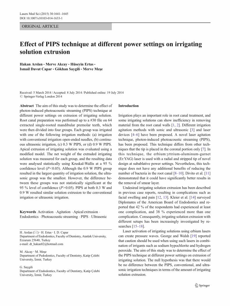
ORIGINAL ARTICLE
Effect of PIPS technique at different power settings on irrigatingsolution extrusion
Hakan Arslan & Merve Akcay & Hüseyin Ertas &Ismail Davut Capar & Gökhan Saygili & Merve Meşe
Received: 5 March 2014 /Accepted: 8 July 2014 /Published online: 19 July 2014# Springer-Verlag London 2014
Abstract The aim of this study was to determine the effect ofphoton-induced photoacoustic streaming (PIPS) technique atdifferent power settings on extrusion of irrigating solution.Root canal preparation was performed up to a #30 file on 64extracted single-rooted mandibular premolar teeth, whichwere then divided into four groups. Each group was irrigatedwith one of the following irrigation methods: (a) irrigationwith conventional irrigation open-ended needles, (b) continu-ous ultrasonic irrigation, (c) 0.3 W PIPS, or (d) 0.9 W PIPS.Apical extrusion of irrigating solution was evaluated using amodified model. The net weight of the extruded irrigatingsolution was measured for each group, and the resulting datawere analysed statistically using Kruskal-Wallis at a 95 %confidence level (P<0.05). Although the 0.9 W PIPS groupresulted in the largest quantity of irrigation solution, the ultra-sonic group was the smallest. However, the difference be-tween these groups was not statistically significant at the95 % level of confidence (P>0.05). PIPS at both 0.3 W and0.9 W resulted similar solution extrusion to the conventionalirrigation or ultrasonic irrigation.
Keywords Activation . Agitation . Apical extrusion .
Endodontics . Photoacoustic streaming . PIPS . Ultrasonic
Introduction
Irrigation plays an important role in root canal treatment, andsome irrigating solutions can show inefficiency in removingmaterial from the root canal walls [1, 2]. Different irrigationagitation methods with sonic and ultrasonic [3] and laserdevices [4–6] have been proposed. A novel laser agitationtechnique, photon-induced photoacoustic streaming (PIPS),has been proposed. This technique differs from other tech-niques that the tip is placed in the coronal portion only [7]. Inthis technique, the erbium:yttrium-aluminum-garnet(Er:YAG) laser is used with a radial and stripped tip of noveldesign at subablative power settings. Nevertheless, this tech-nique does not have any additional benefits of reducing thenumber of bacteria in the root canal [8–10]. Divito et al. [11]demonstrated that it could have significantly better results inthe removal of smear layer.
Undesired irrigating solution extrusion has been describedin previous case reports, resulting in complications such asfacial swelling and pain [12, 13]. Kleier et al. [14] surveyedDiplomates of the American Board of Endodontics and re-ported that 42 % of the respondents had experienced at leastone complication, and 38 % experienced more than onecomplication. Consequently, irrigating solution extrusion withdifferent setups has been increasingly investigated by re-searches [15–18].
Laser activation of irrigating solutions using erbium laserscan create pressure waves. George and Walsh [19] reportedthat caution should be used when using such lasers in combi-nation of irrigants such as sodium hypochlorite and hydrogenperoxide. The aim of this study was to determine the effect ofthe PIPS technique at different power settings on extrusion ofirrigating solution. The null hypothesis was that there wouldbe no difference between the PIPS, conventional, and ultra-sonic irrigation techniques in terms of the amount of irrigatingsolution extrusion.
H. Arslan (*) :H. Ertas : I. D. CaparDepartment of Endodontics, Faculty of Dentistry, Atatürk University,Erzurum 25640, Turkeye-mail: [email protected]
M. Akcay :M. MeşeDepartment of Pedodontics, Faculty of Dentistry, Katip ÇelebiUniversity, İzmir, Turkey
G. SaygiliDepartment of Endodontics, Faculty of Dentistry, Katip ÇelebiUniversity, İzmir, Turkey
Lasers Med Sci (2015) 30:1641–1645DOI 10.1007/s10103-014-1633-1
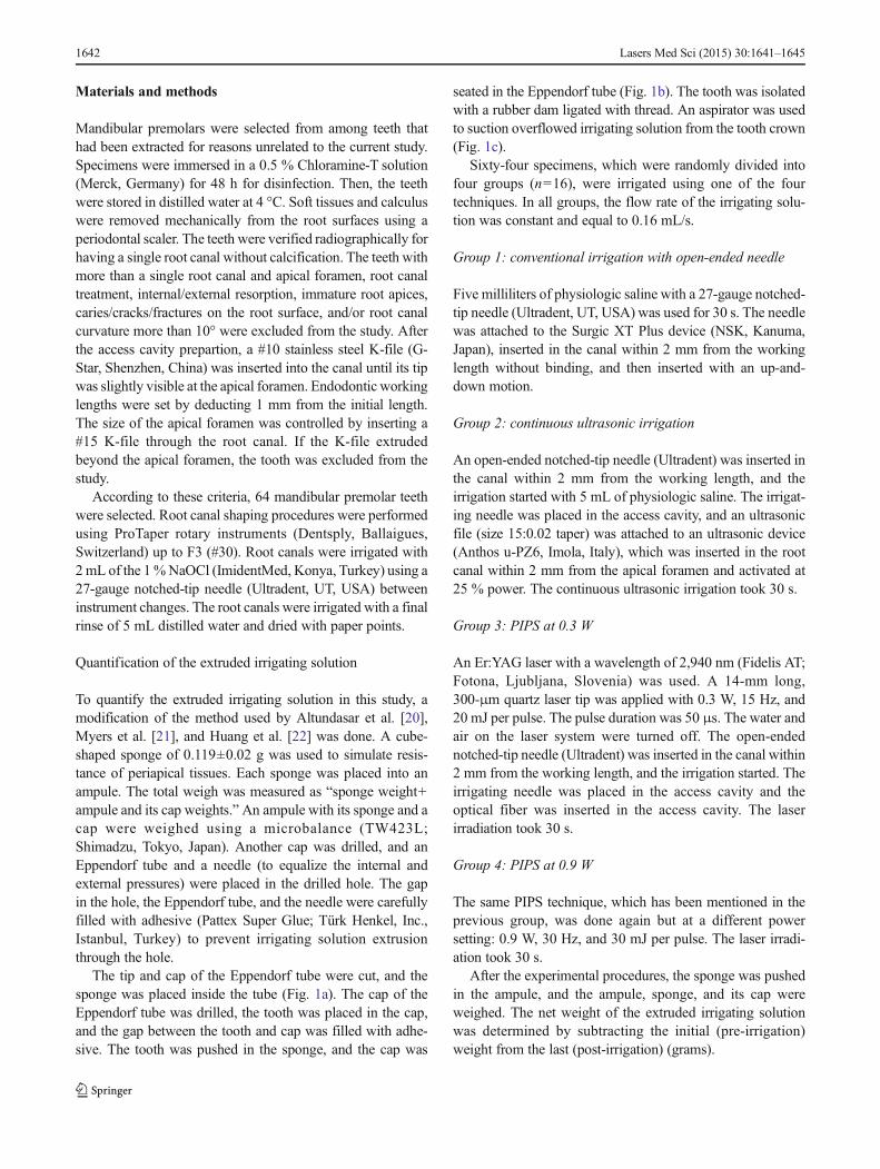
Materials and methods
Mandibular premolars were selected from among teeth thathad been extracted for reasons unrelated to the current study.Specimens were immersed in a 0.5 % Chloramine-T solution(Merck, Germany) for 48 h for disinfection. Then, the teethwere stored in distilled water at 4 °C. Soft tissues and calculuswere removed mechanically from the root surfaces using aperiodontal scaler. The teeth were verified radiographically forhaving a single root canal without calcification. The teeth withmore than a single root canal and apical foramen, root canaltreatment, internal/external resorption, immature root apices,caries/cracks/fractures on the root surface, and/or root canalcurvature more than 10° were excluded from the study. Afterthe access cavity prepartion, a #10 stainless steel K-file (G-Star, Shenzhen, China) was inserted into the canal until its tipwas slightly visible at the apical foramen. Endodontic workinglengths were set by deducting 1 mm from the initial length.The size of the apical foramen was controlled by inserting a#15 K-file through the root canal. If the K-file extrudedbeyond the apical foramen, the tooth was excluded from thestudy.
According to these criteria, 64 mandibular premolar teethwere selected. Root canal shaping procedures were performedusing ProTaper rotary instruments (Dentsply, Ballaigues,Switzerland) up to F3 (#30). Root canals were irrigated with2mL of the 1%NaOCl (ImidentMed, Konya, Turkey) using a27-gauge notched-tip needle (Ultradent, UT, USA) betweeninstrument changes. The root canals were irrigated with a finalrinse of 5 mL distilled water and dried with paper points.
Quantification of the extruded irrigating solution
To quantify the extruded irrigating solution in this study, amodification of the method used by Altundasar et al. [20],Myers et al. [21], and Huang et al. [22] was done. A cube-shaped sponge of 0.119±0.02 g was used to simulate resis-tance of periapical tissues. Each sponge was placed into anampule. The total weigh was measured as “sponge weight+ampule and its cap weights.”An ampule with its sponge and acap were weighed using a microbalance (TW423L;Shimadzu, Tokyo, Japan). Another cap was drilled, and anEppendorf tube and a needle (to equalize the internal andexternal pressures) were placed in the drilled hole. The gapin the hole, the Eppendorf tube, and the needle were carefullyfilled with adhesive (Pattex Super Glue; Türk Henkel, Inc.,Istanbul, Turkey) to prevent irrigating solution extrusionthrough the hole.
The tip and cap of the Eppendorf tube were cut, and thesponge was placed inside the tube (Fig. 1a). The cap of theEppendorf tube was drilled, the tooth was placed in the cap,and the gap between the tooth and cap was filled with adhe-sive. The tooth was pushed in the sponge, and the cap was
seated in the Eppendorf tube (Fig. 1b). The tooth was isolatedwith a rubber dam ligated with thread. An aspirator was usedto suction overflowed irrigating solution from the tooth crown(Fig. 1c).
Sixty-four specimens, which were randomly divided intofour groups (n=16), were irrigated using one of the fourtechniques. In all groups, the flow rate of the irrigating solu-tion was constant and equal to 0.16 mL/s.
Group 1: conventional irrigation with open-ended needle
Five milliliters of physiologic saline with a 27-gauge notched-tip needle (Ultradent, UT, USA) was used for 30 s. The needlewas attached to the Surgic XT Plus device (NSK, Kanuma,Japan), inserted in the canal within 2 mm from the workinglength without binding, and then inserted with an up-and-down motion.
Group 2: continuous ultrasonic irrigation
An open-ended notched-tip needle (Ultradent) was inserted inthe canal within 2 mm from the working length, and theirrigation started with 5 mL of physiologic saline. The irrigat-ing needle was placed in the access cavity, and an ultrasonicfile (size 15:0.02 taper) was attached to an ultrasonic device(Anthos u-PZ6, Imola, Italy), which was inserted in the rootcanal within 2 mm from the apical foramen and activated at25 % power. The continuous ultrasonic irrigation took 30 s.
Group 3: PIPS at 0.3 W
An Er:YAG laser with a wavelength of 2,940 nm (Fidelis AT;Fotona, Ljubljana, Slovenia) was used. A 14-mm long,300-μm quartz laser tip was applied with 0.3 W, 15 Hz, and20 mJ per pulse. The pulse duration was 50 μs. The water andair on the laser system were turned off. The open-endednotched-tip needle (Ultradent) was inserted in the canal within2 mm from the working length, and the irrigation started. Theirrigating needle was placed in the access cavity and theoptical fiber was inserted in the access cavity. The laserirradiation took 30 s.
Group 4: PIPS at 0.9 W
The same PIPS technique, which has been mentioned in theprevious group, was done again but at a different powersetting: 0.9 W, 30 Hz, and 30 mJ per pulse. The laser irradi-ation took 30 s.
After the experimental procedures, the sponge was pushedin the ampule, and the ampule, sponge, and its cap wereweighed. The net weight of the extruded irrigating solutionwas determined by subtracting the initial (pre-irrigation)weight from the last (post-irrigation) (grams).
1642 Lasers Med Sci (2015) 30:1641–1645
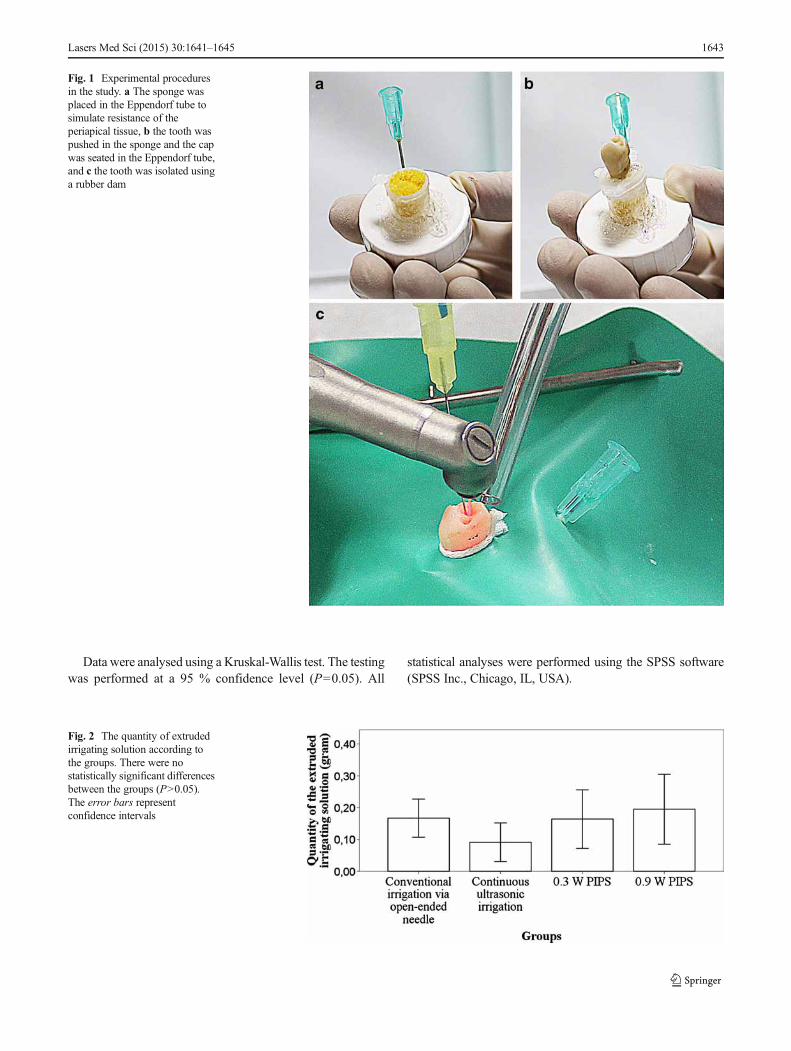
Data were analysed using a Kruskal-Wallis test. The testingwas performed at a 95 % confidence level (P=0.05). All
statistical analyses were performed using the SPSS software(SPSS Inc., Chicago, IL, USA).
Fig. 1 Experimental proceduresin the study. a The sponge wasplaced in the Eppendorf tube tosimulate resistance of theperiapical tissue, b the tooth waspushed in the sponge and the capwas seated in the Eppendorf tube,and c the tooth was isolated usinga rubber dam
Fig. 2 The quantity of extrudedirrigating solution according tothe groups. There were nostatistically significant differencesbetween the groups (P>0.05).The error bars representconfidence intervals
Lasers Med Sci (2015) 30:1641–1645 1643
Results
Figure 2 shows the quantity of apically extruded irrigatingsolution for each group. Mean and standard deviations of theextruded irrigating solution amount was 0.16±0.11 g forconventional irrigation, 0.09±0.11 g for continuous ultrasonicirrigation, 0.16±0.17 g for 0.3 W PIPS, and 0.19±0.20 g for0.9W PIPS groups. The largest quantity of extruded irrigatingsolution occurred in the 0.9 W PIPS group, and the smallestwas in the ultrasonic irrigation group. However, according tothe Kruskal-Wallis test there were not statistically significantbetween the groups at a confidence level of 95 % (P>0.05)(effect size=1.1286).
Discussion
The apical extrusion of irrigating solution in root canal treat-ment can lead to complications, such as damage of periapicaltissues, pain, and burning sensation [12, 23, 24]. Thus, irriga-tion techniques need to be evaluated to determine the one,which results in less extrusion. In vitro models try to simulateclinical situations. In previous studies, in vitro models includ-ed the root apex suspended in air or water [22, 25]. Altundasaret al. [20] used a flower-arranged foam to simulate the resis-tance of the periapical tissues and to collect any extrudedirrigating solution. This study presented a modified modelbased on previous studies [20–22]. In this modified model, asponge simulates the periapical tissue resistance [20], and theextruded irrigating solution is collected in an ampule [21, 22].
In a recent study, Boutsioukis et al. [25] evaluated the effectof ultrasonic, sonic, and manual dynamic agitation on irrigat-ing solution extrusion. They found that there was not statisti-cally significant difference between the ultrasonic agitationand the control group. Desai and Himel [17] evaluated thesafety of various intracanal irrigation systems, and they foundno statistically significant difference between conventional(manual syringe and Max-I-probe needle) and ultrasonic irri-gation. These findings are in harmony with our results. In thisstudy, the ultrasonic irrigation did not result in more irrigatingsolution extrusion when compared to the control group.
In the present study, the PIPS and ultrasonic techniqueswere compared with the conventional irrigation technique.The ultrasonic and PIPS techniques require prior filling ofthe root canals with irrigating solution, followed by continu-ous solution activation through the optical or ultrasonic tip.When such activation occurred, less irrigating solution extru-sion was observed compared to the conventional technique.Moreover, in the conventional technique the needle gets closerto the apex than in the PIPS and ultrasonic groups. Thesefactors could affect the results of studies evaluating the extru-sion of irrigating solution [25].
Our results show that PIPS at both 0.3 and 0.9 W did notresult in more solution extrusion than conventional irrigationand ultrasonic irrigation. Therefore, the null hypothesis thatthere is no difference between the PIPS, conventional, andultrasonic irrigation techniques in terms of irrigating solutionextrusion can be accepted.
Extrusion of irrigating solution can be affected by manyfactors, some of which were controlled in this study. In thepresent study, specimens with an apical diameter more than#15 K-file were excluded from the study. The root canals wereinstrumented up to the size 30 using the ProTaper Universalrotary files. The volume and flow rate of the irrigating solutionhad equal values in all groups. To standardize the irrigationflow rate and the pressure, a Surgic XT Plus device was used.A standard needle type was used. Precautions were taken toavoid extrusion of the irrigating solution in the ampulethrough the gaps in the modified model. Finally, sponges withthe standardized weight were used to simulate resistance ofperiapical tissues.
Conclusion
Within the limitations of this study, all techniques led toextrusion of irrigating solution beyond the apical foramen.Nevertheless, PIPS at both 0.3 and 0.9 W resulted similarextrusion to conventional irrigation and ultrasonic irrigation.The modified model used here can be beneficial for furtherevaluation of apical extrusion.
Conflict of interest There are no conflicts of interest.
References
1. Torabinejad M, Cho Y, Khademi AA, Bakland LK, Shabahang S(2003) The effect of various concentrations of sodium hypochloriteon the ability of MTAD to remove the smear layer. J Endod 29:233–239. doi:10.1097/00004770-200304000-00001
2. Mancini M, Armellin E, Casaglia A, Cerroni L, Cianconi L (2009) Acomparative study of smear layer removal and erosion in apicalintraradicular dentine with three irrigating solutions: a scanning elec-tron microscopy evaluation. J Endod 35:900–903. doi:10.1016/j.joen.2009.03.052
3. Guerisoli DM, Marchesan MA, Walmsley AD, Lumley PJ, PecoraJD (2002) Evaluation of smear layer removal by EDTAC and sodiumhypochlorite with ultrasonic agitation. Int Endod J 35:418–421
4. De Moor RJ, Blanken J, Meire M, Verdaasdonk R (2009) Laserinduced explosive vapor and cavitation resulting in effective irriga-tion of the root canal. Part 2: evaluation of the efficacy. Lasers SurgMed 41:520–523. doi:10.1002/lsm.20797
5. de Groot SD, Verhaagen B, Versluis M, Wu MK, Wesselink PR, vander Sluis LW (2009) Laser-activated irrigation within root canals:cleaning efficacy and flow visualization. Int Endod J 42:1077–1083.doi:10.1111/j.1365-2591.2009.01634.x
1644 Lasers Med Sci (2015) 30:1641–1645
6. Moon YM, Kim HC, Bae KS, Baek SH, Shon WJ, Lee W (2012)Effect of laser-activated irrigation of 1320-nanometer Nd:YAG laseron sealer penetration in curved root canals. J Endod 38:531–535. doi:10.1016/j.joen.2011.12.008
7. DiVito E, Lloyd A (2012) ER:YAG laser for 3-dimensional debride-ment of canal systems: use of photon-induced photoacoustic stream-ing. Dent Today 31(122):4–7
8. Peters OA, Bardsley S, Fong J, Pandher G, Divito E (2011)Disinfection of root canals with photon-initiated photoacousticstreaming. J Endod 37:1008–1012. doi:10.1016/j.joen.2011.03.016
9. Zhu X, Yin X, Chang JW, Wang Y, Cheung GS, Zhang C(2013) Comparison of the antibacterial effect and smear layerremoval using photon-initiated photoacoustic streaming aided ir-rigation versus a conventional irrigation in single-rooted canals:an in vitro study. Photomed Laser Surg 31:371–377. doi:10.1089/pho.2013.3515
10. Pedullà E, Genovese C, Campagna E, Tempera G, Rapisarda E(2012) Decontamination efficacy of photon-initiated photoacousticstreaming (PIPS) of irrigants using low-energy laser settings: anex vivo study. Int Endod J 45:865–870. doi:10.1111/j.1365-2591.2012.02044.x
11. DiVito E, Peters OA, Olivi G (2012) Effectiveness of the erbium:YAG laser and new design radial and stripped tips in removing thesmear layer after root canal instrumentation. Lasers Med Sci 27:273–280. doi:10.1007/s10103-010-0858-x
12. Behrents KT, Speer ML, Noujeim M (2012) Sodium hypochloriteaccident with evaluation by cone beam computed tomography. IntEndod J 45:492–498. doi:10.1111/j.1365-2591.2011.02009.x
13. Motta MV, Chaves-Mendonca MA, Stirton CG, Cardozo HF (2009)Accidental injection with sodium hypochlorite: report of a case. IntEndod J 42:175–182. doi:10.1111/j.1365-2591.2008.01493.x
14. Kleier DJ, Averbach RE, Mehdipour O (2008) The sodiumhypochlorite accident: experience of diplomates of the AmericanBoard of Endodontics. J Endod 34:1346–1350. doi:10.1016/j.joen.2008.07.021
15. Hauser V, Braun A, Frentzen M (2007) Penetration depth of a dyemarker into dentine using a novel hydrodynamic system (RinsEndo).Int Endod J 40:644–652. doi:10.1111/j.1365-2591.2007.01264.x
16. Tasdemir T, Er K, Celik D, Yildirim T (2008) Effect of passiveultrasonic irrigation on apical extrusion of irrigating solution. Eur JDent 2:198–203
17. Desai P, Himel V (2009) Comparative safety of various intracanalirrigation systems. J Endod 35:545–549. doi:10.1016/j.joen.2009.01.011
18. Mitchell RP, Baumgartner JC, Sedgley CM (2011) Apical extrusionof sodium hypochlorite using different root canal irrigation systems. JEndod 37:1677–1681. doi:10.1016/j.joen.2011.09.004
19. George R, Walsh LJ (2008) Apical extrusion of root canal irrigantswhen using Er:YAG and Er, Cr:YSGG lasers with optical fibers: anin vitro dye study. J Endod 34:706–708. doi:10.1016/j.joen.2008.03.003
20. Altundasar E, Nagas E, Uyanik O, Serper A (2011) Debris andirrigant extrusion potential of 2 rotary systems and irrigation needles.Oral Surg Oral Med Oral Pathol Oral Radiol Endod 112:e31–e35.doi:10.1016/j.tripleo.2011.03.044
21. Myers GL,Montgomery S (1991) A comparison of weights of debrisextruded apically by conventional filing and Canal Master tech-niques. J Endod 17:275–279. doi:10.1016/S0099-2399(06)81866-2
22. Huang X, Ling J, Wei X, Gu L (2007) Quantitative evaluation ofdebris extruded apically by using ProTaper Universal Tulsa rotarysystem in endodontic retreatment. J Endod 33:1102–1105. doi:10.1016/j.joen.2007.05.019
23. Serper A, Ozbek M, Calt S (2004) Accidental sodium hypochlorite-induced skin injury during endodontic treatment. J Endod 30:180–181. doi:10.1097/00004770-200403000-00013
24. Chang YC, Huang FM, Tai KW, Chou MY (2001) The effect ofsodium hypochlorite and chlorhexidine on cultured human periodon-tal ligament cells. Oral Surg Oral Med Oral Pathol Oral Radiol Endod92:446–450. doi:10.1067/moe.2001.116812
25. Boutsioukis C, Psimma Z, Kastrinakis E (2013) The effect of flowrate and agitation technique on irrigant extrusion ex vivo. Int Endod J.2013/09/17 edn
Lasers Med Sci (2015) 30:1641–1645 1645
Full Terms & Conditions of access and use can be found athttp://www.tandfonline.com/action/journalInformation?journalCode=iode20
Download by: [UCSF Library] Date: 14 April 2016, At: 09:09
Acta Odontologica Scandinavica
ISSN: 0001-6357 (Print) 1502-3850 (Online) Journal homepage: http://www.tandfonline.com/loi/iode20
Bond strength of self-adhesive resin cementto root dentin. Comparison of photon-initiatedphotoacoustic streaming technique with needleand ultrasonic irrigation
Hakan Arslan, Merve Akcay, Gokhan Saygili, Ahmet Keskı, İbrahim TalhaMeŞe, Adem Gok & Mehmet Dalli
To cite this article: Hakan Arslan, Merve Akcay, Gokhan Saygili, Ahmet Keskı, İbrahim TalhaMeŞe, Adem Gok & Mehmet Dalli (2015) Bond strength of self-adhesive resin cement toroot dentin. Comparison of photon-initiated photoacoustic streaming technique withneedle and ultrasonic irrigation, Acta Odontologica Scandinavica, 73:5, 348-352, DOI:10.3109/00016357.2014.967717
To link to this article: http://dx.doi.org/10.3109/00016357.2014.967717
Published online: 02 Feb 2015.
Submit your article to this journal
Article views: 151
View related articles
View Crossmark data
Acta Odontologica Scandinavica. 2015; 73: 348–352
ORIGINAL ARTICLE
Bond strength of self-adhesive resin cement to root dentin. Comparisonof photon-initiated photoacoustic streaming technique with needle andultrasonic irrigation
HAKAN ARSLAN1, MERVE AKCAY2, GOKHAN SAYGILI3, AHMET KESKI4,_IBRAHIM TALHA MESE4, ADEM GOK5 & MEHMET DALLI5
1Department of Endodontics, Faculty of Dentistry, Atatürk University, Erzurum, Turkey, 2Department of Pedodontics,3Department of Endodontics, 4Department of Prosthodontics, and 5Department of Restorative Dentistry, Faculty ofDentistry, Izmir Katip Celebi University, _Izmir, Turkey
AbstractObjective.The aim of this study was to investigate the effects of photon-initiated photoacoustic streaming (PIPS) with variousirrigating solutions on the bond strength of a self-adhesive resin cement to root dentin.Materials andmethods. Seventy-twomandibular premolar roots were divided into six groups after post space preparation and treated with a needle irrigation withdistilled water and NaOCl, ultrasonic irrigation with NaOCl, PIPS with NaOCl, PIPS with EDTA and PIPS with distilledwater at 0.3 W, 15 Hz and 20 mJ per pulse for 60 s. Fiber posts were cemented with a newly marketed, self-adhesive resincement. The data obtained from the push-out tests were analyzed using analysis of variance (ANOVA) and LSD post-hoc tests(p = 0.05). Results. PIPS with distilled water resulted in higher push-out values than those of needle (with both distilled waterand NaOCl) and ultrasonic irrigation (p < 0.05). Conclusions. The use of PIPS may provide higher bond strength of self-adhesive resin cement to root dentin than needle and ultrasonic irrigation techniques.
Key Words: bond strength, fiber post, photoacoustic streaming, self-adhesive resin cement, ultrasonic
Introduction
Fiber reinforced composite (FRC) posts have beenwidely used in restoring endodontically treated teethto retain coronal restoration. FRC posts have amodulus of elasticity close to that of dentin [1,2].They also have a good esthetic appearance, with norisk of gingival discoloration of the root surfaces dueto corrosive products [3]. The longevity of the restora-tions depends on many factors, such as the effective-ness and durability of the bonding between the post,dentin, and the adhesive resin cement [4]. Effectivebonding can contribute to reduced stress beinggenerated on the root canal walls, thereby strength-ening the remaining tooth structure and decreasingthe risk of fracture [5,6].The efficacies of various irrigating solutions were
investigated as pre-treatment agents before the FRCpost placement [3,7]. Recently, a novel laser agitationtechnique, photon-induced photoacoustic streaming
(PIPS), has been proposed. This technique differsfrom other agitation techniques insofar as it involvesthe placement of only the tip into the coronal portion[8]. In this technique, an erbium:yttrium-aluminum-garnet (Er:YAG) laser is used with a radial andstripped tip of novel design at sub-ablative powersettings. DiVito et al. [9] demonstrated that it resultsin a significantly better cleaning of the root canalwalls.Various strategies have been proposed to lute FRC
posts to root dentin. Conventional dual-cured resincements require the prior application of adhesivesystems. Self-adhesive cements do not require anysurface treatment of the root canals. They are easier tohandle and have clinically effective bond strength[10]. The chemical reaction between the phosphatemethacrylates in the self-adhesive cements andhydroxyapatite in dentin provides bonding [11].The aim of this study was to evaluate the effect of
PIPS with various irrigating solutions on the bond
Correspondence: Assistant Professor Dr Hakan Arslan, Department of Endodontics, Faculty of Dentistry, Atatürk University, Erzurum, 25620, Turkey.Tel: +90 442 325 4040 2352. Fax: +90 442 325 2535. E-mail: [email protected]
(Received 11 April 2014; accepted 3 September 2014)
ISSN 0001-6357 print/ISSN 1502-3850 online � 2015 Informa HealthcareDOI: 10.3109/00016357.2014.967717
Dow
nloa
ded
by [
UC
SF L
ibra
ry]
at 0
9:09
14
Apr
il 20
16
strength to root dentin of a self-adhesive resin cementas compared to needle and ultrasonic irrigation. Thenull hypothesis of this study was that there were nosignificant differences among the techniques in termsof adhesion between the self-adhesive resin cementand root dentin.
Materials and methods
Human mandibular premolar teeth were selectedfrom a collection of teeth that had been recentlyextracted for reasons unrelated to this study andstored in distilled water until use. Soft tissue andcalculus were removed mechanically from the rootsurfaces with a periodontal scaler. Teeth with rootcanal treatment, restoration, immature root apicesand/or coronal defects were excluded from the study.The teeth were verified radiographically as having asingle root canal without calcification. According tothese criteria, 72 mandibular premolar teeth wereselected.Each tooth was decoronated using a diamond disc
(Diamond disc superflex 910S/220; North Bel, Italy)operated perpendicularly to its longitudinal axis toobtain a standardized root length of 15 mm. A size#15stainless steel K-file (Dentsply-Maillefer, Ballai-gues, Switzerland) was moved down in the canal untilthe file was just visible. The working length was set bydeducting 1 mm from this. The root canals wereshaped with ProTaper rotary instruments (Dents-ply-Maillefer) up to a size #40 (F4) at working length.The root canals were irrigated with 2 mL of 5%NaOCl (ImidentMedEndosolve-HP, Konya, Turkey)between instrument changes. The apexes of the speci-mens were closed with boxing wax and the root canalswere irrigated first for 120 s with 5 mL of 17%ethylenediaminetetraacetic acid (EDTA) as a finalflush and then for 120 s with 5 mL of 5% NaOCl,followed by a final rinse with 5 mL of distilled water toremove the smear layer.The specimens were dried using paper points, with
a single gutta-percha cone (F4; Dentsply-Maillefer)slightly coated with AH Plus (Dentsply DeTrey,Kontanz, Germany) and placed into the root canalto the working length. Temporary filling material(Cavit G; 3M ESPE, Seefeld, Germany) was usedto seal the coronal orifice. After storage at 100%humidity for 1 week at 37�C; a portion of the rootcanal filling material was removed with gates-gliddendrills. A 10-mm deep post space was made usinga 1.4-mm size drill (Cytec Carbon; HahnenkrattGmbH, Königsbach-Stein, Germany). The speci-mens were randomly divided into six groups (n =12), as follows:
. Needle irrigation with distilled water: 5 mL of distilledwater via a size 27 gauge blunt-tip needle (Ultra-dent, UT) was used for 60 s in this group.
. Needle irrigation with NaOCl: 5 mL of 5% NaOClwas used for 60 s in this group.
. Ultrasonic irrigation with NaOCl: 5 mL of 5%NaOCl was continuously activated via an ultra-sonic device (Anthos u-PZ6, Imola, Italy). Anultrasonic file (size 15: 0.02 taper) was placedinto the post space without touching the walls,enabling it to vibrate freely. The ultrasonic filewas activated at 25% power for a total of 60 s.
. PIPS with NaOCl: 5 mL of 5% NaOCl was con-tinuously activated via an Er:YAG laser with awavelength of 2940 nm (Fidelis AT; Fotona,Ljubljana, Slovenia). A 14-mm long, 400-micronquartz tip was inserted into the post space ascoronally as possible and applied at 0.3 W,15 Hz and 20 mJ per pulse for 60 s. The waterand air in the laser system were turned off.
. PIPS with EDTA: 5 mL of 17% EDTA was acti-vated as stated above.
. PIPS with distilled water: 5 mL of distilled water wasactivated as stated above.
The flow rate of the irrigating solutions in all groupswas 0.08 mL/s. After the procedures, the post spacewas irrigated with 5mL distilled water and dried usingpaper points. Self-adhesive resin cement (RelyXU200; 3M ESPE, Seefeld, Germany) was applieddirectly into the post space. Size #2 carbon fiber posts(1.4 mm diameter) (Cytec Carbon; HahnenkrattGmbH, Königsbach-Stein, Germany) were insertedinto the post space. Excess cement was removed andlight-cured for 40 s. The coronal opening was sealedusing composite resin and the specimens were storedat 100% humidity, 37�C for 24 h to completely set.Each specimen was sectioned perpendicular to itslong axis using a diamond disc (Arbor; Extec, Enfield,CT) and a precision saw (Isomet 1000; Buehler, LakeBluff, IL) at a low speed with water cooling. Twoslices of a 1 ± 0.1-mm thickness were obtained fromeach tooth (n = 24 for each group). The definitivethicknesses of the slices were recorded after measure-ment using a digital caliper. The diameter of eachpost was measured under a stereomicroscope (ZeissStemi 2000C; Carl Zeiss, Jena, Germany) at 32�magnification.A push-out test was performed on each specimen
with a universal test machine (AGS-X; ShimadzuCorporation, Tokyo, Japan) at a crosshead speed of0.5 mm per minute. The maximum load at failure wasrecorded in Newtons (N) and converted to MPa bydividing the load by A, the bonded area. The bondedarea was calculated using the formula A = 2prh, whereh represents the thickness of the section (mm) and rthe radius of the post segment (mm) [12–14].After the test procedure, each specimen was visually
examined under a stereomicroscope (Zeiss Stemi2000C; Carl Zeiss) at 32� magnification to evaluate
Bond strength of self-adhesive resin 349
Dow
nloa
ded
by [
UC
SF L
ibra
ry]
at 0
9:09
14
Apr
il 20
16
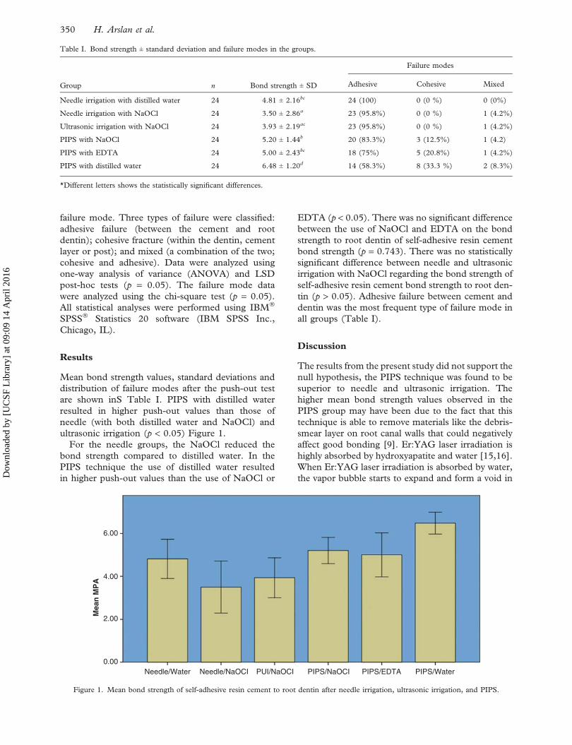
failure mode. Three types of failure were classified:adhesive failure (between the cement and rootdentin); cohesive fracture (within the dentin, cementlayer or post); and mixed (a combination of the two;cohesive and adhesive). Data were analyzed usingone-way analysis of variance (ANOVA) and LSDpost-hoc tests (p = 0.05). The failure mode datawere analyzed using the chi-square test (p = 0.05).All statistical analyses were performed using IBM�
SPSS� Statistics 20 software (IBM SPSS Inc.,Chicago, IL).
Results
Mean bond strength values, standard deviations anddistribution of failure modes after the push-out testare shown inS Table I. PIPS with distilled waterresulted in higher push-out values than those ofneedle (with both distilled water and NaOCl) andultrasonic irrigation (p < 0.05) Figure 1.For the needle groups, the NaOCl reduced the
bond strength compared to distilled water. In thePIPS technique the use of distilled water resultedin higher push-out values than the use of NaOCl or
EDTA (p < 0.05). There was no significant differencebetween the use of NaOCl and EDTA on the bondstrength to root dentin of self-adhesive resin cementbond strength (p = 0.743). There was no statisticallysignificant difference between needle and ultrasonicirrigation with NaOCl regarding the bond strength ofself-adhesive resin cement bond strength to root den-tin (p > 0.05). Adhesive failure between cement anddentin was the most frequent type of failure mode inall groups (Table I).
Discussion
The results from the present study did not support thenull hypothesis, the PIPS technique was found to besuperior to needle and ultrasonic irrigation. Thehigher mean bond strength values observed in thePIPS group may have been due to the fact that thistechnique is able to remove materials like the debris-smear layer on root canal walls that could negativelyaffect good bonding [9]. Er:YAG laser irradiation ishighly absorbed by hydroxyapatite and water [15,16].When Er:YAG laser irradiation is absorbed by water,the vapor bubble starts to expand and form a void in
Table I. Bond strength ± standard deviation and failure modes in the groups.
Group n Bond strength ± SD
Failure modes
Adhesive Cohesive Mixed
Needle irrigation with distilled water 24 4.81 ± 2.16bc 24 (100) 0 (0 %) 0 (0%)
Needle irrigation with NaOCl 24 3.50 ± 2.86a 23 (95.8%) 0 (0 %) 1 (4.2%)
Ultrasonic irrigation with NaOCl 24 3.93 ± 2.19ac 23 (95.8%) 0 (0 %) 1 (4.2%)
PIPS with NaOCl 24 5.20 ± 1.44b 20 (83.3%) 3 (12.5%) 1 (4.2)
PIPS with EDTA 24 5.00 ± 2.43bc 18 (75%) 5 (20.8%) 1 (4.2%)
PIPS with distilled water 24 6.48 ± 1.20d 14 (58.3%) 8 (33.3 %) 2 (8.3%)
*Different letters shows the statistically significant differences.
Mea
n M
PA
6.00
4.00
2.00
0.00Needle/Water PIPS/WaterNeedle/NaOCI PUI/NaOCI PIPS/NaOCI PIPS/EDTA
Figure 1. Mean bond strength of self-adhesive resin cement to root dentin after needle irrigation, ultrasonic irrigation, and PIPS.
350 H. Arslan et al.
Dow
nloa
ded
by [
UC
SF L
ibra
ry]
at 0
9:09
14
Apr
il 20
16
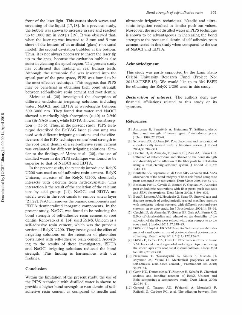
front of the laser light. This causes shock waves andstreaming of the liquid [17,18]. In a previous study,the bubble was shown to increase in size and reachedup to 1800 mm in 220 ms [19]. It was observed that,when the laser tip was inserted to 2 mm and 5 mmshort of the bottom of an artificial (glass) root canalmodel, the second cavitation bubbled at the bottom.Thus, it is not always necessary to insert the laser tipup to the apex, because the cavitation bubbles alsoassist in cleaning the apical region. The present studyhas confirmed this finding in real human teeth.Although the ultrasonic file was inserted into theapical part of the post space, PIPS was found to bethe most effective technique. This suggests that PIPSmay be beneficial in obtaining high bond strengthbetween self-adhesive resin cement and root dentin.Meire et al. [20] investigated the absorption of
different endodontic irrigating solutions includingwater, NaOCl, and EDTA at wavelengths between300–3000 nm. They found that water and NaOClshowed a markedly high absorption (> 40) at 2.940nm (Er:YAG laser), while EDTA showed less absorp-tion (= 33.5). Thus, in the present study, PIPS tech-nique described for Er:YAG laser (2.940 nm) wasused with different irrigating solutions and the effec-tiveness of the PIPS technique on the bond strength tothe root canal dentin of a self-adhesive resin cementwas evaluated for different irrigating solutions. Sim-ilar to the findings of Meire et al. [20], the use ofdistilled water in the PIPS technique was found to besuperior to that of NaOCl and EDTA.In the present study, the recently introduced RelyX
U200 was used as self-adhesive resin cement. RelyXUnicem, ancestor of the RelyX U200, chemicallyinteracts with calcium from hydroxyapatite. Thisinteraction is the result of the chelation of the calciumions by acid groups [11]. NaOCl and EDTA arewidely used in the root canals to remove smear layer[21,22]. NaOCl removes the organic components andEDTA demineralized inorganic components. In thepresent study, NaOCl was found to be reducing thebond strength of self-adhesive resin cement to rootdentin. Renovato et al. [14] used RelyX Unicem as aself-adhesive resin cement, which was the previousversion of RelyX U200. They investigated the effect ofirrigating solutions on the retention of glass-fiberposts luted with self-adhesive resin cement. Accord-ing to the results of these investigators, EDTAand NaOCl irrigating solutions reduced the bondstrength. This finding is harmonious with ourfindings.
Conclusion
Within the limitation of the present study, the use ofthe PIPS technique with distilled water is shown toprovide a higher bond strength to root dentin of self-adhesive resin cement as compared to needle and
ultrasonic irrigation techniques. Needle and ultra-sonic irrigation resulted in similar push-out values.Moreover, the use of distilled water in PIPS techniqueis shown to be advantageous in increasing the bondstrength to the root canal dentin of self-adhesive resincement tested in this study when compared to the useof NaOCl and EDTA.
Acknowledgment
This study was partly supported by the Izmir KatipCelebi University Research Fund (Project No:2013-2-TSBP-15). We would like to to 3M ESPEfor obtaining the RelyX U200 used in this study.
Declaration of interest: The authors deny anyfinancial affiliations related to this study or itssponsors.
References
[1] Asmussen E, Peutzfeldt A, Heitmann T. Stiffness, elasticlimit, and strength of newer types of endodontic posts.J Dent 1999;27:275–8.
[2] Schwartz RS, Robbins JW. Post placement and restoration ofendodontically treated teeth: a literature review. J Endod2004;30:289–301.
[3] Cecchin D, de Almeida JF, Gomes BP, Zaia AA, Ferraz CC.Influence of chlorhexidine and ethanol on the bond strengthand durability of the adhesion of the fiber posts to root dentinusing a total etching adhesive system. J Endod 2011;37:1310–15.
[4] Bonfante EA, Pegoraro LF, de GoesMF, Carvalho RM. SEMobservation of the bond integrity of fiber-reinforced compositeposts cemented into root canals. DentMater 2008;24:483–91.
[5] Boschian Pest L, Cavalli G, Bertani P, Gagliani M. Adhesivepost-endodontic restorations with fiber posts: push-out testsand SEM observations. Dent Mater 2002;18:596–602.
[6] Butz F, Lennon AM, Heydecke G, Strub JR. Survival rate andfracture strength of endodontically treated maxillary incisorswith moderate defects restored with different post-and-coresystems: an in vitro study. Int J Prosthodont 2001;14:58–64.
[7] Cecchin D, de Almeida JF, Gomes BP, Zaia AA, Ferraz CC.Effect of chlorhexidine and ethanol on the durability of theadhesion of the fiber post relined with resin composite to theroot canal. J Endod 2011;37:678–83.
[8] DiVito E, Lloyd A. ER:YAG laser for 3-dimensional debride-ment of canal systems: use of photon-induced photoacousticstreaming. Dent Today 2012;31(11):122,124–7.
[9] DiVito E, Peters OA, Olivi G. Effectiveness of the erbium:YAG laser and new design radial and stripped tips in removingthe smear layer after root canal instrumentation. Lasers MedSci 2012;27:273–80.
[10] Nakamura T, Wakabayashi K, Kinuta S, Nishida H,Miyamae M, Yatani H. Mechanical properties of newself-adhesive resin-based cement. J Prosthodont Res 2010;54:59–64.
[11] Gerth HU, Dammaschke T, Zuchner H, Schafer E. Chemicalanalysis and bonding reaction of RelyX Unicem andBifix composites–a comparative study. Dent Mater 2006;22:934–41.
[12] Goracci C, Tavares AU, Fabianelli A, Monticelli F,Raffaelli O, Cardoso PC, et al. The adhesion between fiber
Bond strength of self-adhesive resin 351
Dow
nloa
ded
by [
UC
SF L
ibra
ry]
at 0
9:09
14
Apr
il 20
16
posts and root canal walls: comparison between microtensileand push-out bond strength measurements. Eur J Oral Sci2004;112:353–61.
[13] Soares CJ, Santana FR, Castro CG, Santos-Filho PC,Soares PV, Qian F, et al. Finite element analysis and bondstrength of a glass post to intraradicular dentin: comparisonbetween microtensile and push-out tests. Dent Mater 2008;24:1405–11.
[14] Renovato SR, Santana FR, Ferreira JM, Souza JB, Soares CJ,Estrela C. Effect of calcium hydroxide and endodontic irri-gants on fibre post bond strength to root canal dentine. IntEndod J 2013;46:738–46.
[15] Paghdiwala AF. Does the laser work on hard dental tissue?J Am Dent Assoc 1991;122:79–80.
[16] Armengol V, Jean A, Rohanizadeh R, Hamel H. Scanningelectron microscopic analysis of diseased and healthy dentalhard tissues after Er:YAG laser irradiation: in vitro study.J Endod 1999;25:543–6.
[17] Brugnera A Jr, Zanin F, Barbin EL, Spanó JC, Santana R,Pécora JD. Effects of Er:YAG and Nd:YAG laser irradiation
on radicular dentine permeability using different irrigatingsolutions. Lasers Surg Med 2003;33:256–9.
[18] Kivanc BH, Ulusoy OI, Gorgul G. Effects of Er:YAG laserand Nd:YAG laser treatment on the root canal dentinof human teeth: a SEM study. Lasers Med Sci 2008;23:247–52.
[19] Matsumoto H, Yoshimine Y, Akamine A. Visualization ofirrigant flow and cavitation induced by Er:YAG laser within aroot canal model. J Endod 2011;37:839–43.
[20] Meire MA, Poelman D, De Moor RJ. Optical properties ofroot canal irrigants in the 300-3,000-nm wavelength region.Lasers Med Sci 2013;27:1307–14.
[21] Ahmetoglu F, Keles A, Yalcin M, Simsek N. Effectivenessof different irrigation systems on smear layer removal:a scanning electron microscopic study. Eur J Dent 2014;8:53–7.
[22] Kalyoncuoglu E, Demiryurek EO. A comparative scanningelectron microscopy evaluation of smear layer removal fromteeth with different irrigation solutions and lasers. MicroscMicroanal 2013;19:1465–9.
352 H. Arslan et al.
Dow
nloa
ded
by [
UC
SF L
ibra
ry]
at 0
9:09
14
Apr
il 20
16
ORIGINAL ARTICLE
Bleaching effect of activation of hydrogen peroxide usingphoton-initiated photoacoustic streaming technique
Hakan Arslan & Merve Akcay & Bilal Yasa &
Huseyin Hatirli & Gökhan Saygili
Received: 3 December 2013 /Accepted: 4 May 2014 /Published online: 25 May 2014# Springer-Verlag Berlin Heidelberg 2014
AbstractObjectives This study aims to investigate the bleaching effec-tiveness of photon-initiated photoacoustic streaming (PIPS)using 35 % hydrogen peroxide on discolored teeth as com-pared with different devital bleaching techniques.Materials and methods Fifty extracted human mandibularincisors were collected and artificially stained using sheep’sblood. The teeth were then randomly divided into five groupsaccording to the different bleaching procedures to be tested:walking bleach with sodium perborate and with 35 % hydro-gen peroxide gel, both for 1 week; PIPS using 35 % hydrogenperoxide liquid for 30 min; and just 35 % hydrogen peroxide,as a liquid and as a gel (again, for 30min). Spectrophotometricmeasurements were obtained on the buccal surfaces of thecrowns, at the beginning, just after the bleaching procedureshad been performed, and the following first, third, and seventhdays. The ΔE values were calculated, and the data wereanalyzed with a two-way analysis of variance (P=0.05).Results There were statistically significant differences be-tween the PIPS technique using 35 % hydrogen peroxideliquid and the 35 % hydrogen peroxide liquid and gel withoutPIPS immediately after the procedures (P<0.05). On Days 1,
3, and 7, the PIPS technique further bleached specimens morethan all of the other techniques (P<0.05).Conclusions The PIPS technique using 35 % hydrogen per-oxide was found to be more effective than all of the conven-tional techniques.Clinical relevance Within limitations of this study, PIPS tech-nique using hydrogen peroxide was superior to the conven-tional techniques. Further studies should be conducted todetermine if the PIPS technique results in any complications,particularly cervical resorption.
Keywords Bleaching . Discoloration . Er/YAG . Hydrogenperoxide . Internal bleaching . Laser . Photon-initiatedphotoacoustic streaming . Sodium perborate
Introduction
Systemic and local factors can cause intrinsic changes to thetooth surface [1]. The main intrinsic changes related to theendodontic process may result in a serious esthetic complaint.Internal bleaching is widely used to resolve this, as it is aminimally invasive, simple, and cost-effective intervention fordiscolored nonvital teeth [2].
Discolored nonvital teeth can be bleached with chemicals,such as sodium perborate, hydrogen peroxide, and carbamideperoxide. Hydrogen peroxide seems to be the optimumchoice, as it produces free radicals, such as hydroperoxyland hydroxyl. Moreover, it can penetrate to the enamel anddentin and release oxygen that breaks the double bonds of theorganic and inorganic compounds [1, 3].
Bleaching agents can be applied in the pulp chamberfollowed by a heat source to catalyze and accelerate thebreaking reaction. Photoxidation, the process for activatingbleaching by light, causes the decomposition of the bleachingagents, thus releasing free radicals [4]. This mechanism may
H. ArslanDepartment of Endodontics, Faculty of Dentistry, Ataturk University,Erzurum, Turkey
M. AkcayDepartment of Pedodontics, Faculty of Dentistry, Izmir Katip CelebiUniversity, Izmir, Turkey
B. Yasa (*) :H. HatirliDepartment of Restorative Dentistry, Faculty of Dentistry, IzmirKatip Celebi University, Izmir, Turkeye-mail: [email protected]
G. SaygiliDepartment of Endodontics, Faculty of Dentistry, Izmir Katip CelebiUniversity, Izmir, Turkey
Clin Oral Invest (2015) 19:253–259DOI 10.1007/s00784-014-1255-9
be induced with halogen lamp appliances or lasers [3]. Inorder to accelerate the bleaching process, different laser de-vices have been used as light sources subsequent to thebleaching agent application [5, 6]. Laser devices also havebeen applied (internal bleaching) prior to the bleaching agentapplication, in order to increase the permeability of tubulesand remove the smear layer (as a conditioner) [7]. However,laser energy has not yet been used at the same time as ableaching agent for the purpose of agitating it.
A novel laser agitation technique, photon-induced photo-acoustic streaming (PIPS), has been proposed. In this tech-nique, an erbium/yttrium–aluminum–garnet (Er/YAG) laserwith a radial and stripped tip of novel design is used atsubablative power settings. This technique differs from otheragitation techniques in the placement of only the tip into thecoronal portion [8].
Cardoso et al. [9] evaluated the effect of the ultrasonicactivation of bleaching agents on internal bleaching. To ourknowledge, however, no studies have investigated thebleaching effect of laser agitation of bleaching agents ondiscolored teeth. Therefore, the aim of the present study wasto investigate the bleaching effect of PIPS using 35 % hydro-gen peroxide on discolored teeth as compared with walkingbleaching techniques (sodium perborate and 35 % hydrogenperoxide gel),and 35 % hydrogen peroxide liquid and gelapplications (without PIPS). The null hypothesis was thatthere is no difference between the PIPS using 35 % hydrogenperoxide and the other internal bleaching techniques.
Materials and methods
Specimen selection
Mandibular incisors were selected from a collection of teeththat had recently been extracted for reasons unrelated to thisstudy. They were stored in distilled water until use. Fifty wereselected from among those with similar mesiodistal (3.3±0.3)and buccolingual width (5.6±0.4) to obtain standard accesscavity dimensions. Teeth with root canal treatment, restora-tion, immature root apices, and/or coronal defects were ex-cluded from the study. The soft tissue and calculus wereremoved mechanically from the root surfaces with an ultra-sonic scaler (Anthos u-PZ6; Imola, Italy), and then polishedwith pumice.
Preparation of specimens
A standard oval coronal access was performed, and the thick-ness of the buccal wall was gradually decreased with a size 6number carbide bur and standardized at 2.6±0.3 by using anelectronic digital caliper. Root canals were enlarged up to F3using ProTaper rotary instruments (Dentsply Maillefer,
Ballaigues, Switzerland). Coronal flaring was performedusing size 4–5 Gates Glidden burs (Mani, Mani Inc.,Takanezawa, Japan) to ease the placement of cement as acervical plug. Specimens were irrigated to open dentinal tu-bules with combination of 5 mL of 17 % EDTA (Werax; SpotDis Deposu A.S., Izmir, Turkey) and 5 mL of 5 % NaOCl,each for 60 s. Final rinse was performed using 5 mL ofdistilled water.
Artificial staining
The specimens were artificially stained as described by previ-ous studies [9, 10], with a modified procedure based on thatemployed by Freccia and Peters [11]. First, the specimenswere immersed in Eppendorf tubes containing sheep bloodand centrifuged at 3,400 rpm for 20 min at 37 °C (Micro220R; Hettich, Germany). After the plasma (supernatant) andprecipitate were yielded, the plasma was removed. TheEppendorf tubes were then centrifuged for a further 20 min,followed by a centrifugation twice a day for a further 2 days.After each centrifugation, the teeth were irrigated with dis-tilled water, reinserted into the Eppendorf tubes, and stored at37 °C in 100 % humidity.
On the fourth day, the teeth were removed from theEppendorf tubes and placed in clean ones. A total of 0.5 mLof distilled water was added to the original Eppendorf tubes(i.e., including blood), which were then centrifuged for afurther 20 min to pioneer the hemolysis of erythrocytes. Thisresulted in a membranous precipitate and hemolysate, and thesupernatant layer was removed. The teeth were then trans-ferred back to these Eppendorf tubes, and the tubes werecentrifuged again, for 20 min on three consecutive days. After6 days, the blood was changed, and all the aforementionedprocedures were repeated, over a further 6 days.
Baseline color measurement
After the artificial staining procedure, the teeth were cleanedin running water and dried with air spray, after which 2 mmthick glass-ionomer cement (KetacMolar; 3M ESPE, Seefeld,Germany) was placed 1 mm apical to the cementoenameljunction (CEJ). A standardized circular strip with an internaldiameter of 4 mm (external diameter=8 mm) was bonded tothe buccal surface of the crown coronal to the CEJ to ensurethat color measurement was performed on the same region atevery turn with a vertical angle (Fig. 1) [12]. A baseline colormeasurement was performed using a noncontact-type spectro-photometer (Spectro Shade™ Micro; MHT, Milan, Italy) onthe buccal surfaces of the crowns. The specimens were dis-tributed equally according to the baseline color measurementsacross the five groups to be tested. Statistical analysis by one-way ANOVA confirmed no significant differences among thegroups in terms of their baseline color (P=0.682).
254 Clin Oral Invest (2015) 19:253–259
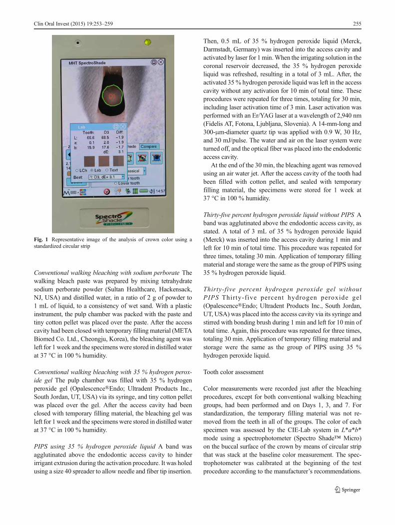
Conventional walking bleaching with sodium perborate Thewalking bleach paste was prepared by mixing tetrahydratesodium perborate powder (Sultan Healthcare, Hackensack,NJ, USA) and distilled water, in a ratio of 2 g of powder to1 mL of liquid, to a consistency of wet sand. With a plasticinstrument, the pulp chamber was packed with the paste andtiny cotton pellet was placed over the paste. After the accesscavity had been closed with temporary filling material (METABiomed Co. Ltd., Cheongju, Korea), the bleaching agent wasleft for 1 week and the specimens were stored in distilled waterat 37 °C in 100 % humidity.
Conventional walking bleaching with 35 % hydrogen perox-ide gel The pulp chamber was filled with 35 % hydrogenperoxide gel (Opalescence®Endo; Ultradent Products Inc.,South Jordan, UT, USA) via its syringe, and tiny cotton pelletwas placed over the gel. After the access cavity had beenclosed with temporary filling material, the bleaching gel wasleft for 1 week and the specimens were stored in distilled waterat 37 °C in 100 % humidity.
PIPS using 35 % hydrogen peroxide liquid A band wasagglutinated above the endodontic access cavity to hinderirrigant extrusion during the activation procedure. It was holedusing a size 40 spreader to allow needle and fiber tip insertion.
Then, 0.5 mL of 35 % hydrogen peroxide liquid (Merck,Darmstadt, Germany) was inserted into the access cavity andactivated by laser for 1 min.When the irrigating solution in thecoronal reservoir decreased, the 35 % hydrogen peroxideliquid was refreshed, resulting in a total of 3 mL. After, theactivated 35% hydrogen peroxide liquid was left in the accesscavity without any activation for 10 min of total time. Theseprocedures were repeated for three times, totaling for 30 min,including laser activation time of 3 min. Laser activation wasperformed with an Er/YAG laser at a wavelength of 2,940 nm(Fidelis AT, Fotona, Ljubljana, Slovenia). A 14-mm-long and300-μm-diameter quartz tip was applied with 0.9 W, 30 Hz,and 30 mJ/pulse. The water and air on the laser system wereturned off, and the optical fiber was placed into the endodonticaccess cavity.
At the end of the 30 min, the bleaching agent was removedusing an air water jet. After the access cavity of the tooth hadbeen filled with cotton pellet, and sealed with temporaryfilling material, the specimens were stored for 1 week at37 °C in 100 % humidity.
Thirty-five percent hydrogen peroxide liquid without PIPS Aband was agglutinated above the endodontic access cavity, asstated. A total of 3 mL of 35 % hydrogen peroxide liquid(Merck) was inserted into the access cavity during 1 min andleft for 10 min of total time. This procedure was repeated forthree times, totaling 30 min. Application of temporary fillingmaterial and storage were the same as the group of PIPS using35 % hydrogen peroxide liquid.
Thirty-five percent hydrogen peroxide gel withoutPIPS Thirty-f ive percent hydrogen peroxide gel(Opalescence®Endo; Ultradent Products Inc., South Jordan,UT, USA) was placed into the access cavity via its syringe andstirred with bonding brush during 1 min and left for 10 min oftotal time. Again, this procedure was repeated for three times,totaling 30 min. Application of temporary filling material andstorage were the same as the group of PIPS using 35 %hydrogen peroxide liquid.
Tooth color assessment
Color measurements were recorded just after the bleachingprocedures, except for both conventional walking bleachinggroups, had been performed and on Days 1, 3, and 7. Forstandardization, the temporary filling material was not re-moved from the teeth in all of the groups. The color of eachspecimen was assessed by the CIE-Lab system in L*a*b*mode using a spectrophotometer (Spectro Shade™ Micro)on the buccal surface of the crown by means of circular stripthat was stack at the baseline color measurement. The spec-trophotometer was calibrated at the beginning of the testprocedure according to the manufacturer’s recommendations.
Fig. 1 Representative image of the analysis of crown color using astandardized circular strip
Clin Oral Invest (2015) 19:253–259 255
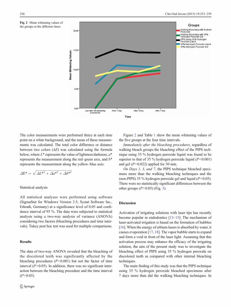
The color measurements were performed thrice at each timepoint on a white background, and the mean of these measure-ments was calculated. The total color difference or distancebetween two colors (ΔE) was calculated using the formulabelow, where L* represents the value of lightness/darkness, a*represents the measurement along the red–green axis, and b*represents the measurement along the yellow–blue axis:
ΔE* ¼ffiffiffiffiffiffiffiffiffiffiffiffiffiffiffiffiffiffiffiffiffiffiffiffiffiffiffiffiffiffiffiffiffiffiffiffiffiffiffiffiffiffiffiffi
ΔL*2 þΔa*2 þΔb*2p
Statistical analysis
All statistical analyses were performed using software(SigmaStat for Windows Version 3.5; Systat Software Inc.,Erkrath, Germany) at a significance level of 0.05 and confi-dence interval of 95 %. The data were subjected to statisticalanalysis using a two-way analysis of variance (ANOVA)considering two factors (bleaching procedures and time inter-vals). Tukey post hoc test was used for multiple comparisons.
Results
The data of two-way ANOVA revealed that the bleaching ofthe discolored teeth was significantly affected by thebleaching procedures (P<0.001) but not the factor of timeinterval (P>0.05). In addition, there was no significant inter-action between the bleaching procedure and the time interval(P>0.05).
Figure 2 and Table 1 show the mean whitening values ofthe five groups at the four time intervals.
Immediately after the bleaching procedures, regardless ofwalking bleach groups the bleaching effect of the PIPS tech-nique using 35 % hydrogen peroxide liquid was found to besuperior to that of 35 % hydrogen peroxide liquid (P<0.001)and gel (P=0.022) applied for 30 min.
On Days 1, 3, and 7, the PIPS technique bleached speci-mens more than the walking bleaching techniques and the(non-PIPS) 35 % hydrogen peroxide gel and liquid (P<0.05).There were no statistically significant differences between theother groups (P>0.05) (Fig. 3).
Discussion
Activation of irrigating solutions with laser tips has recentlybecome popular in endodontics [13–15]. The mechanism oflaser-activated irrigation is based on the formation of bubbles[16].When the energy of erbium lasers is absorbed bywater, itcauses evaporation [17, 18]. The vapor bubble starts to expandand form a void in front of the laser light. Assuming that thisactivation process may enhance the efficacy of the irrigatingsolution, the aim of the present study was to investigate thebleaching effect of PIPS using 35 % hydrogen peroxide ondiscolored teeth as compared with other internal bleachingtechniques.
The main finding of this study was that the PIPS techniqueusing 35 % hydrogen peroxide bleached specimens after7 days more than did the walking bleaching techniques. In
Fig. 2 Mean whitening values ofthe groups at the different times
256 Clin Oral Invest (2015) 19:253–259
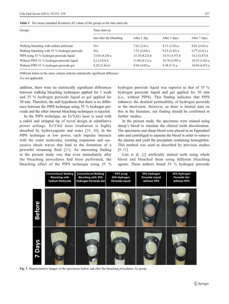
addition, there were no statistically significant differencesbetween walking bleaching techniques applied for 1 weekand 35 % hydrogen peroxide liquid or gel applied for30 min. Therefore, the null hypothesis that there is no differ-ence between the PIPS technique using 35 % hydrogen per-oxide and the other internal bleaching techniques is rejected.
In the PIPS technique, an Er/YAG laser is used witha radial and stripped tip of novel design at subablativepower settings. Er/YAG laser irradiation is highlyabsorbed by hydroxyapatite and water [19, 20]. In thePIPS technique at low power, each impulse interactswith the water molecules, creating expansion and suc-cessive shock waves that lead to the formation of apowerful streaming fluid [21]. An interesting findingin the present study was that even immediately afterthe bleaching procedures had been performed, thebleaching effect of the PIPS technique using 35 %
hydrogen peroxide liquid was superior to that of 35 %hydrogen peroxide liquid and gel applied for 30 min(i.e., without PIPS). This finding indicates that PIPSenhances the dentinal permeability of hydrogen peroxidein the short-term. However, as there is limited data onthis in the literature, our finding should be confirmed infurther studies.
In the present study, the specimens were stained usingsheep’s blood to simulate the clinical tooth discoloration.The specimens and sheep blood were placed in an Eppendorftube and centrifuged to separate the blood in order to removethe plasma and yield the precipitate containing hemoglobin.This method was used as described by previous studies[9–11].
Lim et al. [2] artificially stained teeth using wholeblood and bleached them using different bleachingagents. These authors found 35 % hydrogen peroxide
Fig. 3 Representative images of the specimens before and after the bleaching procedures, by group
Table 1 The mean (standard deviation) ΔE values of the groups at the time intervals
Groups Time interval
Just after the bleaching After 1 day After 3 days After 7 days
Walking bleaching with sodium perborate N/a 7.62 (2.8) a 8.31 (3.43) a 9.01 (3.62) a
Walking bleaching with 35 % hydrogen peroxide N/a 7.51 (2.68) a 9.25 (2.43) a 9.77 (2.61) a
PIPS using 35 % hydrogen peroxide liquid 13.03 (4.24) a 15.18 (4.23) b 14.35 (3.97) b 14.3 (3.47) b
Without PIPS 35 % hydrogen peroxide liquid 6.2 (3.83) b 11.08 (4.11) a 10.78 (3.99) a 10.55 (3.43) a
Without PIPS 35 % hydrogen peroxide gel 8.28 (3.36) b 9.68 (4.02) a 9.38 (3.7) a 10.94 (4.07) a
Different letters in the same column indicate statistically significant difference
N/a not applicable
Clin Oral Invest (2015) 19:253–259 257
to be more effective than sodium perborate for internalbleaching. In the present study, all of the hydrogenperoxide groups apart from the PIPS technique werenot superior to the sodium perborate in terms ofbleaching discolored teeth.
In in vitro bleaching efficacy studies, human maxillaryincisors, human premolars, or bovine teeth were used [2, 7,9, 10, 12]. In the present study, mandibular incisors were usedto evaluate the bleaching effectiveness of PIPS using 35 %hydrogen peroxide. When compared with maxillary incisors,mandibular incisors are easy to obtain, and also, their diame-ters were suitable and easy to use for immersing in Eppendorftubes for centrifugation. Previously, bleaching efficacy wasevaluated using contact-type spectrophotometers whose mea-surement tips are suitable for wide teeth like maxillary inci-sors, premolars, or bovine teeth [7, 9]. However, the contact-type spectrophotometer is not suitable for mandibular incisors,which are smaller than the teeth aforementioned. In the pres-ent study, a noncontact-type spectrophotometer was used. Theimage was taken on the surface of tooth, and color wasevaluated on this image (Fig. 1) [12]. The noncontact-typespectrophotometer could be beneficial for evaluating colorchanges in relatively smaller teeth.
It is well established that hydrogen peroxide iscaustic, burning tissue on contact [1]. In the presentstudy, therefore, a band was agglutinated above theendodontic access cavity to hinder hydrogen peroxideextrusion during the activation procedure. The PIPStechnique in activating hydrogen peroxide was exper-imentally evaluated in the present study. Clinical us-age cannot be suggested according to the presentstudy, however, as further studies should be conduct-ed to investigate further hindrance of hydrogen per-oxide extrusion during the activation procedure.
Conclusions
Within the limitations of the in vitro study, the PIPS techniquewas more effective than both walking bleach techniques aswell as both hydrogen peroxide groups. Bleaching materialshave adverse effects, however, both are localized and system-ic, such as cervical external resorption [22–24], reduction inmicrohardness of hard dental tissues [25], and toxicity [26].Thus, further studies should be conducted to determine if thePIPS technique results in any complications, including cervi-cal resorption.
Conflict of interest The authors declare that there are no conflicts ofinterests in writing this article.
References
1. Plotino G, Buono L, Grande NM, Pameijer CH, Somma F (2008)Nonvital tooth bleaching: a review of the literature and clinicalprocedures. J Endod 34:394–407. doi:10.1016/j.joen.2007.12.020
2. Lim MY, Lum SO, Poh RS, Lee GP, Lim KC (2004) An in vitrocomparison of the bleaching efficacy of 35 % carbamide peroxidewith established intracoronal bleaching agents. Int Endod J 37:483–488. doi:10.1111/j.1365-2591.2004.00829.x
3. Goldstein RE GD (1995) Complete dental bleaching. 3rd edn.Quintessence, Chicago
4. Blankenau R, Goldstein RE, Haywood VB (1999) The current statusof vital tooth whitening techniques. Compend Contin Educ Dent 20:781–784, 786, 788 passim; quiz 796
5. Gontijo IT, Navarro RS, Ciamponi AL, Miyakawa W, Zezell DM(2008) Color and surface temperature variation during bleaching inhuman devitalized primary teeth: an in vitro study. J Dent Child(Chicago) 75:229–234
6. Camargo SE, Cardoso PE, Valera MC, de Araujo MA, Kojima AN(2009) Penetration of 35 % hydrogen peroxide into the pulp chamberin bovine teeth after LED or Nd:YAG laser activation. Eur J EsthetDent Off J Eur Acad Esthet Dent 4:82–88
7. Mohammadi N, Kimyai S, Navimipour EJ, Soleimanzadeh R, BonabSS (2010) Effect of acid etching and laser treatment of dentin surfaceon intracoronal bleaching efficacy. Photomed Laser Surg 28(Suppl2):S51–S55. doi:10.1089/pho.2009.2606
8. DiVito E, Lloyd A (2012) ER:YAG laser for 3-dimensional debride-ment of canal systems: use of photon-induced photoacoustic stream-ing. Dent Today 31(122):124–127
9. Cardoso M, Martinelli CS, Carvalho CA, Borges AB, Torres CR(2013) Ultrasonic activation of internal bleaching agents. Int Endod J46:40–46. doi:10.1111/j.1365-2591.2012.02090.x
10. Yui KC, Rodrigues JR, Mancini MN, Balducci I, Goncalves SE(2008) Ex vivo evaluation of the effectiveness of bleaching agentson the shade alteration of blood-stained teeth. Int Endod J 41:485–492. doi:10.1111/j.1365-2591.2008.01379.x
11. Freccia WF, Peters DD (1982) A technique for staining extractedteeth: a research and teaching aid for bleaching. J Endod 8:67–69.doi:10.1016/S0099-2399(82)80260-4
12. Akcay M, Arslan H, Yasa B, Kavrik F, Yasa E (2013)Spectrophotometric analysis of crown discoloration induced by var-ious antibiotic pastes used in revascularization. J Endod. doi:10.1016/j.joen.2013.09.019
13. De Moor RJ, Blanken J, Meire M, Verdaasdonk R (2009) Laserinduced explosive vapor and cavitation resulting in effective irriga-tion of the root canal. Part 2: evaluation of the efficacy. Lasers SurgMed 41:520–523. doi:10.1002/lsm.20797
14. de Groot SD, Verhaagen B, Versluis M, Wu MK, Wesselink PR, vander Sluis LW (2009) Laser-activated irrigation within root canals:cleaning efficacy and flow visualization. Int Endod J 42:1077–1083.doi:10.1111/j.1365-2591.2009.01634.x
15. Moon YM, Kim HC, Bae KS, Baek SH, Shon WJ, Lee W (2012)Effect of laser-activated irrigation of 1320-nanometer Nd:YAG laseron sealer penetration in curved root canals. J Endod 38:531–535. doi:10.1016/j.joen.2011.12.008
16. MeireMA, Poelman D, DeMoor RJ (2013) Optical properties of rootcanal irrigants in the 300–3,000-nm wavelength region. Lasers MedSci 27:1307–1314. doi:10.1007/s10103-013-1307-4
17. Brugnera A Jr, Zanin F, Barbin EL, Spano JC, Santana R, Pecora JD(2003) Effects of Er:YAG and Nd:YAG laser irradiation on radiculardentine permeability using different irrigating solutions. Lasers SurgMed 33:256–259. doi:10.1002/lsm.10214
18. Kivanc BH, UlusoyOI, Gorgul G (2008) Effects of Er:YAG laser andNd:YAG laser treatment on the root canal dentin of human teeth: a
258 Clin Oral Invest (2015) 19:253–259
SEM study. Lasers Med Sci 23:247–252. doi:10.1007/s10103-007-0474-6
19. Paghdiwala AF (1991) Does the laser work on hard dental tissue? JAm Dent Assoc 122:79–80
20. Armengol V, Jean A, Rohanizadeh R, Hamel H (1999) Scanningelectron microscopic analysis of diseased and healthy dental hardtissues after Er:YAG laser irradiation: in vitro study. J Endod 25:543–546. doi:10.1016/S0099-2399(99)80376-8
21. DiVito E, Peters OA, Olivi G (2012) Effectiveness of the erbium:YAG laser and new design radial and stripped tips in removing thesmear layer after root canal instrumentation. Lasers Med Sci 27:273–280. doi:10.1007/s10103-010-0858-x
22. Feiglin B (1987) A 6-year recall study of clinically chemi-cally bleached teeth. Oral Surg Oral Med Oral Pathol 63:610–613
23. Heller D, Skriber J, Lin LM (1992) Effect of intracoronal bleachingon external cervical root resorption. J Endod 18:145–148. doi:10.1016/S0099-2399(06)81407-X
24. Rotstein I (1991) In vitro determination and quantification of30 % hydrogen peroxide penetration through dentin and ce-mentum during bleaching. Oral Surg Oral Med Oral Pathol72:602–606
25. Lewinstein I, Hirschfeld Z, Stabholz A, Rotstein I (1994) Effect ofhydrogen peroxide and sodium perborate on the microhardness ofhuman enamel and dentin. J Endod 20:61–63. doi:10.1016/S0099-2399(06)81181-7
26. Anderson DG, Chiego DJ Jr, Glickman GN, McCauley LK (1999) Aclinical assessment of the effects of 10 % carbamide peroxide gel onhuman pulp tissue. J Endod 25:247–250. doi:10.1016/S0099-2399(99)80152-6
Clin Oral Invest (2015) 19:253–259 259
Effect of photon-initiated photoacoustic streamingon removal of apically placed dentinal debris
H. Arslan1, I. D. Capar2, G. Saygili2, T. Gok2 & M. Akcay3
1Department of Endodontics, Faculty of Dentistry, Atat€urk University, Erzurum; 2Department of Endodontics, Faculty of
Dentistry, Izmir Katip Celebi University, _Izmir; and 3Department of Pedodontics, Faculty of Dentistry, Izmir Katip Celebi
University, _Izmir, Turkey
Abstract
Arslan H, Capar ID, Saygili G, Gok T, Akcay M. Effect
of photon-initiated photoacoustic streaming on removal of
apically placed dentinal debris. International Endodontic
Journal, 47, 1072–1077, 2014.
Aim To compare the efficacy of photon-induced
photoacoustic streaming (PIPS) technique with con-
ventional, sonic and ultrasonic irrigation on the
removal of apically placed dentinal debris from an
artificial groove created in a root canal.
Methodology Root canal preparation was per-
formed up to size 40 on 48 extracted single-rooted
teeth using ProTaper rotary instruments. The speci-
mens were then split longitudinally, and a standard-
ized groove was prepared in the apical part of each
segment. Each groove was filled with dentinal debris
mixed with 5% NaOCl. Each tooth was reassembled
and irrigated as follows: (i) conventional irrigation
with 1% NaOCl, (ii) sonic, (iii) ultrasonic irrigation,
and (iv) PIPS. The root segments were disassembled,
and the amount of remaining dentinal debris was
evaluated under a stereomicroscope at 209 magnifi-
cation, using a four-grade scoring system. The data
were evaluated statistically using Kruskal–Wallis and
Mann–Whitney U-tests with a 95% confidence level
(P = 0.05).
Results Photon-induced photoacoustic streaming
removed significantly more dentinal debris than con-
ventional irrigation (P < 0.001), sonic irrigation
(P < 0.001) or ultrasonic irrigation (P = 0.005).
There was no significant difference between sonic and
ultrasonic irrigation (P = 0.377).
Conclusions Photon-induced photoacoustic stre-
aming was more effective than conventional, sonic
and ultrasonic irrigation in the removal of apically
placed dentinal debris.
Keywords: EndoActivator, endodontics, photoa-
coustic streaming, PIPS, sonic, ultrasonic.
Received 17 October 2013; accepted 18 January 2014
Introduction
The goal of biomechanical preparation is to clean,
shape and disinfect the root canal system. However,
chemomechanical preparation leaves untouched
zones, debris, the smear layer and microorganisms
and their by-products, which can result in persistent
inflammation (Vertucci 1984, Wu & Wesselink
2001b, Wu et al. 2001a). That is why irrigation plays
an essential role in root canal treatment. However,
because irrigating solutions can be ineffective in
removing material from the root canal walls (Tora-
binejad et al. 2003, Mancini et al. 2009), improved
irrigation agitation methods such as sonic and ultra-
sonic devices have been proposed (Guerisoli et al.
2002). Recently, agitation of irrigants using lasers
has gained popularity (De Moor et al. 2009, de Groot
et al. 2009, Moon et al. 2012).
A novel laser agitation technique, photon-induced
photoacoustic streaming (PIPS), has been proposed.
This technique differs from other agitation techniques
in that only the tip is placed into the canal orifice
Correspondence: Hakan Arslan, Department of Endodontics,
Faculty of Dentistry, Ataturk University, Erzurum 25240,
Turkey (Tel.: +90.442.231.1900; fax: +90.442.236.0945;
e-mail: [email protected]).
© 2014 International Endodontic Journal. Published by John Wiley & Sons LtdInternational Endodontic Journal, 47, 1072–1077, 2014
doi:10.1111/iej.12251
1072
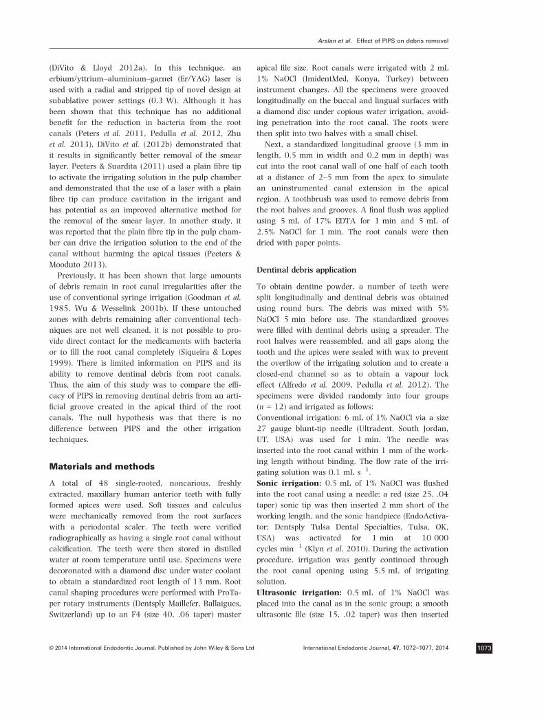
(DiVito & Lloyd 2012a). In this technique, an
erbium/yttrium–aluminium–garnet (Er/YAG) laser is
used with a radial and stripped tip of novel design at
subablative power settings (0.3 W). Although it has
been shown that this technique has no additional
benefit for the reduction in bacteria from the root
canals (Peters et al. 2011, Pedulla et al. 2012, Zhu
et al. 2013), DiVito et al. (2012b) demonstrated that
it results in significantly better removal of the smear
layer. Peeters & Suardita (2011) used a plain fibre tip
to activate the irrigating solution in the pulp chamber
and demonstrated that the use of a laser with a plain
fibre tip can produce cavitation in the irrigant and
has potential as an improved alternative method for
the removal of the smear layer. In another study, it
was reported that the plain fibre tip in the pulp cham-
ber can drive the irrigation solution to the end of the
canal without harming the apical tissues (Peeters &
Mooduto 2013).
Previously, it has been shown that large amounts
of debris remain in root canal irregularities after the
use of conventional syringe irrigation (Goodman et al.
1985, Wu & Wesselink 2001b). If these untouched
zones with debris remaining after conventional tech-
niques are not well cleaned, it is not possible to pro-
vide direct contact for the medicaments with bacteria
or to fill the root canal completely (Siqueira & Lopes
1999). There is limited information on PIPS and its
ability to remove dentinal debris from root canals.
Thus, the aim of this study was to compare the effi-
cacy of PIPS in removing dentinal debris from an arti-
ficial groove created in the apical third of the root
canals. The null hypothesis was that there is no
difference between PIPS and the other irrigation
techniques.
Materials and methods
A total of 48 single-rooted, noncarious, freshly
extracted, maxillary human anterior teeth with fully
formed apices were used. Soft tissues and calculus
were mechanically removed from the root surfaces
with a periodontal scaler. The teeth were verified
radiographically as having a single root canal without
calcification. The teeth were then stored in distilled
water at room temperature until use. Specimens were
decoronated with a diamond disc under water coolant
to obtain a standardized root length of 13 mm. Root
canal shaping procedures were performed with ProTa-
per rotary instruments (Dentsply Maillefer, Ballaigues,
Switzerland) up to an F4 (size 40, .06 taper) master
apical file size. Root canals were irrigated with 2 mL
1% NaOCl (ImidentMed, Konya, Turkey) between
instrument changes. All the specimens were grooved
longitudinally on the buccal and lingual surfaces with
a diamond disc under copious water irrigation, avoid-
ing penetration into the root canal. The roots were
then split into two halves with a small chisel.
Next, a standardized longitudinal groove (3 mm in
length, 0.5 mm in width and 0.2 mm in depth) was
cut into the root canal wall of one half of each tooth
at a distance of 2–5 mm from the apex to simulate
an uninstrumented canal extension in the apical
region. A toothbrush was used to remove debris from
the root halves and grooves. A final flush was applied
using 5 mL of 17% EDTA for 1 min and 5 mL of
2.5% NaOCl for 1 min. The root canals were then
dried with paper points.
Dentinal debris application
To obtain dentine powder, a number of teeth were
split longitudinally and dentinal debris was obtained
using round burs. The debris was mixed with 5%
NaOCl 5 min before use. The standardized grooves
were filled with dentinal debris using a spreader. The
root halves were reassembled, and all gaps along the
tooth and the apices were sealed with wax to prevent
the overflow of the irrigating solution and to create a
closed-end channel so as to obtain a vapour lock
effect (Alfredo et al. 2009, Pedulla et al. 2012). The
specimens were divided randomly into four groups
(n = 12) and irrigated as follows:
Conventional irrigation: 6 mL of 1% NaOCl via a size
27 gauge blunt-tip needle (Ultradent, South Jordan,
UT, USA) was used for 1 min. The needle was
inserted into the root canal within 1 mm of the work-
ing length without binding. The flow rate of the irri-
gating solution was 0.1 mL s�1.
Sonic irrigation: 0.5 mL of 1% NaOCl was flushed
into the root canal using a needle; a red (size 25, .04
taper) sonic tip was then inserted 2 mm short of the
working length, and the sonic handpiece (EndoActiva-
tor; Dentsply Tulsa Dental Specialties, Tulsa, OK,
USA) was activated for 1 min at 10 000
cycles min�1 (Klyn et al. 2010). During the activation
procedure, irrigation was gently continued through
the root canal opening using 5.5 mL of irrigating
solution.
Ultrasonic irrigation: 0.5 mL of 1% NaOCl was
placed into the canal as in the sonic group; a smooth
ultrasonic file (size 15, .02 taper) was then inserted
Arslan et al. Effect of PIPS on debris removal
© 2014 International Endodontic Journal. Published by John Wiley & Sons Ltd International Endodontic Journal, 47, 1072–1077, 2014 1073
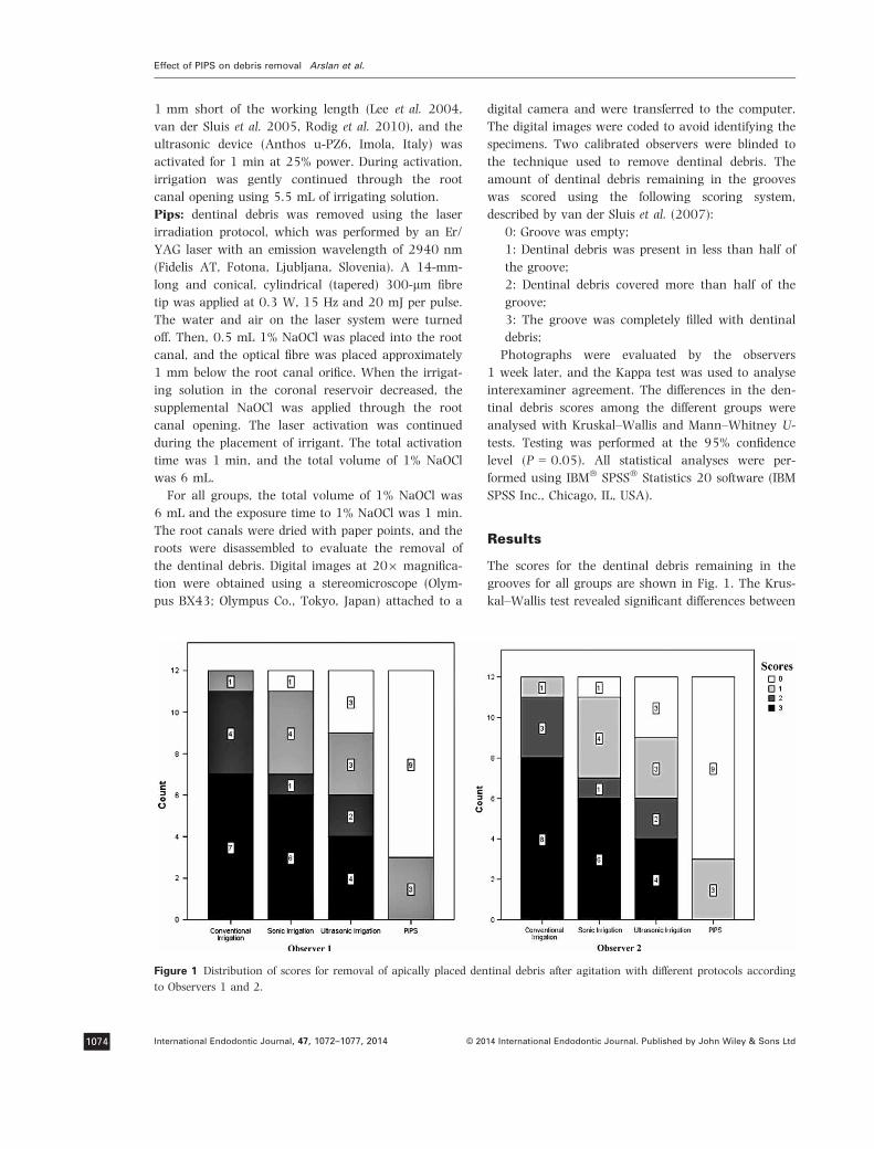
1 mm short of the working length (Lee et al. 2004,
van der Sluis et al. 2005, Rodig et al. 2010), and the
ultrasonic device (Anthos u-PZ6, Imola, Italy) was
activated for 1 min at 25% power. During activation,
irrigation was gently continued through the root
canal opening using 5.5 mL of irrigating solution.
Pips: dentinal debris was removed using the laser
irradiation protocol, which was performed by an Er/
YAG laser with an emission wavelength of 2940 nm
(Fidelis AT, Fotona, Ljubljana, Slovenia). A 14-mm-
long and conical, cylindrical (tapered) 300-lm fibre
tip was applied at 0.3 W, 15 Hz and 20 mJ per pulse.
The water and air on the laser system were turned
off. Then, 0.5 mL 1% NaOCl was placed into the root
canal, and the optical fibre was placed approximately
1 mm below the root canal orifice. When the irrigat-
ing solution in the coronal reservoir decreased, the
supplemental NaOCl was applied through the root
canal opening. The laser activation was continued
during the placement of irrigant. The total activation
time was 1 min, and the total volume of 1% NaOCl
was 6 mL.
For all groups, the total volume of 1% NaOCl was
6 mL and the exposure time to 1% NaOCl was 1 min.
The root canals were dried with paper points, and the
roots were disassembled to evaluate the removal of
the dentinal debris. Digital images at 209 magnifica-
tion were obtained using a stereomicroscope (Olym-
pus BX43; Olympus Co., Tokyo, Japan) attached to a
digital camera and were transferred to the computer.
The digital images were coded to avoid identifying the
specimens. Two calibrated observers were blinded to
the technique used to remove dentinal debris. The
amount of dentinal debris remaining in the grooves
was scored using the following scoring system,
described by van der Sluis et al. (2007):
0: Groove was empty;
1: Dentinal debris was present in less than half of
the groove;
2: Dentinal debris covered more than half of the
groove;
3: The groove was completely filled with dentinal
debris;
Photographs were evaluated by the observers
1 week later, and the Kappa test was used to analyse
interexaminer agreement. The differences in the den-
tinal debris scores among the different groups were
analysed with Kruskal–Wallis and Mann–Whitney U-
tests. Testing was performed at the 95% confidence
level (P = 0.05). All statistical analyses were per-
formed using IBM� SPSS� Statistics 20 software (IBM
SPSS Inc., Chicago, IL, USA).
Results
The scores for the dentinal debris remaining in the
grooves for all groups are shown in Fig. 1. The Krus-
kal–Wallis test revealed significant differences between
Figure 1 Distribution of scores for removal of apically placed dentinal debris after agitation with different protocols according
to Observers 1 and 2.
Effect of PIPS on debris removal Arslan et al.
© 2014 International Endodontic Journal. Published by John Wiley & Sons LtdInternational Endodontic Journal, 47, 1072–1077, 20141074
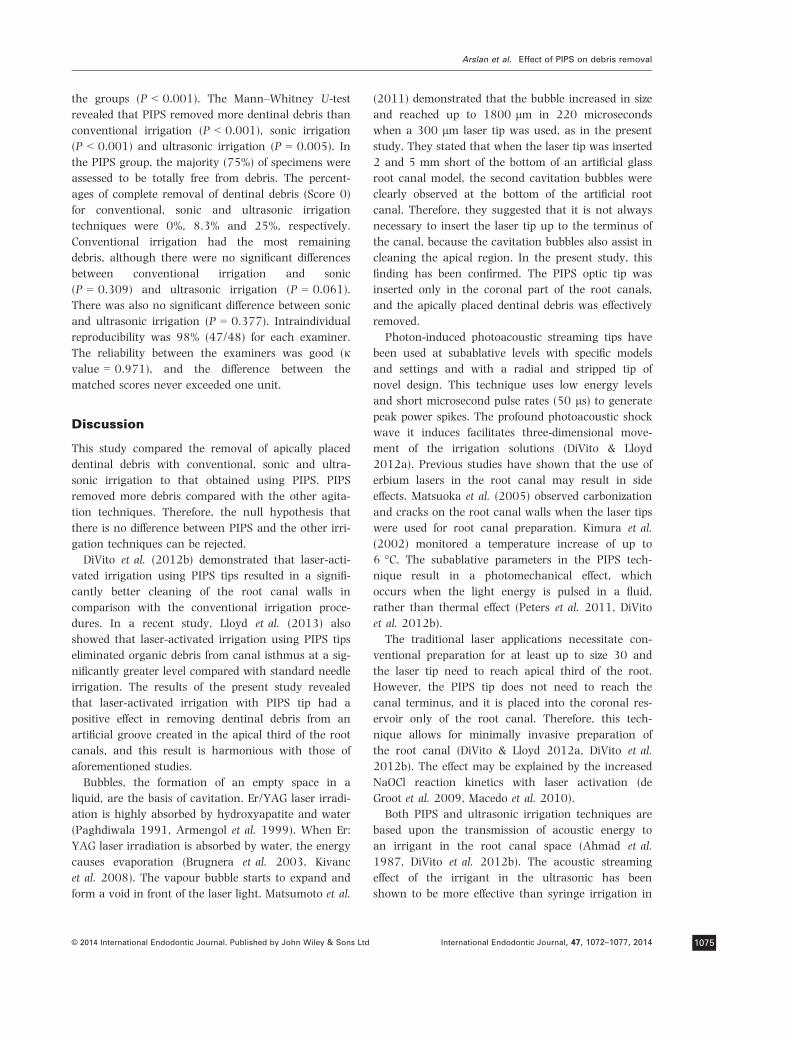
the groups (P < 0.001). The Mann–Whitney U-test
revealed that PIPS removed more dentinal debris than
conventional irrigation (P < 0.001), sonic irrigation
(P < 0.001) and ultrasonic irrigation (P = 0.005). In
the PIPS group, the majority (75%) of specimens were
assessed to be totally free from debris. The percent-
ages of complete removal of dentinal debris (Score 0)
for conventional, sonic and ultrasonic irrigation
techniques were 0%, 8.3% and 25%, respectively.
Conventional irrigation had the most remaining
debris, although there were no significant differences
between conventional irrigation and sonic
(P = 0.309) and ultrasonic irrigation (P = 0.061).
There was also no significant difference between sonic
and ultrasonic irrigation (P = 0.377). Intraindividual
reproducibility was 98% (47/48) for each examiner.
The reliability between the examiners was good (jvalue = 0.971), and the difference between the
matched scores never exceeded one unit.
Discussion
This study compared the removal of apically placed
dentinal debris with conventional, sonic and ultra-
sonic irrigation to that obtained using PIPS. PIPS
removed more debris compared with the other agita-
tion techniques. Therefore, the null hypothesis that
there is no difference between PIPS and the other irri-
gation techniques can be rejected.
DiVito et al. (2012b) demonstrated that laser-acti-
vated irrigation using PIPS tips resulted in a signifi-
cantly better cleaning of the root canal walls in
comparison with the conventional irrigation proce-
dures. In a recent study, Lloyd et al. (2013) also
showed that laser-activated irrigation using PIPS tips
eliminated organic debris from canal isthmus at a sig-
nificantly greater level compared with standard needle
irrigation. The results of the present study revealed
that laser-activated irrigation with PIPS tip had a
positive effect in removing dentinal debris from an
artificial groove created in the apical third of the root
canals, and this result is harmonious with those of
aforementioned studies.
Bubbles, the formation of an empty space in a
liquid, are the basis of cavitation. Er/YAG laser irradi-
ation is highly absorbed by hydroxyapatite and water
(Paghdiwala 1991, Armengol et al. 1999). When Er:
YAG laser irradiation is absorbed by water, the energy
causes evaporation (Brugnera et al. 2003, Kivanc
et al. 2008). The vapour bubble starts to expand and
form a void in front of the laser light. Matsumoto et al.
(2011) demonstrated that the bubble increased in size
and reached up to 1800 lm in 220 microseconds
when a 300 lm laser tip was used, as in the present
study. They stated that when the laser tip was inserted
2 and 5 mm short of the bottom of an artificial glass
root canal model, the second cavitation bubbles were
clearly observed at the bottom of the artificial root
canal. Therefore, they suggested that it is not always
necessary to insert the laser tip up to the terminus of
the canal, because the cavitation bubbles also assist in
cleaning the apical region. In the present study, this
finding has been confirmed. The PIPS optic tip was
inserted only in the coronal part of the root canals,
and the apically placed dentinal debris was effectively
removed.
Photon-induced photoacoustic streaming tips have
been used at subablative levels with specific models
and settings and with a radial and stripped tip of
novel design. This technique uses low energy levels
and short microsecond pulse rates (50 ls) to generate
peak power spikes. The profound photoacoustic shock
wave it induces facilitates three-dimensional move-
ment of the irrigation solutions (DiVito & Lloyd
2012a). Previous studies have shown that the use of
erbium lasers in the root canal may result in side
effects. Matsuoka et al. (2005) observed carbonization
and cracks on the root canal walls when the laser tips
were used for root canal preparation. Kimura et al.
(2002) monitored a temperature increase of up to
6 °C. The subablative parameters in the PIPS tech-
nique result in a photomechanical effect, which
occurs when the light energy is pulsed in a fluid,
rather than thermal effect (Peters et al. 2011, DiVito
et al. 2012b).
The traditional laser applications necessitate con-
ventional preparation for at least up to size 30 and
the laser tip need to reach apical third of the root.
However, the PIPS tip does not need to reach the
canal terminus, and it is placed into the coronal res-
ervoir only of the root canal. Therefore, this tech-
nique allows for minimally invasive preparation of
the root canal (DiVito & Lloyd 2012a, DiVito et al.
2012b). The effect may be explained by the increased
NaOCl reaction kinetics with laser activation (de
Groot et al. 2009, Macedo et al. 2010).
Both PIPS and ultrasonic irrigation techniques are
based upon the transmission of acoustic energy to
an irrigant in the root canal space (Ahmad et al.
1987, DiVito et al. 2012b). The acoustic streaming
effect of the irrigant in the ultrasonic has been
shown to be more effective than syringe irrigation in
Arslan et al. Effect of PIPS on debris removal
© 2014 International Endodontic Journal. Published by John Wiley & Sons Ltd International Endodontic Journal, 47, 1072–1077, 2014 1075
removing artificially created dentine debris placed in
simulated uninstrumented extensions and irregulari-
ties in root canals (Lee et al. 2004). In a recent
study, De Moor et al. (2010) evaluated the efficacy of
laser-activated irrigation with erbium lasers and pas-
sive ultrasonic irrigation in terms of removing artifi-
cially placed dentine debris in root canals. They
showed that the application of the laser-activated
irrigation technique for 20 s was as efficient as pas-
sive ultrasonic irrigation for 3 9 20 s. Similarly, de
Groot et al. (2009) revealed that laser-activated irri-
gation was significantly more effective in removing
dentine debris from the apical part of the root canal
than passive ultrasonic irrigation when the irrigant
was activated for 20 s. In the present study, laser-
activated irrigation with PIPS tip removed more den-
tinal debris than ultrasonic irrigation. This result can
be explained by the high amounts of energy being
transferred to the irrigant with laser activation com-
pared with passive ultrasonic irrigation (de Groot
et al. 2009).
Conclusion
Photon-induced photoacoustic streaming technique
was significantly more effective than both sonic and
ultrasonic irrigation in removing apically placed den-
tinal debris.
References
Ahmad M, Pitt Ford TJ, Crum LA (1987) Ultrasonic debride-
ment of root canals: acoustic streaming and its possible
role. Journal of Endodontics 13, 490–9.
Alfredo E, Souza-Gabriel AE, Silva SR, Sousa-Neto MD, Brug-
nera-Junior A, Silva-Sousa YT (2009) Morphological alter-
ations of radicular dentine pretreated with different
irrigating solutions and irradiated with 980-nm diode
laser. Microscopy Research and Technique 72, 22–7.
Armengol V, Jean A, Rohanizadeh R, Hamel H (1999) Scan-
ning electron microscopic analysis of diseased and healthy
dental hard tissues after Er:YAG laser irradiation: in vitro
study. Journal of Endodontics 25, 543–6.
Brugnera A Jr, Zanin F, Barbin EL, Spano JC, Santana R,
Pecora JD (2003) Effects of Er:YAG and Nd:YAG laser irra-
diation on radicular dentine permeability using different
irrigating solutions. Lasers in Surgery and Medicine 33,
256–9.
De Moor RJ, Blanken J, Meire M, Verdaasdonk R (2009)
Laser induced explosive vapor and cavitation resulting in
effective irrigation of the root canal. Part 2: evaluation of
the efficacy. Lasers in Surgery and Medicine 41, 520–3.
De Moor RJ, Meire M, Goharkhay K, Moritz A, Vanobbergen
J (2010) Efficacy of ultrasonic versus laser-activated irriga-
tion to remove artificially placed dentin debris plugs. Jour-
nal of Endodontics 36, 1580–3.
DiVito E, Lloyd A (2012a) ER:YAG laser for 3-dimensional
debridement of canal systems: use of photon-induced
photoacoustic streaming. Dentistry Today 31, 122 4–7.
DiVito E, Peters OA, Olivi G (2012b) Effectiveness of the
erbium:YAG laser and new design radial and stripped tips
in removing the smear layer after root canal instrumenta-
tion. Lasers in Medical Science 27, 273–80.
Goodman A, Reader A, Beck M, Melfi R, Meyers W (1985)
An in vitro comparison of the efficacy of the step-back
technique versus a step-back/ultrasonic technique in
human mandibular molars. Journal of Endodontics 11,
249–56.
de Groot SD, Verhaagen B, Versluis M, Wu MK, Wesselink
PR, van der Sluis LW (2009) Laser-activated irrigation
within root canals: cleaning efficacy and flow visualiza-
tion. International Endodontic Journal 42, 1077–83.
Guerisoli DM, Marchesan MA, Walmsley AD, Lumley PJ,
Pecora JD (2002) Evaluation of smear layer removal by
EDTAC and sodium hypochlorite with ultrasonic agitation.
International Endodontic Journal 35, 418–21.
Kimura Y, Yonaga K, Yokoyama K, Kinoshita J, Ogata Y,
Matsumoto K (2002) Root surface temperature increase
during Er:YAG laser irradiation of root canals. Journal of
Endodontics 28, 76–8.
Kivanc BH, Ulusoy OI, Gorgul G (2008) Effects of Er:YAG
laser and Nd:YAG laser treatment on the root canal dentin
of human teeth: a SEM study. Lasers in Medical Science 23,
247–52.
Klyn SL, Kirkpatrick TC, Rutledge RE (2010) In vitro com-
parisons of debris removal of the EndoActivator system,
the F file, ultrasonic irrigation, and NaOCl irrigation alone
after hand-rotary instrumentation in human mandibular
molars. Journal of Endodontics 36, 1367–71.
Lee SJ, Wu MK, Wesselink PR (2004) The effectiveness of
syringe irrigation and ultrasonics to remove debris from
simulated irregularities within prepared root canal walls.
International Endodontic Journal 37, 672–8.
Lloyd A, Uhles JP, Clement DJ, Garcia-Godoy F (2013) Elimi-
nation of intracanal tissue and debris through a novel
laser-activated system assessed using high-resolution
micro–computed tomography: a Pilot Study. Journal of
Endodontics. Doi: 10.1016/j.joen.2013.10.040
Macedo RG, Wesselink PR, Zaccheo F, Fanali D, Van Der Sluis
LW (2010) Reaction rate of NaOCl in contact with bovine
dentine: effect of activation, exposure time, concentration
and pH. International Endodontic Journal 43, 1108–15.
Mancini M, Armellin E, Casaglia A, Cerroni L, Cianconi L
(2009) A comparative study of smear layer removal and
erosion in apical intraradicular dentine with three irrigating
solutions: a scanning electron microscopy evaluation.
Journal of Endodontics 35, 900–3.
Effect of PIPS on debris removal Arslan et al.
© 2014 International Endodontic Journal. Published by John Wiley & Sons LtdInternational Endodontic Journal, 47, 1072–1077, 20141076
Matsumoto H, Yoshimine Y, Akamine A (2011) Visualiza-
tion of irrigant flow and cavitation induced by Er:YAG
laser within a root canal model. Journal of Endodontics 37,
839–43.
Matsuoka E, Jayawardena JA, Matsumoto K (2005) Morpho-
logical study of the Er, Cr:YSGG laser for root canal prepa-
ration in mandibular incisors with curved root canals.
Photomedicine and Laser Surgery 23, 480–4.
Moon YM, Kim HC, Bae KS, Baek SH, Shon WJ, Lee W
(2012) Effect of laser-activated irrigation of 1320-nanome-
ter Nd:YAG laser on sealer penetration in curved root
canals. Journal of Endodontics 38, 531–5.
Paghdiwala AF (1991) Does the laser work on hard dental tis-
sue? Journal of the American Dental Association 122, 79–80.
Pedulla E, Genovese C, Campagna E, Tempera G, Rapisarda
E (2012) Decontamination efficacy of photon-initiated
photoacoustic streaming (PIPS) of irrigants using low-
energy laser settings: an ex vivo study. International End-
odontic Journal 45, 865–70.
Peeters HH, Mooduto L (2013) Radiographic examination of
apical extrusion of root canal irrigants during cavitation
induced by Er, Cr:YSGG laser irradiation: an in vivo study.
Clinical Oral Investigations 17, 2105–12.
Peeters HH, Suardita K (2011) Efficacy of smear layer
removal at the root tip by using ethylenediaminetetraace-
tic acid and erbium, chromium: yttrium, scandium, gal-
lium garnet laser. Journal of Endodontics 37, 1585–9.
Peters OA, Bardsley S, Fong J, Pandher G, Divito E (2011)
Disinfection of root canals with photon-initiated photoa-
coustic streaming. Journal of Endodontics 37, 1008–12.
Rodig T, Bozkurt M, Konietschke F, Hulsmann M (2010) Com-
parison of the Vibringe system with syringe and passive
ultrasonic irrigation in removing debris from simulated root
canal irregularities. Journal of Endodontics 36, 1410–3.
Siqueira JF Jr, Lopes HP (1999) Mechanisms of antimicrobial
activity of calcium hydroxide: a critical review. Interna-
tional Endodontic Journal 32, 361–9.
van der Sluis LW, Wu MK, Wesselink PR (2005) A compari-
son between a smooth wire and a K-file in removing artifi-
cially placed dentine debris from root canals in resin
blocks during ultrasonic irrigation. International Endodontic
Journal 38, 593–6.
van der Sluis LW, Wu MK, Wesselink PR (2007) The evalu-
ation of removal of calcium hydroxide paste from an artifi-
cial standardized groove in the apical root canal using
different irrigation methodologies. International Endodontic
Journal 40, 52–7.
Torabinejad M, Cho Y, Khademi AA, Bakland LK, Shaba-
hang S (2003) The effect of various concentrations of
sodium hypochlorite on the ability of MTAD to remove the
smear layer. Journal of Endodontics 29, 233–9.
Vertucci FJ (1984) Root canal anatomy of the human per-
manent teeth. Oral Surgery, Oral Medicine, and Oral Pathol-
ogy 58, 589–99.
Wu MK, Wesselink PR (2001b) A primary observation on
the preparation and obturation of oval canals. International
Endodontic Journal 34, 137–41.
Wu MK, de Schwartz FB, van der Sluis LW, Wesselink PR
(2001a) The quality of root fillings remaining in mandibu-
lar incisors after root-end cavity preparation. International
Endodontic Journal 34, 613–9.
Zhu X, Yin X, Chang JW, Wang Y, Cheung GS, Zhang C
(2013) Comparison of the antibacterial effect and smear
layer removal using photon-initiated photoacoustic
streaming aided irrigation versus a conventional irrigation
in single-rooted canals: an in vitro study. Photomedicine and
Laser Surgery 31, 371–7.
Arslan et al. Effect of PIPS on debris removal
© 2014 International Endodontic Journal. Published by John Wiley & Sons Ltd International Endodontic Journal, 47, 1072–1077, 2014 1077
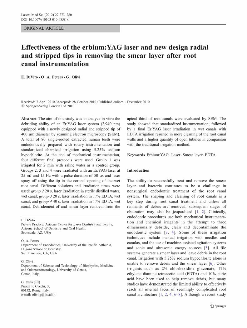
ORIGINAL ARTICLE
Received: 7 April 2010 /Accepted: 28 October 2010 /Published online: 1 December 2010# Springer-Verlag London Ltd 2010
Abstract The aim of this study was to analyze in vitro thedebriding ability of an Er:YAG laser system (2,940 nm)equipped with a newly designed radial and stripped tip of400 µm diameter by scanning electron microscopy (SEM).A total of 80 single-rooted extracted human teeth wereendodontically prepared with rotary instrumentation andstandardized chemical irrigation using 5.25% sodiumhypochlorite. At the end of mechanical instrumentation,four different final protocols were used. Group 1 wasirrigated for 2 min with saline water as a control group.Groups 2, 3 and 4 were irradiated with an Er:YAG laser at25 mJ and 15 Hz with a pulse duration of 50 μs and laserspray off using the tip in the coronal opening of the wetroot canal. Different solutions and irradiation times wereused: group 2 20 s, laser irradiation in sterile distilled water,wet canal; group 3 20 s, laser irradiation in 17% EDTA, wetcanal; and group 4 40 s, laser irradiation in 17% EDTA, wetcanal. Debridement of and smear layer removal from the
apical third of root canals were evaluated by SEM. Thestudy showed that standardized instrumentation, followedby a final Er:YAG laser irradiation in wet canals withEDTA irrigation resulted in more cleaning of the root canalwalls and a higher quantity of open tubules in comparisonwith the traditional irrigation method.
Keywords Erbium:YAG . Laser . Smear layer . EDTA
Introduction
The ability to successfully treat and remove the smearlayer and bacteria continues to be a challenge innonsurgical endodontic treatment of the root canalsystem. The shaping and cleaning of root canals is akey step during root canal treatment and unless allremnants of debris are removed, subsequent stages ofobturation may also be jeopardized [1, 2]. Clinically,endodontic procedures use both mechanical instrumenta-tion and chemical irrigants in the attempt to threedimensionally debride, clean and decontaminate theendodontic system [3, 4]. Some of these irrigationtechniques include manual irrigation with needles andcanulas, and the use of machine-assisted agitation systemsand sonic and ultrasonic energy sources [5]. All filesystems generate a smear layer and leave debris in the rootcanal. Irrigation with 5.25% sodium hypochlorite alone isunable to remove debris and the smear layer [6]. Otherirrigants such as 2% chlorhexidine gluconate, 17%ethylene diamine tetraacetic acid (EDTA) and 10% citricacid have been used to help remove debris, but manystudies have demonstrated the limited ability to effectivelyreach all internal faces of seemingly complicated rootcanal architecture [1, 2, 4, 6–8]. Although a recent study
E. DiVitoPrivate Practice, Arizona Center for Laser Dentistry and faculty,Arizona School of Dentistry and Oral Health,Scottsdale, AZ, USA
O. A. PetersDepartment of Endodontics, University of the Pacific Arthur A,Dugoni School of Dentistry,San Francisco, CA, USA
G. OliviDepartment of Science and Technology of Biophysics, Medicineand Odontostomatology, University of Genoa,Genoa, Italy
G. Olivi (*)Piazza F. Cucchi, 3,00152, Rome, Italye-mail: [email protected]
Lasers Med Sci (2012) 27:273–280DOI 10.1007/s10103-010-0858-x
Effectiveness of the erbium:YAG laser and new design radialand stripped tips in removing the smear layer after rootcanal instrumentation
E. DiVito & O. A. Peters & G. Olivi
[9] has shown excellent ability of a new file systemoperated with a continuous irrigation device to removedebris and the smear layer also from the apical third of theroot canal, the literature shows that when compared to thecoronal and middle thirds of relatively clean canals, theapical third of the root canal always presents a problem inregard to the ability to achieve the same cleanliness [5, 6,10–12]. This fact may be of significance during root canaltreatment because the presence of a smear layer and debrismay prevent sealer adaptation to the canal walls andimpede penetration of irrigants into dentinal tubules andaccessory canals. Accordingly, some alternative, moreeffective method to debride, clean and penetrate thedentinal walls should be explored.
The effectiveness of lasers in dentistry continues to be anarea of discussion. Although the use of lasers for nonsurgicalendodontic treatment of the root canal system has been reportedsince the early 1970s [13, 14], acceptance has been slow. Acommon feature of dissatisfaction has been the thermaldamage associated with the application of laser photonicenergy [15–19]. Laser treatment can be a valuable tool for theremoval of the dentinal smear layer, as a debridement deviceduring endodontic treatment. The Er:YAG laser (wavelength2,940 nm) is approved by the FDA for cleaning, shaping andenlarging the root canal [20]. Previous studies have tested theability and the effects of this laser on root canal walls andhave indicated that the Er:YAG laser is a suitable instrumentfor removal of the smear layer in root canals [21–26].Furthermore, George et al. in an investigation of the abilityof both the Er:YAG and Er,Cr:YSGG lasers equipped withconical shaped radially firing tips and plain tips to remove thesmear layer from the apical third of the root canal showed alaser activation of EDTA and a better performance of conicalfibers compared to plain fibers for improving the action ofEDTAC in dissolving smear layer [27].
The aim of this in vitro study was to evaluate byscanning electron microscopy (SEM) the ability andeffectiveness of the Er:YAG laser in removing the smearlayer and debriding the root canal. A newly designed tipwith a tapered radial firing end and 3 mm of the polyamidesheath removed was used. Using specific pulse rates, ashort microsecond pulse duration and low energy duringapplication, the thermal morphological effects described inthe literature were minimized [21–29].
Materials and methods
Sample preparation
In this study, 80 recently extracted single-rooted humanteeth were used. They were stored in physiological salinesolution until use.
Root canal treatment
The access cavity to the canal orifice was first preparedwith a tapered diamond bur creating a glide path forinsertion of the first instrument (size #10 K file). The teethwere then minimally prepared using nickel/titanium rotaryinstruments in a sequential crown down method to a size20/.06 (Profile GT; Dentsply Tulsa Dental, Tulsa, OK). Thecanals were irrigated during preparation with sodiumhypochlorite. After reaching the final instrumentation sizeof 20/.06, an additional two 30-s cycles of irrigation withsaline only were applied. The samples were then ready tobe treated with the various laser protocols described.
Laser parameters
An Er:YAG laser with a wavelength of 2,940 nm (Fidelis;Fotona, Ljubljana, Slovenia) was used to irradiate the rootcanals after traditional instrumentation. A newly designed 12-mm long 400-µm quartz tip was used. The tip, as receiveddirectly from the manufacturer, was tapered and had 3 mm ofthe polyamide sheath stripped back from its end (Fig. 1a). Thelaser operating parameters used for all of the treatmentgroups (using the free-running emission mode) were asfollows: 20 mJ per pulse, 15 Hz, and 50 μs pulse duration.The coaxial water spray feature of the handpiece was set to‘off’. The tip was placed into the coronal access opening ofchamber only, and was kept stationary and not advanced intothe orifice of the canal (Fig. 1b).
Laser irradiation and irrigation methods
After the mechanical preparation, the teeth were randomlydivided into four groups (20 teeth each) and treatedaccording to the following protocol:
Group 1 Saline water irrigation for 2 min ascontrol group
Group 2 Laser irradiation, 20-s cycle in steriledistilled water
Group 3 Laser irradiation, 20-s cycle in 17% EDTAGroup 4 Laser irradiation, 40-s cycle in 17% EDTA
During the laser irradiation cycles, the root canals werecontinuously irrigated with 2 ml of fluid to maintainhydration and levels using a hand syringe with a 25 gaugeneedle positioned above the laser tip in the coronal aspectof the access opening, accordingly to the above protocol.
Temperature measurements
To identify possible thermal side effects, the temperaturechanges on the external root surface of three teeth in eachlaser group (for a total of nine teeth) were measured.
274 Lasers Med Sci (2012) 27:273–280
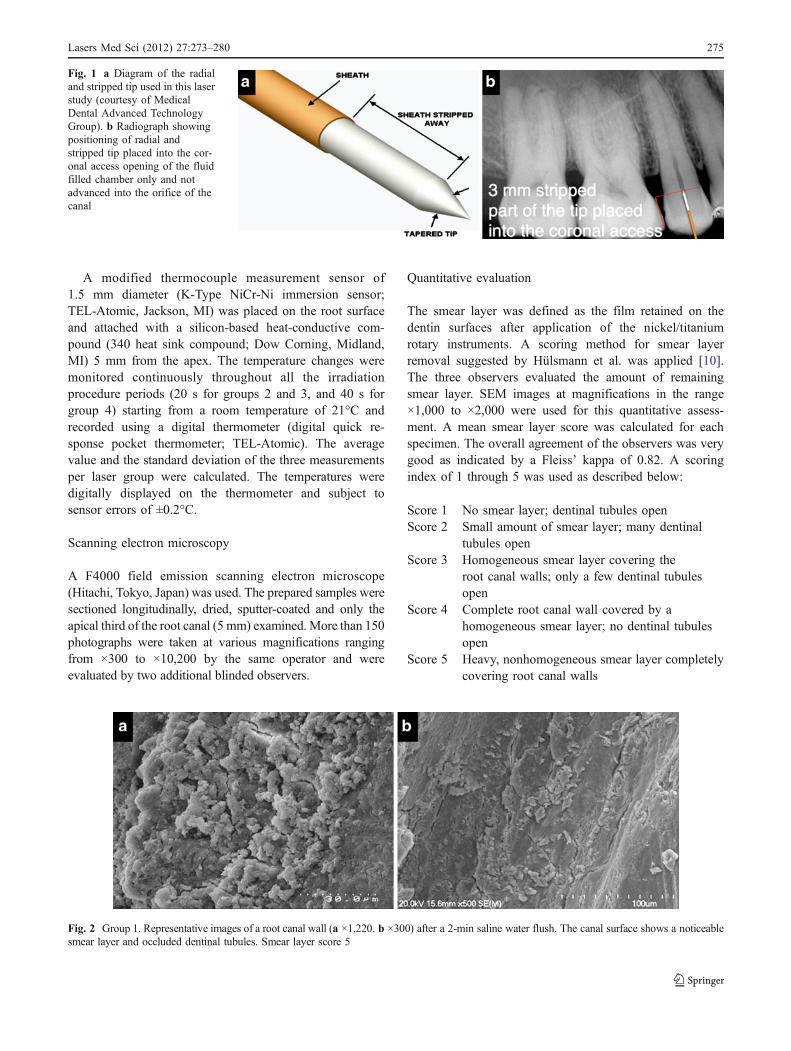
A modified thermocouple measurement sensor of1.5 mm diameter (K-Type NiCr-Ni immersion sensor;TEL-Atomic, Jackson, MI) was placed on the root surfaceand attached with a silicon-based heat-conductive com-pound (340 heat sink compound; Dow Corning, Midland,MI) 5 mm from the apex. The temperature changes weremonitored continuously throughout all the irradiationprocedure periods (20 s for groups 2 and 3, and 40 s forgroup 4) starting from a room temperature of 21°C andrecorded using a digital thermometer (digital quick re-sponse pocket thermometer; TEL-Atomic). The averagevalue and the standard deviation of the three measurementsper laser group were calculated. The temperatures weredigitally displayed on the thermometer and subject tosensor errors of ±0.2°C.
Scanning electron microscopy
A F4000 field emission scanning electron microscope(Hitachi, Tokyo, Japan) was used. The prepared samples weresectioned longitudinally, dried, sputter-coated and only theapical third of the root canal (5 mm) examined. More than 150photographs were taken at various magnifications rangingfrom ×300 to ×10,200 by the same operator and wereevaluated by two additional blinded observers.
Quantitative evaluation
The smear layer was defined as the film retained on thedentin surfaces after application of the nickel/titaniumrotary instruments. A scoring method for smear layerremoval suggested by Hülsmann et al. was applied [10].The three observers evaluated the amount of remainingsmear layer. SEM images at magnifications in the range×1,000 to ×2,000 were used for this quantitative assess-ment. A mean smear layer score was calculated for eachspecimen. The overall agreement of the observers was verygood as indicated by a Fleiss’ kappa of 0.82. A scoringindex of 1 through 5 was used as described below:
Score 1 No smear layer; dentinal tubules openScore 2 Small amount of smear layer; many dentinal
tubules openScore 3 Homogeneous smear layer covering the
root canal walls; only a few dentinal tubulesopen
Score 4 Complete root canal wall covered by ahomogeneous smear layer; no dentinal tubulesopen
Score 5 Heavy, nonhomogeneous smear layer completelycovering root canal walls
Fig. 1 a Diagram of the radialand stripped tip used in this laserstudy (courtesy of MedicalDental Advanced TechnologyGroup). b Radiograph showingpositioning of radial andstripped tip placed into the cor-onal access opening of the fluidfilled chamber only and notadvanced into the orifice of thecanal
Fig. 2 Group 1. Representative images of a root canal wall (a ×1,220. b ×300) after a 2-min saline water flush. The canal surface shows a noticeablesmear layer and occluded dentinal tubules. Smear layer score 5
Lasers Med Sci (2012) 27:273–280 275
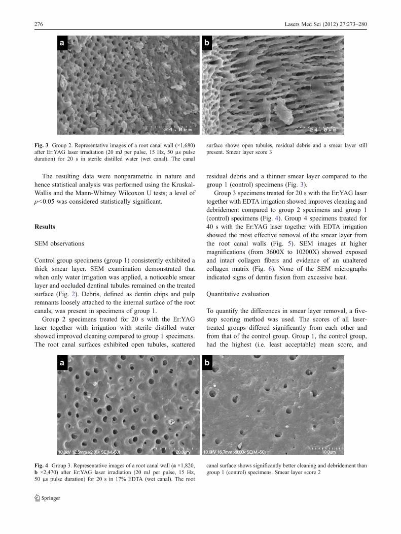
The resulting data were nonparametric in nature andhence statistical analysis was performed using the Kruskal-Wallis and the Mann-Whitney Wilcoxon U tests; a level ofp<0.05 was considered statistically significant.
Results
SEM observations
Control group specimens (group 1) consistently exhibited athick smear layer. SEM examination demonstrated thatwhen only water irrigation was applied, a noticeable smearlayer and occluded dentinal tubules remained on the treatedsurface (Fig. 2). Debris, defined as dentin chips and pulpremnants loosely attached to the internal surface of the rootcanals, was present in specimens of group 1.
Group 2 specimens treated for 20 s with the Er:YAGlaser together with irrigation with sterile distilled watershowed improved cleaning compared to group 1 specimens.The root canal surfaces exhibited open tubules, scattered
residual debris and a thinner smear layer compared to thegroup 1 (control) specimens (Fig. 3).
Group 3 specimens treated for 20 s with the Er:YAG lasertogether with EDTA irrigation showed improves cleaning anddebridement compared to group 2 specimens and group 1(control) specimens (Fig. 4). Group 4 specimens treated for40 s with the Er:YAG laser together with EDTA irrigationshowed the most effective removal of the smear layer fromthe root canal walls (Fig. 5). SEM images at highermagnifications (from 3600X to 10200X) showed exposedand intact collagen fibers and evidence of an unalteredcollagen matrix (Fig. 6). None of the SEM micrographsindicated signs of dentin fusion from excessive heat.
Quantitative evaluation
To quantify the differences in smear layer removal, a five-step scoring method was used. The scores of all laser-treated groups differed significantly from each other andfrom that of the control group. Group 1, the control group,had the highest (i.e. least acceptable) mean score, and
Fig. 3 Group 2. Representative images of a root canal wall (×1,680)after Er:YAG laser irradiation (20 mJ per pulse, 15 Hz, 50 μs pulseduration) for 20 s in sterile distilled water (wet canal). The canal
surface shows open tubules, residual debris and a smear layer stillpresent. Smear layer score 3
Fig. 4 Group 3. Representative images of a root canal wall (a ×1,820,b ×2,470) after Er:YAG laser irradiation (20 mJ per pulse, 15 Hz,50 μs pulse duration) for 20 s in 17% EDTA (wet canal). The root
canal surface shows significantly better cleaning and debridement thangroup 1 (control) specimens. Smear layer score 2
276 Lasers Med Sci (2012) 27:273–280
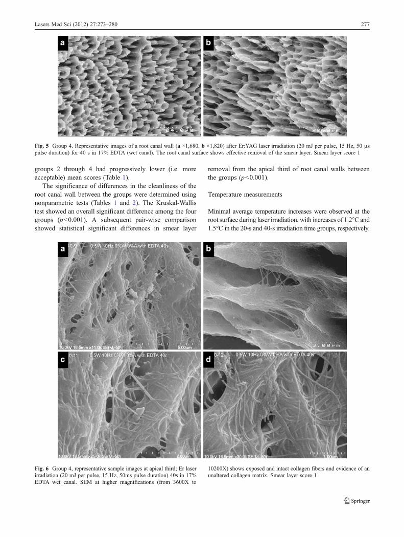
groups 2 through 4 had progressively lower (i.e. moreacceptable) mean scores (Table 1).
The significance of differences in the cleanliness of theroot canal wall between the groups were determined usingnonparametric tests (Tables 1 and 2). The Kruskal-Wallistest showed an overall significant difference among the fourgroups (p<0.001). A subsequent pair-wise comparisonshowed statistical significant differences in smear layer
removal from the apical third of root canal walls betweenthe groups (p<0.001).
Temperature measurements
Minimal average temperature increases were observed at theroot surface during laser irradiation, with increases of 1.2°C and1.5°C in the 20-s and 40-s irradiation time groups, respectively.
Fig. 5 Group 4. Representative images of a root canal wall (a ×1,680, b ×1,820) after Er:YAG laser irradiation (20 mJ per pulse, 15 Hz, 50 μspulse duration) for 40 s in 17% EDTA (wet canal). The root canal surface shows effective removal of the smear layer. Smear layer score 1
Fig. 6 Group 4, representative sample images at apical third; Er laserirradiation (20 mJ per pulse, 15 Hz, 50ms pulse duration) 40s in 17%EDTA wet canal. SEM at higher magnifications (from 3600X to
10200X) shows exposed and intact collagen fibers and evidence of anunaltered collagen matrix. Smear layer score 1
Lasers Med Sci (2012) 27:273–280 277
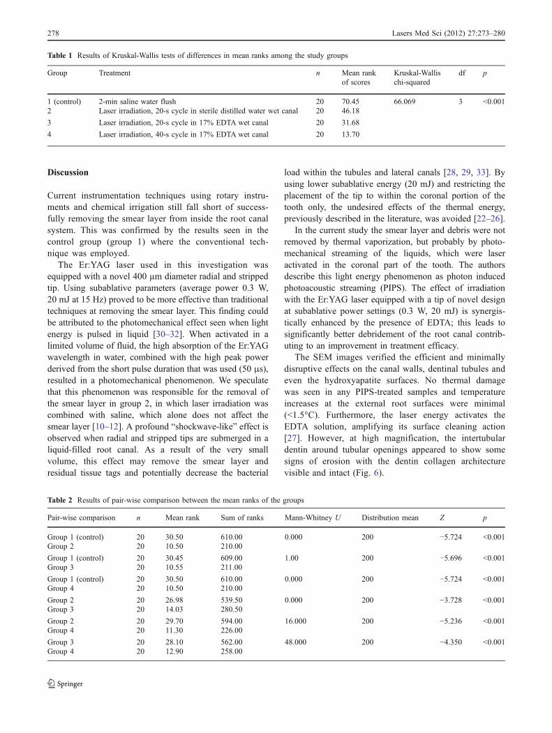
Discussion
Current instrumentation techniques using rotary instru-ments and chemical irrigation still fall short of success-fully removing the smear layer from inside the root canalsystem. This was confirmed by the results seen in thecontrol group (group 1) where the conventional tech-nique was employed.
The Er:YAG laser used in this investigation wasequipped with a novel 400 µm diameter radial and strippedtip. Using subablative parameters (average power 0.3 W,20 mJ at 15 Hz) proved to be more effective than traditionaltechniques at removing the smear layer. This finding couldbe attributed to the photomechanical effect seen when lightenergy is pulsed in liquid [30–32]. When activated in alimited volume of fluid, the high absorption of the Er:YAGwavelength in water, combined with the high peak powerderived from the short pulse duration that was used (50 μs),resulted in a photomechanical phenomenon. We speculatethat this phenomenon was responsible for the removal ofthe smear layer in group 2, in which laser irradiation wascombined with saline, which alone does not affect thesmear layer [10–12]. A profound “shockwave-like” effect isobserved when radial and stripped tips are submerged in aliquid-filled root canal. As a result of the very smallvolume, this effect may remove the smear layer andresidual tissue tags and potentially decrease the bacterial
load within the tubules and lateral canals [28, 29, 33]. Byusing lower subablative energy (20 mJ) and restricting theplacement of the tip to within the coronal portion of thetooth only, the undesired effects of the thermal energy,previously described in the literature, was avoided [22–26].
In the current study the smear layer and debris were notremoved by thermal vaporization, but probably by photo-mechanical streaming of the liquids, which were laseractivated in the coronal part of the tooth. The authorsdescribe this light energy phenomenon as photon inducedphotoacoustic streaming (PIPS). The effect of irradiationwith the Er:YAG laser equipped with a tip of novel designat subablative power settings (0.3 W, 20 mJ) is synergis-tically enhanced by the presence of EDTA; this leads tosignificantly better debridement of the root canal contrib-uting to an improvement in treatment efficacy.
The SEM images verified the efficient and minimallydisruptive effects on the canal walls, dentinal tubules andeven the hydroxyapatite surfaces. No thermal damagewas seen in any PIPS-treated samples and temperatureincreases at the external root surfaces were minimal(<1.5°C). Furthermore, the laser energy activates theEDTA solution, amplifying its surface cleaning action[27]. However, at high magnification, the intertubulardentin around tubular openings appeared to show somesigns of erosion with the dentin collagen architecturevisible and intact (Fig. 6).
Table 1 Results of Kruskal-Wallis tests of differences in mean ranks among the study groups
Group Treatment n Mean rankof scores
Kruskal-Wallischi-squared
df p
1 (control) 2-min saline water flush 20 70.45 66.069 3 <0.0012 Laser irradiation, 20-s cycle in sterile distilled water wet canal 20 46.18
3 Laser irradiation, 20-s cycle in 17% EDTA wet canal 20 31.68
4 Laser irradiation, 40-s cycle in 17% EDTA wet canal 20 13.70
Table 2 Results of pair-wise comparison between the mean ranks of the groups
Pair-wise comparison n Mean rank Sum of ranks Mann-Whitney U Distribution mean Z p
Group 1 (control) 20 30.50 610.00 0.000 200 −5.724 <0.001Group 2 20 10.50 210.00
Group 1 (control) 20 30.45 609.00 1.00 200 −5.696 <0.001Group 3 20 10.55 211.00
Group 1 (control) 20 30.50 610.00 0.000 200 −5.724 <0.001Group 4 20 10.50 210.00
Group 2 20 26.98 539.50 0.000 200 −3.728 <0.001Group 3 20 14.03 280.50
Group 2 20 29.70 594.00 16.000 200 −5.236 <0.001Group 4 20 11.30 226.00
Group 3 20 28.10 562.00 48.000 200 −4.350 <0.001Group 4 20 12.90 258.00
278 Lasers Med Sci (2012) 27:273–280
With conventional treatment protocols (without a laser),an irrigation syringe is more effective when the tip is placedcloser to the working length. With this new laser system,the laser tip is not placed within the canal itself, but israther confined to the coronal chamber above the orifice. Itis suggested that this allows easy access for the photome-chanical effects to occur within the root canal, which mayassist in cleaning canals of various shapes.
A standard ISO size #30 file preparation is needed toallow traditional laser tips (200–320 µm) to reach close tothe apex [28, 29, 33]. Using the radial and stripped designwith PIPS, the apex can be reached without the need tonegotiate the tip close to the apex. Correspondingly, thiswould allow a less-invasive preparation using an ISO size#20/.06 file, according to the method described.
Irrigation with chelating agents following the currentconventional instrumentation procedure requires more timeto initiate a satisfactory debridement (EDTA placedpassively into the prepared root canal) [11, 34]. The PIPStechnique resulted in pronounced smear layer removalwhen used together with EDTA and at the settings outlined.
Published material on endodontic techniques using theEr:YAG laser provides differing operating parameters [35].These authors recommend the use of higher average power(1.125–1.5 W) delivered through end-firing laser tips.Additionally, these tips need to be placed 1–2 mm fromthe root apex.
Conclusion
The Er:YAG laser used in this study showed significantlybetter smear layer removal than traditional syringe irrigation.At the energy levels and with the operating parameters used,no thermal effects or damage to the dentin surface wasobserved. In this study the Er:YAG laser with the currentsettings produced a photomechanical effect demonstrating itspotential as an improved alternative method for debriding theroot canal system in a minimally invasive manner.
Acknowledgments This study was financially supported by theMedical Dental Advanced Technology Group, L.L.C. Scottsdale, AZ,USA. The authors thank Jan DiLoreto for her administrative support.
Disclosure The authors hereby disclose that they are working withFotona to manufacture and distribute this new and improved deliverysystem for endodontic treatment. Dr. Giovanni Olivi is an independentresearcher affiliated with the University of Genoa where he performslaser studies.
References
1. Wu M-K, Wesselink PR (2001) A primary observation on thepreparation and obturation in oval canals. Int Endod J 34:137–141
2. De Deus G, Gurgel-Filho ED, Magalhaes KM et al (2006) Alaboratory analysis of gutta-percha filled area obtained usingThermafil, System B and lateral condensation. Int Endod J39:378–383
3. Peters OA (2004) Challenges in root canal preparation. J Endod30:539–567
4. Hülsmann M, Peters OA, Dummer PMH (2005) Mechanicalpreparation of root canals: shaping goals, techniques and means.Endod Top 10:30–76
5. Gu L, Kim JR, Ling J, Choi KK, Pashley DH, Tay FR (2009)Review of contemporary irrigant agitation techniques and devices.J Endod 35:791–804
6. Baumgartner JC, Cuenin PR (1992) Efficacy of several concen-trations of sodium hypochlorite for root canal irrigation. J Endod18:605–612
7. White RR, Hays GL, Janer LR (1997) Residual antimicrobial activityafter canal irrigation with chlorhexidine. J Endod 23:229–231
8. Berutti E, Marini R, Angeretti A (1997) Penetration ability ofdifferent irrigants into dentinal tubules. J Endod 23:725–727
9. Metzger Z, Teperovich E, Cohen R, Zary R, Paqué F, HülsmannM (2010) The self-adjusting file (SAF). Part 3: removal of debrisand smear layer – a scanning electron microscope study. J Endod36:697–702
10. Hülsmann M, Rümmelin C, Schäfers F (1997) Root canalcleanliness after preparation with different endodontic handpiecesand hand instruments: a comparative SEM investigation. J Endod23:301–306
11. Khedmat S, Shokouhinejad N (2008) Comparison of the efficacy ofthree chelating agents in smear layer removal. J Endod 34:599–602
12. Caron G, Nham K, Bronnec F, Machtou P (2010) Effectiveness ofdifferent final irrigant activation protocols on smear layer removalin curved canals. J Endod 36:1361–1366
13. Weichman JA, Johnson FM, Nitta LK (1972) Laser use inendodontics. II. Oral Surg Oral Med Oral Pathol 34:828–830
14. Weichman JA, Johnson FM (1971) Laser use in endodontics. Apreliminary investigation. Oral Surg Oral Med Oral Pathol31:416–420
15. Yamazaki R, Goya C, Yu DG, Kimura Y, Matsumoto K (2001)Effects of erbium,chromium:YSGG laser irradiation on root canalwalls: a scanning electron microscopic and thermographic study. JEndod 27:9–12
16. Kimura Y, Yonaga K, Yokoyama K, Kinoshita J, Ogata Y,Matsumoto K (2002) Root surface temperature increase duringEr:YAG laser irradiation of root canals. J Endod 28:76–78
17. Ebihara A, Majaron B, Liaw LH, Krasieva TB, Wilder-Smith P(2002) Er:YAG laser modification of root canal dentine: influenceof pulse duration, repetitive irradiation and water spray. PhotomedLaser Surg 17:198–207
18. Ishizaki NT, Matsumoto K, Kimura Y, Wang X, Kinoshita J,Okano SM, Jayawardena JA (2004) Thermographical and mor-phological studies of Er,Cr:YSGG laser irradiation on root canalwalls. Photomed Laser Surg 22:291–297
19. Altundasar E, Ozçelik B, Cehreli ZC, Matsumoto K (2006)Ultramorphological and histochemical changes after ErCr:YSGGlaser irradiation and two different irrigation regimes. J Endod32:465–468
20. Sulewski JG. Making the most of the 16th Annual Conference andExhibition: a practical orientation for attendees. Academy of LaserDentistry 16th Annual Conference and Exhibition, 22–26 April2009, Las Vegas, Nevada
21. Takeda FH, Harashima T, Kimura Y, Matsumoto K (1998)Comparative study about the removal of smear layer by threetypes of laser devices. J Clin Laser Med Surg 16:117–122
22. Matsuoka E, Kimura Y, Matsumoto K (1998) Studies on theremoval of debris near the apical seats by Er:YAG laser andassessment with a fiberscope. J Clin Laser Med Surg 16:255–261
Lasers Med Sci (2012) 27:273–280 279
23. Takeda FH, Harashima T, Eto JN, Kimura Y, Matsumoto K (1998)Effect of Er:YAG laser treatment on the root canal walls of humanteeth: an SEM study. Endod Dent Traumatol 14:270–273
24. Takeda FH, Harashima T, Kimura Y, Matsumoto K (1998)Efficacy of Er:YAG laser irradiation in removing debris andsmear layer on root canal walls. J Endod 24:548–551
25. Takeda FH, Harashima T, Kimura Y, Matsumoto K (1999) Acomparative study of the removal of smear layer by threeendodontic irrigants and two types of laser. Int Endod J 32:32–39
26. Pecora JD, Brugnera-Júnior A, Cussioli AL, Zanin F, Silva R (2000)Evaluation of dentin root canal permeability after instrumentationand Er:YAG laser application. Lasers Surg Med 26:277–281
27. George R, Meyers IA, Walsh LJ (2008) Laser activation ofendodontic irrigants with improved conical laser fiber tips forremoving smear layer in the apical third of the root canal. J Endod34:1524–1527
28. Schoop U, Goharkhay K, Klimscha J, Zagler M, Wernisch J,Georgopoulos A, Sperr W, Moritz A (2007) The use of theerbium,chromium:yttrium-scandium-gallium-garnet laser in end-odontic treatment: the results of an in vitro study. J Am DentAssoc 138:949–955
29. Schoop U, Barylyak A, Goharkhay K, Beer F, Wernisch J,Georgopoulos A, Sperr W, Moritz A (2009) The impact of anerbium,chromium:yttrium-scandium-gallium-garnet laser with
radial-firing tips on endodontic treatment. Lasers Med Sci24:59–65
30. Blanken J, De Moor RJ, Meire M, Verdaasdonk R (2009) Laserinduced explosive vapor and cavitation resulting in effectiveirrigation of the root canal. Part 1: a visualization study. LasersSurg Med 41(7):514–519
31. De Moor RJ, Blanken J, Meire M, Verdaasdonk R (2009) Laserinduced explosive vapor and cavitation resulting in effectiveirrigation of the root canal. Part 2: evaluation of the efficacy.Lasers Surg Med 41(7):520–523
32. de Groot SD, Verhaagen B, Versluis M, Wu MK, Wesselink PR,van der Sluis LW (2009) Laser activated irrigation within rootcanals: cleaning efficacy and flow visualization. Int Endod J 42(12):1077–1083
33. Gordon W, Atabakhsh VA, Meza F, Doms A, Nissan R, Rizoiu I,Stevens RH (2007) The antimicrobial efficacy of the erbium,chromium:yttrium-scandium-gallium-garnet laser with radial emit-ting tips on root canal dentin walls infected with Enterococcusfaecalis. J Am Dent Assoc 138:992–1002
34. Hülsmann M, Heckendorff M, Lennon A (2003) Chelating agentsin root canal treatment: mode of action and indications for theiruse. Int Endod J 36:810–830
35. Moritz A (2006) Oral laser application. Quintessence, Berlin, pp284–287
280 Lasers Med Sci (2012) 27:273–280
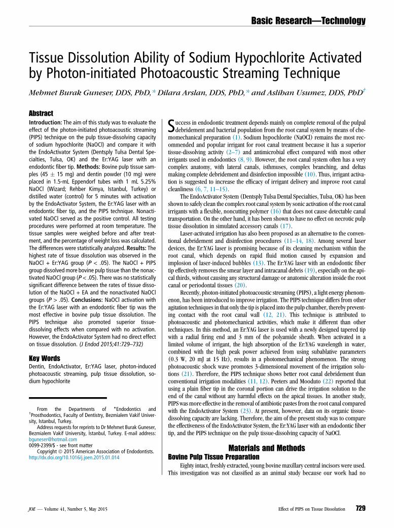
Basic Research—Technology
Tissue Dissolution Ability of Sodium Hypochlorite Activatedby Photon-initiated Photoacoustic Streaming TechniqueMehmet Burak Guneser, DDS, PhD,* Dilara Arslan, DDS, PhD,* and Aslihan Usumez, DDS, PhD†
Abstract
Introduction: The aim of this study was to evaluate theeffect of the photon-initiated photoacoustic streaming(PIPS) technique on the pulp tissue-dissolving capacityof sodium hypochlorite (NaOCl) and compare it withthe EndoActivator System (Dentsply Tulsa Dental Spe-cialties, Tulsa, OK) and the Er:YAG laser with anendodontic fiber tip.Methods: Bovine pulp tissue sam-ples (45 � 15 mg) and dentin powder (10 mg) wereplaced in 1.5-mL Eppendorf tubes with 1 mL 5.25%NaOCl (Wizard; Rehber Kimya, Istanbul, Turkey) ordistilled water (control) for 5 minutes with activationby the EndoActivator System, the Er:YAG laser with anendodontic fiber tip, and the PIPS technique. Nonacti-vated NaOCl served as the positive control. All testingprocedures were performed at room temperature. Thetissue samples were weighed before and after treat-ment, and the percentage of weight loss was calculated.The differences were statistically analyzed. Results: Thehighest rate of tissue dissolution was observed in theNaOCl + Er:YAG group (P < .05). The NaOCl + PIPSgroup dissolved more bovine pulp tissue than the nonac-tivated NaOCl group (P < .05). There was no statisticallysignificant difference between the rates of tissue disso-lution of the NaOCl + EA and the nonactivated NaOClgroups (P > .05). Conclusions: NaOCl activation withthe Er:YAG laser with an endodontic fiber tip was themost effective in bovine pulp tissue dissolution. ThePIPS technique also promoted superior tissue-dissolving effects when compared with no activation.However, the EndoActivator System had no direct effecton tissue dissolution. (J Endod 2015;41:729–732)Key WordsDentin, EndoActivator, Er:YAG laser, photon-inducedphotoacoustic streaming, pulp tissue dissolution, so-dium hypochlorite
From the Departments of *Endodontics and†Prosthodontics, Faculty of Dentistry, Bezmialem Vakif Univer-sity, Istanbul, Turkey.
Address requests for reprints to Dr Mehmet Burak Guneser,Bezmialem Vakif University, _Istanbul, Turkey. E-mail address:[email protected]/$ - see front matter
Copyright ª 2015 American Association of Endodontists.http://dx.doi.org/10.1016/j.joen.2015.01.014
JOE — Volume 41, Number 5, May 2015
Success in endodontic treatment depends mainly on complete removal of the pulpaldebridement and bacterial population from the root canal system by means of che-
momechanical preparation (1). Sodium hypochlorite (NaOCl) remains the most rec-ommended and popular irrigant for root canal treatment because it has a superiortissue-dissolving activity (2–7) and antimicrobial effect compared with most otherirrigants used in endodontics (8, 9). However, the root canal system often has a verycomplex anatomy, with lateral canals, isthmuses, complex branching, and deltasmaking complete debridement and disinfection impossible (10). Thus, irrigant activa-tion is suggested to increase the efficacy of irrigant delivery and improve root canalcleanliness (6, 7, 11–15).
The EndoActivator System (Dentsply Tulsa Dental Specialties, Tulsa, OK) has beenshown to safely clean the complex root canal system by sonic activation of the root canalirrigants with a flexible, noncutting polymer (16) that does not cause detectable canaltransportation. On the other hand, it has been shown to have no effect on necrotic pulptissue dissolution in simulated accessory canals (17).
Laser-activated irrigation has also been proposed as an alternative to the conven-tional debridement and disinfection procedures (11–14, 18). Among several laserdevices, the Er:YAG laser is promising because of its cleaning mechanism within theroot canal, which depends on rapid fluid motion caused by expansion andimplosion of laser-induced bubbles (13). The Er:YAG laser with an endodontic fibertip effectively removes the smear layer and intracanal debris (19), especially on the api-cal thirds, without causing any structural damage or anatomic alteration inside the rootcanal or periodontal tissues (20).
Recently, photon-initiated photoacoustic streaming (PIPS), a light energy phenom-enon, has been introduced to improve irrigation. The PIPS technique differs from otheragitation techniques in that only the tip is placed into the pulp chamber, thereby prevent-ing contact with the root canal wall (12, 21). This technique is attributed tophotoacoustic and photomechanical activities, which make it different than othertechniques. In this method, an Er:YAG laser is used with a newly designed tapered tipwith a radial firing end and 3 mm of the polyamide sheath. When activated in alimited volume of irrigant, the high absorption of the Er:YAG wavelength in water,combined with the high peak power achieved from using subablative parameters(0.3 W, 20 mJ at 15 Hz), results in a photomechanical phenomenon. The strongphotoacoustic shock wave promotes 3-dimensional movement of the irrigation solu-tions (21). Therefore, the PIPS technique shows better root canal debridement thanconventional irrigation modalities (11, 12). Peeters and Mooduto (22) reported thatusing a plain fiber tip in the coronal portion can drive the irrigation solution to theend of the canal without any harmful effects on the apical tissues. In another study,PIPS wasmore effective in the removal of antibiotic pastes from the root canal comparedwith the EndoActivator System (23). At present, however, data on its organic tissue-dissolving capacity are lacking. Therefore, the aim of the present study was to comparethe effectiveness of the EndoActivator System, the Er:YAG laser with an endodontic fibertip, and the PIPS technique on the pulp tissue-dissolving capacity of NaOCl.
Materials and MethodsBovine Pulp Tissue Preparation
Eighty intact, freshly extracted, young bovine maxillary central incisors were used.This investigation was not classified as an animal study because our work had no
Effect of PIPS on Tissue Dissolution 729
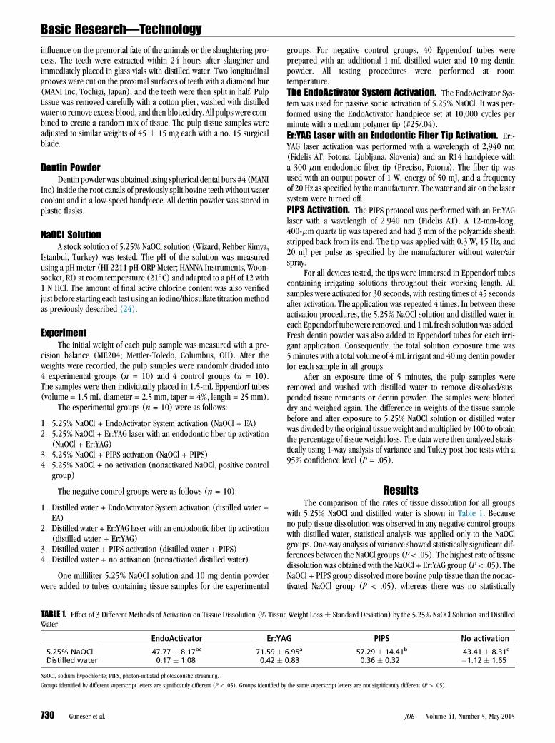
Basic Research—Technology
influence on the premortal fate of the animals or the slaughtering pro-cess. The teeth were extracted within 24 hours after slaughter andimmediately placed in glass vials with distilled water. Two longitudinalgrooves were cut on the proximal surfaces of teeth with a diamond bur(MANI Inc, Tochigi, Japan), and the teeth were then split in half. Pulptissue was removed carefully with a cotton plier, washed with distilledwater to remove excess blood, and then blotted dry. All pulps were com-bined to create a random mix of tissue. The pulp tissue samples wereadjusted to similar weights of 45 � 15 mg each with a no. 15 surgicalblade.Dentin PowderDentin powder was obtained using spherical dental burs #4 (MANI
Inc) inside the root canals of previously split bovine teeth without watercoolant and in a low-speed handpiece. All dentin powder was stored inplastic flasks.
NaOCl SolutionA stock solution of 5.25% NaOCl solution (Wizard; Rehber Kimya,
Istanbul, Turkey) was tested. The pH of the solution was measuredusing a pH meter (HI 2211 pH-ORP Meter; HANNA Instruments, Woon-socket, RI) at room temperature (21�C) and adapted to a pH of 12 with1 N HCl. The amount of final active chlorine content was also verifiedjust before starting each test using an iodine/thiosulfate titration methodas previously described (24).
ExperimentThe initial weight of each pulp sample was measured with a pre-
cision balance (ME204; Mettler-Toledo, Columbus, OH). After theweights were recorded, the pulp samples were randomly divided into4 experimental groups (n = 10) and 4 control groups (n = 10).The samples were then individually placed in 1.5-mL Eppendorf tubes(volume = 1.5 mL, diameter = 2.5 mm, taper = 4%, length = 25 mm).
The experimental groups (n = 10) were as follows:
1. 5.25% NaOCl + EndoActivator System activation (NaOCl + EA)2. 5.25% NaOCl + Er:YAG laser with an endodontic fiber tip activation
(NaOCl + Er:YAG)3. 5.25% NaOCl + PIPS activation (NaOCl + PIPS)4. 5.25% NaOCl + no activation (nonactivated NaOCl, positive control
group)
The negative control groups were as follows (n = 10):
1. Distilled water + EndoActivator System activation (distilled water +EA)
2. Distilled water + Er:YAG laser with an endodontic fiber tip activation(distilled water + Er:YAG)
3. Distilled water + PIPS activation (distilled water + PIPS)4. Distilled water + no activation (nonactivated distilled water)
One milliliter 5.25% NaOCl solution and 10 mg dentin powderwere added to tubes containing tissue samples for the experimental
TABLE 1. Effect of 3 Different Methods of Activation on Tissue Dissolution (% TissueWater
EndoActivator Er:YA
5.25% NaOCl 47.77 � 8.17bc 71.59 �Distilled water 0.17 � 1.08 0.42 �
NaOCl, sodium hypochlorite; PIPS, photon-initiated photoacoustic streaming.
Groups identified by different superscript letters are significantly different (P < .05). Groups identified by
730 Guneser et al.
groups. For negative control groups, 40 Eppendorf tubes wereprepared with an additional 1 mL distilled water and 10 mg dentinpowder. All testing procedures were performed at roomtemperature.
The EndoActivator System Activation. The EndoActivator Sys-tem was used for passive sonic activation of 5.25% NaOCl. It was per-formed using the EndoActivator handpiece set at 10,000 cycles perminute with a medium polymer tip (#25/.04).
Er:YAG Laser with an Endodontic Fiber Tip Activation. Er:-YAG laser activation was performed with a wavelength of 2,940 nm(Fidelis AT; Fotona, Ljubljana, Slovenia) and an R14 handpiece witha 300-mm endodontic fiber tip (Preciso, Fotona). The fiber tip wasused with an output power of 1 W, energy of 50 mJ, and a frequencyof 20 Hz as specified by the manufacturer. The water and air on the lasersystem were turned off.
PIPS Activation. The PIPS protocol was performed with an Er:YAGlaser with a wavelength of 2.940 nm (Fidelis AT). A 12-mm-long,400-mm quartz tip was tapered and had 3 mm of the polyamide sheathstripped back from its end. The tip was applied with 0.3 W, 15 Hz, and20 mJ per pulse as specified by the manufacturer without water/airspray.
For all devices tested, the tips were immersed in Eppendorf tubescontaining irrigating solutions throughout their working length. Allsamples were activated for 30 seconds, with resting times of 45 secondsafter activation. The application was repeated 4 times. In between theseactivation procedures, the 5.25% NaOCl solution and distilled water ineach Eppendorf tube were removed, and 1mL fresh solution was added.Fresh dentin powder was also added to Eppendorf tubes for each irri-gant application. Consequently, the total solution exposure time was5 minutes with a total volume of 4 mL irrigant and 40 mg dentin powderfor each sample in all groups.
After an exposure time of 5 minutes, the pulp samples wereremoved and washed with distilled water to remove dissolved/sus-pended tissue remnants or dentin powder. The samples were blotteddry and weighed again. The difference in weights of the tissue samplebefore and after exposure to 5.25% NaOCl solution or distilled waterwas divided by the original tissue weight and multiplied by 100 to obtainthe percentage of tissue weight loss. The data were then analyzed statis-tically using 1-way analysis of variance and Tukey post hoc tests with a95% confidence level (P = .05).
ResultsThe comparison of the rates of tissue dissolution for all groups
with 5.25% NaOCl and distilled water is shown in Table 1. Becauseno pulp tissue dissolution was observed in any negative control groupswith distilled water, statistical analysis was applied only to the NaOClgroups. One-way analysis of variance showed statistically significant dif-ferences between the NaOCl groups (P< .05). The highest rate of tissuedissolution was obtained with the NaOCl + Er:YAG group (P< .05). TheNaOCl + PIPS group dissolved more bovine pulp tissue than the nonac-tivated NaOCl group (P < .05), whereas there was no statistically
Weight Loss� Standard Deviation) by the 5.25% NaOCl Solution and Distilled
G PIPS No activation
6.95a 57.29 � 14.41b 43.41 � 8.31c
0.83 0.36 � 0.32 �1.12 � 1.65
the same superscript letters are not significantly different (P > .05).
JOE — Volume 41, Number 5, May 2015
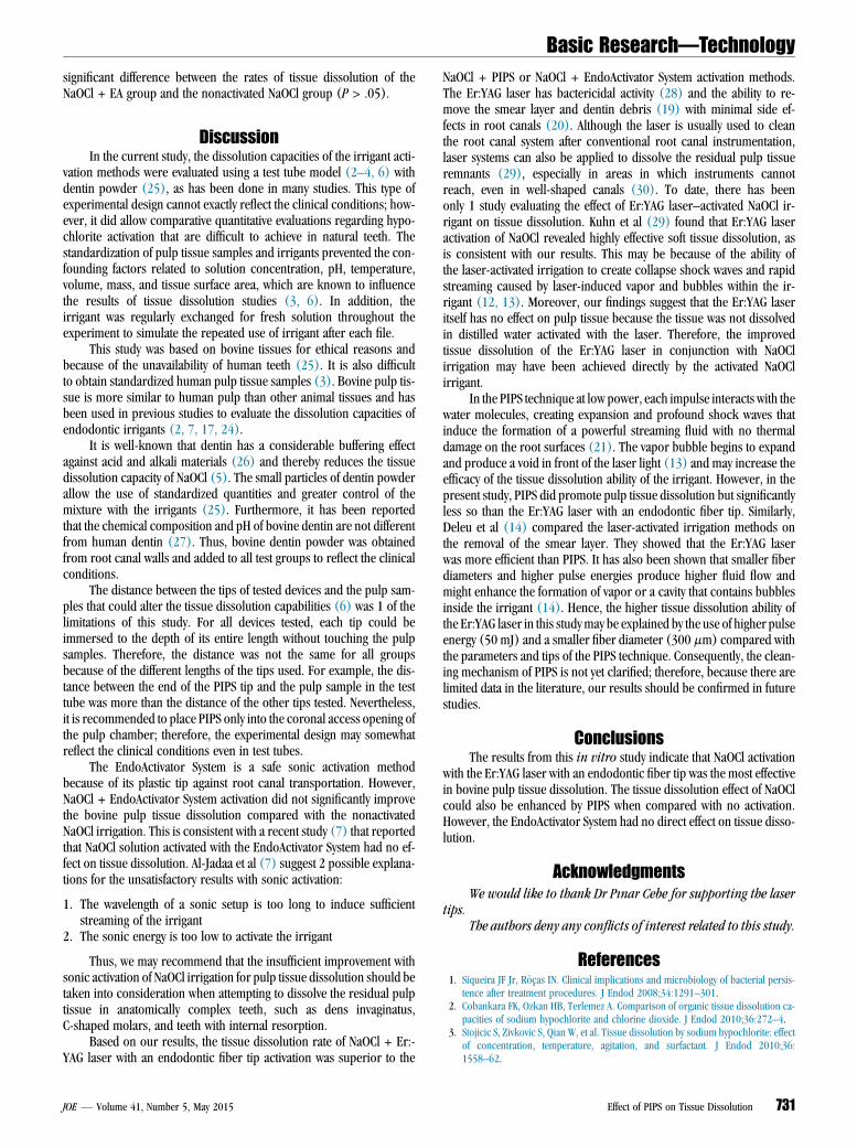
Basic Research—Technology
significant difference between the rates of tissue dissolution of theNaOCl + EA group and the nonactivated NaOCl group (P > .05).DiscussionIn the current study, the dissolution capacities of the irrigant acti-
vation methods were evaluated using a test tube model (2–4, 6) withdentin powder (25), as has been done in many studies. This type ofexperimental design cannot exactly reflect the clinical conditions; how-ever, it did allow comparative quantitative evaluations regarding hypo-chlorite activation that are difficult to achieve in natural teeth. Thestandardization of pulp tissue samples and irrigants prevented the con-founding factors related to solution concentration, pH, temperature,volume, mass, and tissue surface area, which are known to influencethe results of tissue dissolution studies (3, 6). In addition, theirrigant was regularly exchanged for fresh solution throughout theexperiment to simulate the repeated use of irrigant after each file.
This study was based on bovine tissues for ethical reasons andbecause of the unavailability of human teeth (25). It is also difficultto obtain standardized human pulp tissue samples (3). Bovine pulp tis-sue is more similar to human pulp than other animal tissues and hasbeen used in previous studies to evaluate the dissolution capacities ofendodontic irrigants (2, 7, 17, 24).
It is well-known that dentin has a considerable buffering effectagainst acid and alkali materials (26) and thereby reduces the tissuedissolution capacity of NaOCl (5). The small particles of dentin powderallow the use of standardized quantities and greater control of themixture with the irrigants (25). Furthermore, it has been reportedthat the chemical composition and pH of bovine dentin are not differentfrom human dentin (27). Thus, bovine dentin powder was obtainedfrom root canal walls and added to all test groups to reflect the clinicalconditions.
The distance between the tips of tested devices and the pulp sam-ples that could alter the tissue dissolution capabilities (6) was 1 of thelimitations of this study. For all devices tested, each tip could beimmersed to the depth of its entire length without touching the pulpsamples. Therefore, the distance was not the same for all groupsbecause of the different lengths of the tips used. For example, the dis-tance between the end of the PIPS tip and the pulp sample in the testtube was more than the distance of the other tips tested. Nevertheless,it is recommended to place PIPS only into the coronal access opening ofthe pulp chamber; therefore, the experimental design may somewhatreflect the clinical conditions even in test tubes.
The EndoActivator System is a safe sonic activation methodbecause of its plastic tip against root canal transportation. However,NaOCl + EndoActivator System activation did not significantly improvethe bovine pulp tissue dissolution compared with the nonactivatedNaOCl irrigation. This is consistent with a recent study (7) that reportedthat NaOCl solution activated with the EndoActivator System had no ef-fect on tissue dissolution. Al-Jadaa et al (7) suggest 2 possible explana-tions for the unsatisfactory results with sonic activation:
1. The wavelength of a sonic setup is too long to induce sufficientstreaming of the irrigant
2. The sonic energy is too low to activate the irrigant
Thus, we may recommend that the insufficient improvement withsonic activation of NaOCl irrigation for pulp tissue dissolution should betaken into consideration when attempting to dissolve the residual pulptissue in anatomically complex teeth, such as dens invaginatus,C-shaped molars, and teeth with internal resorption.
Based on our results, the tissue dissolution rate of NaOCl + Er:-YAG laser with an endodontic fiber tip activation was superior to the
JOE — Volume 41, Number 5, May 2015
NaOCl + PIPS or NaOCl + EndoActivator System activation methods.The Er:YAG laser has bactericidal activity (28) and the ability to re-move the smear layer and dentin debris (19) with minimal side ef-fects in root canals (20). Although the laser is usually used to cleanthe root canal system after conventional root canal instrumentation,laser systems can also be applied to dissolve the residual pulp tissueremnants (29), especially in areas in which instruments cannotreach, even in well-shaped canals (30). To date, there has beenonly 1 study evaluating the effect of Er:YAG laser–activated NaOCl ir-rigant on tissue dissolution. Kuhn et al (29) found that Er:YAG laseractivation of NaOCl revealed highly effective soft tissue dissolution, asis consistent with our results. This may be because of the ability ofthe laser-activated irrigation to create collapse shock waves and rapidstreaming caused by laser-induced vapor and bubbles within the ir-rigant (12, 13). Moreover, our findings suggest that the Er:YAG laseritself has no effect on pulp tissue because the tissue was not dissolvedin distilled water activated with the laser. Therefore, the improvedtissue dissolution of the Er:YAG laser in conjunction with NaOClirrigation may have been achieved directly by the activated NaOClirrigant.
In the PIPS technique at low power, each impulse interacts with thewater molecules, creating expansion and profound shock waves thatinduce the formation of a powerful streaming fluid with no thermaldamage on the root surfaces (21). The vapor bubble begins to expandand produce a void in front of the laser light (13) and may increase theefficacy of the tissue dissolution ability of the irrigant. However, in thepresent study, PIPS did promote pulp tissue dissolution but significantlyless so than the Er:YAG laser with an endodontic fiber tip. Similarly,Deleu et al (14) compared the laser-activated irrigation methods onthe removal of the smear layer. They showed that the Er:YAG laserwas more efficient than PIPS. It has also been shown that smaller fiberdiameters and higher pulse energies produce higher fluid flow andmight enhance the formation of vapor or a cavity that contains bubblesinside the irrigant (14). Hence, the higher tissue dissolution ability ofthe Er:YAG laser in this studymay be explained by the use of higher pulseenergy (50 mJ) and a smaller fiber diameter (300 mm) compared withthe parameters and tips of the PIPS technique. Consequently, the clean-ing mechanism of PIPS is not yet clarified; therefore, because there arelimited data in the literature, our results should be confirmed in futurestudies.
ConclusionsThe results from this in vitro study indicate that NaOCl activation
with the Er:YAG laser with an endodontic fiber tip was the most effectivein bovine pulp tissue dissolution. The tissue dissolution effect of NaOClcould also be enhanced by PIPS when compared with no activation.However, the EndoActivator System had no direct effect on tissue disso-lution.
AcknowledgmentsWe would like to thank Dr Pınar Cebe for supporting the laser
tips.The authors deny any conflicts of interest related to this study.
References1. Siqueira JF Jr, Rocas IN. Clinical implications and microbiology of bacterial persis-
tence after treatment procedures. J Endod 2008;34:1291–301.2. Cobankara FK, Ozkan HB, Terlemez A. Comparison of organic tissue dissolution ca-
pacities of sodium hypochlorite and chlorine dioxide. J Endod 2010;36:272–4.3. Stojicic S, Zivkovic S, Qian W, et al. Tissue dissolution by sodium hypochlorite: effect
of concentration, temperature, agitation, and surfactant. J Endod 2010;36:1558–62.
Effect of PIPS on Tissue Dissolution 731
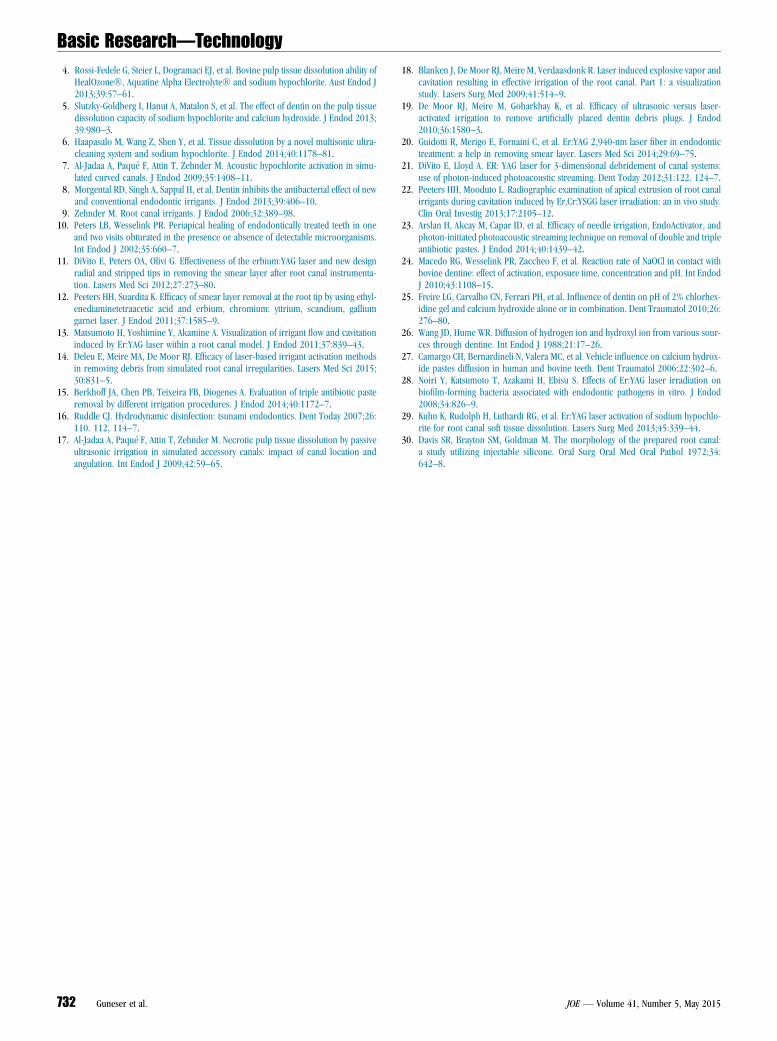
Basic Research—Technology
4. Rossi-Fedele G, Steier L, Dogramaci EJ, et al. Bovine pulp tissue dissolution ability ofHealOzone�, Aquatine Alpha Electrolyte� and sodium hypochlorite. Aust Endod J2013;39:57–61.
5. Slutzky-Goldberg I, Hanut A, Matalon S, et al. The effect of dentin on the pulp tissuedissolution capacity of sodium hypochlorite and calcium hydroxide. J Endod 2013;39:980–3.
6. Haapasalo M, Wang Z, Shen Y, et al. Tissue dissolution by a novel multisonic ultra-cleaning system and sodium hypochlorite. J Endod 2014;40:1178–81.
7. Al-Jadaa A, Paqu�e F, Attin T, Zehnder M. Acoustic hypochlorite activation in simu-lated curved canals. J Endod 2009;35:1408–11.
8. Morgental RD, Singh A, Sappal H, et al. Dentin inhibits the antibacterial effect of newand conventional endodontic irrigants. J Endod 2013;39:406–10.
9. Zehnder M. Root canal irrigants. J Endod 2006;32:389–98.10. Peters LB, Wesselink PR. Periapical healing of endodontically treated teeth in one
and two visits obturated in the presence or absence of detectable microorganisms.Int Endod J 2002;35:660–7.
11. DiVito E, Peters OA, Olivi G. Effectiveness of the erbium:YAG laser and new designradial and stripped tips in removing the smear layer after root canal instrumenta-tion. Lasers Med Sci 2012;27:273–80.
12. Peeters HH, Suardita K. Efficacy of smear layer removal at the root tip by using ethyl-enediaminetetraacetic acid and erbium, chromium: yttrium, scandium, galliumgarnet laser. J Endod 2011;37:1585–9.
13. Matsumoto H, Yoshimine Y, Akamine A. Visualization of irrigant flow and cavitationinduced by Er:YAG laser within a root canal model. J Endod 2011;37:839–43.
14. Deleu E, Meire MA, De Moor RJ. Efficacy of laser-based irrigant activation methodsin removing debris from simulated root canal irregularities. Lasers Med Sci 2015;30:831–5.
15. Berkhoff JA, Chen PB, Teixeira FB, Diogenes A. Evaluation of triple antibiotic pasteremoval by different irrigation procedures. J Endod 2014;40:1172–7.
16. Ruddle CJ. Hydrodynamic disinfection: tsunami endodontics. Dent Today 2007;26:110. 112, 114–7.
17. Al-Jadaa A, Paqu�e F, Attin T, Zehnder M. Necrotic pulp tissue dissolution by passiveultrasonic irrigation in simulated accessory canals: impact of canal location andangulation. Int Endod J 2009;42:59–65.
732 Guneser et al.
18. Blanken J, De Moor RJ, Meire M, Verdaasdonk R. Laser induced explosive vapor andcavitation resulting in effective irrigation of the root canal. Part 1: a visualizationstudy. Lasers Surg Med 2009;41:514–9.
19. De Moor RJ, Meire M, Goharkhay K, et al. Efficacy of ultrasonic versus laser-activated irrigation to remove artificially placed dentin debris plugs. J Endod2010;36:1580–3.
20. Guidotti R, Merigo E, Fornaini C, et al. Er:YAG 2,940-nm laser fiber in endodontictreatment: a help in removing smear layer. Lasers Med Sci 2014;29:69–75.
21. DiVito E, Lloyd A. ER: YAG laser for 3-dimensional debridement of canal systems:use of photon-induced photoacoustic streaming. Dent Today 2012;31:122. 124–7.
22. Peeters HH, Mooduto L. Radiographic examination of apical extrusion of root canalirrigants during cavitation induced by Er,Cr:YSGG laser irradiation: an in vivo study.Clin Oral Investig 2013;17:2105–12.
23. Arslan H, Akcay M, Capar ID, et al. Efficacy of needle irrigation, EndoActivator, andphoton-initiated photoacoustic streaming technique on removal of double and tripleantibiotic pastes. J Endod 2014;40:1439–42.
24. Macedo RG, Wesselink PR, Zaccheo F, et al. Reaction rate of NaOCl in contact withbovine dentine: effect of activation, exposure time, concentration and pH. Int EndodJ 2010;43:1108–15.
25. Freire LG, Carvalho CN, Ferrari PH, et al. Influence of dentin on pH of 2% chlorhex-idine gel and calcium hydroxide alone or in combination. Dent Traumatol 2010;26:276–80.
26. Wang JD, Hume WR. Diffusion of hydrogen ion and hydroxyl ion from various sour-ces through dentine. Int Endod J 1988;21:17–26.
27. Camargo CH, Bernardineli N, Valera MC, et al. Vehicle influence on calcium hydrox-ide pastes diffusion in human and bovine teeth. Dent Traumatol 2006;22:302–6.
28. Noiri Y, Katsumoto T, Azakami H, Ebisu S. Effects of Er:YAG laser irradiation onbiofilm-forming bacteria associated with endodontic pathogens in vitro. J Endod2008;34:826–9.
29. Kuhn K, Rudolph H, Luthardt RG, et al. Er:YAG laser activation of sodium hypochlo-rite for root canal soft tissue dissolution. Lasers Surg Med 2013;45:339–44.
30. Davis SR, Brayton SM, Goldman M. The morphology of the prepared root canal:a study utilizing injectable silicone. Oral Surg Oral Med Oral Pathol 1972;34:642–8.
JOE — Volume 41, Number 5, May 2015
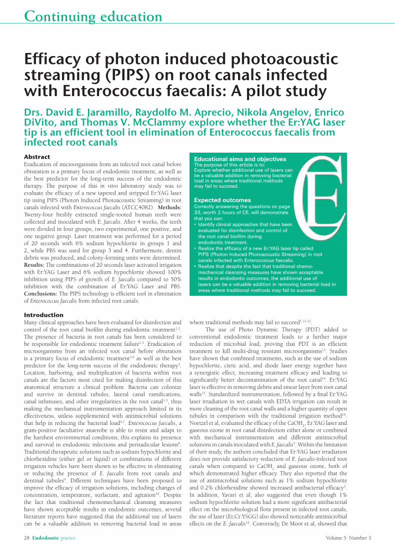
28 Endodontic practice Volume 5 Number 3
Continuing education
Abstract Eradication of microorganisms from an infected root canal before obturation is a primary focus of endodontic treatment, as well as the best predictor for the long-term success of the endodontic therapy. The purpose of this in vitro laboratory study was to evaluate the efficacy of a new tapered and stripped Er:YAG laser tip using PIPS (Photon Induced Photoacoustic Streaming) in root canals infected with Enterococcus faecalis (ATCC4082). Methods: Twenty-four freshly extracted single-rooted human teeth were collected and inoculated with E. faecalis. After 4 weeks, the teeth were divided in four groups, two experimental, one positive, and one negative group. Laser treatment was performed for a period of 20 seconds with 6% sodium hypochlorite in groups 1 and 2, while PBS was used for group 3 and 4. Furthermore, dentin debris was produced, and colony-forming units were determined. Results: The combinations of 20 seconds laser activated irrigation with Er:YAG Laser and 6% sodium hypochlorite showed 100% inhibition using PIPS of growth of E. faecalis compared to 50% inhibition with the combination of Er:YAG Laser and PBS. Conclusions: The PIPS technology is efficient tool in elimination of Enterococcus faecalis from infected root canals.
IntroductionMany clinical approaches have been evaluated for disinfection and control of the root canal biofilm during endodontic treatment1,2. The presence of bacteria in root canals has been considered to be responsible for endodontic treatment failure1,3. Eradication of microorganisms from an infected root canal before obturation is a primary focus of endodontic treatment3,4 as well as the best predictor for the long-term success of the endodontic therapy5. Location, harboring, and multiplication of bacteria within root canals are the factors most cited for making disinfection of this anatomical structure a clinical problem. Bacteria can colonize and survive in dentinal tubules, lateral canal ramifications, canal isthmuses, and other irregularities in the root canal1,3, thus making the mechanical instrumentation approach limited in its effectiveness, unless supplemented with antimicrobial solutions that help in reducing the bacterial load6,7. Enterococcus faecalis, a gram-positive facultative anaerobe is able to resist and adapt to the harshest environmental conditions; this explains its presence and survival in endodontic infections and periradicular lesions8. Traditional therapeutic solutions such as sodium hypochlorite and chlorhexidine (either gel or liquid) or combinations of different irrigation vehicles have been shown to be effective in eliminating or reducing the presence of E. faecalis from root canals and dentinal tubules9. Different techniques have been proposed to improve the efficacy of irrigation solutions, including changes of concentration, temperature, surfactant, and agitation10. Despite the fact that traditional chemomechanical cleansing measures have shown acceptable results in endodontic outcomes, several literature reports have suggested that the additional use of lasers can be a valuable addition in removing bacterial load in areas
Efficacy of photon induced photoacoustic streaming (PIPS) on root canals infected with Enterococcus faecalis: A pilot studyDrs. David E. Jaramillo, Raydolfo M. Aprecio, Nikola Angelov, Enrico DiVito, and Thomas V. McClammy explore whether the Er:YAG laser tip is an efficient tool in elimination of Enterococcus faecalis from infected root canals
Educational aims and objectivesThe purpose of this article is to:Explore whether additional use of lasers can be a valuable addition in removing bacterial load in areas where traditional methods may fail to succeed.
Expected outcomesCorrectly answering the questions on page 33, worth 2 hours of CE, will demonstrate that you can:
evaluated for disinfection and control of the root canal biofilm during endodontic treatment.
canals infected with Enteroccocus faecalis.
mechanical cleansing measures have shown acceptable results in endodontic outcomes, the additional use of lasers can be a valuable addition in removing bacterial load in areas where traditional methods may fail to succeed.
where traditional methods may fail to succeed1,11,12. The use of Photo Dynamic Therapy (PDT) added to conventional endodontic treatment leads to a further major reduction of microbial load, proving that PDT is an efficient treatment to kill multi-drug resistant microorganisms13. Studies have shown that combined treatments, such as the use of sodium hypochlorite, citric acid, and diode laser energy together have a synergistic effect, increasing treatment efficacy and leading to significantly better decontamination of the root canal14. Er:YAG laser is effective in removing debris and smear layer from root canal walls15. Standardized instrumentation, followed by a final Er:YAG laser irradiation in wet canals with EDTA irrigation can result in more cleaning of the root canal walls and a higher quantity of open tubules in comparison with the traditional irrigation method16. Noetzel et al, evaluated the efficacy of the CaOH2, Er:YAG laser and gaseous ozone in root canal disinfection either alone or combined with mechanical instrumentation and different antimicrobial solutions in canals inoculated with E. faecalis3. Within the limitation of their study, the authors concluded that Er:YAG laser irradiation does not provide satisfactory reduction of E. faecalis-infected root canals when compared to CaOH2 and gaseous ozone, both of which demonstrated higher efficacy. They also reported that the use of antimicrobial solutions such as 1% sodium hypochlorite and 0.2% chlorhexidine showed increased antibacterial efficacy3. In addition, Yavari et al, also suggested that even though 1% sodium hypochlorite solution had a more significant antibacterial effect on the microbiological flora present in infected root canals, the use of laser (Er,Cr:YSGG) also showed noticeable antimicrobial effects on the E. faecalis12. Conversely, De Moor et al, showed that
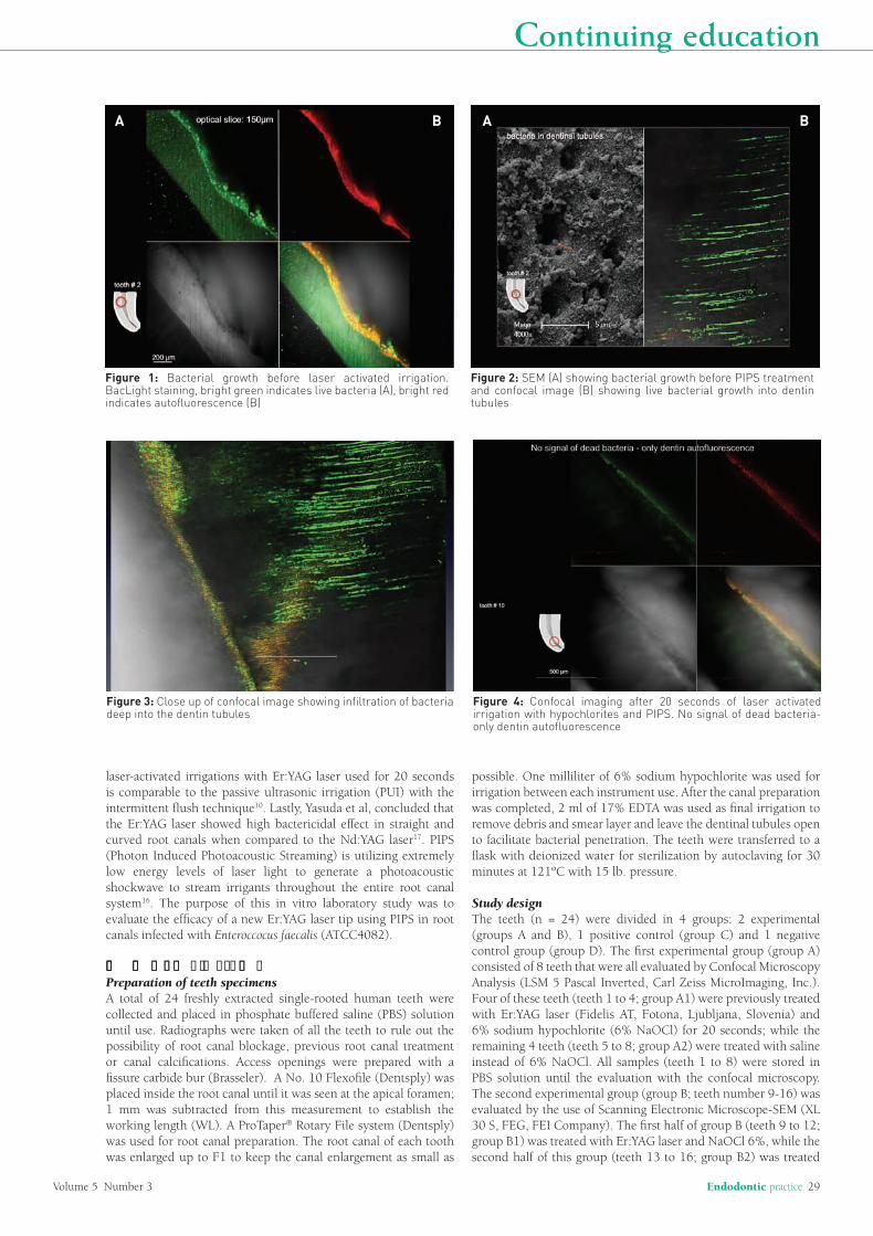
Volume 5 Number 3 Endodontic practice 29
Continuing education
laser-activated irrigations with Er:YAG laser used for 20 seconds is comparable to the passive ultrasonic irrigation (PUI) with the intermittent flush technique10. Lastly, Yasuda et al, concluded that the Er:YAG laser showed high bactericidal effect in straight and curved root canals when compared to the Nd:YAG laser17. PIPS (Photon Induced Photoacoustic Streaming) is utilizing extremely low energy levels of laser light to generate a photoacoustic shockwave to stream irrigants throughout the entire root canal system16. The purpose of this in vitro laboratory study was to evaluate the efficacy of a new Er:YAG laser tip using PIPS in root canals infected with Enteroccocus faecalis (ATCC4082).
��� ��� �� �� �� � � � � � �� �Preparation of teeth specimensA total of 24 freshly extracted single-rooted human teeth were collected and placed in phosphate buffered saline (PBS) solution until use. Radiographs were taken of all the teeth to rule out the possibility of root canal blockage, previous root canal treatment or canal calcifications. Access openings were prepared with a fissure carbide bur (Brasseler). A No. 10 Flexofile (Dentsply) was placed inside the root canal until it was seen at the apical foramen; 1 mm was subtracted from this measurement to establish the working length (WL). A ProTaper® Rotary File system (Dentsply) was used for root canal preparation. The root canal of each tooth was enlarged up to F1 to keep the canal enlargement as small as
possible. One milliliter of 6% sodium hypochlorite was used for irrigation between each instrument use. After the canal preparation was completed, 2 ml of 17% EDTA was used as final irrigation to remove debris and smear layer and leave the dentinal tubules open to facilitate bacterial penetration. The teeth were transferred to a flask with deionized water for sterilization by autoclaving for 30 minutes at 121ºC with 15 lb. pressure.
Study designThe teeth (n = 24) were divided in 4 groups: 2 experimental (groups A and B), 1 positive control (group C) and 1 negative control group (group D). The first experimental group (group A) consisted of 8 teeth that were all evaluated by Confocal Microscopy Analysis (LSM 5 Pascal Inverted, Carl Zeiss MicroImaging, Inc.). Four of these teeth (teeth 1 to 4; group A1) were previously treated with Er:YAG laser (Fidelis AT, Fotona, Ljubljana, Slovenia) and 6% sodium hypochlorite (6% NaOCl) for 20 seconds; while the remaining 4 teeth (teeth 5 to 8; group A2) were treated with saline instead of 6% NaOCl. All samples (teeth 1 to 8) were stored in PBS solution until the evaluation with the confocal microscopy. The second experimental group (group B; teeth number 9-16) was evaluated by the use of Scanning Electronic Microscope-SEM (XL 30 S, FEG, FEI Company). The first half of group B (teeth 9 to 12; group B1) was treated with Er:YAG laser and NaOCl 6%, while the second half of this group (teeth 13 to 16; group B2) was treated
Figure 2: SEM (A) showing bacterial growth before PIPS treatment and confocal image (B) showing live bacterial growth into dentin tubules
Figure 3: Close up of confocal image showing infiltration of bacteria deep into the dentin tubules
Figure 1: Bacterial growth before laser activated irrigation. BacLight staining, bright green indicates live bacteria (A), bright red indicates autofluorescence (B)
A B A B
Figure 4: Confocal imaging after 20 seconds of laser activated irrigation with hypochlorites and PIPS. No signal of dead bacteria-only dentin autofluorescence
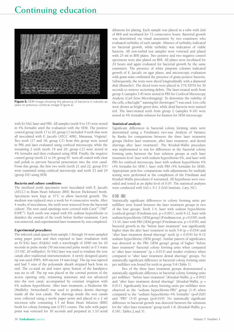
30 Endodontic practice Volume 5 Number 3
Continuing education
with Er:YAG laser and PBS. All samples (teeth 9 to 15) were stored in 4% formalin until the evaluation with the SEM. The positive control group (teeth 17 to 20; group C) included 4 teeth that were all inoculated with E. faecalis (ATCC 4082, Manassas, Virginia). Two teeth (17 and 18; group C1) from this group were stored in PBS and later evaluated using confocal microscopy, while the remaining 2 teeth (teeth 19 and 20; group C2) were stored in 4% formalin and then evaluated using SEM. Finally, the negative control group (teeth 21 to 24; group D) were all coated with clear nail polish to prevent bacterial penetration into the root canal. From this group, the first two teeth (teeth 21 and 22; group D1) were examined using confocal microscopy and teeth 23 and 24 (group D2) using SEM.
Bacteria and culture conditionsThe sterilized tooth specimens were inoculated with E. faecalis (ATCC) in Brain Heart Infusion (BHI; Becton Dickinson) broth. Specimens were kept at 37˚C to allow bacterial growth. The medium was replaced once a week for 4 consecutive weeks. After 4 weeks of inoculation, the teeth were removed from the bacterial culture. The root canal openings were covered with Cavit™ (3M™
ESPE™). Each tooth was wiped with 6% sodium hypochlorite to disinfect the outside of the tooth before further treatment. Cavit was removed, and experimental laser treatment by was completed.
Experimental proceduresThe infected canal spaces from sample 1 through 16 were sampled using paper point and then exposed to laser irradiation with an Er:YAG laser (Fidelis) with a wavelength of 2940 nm for 20 seconds in pulse mode (50 microsecond pulse mode) at 0.3 watts (15 HZ, 20 millijules). Er:YAG laser was used to irradiate the root canals after traditional instrumentation. A newly designed quartz tip was used (PIPS, 400 micron 14 mm long). The tip was tapered and had 3 mm of the polyamide sheath stripped back from its end. The co-axial air and water spray feature of the handpiece was set to off. The tip was placed in the coronal portion of the access opening only, remaining stationary and not advanced into the root canal, accompanied by constant irrigation with 6% sodium hypochlorite. After laser treatment, a Hedström file (Maillefer, Switzerland) was used to produce dentin shavings inside all the root canals. The shavings inside the root canals were collected using a sterile paper point and placed in a 2 ml microtest tube containing 1.5 ml Brain Heart Infusion (BHI) broth for colony forming unit (CFU) counts. The tube with paper point was vortexed for 30 seconds and prepared in 1:10 serial
Figure 5: SEM image showing the absence of bacteria in tubules as seen on previous confocal image (Figure 4)
dilutions for plating. Each sample was placed in a tube with 2ml of BHI and incubated for 72 consecutive hours. Bacterial growth was determined via visual assessment by two examiners who evaluated turbidity of each sample. Absence of turbidity indicated no bacterial growth, while turbidity was indication of viable bacteria. All non-turbid test samples were vortexed and plated with 25 ml in BHI plates. Two positive and two negative control specimens were also plated on BHI. All plates were incubated for 24 hours and again evaluated for bacterial growth by the same examiners. The presence of white pinpoint colonies indicated growth of E. faecalis on agar plates, and microscopic evaluation with gram stain confirmed the presence of gram-positive bacteria. Subsequently, the roots were sliced longitudinally with a diamond disk (Brasseler). The sliced roots were placed in 17% EDTA for 30 seconds to remove sectioning debris. The laser-treated teeth from group A (samples 1-8) were stored in PBS for Confocal Microscopy Analysis (Carl Zeiss MicroImaging). To determine the viability of the cells, a BacLight™ staining kit (Invitrogen™) was used. Live cells were shown as bright green dots, while dead bacteria were stained red. The laser-treated teeth from group 2 (samples 9-16) were stored in 4% formalin solution for fixation for SEM microscopy.
Statistical analysisSignificant differences in bacterial colony forming units were determined using a Friedman’s two-way Analysis of Variance by Ranks for comparisons between the three laser treatment groups (before laser treatment, after laser treatment, and dental shavings after laser treatment). The Kruskal-Wallis procedure was implemented to test for differences in the bacterial colony forming units between the four solution groups at each laser treatment level: laser with sodium hypochlorite 6%, and laser with PBS for confocal microscopy, laser with sodium hypochlorite 6% (4% formalin for SEM ), laser with PBS (4% formalin for SEM), Appropriate post-hoc comparisons with adjustments for multiple testing were performed at the completion of the Friedman and Kruskal-Wallis procedures if warranted. All hypotheses were two-sided and tested at an alpha level of 0.05. The statistical analyses were conducted with SAS v. 9.1.3 (SAS Institute, Cary, NC).
� � �� � � �Statistically significant differences in colony forming units per milliliter were found between the laser treatment groups in two of the four groups; Teeth 1-4, laser with sodium hypochlorite (confocal group) (Friedman test, p < 0.001), teeth 9-12, laser with sodium hypochlorite (SEM group) (Friedman test, p = 0.018), teeth 13-16, laser with PBS (SEM group) (Friedman test, p = 0.039), and bacterial growth in the “before laser treatment” was significantly higher than the after laser treatment in teeth 5-8 (p = 0.034) and “after laser treatment dental shavings” teeth (p = 0.034) for 9-12 sodium hypochlorite (SEM group). Similar pattern of significance was detected in the PBS (SEM group) group of higher “before laser treatment” bacterial colony forming units when compared to “after laser treatment” (p = 0.013) and weaker evidence when compared to “after laser treatment dental shavings” groups. No statistically significant difference in bacterial colony forming units per milliliter was found for teeth in group 5-8 (Table 1). Two of the three laser treatment groups demonstrated a statistically significant difference in bacterial colony forming units per milliliter: “before laser treatment” (Kruskal-Wallis, p = 0.042) and “after laser treatment dental shavings” (Kruskal-Wallis, p = 0.011). Significantly less colony forming units per milliliter were observed in the “sodium hypochlorite-PBS” group (1-4) when compared to the “sodium hypochlorite–4% formalin” (p=0.026) and “PBS” (5-9) groups (p=0.019). No statistically significant difference in bacterial growth was detected between the solutions for the “after laser treatment” group teeth 1-8, (Kruskal-Wallis, p = 0.181; Tables 2 and 3).
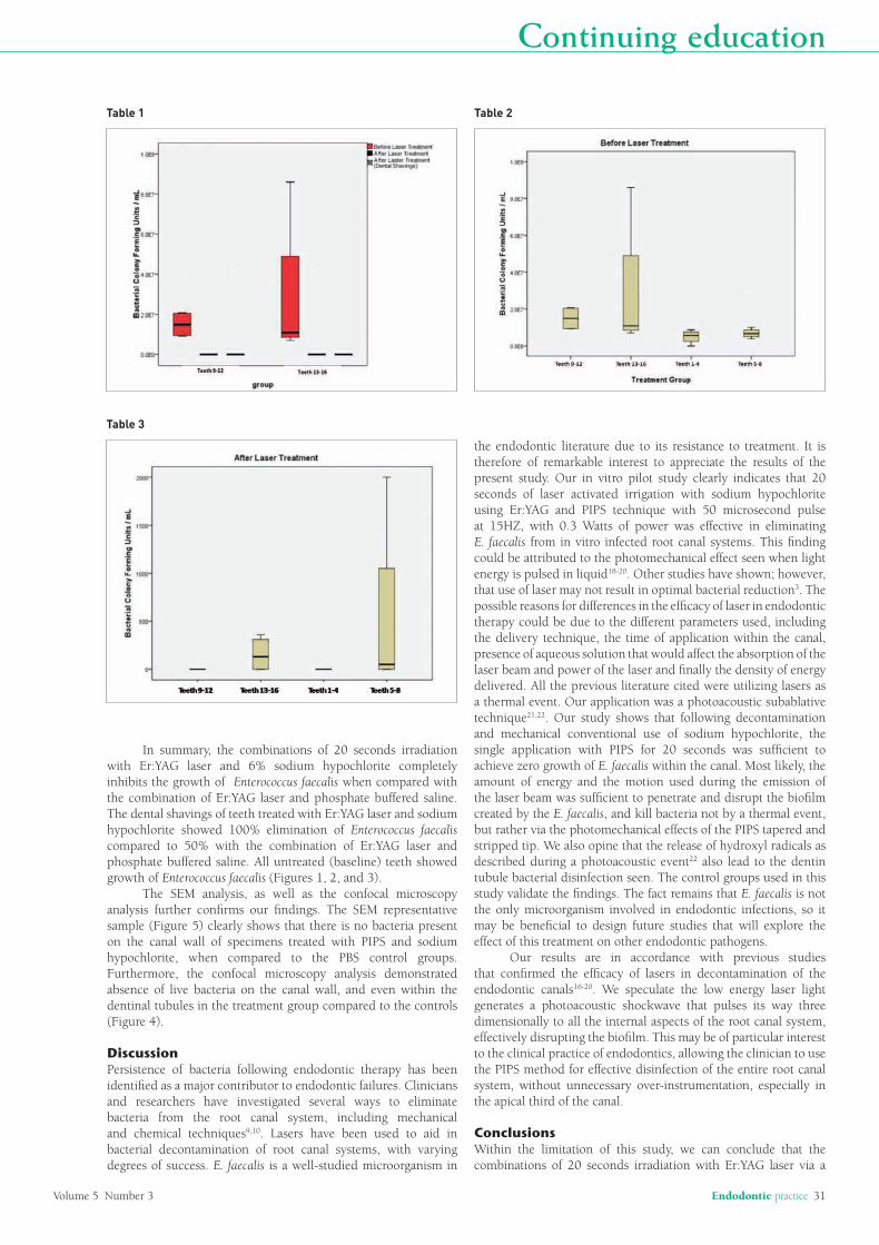
Volume 5 Number 3 Endodontic practice 31
Continuing education
Table 3
Table 2Table 1
In summary, the combinations of 20 seconds irradiation with Er:YAG laser and 6% sodium hypochlorite completely inhibits the growth of Enterococcus faecalis when compared with the combination of Er:YAG laser and phosphate buffered saline. The dental shavings of teeth treated with Er:YAG laser and sodium hypochlorite showed 100% elimination of Enterococcus faecalis compared to 50% with the combination of Er:YAG laser and phosphate buffered saline. All untreated (baseline) teeth showed growth of Enterococcus faecalis (Figures 1, 2, and 3). The SEM analysis, as well as the confocal microscopy analysis further confirms our findings. The SEM representative sample (Figure 5) clearly shows that there is no bacteria present on the canal wall of specimens treated with PIPS and sodium hypochlorite, when compared to the PBS control groups. Furthermore, the confocal microscopy analysis demonstrated absence of live bacteria on the canal wall, and even within the dentinal tubules in the treatment group compared to the controls (Figure 4).
DiscussionPersistence of bacteria following endodontic therapy has been identified as a major contributor to endodontic failures. Clinicians and researchers have investigated several ways to eliminate bacteria from the root canal system, including mechanical and chemical techniques9,10. Lasers have been used to aid in bacterial decontamination of root canal systems, with varying degrees of success. E. faecalis is a well-studied microorganism in
the endodontic literature due to its resistance to treatment. It is therefore of remarkable interest to appreciate the results of the present study. Our in vitro pilot study clearly indicates that 20 seconds of laser activated irrigation with sodium hypochlorite using Er:YAG and PIPS technique with 50 microsecond pulse at 15HZ, with 0.3 Watts of power was effective in eliminating E. faecalis from in vitro infected root canal systems. This finding could be attributed to the photomechanical effect seen when light energy is pulsed in liquid18-20. Other studies have shown; however, that use of laser may not result in optimal bacterial reduction3. The possible reasons for differences in the efficacy of laser in endodontic therapy could be due to the different parameters used, including the delivery technique, the time of application within the canal, presence of aqueous solution that would affect the absorption of the laser beam and power of the laser and finally the density of energy delivered. All the previous literature cited were utilizing lasers as a thermal event. Our application was a photoacoustic subablative technique21,22. Our study shows that following decontamination and mechanical conventional use of sodium hypochlorite, the single application with PIPS for 20 seconds was sufficient to achieve zero growth of E. faecalis within the canal. Most likely, the amount of energy and the motion used during the emission of the laser beam was sufficient to penetrate and disrupt the biofilm created by the E. faecalis, and kill bacteria not by a thermal event, but rather via the photomechanical effects of the PIPS tapered and stripped tip. We also opine that the release of hydroxyl radicals as described during a photoacoustic event22 also lead to the dentin tubule bacterial disinfection seen. The control groups used in this study validate the findings. The fact remains that E. faecalis is not the only microorganism involved in endodontic infections, so it may be beneficial to design future studies that will explore the effect of this treatment on other endodontic pathogens. Our results are in accordance with previous studies that confirmed the efficacy of lasers in decontamination of the endodontic canals16-20. We speculate the low energy laser light generates a photoacoustic shockwave that pulses its way three dimensionally to all the internal aspects of the root canal system, effectively disrupting the biofilm. This may be of particular interest to the clinical practice of endodontics, allowing the clinician to use the PIPS method for effective disinfection of the entire root canal system, without unnecessary over-instrumentation, especially in the apical third of the canal.
ConclusionsWithin the limitation of this study, we can conclude that the combinations of 20 seconds irradiation with Er:YAG laser via a
32 Endodontic practice Volume 5 Number 3
Continuing education
References
1. Meire MA, De Prijck K, Coenye T, Nelis HJ, De Moor RJ (2009. Effectiveness of different laser systems to kill Enterococcus faecalis in aqueous suspension and in an infected tooth model. Int Endod J. Apr:42(4):351-9.
2. Chávez de Paz LE, Bergenholtz G, Svensäter G (2010). The effects of antimicrobials on endodontic biofilm bacteria. J Endod. Jan;36(1):70-7.
3. Noetzel J, Nonhoff J, Bitter K, Wagner J, Neumann K, Kielbassa AM (2009). Efficacy of calcium hydroxide, Er:YAG laser or gaseous ozone against Enterococcus faecalis in root canals. Am J Dent. Feb;22(1):14-8.
4. Dewsnup N, Pileggi R, Haddix J, Nair U, Walker C, Varella CH (2010). Comparison of bacterial reduction in straight and curved canals using erbium, chromium:yttrium-scandium-gallium-garnet laser treatment versus a traditional irrigation technique with sodium hypochlorite. J Endod. Apr:36(4):725-8.
5. Franzen R, Gutknecht N, Falken S, Heussen N, Meister J (2011). Bactericidal effect of a Nd:YAG laser on Enterococcus faecalis at pulse durations of 15 and 25 ms in dentine depths of 500 and 1,000 µm. Lasers Med Sci. Jan:26(1):95-101. Epub 2010 Aug 31.
6. Mohammadi Z (2010). Chemomechanical strategies to manage endodontic infections. Dent Today. Feb:29(2):91-2, 94, 96 passim; quiz 99.
7. Upadya MH, Kishen A (2010). Influence of bacterial growth modes on the susceptibility to light-activated disinfection. Int Endod J. Nov:43(11):978-87.
8. Kayaoglu G, Ørstavik D (2004).Virulence factors of Enterococcus faecalis: relationship to endodontic disease. Crit Rev Oral Biol Med. Sep 1:15(5):308-20.
9. Stuart CH, Schwartz SA, Beeson TJ, Owatz CB (2006). Enterococcus faecalis: its role in root canal treatment failure and current concepts in retreatment. J Endod. Feb32(2):93-8.
10. Stojicic S, Zivkovic S, Qian W, Zhang H, Haapasalo M (2010). Tissue dissolution by sodium hypochlorite: effect of concentration, temperature, agitation, and surfactant. J Endod. Sep:36(9):1558-62.
11. De Moor RJ, Meire M, Goharkhay K, Moritz A, Vanobbergen J (2010). Efficacy of ultrasonic versus laser-activated irrigation to remove artificially placed dentin debris plugs. J Endod. Sep:36(9):1580-3.
12. Yavari HR, Rahimi S, Shahi S, Lotfi M, Barhaghi MH, Fatemi A, Abdolrahimi M (2010). Effect of Er, Cr: YSGG laser irradiation on Enterococcus faecalis in infected root canals. Photomed Laser Surg. Aug:28 Suppl 1:S91-6.
13. Garcez AS, Nuñez SC, Hamblim MR, Suzuki H, Ribeiro MS (2010). Photodynamic therapy associated with conventional endodontic treatment in patients with antibiotic-resistant microflora: a preliminary report. J Endod. Sep:36(9):1463-6
14. Benedicenti S, Cassanelli C, Signore A, Ravera G, Angiero F (2008). Decontamination of root canals with the gallium-aluminum-arsenide laser: an in vitro study. Photomed Laser Surg. Aug:26(4):367-70.
15. Takeda FH, Harashima T, Kimura Y, Matsumoto K (1998). Efficacy of Er:YAG laser irradiation in removing debris and smear layer on root canal walls. J Endod. Aug:24(8):548-51.
16. Divito E, Peters OA, Olivi G (2010). Effectiveness of the erbium:YAG laser and new design radial and stripped tips in removing the smear layer after root canal instrumentation. Lasers Med Sci. Dec 1.
17. Yasuda Y, Kawamorita T, Yamaguchi H, Saito T (2010). Bactericidal effect of Nd:YAG and Er:YAG lasers in experimentally infected curved root canals. Photomed Laser Surg. Oct:28 Suppl 2:S75-8.
18. Blanken J, De Moor RJ, Meire M, Verdaasdonk R (2009). Laser induced explosive vapor and cavitation resulting in effective irrigation of the root canal. Part 1: a visualization study. Lasers Surg Med 41(7):514–519
19. De Moor RJ, Blanken J, Meire M, Verdaasdonk R (2009). Laser induced explosive vapor and cavitation resulting in effective irrigation of the root canal. Part 2: evaluation of the efficacy. Lasers Surg Med 41(7):520–523
20. de Groot SD, Verhaagen B, Versluis M, Wu MK, Wesselink PR, van der Sluis LW (2009). Laser activated irrigation within root canals: cleaning efficacy and flow visualization. Int Endod J 42 (12):1077–108.
21. Doukas AG, Birngruber R, Deutsch TF (1990). Determination of the shock wave pressures generated by laser-induced breakdown in water. In: Jacques FL (ed) Laser-Tissue Interaction. SPIE , Vol. 1202, pp. 61-70.
22. Müller P, Guggenheim B, Attin T, Marlinghaus E, Schmidlin, PR (2011). Potential of shock waves to remove calculus and biofilm. Clin Oral Invest 15:959–965.
Nikola Angelov, DDS, MS, PhD, is a professor and a Program Director of Undergraduate Periodontics at Loma Linda University School of Dentistry. Dr. Angelov was born in Skopje, Macedonia in 1968. He earned his DDS, MS and PhD degree from St. Cyril and Methodius University and has been teaching for the Department of Oral Pathology and Periodontology in Macedonia since 1993. In 2001, he received
a postdoctoral fellowship at the National Institute of Health, National Institute of Dental and Craniofacial Research (NIH/NIDCR). At NIH/NIDCR, Dr. Angelov successfully developed a new model of oral mucosal wound healing and was also involved in collaborative clinical studies, clinical trial evaluations, as well as other studies. Dr. Angelov also holds a Certificate of Specialty in Periodontics and is a Diplomate of the American Board of Periodontology. In 2008, Dr. Angelov received the prestigious annual Educator of the Year Award from the American Academy of Periodontics for outstanding results in teaching in the area of Periodontics. Dr. Angelov has several other academic awards and is actively involved in teaching and research. His research area includes oral infection and immunity, mucosal wound healing, periodontal microbiology, as well as dental implants. Dr. Angelov has extensively been published in peer review articles, and has been an invited speaker to prominent professional and research meetings. He is a member of numerous professional societies in the USA and abroad. Dr. Angelov also serves as a reviewer on the Journal of Periodontology, an official journal of the American Academy of Periodontology, as well as a School of Dentistry representative to the Institutional Review Board, and the Institutional Animal Care and Use Committee at Loma Linda University in California.
Dr. Enrico DiVito formed his dental practice in 1980 in Scottsdale. In 2004, he formed the Arizona Center for Laser Dentistry. He is also the founder and director of the state-accredited Arizona School of Dental Assisting (ASDA). In addition to teaching at ASDA, DiVito is also a clinical instructor at the Arizona School of Dentistry and Oral Health and is responsible for helping to create the Department of
Laser Dentistry. He earned his undergraduate degree from Arizona State University in 1980 and is a graduate from the University of the Pacific, Arthur A. Dugoni School of Dentistry with honors receiving several clinical excellence awards. He can be reached at [email protected].
Thomas V. McClammy, DMD, MS, graduated from Oregon Health Sciences University, School of Dentistry, in 1982. During the next 15 years, he created a remarkable general practice in Oregon and passionately pursued continuing education courses to fulfill his insatiable desire to learn. In 1996, Dr. McClammy was accepted as a post graduate endodontic resident at Boston University’s Goldman Graduate School
of Dentistry. He earned a Master’s degree in 1999 from his mentor, Dr. Herbert Schilder, who is widely recognized as the “father of modern day endodontics.” Currently, Dr. McClammy has a full-time practice dedicated to saving teeth with endodontics and implants in Scottsdale, Arizona, and is well known for his expertise, passion, and extraordinary teaching skills.
David E. Jaramillo, DDS, completed dental school and an advanced program in endodontics in Mexico in 1990 and has been teaching since then. He has also successfully practiced endodontics for 15 years. In 2004, he accepted the faculty position at USC Endodontic advanced program. In 2005, he started working at the Center for Biofilms at USC, run by Dr. Bill Costerton where he got trained in the use of SEM
(Scanning Electron Microscopy) and Confocal (Laser Scanning Microscopy). In 2006, he accepted a full-time position at Loma Linda University School of Dentistry where he is an associate professor and the clinical director of endodontics. Dr. Jaramillo also works at the Center for Dental Research LLUSD. He teaches at six different advanced endodontic programs in Mexico along with USC and Long Beach VA Hospital. He has lectured in the US, Mexico, Europe and South America and has been published in numerous journals. He has co-authored several papers with his colleagues from the Department of Electrical Engineering-Electrophysics, Viterbi School of Engineering University of Southern California on the use of new plasma technology for biofilm removal from root canal system, as well as a chapter in the 6th edition of Ingle’s Endodontics. Dr. Jaramillo has donated scanning electronic microscopy images for illustration for a chapter entitled “The Microbiology of the Necrotic Pulp” for Textbook of Endodontology by Dr. Gunnar Bergenholtz- 2nd edition. Dr. Jaramillo has done extensive research in the field of biofilms and has two temporary patents filed through the university patent office.
Raydolfo M. Aprecio Sr., BS, OD, is a Research Associate with extensive research experience and expertise in microbiology. Before joining the Center for Dental Research, he was a Senior Research Assistant with the Department of Microbiology and Molecular Genetics, LLU School of Medicine for 25 years. He has worked on numerous research projects in the areas of endodontics and periodontics and
helped numerous graduate students and residents from Departments of Endodontics, Pediatrics and Implant Dentistry for their thesis research projects. He has served as a mentor to dental and hygiene students for their table clinic research. Each year, his student table clinics have won awards during the annual Student and Alumni Convention, California Dental Association and national meetings. He was an original member of the research team that developed the MTA and MTAD.
photoacoustic delivery system and 6% sodium hypochlorite is very effective in inhibiting Enterococcus faecalis growth. The results are encouraging in terms of the possible application of the Er:YAG laser in endodontics. The PIPS technology can be used as an efficient additional tool in the decontamination of infected root canals during endodontic treatment. EP
Original Research
Effectiveness of Sonic, Ultrasonic, and Photon-InducedPhotoacoustic Streaming Activation of NaOCl
on Filling Material Removal Following Retreatmentin Oval Canal Anatomy
Shan Jiang, MDS,1 Ting Zou, MSc,2 Dongxia Li, DDS,2,3 Jeffery W.W. Chang, BDS, MDS,2
Xiaojing Huang, DDS, PhD,1 and Chengfei Zhang, DDS, PhD2
Abstract
Objective: This study aimed to assess the effectiveness of sonic, ultrasonic and laser [photon-induced photo-acoustic streaming (PIPS)] irrigation activation in removing filling remnants from oval root canals after standardcanal retreatment procedures with the ProTaper universal rotary retreatment system. Methods: Twenty-eightmaxillary first premolars were instrumented with ProTaper NiTi rotary instruments and obturated with gutta-percha and AH Plus sealer using the continuous wave of condensation technique. After storage at 37�C and 100%humidity for 1 week, the specimens were retreated with the ProTaper universal retreatment system for the removalof filling material. Teeth were then randomly assigned into four groups (n = 7): group 1, positive control; group 2,retreated with sonic irrigation; group 3, retreated with ultrasonic irrigation; and group 4, retreated with laserirradiation. The specimens were scanned using micro-CT before instrumentation, after obturation and mechanicalretreatment, and after additional activation procedures. The percentage volume of the filling remnants wasmeasured. Specimens were split longitudinally after micro-CT scan, canal walls were examined using scanningelectron microscopy (SEM), and the amount of residual filling material was scored. Results: The filling materials’removal efficacy in the three experimental groups was higher than that of the control group ( p < 0.05), whereasfilling materials ranging from 1.46 – 0.30 to 2.21 – 0.46 mm3 remained in the canal in all three experimentalgroups. Additionally, there was a significantly greater reduction in the amount of filling remnants in the PIPSgroup than in the sonic and ultrasonic groups (both p < 0.05), and significantly greater reduction in the ultrasonicgroup than the sonic group ( p < 0.05). Conclusions: Activation of NaOCl with PIPS showed significantly betterperformance than sonic and ultrasonic techniques in removing the filling remnants following mechanical re-treatment of oval root canals. The ultrasonic technique also performed better than the sonic technique. However,none of the additional activation procedures was able to completely eliminate the filling remnants.
Introduction
Endodontic retreatment aims at completely remov-
ing the previous root canal filling materials (gutta-percha and endodontic sealer) and creating the necessarypathways, which will facilitate further shaping, cleaning, re-disinfection and re-obturation of the canal system to establishhealthy periapical tissues.1 The remnants of the infected rootcanal filling materials could compromise the effectiveness ofcleaning and disinfecting using mechanical and chemical
methods.2 Therefore, the complete removal of obturationmaterials from previously filled root canals may be consid-ered an important step for endodontic retreatment outcomes.With appropriate instruments and their corresponding pro-cedures to enhance the removal of gutta-percha and end-odontic sealer, an improved disinfection of the root canalsystem could be achieved.3
Traditionally, numerous strategies including mechanical(stainless steel hand files, NiTi rotary systems), chemical(solvents), and thermal (heat carrying instruments) techniques,
1Department of Endodontics and Operative Dentistry I, School and Hospital of Stomatology, Fujian Medical University, Fujian, China.2Comprehensive Dental Care, Endodontics, Faculty of Dentistry, The University of Hong Kong, Prince Philip Dental Hospital, Hong
Kong SAR, China.3Department of Stomatology, Air Force General Hospital of the PLA, Haidian District, Beijing, China.
Photomedicine and Laser SurgeryVolume 34, Number 1, 2016ª Mary Ann Liebert, Inc.Pp. 3–10DOI: 10.1089/pho.2015.3937
3
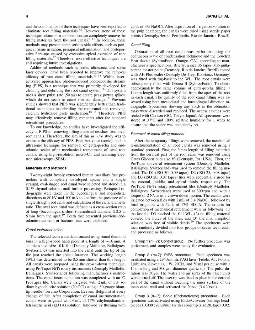
and the combination of these techniques have been reported toeliminate root filling materials.4,5 However, none of thesetechniques alone or in combination can completely remove thefilling materials from the root canals.6–11 In addition, thesemethods may present some serious side effects, such as peri-apical tissue irritation, periapical inflammation, and postoper-ative flare-ups caused by excessive apical extrusion of rootfilling materials.12 Therefore, more effective techniques arestill requiring future investigations.
Additional methods, such as sonic, ultrasonic, and somelaser devices, have been reported to improve the removalefficacy of root canal filling materials.13–15 Within laser-activated approaches, photon-induced photoacoustic stream-ing (PIPS) is a technique that was primarily developed forcleaning and debriding the root canal system.16 This systemuses a short pulse rate (50 ms) to create peak power spikes,which do not seem to cause thermal damage.16 Previousstudies showed that PIPS was significantly better than tradi-tional techniques in debriding the root canal and removingcalcium hydroxide paste medication.16–18 Therefore, PIPSmay effectively remove filling remnants after the standardretreatment procedures.
To our knowledge, no studies have investigated the effi-cacy of PIPS in removing filling material residues from ovalroot canals. Therefore, the aim of this in vitro study was toevaluate the efficacy of PIPS, EndoActivator (sonic), and anultrasonic technique for removal of gutta-percha and end-odontic sealer after mechanical retreatment of oval rootcanals, using high-resolution micro-CT and scanning elec-tron microscopy (SEM).
Materials and Methods
Twenty-eight freshly extracted human maxillary first pre-molars with completely developed apices and a singlestraight, oval-shaped root canal were selected and stored in a0.1% thymol solution until further processing. Periapical ra-diographs were taken in the buccolingual and mesiodistaldirections at 80 kV and 100 mA to confirm the presence of asingle straight root canal and calculation of the canal diameterratio. The oval root canal was defined as a cross-section ratioof long (buccolingual): short (mesiodistal) diameter ‡ 2.5 at5 mm from the apex.19 Teeth that presented previous end-odontic treatment or fracture lines were excluded.
Canal instrumentation
The selected teeth were decoronated using round diamondburs in a high-speed hand piece at a length of *16 mm. Astainless steel size 10 K-file (Dentsply Maillefer, Ballaigues,Switzerland) was inserted into the canal until the tip of thefile just reached the apical foramen. The working length(WL) was determined to be 0.5 mm shorter than this length.All canals were prepared using the crown-down technique,using ProTaper NiTi rotary instruments (Dentsply Maillefer,Ballaigues, Switzerland) following manufacturer’s instruc-tions. The canal instrumentation was completed with an F2ProTaper file. Canals were irrigated with 2 mL of 3% so-dium hypochlorite solution (NaOCl) using a 30-gauge blunt-tip needle (Terumo Corporation, Leaven, Belgium) at everychange of file. After completion of canal instrumentation,canals were irrigated with 6 mL of 17% ethylenediamine-tetraacetic acid (EDTA) solution, followed by flushing with
2 mL of 3% NaOCl. After aspiration of irrigation solution inthe pulp chamber, the canals were dried using sterile paperpoints (Dentsply/Herpo, Petropolis, Rio de Janeiro, Brazil).
Canal filling
Obturation of all root canals was performed using thecontinuous wave of condensation technique and the Touch’nHeat device (SybronEndo, Orange, CA), according to man-ufacturer’s specifications. Briefly, a size 35 taper 0.06 gutta-percha master point (Dentsply, Rio de Janeiro, Brazil) coatedwith AH Plus sealer (Dentsply De Trey, Konstanz, Germany)was fitted with tug-back to the WL. The root canals weresubsequently filled with Obtura II (SybronEndo). To obtainapproximately the same volume of gutta-percha filling, a14 mm length was uniformly filled from the apex of the rootin each canal. The quality of the root canal filling was as-sessed using both mesiodistal and buccolingual direction ra-diographs. Specimens showing any voids in the obturationmass were discarded and replaced. The access cavities weresealed with Caviton (GC, Tokyo, Japan). All specimens werestored at 37�C and 100% relative humidity for 1 week toensure that the sealer was completely set.
Removal of canal filling material
After the temporary fillings were removed, the mechanicalre-instrumentation of all root canals was removed using astandard protocol. First, the 3 mm length of filling materialsfrom the cervical part of the root canal was removed usingGates Glidden burs size #3 (Dentsply, PA, USA). Then, theProTaper universal retreatment system (Dentsply Maillefer,Ballaigues, Switzerland) was used to remove the filling ma-terial. The D1 (ISO 30, 0.09 taper), D2 (ISO 25, 0.08 taper)and D3 (ISO 20, 0.07 taper) files were sequentially used forthe coronal, middle, and apical thirds, respectively. TheProTaper Ni-Ti rotary retreatment files (Dentsply Maillefer,Ballaigues, Switzerland) were used at 300 rpm and with atorque of 2 N/cm in a crown-down motion. The canals wereirrigated between files with 2 mL of 3% NaOCl, followed byfinal irrigation with 5 mL of 17% EDTA. The criteria forcompletion of mechanical retreatment were as following: (1)the last file D3 reached the full WL, (2) no filling materialcovered the flutes of the files, and (3) the final irrigationsolution was free of visible debris.20 The specimens werethen randomly divided into four groups of seven teeth each,and processed as follows.
Group 1 (n = 7): Control group. No further procedure wasperformed, and samples were ready for evaluation.
Group 2 (n = 7): PIPS procedure. Each specimen wasirradiated using a 2940 nm Er.YAG laser (Fidelis AT, Fotona,Ljubljana, Slovenia), 1 W, 20 Hz, and 50 mJ per pulse with a14 mm long and 300 lm diameter quartz tip. The pulse du-ration was 50 ls. The water and air spray of the laser unitswere turned off. The laser tip was fixed in place in the coronalpart of the canal without touching the inner surface of themain canal wall and activated for 20 sec (3 · 20 sec).
Group 3 (n = 7): Sonic (EndoActivator) procedure. Eachspecimen was activated using EndoActivator (setting: head-pieces 10,000 cycles/min) with a sonic tip (size 20, taper 0.02)
4 JIANG ET AL.
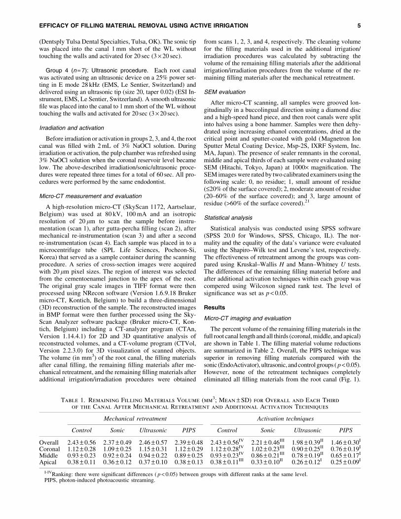
(Dentsply Tulsa Dental Specialties, Tulsa, OK). The sonic tipwas placed into the canal 1 mm short of the WL withouttouching the walls and activated for 20 sec (3 · 20 sec).
Group 4 (n = 7): Ultrasonic procedure. Each root canalwas activated using an ultrasonic device on a 25% power set-ting in E mode 28 kHz (EMS, Le Sentier, Switzerland) anddelivered using an ultrasonic tip (size 20, taper 0.02) (ESI In-strument, EMS, Le Sentier, Switzerland). A smooth ultrasonicfile was placed into the canal to 1 mm short of the WL withouttouching the walls and activated for 20 sec (3 · 20 sec).
Irradiation and activation
Before irradiation or activation in groups 2, 3, and 4, the rootcanal was filled with 2 mL of 3% NaOCl solution. Duringirradiation or activation, the pulp chamber was refreshed using3% NaOCl solution when the coronal reservoir level becamelow. The above-described irradiation/sonic/ultrasonic proce-dures were repeated three times for a total of 60 sec. All pro-cedures were performed by the same endodontist.
Micro-CT measurement and evaluation
A high-resolution micro-CT (SkyScan 1172, Aartselaar,Belgium) was used at 80 kV, 100 mA and an isotropicresolution of 20 lm to scan the sample before instru-mentation (scan 1), after gutta-percha filling (scan 2), aftermechanical re-instrumentation (scan 3) and after a secondre-instrumentation (scan 4). Each sample was placed in to amicrocentrifuge tube (SPL Life Sciences, Pocheon-Si,Korea) that served as a sample container during the scanningprocedure. A series of cross-section images were acquiredwith 20 lm pixel sizes. The region of interest was selectedfrom the cementoenamel junction to the apex of the root.The original gray scale images in TIFF format were thenprocessed using NRecon software (Version 1.6.9.18 Brukermicro-CT, Kontich, Belgium) to build a three-dimensional(3D) reconstruction of the sample. The reconstructed imagesin BMP format were then further processed using the Sky-Scan Analyzer software package (Bruker micro-CT, Kon-tich, Belgium) including a CT-analyzer program (CTAn,Version 1.14.4.1) for 2D and 3D quantitative analysis ofreconstructed volumes, and a CT-volume program (CTVol,Version 2.2.3.0) for 3D visualization of scanned objects.The volume (in mm3) of the root canal, the filling materialsafter canal filling, the remaining filling materials after me-chanical retreatment, and the remaining filling materials afteradditional irrigation/irradiation procedures were obtained
from scans 1, 2, 3, and 4, respectively. The cleaning volumefor the filling materials used in the additional irrigation/irradiation procedures was calculated by subtracting thevolume of the remaining filling materials after the additionalirrigation/irradiation procedures from the volume of the re-maining filling materials after the mechanical retreatment.
SEM evaluation
After micro-CT scanning, all samples were grooved lon-gitudinally in a buccolingual direction using a diamond discand a high-speed hand piece, and then root canals were splitinto halves using a bone hammer. Samples were then dehy-drated using increasing ethanol concentrations, dried at thecritical point and sputter-coated with gold (Magnetron IonSputter Metal Coating Device, Msp-2S, IXRF System, Inc.MA, Japan). The presence of sealer remnants in the coronal,middle and apical thirds of each sample were evaluated usingSEM (Hitachi, Tokyo, Japan) at 1000· magnification. TheSEM images were rated by two calibrated examiners using thefollowing scale: 0, no residue; 1, small amount of residue(£20% of the surface covered); 2, moderate amount of residue(20–60% of the surface covered); and 3, large amount ofresidue (>60% of the surface covered).21
Statistical analysis
Statistical analysis was conducted using SPSS software(SPSS 20.0 for Windows, SPSS, Chicago, IL). The nor-mality and the equality of the data’s variance were evaluatedusing the Shapiro–Wilk test and Levene’s test, respectively.The effectiveness of retreatment among the groups was com-pared using Kruskal–Wallis H and Mann–Whitney U tests.The differences of the remaining filling material before andafter additional activation techniques within each group wascompered using Wilcoxon signed rank test. The level ofsignificance was set as p < 0.05.
Results
Micro-CT imaging and evaluation
The percent volume of the remaining filling materials in thefull root canal length and all thirds (coronal, middle, and apical)are shown in Table 1. The filling material volume reductionsare summarized in Table 2. Overall, the PIPS technique wassuperior in removing filling materials compared with thesonic (EndoActivator), ultrasonic, and control groups ( p < 0.05).However, none of the retreatment techniques completelyeliminated all filling materials from the root canal (Fig. 1).
Table 1. Remaining Filling Materials Volume (mm3; Mean – SD) for Overall and Each Third
of the Canal After Mechanical Retreatment and Additional Activation Techniques
Mechanical retreatment Activation techniques
Control Sonic Ultrasonic PIPS Control Sonic Ultrasonic PIPS
Overall 2.43 – 0.56 2.37 – 0.49 2.46 – 0.57 2.39 – 0.48 2.43 – 0.56IV 2.21 – 0.46III 1.98 – 0.39II 1.46 – 0.30I
Coronal 1.12 – 0.28 1.09 – 0.25 1.15 – 0.31 1.12 – 0.29 1.12 – 0.28IV 1.02 – 0.23III 0.90 – 0.25II 0.76 – 0.19I
Middle 0.93 – 0.23 0.92 – 0.24 0.94 – 0.22 0.89 – 0.25 0.93 – 0.23IV 0.86 – 0.21III 0.78 – 0.19II 0.65 – 0.17I
Apical 0.38 – 0.11 0.36 – 0.12 0.37 – 0.10 0.38 – 0.13 0.38 – 0.11III 0.33 – 0.10II 0.26 – 0.12I 0.25 – 0.09I
I-IVRanking: there were significant differences ( p < 0.05) between groups with different ranks at the same level.PIPS, photon-induced photoacoustic streaming.
EFFICACY OF FILLING MATERIAL REMOVAL USING ACTIVE IRRIGATION 5
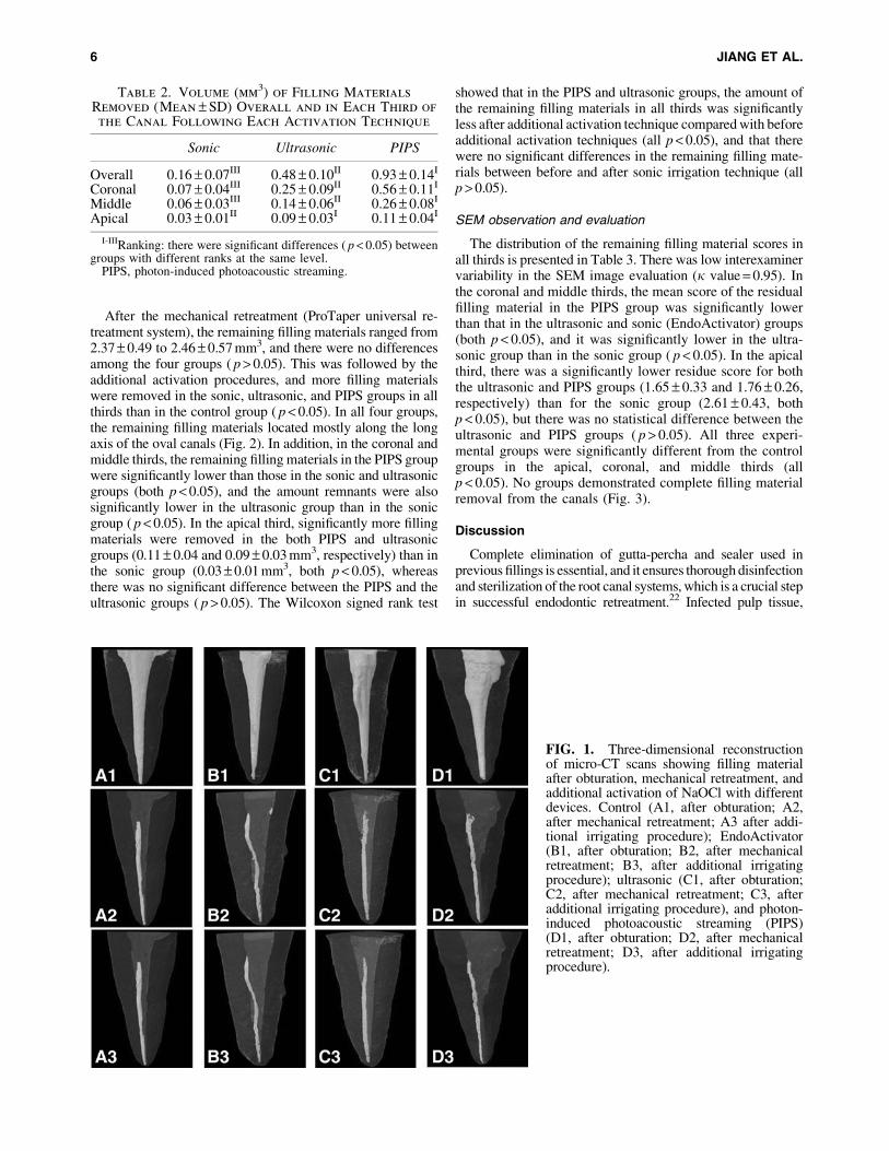
After the mechanical retreatment (ProTaper universal re-treatment system), the remaining filling materials ranged from2.37 – 0.49 to 2.46 – 0.57 mm3, and there were no differencesamong the four groups ( p > 0.05). This was followed by theadditional activation procedures, and more filling materialswere removed in the sonic, ultrasonic, and PIPS groups in allthirds than in the control group ( p < 0.05). In all four groups,the remaining filling materials located mostly along the longaxis of the oval canals (Fig. 2). In addition, in the coronal andmiddle thirds, the remaining filling materials in the PIPS groupwere significantly lower than those in the sonic and ultrasonicgroups (both p < 0.05), and the amount remnants were alsosignificantly lower in the ultrasonic group than in the sonicgroup ( p < 0.05). In the apical third, significantly more fillingmaterials were removed in the both PIPS and ultrasonicgroups (0.11 – 0.04 and 0.09 – 0.03 mm3, respectively) than inthe sonic group (0.03 – 0.01 mm3, both p < 0.05), whereasthere was no significant difference between the PIPS and theultrasonic groups ( p > 0.05). The Wilcoxon signed rank test
showed that in the PIPS and ultrasonic groups, the amount ofthe remaining filling materials in all thirds was significantlyless after additional activation technique compared with beforeadditional activation techniques (all p < 0.05), and that therewere no significant differences in the remaining filling mate-rials between before and after sonic irrigation technique (allp > 0.05).
SEM observation and evaluation
The distribution of the remaining filling material scores inall thirds is presented in Table 3. There was low interexaminervariability in the SEM image evaluation (j value = 0.95). Inthe coronal and middle thirds, the mean score of the residualfilling material in the PIPS group was significantly lowerthan that in the ultrasonic and sonic (EndoActivator) groups(both p < 0.05), and it was significantly lower in the ultra-sonic group than in the sonic group ( p < 0.05). In the apicalthird, there was a significantly lower residue score for boththe ultrasonic and PIPS groups (1.65 – 0.33 and 1.76 – 0.26,respectively) than for the sonic group (2.61 – 0.43, bothp < 0.05), but there was no statistical difference between theultrasonic and PIPS groups ( p > 0.05). All three experi-mental groups were significantly different from the controlgroups in the apical, coronal, and middle thirds (allp < 0.05). No groups demonstrated complete filling materialremoval from the canals (Fig. 3).
Discussion
Complete elimination of gutta-percha and sealer used inprevious fillings is essential, and it ensures thorough disinfectionand sterilization of the root canal systems, which is a crucial stepin successful endodontic retreatment.22 Infected pulp tissue,
Table 2. Volume (mm3) of Filling Materials
Removed (Mean – SD) Overall and in Each Third of
the Canal Following Each Activation Technique
Sonic Ultrasonic PIPS
Overall 0.16 – 0.07III 0.48 – 0.10II 0.93 – 0.14I
Coronal 0.07 – 0.04III 0.25 – 0.09II 0.56 – 0.11I
Middle 0.06 – 0.03III 0.14 – 0.06II 0.26 – 0.08I
Apical 0.03 – 0.01II 0.09 – 0.03I 0.11 – 0.04I
I-IIIRanking: there were significant differences ( p < 0.05) betweengroups with different ranks at the same level.
PIPS, photon-induced photoacoustic streaming.
FIG. 1. Three-dimensional reconstructionof micro-CT scans showing filling materialafter obturation, mechanical retreatment, andadditional activation of NaOCl with differentdevices. Control (A1, after obturation; A2,after mechanical retreatment; A3 after addi-tional irrigating procedure); EndoActivator(B1, after obturation; B2, after mechanicalretreatment; B3, after additional irrigatingprocedure); ultrasonic (C1, after obturation;C2, after mechanical retreatment; C3, afteradditional irrigating procedure), and photon-induced photoacoustic streaming (PIPS)(D1, after obturation; D2, after mechanicalretreatment; D3, after additional irrigatingprocedure).
6 JIANG ET AL.
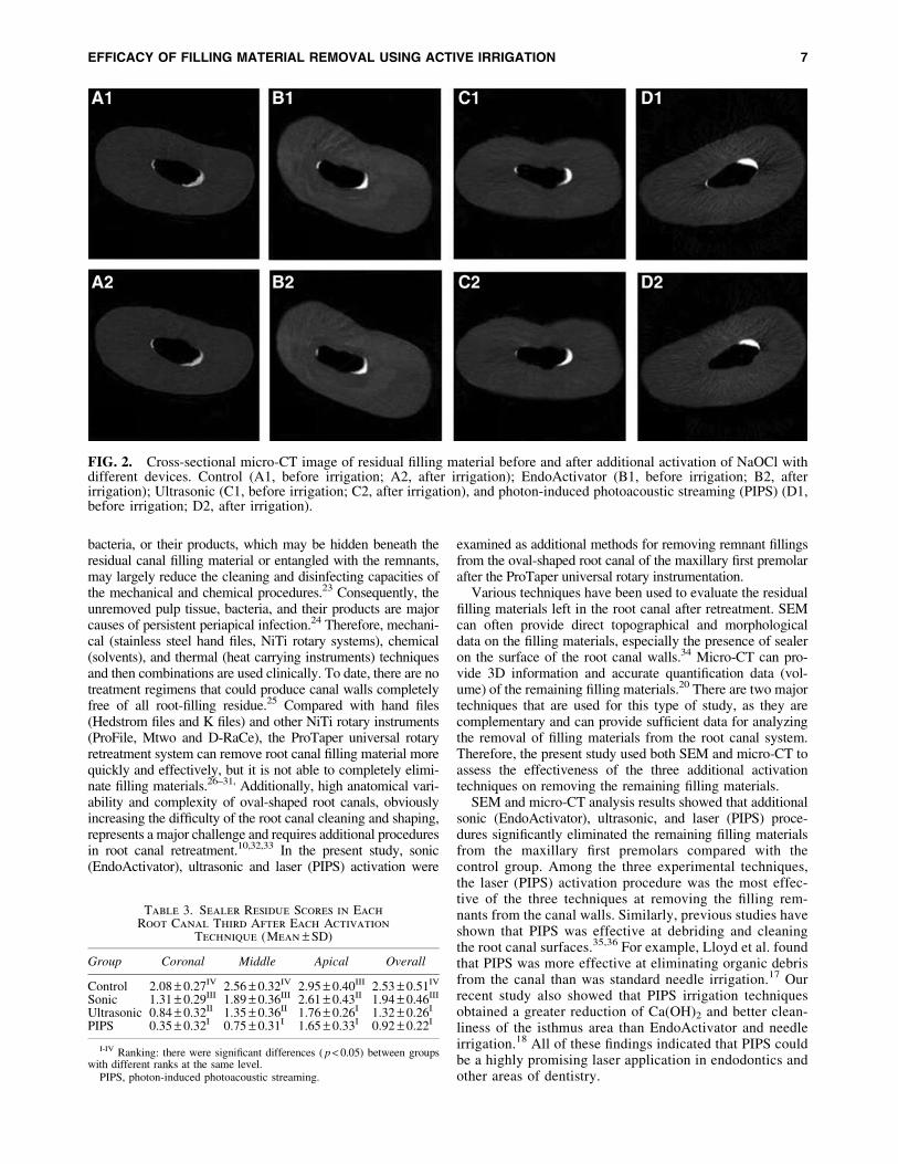
bacteria, or their products, which may be hidden beneath theresidual canal filling material or entangled with the remnants,may largely reduce the cleaning and disinfecting capacities ofthe mechanical and chemical procedures.23 Consequently, theunremoved pulp tissue, bacteria, and their products are majorcauses of persistent periapical infection.24 Therefore, mechani-cal (stainless steel hand files, NiTi rotary systems), chemical(solvents), and thermal (heat carrying instruments) techniquesand then combinations are used clinically. To date, there are notreatment regimens that could produce canal walls completelyfree of all root-filling residue.25 Compared with hand files(Hedstrom files and K files) and other NiTi rotary instruments(ProFile, Mtwo and D-RaCe), the ProTaper universal rotaryretreatment system can remove root canal filling material morequickly and effectively, but it is not able to completely elimi-nate filling materials.26–31, Additionally, high anatomical vari-ability and complexity of oval-shaped root canals, obviouslyincreasing the difficulty of the root canal cleaning and shaping,represents a major challenge and requires additional proceduresin root canal retreatment.10,32,33 In the present study, sonic(EndoActivator), ultrasonic and laser (PIPS) activation were
examined as additional methods for removing remnant fillingsfrom the oval-shaped root canal of the maxillary first premolarafter the ProTaper universal rotary instrumentation.
Various techniques have been used to evaluate the residualfilling materials left in the root canal after retreatment. SEMcan often provide direct topographical and morphologicaldata on the filling materials, especially the presence of sealeron the surface of the root canal walls.34 Micro-CT can pro-vide 3D information and accurate quantification data (vol-ume) of the remaining filling materials.20 There are two majortechniques that are used for this type of study, as they arecomplementary and can provide sufficient data for analyzingthe removal of filling materials from the root canal system.Therefore, the present study used both SEM and micro-CT toassess the effectiveness of the three additional activationtechniques on removing the remaining filling materials.
SEM and micro-CT analysis results showed that additionalsonic (EndoActivator), ultrasonic, and laser (PIPS) proce-dures significantly eliminated the remaining filling materialsfrom the maxillary first premolars compared with thecontrol group. Among the three experimental techniques,the laser (PIPS) activation procedure was the most effec-tive of the three techniques at removing the filling rem-nants from the canal walls. Similarly, previous studies haveshown that PIPS was effective at debriding and cleaningthe root canal surfaces.35,36 For example, Lloyd et al. foundthat PIPS was more effective at eliminating organic debrisfrom the canal than was standard needle irrigation.17 Ourrecent study also showed that PIPS irrigation techniquesobtained a greater reduction of Ca(OH)2 and better clean-liness of the isthmus area than EndoActivator and needleirrigation.18 All of these findings indicated that PIPS couldbe a highly promising laser application in endodontics andother areas of dentistry.
FIG. 2. Cross-sectional micro-CT image of residual filling material before and after additional activation of NaOCl withdifferent devices. Control (A1, before irrigation; A2, after irrigation); EndoActivator (B1, before irrigation; B2, afterirrigation); Ultrasonic (C1, before irrigation; C2, after irrigation), and photon-induced photoacoustic streaming (PIPS) (D1,before irrigation; D2, after irrigation).
Table 3. Sealer Residue Scores in Each
Root Canal Third After Each Activation
Technique (Mean – SD)
Group Coronal Middle Apical Overall
Control 2.08 – 0.27IV 2.56 – 0.32IV 2.95 – 0.40III 2.53 – 0.51IV
Sonic 1.31 – 0.29III 1.89 – 0.36III 2.61 – 0.43II 1.94 – 0.46III
Ultrasonic 0.84 – 0.32II 1.35 – 0.36II 1.76 – 0.26I 1.32 – 0.26I
PIPS 0.35 – 0.32I 0.75 – 0.31I 1.65 – 0.33I 0.92 – 0.22I
I-IV Ranking: there were significant differences ( p < 0.05) between groupswith different ranks at the same level.
PIPS, photon-induced photoacoustic streaming.
EFFICACY OF FILLING MATERIAL REMOVAL USING ACTIVE IRRIGATION 7
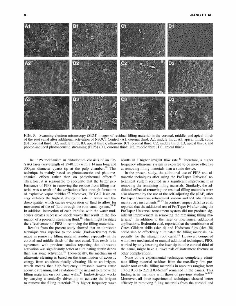
The PIPS mechanism in endodontics consists of an Er:-YAG laser (wavelength of 2940 nm) with a 14 mm long and300 lm diameter quartz tip at the pulp chamber.16 Thistechnique is mainly based on photoacoustic and photome-chanical effects rather than on photothermal effects.37
Therefore, it is reasonable to speculate that the better per-formance of PIPS in removing the residue from filling ma-terial was a result of the cavitation effect through formationof explosive vapor bubbles.36 Moreover, Er:YAG laser en-ergy exhibits the highest absorption rate in water and hy-droxyapatite, which causes evaporation of fluid to allow formovement of the of fluid through the root canal system.38,39
In addition, interaction of each impulse with the water mol-ecules creates successive shock waves that result in the for-mation of a powerful streaming fluid,16 which might facilitatethe effectiveness of PIPS in removing the filling materials.
Results from the present study showed that an ultrasonictechnique was superior to the sonic (EndoActivator) tech-nique in removing filling material residue, especially in thecoronal and middle thirds of the root canal. This result is inagreement with previous studies reporting that ultrasonicactivation was significantly better at eliminating dentin debristhan was sonic activation.40 Theoretically, the mechanism ofultrasonic cleaning is based on the transmission of acousticenergy from an ultrasonically vibrating file to an irrigant,which means that high-frequency ultrasonic waves causeacoustic streaming and cavitation of the irrigant to remove thefilling materials on root canal walls.41 EndoActivator worksby carrying a sonically driven tip to activate the irrigantto remove the filling materials.42 A higher frequency wave
results in a higher irrigant flow rate.43 Therefore, a higherfrequency ultrasonic system is expected to be more effectiveat removing filling materials than a sonic device.
In the present study, the additional use of PIPS and ul-trasonic techniques after using the ProTaper Universal re-treatment system resulted in a significant improvement inremoving the remaining filling materials. Similarly, the ad-ditional effect of removing the residual filling materials werealso observed by the use of the self-adjusting file (SAF) afterProTaper Universal retreatment system and R-Endo retreat-ment rotary instruments.44,45 In contrast, arques da Silva et al.reported that the additional use of ProTaper F4 after using theProTaper Universal retreatment system did not produce sig-nificant improvement in removing the remaining filling ma-terials.31 In addition to the laser or mechanical additionalapplications, Bodrumlu et al. reported that the combination ofGates Glidden drills (size 4) and Hedstrom files (size 30)could also be effectively eliminated the filling materials, es-pecially for the straight root canal.46 However, comparedwith these mechanical or manual additional techniques, PIPS,worked by only inserting the laser tip into the coronal third ofthe canal, might have a lower risk of instrument fracture orother complications.
None of the experimental techniques completely elimi-nate filling material residues from the maxillary first pre-molar root canals; filling materials in amounts ranging from1.46 – 0.30 to 2.21 – 0.46 mm3 remained in the canals. Thisfinding is in harmony with those of previous studies.6–8,30
Moreover, all three experimental techniques showed betterefficacy in removing filling materials from the coronal and
FIG. 3. Scanning electron microscopy (SEM) images of residual filling material in the coronal, middle, and apical thirdsof the root canal after additional activation of NaOCl. Control (A1, coronal third; A2, middle third; A3, apical third); sonic(B1, coronal third; B2, middle third; B3, apical third); ultrasonic (C1, coronal third; C2, middle third; C3, apical third), andphoton-induced photoacoustic streaming (PIPS) (D1, coronal third; D2, middle third; D3, apical third).
8 JIANG ET AL.
middle thirds compared with the apical third of the canal.Conversely, Abramovitz et al. reported that a self-adjustingfile was more effective in removing residual gutta-perchafrom the apical section than the coronal and middle sectionsof the mesial canals of mandibular molar after using Pro-Taper Universal retreatment files.30 This is likely because ofthe different techniques applied. When the PIPS tip wasplaced only in the coronal part, its photoacoustic shockwave may weaken over the distance to the apical one third,and, therefore, the effectiveness of PIPS in removing fillingmaterials from the apical part was decreased. Similarly, Zhuet al. found that PIPS was more effective in removing thesmear layer and debris in the coronal and middle thirds thanin the apical third.47 It is expected that ultrasonic and sonictechniques will be less effective in removing filling materialin the apical region, and this was likely a result of the re-duction in the acoustic microstreaming and/or cavitationeffect after the ultrasonic file or sonic tips entered the apicalvapor lock.41,42 As mentioned, the optic tip of the PIPSdevice was only placed in the coronal third of the canal,which was likely to preserve the root structure.16 To suffi-ciently remove the filling materials in the apical third of theroot canal, further studies shoud be conducted to optimizePIPS parameters and the PIPS tip location in the root canal.
Limitations
Limitations of the present study should be noted. First, thisstudy was conducted on teeth with straight oval root canals,and the findings cannot be directly applied to teeth withcurved root canal systems, because root curvature is a crucialfactor in affecting the efficacy of root canal instrumentation.9
Second, similar to other similar studies,24,31,35 decoronatingteeth, which is impossible in the daily practice, were done inthe present study in order to standardize the specimens;therefore, the conclusion of the current study cannot be di-rectly extended to clinical conditions. Further research isneeded to complement the results of the present study.
Conclusions
The additional use of PIPS for the activation of NaOCl wassuperior to sonic and ultrasonic techniques in removing theremaining filling materials after standard retreatment proce-dures using the ProTaper universal retreatment system. How-ever, none of the experimental techniques completely removedthe filling remnants from the root canal of the maxillary firstpremolars.
Author Disclosure Statement
The authors declare no competing financial interests.
References
1. Stabholz A, Friedman S. Endodontic retreatment-case se-lection and technique. Part 2: treatment planning for re-treatment. J Endod 1988;14:607–614.
2. Hulsmann M, Bluhm V. Efficacy, cleaning ability andsafety of different rotary NiTi instruments in root canalretreatment. Int Endod J 2004;37:468–476.
3. Bueno CE, Delboni MG, deAraujo RA, Carrara HJ, CunhaRS. Effectiveness of rotary and hand files in gutta-percha
and sealer removal using chloroform or chlorhexidine gel.Braz Dent J 2006;17:139–143.
4. Bodrumlu E, Uzun O, Topuz O, Semiz M. Efficacy of 3techniques in removing root canal filling material. J CanDent Assoc 2008;74:72.
5. Good ML, McCammon A. An removal of gutta-percha androot canal sealer: a literature review and an audit comparingcurrent practice in dental schools. Dent Update 2012;39:703–708.
6. Somma F, Cammarota G, Plotino G, Grande NM, PameijerCH. The effectiveness of manual and mechanical instru-mentation for the retreatment of three different root canalfilling materials. J Endod 2008;34:466–469.
7. Bramante CM, Fidelis NS, Assumpcao TS, et al. Heat re-lease, time required, and cleaning ability of MTwo R andProTaper Universal retreatment systems in the removal offilling material. J Endod 2010;36:1870–1873.
8. Rios MA, Villela AM, Cunha RS, et al. Efficacy of 2 re-ciprocating systems compared with a rotary retreatmentsystem for gutta-percha removal. J Endod 2014;40:543–546.
9. Khalilak Z, Vatanpour M, Dadresanfar B, Moshkelgosha P,Nourbakhsh H. In vitro comparison of gutta-percha re-moval with H-file and ProTaper with or without chloro-form. Iran Endod J 2013;8:6–9.
10. Ma J, Al-Ashaw AJ, Shen Y, et al. Efficacy of ProTaperUniversal Rotary Retreatment system for gutta-percha re-moval from oval root canals: a micro-computed tomogra-phy study. J Endod 2012;38:1516–1520.
11. Hiltner RS, Kulild JC, Weller RN. Effect of mechanicalversus thermal removal of gutta-percha on the quality of theapical seal following post space preparation. J Endod 1992;18:451–454.
12. Seltzer S, Naidorf IJ. Flare-ups in endodontics: I. Etiolo-gical factors. J Endod 1985;11:472–478.
13. Cavenago BC, Ordinola–Zapata R, Duarte MA, et al. Effi-cacy of xylene and passive ultrasonic irrigation on remainingroot filling material during retreatment of anatomicallycomplex teeth. Int Endod J 2014;47:1078–1083.
14. Tachinami H, Katsuumi I. Removal of root canal fillingmaterials using Er:YAG laser irradiation. Dent Mater J2010;29:246–252.
15. Anjo T, Ebihara A, Takeda A, Takashina M, Sunakawa M,Suda H. Removal of two types of root canal filling materialusing pulsed Nd:YAG laser irradiation. Photomed LaserSurg 2004;22:470–476.
16. DiVito E, Peters OA, Olivi G. Effectiveness of the erbi-um:YAG laser and new design radial and stripped tips inremoving the smear layer after root canal instrumentation.Lasers Med Sci 2012;27:273–280.
17. Lloyd A, Uhles JP, Clement DJ, Garcia–Godoy F. Eliminationof intracanal tissue and debris through a novel laser-activatedsystem assessed using high-resolution micro-computed to-mography: a pilot study. Int Endod J 2014;40:584–587.
18. Li D, Jiang S, Yin X, Chang JW, Ke J, Zhang C. Efficacyof needle, ultrasonic, and EndoActivator irrigation andphoton-induced photoacoustic streaming in removing cal-cium hydroxide from the main canal and isthmus: anin vitro micro-computed tomography and scanning electronmicroscopy study. Photomed Laser Surg 2015;33:330–337.
19. De–Deus G, Reis C, Beznos D. Limited ability of threecommonly used thermoplasticized gutta-percha techniquesin filling. J Endod 2008;34:1401–1405.
20. Asheibi F, Qualtrough AJ, Mellor A, Withers PJ, Lowe T.Micro-CT evaluation of the effectiveness of the combined
EFFICACY OF FILLING MATERIAL REMOVAL USING ACTIVE IRRIGATION 9
use of rotary and hand instrumentation in removal of Re-silon. Dent Mater J 2014;33:1–6.
21. Kuga MC, Tanomaru–Filho M, Faria G, So MV, Galletti T,Bavello JR. Calcium hydroxide intracanal dressing removalwith different rotary instruments and irrigating solutions: ascanning electron microscopy study. Braz Dent J 2010;21:310–314.
22. Gordon MP. The removal of gutta-percha and root canalsealers from root canals. N Z Dent J 2005;101:44–52.
23. Fabricius L, Dahlen G, Sundqvist G, Happonen RP, MollerAJ. Influence of residual bacteria on periapical tissuehealing after chemomechanical treatment and root filling ofexperimentally infected monkey teeth. Eur J Oral Sci2006;114:278–285.
24. Silva EJ, Orlowsky NB, Herrera DR, Machado R, KrebsRL, Coutinho–Filho Tde S. Effectiveness of rotatory andreciprocating movements in root canal filling material re-moval. Braz Oral Res 2015;29:1–6.
25. Pirani C, Pelliccioni GA, Marchionni S, Montebugnoli L,Piana G, Prati C. Effectiveness of three different retreat-ment techniques in canals filled with compacted gutta-percha or Thermafil: a scanning electron microscope study.J Endod 2009;35:1433–1440.
26. Kumar MSR, Sajjan GS, Satish K, Varma KM. A com-parative evaluation of efficacy of protaper universal rotaryretreatment system for gutta-percha removal with or with-out a solvent. Contemp Clin Dent 2012;3:S160–S163.
27. Gu LS, Ling JQ, Wei X, Huang XY. Efficacy of ProTaperUniversal rotary retreatment system for gutta-percha re-moval from root canals. Int Endod J 2008;41:288–295.
28. Shivanand S, Patil CR, Thangala V, Kumar PR, SachdevaJ, Krishna AJ. To evaluate and compare the efficacy,cleaning ability of hand and two rotary systems in rootcanal retreatment. J Contemp Dent Pract 2013;14:440–444;PMID: 24171986.
29. Kumar MS, Sajjan GS, Satish K, Varma KM. A compar-ative evaluation of efficacy of protaper universal rotaryretreatment system for gutta-percha removal with or with-out a solvent. Contemp Clin Dent 2012;3:S160–S163;PMID: 23230353.
30. Abramovitz I, Relles–Bonar S, Baransi B, Kfir A. The ef-fectiveness of a self-adjusting file to remove residual gutta-percha after retreatment with rotary files. Int Endod J2012;45:386–392.
31. Marques da Silva B, Baratto–Filho F, Leonardi DP, Hen-rique Borges A, Volpato L, Branco Barletta F. Effective-ness of ProTaper, D-RaCe, and Mtwo retreatment files withand without supplementary instruments in the removal ofroot canal filling material. Int Endod J 2012;45:927–932.
32. Zmener O, Pameijer CH, Banegas G. Retreatment efficacy ofhand versus automated instrumentation in oval-shaped rootcanals: an ex vivo study. Int Endod J 2006;39:521–526.
33. Rechenberg DK, Paque F. Impact of cross-sectional rootcanal shape on filled canal volume and remaining root fillingmaterial after retreatment. Int Endod J 2013;46:547–555.
34. Upadhyay V, Upadhyay M, Panday RK, Chturvedi TP,Bajpai U. A SEM evaluation of dentinal adaptation of rootcanal obturation with GuttaFlow and conventional obtur-ating material. Indian J Dent Res 2011;22:881.
35. Arslan H, Akcay M, Capar ID, Saygili G, Gok T, Ertas H. Anin vitro comparison of irrigation using photon-initiatedphotoacoustic streaming, ultrasonic, sonic and needle tech-niques in removing calcium hydroxide. Int Endod J2015;48:246–251.
36. Arslan H, Capar I, Saygili G, Gok T, Akcay M. Effect ofphoton-initiated photoacoustic streaming on removal of api-cally placed dentinal debris. Int Endod J 2014;47:1072–1077.
37. Peters OA, Bardsley S, Fong J, Pandher G, Divito E. Dis-infection of root canals with photon-initiated photoacousticstreaming. J Endod 2011;37:1008–1012.
38. Hoke JA, Burkes EJ Jr, Gomes ED, Wolbarsht ML. Er-bium:YAG (2.94 mum) laser effects on dental tissues. JLaser Appl 1990;2:61–65.
39. Armengol V, Jean A, Rohanizadeh R, Hamel H. Scanningelectron microscopic analysis of diseased and healthydental hard tissues after Er:YAG laser irradiation: in vitrostudy. J Endod 1999;25:543–546.
40. Rodig T, Bozkurt M, Konietschke F, Hulsmann M. Com-parison of the Vibringe system with syringe and passiveultrasonic irrigation in removing debris from simulated rootcanal irregularities. J Endod 2010;36:1410–1413.
41. Yoo YJ, Lee W, Kim HC, Shon WJ, Baek SH. Multivariateanalysis of the cleaning efficacy of different final irrigationtechniques in the canal and isthmus of mandibular posteriorteeth. Restor Dent Endod 2013;38:154–159.
42. Jiang LM, Verhaagen B, Versluis M, van der Sluis LW.Evaluation of a sonic device designed to activate irrigant inthe root canal. J Endod 2010;36:143–146.
43. _Iriboz E, Bayraktar K, Turkaydın D, Tarcın B. Comparisonof apical extrusion of sodium hypochlorite using 4 differentroot canal irrigation techniques. J Endod 2015;41:380–384.
44. Voet KC, Wu MK, Wesselink PR, Shemesh H. Removal ofgutta-percha from root canals using the self-adjusting file. JEndod 2012;38:1004–1006.
45. Kelesx A, Alcin H, Kamalak A, Versiani MA. Oval-shapedcanal retreatment with self-adjusting file: a micro-computedtomography study. Clin Oral Investig 2014;18:1147–1153.
46. Bodrumlu E1, Uzun O, Topuz O, Semiz M. Efficacy of 3techniques in removing root canal filling material. J CanDent Assoc 2008;74:721.
47. Zhu X, Yin X, Chang JW, Wang Y, Cheung GS, Zhang C.Comparison of the antibacterial effect and smear layer removalusing photon-initiated photoacoustic streaming aided irriga-tion versus a conventional irrigation in single-rooted canals:an in vitro study. Photomed Laser Surg 2013;31:371–377.
Address correspondence to:Chengfei Zhang
Comprehensive Dental Care, EndodonticsFaculty of Dentistry
The University of Hong KongPrince Philip Dental Hospital
34 Hospital Road, Sai Ying PunHong Kong SAR, 999077
China
E-mail: [email protected]
or
Xiaojing HuangDepartment of Endodontics and Operative Dentistry I
School and Hospital of StomatologyFujian Medical University246 Yangqiao Zhong Road
Fuzhou, Fujian 350002China
E-mail: [email protected]
10 JIANG ET AL.
SHORT COMMUNICATION
Irrigant flow during photon-induced photoacoustic streaming(PIPS) using Particle Image Velocimetry (PIV)
Jon D. Koch1& David E. Jaramillo2 & Enrico DiVito3,4 & Ove A. Peters5
Received: 13 March 2015 /Accepted: 4 August 2015 /Published online: 26 August 2015# Springer-Verlag Berlin Heidelberg 2015
AbstractObjectives This study aimed to compare fluid movementsgenerated from photon-induced photoacoustic streaming(PIPS) and passive ultrasonic irrigation (PUI).Materials and methods Particle Image Velocimetry (PIV) wasperformed using 6-μm melamine spheres in water. Measure-ment areas were 3-mm-long sections of the canal in the coro-nal, midroot and apical regions for PIPS (erbium/yttrium-alu-minium garnet (Er:YAG) laser set at 15 Hz with 20 mJ), orpassive ultrasonic irrigation (PUI, non-cutting insert at 30 %unit power) was performed in simulated root canals preparedto an apical size #30/0.04 taper. Fluid movement was analyseddirectly subjacent to the apical ends of ultrasonic insert or fiberoptic tips as well as at midroot and apically.Results During PUI,measured average velocities were around0.03 m/s in the immediate vicinity of the sides and tip of theultrasonic file. Speeds decayed to non-measureable values at adistance of about 2 mm from the sides and tip. During PIPS,typical average speeds were about ten times higher than those
measured for PUI, and they were measured throughout thelength of the canal, at distances up to 20 mm away.Conclusions PIPS caused higher average fluid speeds whencompared to PUI, both close and distant from the instrument.The findings of this study could be relevant to the debridingand disinfecting stage of endodontic therapy.Clinical relevance Irrigation enhancement beyond needle ir-rigation is relevant to more effectively eradicate microorgan-isms from root canal systems. PIPS may be an alternativeapproach due to its ability to create high streaming velocitiesfurther away from the activation source compared to ultrason-ic activation.
Keywords Particle image velocimetry . Photon-inducedphotoacoustic streaming . Ultrasonics . Endodontics .
Irrigation
Introduction
An important aim of root canal treatment is the elimination orprevention of periradicular periodontitis; it is well establishedthat bacteria and their toxins are the cause of this disease [13]and therefore eradication, or at least reduction to a biologicallyacceptable number, of intracanal microorganisms is required[23]. Enlargement of root canals with current root canal in-struments reduces bacterial counts even in the case ofbuccolingually wide root canals [24]. However, preparationdoes not eliminate all microorganisms from the root canalsystem. Therefore, antimicrobial irrigants are commonly usedand it is believed that enhancement of the flushing action iseffective in improving root canal cleanliness [2, 11]. Differentagitation techniques have been proposed to improve the effi-cacy of irrigation solutions, including agitation with hand
* Ove A. [email protected]
1 Department ofMechanical and Aerospace Engineering, Allen Schoolof Engineering, Trine University, Angola, IN, USA
2 Department of Endodontics, University of Texas Health ScienceCenter at Houston, School of Dentistry, Houston, TX, USA
3 Private Practice, Arizona Center for Laser Dentistry, Scottsdale, AZ,USA
4 Arizona School of Dentistry and Oral Health, Mesa, AZ, USA5 Department of Endodontics, University of the Pacific Arthur A.
Dugoni School of Dentistry, 155 5th St, San Francisco, CA 94103,USA
Clin Oral Invest (2016) 20:381–386DOI 10.1007/s00784-015-1562-9
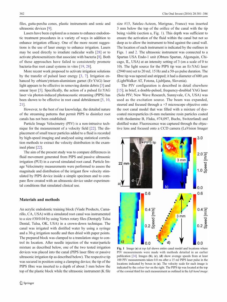
files, gutta-percha cones, plastic instruments and sonic andultrasonic devices [9].
Lasers have been explored as a means to enhance endodon-tic treatment procedures in a variety of ways in addition toenhance irrigation efficacy. One of the more recent sugges-tions is the use of laser energy to enhance irrigation. Lasersmay be used directly to irradiate radicular walls [28] or toactivate photosensitizers that associate with bacteria [8]. Bothof these approaches have failed to consistently promotebacteria-free root canal systems in vitro [19, 28].
More recent work proposed to activate irrigation solutionsby the transfer of pulsed laser energy [3, 7]. Irrigation en-hanced by erbium/yttrium-aluminium garnet (Er:YAG) laserlight appears to be effective in removing dentin debris [3] andsmear layer [5]. Specifically, the action of a pulsed Er:YAGlaser via photon-induced photoacoustic streaming (PIPS) hasbeen shown to be effective in root canal debridement [5, 10,21].
However, to the best of our knowledge, the detailed natureof the streaming patterns that permit PIPS to disinfect rootcanals has not been established.
Particle Image Velocimetry (PIV) is a non-intrusive tech-nique for the measurement of a velocity field [22]. The dis-placement of small tracer particles added to a fluid is recordedby high-speed imaging and analysed using statistical correla-tion methods to extract the velocity distribution in the exam-ined plane [22].
The aim of the present study was to compare differences influid movement generated from PIPS and passive ultrasonicirrigation (PUI) in a curved simulated root canal. Particle Im-age Velocimetry measurements were performed to assess themagnitude and distribution of the irrigant flow velocity stim-ulated by PIPS device inside a simple specimen and to com-pare flow created with an ultrasonic device under experimen-tal conditions that simulated clinical use.
Materials and methods
An acrylic endodontic training block (Viade Products, Cama-rillo, CA, USA) with a simulated root canal was instrumentedto a size #30/0.04 by using Vortex rotary files (Dentsply TulsaDental, Tulsa, OK, USA) in a crown-down technique. Thecanal was irrigated with distilled water by using a syringeand a 30-g irrigation needle and then dried with paper points.The prepared block was clamped to a translation stage to con-trol its location. After needle injection of the water/particlemixture as described below, one of the two tested irrigationdevices was placed into the canal (PIPS laser fibre or passiveultrasonic irrigation tip as described below). The respective tipwas secured in position using a clamping device; the tip of thePIPS fibre was inserted to a depth of about 3 mm below thetop of the plastic block while the ultrasonic instrument (K file
size #15, Satelec-Acteon, Merignac, France) was inserted3 mm below the top of the orifice of the canal with the tipbeing visible (section a, Fig. 1). This depth was sufficient toensure the activation of the fluid within the canal but not sodeep as to allow the instrument to bind against the canal wall.The location of each instrument is indicated by the outlines inFigs. 1 and 2. The ultrasonic instrument was connected to aSpartan USA Endo-1 unit (Obtura Spartan, Algonquin, Chi-cago, IL, USA) at an intensity setting of 3 (on a scale of 0 to10). The light source for the PIPS tip was an Er:YAG laser(2940 nm) set to 20mJ, 15Hz and a 50-μs pulse duration. Thefibre tip was tapered and stripped; it had a diameter of 600 μm(LightWalker AT, Fotona, Ljubljana, Slovenia).
The PIV configuration is described in detail elsewhere[15]; in brief, a double-pulsed, frequency-doubled YAG laser(Solo PIV; New Wave Research, Sunnyvale, CA, USA) wasused as the excitation source. The beam was expanded,steered and focused through a ×5 microscope objective ontothe root canal model that was filled with a mixture of dye-coated microparticles (6-mm melamine resin particles coatedwith rhodamine B, Fluka, #74,097, Buchs, Switzerland) anddistilled water. Fluorescence was captured through the objec-tive lens and focused onto a CCD camera (LaVision Imager
Fig. 1 Image (a) at top left shows entire canal model and locations wherePIV measurements were made with methods detailed in an earlierpublication [24]. Images (b), (c), (d) show average speeds from at least100 PIV measurements taken 0.8 ms after a 15 mJ PIPS laser pulse in thelocations indicated by boxes in (a). The velocity scale for each image isindicated by the colour bar on the right. The PIPS tip was located at the topof the coronal third for each measurement as outlined in the left hand image
382 Clin Oral Invest (2016) 20:381–386
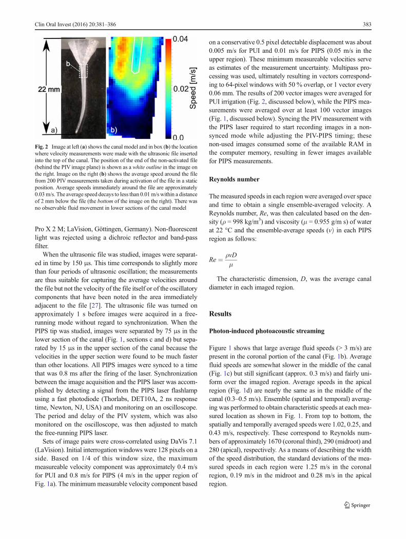
Pro X 2 M; LaVision, Göttingen, Germany). Non-fluorescentlight was rejected using a dichroic reflector and band-passfilter.
When the ultrasonic file was studied, images were separat-ed in time by 150 μs. This time corresponds to slightly morethan four periods of ultrasonic oscillation; the measurementsare thus suitable for capturing the average velocities aroundthe file but not the velocity of the file itself or of the oscillatorycomponents that have been noted in the area immediatelyadjacent to the file [27]. The ultrasonic file was turned onapproximately 1 s before images were acquired in a free-running mode without regard to synchronization. When thePIPS tip was studied, images were separated by 75 μs in thelower section of the canal (Fig. 1, sections c and d) but sepa-rated by 15 μs in the upper section of the canal because thevelocities in the upper section were found to be much fasterthan other locations. All PIPS images were synced to a timethat was 0.8 ms after the firing of the laser. Synchronizationbetween the image acquisition and the PIPS laser was accom-plished by detecting a signal from the PIPS laser flashlampusing a fast photodiode (Thorlabs, DET10A, 2 ns responsetime, Newton, NJ, USA) and monitoring on an oscilloscope.The period and delay of the PIV system, which was alsomonitored on the oscilloscope, was then adjusted to matchthe free-running PIPS laser.
Sets of image pairs were cross-correlated using DaVis 7.1(LaVision). Initial interrogation windows were 128 pixels on aside. Based on 1/4 of this window size, the maximummeasureable velocity component was approximately 0.4 m/sfor PUI and 0.8 m/s for PIPS (4 m/s in the upper region ofFig. 1a). The minimummeasurable velocity component based
on a conservative 0.5 pixel detectable displacement was about0.005 m/s for PUI and 0.01 m/s for PIPS (0.05 m/s in theupper region). These minimum measureable velocities serveas estimates of the measurement uncertainty. Multipass pro-cessing was used, ultimately resulting in vectors correspond-ing to 64-pixel windows with 50 % overlap, or 1 vector every0.06 mm. The results of 200 vector images were averaged forPUI irrigation (Fig. 2, discussed below), while the PIPS mea-surements were averaged over at least 100 vector images(Fig. 1, discussed below). Syncing the PIV measurement withthe PIPS laser required to start recording images in a non-synced mode while adjusting the PIV-PIPS timing; thesenon-used images consumed some of the available RAM inthe computer memory, resulting in fewer images availablefor PIPS measurements.
Reynolds number
Themeasured speeds in each region were averaged over spaceand time to obtain a single ensemble-averaged velocity. AReynolds number, Re, was then calculated based on the den-sity (ρ = 998 kg/m3) and viscosity (μ = 0.955 g/m s) of waterat 22 °C and the ensemble-average speeds (vÞ in each PIPSregion as follows:
Re ¼ ρvD
μ
The characteristic dimension, D, was the average canaldiameter in each imaged region.
Results
Photon-induced photoacoustic streaming
Figure 1 shows that large average fluid speeds (> 3 m/s) arepresent in the coronal portion of the canal (Fig. 1b). Averagefluid speeds are somewhat slower in the middle of the canal(Fig. 1c) but still significant (approx. 0.3 m/s) and fairly uni-form over the imaged region. Average speeds in the apicalregion (Fig. 1d) are nearly the same as in the middle of thecanal (0.3–0.5 m/s). Ensemble (spatial and temporal) averag-ing was performed to obtain characteristic speeds at each mea-sured location as shown in Fig. 1. From top to bottom, thespatially and temporally averaged speeds were 1.02, 0.25, and0.43 m/s, respectively. These correspond to Reynolds num-bers of approximately 1670 (coronal third), 290 (midroot) and280 (apical), respectively. As a means of describing the widthof the speed distribution, the standard deviations of the mea-sured speeds in each region were 1.25 m/s in the coronalregion, 0.19 m/s in the midroot and 0.28 m/s in the apicalregion.
Fig. 2 Image at left (a) shows the canal model and in box (b) the locationwhere velocity measurements were made with the ultrasonic file insertedinto the top of the canal. The position of the end of the non-activated file(behind the PIV image plane) is shown as a white outline in the image onthe right. Image on the right (b) shows the average speed around the filefrom 200 PIV measurements taken during activation of the file in a staticposition. Average speeds immediately around the file are approximately0.03m/s. The average speed decays to less than 0.01m/swithin a distanceof 2 mm below the file (the bottom of the image on the right). There wasno observable fluid movement in lower sections of the canal model
Clin Oral Invest (2016) 20:381–386 383
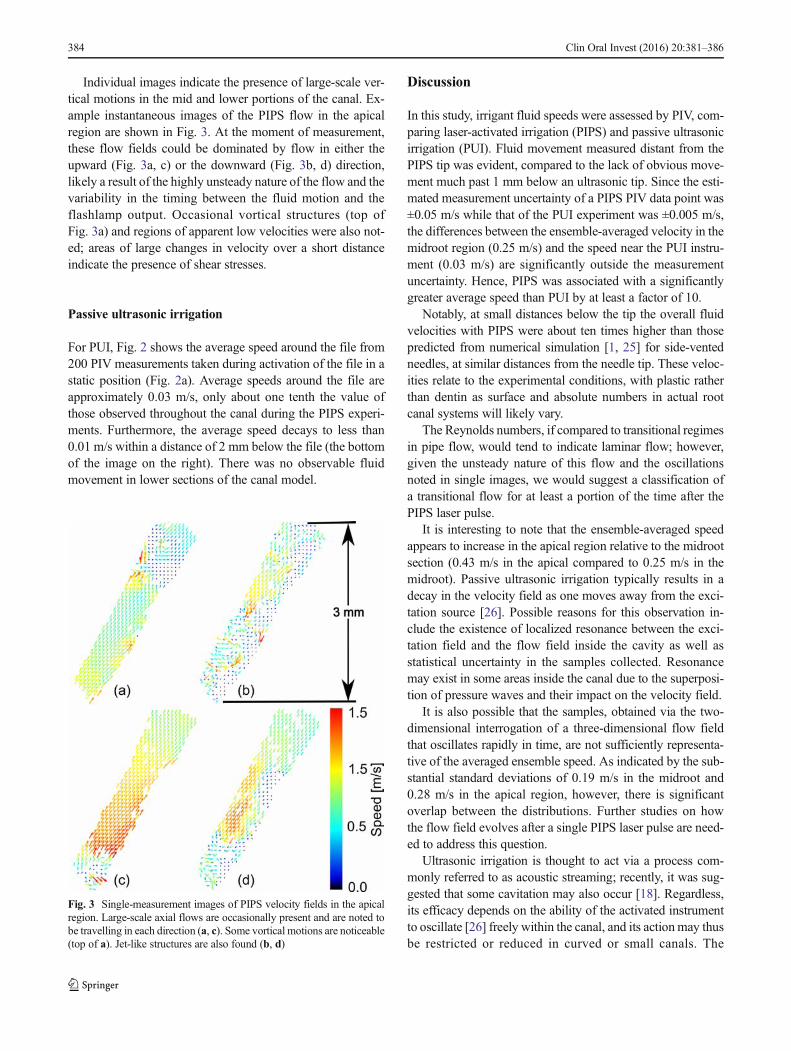
Individual images indicate the presence of large-scale ver-tical motions in the mid and lower portions of the canal. Ex-ample instantaneous images of the PIPS flow in the apicalregion are shown in Fig. 3. At the moment of measurement,these flow fields could be dominated by flow in either theupward (Fig. 3a, c) or the downward (Fig. 3b, d) direction,likely a result of the highly unsteady nature of the flow and thevariability in the timing between the fluid motion and theflashlamp output. Occasional vortical structures (top ofFig. 3a) and regions of apparent low velocities were also not-ed; areas of large changes in velocity over a short distanceindicate the presence of shear stresses.
Passive ultrasonic irrigation
For PUI, Fig. 2 shows the average speed around the file from200 PIV measurements taken during activation of the file in astatic position (Fig. 2a). Average speeds around the file areapproximately 0.03 m/s, only about one tenth the value ofthose observed throughout the canal during the PIPS experi-ments. Furthermore, the average speed decays to less than0.01 m/s within a distance of 2 mm below the file (the bottomof the image on the right). There was no observable fluidmovement in lower sections of the canal model.
Discussion
In this study, irrigant fluid speeds were assessed by PIV, com-paring laser-activated irrigation (PIPS) and passive ultrasonicirrigation (PUI). Fluid movement measured distant from thePIPS tip was evident, compared to the lack of obvious move-ment much past 1 mm below an ultrasonic tip. Since the esti-mated measurement uncertainty of a PIPS PIV data point was±0.05 m/s while that of the PUI experiment was ±0.005 m/s,the differences between the ensemble-averaged velocity in themidroot region (0.25 m/s) and the speed near the PUI instru-ment (0.03 m/s) are significantly outside the measurementuncertainty. Hence, PIPS was associated with a significantlygreater average speed than PUI by at least a factor of 10.
Notably, at small distances below the tip the overall fluidvelocities with PIPS were about ten times higher than thosepredicted from numerical simulation [1, 25] for side-ventedneedles, at similar distances from the needle tip. These veloc-ities relate to the experimental conditions, with plastic ratherthan dentin as surface and absolute numbers in actual rootcanal systems will likely vary.
The Reynolds numbers, if compared to transitional regimesin pipe flow, would tend to indicate laminar flow; however,given the unsteady nature of this flow and the oscillationsnoted in single images, we would suggest a classification ofa transitional flow for at least a portion of the time after thePIPS laser pulse.
It is interesting to note that the ensemble-averaged speedappears to increase in the apical region relative to the midrootsection (0.43 m/s in the apical compared to 0.25 m/s in themidroot). Passive ultrasonic irrigation typically results in adecay in the velocity field as one moves away from the exci-tation source [26]. Possible reasons for this observation in-clude the existence of localized resonance between the exci-tation field and the flow field inside the cavity as well asstatistical uncertainty in the samples collected. Resonancemay exist in some areas inside the canal due to the superposi-tion of pressure waves and their impact on the velocity field.
It is also possible that the samples, obtained via the two-dimensional interrogation of a three-dimensional flow fieldthat oscillates rapidly in time, are not sufficiently representa-tive of the averaged ensemble speed. As indicated by the sub-stantial standard deviations of 0.19 m/s in the midroot and0.28 m/s in the apical region, however, there is significantoverlap between the distributions. Further studies on howthe flow field evolves after a single PIPS laser pulse are need-ed to address this question.
Ultrasonic irrigation is thought to act via a process com-monly referred to as acoustic streaming; recently, it was sug-gested that some cavitation may also occur [18]. Regardless,its efficacy depends on the ability of the activated instrumentto oscillate [26] freely within the canal, and its actionmay thusbe restricted or reduced in curved or small canals. The
Fig. 3 Single-measurement images of PIPS velocity fields in the apicalregion. Large-scale axial flows are occasionally present and are noted tobe travelling in each direction (a, c). Some vortical motions are noticeable(top of a). Jet-like structures are also found (b, d)
384 Clin Oral Invest (2016) 20:381–386
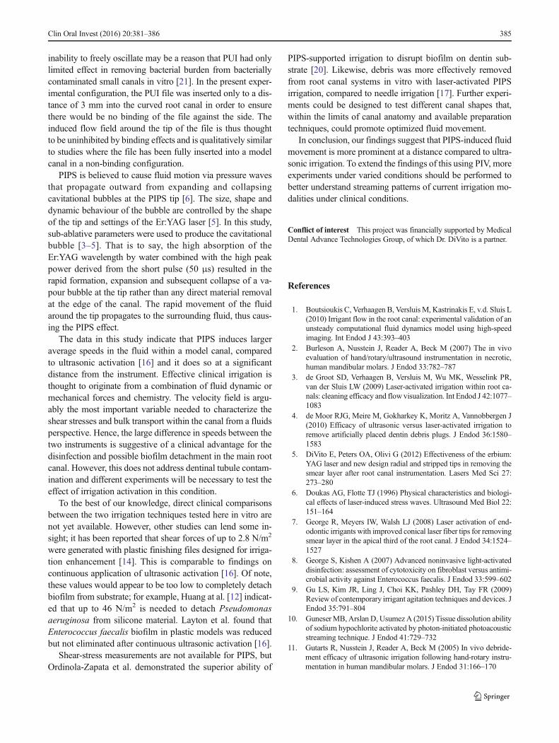
inability to freely oscillate may be a reason that PUI had onlylimited effect in removing bacterial burden from bacteriallycontaminated small canals in vitro [21]. In the present exper-imental configuration, the PUI file was inserted only to a dis-tance of 3 mm into the curved root canal in order to ensurethere would be no binding of the file against the side. Theinduced flow field around the tip of the file is thus thoughtto be uninhibited by binding effects and is qualitatively similarto studies where the file has been fully inserted into a modelcanal in a non-binding configuration.
PIPS is believed to cause fluid motion via pressure wavesthat propagate outward from expanding and collapsingcavitational bubbles at the PIPS tip [6]. The size, shape anddynamic behaviour of the bubble are controlled by the shapeof the tip and settings of the Er:YAG laser [5]. In this study,sub-ablative parameters were used to produce the cavitationalbubble [3–5]. That is to say, the high absorption of theEr:YAG wavelength by water combined with the high peakpower derived from the short pulse (50 μs) resulted in therapid formation, expansion and subsequent collapse of a va-pour bubble at the tip rather than any direct material removalat the edge of the canal. The rapid movement of the fluidaround the tip propagates to the surrounding fluid, thus caus-ing the PIPS effect.
The data in this study indicate that PIPS induces largeraverage speeds in the fluid within a model canal, comparedto ultrasonic activation [16] and it does so at a significantdistance from the instrument. Effective clinical irrigation isthought to originate from a combination of fluid dynamic ormechanical forces and chemistry. The velocity field is argu-ably the most important variable needed to characterize theshear stresses and bulk transport within the canal from a fluidsperspective. Hence, the large difference in speeds between thetwo instruments is suggestive of a clinical advantage for thedisinfection and possible biofilm detachment in the main rootcanal. However, this does not address dentinal tubule contam-ination and different experiments will be necessary to test theeffect of irrigation activation in this condition.
To the best of our knowledge, direct clinical comparisonsbetween the two irrigation techniques tested here in vitro arenot yet available. However, other studies can lend some in-sight; it has been reported that shear forces of up to 2.8 N/m2
were generated with plastic finishing files designed for irriga-tion enhancement [14]. This is comparable to findings oncontinuous application of ultrasonic activation [16]. Of note,these values would appear to be too low to completely detachbiofilm from substrate; for example, Huang at al. [12] indicat-ed that up to 46 N/m2 is needed to detach Pseudomonasaeruginosa from silicone material. Layton et al. found thatEnterococcus faecalis biofilm in plastic models was reducedbut not eliminated after continuous ultrasonic activation [16].
Shear-stress measurements are not available for PIPS, butOrdinola-Zapata et al. demonstrated the superior ability of
PIPS-supported irrigation to disrupt biofilm on dentin sub-strate [20]. Likewise, debris was more effectively removedfrom root canal systems in vitro with laser-activated PIPSirrigation, compared to needle irrigation [17]. Further experi-ments could be designed to test different canal shapes that,within the limits of canal anatomy and available preparationtechniques, could promote optimized fluid movement.
In conclusion, our findings suggest that PIPS-induced fluidmovement is more prominent at a distance compared to ultra-sonic irrigation. To extend the findings of this using PIV, moreexperiments under varied conditions should be performed tobetter understand streaming patterns of current irrigation mo-dalities under clinical conditions.
Conflict of interest This project was financially supported by MedicalDental Advance Technologies Group, of which Dr. DiVito is a partner.
References
1. Boutsioukis C, Verhaagen B, VersluisM, Kastrinakis E, v.d. Sluis L(2010) Irrigant flow in the root canal: experimental validation of anunsteady computational fluid dynamics model using high-speedimaging. Int Endod J 43:393–403
2. Burleson A, Nusstein J, Reader A, Beck M (2007) The in vivoevaluation of hand/rotary/ultrasound instrumentation in necrotic,human mandibular molars. J Endod 33:782–787
3. de Groot SD, Verhaagen B, Versluis M, Wu MK, Wesselink PR,van der Sluis LW (2009) Laser-activated irrigation within root ca-nals: cleaning efficacy and flow visualization. Int Endod J 42:1077–1083
4. de Moor RJG, Meire M, Gokharkey K, Moritz A, Vannobbergen J(2010) Efficacy of ultrasonic versus laser-activated irrigation toremove artificially placed dentin debris plugs. J Endod 36:1580–1583
5. DiVito E, Peters OA, Olivi G (2012) Effectiveness of the erbium:YAG laser and new design radial and stripped tips in removing thesmear layer after root canal instrumentation. Lasers Med Sci 27:273–280
6. Doukas AG, Flotte TJ (1996) Physical characteristics and biologi-cal effects of laser-induced stress waves. Ultrasound Med Biol 22:151–164
7. George R, Meyers IW, Walsh LJ (2008) Laser activation of end-odontic irrigants with improved conical laser fiber tips for removingsmear layer in the apical third of the root canal. J Endod 34:1524–1527
8. George S, Kishen A (2007) Advanced noninvasive light-activateddisinfection: assessment of cytotoxicity on fibroblast versus antimi-crobial activity against Enterococcus faecalis. J Endod 33:599–602
9. Gu LS, Kim JR, Ling J, Choi KK, Pashley DH, Tay FR (2009)Review of contemporary irrigant agitation techniques and devices. JEndod 35:791–804
10. GuneserMB, Arslan D, UsumezA (2015) Tissue dissolution abilityof sodium hypochlorite activated by photon-initiated photoacousticstreaming technique. J Endod 41:729–732
11. Gutarts R, Nusstein J, Reader A, Beck M (2005) In vivo debride-ment efficacy of ultrasonic irrigation following hand-rotary instru-mentation in human mandibular molars. J Endod 31:166–170
Clin Oral Invest (2016) 20:381–386 385
12. Huang Z, McLamore E, Chuang H, Zhang W, Wereley S, Leon J,Banks M (2013) Shear-induced detachment of biofilms from hol-low fiber silicone membranes. Biotechnol Bioeng 110:525–534
13. Kakehashi S, Stanley HR, Fitzgerald RJ (1965) The effects of sur-gical exposures of dental pulps in germ-free and conventional lab-oratory rats. Oral Surg Oral Med Oral Path 20:340–349
14. Koch J, Borg J, Matteson A, Olsen F, Bahcall J (2012) An in vitrocomparative study of intracanal fluid motion and wall shear stressinduced by ultrasonic and polymer rotary finishing files in a simu-lated root canal model. ISRN Dent 2012:764041
15. Koch J, Smith N, Garces D, Gao L, Olsen F (2014) In vitro particleimage velocity measurements in a model root canal: flow around apolymer rotary finishing file. J Endod 40:412–416
16. Layton G, Wu W-I, Selvaganapthy RR, Friedman S, Kishen A(2015) Fluid dynamics and biofilm removal generated by syringe-delivered and two ultrasonic-assisted irrigation methods: a novelexperimental approach. J Endod 41:884–889
17. Lloyd A, Uhles JP, Clement DJ, Garcia-Godoy F (2014)Elimination of intracanal tissue and debris through a novel laser-activated system assessed using high-resolution micro-computedtomography: a pilot study. J Endod 40:584–587
18. Macedo R, Verhaagen B, Rivas DF, Versluis M, Wesselink P, v. d.Sluis LW (2013) Cavitation measurement during sonic and ultra-sonic activated irrigation. J Endod 40:580–583
19. Meire MA, De Prijck K, Coenye T, Nelis HJ, DeMoor RJG (2009)Effectiveness of different laser systems to kill Enterococcus faecalisin aqueous suspension and in an infected tooth model. Int Endod J42:351–359
20. Ordinola-Zapata R, Bramante CM, Aprecio RM, Handysides R,Jaramillo DE (2014) Biofilm removal by 6 % sodium hypochloriteactivated by different irrigation techniques. Int Endod J 47:659–666
21. Peters OA, Bardsley S, Fong J, Pandher G, Divito E (2011)Disinfection of root canals with photon-initiated photoacousticstreaming. J Endod 37:1008–1012
22. Raffel M, Willert CE, Kompenhans J (1998) Particle imagevelocimetry: a practical guide. Springer, Berlin, Heidelberg
23. Ricucci D, Siqueira JF (2010) Fate of the tissue in lateral canals andapical ramifications in response to pathologic conditions and treat-ment procedures. J Endod 36:1–15
24. Siqueira JF, Alves FRF, Almeida BM, de Oliveira JCM, Rocas IN(2010) Ability of chemomechanical preparation with either rotaryinstruments or self-adjusting file to disinfect oval-shaped root ca-nals. J Endod 36:1860–1865
25. Snjaric D, Carija Z, Braut A, Halaji A, KovacevicM,Kuis D (2012)Irrigation of human prepared root canal—ex vivo based computa-tional fluid dynamics analysis. Croat Med J 53:470–479
26. v. d. Sluis LW, Versluis M, Wu MK, Wesselink PR (2007) Passiveultrasonic irrigation of the root canal: a review of the literature. IntEndod J 40:415–426
27. Verhaagen B, Boutsioukis C, van der Sluis LW, Versluis M (2014)Acoustic streaming induced by an ultrasonically oscillating end-odontic file. J Acoust Soc Am 135:1717–1730
28. Yasuda Y, Kawamorita T, Yamaguchi H, Saito T (2010)Bactericidal effect of Nd:YAG and Er:YAG lasers in experimental-ly infected curved root canals. Photomed Laser Surg 28:S75–S78
386 Clin Oral Invest (2016) 20:381–386
Basic Research—Technology
Elimination of Intracanal Tissue and Debris through a NovelLaser-activated System Assessed Using High-resolutionMicro–computed Tomography: A Pilot StudyAdam Lloyd, BDS, MS,* Jonathan P. Uhles, DDS,* David J. Clement, DDS,*and Franklin Garcia-Godoy, DDS, MS, PhD, PhD†
Abstract
Introduction: Laser-activated irrigation to removeorganic debris from canal isthmuses was investigatedusing x-ray microfocus computed tomographic imaging.Methods: A total of 14 extracted human mandibularmolars were used. The mesial canals were preparedusing a standardized instrumentation protocol. Twogroups (n = 7) underwent final irrigation using eitherstandard needle irrigation (SNI) or photon-induced pho-toacoustic streaming (PIPS). After enlarging canals to30/.06, canal volumes were reconstructed from micro–computed tomographic scans before and after irrigationto assess removal of organic tissue and inorganic debrisby quantitative analysis of the superimposed volumes.Comparisons of the volumes were made using 2-wayanalysis of variance and Tukey method, with statisticaldifferences considered significant at the alpha = 0.05level. Results: Debris removal and an increase in rootcanal system volume for the laser-activated PIPS groupwas more significant (P < .001) than for the SNI group(P = .04). Irrigation using PIPS increased the canal vol-ume and eliminated debris from the canal system2.6 times greater than SNI. Conclusions: Eliminatingdebris from complex canal spaces found in mandibularmolars was achieved at a significantly greater levelusing laser-activated PIPS irrigation compared withSNI. (J Endod 2014;40:584–587)Key WordsEr:YAG laser, photon-induced photoacoustic streaming,root canal disinfection
From the *Department of Endodontics and †BioscienceResearch Center, College of Dentistry, University of TennesseeHealth Science Center, Memphis, Tennessee.
Address requests for reprints to Dr Adam Lloyd, Departmentof Endodontics, University of Tennessee Health Science CenterCollege of Dentistry, 875 Union Avenue, Memphis, TN 38163.E-mail address: [email protected]/$ - see front matter
Copyright ª 2014 American Association of Endodontists.http://dx.doi.org/10.1016/j.joen.2013.10.040
584 Lloyd et al.
Essential to the success of endodontic treatment for teeth with apical pathosis is theelimination of microorganisms and their byproducts from the root canal system
(1, 2). It would appear that a limiting factor in canal disinfection is the inability toadequately have irrigant enter canal isthmuses because they are blocked by hardtissue created during mechanical preparation (3) or infected pulp tissues and theirassociated microbes. Additionally, the blockage of canal isthmuses prevents fluidinterchange regardless of the volume of irrigant (4–6). Examination of theintricacies associated with molars, the most commonly treated teeth even amonggeneral dentists (7), has shown that the isthmus remains untouched during canalpreparation (8). Examination of the apical 5 mm of molars found the incidence ofcanal isthmuses and complex systems to range from 17.25% to 80% (4, 5, 9, 10),which if left untreated could account for post-treatment disease (11). Enlarging canalsto include isthmus preparation would result in gross enlargement and likely rootperforation while not significantly increasing the amount of contact of the instrumentto dentin in the canal (12).
The elimination of microbes has been shown to be enhanced when using lasers incanal disinfection (13). Recently, canal disinfection protocols such as photodynamictherapy have been investigated to decrease the intracanal bacterial load (14). Targeteddelivery of photosensitizer into the root canal system complexities still proves chal-lenging, and elimination of bacteria is not guaranteed. Another laser-activatedapproach, photon-induced photoacoustic streaming (PIPS) involving agitation of stan-dard intracanal irrigants, has been shown to create explosive vapor bubbles with sec-ondary cavitation effects, enhancing fluid interchange and the removal of debris(15–17). Laser activation using a modified tip design has shown the removal of thesmear layer in the presence of EDTA (18, 19).
A novel 9-mm-long, 600-mm quartz tip for use in an Er:YAG laser has been devel-oped that transfers energy into the irrigant causing removal of organic debris with only aminor increase in tooth temperature. PIPS is a nonthermal subablative phenomenonthat has also been shown to eliminate the smear layer in the presence of EDTA (19)and provide more negative bacterial samples than ultrasonic activation (20). The tipis tapered and stripped of its polyamide sheath 3mm from its end. PIPS has the potentialto remove tissue from intricate canal anatomy and, with the use of an appropriate irri-gant, provide better canal disinfection (19). The aim of this study was to assess thechange in root canal system volume by the removal of tissue from the radicular pulpspace using PIPS laser activation assessed using micro–computed tomographic (mi-cro-CT) scanning.
Materials and MethodsFor the purpose of this study, 16 recently extracted vital human mandibular molars
had only their mesial roots prepared. Tooth length was standardized by grinding theocclusal plane flat and embedding each tooth in a matrix that allowed precise positioningon the micro-CT stage. Scanning of all specimens before access cavity preparation wasperformed using micro-CT imaging (Varian Medical Systems, Palo Alto, CA) at 75 kVand 100 mA through 180� of rotation around the vertical axis and a single rotationstep of 0.9� during a 15-minute scan with a source to object distance of 300 mm and
JOE — Volume 40, Number 4, April 2014
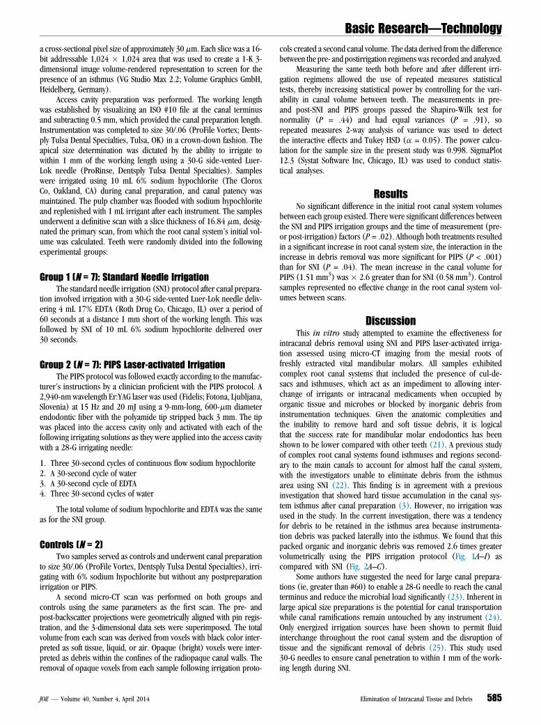
Basic Research—Technology
a cross-sectional pixel size of approximately 30mm. Each slice was a 16-bit addressable 1,024 � 1,024 area that was used to create a 1-K 3-dimensional image volume-rendered representation to screen for thepresence of an isthmus (VG Studio Max 2.2; Volume Graphics GmbH,Heidelberg, Germany).Access cavity preparation was performed. The working lengthwas established by visualizing an ISO #10 file at the canal terminusand subtracting 0.5 mm, which provided the canal preparation length.Instrumentation was completed to size 30/.06 (ProFile Vortex; Dents-ply Tulsa Dental Specialties, Tulsa, OK) in a crown-down fashion. Theapical size determination was dictated by the ability to irrigate towithin 1 mm of the working length using a 30-G side-vented Luer-Lok needle (ProRinse, Dentsply Tulsa Dental Specialties). Sampleswere irrigated using 10 mL 6% sodium hypochlorite (The CloroxCo, Oakland, CA) during canal preparation, and canal patency wasmaintained. The pulp chamber was flooded with sodium hypochloriteand replenished with 1 mL irrigant after each instrument. The samplesunderwent a definitive scan with a slice thickness of 16.84 mm, desig-nated the primary scan, from which the root canal system’s initial vol-ume was calculated. Teeth were randomly divided into the followingexperimental groups:
Group 1 (N = 7): Standard Needle IrrigationThe standard needle irrigation (SNI) protocol after canal prepara-
tion involved irrigation with a 30-G side-vented Luer-Lok needle deliv-ering 4 mL 17% EDTA (Roth Drug Co, Chicago, IL) over a period of60 seconds at a distance 1 mm short of the working length. This wasfollowed by SNI of 10 mL 6% sodium hypochlorite delivered over30 seconds.
Group 2 (N = 7): PIPS Laser-activated IrrigationThe PIPS protocol was followed exactly according to the manufac-
turer’s instructions by a clinician proficient with the PIPS protocol. A2,940-nm wavelength Er:YAG laser was used (Fidelis; Fotona, Ljubljana,Slovenia) at 15 Hz and 20 mJ using a 9-mm-long, 600-mm diameterendodontic fiber with the polyamide tip stripped back 3 mm. The tipwas placed into the access cavity only and activated with each of thefollowing irrigating solutions as they were applied into the access cavitywith a 28-G irrigating needle:
1. Three 30-second cycles of continuous flow sodium hypochlorite2. A 30-second cycle of water3. A 30-second cycle of EDTA4. Three 30-second cycles of water
The total volume of sodium hypochlorite and EDTA was the sameas for the SNI group.
Controls (N = 2)Two samples served as controls and underwent canal preparation
to size 30/.06 (ProFile Vortex, Dentsply Tulsa Dental Specialties), irri-gating with 6% sodium hypochlorite but without any postpreparationirrigation or PIPS.
A second micro-CT scan was performed on both groups andcontrols using the same parameters as the first scan. The pre- andpost-backscatter projections were geometrically aligned with pin regis-tration, and the 3-dimensional data sets were superimposed. The totalvolume from each scan was derived from voxels with black color inter-preted as soft tissue, liquid, or air. Opaque (bright) voxels were inter-preted as debris within the confines of the radiopaque canal walls. Theremoval of opaque voxels from each sample following irrigation proto-
JOE — Volume 40, Number 4, April 2014
cols created a second canal volume. The data derived from the differencebetween the pre- and postirrigation regimenswas recorded and analyzed.
Measuring the same teeth both before and after different irri-gation regimens allowed the use of repeated measures statisticaltests, thereby increasing statistical power by controlling for the vari-ability in canal volume between teeth. The measurements in pre-and post-SNI and PIPS groups passed the Shapiro-Wilk test fornormality (P = .44) and had equal variances (P = .91), sorepeated measures 2-way analysis of variance was used to detectthe interactive effects and Tukey HSD (a = 0.05). The power calcu-lation for the sample size in the present study was 0.998. SigmaPlot12.3 (Systat Software Inc, Chicago, IL) was used to conduct statis-tical analyses.
ResultsNo significant difference in the initial root canal system volumes
between each group existed. There were significant differences betweenthe SNI and PIPS irrigation groups and the time of measurement (pre-or post-irrigation) factors (P= .02). Although both treatments resultedin a significant increase in root canal system size, the interaction in theincrease in debris removal was more significant for PIPS (P < .001)than for SNI (P = .04). The mean increase in the canal volume forPIPS (1.51 mm3) was� 2.6 greater than for SNI (0.58 mm3). Controlsamples represented no effective change in the root canal system vol-umes between scans.
DiscussionThis in vitro study attempted to examine the effectiveness for
intracanal debris removal using SNI and PIPS laser-activated irriga-tion assessed using micro-CT imaging from the mesial roots offreshly extracted vital mandibular molars. All samples exhibitedcomplex root canal systems that included the presence of cul-de-sacs and isthmuses, which act as an impediment to allowing inter-change of irrigants or intracanal medicaments when occupied byorganic tissue and microbes or blocked by inorganic debris frominstrumentation techniques. Given the anatomic complexities andthe inability to remove hard and soft tissue debris, it is logicalthat the success rate for mandibular molar endodontics has beenshown to be lower compared with other teeth (21). A previous studyof complex root canal systems found isthmuses and regions second-ary to the main canals to account for almost half the canal system,with the investigators unable to eliminate debris from the isthmusarea using SNI (22). This finding is in agreement with a previousinvestigation that showed hard tissue accumulation in the canal sys-tem isthmus after canal preparation (3). However, no irrigation wasused in the study. In the current investigation, there was a tendencyfor debris to be retained in the isthmus area because instrumenta-tion debris was packed laterally into the isthmus. We found that thispacked organic and inorganic debris was removed 2.6 times greatervolumetrically using the PIPS irrigation protocol (Fig. 1A–I) ascompared with SNI (Fig. 2A–C).
Some authors have suggested the need for large canal prepara-tions (ie, greater than #60) to enable a 28-G needle to reach the canalterminus and reduce the microbial load significantly (23). Inherent inlarge apical size preparations is the potential for canal transportationwhile canal ramifications remain untouched by any instrument (24).Only energized irrigation sources have been shown to permit fluidinterchange throughout the root canal system and the disruption oftissue and the significant removal of debris (25). This study used30-G needles to ensure canal penetration to within 1 mm of the work-ing length during SNI.
Elimination of Intracanal Tissue and Debris 585
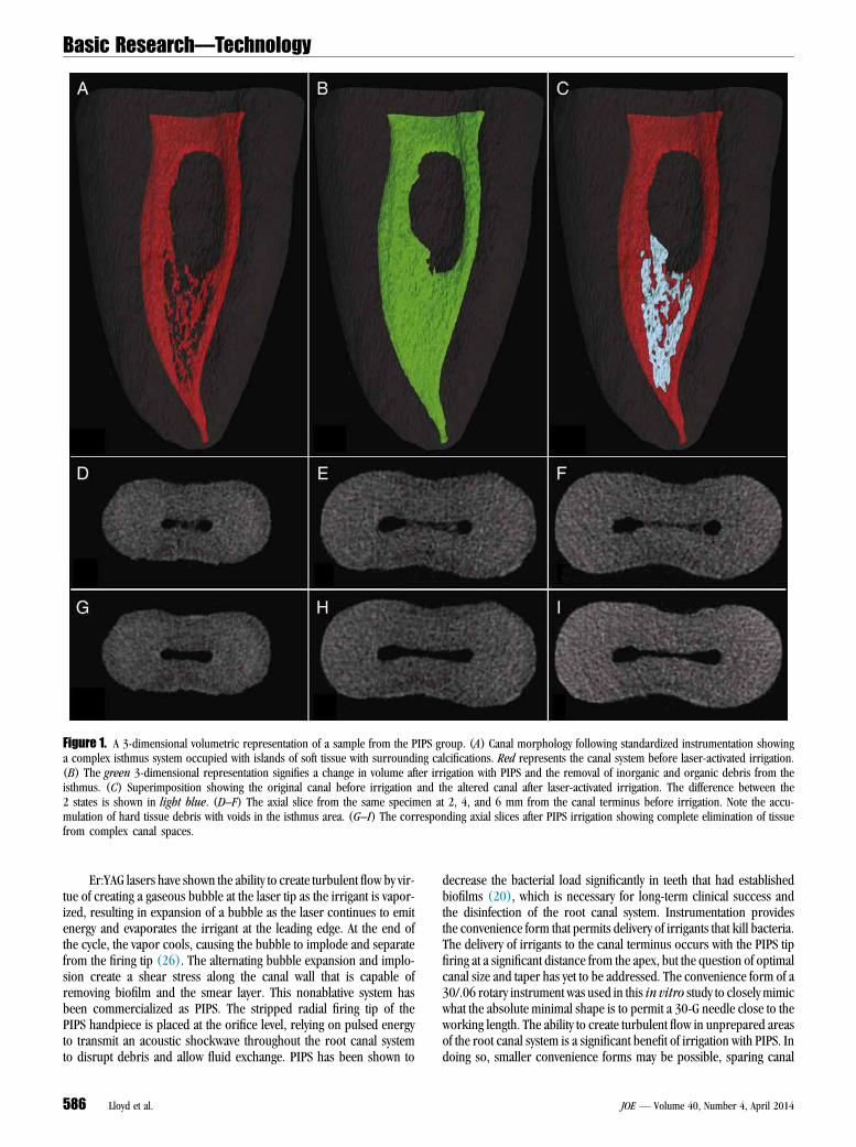
Figure 1. A 3-dimensional volumetric representation of a sample from the PIPS group. (A) Canal morphology following standardized instrumentation showinga complex isthmus system occupied with islands of soft tissue with surrounding calcifications. Red represents the canal system before laser-activated irrigation.(B) The green 3-dimensional representation signifies a change in volume after irrigation with PIPS and the removal of inorganic and organic debris from theisthmus. (C) Superimposition showing the original canal before irrigation and the altered canal after laser-activated irrigation. The difference between the2 states is shown in light blue. (D–F) The axial slice from the same specimen at 2, 4, and 6 mm from the canal terminus before irrigation. Note the accu-mulation of hard tissue debris with voids in the isthmus area. (G–I) The corresponding axial slices after PIPS irrigation showing complete elimination of tissuefrom complex canal spaces.
Basic Research—Technology
Er:YAG lasers have shown the ability to create turbulent flow by vir-tue of creating a gaseous bubble at the laser tip as the irrigant is vapor-ized, resulting in expansion of a bubble as the laser continues to emitenergy and evaporates the irrigant at the leading edge. At the end ofthe cycle, the vapor cools, causing the bubble to implode and separatefrom the firing tip (26). The alternating bubble expansion and implo-sion create a shear stress along the canal wall that is capable ofremoving biofilm and the smear layer. This nonablative system hasbeen commercialized as PIPS. The stripped radial firing tip of thePIPS handpiece is placed at the orifice level, relying on pulsed energyto transmit an acoustic shockwave throughout the root canal systemto disrupt debris and allow fluid exchange. PIPS has been shown to
586 Lloyd et al.
decrease the bacterial load significantly in teeth that had establishedbiofilms (20), which is necessary for long-term clinical success andthe disinfection of the root canal system. Instrumentation providesthe convenience form that permits delivery of irrigants that kill bacteria.The delivery of irrigants to the canal terminus occurs with the PIPS tipfiring at a significant distance from the apex, but the question of optimalcanal size and taper has yet to be addressed. The convenience form of a30/.06 rotary instrument was used in this in vitro study to closely mimicwhat the absolute minimal shape is to permit a 30-G needle close to theworking length. The ability to create turbulent flow in unprepared areasof the root canal system is a significant benefit of irrigation with PIPS. Indoing so, smaller convenience forms may be possible, sparing canal
JOE — Volume 40, Number 4, April 2014
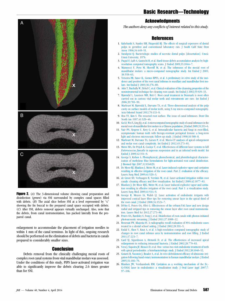
Figure 2. (A) The 3-dimensional volume showing canal preparation anddisinfection (green) via SNI surrounded by complex canal spaces filledwith debris. (B) The axial slice before SNI at a level represented by ‘‘x’’showing the fin buccal to the prepared canal space occupied with debris.(C) After SNI, debris removal appears virtually unchanged. Also, note thatthe debris, from canal instrumentation, has packed laterally from the pre-pared canal.
Basic Research—Technology
enlargement to accommodate the placement of irrigation needles towithin 1 mm of the canal terminus. In light of this, ongoing researchshould be performed on the elimination of debris and bacteria in canalsprepared to considerably smaller sizes.
ConclusionDebris removal from the clinically challenging mesial roots of
complex root canal systems from vital mandibular molars was assessed.Under the conditions of this study, PIPS laser-activated irrigation wasable to significantly improve the debris clearing 2.6 times greaterthan for SNI.
JOE — Volume 40, Number 4, April 2014
AcknowledgmentsThe authors deny any conflicts of interest related to this study.
References1. Kakehashi S, Stanley HR, Fitzgerald RJ. The effects of surgical exposures of dental
pulps in germfree and conventional laboratory rats. J South Calif State DentAssoc 1966;34:449–51.
2. Sundqvist Q. Bacteriologic studies of necrotic dental pulps [dissertation]. Ume�a:Ume�a University; 1976.
3. Paqu�e F, Laib A, Gautschi H, et al. Hard-tissue debris accumulation analysis by high-resolution computed tomography scans. J Endod 2009;35:1044–7.
4. Mannocci F, Peru M, Sherriff M, et al. The isthmuses of the mesial root ofmandibular molars: a micro-computed tomographic study. Int Endod J 2005;38:558–63.
5. Teixeira FB, Sano CL, Gomes BPFA, et al. A preliminary in vitro study of the inci-dence and position of the root canal isthmus in maxillary and mandibular first mo-lars. Int Endod J 2003;36:276–80.
6. Attin T, Buchalla W, Zirkel C, et al. Clinical evaluation of the cleansing properties of thenoninstrumental technique for cleaning root canals. Int Endod J 2002;35:929–33.
7. Bjørndal L, Laustsen MH, Reit C. Root canal treatment in Denmark is most oftencarried out in carious vital molar teeth and retreatments are rare. Int Endod J2006;39:785–90.
8. Markvart M, Bjørndal L, Darvann TA, et al. Three-dimensional analysis of the pulpcavity on surface models of molar teeth, using X-ray micro-computed tomography.Acta Odontol Scand 2012;70:133–9.
9. Hsu YY, Kim S. The resected root surface. The issue of canal isthmuses. Dent ClinNorth Am 1997;41:529–40.
10. Gu LS,Wei X, Ling JQ, et al. Amicrocomputed tomographic study of canal isthmuses in themesial root of mandibular first molars in a Chinese population. J Endod 2009;35:353–6.
11. Nair PN, Sjogren U, Krey G, et al. Intraradicular bacteria and fungi in root-filled,asymptomatic human teeth with therapy-resistant periapical lesions: a long-termlight and electron microscopic follow-up study. J Endod 1990;16:580–8.
12. Markvart M, Darvann TA, Larsen P, et al. Micro-CT analyses of apical enlargementand molar root canal complexity. Int Endod J 2012;45:273–81.
13. Meire MA, De Prijck K, Coenye T, et al. Effectiveness of different laser systems to killEnterococcus faecalis in aqueous suspension and in an infected tooth model. IntEndod J 2009;42:351–9.
14. George S, Kishen A. Photophysical, photochemical, and photobiological character-ization of methylene blue formulations for light-activated root canal disinfection.J Biomed Opt 2007;12:034029.
15. De Moor RJ, Blanken J, Meire M, et al. Laser induced explosive vapor and cavitationresulting in effective irrigation of the root canal. Part 2: evaluation of the efficacy.Lasers Surg Med 2009;41:520–3.
16. de Groot SD, Verhaagen B, Versluis M, et al. Laser-activated irrigation within rootcanals: cleaning efficacy and flow visualization. Int Endod J 2009;42:1077–83.
17. Blanken J, De Moor RJG, Meire M, et al. Laser induced explosive vapor and cavita-tion resulting in effective irrigation of the root canal. Part 1: a visualization study.Lasers Surg Med 2009;41:514–9.
18. George R, Meyers IA, Walsh LJ. Laser activation of endodontic irrigants withimproved conical laser fiber tips for removing smear layer in the apical third ofthe root canal. J Endod 2008;34:1524–7.
19. DiVito E, Peters OA, Olivi G. Effectiveness of the erbium:YAG laser and new designradial and stripped tips in removing the smear layer after root canal instrumenta-tion. Lasers Med Sci 2012;27:273–80.
20. Peters OA, Bardsley S, Fong J, et al. Disinfection of root canals with photon-initiatedphotoacoustic streaming. J Endod 2011;37:1008–12.
21. Benenati FW, Khajotia SS. A radiographic recall evaluation of 894 endodontic casestreated in a dental school setting. J Endod 2002;28:391–5.
22. Endal U, Shen Y, Knut A, et al. A high-resolution computed tomographic study ofchanges in root canal isthmus area by instrumentation and root filling. J Endod2011;37:223–7.
23. Card SJ, Sigurdsson A, Ørstavik D, et al. The effectiveness of increased apicalenlargement in reducing intracanal bacteria. J Endod 2002;28:779–83.
24. Vera J, Siqueira JF, Ricucci D, et al. One- versus two-visit endodontic treatment of teethwith apical periodontitis: a histobacteriologic study. J Endod 2012;38:1040–52.
25. Gutarts R, Nusstein J, Reader A, et al. In vivo debridement efficacy of ultrasonic irri-gation following hand-rotary instrumentation in human mandibular molars. J Endod2005;31:166–70.
26. Blanken JW, Verdaasdonk RM. Cavitation as a working mechanism of the Er,-Cr:YSGG laser in endodontics: a visualization study. J Oral Laser Appl 2007;7:97–106.
Elimination of Intracanal Tissue and Debris 587
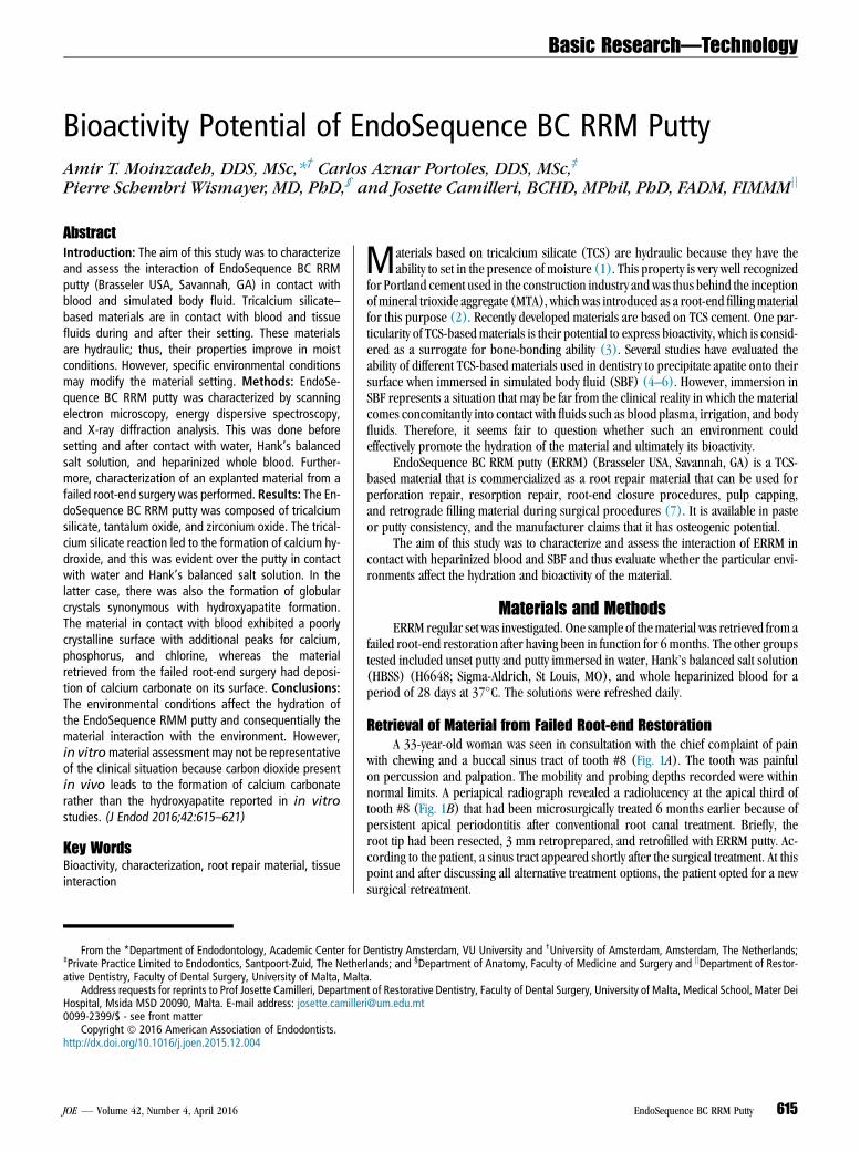
Basic Research—Technology
Bioactivity Potential of EndoSequence BC RRM PuttyAmir T. Moinzadeh, DDS, MSc,*† Carlos Aznar Portoles, DDS, MSc,‡
Pierre Schembri Wismayer, MD, PhD,§ and Josette Camilleri, BCHD, MPhil, PhD, FADM, FIMMMjj
Abstract
Introduction: The aim of this study was to characterizeand assess the interaction of EndoSequence BC RRMputty (Brasseler USA, Savannah, GA) in contact withblood and simulated body fluid. Tricalcium silicate–based materials are in contact with blood and tissuefluids during and after their setting. These materialsare hydraulic; thus, their properties improve in moistconditions. However, specific environmental conditionsmay modify the material setting. Methods: EndoSe-quence BC RRM putty was characterized by scanningelectron microscopy, energy dispersive spectroscopy,and X-ray diffraction analysis. This was done beforesetting and after contact with water, Hank’s balancedsalt solution, and heparinized whole blood. Further-more, characterization of an explanted material from afailed root-end surgery was performed. Results: The En-doSequence BC RRM putty was composed of tricalciumsilicate, tantalum oxide, and zirconium oxide. The trical-cium silicate reaction led to the formation of calcium hy-droxide, and this was evident over the putty in contactwith water and Hank’s balanced salt solution. In thelatter case, there was also the formation of globularcrystals synonymous with hydroxyapatite formation.The material in contact with blood exhibited a poorlycrystalline surface with additional peaks for calcium,phosphorus, and chlorine, whereas the materialretrieved from the failed root-end surgery had deposi-tion of calcium carbonate on its surface. Conclusions:The environmental conditions affect the hydration ofthe EndoSequence RMM putty and consequentially thematerial interaction with the environment. However,in vitromaterial assessment may not be representativeof the clinical situation because carbon dioxide presentin vivo leads to the formation of calcium carbonaterather than the hydroxyapatite reported in in vitrostudies. (J Endod 2016;42:615–621)Key WordsBioactivity, characterization, root repair material, tissueinteraction
From the *Department of Endodontology, Academic Center for‡Private Practice Limited to Endodontics, Santpoort-Zuid, The Netheative Dentistry, Faculty of Dental Surgery, University of Malta, Malt
Address requests for reprints to Prof Josette Camilleri, DepartmenHospital, Msida MSD 20090, Malta. E-mail address: josette.camiller0099-2399/$ - see front matter
Copyright ª 2016 American Association of Endodontists.http://dx.doi.org/10.1016/j.joen.2015.12.004
JOE — Volume 42, Number 4, April 2016
Materials based on tricalcium silicate (TCS) are hydraulic because they have theability to set in the presence of moisture (1). This property is very well recognized
for Portland cement used in the construction industry and was thus behind the inceptionof mineral trioxide aggregate (MTA), whichwas introduced as a root-end fillingmaterialfor this purpose (2). Recently developed materials are based on TCS cement. One par-ticularity of TCS-basedmaterials is their potential to express bioactivity, which is consid-ered as a surrogate for bone-bonding ability (3). Several studies have evaluated theability of different TCS-based materials used in dentistry to precipitate apatite onto theirsurface when immersed in simulated body fluid (SBF) (4–6). However, immersion inSBF represents a situation that may be far from the clinical reality in which the materialcomes concomitantly into contact with fluids such as blood plasma, irrigation, and bodyfluids. Therefore, it seems fair to question whether such an environment couldeffectively promote the hydration of the material and ultimately its bioactivity.
EndoSequence BC RRM putty (ERRM) (Brasseler USA, Savannah, GA) is a TCS-based material that is commercialized as a root repair material that can be used forperforation repair, resorption repair, root-end closure procedures, pulp capping,and retrograde filling material during surgical procedures (7). It is available in pasteor putty consistency, and the manufacturer claims that it has osteogenic potential.
The aim of this study was to characterize and assess the interaction of ERRM incontact with heparinized blood and SBF and thus evaluate whether the particular envi-ronments affect the hydration and bioactivity of the material.
Materials and MethodsERRM regular set was investigated. One sample of thematerial was retrieved from a
failed root-end restoration after having been in function for 6 months. The other groupstested included unset putty and putty immersed in water, Hank’s balanced salt solution(HBSS) (H6648; Sigma-Aldrich, St Louis, MO), and whole heparinized blood for aperiod of 28 days at 37�C. The solutions were refreshed daily.
Retrieval of Material from Failed Root-end RestorationA 33-year-old woman was seen in consultation with the chief complaint of pain
with chewing and a buccal sinus tract of tooth #8 (Fig. 1A). The tooth was painfulon percussion and palpation. The mobility and probing depths recorded were withinnormal limits. A periapical radiograph revealed a radiolucency at the apical third oftooth #8 (Fig. 1B) that had been microsurgically treated 6 months earlier because ofpersistent apical periodontitis after conventional root canal treatment. Briefly, theroot tip had been resected, 3 mm retroprepared, and retrofilled with ERRM putty. Ac-cording to the patient, a sinus tract appeared shortly after the surgical treatment. At thispoint and after discussing all alternative treatment options, the patient opted for a newsurgical retreatment.
Dentistry Amsterdam, VU University and †University of Amsterdam, Amsterdam, The Netherlands;rlands; and §Department of Anatomy, Faculty of Medicine and Surgery and jjDepartment of Restor-a.t of Restorative Dentistry, Faculty of Dental Surgery, University of Malta, Medical School, Mater [email protected]
EndoSequence BC RRM Putty 615
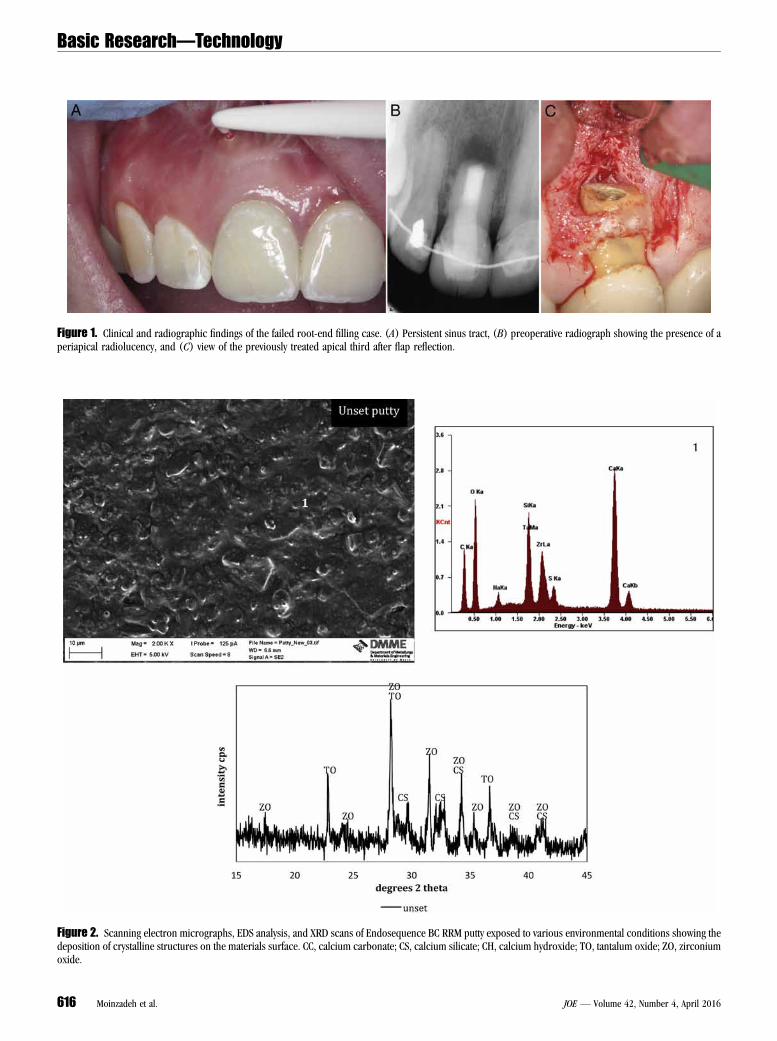
Figure 1. Clinical and radiographic findings of the failed root-end filling case. (A) Persistent sinus tract, (B) preoperative radiograph showing the presence of aperiapical radiolucency, and (C) view of the previously treated apical third after flap reflection.
Figure 2. Scanning electron micrographs, EDS analysis, and XRD scans of Endosequence BC RRM putty exposed to various environmental conditions showing thedeposition of crystalline structures on the materials surface. CC, calcium carbonate; CS, calcium silicate; CH, calcium hydroxide; TO, tantalum oxide; ZO, zirconiumoxide.
Basic Research—Technology
616 Moinzadeh et al. JOE — Volume 42, Number 4, April 2016
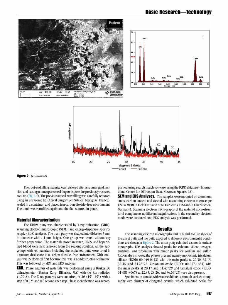
Figure 2. (Continued).
Basic Research—Technology
The root-end fillingmaterial was retrieved after a submarginal inci-sion and raising a mucoperiosteal flap to expose the previously resectedroot tip (Fig. 1C). The previous apical retrofilling was carefully removedusing an ultrasonic tip (Apical Surgery Set; Satelec, M�erignac, France),sealed in a container, and placed in a carbon dioxide–free environment.The tooth was retrofilled again and the flap sutured in place.
Material CharacterizationThe ERRM putty was characterized by X-ray diffraction (XRD),
scanning electron microscopic (SEM), and energy-dispersive spectro-scopic (EDS) analyses. The fresh putty was shaped into diskettes 5 mmin diameter with a 1-mm height. One group was tested without anyfurther preparation. The materials stored in water, HBSS, and heparin-ized blood were first removed from the soaking solution. All the sub-groups with set materials including the explanted putty were dried ina vacuum desiccator in a carbon dioxide–free environment. XRD anal-ysis was performed first because this was a nondestructive technique.This was followed by SEM and EDS analyses.
XRD. Phase analysis of materials was performed using a Bruker D8diffractometer (Bruker Corp, Billerica, MA) with Co Ka radiation(1.79�A). The X-ray patterns were acquired in 2q (15�–45�) with astep of 0.02� and 0.6 seconds per step. Phase identification was accom-
JOE — Volume 42, Number 4, April 2016
plished using search match software using the ICDD database (Interna-tional Centre for Diffraction Data, Newtown Square, PA).
SEM and EDS Analyses. The samples were mounted on aluminumstubs, carbon coated, and viewed with a scanning electron microscope(Zeiss MERLIN Field Emission SEM; Carl Zeiss NTS GmbH, Oberkochen,Germany). Scanning electron micrographs of the material microstruc-tural components at different magnifications in the secondary electronmode were captured, and EDS analysis was performed.
ResultsThe scanning electron micrographs and EDS and XRD analyses of
the unset putty and the putty exposed to different environmental condi-tions are shown in Figure 2. The unset putty exhibited a smooth surfacetopography. EDS analysis showed peaks for calcium, silicon, oxygen,tantalum, and zirconium with minor peaks for sodium and sulfur.XRD analysis showed the phases present, namely monoclinic tricalciumsilicate (ICDD: 00-049-0442) with the main peaks at 29.30, 32.13,32.46, and 34.28�2q. Zirconium oxide (ICDD: 00-037-1484) withthe main peaks at 28.17 and 31.47�2q and tantalum oxide (ICDD:01-081-8067) at 22.83, 28.26, and 36.64�2q were also present.
Specimens in contact with water exhibited a smooth surface topog-raphy with clusters of elongated crystals, which exhibited peaks for
EndoSequence BC RRM Putty 617
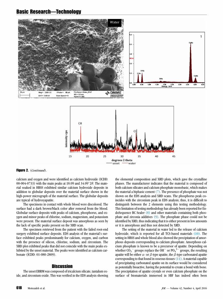
Figure 2. (Continued).
Basic Research—Technology
calcium and oxygen and were identified as calcium hydroxide (ICDD:00-004-0733) with the main peaks at 18.09 and 34.09�2q. The mate-rial soaked in HBSS exhibited similar calcium hydroxide deposits inaddition to globular deposits over the material surface shown in thehigh-power micrograph of the material surface. The globular depositsare typical of hydroxyapatite.
The specimens in contact with whole blood were discolored. Thesurface had a dark brown/black color after removal from the blood.Globular surface deposits with peaks of calcium, phosphorus, and ox-ygen and minor peaks of chlorine, sodium, magnesium, and potassiumwere present. The material surface deposit was amorphous as seen bythe lack of specific peaks present on the XRD scan.
The specimen retrieved from the patient with the failed root-endsurgery exhibited surface deposits. EDS analysis of the material’s sur-face exhibited peaks predominantly for calcium, oxygen, and carbonwith the presence of silicon, chlorine, sodium, and zirconium. TheXRD plot exhibited peaks that did not coincide with the main peaks ex-hibited by the unset material. The peaks were identified as calcium car-bonate (ICDD: 01-080-2809).
DiscussionThe unset ERRMwas composed of tricalcium silicate, tantalum ox-
ide, and zirconium oxide. This was verified in the EDS analysis showing
618 Moinzadeh et al.
the elemental composition and XRD plots, which gave the crystallinephases. The manufacturer indicates that the material is composed ofboth calcium silicates and calcium phosphate monobasic, which makesthe material a biphasic cement (7). The presence of phosphate was notshown on the EDS analysis and XRD scans. The phosphorus peak co-incides with the zirconium peak in EDS analysis; thus, it is difficult todistinguish between the 2 elements using this testing methodology.This limitation of testing methodology has already been reported for En-doSequence BC Sealer (8) and other materials containing both phos-phate and zirconia additives (9). The phosphate phase could not beidentified by XRD, thus indicating that it is either present in low amountsor it is amorphous and thus not detected by XRD.
The setting of the material in water led to the release of calciumhydroxide, which is reported for all TCS-based materials (10). Thesetting in HBSS and whole blood also showed the precipitation of amor-phous deposits corresponding to calcium phosphate. Amorphous cal-cium phosphate is known to be a precursor of apatite. Depending onwhether CO3
� groups replace the OH� or PO43� groups, the resulting
apatite will be either a- or b-type apatite, the b-type carbonated apatitecorresponding to that found in osseous tissues (11). A material capableof precipitating carbonated apatite on its surface would be consideredas potentially bioactive, having the potential to create a bond with bone.The precipitation of apatite crystals or even calcium phosphate on thesurface of biomaterials immersed in SBF has indeed often been
JOE — Volume 42, Number 4, April 2016
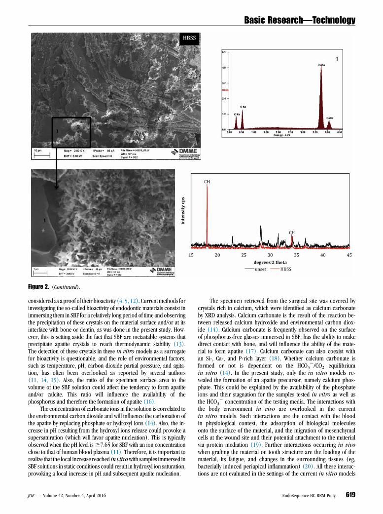
Figure 2. (Continued).
Basic Research—Technology
considered as a proof of their bioactivity (4, 5, 12). Current methods forinvestigating the so-called bioactivity of endodontic materials consist inimmersing them in SBF for a relatively long period of time and observingthe precipitation of these crystals on the material surface and/or at itsinterface with bone or dentin, as was done in the present study. How-ever, this is setting aside the fact that SBF are metastable systems thatprecipitate apatite crystals to reach thermodynamic stability (13).The detection of these crystals in these in vitro models as a surrogatefor bioactivity is questionable, and the role of environmental factors,such as temperature, pH, carbon dioxide partial pressure, and agita-tion, has often been overlooked as reported by several authors(11, 14, 15). Also, the ratio of the specimen surface area to thevolume of the SBF solution could affect the tendency to form apatiteand/or calcite. This ratio will influence the availability of thephosphorus and therefore the formation of apatite (16).
The concentration of carbonate ions in the solution is correlated tothe environmental carbon dioxide and will influence the carbonation ofthe apatite by replacing phosphate or hydroxyl ions (14). Also, the in-crease in pH resulting from the hydroxyl ions release could provoke asupersaturation (which will favor apatite nucleation). This is typicallyobserved when the pH level is$7.65 for SBF with an ion concentrationclose to that of human blood plasma (11). Therefore, it is important torealize that the local increase reached in vitrowith samples immersed inSBF solutions in static conditions could result in hydroxyl ion saturation,provoking a local increase in pH and subsequent apatite nucleation.
JOE — Volume 42, Number 4, April 2016
The specimen retrieved from the surgical site was covered bycrystals rich in calcium, which were identified as calcium carbonateby XRD analysis. Calcium carbonate is the result of the reaction be-tween released calcium hydroxide and environmental carbon diox-ide (14). Calcium carbonate is frequently observed on the surfaceof phosphorus-free glasses immersed in SBF, has the ability to makedirect contact with bone, and will influence the ability of the mate-rial to form apatite (17). Calcium carbonate can also coexist withan Si-, Ca-, and P-rich layer (18). Whether calcium carbonate isformed or not is dependent on the HCO3
�/CO2 equilibriumin vitro (14). In the present study, only the in vitro models re-vealed the formation of an apatite precursor, namely calcium phos-phate. This could be explained by the availability of the phosphateions and their stagnation for the samples tested in vitro as well asthe HCO3
� concentration of the testing media. The interactions withthe body environment in vivo are overlooked in the currentin vitro models. Such interactions are the contact with the bloodin physiological context, the adsorption of biological moleculesonto the surface of the material, and the migration of mesenchymalcells at the wound site and their potential attachment to the materialvia protein mediation (19). Further interactions occurring in vivowhen grafting the material on tooth structure are the loading of thematerial, its fatigue, and changes in the surrounding tissues (eg,bacterially induced periapical inflammation) (20). All these interac-tions are not evaluated in the settings of the current in vitro models
EndoSequence BC RRM Putty 619
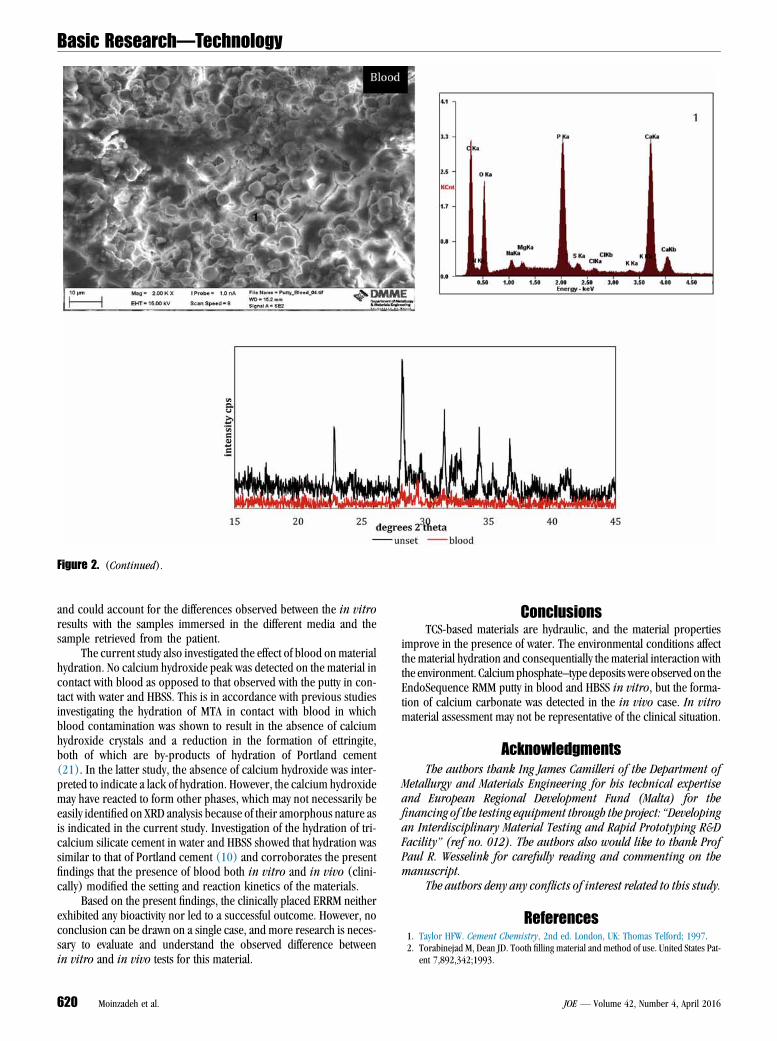
Figure 2. (Continued).
Basic Research—Technology
and could account for the differences observed between the in vitroresults with the samples immersed in the different media and thesample retrieved from the patient.
The current study also investigated the effect of blood on materialhydration. No calcium hydroxide peak was detected on the material incontact with blood as opposed to that observed with the putty in con-tact with water and HBSS. This is in accordance with previous studiesinvestigating the hydration of MTA in contact with blood in whichblood contamination was shown to result in the absence of calciumhydroxide crystals and a reduction in the formation of ettringite,both of which are by-products of hydration of Portland cement(21). In the latter study, the absence of calcium hydroxide was inter-preted to indicate a lack of hydration. However, the calcium hydroxidemay have reacted to form other phases, which may not necessarily beeasily identified on XRD analysis because of their amorphous nature asis indicated in the current study. Investigation of the hydration of tri-calcium silicate cement in water and HBSS showed that hydration wassimilar to that of Portland cement (10) and corroborates the presentfindings that the presence of blood both in vitro and in vivo (clini-cally) modified the setting and reaction kinetics of the materials.
Based on the present findings, the clinically placed ERRM neitherexhibited any bioactivity nor led to a successful outcome. However, noconclusion can be drawn on a single case, and more research is neces-sary to evaluate and understand the observed difference betweenin vitro and in vivo tests for this material.
620 Moinzadeh et al.
ConclusionsTCS-based materials are hydraulic, and the material properties
improve in the presence of water. The environmental conditions affectthe material hydration and consequentially the material interaction withthe environment. Calciumphosphate–type deposits were observed on theEndoSequence RMM putty in blood and HBSS in vitro, but the forma-tion of calcium carbonate was detected in the in vivo case. In vitromaterial assessment may not be representative of the clinical situation.
AcknowledgmentsThe authors thank Ing James Camilleri of the Department of
Metallurgy and Materials Engineering for his technical expertiseand European Regional Development Fund (Malta) for thefinancing of the testing equipment through the project: ‘‘Developingan Interdisciplinary Material Testing and Rapid Prototyping R&DFacility’’ (ref no. 012). The authors also would like to thank ProfPaul R. Wesselink for carefully reading and commenting on themanuscript.
The authors deny any conflicts of interest related to this study.
References1. Taylor HFW. Cement Chemistry, 2nd ed. London, UK: Thomas Telford; 1997.2. Torabinejad M, Dean JD. Tooth filling material and method of use. United States Pat-
ent 7,892,342;1993.
JOE — Volume 42, Number 4, April 2016
Basic Research—Technology
3. Niu LN, Jiao K, Wang TD, et al. A review of the bioactivity of hydraulic calcium silicatecements. J Dent 2014;42:517–33.4. Reyes-Carmona JF, Felippe MA, Felippe WT. A phosphate-buffered saline intracanal
dressing improves the biomineralization ability of mineral trioxide aggregate apicalplugs. J Endod 2010;36:1648–52.
5. Sarkar NK, Caicedo R, Ritwik P, et al. Physicochemical basis of the biologic prop-erties of mineral trioxide aggregate. J Endod 2005;31:97–100.
6. Tay FR, Pashley DH, Rueggeberg FA, et al. Calcium phosphate phase transforma-tion produced by the interaction of the portland cement component of whitemineral trioxide aggregate with a phosphate-containing fluid. J Endod 2007;33:1347–51.
7. EndoSequence BC RRM. Brasseler USA Dental Instrumentation. Available at: http://brasselerusadental.com/products/bc-rrm/. Accessed August 16, 2015.
8. Xuereb M, Vella P, Damidot D, et al. In situ assessment of the setting of tri-calcium silicate-based sealers using a dentin pressure model. J Endod 2015;41:111–24.
9. Viapiana R, Guerreiro-Tanomaru J, Tanomaru-Filho M, et al. Interface of dentine toroot canal sealers. J Dent 2014;42:336–50.
10. Camilleri J. Characterization and hydration kinetics of tricalcium silicate cement foruse as a dental biomaterial. Dent Mater 2011;27:836–44.
11. M€uller L, M€uller FA. Preparation of SBF with different HCO3- content and its influ-
ence on the composition of biomimetic apatites. Acta Biomater 2006;2:181–9.
JOE — Volume 42, Number 4, April 2016
12. Kim JR, Nosrat A, Fouad AF. Interfacial characteristics of Biodentine and MTA withdentine in simulated body fluid. J Dent 2015;43:241–7.
13. Lu X, Leng Y. Theoretical analysis of calcium phosphate precipitation in simulatedbody fluid. Biomaterials 2005;26:1097–108.
14. Bohner M, Lemaitre J. Can bioactivity be tested in vitro with SBF solution? Bioma-terials 2009;30:2175–9.
15. Pan H, Zhao X, Darvell BW, et al. Apatite-formation ability—predictor of ‘‘bioac-tivity’’? Acta Biomater 2010;6:4181–8.
16. Rybarikova L, Had J, Holubova I. Evaluating the phase composition of the surface ofbioactive glasses following their interaction with simulated body fluid, with the use ofX-ray diffraction analysis. Ceram Silik 1995;39:34–9.
17. Neo M, Kotani S, Fujita Y, et al. Differences in ceramic-bone interface betweensurface-active ceramics and resorbable ceramics: a study by scanning and transmis-sion electron microscopy. J Biomed Mater Res 1992;26:255–67.
18. Zhang Y, Takadam H, Mizuno M, et al. Calcite formation in a revised simulated bodyfluid. Ceram Eng Sci Proc 2002;23:789–96.
19. Clarke B. Normal bone anatomy and physiology. Clin J Am Soc Nephrol 2008;3:S131–9.
20. Boyan BD, Hummert TW, Dean DD, et al. Role of material surfaces in regulatingbone and cartilage cell response. Biomaterials 1996;17:137–46.
21. Nekoofar MH, Davies TE, Stone D, et al. Microstructure and chemical analysis ofblood-contaminated mineral trioxide aggregate. Int Endod J 2011;44:1011–8.
EndoSequence BC RRM Putty 621
Biofilm removal by 6% sodium hypochloriteactivated by different irrigation techniques
R. Ordinola-Zapata1, C. M. Bramante1, R. M. Aprecio2, R. Handysides2 & D. E. Jaramillo2
1Department of Endodontics, Dental School of Bauru, University of S~ao Paulo, Bauru, Brazil; and 2Department of Endodontics,
School of Dentistry, Loma Linda University, Loma Linda, CA, USA
Abstract
Ordinola-Zapata R, Bramante CM, Aprecio RM,
Handysides R, Jaramillo DE. Biofilm removal by 6%
sodium hypochlorite activated by different irrigation tech-
niques. International Endodontic Journal, 47, 659–666, 2014.
Aim To compare the removal of biofilm utilizing four
irrigation techniques on a bovine root canal model.
Methodology Fifty dentine specimens (2 9 2 mm)
were infected with biofilm. The samples were then
adapted to previously created cavities in the bovine
model. The root canals were irrigated twice with 2 mL
of 6% sodium hypochlorite for 2 min (4 min total). Fol-
lowing initial irrigation, the different treatment modali-
ties were introduced for 60 s (3 9 20 s intervals). The
evaluated techniques were needle irrigation, Endoacti-
vator (Dentsply Tulsa Dental, Tulsa, OK, USA), passive
ultrasonic irrigation and laser-activated irrigation
(photon-induced photoacoustic streaming). The con-
trols were irrigated with distilled water and conven-
tional needle irrigation. Subsequently, the dentine
samples were separated from the model and analysed
using a scanning electron microscope (SEM). Fifteen
operative fields were scanned per block, and SEM pic-
tures were captured. Two calibrated evaluators exam-
ined the images and collected data using a four-degree
scale. Nonparametric tests were used to evaluate for
statistical significance amongst the groups.
Results The group undergoing laser-activated irri-
gation using photon-induced photoacoustic streaming
exhibited the most favourable results in the removal
of biofilm. Passive ultrasonic irrigation scores were
significantly lower than both the Endoactivator and
needle irrigation scores. Sonic and needle irrigation
were not significantly different. The least favourable
results were found in the control group.
Conclusions Laser activation of 6% sodium
hypochlorite significantly improved the cleaning of
biofilm-infected dentine followed by passive ultrasonic
irrigation.
Keywords: biofilms, irrigant solutions, laser-acti-
vated irrigation, photoacoustic streaming.
Received 19 July 2013; accepted 5 October 2013
Introduction
The aim of irrigation in root canal treatment is to
improve the cleaning and disinfection process within
the root canal system (Siqueira & Rocas 2008). Irri-
gants play multiple roles in endodontic therapy. They
are necessary from an antimicrobial aspect as the
mechanical instrumentation process is insufficient on
its own to remove the microbial load (Bystr€om &
Sundqvist 1983). Sodium hypochlorite (NaOCl) is
considered the main root canal irrigant because of its
tissue dissolution and antimicrobial properties. Whilst
some microscopic studies have shown that complete
dissolution of biofilms by sodium hypochlorite is
possible using the direct contact test (del Carpio-
Perochena et al. 2011), incomplete dissolution and
residual biofilm appears to be common under clinical
conditions following full-strength NaOCl irrigation
(Vera et al. 2012). Residual biofilm may contain
viable bacteria and may decrease the interfacial adap-
tation of root filling materials (Vera et al. 2012).
Significant information regarding the physical effect
of fluids in root canals has been previously reported
Correspondence: Ronald Ordinola-Zapata, Faculdade de
Odontologia de Bauru, University of S~ao Paulo, Al. Oct�avio
Pinheiro Brisolla, 9-75, CEP 17012-901, Bauru, SP, Brazil
(e-mail: [email protected]).
© 2013 International Endodontic Journal. Published by John Wiley & Sons Ltd International Endodontic Journal, 47, 659–666, 2014
doi:10.1111/iej.12202
659
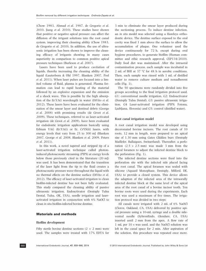
(Chow 1983, Ahmad et al. 1987, de Gregorio et al.
2010, Jiang et al. 2010). These studies have shown
that positive or negative apical pressure can affect the
diffusion of the irrigant solutions into the root canal
system, improving their cleaning ability (Chow 1983,
de Gregorio et al. 2010). In addition, the use of ultra-
sonic irrigation has been shown to improve the clean-
ing efficacy of irrigants showing in many cases
superiority in comparison to common positive apical
pressure techniques (Burleson et al. 2007).
Lasers have been used to produce cavitation of
liquids, thereby increasing the cleaning ability of the
liquid (Lauterborn & Ohl 1997, Blanken 2007, Peel
et al. 2011). When laser pulses are focused into a lim-
ited volume of fluid, plasma is generated. Plasma for-
mation can lead to rapid heating of the material
followed by an explosive expansion and the emission
of a shock wave. This is possible by the high absorp-
tion of the Er:YAG wavelength in water (DiVito et al.
2012). These lasers have been evaluated for the elimi-
nation of the smear layer and dentinal debris (George
et al. 2008) with promising results (de Groot et al.
2009). These techniques, referred to as laser-activated
irrigation (de Groot et al. 2009), have been evaluated
for endodontic irrigation applications basically using
Erbium YAG (Er:YAG) or Er, CrYSGG lasers, with
energy levels that vary from 25 to 300 mJ (Blanken
2007, George et al. 2008, Blanken et al. 2009, Peters
et al. 2011).
In this work, a novel tapered and stripped tip of a
laser-activated irrigation technique called photon-
induced photoacoustic streaming (PIPS) at energy levels
below those previously cited in the literature (20 mJ)
was used. It has been demonstrated that the transition
of the laser light from the tip to the fluid creates a
photoacoustic pressure wave throughout the liquid with
no thermal effects on the dentine surface (DiVito et al.
2012). The efficacy of laser-activated irrigation to clean
biofilm-infected dentine has not been fully evaluated.
This study compared the cleaning ability of passive
ultrasonic irrigation, Endoactivator (Dentsply Tulsa
Dental, Tulsa, OK, USA), needle irrigation and laser-
activated irrigation in conjunction with 6% NaOCl to
clean in situ biofilm-infected bovine dentine.
Materials and methods
Biofilm development
Fifty sterile bovine dentine sections (2 9 2 mm) were
used. The samples were treated with 17% EDTA for
3 min to eliminate the smear layer produced during
the sectioning process. To induce dentine infection,
an in situ model was selected using a Hawleys ortho-
dontic device. The dentine surface exposed to the oral
cavity was fixed 1 mm above the surface to allow the
accumulation of plaque. One volunteer used the
device continuously for 72 h, except during oral
hygiene procedures, to generate biofilm (Human com-
mittee and ethic research approval, CEP134/2010).
Daily food diet was maintained. After the intraoral
contamination process, each sample was incubated in
2 mL of BHI at 37 °C for 48 h in aerobic conditions.
Then, each sample was rinsed with 1 mL of distilled
water to remove culture medium and nonadherent
cells (Fig. 1).
The 50 specimens were randomly divided into five
groups according to the final irrigation protocol used.
G1: conventional needle irrigation, G2: Endoactivator
(Dentsply Tulsa Dental), G3: passive ultrasonic irriga-
tion, G4 Laser-activated irrigation (PIPS; Fotona,
Ljubljana, Slovenia) and G5: control (distilled water).
Root canal irrigation model
A root canal irrigation model was developed using
decoronated bovine incisors. The root canals of 10
roots, 12 mm in length, were prepared to an apical
size of 1.30 mm using Gates Glidden burs (Dentsply
Maillefer, Ballaigues, Switzerland). Thereafter, a perfo-
ration (2.5 9 2.5 mm) was made 3 mm from the
apical foramen to adjust the infected dentine block to
the perforation (Fig. 1).
The infected dentine sections were fixed into the
perforation site with the infected side placed facing
the root canal. The apical foramen was sealed with
silicone (Aquasil Monophase, Dentsply, Milford, DE,
USA) to provide a closed system. This device allows
the adaption of the infected area of the intraorally
infected dentine block at the same level of the apical
area of the root canal of a bovine incisor tooth. Ten
bovine roots were used during the experiments. Each
root was used a maximum of five times. The irriga-
tion protocol was divided in two steps:
All canals were irrigated with 2 mL of 6% NaOCl
(Clorox, Oakland, CA, USA) delivered by positive api-
cal pressure using a 10-mL syringe and a double side-
vented needle (SybronEndo, Glendare, CA, USA)
inserted until 2 mm from the apex. A flow rate of
1 mL per 10 s was used, and the NaOCl solution was
left in the canal space for 2 min. After aspiration of
the solution, this procedure was repeated once more.
Biofilm removal by different irrigation techniques Ordinola-Zapata et al.
© 2013 International Endodontic Journal. Published by John Wiley & Sons LtdInternational Endodontic Journal, 47, 659–666, 2014660
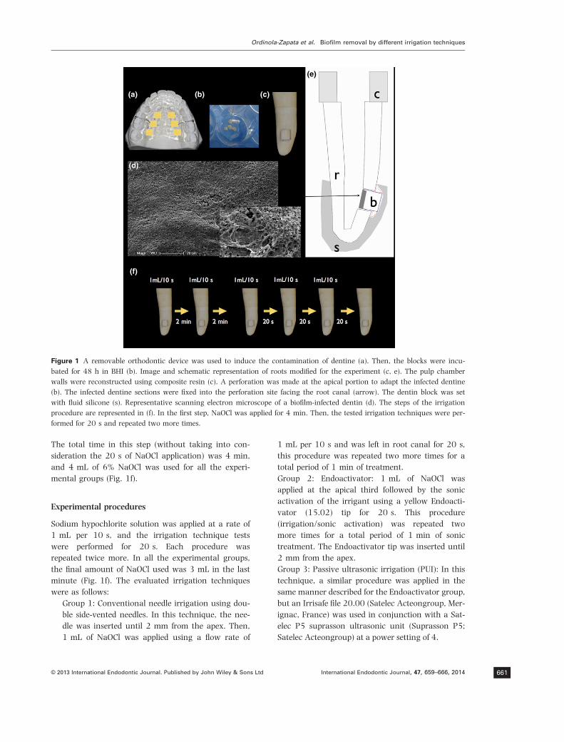
The total time in this step (without taking into con-
sideration the 20 s of NaOCl application) was 4 min,
and 4 mL of 6% NaOCl was used for all the experi-
mental groups (Fig. 1f).
Experimental procedures
Sodium hypochlorite solution was applied at a rate of
1 mL per 10 s, and the irrigation technique tests
were performed for 20 s. Each procedure was
repeated twice more. In all the experimental groups,
the final amount of NaOCl used was 3 mL in the last
minute (Fig. 1f). The evaluated irrigation techniques
were as follows:
Group 1: Conventional needle irrigation using dou-
ble side-vented needles. In this technique, the nee-
dle was inserted until 2 mm from the apex. Then,
1 mL of NaOCl was applied using a flow rate of
1 mL per 10 s and was left in root canal for 20 s,
this procedure was repeated two more times for a
total period of 1 min of treatment.
Group 2: Endoactivator: 1 mL of NaOCl was
applied at the apical third followed by the sonic
activation of the irrigant using a yellow Endoacti-
vator (15.02) tip for 20 s. This procedure
(irrigation/sonic activation) was repeated two
more times for a total period of 1 min of sonic
treatment. The Endoactivator tip was inserted until
2 mm from the apex.
Group 3: Passive ultrasonic irrigation (PUI): In this
technique, a similar procedure was applied in the
same manner described for the Endoactivator group,
but an Irrisafe file 20.00 (Satelec Acteongroup, Mer-
ignac, France) was used in conjunction with a Sat-
elec P5 suprasson ultrasonic unit (Suprasson P5;
Satelec Acteongroup) at a power setting of 4.
(a)
(d)
(f)
(e)
(b) (c)
Figure 1 A removable orthodontic device was used to induce the contamination of dentine (a). Then, the blocks were incu-
bated for 48 h in BHI (b). Image and schematic representation of roots modified for the experiment (c, e). The pulp chamber
walls were reconstructed using composite resin (c). A perforation was made at the apical portion to adapt the infected dentine
(b). The infected dentine sections were fixed into the perforation site facing the root canal (arrow). The dentin block was set
with fluid silicone (s). Representative scanning electron microscope of a biofilm-infected dentin (d). The steps of the irrigation
procedure are represented in (f). In the first step, NaOCl was applied for 4 min. Then, the tested irrigation techniques were per-
formed for 20 s and repeated two more times.
Ordinola-Zapata et al. Biofilm removal by different irrigation techniques
© 2013 International Endodontic Journal. Published by John Wiley & Sons Ltd International Endodontic Journal, 47, 659–666, 2014 661
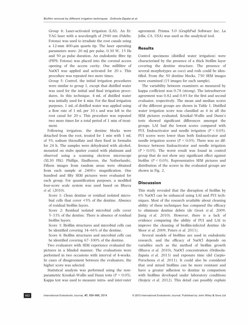
Group 4: Laser-activated irrigation (LAI). An Er:
YAG laser with a wavelength of 2940 nm (Fidelis;
Fotona) was used to irradiate the root canals using
a 12-mm 400-lm quartz tip. The laser operating
parameters were: 20 mJ per pulse, 0.30 W, 15 Hz
and 50 ls pulse duration. An endodontic fibre tip
(PIPS; Fotona) was placed into the coronal access
opening of the access cavity. One millilitre of
NaOCl was applied and activated for 20 s. This
procedure was repeated two more times.
Group 5: Control, the initial irrigation procedures
were similar to group 1, except that distilled water
was used for the initial and final irrigation proce-
dures. In this technique, 4 mL of distilled water
was initially used for 4 min. For the final irrigation
purposes, 1 mL of distilled water was applied using
a flow rate of 1 mL per 10 s and was left in the
root canal for 20 s. This procedure was repeated
two more times for a total period of 1 min of treat-
ment.
Following irrigation, the dentine blocks were
detached from the root, treated for 1 min with 1 mL
of 5% sodium thiosulfate and then fixed in formalin
for 24 h. The samples were dehydrated with alcohol,
mounted on stubs sputter coated with platinum and
observed using a scanning electron microscope
(XL30 FEG; Phillips, Eindhoven, the Netherlands).
Fifteen images from random areas were obtained
from each sample at 24009 magnification. One
hundred and fifty SEM pictures were evaluated for
each group. For quantification purposes, a modified
four-score scale system was used based on Bhuva
et al. (2010).
Score 1: Clean dentine or residual isolated micro-
bial cells that cover <5% of the dentine. Absence
of residual biofilm layers.
Score 2: Residual isolated microbial cells cover
5–33% of the dentine. There is absence of residual
biofilm layers.
Score 3: Biofilm structures and microbial cells can
be identified covering 34–66% of the dentine.
Score 4: Biofilm structures and microbial cells can
be identified covering 67–100% of the dentine.
Two evaluators with SEM experience evaluated the
pictures in a blinded manner. The evaluations were
performed in two occasions with interval of 4-weeks.
In cases of disagreement between the evaluators, the
higher score was selected.
Statistical analysis was performed using the non-
parametric Kruskal–Wallis and Dunn tests (P < 0.05).
Kappa test was used to measure intra- and inter-rater
agreement. Prisma 5.0 (GraphPad Software Inc, La
Jolla, CA, USA) was used as the analytical tool.
Results
Control specimens (distilled water irrigation) were
characterized by the presence of a thick biofilm layer
covering the dentine structure. The presence of
several morphotypes as cocci and rods could be iden-
tified. From the 50 dentine blocks, 750 SEM images
were examined (15 images for each sample).
The variability between examiners as measured by
kappa coefficient was 0.78 (strong). The intraobserver
agreement was 0.82 and 0.85 for the first and second
evaluator, respectively. The mean and median scores
of the different groups are shown in Table 1. Distilled
water irrigation score was classified as 4 in all the
SEM pictures evaluated. Kruskal–Wallis and Dunn’s
tests showed significant differences amongst the
groups. LAI had the lowest scores compared with
PUI, Endoactivator and needle irrigation (P < 0.05).
PUI scores were lower than both Endoactivator and
needle irrigation scores (P < 0.05). There was no dif-
ference between Endoactivator and needle irrigation
(P > 0.05). The worst result was found in control
group that do not show any significant effect against
biofilm (P < 0.05). Representative SEM pictures and
distribution of the scores in the evaluated groups are
shown in Fig. 2.
Discussion
This study revealed that the disruption of biofilm by
6% NaOCl can be enhanced using LAI and PUI tech-
niques. Most of the research available about cleaning
ability of these techniques has compared the efficacy
to eliminate dentine debris (de Groot et al. 2009,
Jiang et al. 2010). However, there is a lack of
evidence comparing the ability of PUI and LAI to
improve the cleaning of biofilm-infected dentine (de
Moor et al. 2009, Peters et al. 2011).
Several models of biofilms are used in endodontic
research, and the efficacy of NaOCl depends on
variables such as the method of biofilm growth
(Bhuva et al. 2010), NaOCl concentration (Ordinola-
Zapata et al. 2013) and exposure time (del Carpio-
Perochena et al. 2011). It could also be considered
that oral mixed biofilms can be more resistant and
have a greater adhesion to dentine in comparison
with biofilms developed under laboratory conditions
(Stojicic et al. 2012). This detail can possibly explain
Biofilm removal by different irrigation techniques Ordinola-Zapata et al.
© 2013 International Endodontic Journal. Published by John Wiley & Sons LtdInternational Endodontic Journal, 47, 659–666, 2014662
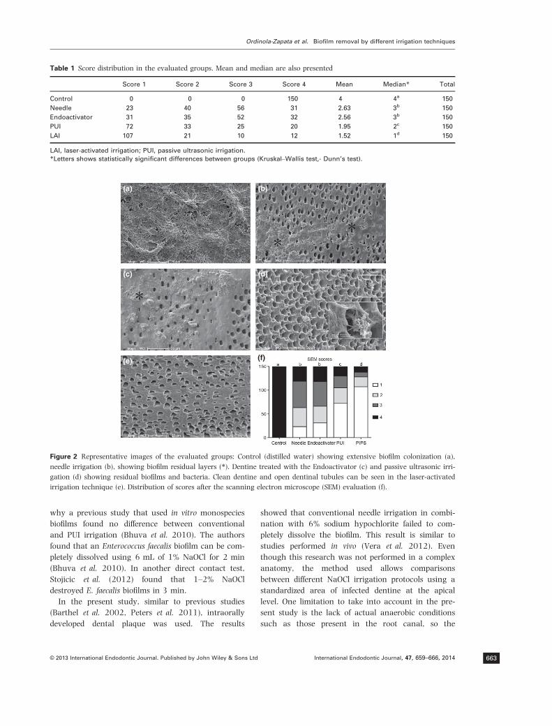
why a previous study that used in vitro monospecies
biofilms found no difference between conventional
and PUI irrigation (Bhuva et al. 2010). The authors
found that an Enterococcus faecalis biofilm can be com-
pletely dissolved using 6 mL of 1% NaOCl for 2 min
(Bhuva et al. 2010). In another direct contact test,
Stojicic et al. (2012) found that 1–2% NaOCl
destroyed E. faecalis biofilms in 3 min.
In the present study, similar to previous studies
(Barthel et al. 2002, Peters et al. 2011), intraorally
developed dental plaque was used. The results
showed that conventional needle irrigation in combi-
nation with 6% sodium hypochlorite failed to com-
pletely dissolve the biofilm. This result is similar to
studies performed in vivo (Vera et al. 2012). Even
though this research was not performed in a complex
anatomy, the method used allows comparisons
between different NaOCl irrigation protocols using a
standardized area of infected dentine at the apical
level. One limitation to take into account in the pre-
sent study is the lack of actual anaerobic conditions
such as those present in the root canal, so the
Table 1 Score distribution in the evaluated groups. Mean and median are also presented
Score 1 Score 2 Score 3 Score 4 Mean Median* Total
Control 0 0 0 150 4 4a 150
Needle 23 40 56 31 2.63 3b 150
Endoactivator 31 35 52 32 2.56 3b 150
PUI 72 33 25 20 1.95 2c 150
LAI 107 21 10 12 1.52 1d 150
LAI, laser-activated irrigation; PUI, passive ultrasonic irrigation.
*Letters shows statistically significant differences between groups (Kruskal–Wallis test,- Dunn’s test).
(a)
(d)
(e) (f)
(b)
(c)
Figure 2 Representative images of the evaluated groups: Control (distilled water) showing extensive biofilm colonization (a),
needle irrigation (b), showing biofilm residual layers (*). Dentine treated with the Endoactivator (c) and passive ultrasonic irri-
gation (d) showing residual biofilms and bacteria. Clean dentine and open dentinal tubules can be seen in the laser-activated
irrigation technique (e). Distribution of scores after the scanning electron microscope (SEM) evaluation (f).
Ordinola-Zapata et al. Biofilm removal by different irrigation techniques
© 2013 International Endodontic Journal. Published by John Wiley & Sons Ltd International Endodontic Journal, 47, 659–666, 2014 663
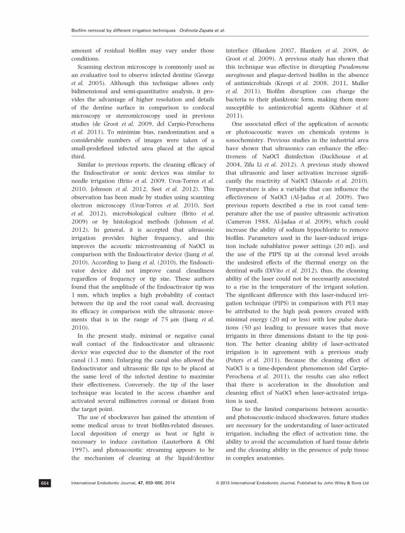
amount of residual biofilm may vary under those
conditions.
Scanning electron microscopy is commonly used as
an evaluative tool to observe infected dentine (George
et al. 2005). Although this technique allows only
bidimensional and semi-quantitative analysis, it pro-
vides the advantage of higher resolution and details
of the dentine surface in comparison to confocal
microscopy or stereomicroscopy used in previous
studies (de Groot et al. 2009, del Carpio-Perochena
et al. 2011). To minimize bias, randomization and a
considerable numbers of images were taken of a
small-predefined infected area placed at the apical
third.
Similar to previous reports, the cleaning efficacy of
the Endoactivator or sonic devices was similar to
needle irrigation (Brito et al. 2009, Uroz-Torres et al.
2010, Johnson et al. 2012, Seet et al. 2012). This
observation has been made by studies using scanning
electron microscopy (Uroz-Torres et al. 2010, Seet
et al. 2012), microbiological culture (Brito et al.
2009) or by histological methods (Johnson et al.
2012). In general, it is accepted that ultrasonic
irrigation provides higher frequency, and this
improves the acoustic microstreaming of NaOCl in
comparison with the Endoactivator device (Jiang et al.
2010). According to Jiang et al. (2010), the Endoacti-
vator device did not improve canal cleanliness
regardless of frequency or tip size. These authors
found that the amplitude of the Endoactivator tip was
1 mm, which implies a high probability of contact
between the tip and the root canal wall, decreasing
its efficacy in comparison with the ultrasonic move-
ments that is in the range of 75 lm (Jiang et al.
2010).
In the present study, minimal or negative canal
wall contact of the Endoactivator and ultrasonic
device was expected due to the diameter of the root
canal (1.3 mm). Enlarging the canal also allowed the
Endoactivator and ultrasonic file tips to be placed at
the same level of the infected dentine to maximize
their effectiveness. Conversely, the tip of the laser
technique was located in the access chamber and
activated several millimetres coronal or distant from
the target point.
The use of shockwaves has gained the attention of
some medical areas to treat biofilm-related diseases.
Local deposition of energy as heat or light is
necessary to induce cavitation (Lauterborn & Ohl
1997), and photoacoustic streaming appears to be
the mechanism of cleaning at the liquid/dentine
interface (Blanken 2007, Blanken et al. 2009, de
Groot et al. 2009). A previous study has shown that
this technique was effective in disrupting Pseudomona
aureginosas and plaque-derived biofilm in the absence
of antimicrobials (Krespi et al. 2008, 2011, Muller
et al. 2011). Biofilm disruption can change the
bacteria to their planktonic form, making them more
susceptible to antimicrobial agents (Kizhner et al.
2011).
One associated effect of the application of acoustic
or photoacoustic waves on chemicals systems is
sonochemistry. Previous studies in the industrial area
have shown that ultrasonics can enhance the effec-
tiveness of NaOCl disinfection (Duckhouse et al.
2004, Zifu Li et al. 2012). A previous study showed
that ultrasonic and laser activation increase signifi-
cantly the reactivity of NaOCl (Macedo et al. 2010).
Temperature is also a variable that can influence the
effectiveness of NaOCl (Al-Jadaa et al. 2009). Two
previous reports described a rise in root canal tem-
perature after the use of passive ultrasonic activation
(Cameron 1988, Al-Jadaa et al. 2009), which could
increase the ability of sodium hypochlorite to remove
biofilm. Parameters used in the laser-induced irriga-
tion include subablative power settings (20 mJ), and
the use of the PIPS tip at the coronal level avoids
the undesired effects of the thermal energy on the
dentinal walls (DiVito et al. 2012), thus, the cleaning
ability of the laser could not be necessarily associated
to a rise in the temperature of the irrigant solution.
The significant difference with this laser-induced irri-
gation technique (PIPS) in comparison with PUI may
be attributed to the high peak powers created with
minimal energy (20 mJ or less) with low pulse dura-
tions (50 ls) leading to pressure waves that move
irrigants in three dimensions distant to the tip posi-
tion. The better cleaning ability of laser-activated
irrigation is in agreement with a previous study
(Peters et al. 2011). Because the cleaning effect of
NaOCl is a time-dependent phenomenon (del Carpio-
Perochena et al. 2011), the results can also reflect
that there is acceleration in the dissolution and
cleaning effect of NaOCl when laser-activated irriga-
tion is used.
Due to the limited comparisons between acoustic-
and photoacoustic-induced shockwaves, future studies
are necessary for the understanding of laser-activated
irrigation, including the effect of activation time, the
ability to avoid the accumulation of hard tissue debris
and the cleaning ability in the presence of pulp tissue
in complex anatomies.
Biofilm removal by different irrigation techniques Ordinola-Zapata et al.
© 2013 International Endodontic Journal. Published by John Wiley & Sons LtdInternational Endodontic Journal, 47, 659–666, 2014664
Conclusions
Under the conditions of the current study, laser-acti-
vated irrigation using the photon-induced photoacou-
stic streaming technique of 6% sodium hypochlorite
significantly improved the cleaning of biofilm-infected
dentine compared with passive ultrasonic, sonic or
mere needle irrigation.
Acknowledgement
Supported by FAPESP grants 2010/16002-4 and
2011/22283-9.
References
Ahmad M, Pitt Ford TJ, Crum LA (1987) Ultrasonic debride-
ment of root canals: acoustic streaming and its possible
role. Journal of Endodontics 13, 490–9.
Al-Jadaa A, Paqu�e F, Attin T, Zehnder M (2009) Necrotic
pulp tissue dissolution by passive ultrasonic irrigation in
simulated accessory canals: impact of canal location and
angulation. International Endodontic Journal 42, 59–65.
Barthel CR, Zimmer S, Zilliges S, Schiller R, Gobel UB, Roulet
JF (2002) In situ antimicrobial effectiveness of chlorhexi-
dine and calcium hydroxide: gel and paste versus gutta-
percha points. Journal of Endodontics 28, 427–30.
Bhuva B, Patel S, Wilson R, Niazi S, Beighton D, Mannocci F
(2010) The effectiveness of passive ultrasonic irrigation on
intraradicular Enterococcus faecalis biofilms in extracted
single-rooted human teeth. International Endodontic Journal
43, 241–50.
Blanken RMV (2007) Cavitation as a working mechanism of
the Er, Cr: YSGG laser in endodontics: a visualization
study. Journal of Oral Laser Applications 7, 97–106.
Blanken J, De Moor RJG, Meire M, Verdaasdonk R (2009)
Laser induced explosive vapor and cavitation resulting in
effective irrigation of the root canal. Part 1: a visualization
study. Lasers in Surgery and Medicine 41, 514–9.
Brito PR, Souza LC, Machado de Oliveira JC et al. (2009)
Comparison of the effectiveness of three irrigation tech-
niques in reducing intracanal Enterococcus faecalis popula-
tions: an in vitro study. Journal of Endodontics 35, 1422–7.
Burleson A, Nusstein J, Reader A, Beck M (2007) The in vivo
evaluation of hand/rotary/ultrasound instrumentation in
necrotic, human mandibular molars. Journal of Endodontics
33, 782–7.
Bystr€om A, Sundqvist G (1983) Bacteriologic evaluation of
the effect of 0.5 percent sodium hypochlorite in endodon-
tic therapy. Oral Surgery, Oral Medicine, and Oral Pathology
55, 307–12.
Cameron JA (1988) The effect of ultrasonic endodontics on
the temperature of the root canal wall. Journal of Endodon-
tics 14, 554–8.
Chow T (1983) Mechanical effectiveness of root canal irriga-
tion. Journal of Endodontics 9, 475–9.
de Gregorio C, Estevez R, Cisneros R, Paranjpe A, Cohenca N
(2010) Efficacy of different irrigation and activation
systems on the penetration of sodium hypochlorite into
simulated lateral canals and up to working length: an in
vitro study. Journal of Endodontics 36, 1216–21.
de Groot SD, Verhaagen B, Versluis M, Wu MK, Wesselink
PR, van der Sluis LWM (2009) Laser-activated irrigation
within root canals: cleaning efficacy and flow visualiza-
tion. International Endodontic Journal 42, 1077–83.
de Moor RJG, Blanken J, Meire M, Verdaasdonk R (2009)
Laser induced explosive vapor and cavitation resulting in
effective irrigation of the root canal. Part 2: evaluation of
the efficacy. Lasers in Surgery and Medicine 41, 520–3.
del Carpio-Perochena AE, Bramante CM, Duarte MA et al.
(2011) Biofilm dissolution and cleaning ability of different
irrigant solutions on intraorally infected dentin. Journal of
Endodontics 37, 1134–8.
DiVito E, Peters OA, Olivi G (2012) Effectiveness of the
erbium:YAG laser and new design radial and stripped tips
in removing the smear layer after root canal instrumenta-
tion. Lasers in Medical Science 27, 273–80.
Duckhouse H, Mason TJ, Phull SS, Lorimer JP (2004) The
effect of sonication on microbial disinfection using
hypochlorite. Ultrasonics Sonochemistry 11, 173–6.
George S, Kishen A, Song KP (2005) The role of environ-
mental changes on monospecies biofilm formation on root
canal wall by Enterococcus faecalis. Journal of Endodontics
31, 867–72.
George R, Meyers IA, Walsh LJ (2008) Laser activation of
endodontic irrigants with improved conical laser fiber tips
for removing smear layer in the apical third of the root
canal. Journal of Endodontics 34, 1524–7.
Jiang LM, Verhaagen B, Versluis M, van der Sluis LWM
(2010) Evaluation of a sonic device designed to activate
irrigant in the root canal. Journal of Endodontics 36, 143–6.
Johnson M, Sidow SJ, Looney SW, Lindsey K, Niu LN, Tay
FR (2012) Canal and isthmus debridement efficacy using
a sonic irrigation technique in a closed-canal system. Jour-
nal of Endodontics 38, 1265–8.
Kizhner V, Krespi YP, Hall-Stoodley L, Stoodley P (2011)
Laser-generated shockwave for clearing medical device
biofilms. Photomedicine and Laser Surgery 29, 277–82.
Krespi YP, Stoodley P, Hall-Stoodley L (2008) Laser disrup-
tion of biofilm. The Laryngoscope 118, 1168–73.
Krespi YP, Kizhner V, Nistico L, Hall-Stoodley L, Stoodley P
(2011) Laser disruption and killing of methicillin-resistant
Staphylococcus aureus biofilms. American Journal of Otolar-
yngology 32, 198–202.
Lauterborn W, Ohl CD (1997) Cavitation bubble dynamics.
Ultrasonic Sonochemistry 4, 65–75.
Macedo RG, Wesselink PR, Zaccheo F, Fanali D, Van Der
Sluis LW (2010) Reaction rate of NaOCl in contact
with bovine dentine: effect of activation, exposure time,
Ordinola-Zapata et al. Biofilm removal by different irrigation techniques
© 2013 International Endodontic Journal. Published by John Wiley & Sons Ltd International Endodontic Journal, 47, 659–666, 2014 665
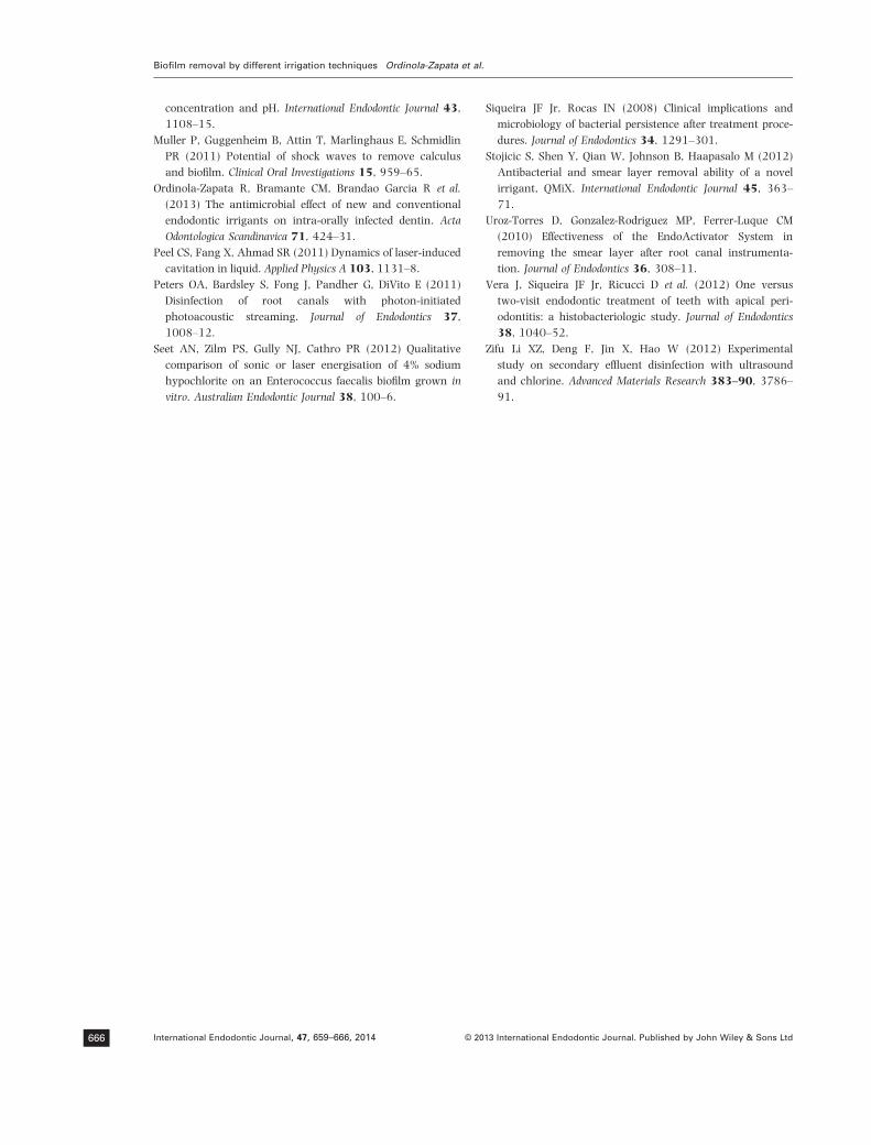
concentration and pH. International Endodontic Journal 43,
1108–15.
Muller P, Guggenheim B, Attin T, Marlinghaus E, Schmidlin
PR (2011) Potential of shock waves to remove calculus
and biofilm. Clinical Oral Investigations 15, 959–65.
Ordinola-Zapata R, Bramante CM, Brandao Garcia R et al.
(2013) The antimicrobial effect of new and conventional
endodontic irrigants on intra-orally infected dentin. Acta
Odontologica Scandinavica 71, 424–31.
Peel CS, Fang X, Ahmad SR (2011) Dynamics of laser-induced
cavitation in liquid. Applied Physics A 103, 1131–8.
Peters OA, Bardsley S, Fong J, Pandher G, DiVito E (2011)
Disinfection of root canals with photon-initiated
photoacoustic streaming. Journal of Endodontics 37,
1008–12.
Seet AN, Zilm PS, Gully NJ, Cathro PR (2012) Qualitative
comparison of sonic or laser energisation of 4% sodium
hypochlorite on an Enterococcus faecalis biofilm grown in
vitro. Australian Endodontic Journal 38, 100–6.
Siqueira JF Jr, Rocas IN (2008) Clinical implications and
microbiology of bacterial persistence after treatment proce-
dures. Journal of Endodontics 34, 1291–301.
Stojicic S, Shen Y, Qian W, Johnson B, Haapasalo M (2012)
Antibacterial and smear layer removal ability of a novel
irrigant, QMiX. International Endodontic Journal 45, 363–
71.
Uroz-Torres D, Gonzalez-Rodriguez MP, Ferrer-Luque CM
(2010) Effectiveness of the EndoActivator System in
removing the smear layer after root canal instrumenta-
tion. Journal of Endodontics 36, 308–11.
Vera J, Siqueira JF Jr, Ricucci D et al. (2012) One versus
two-visit endodontic treatment of teeth with apical peri-
odontitis: a histobacteriologic study. Journal of Endodontics
38, 1040–52.
Zifu Li XZ, Deng F, Jin X, Hao W (2012) Experimental
study on secondary effluent disinfection with ultrasound
and chlorine. Advanced Materials Research 383–90, 3786–
91.
Biofilm removal by different irrigation techniques Ordinola-Zapata et al.
© 2013 International Endodontic Journal. Published by John Wiley & Sons LtdInternational Endodontic Journal, 47, 659–666, 2014666
Clinical Research
A Comparative Investigation of Cone-beam ComputedTomography and Periapical Radiography in the Diagnosisof a Healthy PeriapexOliver Pope, BDSc, DCD, Chankhrit Sathorn, DCD, PhD, and Peter Parashos, MDSc, PhD
Abstract
Introduction: This research aimed to compare theappearance of healthy periapical tissues on cone-beamcomputed tomography (CBCT) with periapical radiog-raphy and to measure the periodontal ligament (PDL)space on CBCT for teeth with healthy and necrotic pulps.Methods: Patient records from specialist endodonticpractices were examined for teeth that had a high-resolution (0.08-mm voxel) and small field-of-viewCBCT scan, a periapical radiograph, and clinical pulptests (CO2 and electric pulp testing). The periapicalregions of the CBCT scans and radiographs were scoredindividually by 2 calibrated, blinded examiners by usinga modified CBCT-periapical index (CBCT-PAI) for bothand represented as CBCT-PAI and PAI, respectively.The Fisher exact and c2 statistics tested the relation-ships between CBCT-PAI, PAI, and pulp status. Results:Of 200 teeth included in the study, 166 showed clinicalsigns of pulpal health, and the CBCT-PAI score wasgreater than the PAI in 72% (119 of 166), with a vitalpulp likely to have a radiographic PDL space wideningof 0–1 mm (P < .001). Although 2 healthy teeth showedradiolucencies 2–4 mm on CBCT scan when the periapicalradiograph showed none, a PDL space of greater than 1–2mm was indicative of a necrotic pulp (P < .001).Conclusions: Teeth with necrotic pulps were more likelyto have PDL widening, but the PDL space of a healthytooth demonstrated significant variation when examinedby CBCT. The radiographic interpretation of health anddisease on CBCT must be further investigated beforeusage in outcome or epidemiologic investigations. Thisresearch questions the traditional radiographic interpreta-tion of the PDL space. (J Endod 2014;40:360–365)Key WordsCone-beam computed tomography, diagnosis, intraoralradiographs, periapical lesions
From the Melbourne Dental School, University of Mel-bourne, Melbourne, Victoria, Australia.
Address requests for reprints to Prof Peter Parashos, Mel-bourne Dental School, University of Melbourne, 720 SwanstonStreet, Melbourne, Victoria 3010, Australia. E-mail address:[email protected]/$ - see front matter
Copyright ª 2014 American Association of Endodontists.http://dx.doi.org/10.1016/j.joen.2013.10.003
360 Pope et al.
Radiographic imaging is essential in all stages of endodontics from the diagnosisthrough long-term assessment of healing outcomes. In conjunction with symptoms,
outcome is assessed by comparison of preoperative and/or immediate postoperativeradiographs, with subsequent radiographs taken at review appointments. The appearanceof the periapical tissues on a radiograph is influenced by the superimposition of anatomicstructures and the variable nature of the overlying bone density and texture; interpretationcan have a high degree of both interexaminer and intraexaminer variability (1, 2).
The diagnostic value of pretreatment radiographs depends on how well they reflectthe histology of apical periodontitis. Studies that have investigated the correlation betweenhistologic appearance and radiographic manifestations (3–6) have found that the absenceof radiographic signs does not preclude apical inflammation, and the radiographicappearance is always smaller than the histologic extent of the lesion. Radiographic signspathognomonic of apical periodontitis include radiolucent changes in periradiculartrabecular pattern and altered shape and width of the periodontal ligament (PDL)space (3–6).
The limitations of periapical radiography have led to significant interest incone-beam computed tomography (CBCT) and specific endodontic applications.Although there are no published data available, anecdotally it seems that the numberof CBCT scans taken every year is increasing as awareness increases, resolution increases,and costs reduce. A similar parallel may be drawn with the use of medical CT scans. Sincethe 1990s, the per-capita use of head CT scans has doubled, and chest CT scans haveincreased 5-fold (7), and it is likely that the use of CBCTmay increase similarly. Althoughan attractive imaging modality, including as a research tool (8), the radiation safety prin-ciples of as low as reasonably achievable require adherence, and an increase in clinicaluse is not yet supported by the development of evidence-based protocols to show animproved outcome for the patient. Along these lines, although CBCT may be a usefultool for observing the bone accumulation in periapical lesions, Kaya et al (9) confirmedthere is a need to reduce the radiation dosage, confirming the American Associationof Endodontists and the American Academy of Oral and Maxillofacial Radiology JointPosition Statement recommending against routine use but limited to assessment andtreatment of complex endodontic conditions (10).
Many clinical and laboratory studies have compared the diagnostic accuracy ofCBCT and traditional periapical radiography (11–19). Although these conclude thatCBCT is more readily able to detect simulated defects, they lack clinical applicabilitybecause of failure to use an appropriate reference standard to delineate the truepresence of disease, and there are still limitations to the accuracy of CBCT that shouldbe considered during interpretation (18). In 2 studies that reported CBCT was a moreaccurate diagnostic method, the CBCT itself was used as the reference standard, withno mention of what was defined as apical periodontitis and whether such an appearancecorrelated to true presence of disease (11, 12). Another study (14) used histopathologyas the reference standard to correlate with radiographic appearance and concludedCBCT was a significantly more accurate diagnostic tool than periapical radiographs.
Although the sensitivity is, or approaches, 100% in in vitro studies, CBCT haslimitations. Comparing the diagnostic accuracy of CBCT complete 360� versus partial180� scans in the detection of artificial periapical lesions reveals similar accuracy,sensitivity, and specificity (20). The sensitivity is the proportion of apical periodontitisthat has been correctly diagnosed, whereas the specificity is the proportion of healthy
JOE — Volume 40, Number 3, March 2014
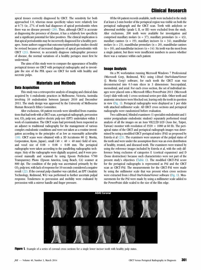
Clinical Research
apical tissues correctly diagnosed by CBCT. The sensitivity for bothapproached 1.0, whereas mean specificity values were relatively lowat 0.73 (ie, 27% of teeth had diagnoses of apical periodontitis whenthere was no disease present) (20). Thus, although CBCT is accurateat diagnosing the presence of disease, it has a relatively low specificityand a significant potential for false positives. The clinical implication isthat apical periodontitis may be incorrectly diagnosed for a healthy peri-apex. Some authors suggest that outcome/epidemiologic studies shouldbe revised because of increased diagnosis of apical periodontitis withCBCT (21). However, to accurately diagnose radiographic presenceof disease, the normal variations of a healthy periapex must first beunderstood.The aims of this study were to compare the appearance of healthyperiapical tissues on CBCT with periapical radiography and to investi-gate the size of the PDL space on CBCT for teeth with healthy andnecrotic pulps.
Materials and MethodsData Acquisition
This study was a retrospective analysis of imaging and clinical dataacquired by 4 endodontic practices in Melbourne, Victoria, Australiainvolving 10 endodontists between January 2010 and December2011. The study design was approved by the University of MelbourneHuman Research Ethics Committee.
After exclusions, 68 patient records were identified from examina-tions that had teeth with a CBCT scan, a periapical radiograph, percussiontest, CO2 pulp test, and/or electric pulp test (EPT) undertaken within 1week of examination. The CBCT scans had previously been requested asan adjunct to traditional radiography for the management of variouscomplex endodontic conditions and were not taken as a routine investi-gation according to the principles of as low as reasonably achievable(10). CBCT scans were obtained with a 3D Accuitomo 80 (J. Morita,Corporation, Kyoto, Japan), small (40 � 40 � 40 mm) field of view,and voxel size of 0.08 � 0.08 � 0.08 mm. The periapicalradiographs were taken according to the paralleling radiographic tech-nique. Sixty of the radiographs were digitally acquired, and 8 were pro-cessed manually and scanned by using Epsom Perfection V700Transparency Photo (Epsom America, Long Beach, CA) scanner at800 dpi. The condition of the pulp was ascertained primarily by theCO2 pulp test, with lack of response for 10 seconds considered a negativeresult (22). If the coronal pulp chamber was calcified, an EPT (AnalyticTechnology, Redmond, WA) was performed to further ascertain pulpalresponse. Tenderness to percussion and mobility were evaluated bypercussion with a mirror handle and finger pressure.
Figure 1. Example of a series of coronal cross sections for a single lower incisor
JOE — Volume 40, Number 3, March 2014
Of the 68 patient records available, teeth were included in the studyif at least a 3-mm border of the periapical region was visible on both theperiapical radiograph and the CBCT scan. Teeth with ankylosis orabnormal mobility (grade I, II, or III) were excluded from the study.After exclusions, 200 teeth were available for investigation andcomprised maxillary molars (n = 37), maxillary premolars (n = 43),maxillary canines (n = 19), maxillary incisors (n = 34), mandibularmolars (n = 23), mandibular premolars (n = 20), mandibular canines(n = 10), andmandibular incisors (n = 14). Six teeth was the most froma single patient, but there were insufficient numbers to assess whetherthere was a variance within each patient.
Image AnalysisOn a PC workstation running Microsoft Windows 7 Professional
(Microsoft Corp, Redmond, WA) using i-Dixel OneVolumeViewer(J. Morita Corp) software, for each tooth the CBCT scan wasdeconstructed into 0.5-mm slices in 3 dimensions: buccopalatal,mesiodistal, and axial. For each cross section, the set of individual im-ages were placed onto a Microsoft Office PowerPoint 2011 (MicrosoftCorp) slide with only 1 cross-sectional series per slide. Other teeth andanatomic structures were blocked out, leaving only the tooth in questionin view (Fig. 1). Periapical radiographs were displayed as 1 per slidewith attached millimeter scale. All CBCT cross sections and periapicalradiographs were randomized before evaluation.
Two calibrated, blinded examiners (1 specialist endodontist and 1senior postgraduate endodontic student) separately performed visualanalysis of all the images on an Asus VH222D LCD (Asus Inc, Taipei,Taiwan) monitor with resolution of 1920 � 1080 at 60 Hz. The peri-apical status of the CBCT and periapical radiograph images was deter-mined by using a modified CBCT-periapical index (PAI) as proposed byEstrela et al (23). The examiners were unaware of the pulpal status ofthe teeth and were under the assumption there was an even distributionof healthy, treated, and diseased teeth. The examiners were trained byusing the reference images included by Estrela et al, with the only dif-ference being exclusion of categories E (cortical expansion) and D(bone destruction) because such characteristics were not part of thepresent study’s objectives (Table 1). The modified CBCT-PAI scorefor the periapical radiographs is represented as PAI and the CBCTscan as CBCT-PAI. The measurements for the CBCT-PAI were madeby using the millimeter scale that was present when cross sectionswere extracted from i-Dixel OneVolumeViewer software (Fig. 1). Mea-surements for the PAI were made by using a millimeter scale added tothe PowerPoint slide scaled to the size of the film edge.
tooth with healthy pulp status.
CBCT Versus Periapical Radiography in Diagnosis 361
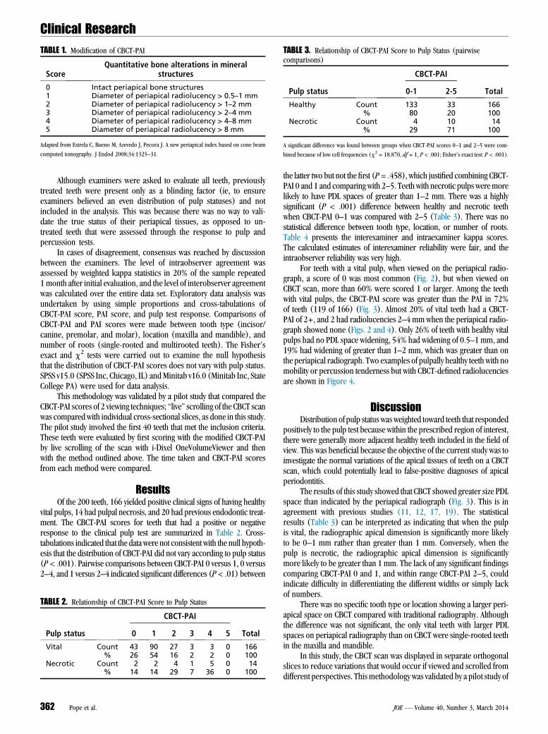
TABLE 1. Modification of CBCT-PAI
ScoreQuantitative bone alterations in mineral
structures
0 Intact periapical bone structures1 Diameter of periapical radiolucency > 0.5–1 mm2 Diameter of periapical radiolucency > 1–2 mm3 Diameter of periapical radiolucency > 2–4 mm4 Diameter of periapical radiolucency > 4–8 mm5 Diameter of periapical radiolucency > 8 mm
Adapted from Estrela C, Bueno M, Azevedo J, Pecora J. A new periapical index based on cone beam
computed tomography. J Endod 2008;34:1325–31.
TABLE 3. Relationship of CBCT-PAI Score to Pulp Status (pairwisecomparisons)
Pulp status
CBCT-PAI
Total0-1 2-5
Healthy Count 133 33 166% 80 20 100
Necrotic Count 4 10 14% 29 71 100
A significant difference was found between groups when CBCT-PAI scores 0–1 and 2–5 were com-
bined because of low cell frequencies (c2 = 18.870, df = 1, P < .001; Fisher’s exact test: P < .001).
Clinical Research
Although examiners were asked to evaluate all teeth, previouslytreated teeth were present only as a blinding factor (ie, to ensureexaminers believed an even distribution of pulp statuses) and notincluded in the analysis. This was because there was no way to vali-date the true status of their periapical tissues, as opposed to un-treated teeth that were assessed through the response to pulp andpercussion tests.
In cases of disagreement, consensus was reached by discussionbetween the examiners. The level of intraobserver agreement wasassessed by weighted kappa statistics in 20% of the sample repeated1month after initial evaluation, and the level of interobserver agreementwas calculated over the entire data set. Exploratory data analysis wasundertaken by using simple proportions and cross-tabulations ofCBCT-PAI score, PAI score, and pulp test response. Comparisons ofCBCT-PAI and PAI scores were made between tooth type (incisor/canine, premolar, and molar), location (maxilla and mandible), andnumber of roots (single-rooted and multirooted teeth). The Fisher’sexact and c2 tests were carried out to examine the null hypothesisthat the distribution of CBCT-PAI scores does not vary with pulp status.SPSS v15.0 (SPSS Inc, Chicago, IL) andMinitab v16.0 (Minitab Inc, StateCollege PA) were used for data analysis.
This methodology was validated by a pilot study that compared theCBCT-PAI scores of 2 viewing techniques; ‘‘live’’ scrolling of the CBCT scanwas compared with individual cross-sectional slices, as done in this study.The pilot study involved the first 40 teeth that met the inclusion criteria.These teeth were evaluated by first scoring with the modified CBCT-PAIby live scrolling of the scan with i-Dixel OneVolumeViewer and thenwith the method outlined above. The time taken and CBCT-PAI scoresfrom each method were compared.
ResultsOf the 200 teeth, 166 yielded positive clinical signs of having healthy
vital pulps, 14 had pulpal necrosis, and 20 had previous endodontic treat-ment. The CBCT-PAI scores for teeth that had a positive or negativeresponse to the clinical pulp test are summarized in Table 2. Cross-tabulations indicated that the data were not consistent with the null hypoth-esis that the distribution of CBCT-PAI did not vary according to pulp status(P< .001). Pairwise comparisons between CBCT-PAI 0 versus 1, 0 versus2–4, and 1 versus 2–4 indicated significant differences (P< .01) between
TABLE 2. Relationship of CBCT-PAI Score to Pulp Status
Pulp status
CBCT-PAI
Total0 1 2 3 4 5
Vital Count 43 90 27 3 3 0 166% 26 54 16 2 2 0 100
Necrotic Count 2 2 4 1 5 0 14% 14 14 29 7 36 0 100
362 Pope et al.
the latter two but not the first (P= .458), which justified combining CBCT-PAI 0 and 1 and comparing with 2–5. Teeth with necrotic pulps weremorelikely to have PDL spaces of greater than 1–2 mm. There was a highlysignificant (P < .001) difference between healthy and necrotic teethwhen CBCT-PAI 0–1 was compared with 2–5 (Table 3). There was nostatistical difference between tooth type, location, or number of roots.Table 4 presents the interexaminer and intraexaminer kappa scores.The calculated estimates of interexaminer reliability were fair, and theintraobserver reliability was very high.
For teeth with a vital pulp, when viewed on the periapical radio-graph, a score of 0 was most common (Fig. 2), but when viewed onCBCT scan, more than 60% were scored 1 or larger. Among the teethwith vital pulps, the CBCT-PAI score was greater than the PAI in 72%of teeth (119 of 166) (Fig. 3). Almost 20% of vital teeth had a CBCT-PAI of 2+, and 2 had radiolucencies 2–4 mmwhen the periapical radio-graph showed none (Figs. 2 and 4). Only 26% of teeth with healthy vitalpulps had no PDL space widening, 54% had widening of 0.5–1 mm, and19% had widening of greater than 1–2 mm, which was greater than onthe periapical radiograph. Two examples of pulpally healthy teeth with nomobility or percussion tenderness but with CBCT-defined radiolucenciesare shown in Figure 4.
DiscussionDistribution of pulp status wasweighted toward teeth that responded
positively to the pulp test because within the prescribed region of interest,there were generally more adjacent healthy teeth included in the field ofview. This was beneficial because the objective of the current study was toinvestigate the normal variations of the apical tissues of teeth on a CBCTscan, which could potentially lead to false-positive diagnoses of apicalperiodontitis.
The results of this study showed that CBCT showed greater size PDLspace than indicated by the periapical radiograph (Fig. 3). This is inagreement with previous studies (11, 12, 17, 19). The statisticalresults (Table 3) can be interpreted as indicating that when the pulpis vital, the radiographic apical dimension is significantly more likelyto be 0–1 mm rather than greater than 1 mm. Conversely, when thepulp is necrotic, the radiographic apical dimension is significantlymore likely to be greater than 1 mm. The lack of any significant findingscomparing CBCT-PAI 0 and 1, and within range CBCT-PAI 2–5, couldindicate difficulty in differentiating the different widths or simply lackof numbers.
There was no specific tooth type or location showing a larger peri-apical space on CBCT compared with traditional radiography. Althoughthe difference was not significant, the only vital teeth with larger PDLspaces on periapical radiography than on CBCT were single-rooted teethin the maxilla and mandible.
In this study, the CBCT scan was displayed in separate orthogonalslices to reduce variations that would occur if viewed and scrolled fromdifferent perspectives. This methodology was validated by a pilot study of
JOE — Volume 40, Number 3, March 2014
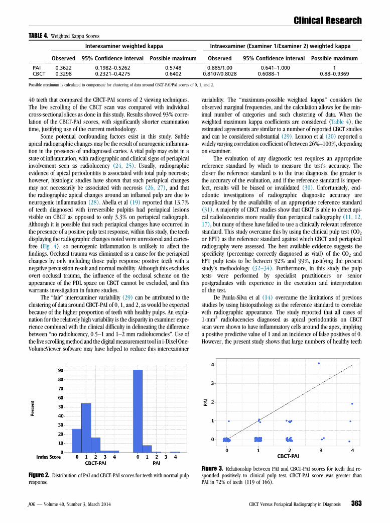
TABLE 4. Weighted Kappa Scores
Interexaminer weighted kappa Intraexaminer (Examiner 1/Examiner 2) weighted kappa
Observed 95% Confidence interval Possible maximum Observed 95% Confidence interval Possible maximum
PAI 0.3622 0.1982–0.5262 0.5748 0.885/1.00 0.641–1.000 1CBCT 0.3298 0.2321–0.4275 0.6402 0.8107/0.8028 0.6088–1 0.88–0.9369
Possible maximum is calculated to compensate for clustering of data around CBCT-PAI/PAI scores of 0, 1, and 2.
Clinical Research
40 teeth that compared the CBCT-PAI scores of 2 viewing techniques.The live scrolling of the CBCT scan was compared with individualcross-sectional slices as done in this study. Results showed 93% corre-lation of the CBCT-PAI scores, with significantly shorter examinationtime, justifying use of the current methodology.
Some potential confounding factors exist in this study. Subtleapical radiographic changes may be the result of neurogenic inflamma-tion in the presence of undiagnosed caries. A vital pulp may exist in astate of inflammation, with radiographic and clinical signs of periapicalinvolvement seen as radiolucency (24, 25). Usually, radiographicevidence of apical periodontitis is associated with total pulp necrosis;however, histologic studies have shown that such periapical changesmay not necessarily be associated with necrosis (26, 27), and thatthe radiographic apical changes around an inflamed pulp are due toneurogenic inflammation (28). Abella et al (19) reported that 13.7%of teeth diagnosed with irreversible pulpitis had periapical lesionsvisible on CBCT as opposed to only 3.3% on periapical radiograph.Although it is possible that such periapical changes have occurred inthe presence of a positive pulp test response, within this study, the teethdisplaying the radiographic changes noted were unrestored and caries-free (Fig. 4), so neurogenic inflammation is unlikely to affect thefindings. Occlusal trauma was eliminated as a cause for the periapicalchanges by only including those pulp response positive teeth with anegative percussion result and normal mobility. Although this excludesovert occlusal trauma, the influence of the occlusal scheme on theappearance of the PDL space on CBCT cannot be excluded, and thiswarrants investigation in future studies.
The ‘‘fair’’ interexaminer variability (29) can be attributed to theclustering of data around CBCT-PAI of 0, 1, and 2, as would be expectedbecause of the higher proportion of teeth with healthy pulps. An expla-nation for the relatively high variability is the disparity in examiner expe-rience combined with the clinical difficulty in delineating the differencebetween ‘‘no radiolucency, 0.5–1 and 1–2 mm radiolucencies’’. Use ofthe live scrollingmethod and the digital measurement tool in i-Dixel One-VolumeViewer software may have helped to reduce this interexaminer
Figure 2. Distribution of PAI and CBCT-PAI scores for teeth with normal pulpresponse.
JOE — Volume 40, Number 3, March 2014
variability. The ‘‘maximum-possible weighted kappa’’ considers theobserved marginal frequencies, and the calculation allows for the min-imal number of categories and such clustering of data. When theweighted maximum kappa coefficients are considered (Table 4), theestimated agreements are similar to a number of reported CBCT studiesand can be considered substantial (29). Lennon et al (20) reported awidely varying correlation coefficient of between 26%–100%, dependingon examiner.
The evaluation of any diagnostic test requires an appropriatereference standard by which to measure the test’s accuracy. Thecloser the reference standard is to the true diagnosis, the greater isthe accuracy of the evaluation, and if the reference standard is imper-fect, results will be biased or invalidated (30). Unfortunately, end-odontic investigations of radiographic diagnostic accuracy arecomplicated by the availability of an appropriate reference standard(31). A majority of CBCT studies show that CBCT is able to detect api-cal radiolucencies more readily than periapical radiography (11, 12,17), but many of these have failed to use a clinically relevant referencestandard. This study overcame this by using the clinical pulp test (CO2or EPT) as the reference standard against which CBCT and periapicalradiography were assessed. The best available evidence suggests thespecificity (percentage correctly diagnosed as vital) of the CO2 andEPT pulp tests to be between 92% and 99%, justifying the presentstudy’s methodology (32–34). Furthermore, in this study the pulptests were performed by specialist practitioners or seniorpostgraduates with experience in the execution and interpretationof the test.
De Paula-Silva et al (14) overcame the limitations of previousstudies by using histopathology as the reference standard to correlatewith radiographic appearance. The study reported that all cases of1-mm3 radiolucencies diagnosed as apical periodontitis on CBCTscan were shown to have inflammatory cells around the apex, implyinga positive predictive value of 1 and an incidence of false positives of 0.However, the present study shows that large numbers of healthy teeth
Figure 3. Relationship between PAI and CBCT-PAI scores for teeth that re-sponded positively to clinical pulp test. CBCT-PAI score was greater thanPAI in 72% of teeth (119 of 166).
CBCT Versus Periapical Radiography in Diagnosis 363
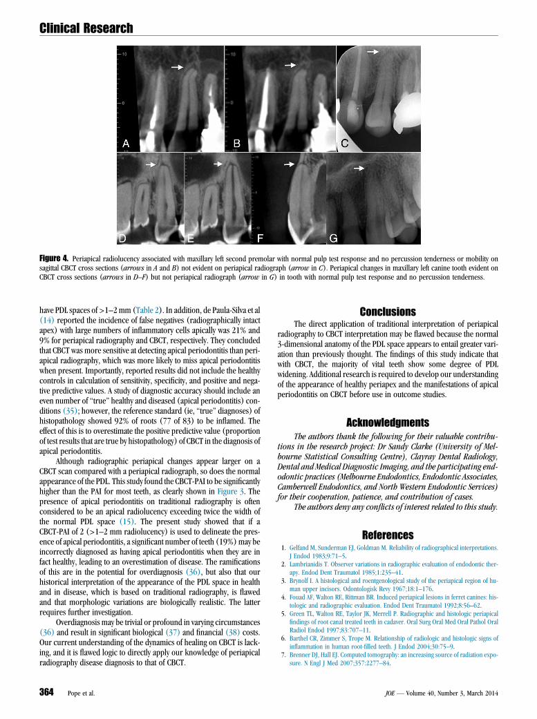
Figure 4. Periapical radiolucency associated with maxillary left second premolar with normal pulp test response and no percussion tenderness or mobility onsagittal CBCT cross sections (arrows in A and B) not evident on periapical radiograph (arrow in C). Periapical changes in maxillary left canine tooth evident onCBCT cross sections (arrows in D–F) but not periapical radiograph (arrow in G) in tooth with normal pulp test response and no percussion tenderness.
Clinical Research
have PDL spaces of >1–2mm (Table 2). In addition, de Paula-Silva et al(14) reported the incidence of false negatives (radiographically intactapex) with large numbers of inflammatory cells apically was 21% and9% for periapical radiography and CBCT, respectively. They concludedthat CBCT was more sensitive at detecting apical periodontitis than peri-apical radiography, which was more likely to miss apical periodontitiswhen present. Importantly, reported results did not include the healthycontrols in calculation of sensitivity, specificity, and positive and nega-tive predictive values. A study of diagnostic accuracy should include aneven number of ‘‘true’’ healthy and diseased (apical periodontitis) con-ditions (35); however, the reference standard (ie, ‘‘true’’ diagnoses) ofhistopathology showed 92% of roots (77 of 83) to be inflamed. Theeffect of this is to overestimate the positive predictive value (proportionof test results that are true by histopathology) of CBCT in the diagnosis ofapical periodontitis.
Although radiographic periapical changes appear larger on aCBCT scan compared with a periapical radiograph, so does the normalappearance of the PDL. This study found the CBCT-PAI to be significantlyhigher than the PAI for most teeth, as clearly shown in Figure 3. Thepresence of apical periodontitis on traditional radiography is oftenconsidered to be an apical radiolucency exceeding twice the width ofthe normal PDL space (15). The present study showed that if aCBCT-PAI of 2 (>1–2 mm radiolucency) is used to delineate the pres-ence of apical periodontitis, a significant number of teeth (19%)may beincorrectly diagnosed as having apical periodontitis when they are infact healthy, leading to an overestimation of disease. The ramificationsof this are in the potential for overdiagnosis (36), but also that ourhistorical interpretation of the appearance of the PDL space in healthand in disease, which is based on traditional radiography, is flawedand that morphologic variations are biologically realistic. The latterrequires further investigation.
Overdiagnosis may be trivial or profound in varying circumstances(36) and result in significant biological (37) and financial (38) costs.Our current understanding of the dynamics of healing on CBCT is lack-ing, and it is flawed logic to directly apply our knowledge of periapicalradiography disease diagnosis to that of CBCT.
364 Pope et al.
ConclusionsThe direct application of traditional interpretation of periapical
radiography to CBCT interpretation may be flawed because the normal3-dimensional anatomy of the PDL space appears to entail greater vari-ation than previously thought. The findings of this study indicate thatwith CBCT, the majority of vital teeth show some degree of PDLwidening. Additional research is required to develop our understandingof the appearance of healthy periapex and the manifestations of apicalperiodontitis on CBCT before use in outcome studies.
AcknowledgmentsThe authors thank the following for their valuable contribu-
tions in the research project: Dr Sandy Clarke (University of Mel-bourne Statistical Consulting Centre), Clayray Dental Radiology,Dental and Medical Diagnostic Imaging, and the participating end-odontic practices (Melbourne Endodontics, Endodontic Associates,Camberwell Endodontics, and North Western Endodontic Services)for their cooperation, patience, and contribution of cases.
The authors deny any conflicts of interest related to this study.
References1. Gelfand M, Sunderman EJ, Goldman M. Reliability of radiographical interpretations.
J Endod 1983;9:71–5.2. Lambrianidis T. Observer variations in radiographic evaluation of endodontic ther-
apy. Endod Dent Traumatol 1985;1:235–41.3. Brynolf I. A histological and roentgenological study of the periapical region of hu-
man upper incisors. Odontologisk Revy 1967;18:1–176.4. Fouad AF, Walton RE, Rittman BR. Induced periapical lesions in ferret canines: his-
tologic and radiographic evaluation. Endod Dent Traumatol 1992;8:56–62.5. Green TL, Walton RE, Taylor JK, Merrell P. Radiographic and histologic periapical
findings of root canal treated teeth in cadaver. Oral Surg Oral Med Oral Pathol OralRadiol Endod 1997;83:707–11.
6. Barthel CR, Zimmer S, Trope M. Relationship of radiologic and histologic signs ofinflammation in human root-filled teeth. J Endod 2004;30:75–9.
7. Brenner DJ, Hall EJ. Computed tomography: an increasing source of radiation expo-sure. N Engl J Med 2007;357:2277–84.
JOE — Volume 40, Number 3, March 2014
Clinical Research
8. Domark JD, Hatton JF, Benison RP, Hildebolt CF. An ex vivo comparison of digitalradiography and cone-beam and micro computed tomography in the detection ofthe number of canals in the mesiobuccal roots of maxillary molars. J Endod2013;39:901–5.
9. Kaya S, Yavuz I, Uysal I, Akkus Z. Measuring bone density in healing periapical le-sions by using cone beam computed tomography: a clinical investigation. J Endod2012;38:28–31.
10. Joint Position Statement of the American Association of Endodontists and the Amer-ican Academy of Oral and Maxillofacial Radiology. Use of cone-beam computed to-mography in endodontics. Oral Surg Oral Med Oral Pathol Oral Radiol Endod 2011;111:234–7.
11. Lofthag-Hansen S, Huumonen S, Grondahl K, Grondahl H. Limited cone-beam CTand intraoral radiography for the diagnosis of periapical pathology. Oral SurgOral Med Oral Pathol Oral Radiol Endod 2007;103:114–9.
12. Estrela C, Bueno MR, Leles CR, et al. Accuracy of cone beam computed tomographyand panoramic and periapical radiography for detection of apical periodontitis.J Endod 2008;34:273–9.
13. Garcia de Paula-Silva FW, Hassan B, Bezerra da Silva LA, et al. Outcome of root canaltreatment in dogs determined by periapical radiography and cone-beam computedtomography scans. J Endod 2009;35:723–6.
14. de Paula-Silva FWG, Wu M-K, Leonardo MR, et al. Accuracy of periapical radiog-raphy and cone-beam computed tomography scans in diagnosing apical peri-odontitis using histopathological findings as a gold standard. J Endod 2009;35:1009–12.
15. Bornstein MM, Lauber R, Sendi P, von Arx T. Comparison of periapical radiographyand limited cone-beam computed tomography in mandibular molars for analysis ofanatomical landmarks before apical surgery. J Endod 2011;37:151–7.
16. Liang YH, Li G, Wesselink PR, Wu MK. Endodontic outcome predictors identifiedwith periapical radiographs and cone-beam computed tomography scans.J Endod 2011;37:326–31.
17. Sogur E, Grondahl HG, Baksi BG, Mert A. Does a combination of two radiographsincrease accuracy in detecting acid-induced periapical lesions and does it approachthe accuracy of cone-beam computed tomography scanning? J Endod 2012;38:131–6.
18. Tsai P, Torabinejad M, Rice D, Azevedo B. Accuracy of cone-beam computed tomog-raphy and periapical radiography in detecting small periapical lesions. J Endod2012;38:965–70.
19. Abella F, Patel S, Duran-Sindreu F, et al. Evaluating the periapical status of teeth withirreversible pulpitis by using cone-beam computed tomography scanning and peri-apical radiographs. J Endod 2012;38:1588–91.
20. Lennon S, Patel S, Foschi F, et al. Diagnostic accuracy of limited-volume cone-beamcomputed tomography in the detection of periapical bone loss: 360 degrees scansversus 180 degrees scans. Int Endod J 2011;44:1118–27.
JOE — Volume 40, Number 3, March 2014
21. Wu M-K, Shemesh H, Wesselink PR. Limitations of previously published systematic re-views evaluating the outcome of endodontic treatment. Int Endod J 2009;42:656–66.
22. Chen E, Abbott PV. Evaluation of accuracy, reliability, and repeatability of five dentalpulp tests. J Endod 2011;37:1619–23.
23. Estrela C, Bueno M, Azevedo J, Pecora J. A new periapical index based on cone beamcomputed tomography. J Endod 2008;34:1325–31.
24. Caliskan MK. Pulpotomy of carious vital teeth with periapical involvement. Int EndodJ 1995;28:172–6.
25. Teixeira LS, Demarco FF, Coppola MC, Bonow ML. Clinical and radiographic eval-uation of pulpotomies performed under intrapulpal injection of anaesthetic solu-tion. Int Endod J 2001;34:440–6.
26. Mitchell DF, Tarplee RE. Painful pulpitis: a clinical and microscopic study. Oral SurgOral Med Oral Pathol 1960;13:1360–70.
27. Langeland K. Management of the inflamed pulp associated with deep carious lesion.J Endod 1981;7:169–81.
28. Kabashima H, Nagata K, Maeda K, Iijima T. Involvement of substance P, mast cells,TNF-alpha and ICAM-1 in the infiltration of inflammatory cells in human periapicalgranulomas. J Oral Pathol Med 2002;31:175–80.
29. Landis JR, Koch GG. The measurement of observer agreement for categorical data.Biometrics 1977;33:159–74.
30. Walter SD, Macaskill P, Lord SJ, Irwig L. Effect of dependent errors in the assessmentof diagnostic or screening test accuracy when the reference standard is imperfect.Stat Med 2012;31:1129–38.
31. Petersson A, Axelsson S, Davidson T, et al. Radiological diagnosis of periapical bonetissue lesions in endodontics: a systematic review. Int Endod J 2012;45:783–801.
32. Petersson K, S€oderstr€om C, Kiani-Anaraki M, L�evy G. Evaluation of the ability of thermaland electrical tests to register pulp vitality. Endod Dent Traumatol 1999;15:127–31.
33. Gopikrishna V, Tinagupta K, Kandaswamy D. Evaluation of efficacy of a new custom-made pulse oximeter dental probe in comparison with the electrical and thermaltests for assessing pulp vitality. J Endod 2007;33:411–4.
34. Weisleder R, Yamauchi S, Caplan DJ, et al. The validity of pulp testing: a clinicalstudy. J Am Dent Assoc 2009;140:1013–7.
35. Greenhalgh T. How to read a paper: papers that report diagnostic or screening tests.BMJ 1997;315:540–3.
36. Welch HG. Genesis. In: Welch HG, Schwarz L, Woloshin S, eds. Overdiagnosed:Making People Sick in the Pursuit of Health. Boston: Beacon Press; 2011:1–15.
37. Yu VS, Messer HH, Yee R, Shen L. Incidence and impact of painful exacerbations in acohort with post-treatment persistent endodontic lesions. J Endod 2012;38:41–6.
38. Brown L, Nash K, Johns B, Warren M. The Economics of Endodontics: Dental HealthPolicy Analysis Series [eBook]. Chicago: ADA, Health Policy Research Centre; 2005.Available at http://catalog.ada.org/ProductCatalog/1047/Economics-Dentistry-Dental-Health-Analysis-Series/The-Economics-of-Endodontics-Downloadable-SC/ECE-2005D. Accessed September 8, 2012.
CBCT Versus Periapical Radiography in Diagnosis 365
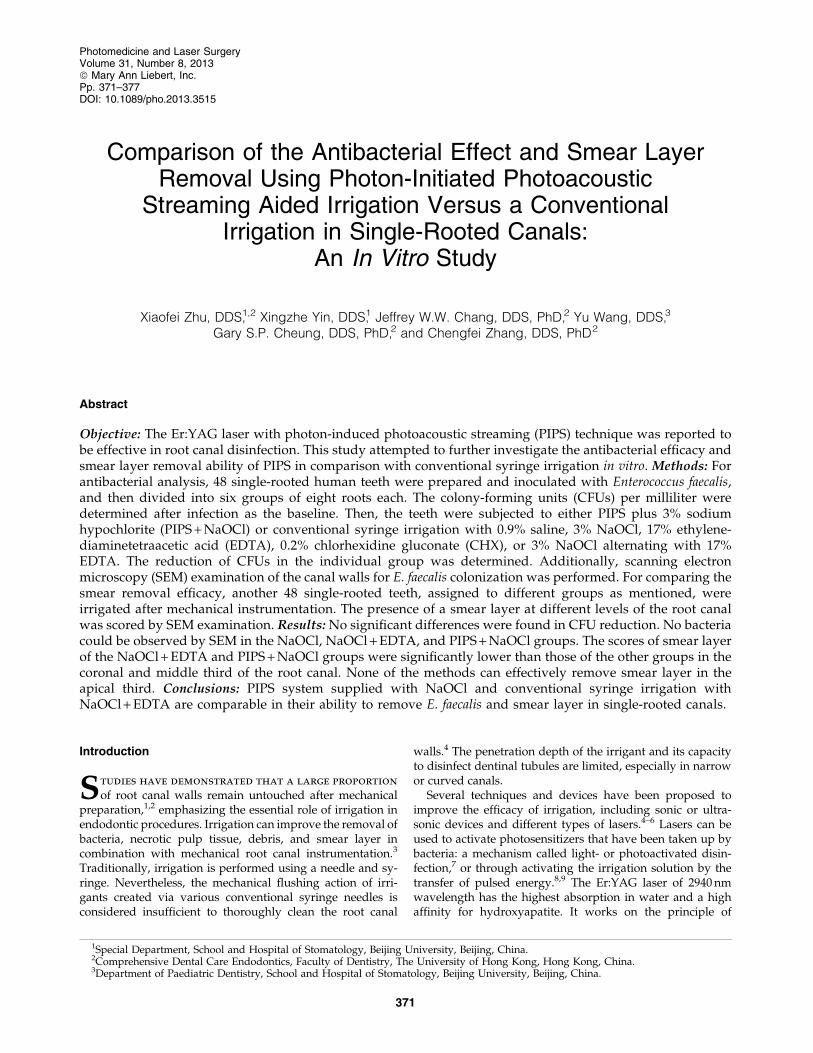
Comparison of the Antibacterial Effect and Smear LayerRemoval Using Photon-Initiated Photoacoustic
Streaming Aided Irrigation Versus a ConventionalIrrigation in Single-Rooted Canals:
An In Vitro Study
Xiaofei Zhu, DDS,1,2 Xingzhe Yin, DDS,1 Jeffrey W.W. Chang, DDS, PhD,2 Yu Wang, DDS,3
Gary S.P. Cheung, DDS, PhD,2 and Chengfei Zhang, DDS, PhD2
Abstract
Objective: The Er:YAG laser with photon-induced photoacoustic streaming (PIPS) technique was reported tobe effective in root canal disinfection. This study attempted to further investigate the antibacterial efficacy andsmear layer removal ability of PIPS in comparison with conventional syringe irrigation in vitro. Methods: Forantibacterial analysis, 48 single-rooted human teeth were prepared and inoculated with Enterococcus faecalis,and then divided into six groups of eight roots each. The colony-forming units (CFUs) per milliliter weredetermined after infection as the baseline. Then, the teeth were subjected to either PIPS plus 3% sodiumhypochlorite (PIPS + NaOCl) or conventional syringe irrigation with 0.9% saline, 3% NaOCl, 17% ethylene-diaminetetraacetic acid (EDTA), 0.2% chlorhexidine gluconate (CHX), or 3% NaOCl alternating with 17%EDTA. The reduction of CFUs in the individual group was determined. Additionally, scanning electronmicroscopy (SEM) examination of the canal walls for E. faecalis colonization was performed. For comparing thesmear removal efficacy, another 48 single-rooted teeth, assigned to different groups as mentioned, wereirrigated after mechanical instrumentation. The presence of a smear layer at different levels of the root canalwas scored by SEM examination. Results: No significant differences were found in CFU reduction. No bacteriacould be observed by SEM in the NaOCl, NaOCl + EDTA, and PIPS + NaOCl groups. The scores of smear layerof the NaOCl + EDTA and PIPS + NaOCl groups were significantly lower than those of the other groups in thecoronal and middle third of the root canal. None of the methods can effectively remove smear layer in theapical third. Conclusions: PIPS system supplied with NaOCl and conventional syringe irrigation withNaOCl + EDTA are comparable in their ability to remove E. faecalis and smear layer in single-rooted canals.
Introduction
Studies have demonstrated that a large proportion
of root canal walls remain untouched after mechanicalpreparation,1,2 emphasizing the essential role of irrigation inendodontic procedures. Irrigation can improve the removal ofbacteria, necrotic pulp tissue, debris, and smear layer incombination with mechanical root canal instrumentation.3
Traditionally, irrigation is performed using a needle and sy-ringe. Nevertheless, the mechanical flushing action of irri-gants created via various conventional syringe needles isconsidered insufficient to thoroughly clean the root canal
walls.4 The penetration depth of the irrigant and its capacityto disinfect dentinal tubules are limited, especially in narrowor curved canals.
Several techniques and devices have been proposed toimprove the efficacy of irrigation, including sonic or ultra-sonic devices and different types of lasers.4–6 Lasers can beused to activate photosensitizers that have been taken up bybacteria: a mechanism called light- or photoactivated disin-fection,7 or through activating the irrigation solution by thetransfer of pulsed energy.8,9 The Er:YAG laser of 2940 nmwavelength has the highest absorption in water and a highaffinity for hydroxyapatite. It works on the principle of
1Special Department, School and Hospital of Stomatology, Beijing University, Beijing, China.2Comprehensive Dental Care Endodontics, Faculty of Dentistry, The University of Hong Kong, Hong Kong, China.3Department of Paediatric Dentistry, School and Hospital of Stomatology, Beijing University, Beijing, China.
Photomedicine and Laser SurgeryVolume 31, Number 8, 2013ª Mary Ann Liebert, Inc.Pp. 371–377DOI: 10.1089/pho.2013.3515
371
transferring the pulsed energy to activate the irrigation so-lutions, which makes it suitable for use in root canal disin-fection and cleaning.10 Recently, a photoacoustic techniquecalled photon-induced photoacoustic streaming (PIPS), wasreported to result in effective debris and smear layer removalwith a newly designed radial and stripped tip11 or a 21 mmlong, 400 lm diameter endodontic fiber.12 With this PIPStechnique, the laser tip was placed into the pulp chamberonly, with no need to advance into the orifice of the canal;therefore, more cleaning of the root canal walls and a higherquantity of open tubules was achieved in comparison withtraditional irrigation.11,12 Therefore, we hypothesize thatEr:YAG laser with PIPS technique may have greater efficacyin promoting root canal irrigation and disinfection.
However, to the best of our knowledge, neither the anti-bacterial effect nor the debridement ability in the apical thirdof root canals has been well established for PIPS. This studyaimed to investigate the antibacterial effect and smear layerremoval of PIPS, compared with conventional syringe irri-gation in the apical area of root canals.
Materials and Methods
Ninety-six single-rooted human teeth extracted for peri-odontal reasons were used. Approval for conducting thestudy was granted by the Beijing University InstitutionalReview Board.
Experiment 1: Antibacterial effect of conventionalversus PIPS-aided irrigation on Enterococcus faecalis
Root canal preparation, inoculation and disinfection. Theexternal root surfaces of 48 single-rooted human teeth werecleaned with a curette to remove calculus and periodontaltissues. Presence of a single canal was determined by ra-diographs. Conventional access cavities were prepared. Afterthe patency was established, the canals were enlarged to anapical size of #40 using stainless steel K-files (DentsplyMaillefer, Ballaigues, Switzerland) and rotary nickel titaniumBioRace instruments (BR5, 4% taper, FKG Dentaire, LaChaux-de-Fonds, Switzerland). Copious irrigation with 3%sodium hypochlorite (NaOCl) was used throughout the rootcanal instrumentation. The smear layer was removed byusing 17% ethylenediaminetetraacetic acid (EDTA) for 3 min.A final flush with sterile 10% sodium thiosulfate solutionwas performed to inactivate any residual NaOCl. A flowablecomposite resin (3M Dental Products, St. Paul, MN) wasused to block the apical foramen by applying it over the rootapex. After that, the teeth were immersed in brain heart in-fusion broth (BHI, Difco, Detroit, MI), ultrasonicated for1 min, and then sterilized by autoclaving for 15 min at 121�.
E. faecalis strain (ATCC 29212) was used to infect the rootcanals. The flasks with infected teeth were incubated aero-bically for 4 weeks at 37� under gentle shaking, and theculture media were replenished every week.
Experimental groups and root canal management. Theinfected teeth were distributed into six groups of eight rootseach as follows. Control group, conventional irrigation with10 mL 0.9% sterile saline (NS); NaOCl group, irrigation with10 mL 3% NaOCl; CHX group, irrigation with 10 mL 0.2%chlorhexidine gluconate (CHX); EDTA group, irrigation with10 mL 17% EDTA; NaOCl + EDTA group, irrigation with 5 mL
3% NaOCl and 5 mL 17% EDTA alternatively;,PIPS + NaOClgroup, canal and pulp chamber bathed in 3% NaOCl andirradiated with Er:YAG laser in the pulp chamber for 1 min.All manual irrigation was performed with a 5 mL syringe andNaviTip 30-gauge safety needles (Ultradent, South Utah,USA). The tip of needle was at 1 mm short of the workinglength. For the PIPS + NaOCl group, an Er:YAG laser with awavelength of 2940 nm (Fidelis, Fotona, Ljubljana, Slovenia)was used with a 12 mm long 400lm diameter quartz tip. Thelaser operating parameters were 20 mJ per pulse, 15 Hz, and50 ls pulse duration.11 The coaxial water spray feature of thehandpiece was set to ‘‘off.’’ The tip was placed into the coronalaccess opening of the pulp chamber only, and kept stationaryand activated for 1 min. During the laser irradiation, addi-tional solution (3% NaOCl) was not deposited except when itwas noted that the pulp chamber was depleted of any irrigant.In such cases, care was taken to replenish the irrigant in thepulp chamber only. Approximately 3 mL NaOCl was neededfor one canal in the PIPS + NaOCl group.
Microbial analysis and scanning electron microscopy(SEM) examination. Root canals were sampled bacterio-logically before (S1) and after (S2) irrigation/irradiation witha ‘‘paper point method.’’13 Briefly, the root canal was gentlyrinsed with 1 mL of NS to remove nonadherent cells, and aninitial sample was taken by the sequential use of three paperpoints placed to the working length. All paper points for thesame tooth were transferred to 1 mL NS and immediatelyprocessed. This sample was labeled as S1. After irrigation/irradiation, the same procedure was conducted to obtainpostoperative sample S2.
In the laboratory, sample was first vortexed for 1 min,followed by 10-fold serial dilutions in saline. Then, aliquotsof 50 lL were plated onto BHI agar plates (Difco, Detroit, MI)and incubated at 37� for 48 h. The colony-forming units(CFUs) were counted and then transformed into actualcounts on the basis of the known dilution factors. Each countwas performed in duplicate on two occasions.
SEM observation of root canal walls after irrigation/irradi-ation. The roots were split longitudinally after S2 sampling.Each specimen was fixed in 4% gluteraldehyde at roomtemperature for 24 h, washed with phosphate-buffered saline(PBS) for 15 min, and post-fixed for 12 h in 1% osmium te-troxide. After a final wash with PBS, serial dehydration wasperformed with increasing concentrations of ethanol. Thespecimens were finally dried by using a SAMDRI PVT-3critical point dryer apparatus (Tousimis Research Corp.,Rockville, MD), coated with a 200 A layer of gold palladium,and examined by using a Hitachi S3400N scanning electronmicroscope (Hitachi, Tokyo, Japan) at 12 kV.
Experiment 2: Removal of smear layer by conventionaland PIPS aided irrigations
Another 48 single-rooted human teeth were recruited andsimilarly prepared as in Experiment 1, except that NS irri-gation was used throughout instrumentation. Experimentalgroup distribution and root canal management were con-ducted as in Experiment 1. After different irrigation/irradi-ation procedures, the roots were split longitudinally for SEMobservation.
372 ZHU ET AL.
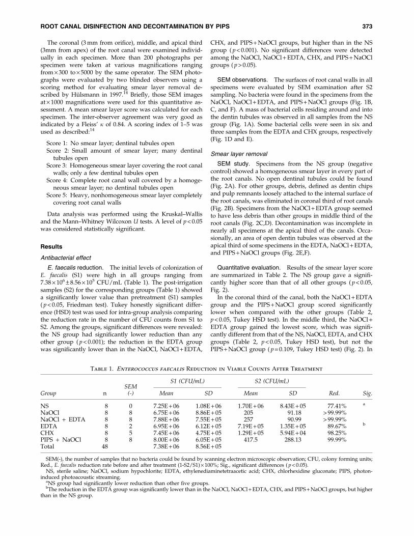
The coronal (3 mm from orifice), middle, and apical third(3mm from apex) of the root canal were examined individ-ually in each specimen. More than 200 photographs perspecimen were taken at various magnifications rangingfrom · 300 to · 5000 by the same operator. The SEM photo-graphs were evaluated by two blinded observers using ascoring method for evaluating smear layer removal de-scribed by Hulsmann in 1997.14 Briefly, those SEM imagesat · 1000 magnifications were used for this quantitative as-sessment. A mean smear layer score was calculated for eachspecimen. The inter-observer agreement was very good asindicated by a Fleiss’ j of 0.84. A scoring index of 1–5 wasused as described:14
Score 1: No smear layer; dentinal tubules openScore 2: Small amount of smear layer; many dentinal
tubules openScore 3: Homogeneous smear layer covering the root canal
walls; only a few dentinal tubules openScore 4: Complete root canal wall covered by a homoge-
neous smear layer; no dentinal tubules openScore 5: Heavy, nonhomogeneous smear layer completely
covering root canal walls
Data analysis was performed using the Kruskal–Wallisand the Mann–Whitney Wilcoxon U tests. A level of p < 0.05was considered statistically significant.
Results
Antibacterial effect
E. faecalis reduction. The initial levels of colonization ofE. faecalis (S1) were high in all groups ranging from7.38 · 106 – 8.56 · 105 CFU/mL (Table 1). The post-irrigationsamples (S2) for the corresponding groups (Table 1) showeda significantly lower value than pretreatment (S1) samples( p < 0.05, Friedman test). Tukey honestly significant differ-ence (HSD) test was used for intra-group analysis comparingthe reduction rate in the number of CFU counts from S1 toS2. Among the groups, significant differences were revealed:the NS group had significantly lower reduction than anyother group ( p < 0.001); the reduction in the EDTA groupwas significantly lower than in the NaOCl, NaOCl + EDTA,
CHX, and PIPS + NaOCl groups, but higher than in the NSgroup ( p < 0.001). No significant differences were detectedamong the NaOCl, NaOCl + EDTA, CHX, and PIPS + NaOClgroups ( p > 0.05).
SEM observations. The surfaces of root canal walls in allspecimens were evaluated by SEM examination after S2sampling. No bacteria were found in the specimens from theNaOCl, NaOCl + EDTA, and PIPS + NaOCl groups (Fig. 1B,C, and F). A mass of bacterial cells residing around and intothe dentin tubules was observed in all samples from the NSgroup (Fig. 1A). Some bacterial cells were seen in six andthree samples from the EDTA and CHX groups, respectively(Fig. 1D and E).
Smear layer removal
SEM study. Specimens from the NS group (negativecontrol) showed a homogeneous smear layer in every part ofthe root canals. No open dentinal tubules could be found(Fig. 2A). For other groups, debris, defined as dentin chipsand pulp remnants loosely attached to the internal surface ofthe root canals, was eliminated in coronal third of root canals(Fig. 2B). Specimens from the NaOCl + EDTA group seemedto have less debris than other groups in middle third of theroot canals (Fig. 2C,D). Decontamination was incomplete innearly all specimens at the apical third of the canals. Occa-sionally, an area of open dentin tubules was observed at theapical third of some specimens in the EDTA, NaOCl + EDTA,and PIPS + NaOCl groups (Fig. 2E,F).
Quantitative evaluation. Results of the smear layer scoreare summarized in Table 2. The NS group gave a signifi-cantly higher score than that of all other groups ( p < 0.05,Fig. 2).
In the coronal third of the canal, both the NaOCl + EDTAgroup and the PIPS + NaOCl group scored significantlylower when compared with the other groups (Table 2,p < 0.05, Tukey HSD test). In the middle third, the NaOCl +EDTA group gained the lowest score, which was signifi-cantly different from that of the NS, NaOCl, EDTA, and CHXgroups (Table 2, p < 0.05, Tukey HSD test), but not thePIPS + NaOCl group ( p = 0.109, Tukey HSD test) (Fig. 2). In
Table 1. Enterococcus faecalis Reduction in Viable Counts After Treatment
S1 (CFU/mL) S2 (CFU/mL)
Group nSEM
(-) Mean SD Mean SD Red. Sig.
NS 8 0 7.25E + 06 1.08E + 06 1.70E + 06 8.43E + 05 77.41% a
NaOCl 8 8 6.75E + 06 8.86E + 05 205 91.18 > 99.99%NaOCl + EDTA 8 8 7.88E + 06 7.55E + 05 257 90.99 > 99.99%EDTA 8 2 6.95E + 06 6.12E + 05 7.19E + 05 1.35E + 05 89.67% b
CHX 8 5 7.45E + 06 4.75E + 05 1.29E + 05 5.94E + 04 98.25%PIPS + NaOCl 8 8 8.00E + 06 6.05E + 05 417.5 288.13 99.99%Total 48 7.38E + 06 8.56E + 05
SEM(-), the number of samples that no bacteria could be found by scanning electron microscopic observation; CFU, colony forming units;Red., E. faecalis reduction rate before and after treatment (1-S2/S1) · 100%; Sig., significant differences ( p < 0.05).
NS, sterile saline; NaOCl, sodium hypochlorite; EDTA, ethylenediaminetetraacetic acid; CHX, chlorhexidine gluconate; PIPS, photon-induced photoacoustic streaming.
aNS group had significantly lower reduction than other five groups.bThe reduction in the EDTA group was significantly lower than in the NaOCl, NaOCl + EDTA, CHX, and PIPS + NaOCl groups, but higher
than in the NS group.
ROOT CANAL DISINFECTION AND DECONTAMINATION BY PIPS 373
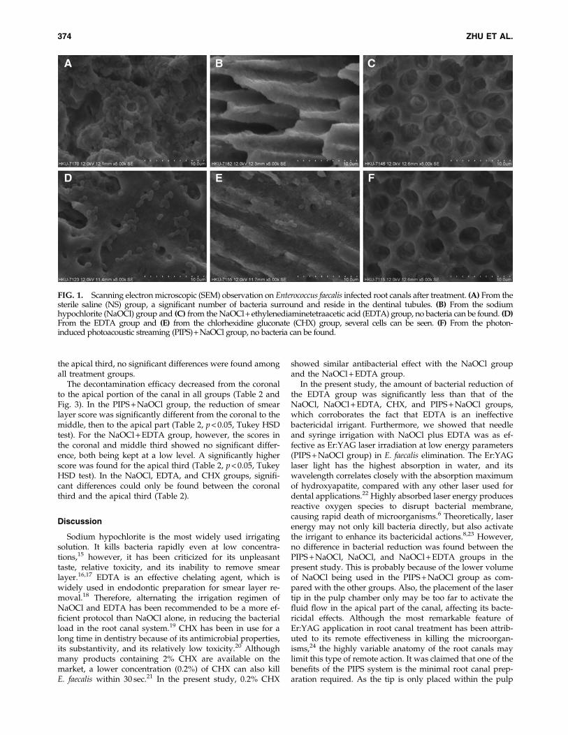
the apical third, no significant differences were found amongall treatment groups.
The decontamination efficacy decreased from the coronalto the apical portion of the canal in all groups (Table 2 andFig. 3). In the PIPS + NaOCl group, the reduction of smearlayer score was significantly different from the coronal to themiddle, then to the apical part (Table 2, p < 0.05, Tukey HSDtest). For the NaOCl + EDTA group, however, the scores inthe coronal and middle third showed no significant differ-ence, both being kept at a low level. A significantly higherscore was found for the apical third (Table 2, p < 0.05, TukeyHSD test). In the NaOCl, EDTA, and CHX groups, signifi-cant differences could only be found between the coronalthird and the apical third (Table 2).
Discussion
Sodium hypochlorite is the most widely used irrigatingsolution. It kills bacteria rapidly even at low concentra-tions,15 however, it has been criticized for its unpleasanttaste, relative toxicity, and its inability to remove smearlayer.16,17 EDTA is an effective chelating agent, which iswidely used in endodontic preparation for smear layer re-moval.18 Therefore, alternating the irrigation regimen ofNaOCl and EDTA has been recommended to be a more ef-ficient protocol than NaOCl alone, in reducing the bacterialload in the root canal system.19 CHX has been in use for along time in dentistry because of its antimicrobial properties,its substantivity, and its relatively low toxicity.20 Althoughmany products containing 2% CHX are available on themarket, a lower concentration (0.2%) of CHX can also killE. faecalis within 30 sec.21 In the present study, 0.2% CHX
showed similar antibacterial effect with the NaOCl groupand the NaOCl + EDTA group.
In the present study, the amount of bacterial reduction ofthe EDTA group was significantly less than that of theNaOCl, NaOCl + EDTA, CHX, and PIPS + NaOCl groups,which corroborates the fact that EDTA is an ineffectivebactericidal irrigant. Furthermore, we showed that needleand syringe irrigation with NaOCl plus EDTA was as ef-fective as Er:YAG laser irradiation at low energy parameters(PIPS + NaOCl group) in E. faecalis elimination. The Er:YAGlaser light has the highest absorption in water, and itswavelength correlates closely with the absorption maximumof hydroxyapatite, compared with any other laser used fordental applications.22 Highly absorbed laser energy producesreactive oxygen species to disrupt bacterial membrane,causing rapid death of microorganisms.6 Theoretically, laserenergy may not only kill bacteria directly, but also activatethe irrigant to enhance its bactericidal actions.8,23 However,no difference in bacterial reduction was found between thePIPS + NaOCl, NaOCl, and NaOCl + EDTA groups in thepresent study. This is probably because of the lower volumeof NaOCl being used in the PIPS + NaOCl group as com-pared with the other groups. Also, the placement of the lasertip in the pulp chamber only may be too far to activate thefluid flow in the apical part of the canal, affecting its bacte-ricidal effects. Although the most remarkable feature ofEr:YAG application in root canal treatment has been attrib-uted to its remote effectiveness in killing the microorgan-isms,24 the highly variable anatomy of the root canals maylimit this type of remote action. It was claimed that one of thebenefits of the PIPS system is the minimal root canal prep-aration required. As the tip is only placed within the pulp
FIG. 1. Scanning electron microscopic (SEM) observation on Enterococcus faecalis infected root canals after treatment. (A) From thesterile saline (NS) group, a significant number of bacteria surround and reside in the dentinal tubules. (B) From the sodiumhypochlorite (NaOCl) group and (C) from the NaOCl + ethylenediaminetetraacetic acid (EDTA) group, no bacteria can be found. (D)From the EDTA group and (E) from the chlorhexidine gluconate (CHX) group, several cells can be seen. (F) From the photon-induced photoacoustic streaming (PIPS) + NaOCl group, no bacteria can be found.
374 ZHU ET AL.
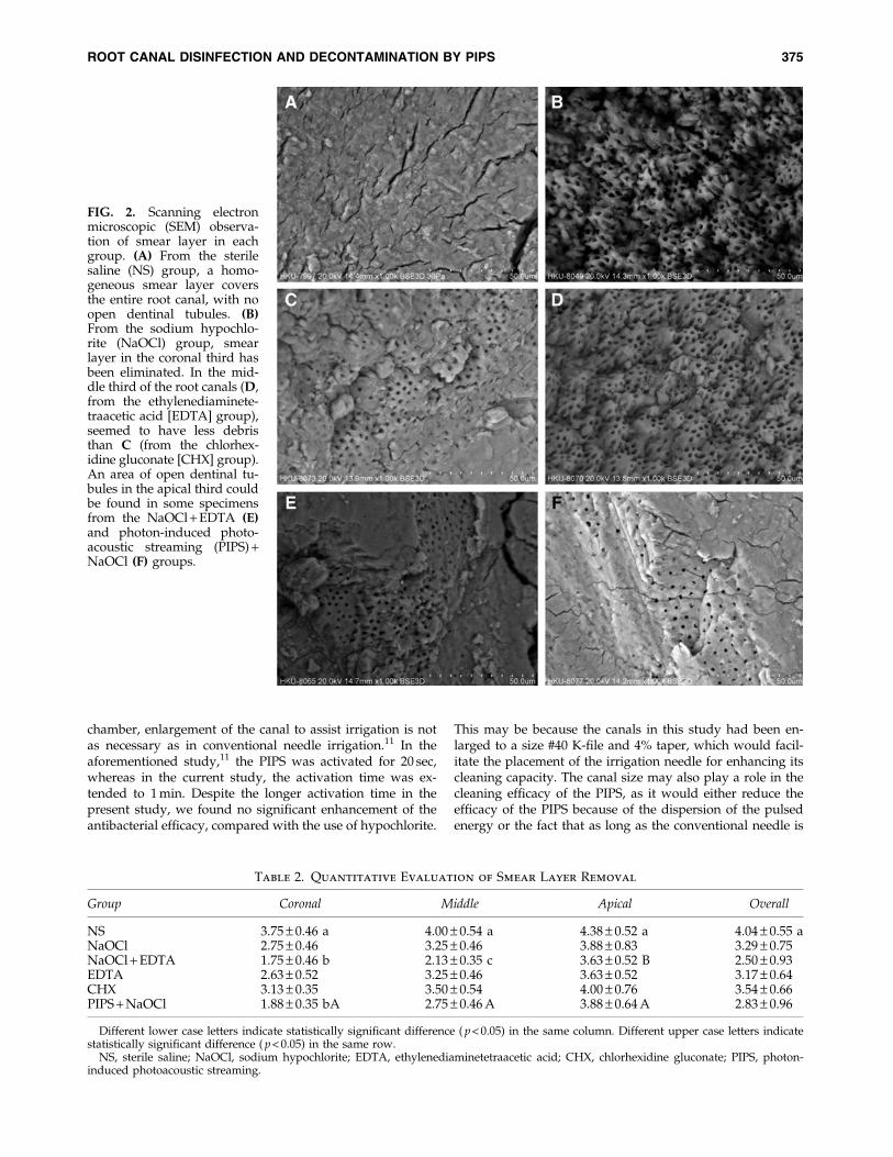
chamber, enlargement of the canal to assist irrigation is notas necessary as in conventional needle irrigation.11 In theaforementioned study,11 the PIPS was activated for 20 sec,whereas in the current study, the activation time was ex-tended to 1 min. Despite the longer activation time in thepresent study, we found no significant enhancement of theantibacterial efficacy, compared with the use of hypochlorite.
This may be because the canals in this study had been en-larged to a size #40 K-file and 4% taper, which would facil-itate the placement of the irrigation needle for enhancing itscleaning capacity. The canal size may also play a role in thecleaning efficacy of the PIPS, as it would either reduce theefficacy of the PIPS because of the dispersion of the pulsedenergy or the fact that as long as the conventional needle is
FIG. 2. Scanning electronmicroscopic (SEM) observa-tion of smear layer in eachgroup. (A) From the sterilesaline (NS) group, a homo-geneous smear layer coversthe entire root canal, with noopen dentinal tubules. (B)From the sodium hypochlo-rite (NaOCl) group, smearlayer in the coronal third hasbeen eliminated. In the mid-dle third of the root canals (D,from the ethylenediaminete-traacetic acid [EDTA] group),seemed to have less debristhan C (from the chlorhex-idine gluconate [CHX] group).An area of open dentinal tu-bules in the apical third couldbe found in some specimensfrom the NaOCl + EDTA (E)and photon-induced photo-acoustic streaming (PIPS) +NaOCl (F) groups.
Table 2. Quantitative Evaluation of Smear Layer Removal
Group Coronal Middle Apical Overall
NS 3.75 – 0.46 a 4.00 – 0.54 a 4.38 – 0.52 a 4.04 – 0.55 aNaOCl 2.75 – 0.46 3.25 – 0.46 3.88 – 0.83 3.29 – 0.75NaOCl + EDTA 1.75 – 0.46 b 2.13 – 0.35 c 3.63 – 0.52 B 2.50 – 0.93EDTA 2.63 – 0.52 3.25 – 0.46 3.63 – 0.52 3.17 – 0.64CHX 3.13 – 0.35 3.50 – 0.54 4.00 – 0.76 3.54 – 0.66PIPS + NaOCl 1.88 – 0.35 bA 2.75 – 0.46 A 3.88 – 0.64 A 2.83 – 0.96
Different lower case letters indicate statistically significant difference ( p < 0.05) in the same column. Different upper case letters indicatestatistically significant difference ( p < 0.05) in the same row.
NS, sterile saline; NaOCl, sodium hypochlorite; EDTA, ethylenediaminetetraacetic acid; CHX, chlorhexidine gluconate; PIPS, photon-induced photoacoustic streaming.
ROOT CANAL DISINFECTION AND DECONTAMINATION BY PIPS 375
able to reach the apical third, there would be no difference intheir disinfecting ability. It is, therefore, essential to furtherinvestigate an appropriate canal size and penetration depthof the PIPS to achieve maximum levels of bactericidal actionwhile at the same time not extruding any irrigant beyond theapical foramen.
Neither conventional nor PIPS-aided irrigation could ef-fectively remove the smear layer in the apical third of thecanal, even for single-canal teeth in this study. It is wellknown that needle irrigation is relatively ineffective in theapical portion of the root canal, because traditional needleirrigation delivers solutions no further than 1 mm past the tipof the needle.25,26 Furthermore, hand needle irrigation alonewas not able to create sufficient volume and flow of the ir-rigant in closed canal systems.27 In the closed canal system,irrigant extrusion beyond 1–1.5 mm of the needle could havegenerated a liquid film along the air bubble–canal wall in-terface. Adequate irrigant replacement cannot be achieved inthis area, resulting in gross debris retention.27
It has been suggested that Er:YAG laser activated in alimited volume of fluid, the high absorption of the laserwavelength in water, combined with the high peak powerderived from the short pulse duration, would have resultedin a photomechanical phenomenon.11 This action may re-move bacteria and smear layer in the root canal. However,the present results indicate that this kind of effectivenessoccurred only in the coronal and middle thirds of the rootcanals. The cleanliness of the intra-canal surface declinedfrom the coronal to the apical portion. Interestingly, withEDTA irrigation, Er:YAG laser irradiation showed more ef-fective removal of smear layer than with non-chelating irri-gants.11 To achieve higher efficacy of apical smear layerremoval, PIPS technique with EDTA irrigant could be a ra-tional combination.
Penetration of the laser tip is another critical factor forapical smear removal. It is still unknown as to what extentthe rapid flow and the action of cavitation bubbles cancontribute to root canal cleaning. When the laser tip wasplaced 2 mm short of the bottom of the root canal model andthe laser was emitted at 50 mJ and 20 pps, an effective fluid
flow could be created within 4 mm from the apex.12 Theliterature is still obscure on how far the laser tip should bekept away from the apex to allow adequate cleaning anddisinfection without injury to periapical tissues from the in-crease in the temperature or the extrusion of the irrigant. Ithas been suggested that the fiber tips (200 and 320 mm indiameter) be kept 2 or 3 mm away from the anatomical apexfor better apical cleaning;28 however, in a dye penetrationstudy, a distance of 5 mm from the apical stop has been re-ported to be better than 4 mm in terms of extrusion of theirrigant.29 The efficacy for cleaning versus apical extrusion ofthe irrigant would require further evaluations to postulatethe ideal balance between cleaning and safety. This forms thetheme of our next study. It is obvious from the presentresults, that laser irradiation from the pulp chamber was notable to clean the apical third, as the tips were placed withinthe pulp chamber. Various depths of penetration of the lasertip into the canal would need to be investigated for theircleaning efficacy in relation to the apical extrusion.
There are potential advantages of PIPS over chemicaldisinfectants with hand irrigation techniques. The bacteri-cidal effect of the pulsed Er:YAG laser is non-thermal, whichcan avoid the undesired effects of thermal energy.30 The ra-pid fluid motion caused by expansion and implosion oflaser-induced bubbles can assist in cleaning the apical region,indicating that it is not always necessary to insert the laser tipup to the apex.31 It implies that PIPS technique allows easyaccess for the photomechanical effects to occur within theroot canal, and improves the success of root canal treatment,especially in narrow curved canals where the irrigationneedle and ultrasonic tip might be restricted by the canalwalls.
Conclusions
PIPS-aided irrigation and conventional syringe irrigationwith NaOCl plus EDTA can significantly reduce E. faecaliscolonization and remove smear layer in the coronal andmiddle thirds of single-rooted teeth, but cannot effectivelyremove the smear layer in the apical third of the root canal.
Acknowledgments
This study was supported by the National Natural ScienceFoundation of China (NSFC) Grant 81000428.
Author Disclosure Statement
No competing financial interests exist.
References
1. Peters, O.A., Schonenberger, K., and Laib, A. (2001). Effectsof four Ni-Ti preparation techniques on root canal geometryassessed by micro computed tomography. Int. Endod. J. 34,221–230.
2. Yin, X., Cheung, G.S., Zhang, C., Masuda, Y.M., Kimura, Y.,et al. (2010). Micro-computed tomographic comparisonof nickel-titanium rotary versus traditional instruments inC-shaped root canal system. J. Endod. 36, 708–712.
3. Zehnder, M. (2006). Root canal irrigants. J. Endod. 32, 389–398.4. Gu, L.S., Kim, J.R., Ling, J., Choi, K.K., Pashley, D.H., et al.
(2009). Review of contemporary irrigant agitation techniquesand devices. J. Endod. 35, 791–804.
FIG. 3. A quantitative evaluation of smear layer at differentpositions within the root canals. All debridement propertieswere less effective in removing smear layer in the apicalthird.
376 ZHU ET AL.
5. Wang, Q.Q., Zhang, C.F., and Yin, X.Z. (2007). Evaluation ofthe bactericidal effect of Er,Cr:YSGG, and Nd:YAG lasers inexperimentally infected root canals. J. Endod. 33, 830–832.
6. Yao, N., Zhang, C., and Chu, C. (2012). Effectiveness ofphotoactivated disinfection (PAD) to kill enterococcus fae-calis in planktonic solution and in an infected tooth model.Photomed. Laser Surg. 30, 699–704.
7. George, S., and Kishen, A. (2007). Advanced noninvasivelight-activated disinfection: assessment of cytotoxicity onfibroblast versus antimicrobial activity against Enterococcusfaecalis. J. Endod. 33, 599–602.
8. de Groot, S.D., Verhaagen, B., Versluis, M., Wu, M.K.,Wesselink, P.R., et al. (2009). Laser-activated irrigationwithin root canals: cleaning efficacy and flow visualization.Int. Endod. J. 42, 1077–1083.
9. George, R., Meyers, I.A., and Walsh, L.J. (2008). Laser acti-vation of endodontic irrigants with improved conical laserfiber tips for removing smear layer in the apical third of theroot canal. J. Endod. 34, 1524–1527.
10. Coluzzi, D.J. (2004). Fundamentals of dental lasers: scienceand instruments. Dent. Clin. North Am. 48, 751–770, v.
11. DiVito, E., Peters, O.A., and Olivi, G. (2012). Effectiveness ofthe erbium:YAG laser and new design radial and strippedtips in removing the smear layer after root canal instru-mentation. Lasers Med. Sci. 27, 273–280.
12. Peters, O.A., Bardsley, S., Fong, J., Pandher, G., and Divito,E. (2011). Disinfection of root canals with photon-initiatedphotoacoustic streaming. J. Endod. 37, 1008–1012.
13. Siqueira, J.F., Jr., Alves, F.R., Almeida, B.M., de Oliveira, J.C.,and Rocas, I.N. (2010). Ability of chemomechanical prepa-ration with either rotary instruments or self-adjusting file todisinfect oval-shaped root canals. J. Endod. 36, 1860–1865.
14. Hulsmann, M., Rummelin, C., and Schafers, F. (1997). Rootcanal cleanliness after preparation with different endodontichandpieces and hand instruments: a comparative SEM in-vestigation. J. Endod. 23, 301–306.
15. Waltimo, T.M., Orstavik, D., Siren, E.K., and Haapasalo, M.P.(1999). In vitro susceptibility of Candida albicans to four dis-infectants and their combinations. Int. Endod. J. 32, 421–429.
16. Spangberg, L., Engstrom, B., and Langeland, K. (1973).Biologic effects of dental materials. 3. Toxicity and antimi-crobial effect of endodontic antiseptics in vitro. Oral Surg.Oral Med. Oral Pathol. 36, 856–871.
17. McComb, D., Smith, D.C., and Beagrie, G.S. (1976). The resultsof in vivo endodontic chemomechanical instrumentation—ascanning electron microscopic study. J. Br. Endod. Soc. 9, 11–18.
18. Czonstkowsky, M., Wilson, E.G., and Holstein, F.A. (1990). Thesmear layer in endodontics. Dent. Clin. North Am. 34, 13–25.
19. Bystrom, A., and Sundqvist, G. (1985). The antibacterial ac-tion of sodium hypochlorite and EDTA in 60 cases of end-odontic therapy. Int. Endod. J. 18, 35–40.
20. Haapasalo, M., Endal, U., Zandi, H., and Coil, J.M. (2005).Eradication of endodontic infection by instrumentation andirrigation solutions. Endod Topics 10, 77–102.
21. Gomes, B.P., Ferraz, C.C., Vianna, M.E., Berber, V.B., Teix-eira, F.B., et al. (2001). In vitro antimicrobial activity ofseveral concentrations of sodium hypochlorite and chlor-hexidine gluconate in the elimination of Enterococcus fae-calis. Int. Endod. J. 34, 424–428.
22. Hoke, J.A., Burkes, E.J., Jr., Gomes, E.D., and Wolbarsht,M.L. (1990). Erbium:YAG (2.94 mum) laser effects on dentaltissues. J. Laser Appl. 2, 61–65.
23. Meire, M.A., De Prijck, K., Coenye, T., Nelis, H.J., and DeMoor, R.J. (2009). Effectiveness of different laser systems tokill Enterococcus faecalis in aqueous suspension and in aninfected tooth model. Int. Endod. J. 42, 351–359.
24. Meire, M.A., Coenye, T., Nelis, H.J., and De Moor, R.J.(2012). Evaluation of Nd:YAG and Er:YAG irradiation, an-tibacterial photodynamic therapy and sodium hypochloritetreatment on Enterococcus faecalis biofilms. Int. Endod. J. 45,482–491.
25. Boutsioukis, C., Lambrianidis, T., and Kastrinakis, E. (2009).Irrigant flow within a prepared root canal using variousflow rates: a Computational Fluid Dynamics study. Int.Endod. J. 42, 144–155.
26. Ram, Z. (1977). Effectiveness of root canal irrigation. OralSurg. Oral Med. Oral Pathol. 44, 306–312.
27. Tay, F.R., Gu, L.S., Schoeffel, G.J., Wimmer, C., Susin, L.,et al. (2010). Effect of vapor lock on root canal debridementby using a side-vented needle for positive-pressure irrigantdelivery. J. Endod. 36, 745–750.
28. Matsuoka, E., Jayawardena, J.A., and Matsumoto, K. (2005).Morphological study of the Er,Cr:YSGG laser for root canalpreparation in mandibular incisors with curved root canals.Photomed. Laser Surg. 23, 480–484.
29. George, R., and Walsh, L.J. (2008). Apical extrusion ofroot canal irrigants when using Er:YAG and Er,Cr:YSGGlasers with optical fibers: an in vitro dye study. J. Endod.34, 706–708.
30. Guidotti, R., Merigo, E., Fornaini, C., Rocca, J.P., Medioni, E.,et al. (2012). Er:YAG 2,940-nm laser fiber in endodontictreatment: a help in removing smear layer. Lasers Med. Sci.December 5 [Epub ahead of print].
31. Matsumoto, H., Yoshimine, Y., and Akamine, A. (2011).Visualization of irrigant flow and cavitation inducedby Er:YAG laser within a root canal model. J. Endod. 37,839–843.
Address correspondence to:Chengfei Zhang
Comprehensive Dental Care EndodonticsFaculty of Dentistry
The University of Hong KongHong Kong
China
E-mail: [email protected]
ROOT CANAL DISINFECTION AND DECONTAMINATION BY PIPS 377


![Nonlinear quantitative photoacoustic tomography with two …kr2002/publication_files/Ren-Zhang-TP-PAT-2016.pdf · Two-photon photoacoustic tomography (TP-PAT) [35,36,51,53,56,57,58,60,59]](https://img.pdfslide.us/doc/110x75/5e26be0daa2e5d594541a49c/nonlinear-quantitative-photoacoustic-tomography-with-two-kr2002publicationfilesren-zhang-tp-pat-2016pdf.jpg)