Embed Size (px)
Citation preview
ISSN 1995�0780, Nanotechnologies in Russia, 2010, Vol. 5, Nos. 7–8, pp. 554–563. © Pleiades Publishing, Ltd., 2010.Original Russian Text © Anh�Tuan Le, P.T. Huy, Tran Quang Huy, Phung Dac Cam, A.A. Kudrinskiy, A.Yu. Olenin, G.V. Lisichkin, Yu.A. Krutyakov, 2010, published in Rossiiskienanotekhnologii, 2010, Vol. 5, Nos. 7–8.
554
INTRODUCTION
Due to a set of unique physical and chemical prop�erties that nanoparticles of noble metals have, as wellas the continuous improvement of instrumental tech�niques for their investigation, more and more newareas in which these objects can be used have been dis�covered over the last decade [1–3]. By virtue of theirsmall linear size, nanoparticles (NPs) have a high sur�face area, a high�capacity electric double layer, chem�ical and biological activity, and many other usefulcharacteristics. Due to the growing prevalence of NPsin industry; medicine; the production (storage) of food;and, consequently, throughout daily life, there is definiteconcern regarding the safety of nanomaterials for theenvironment in general and their toxicological charac�teristics to warm�blooded organisms in particular.
One can state that a nanoscale silver takes a specialplace among colloid metals due to its unique opticalproperties, relative chemical inertness, and high bio�cidal activity with respect to gram� negative and gram�positive bacteria, as well as to viruses, spores and fungi[7]. The antibacterial properties of bulk silver havebeen known for a long time, but for more than a thou�sand years of its usage, most microorganisms have notbecome resistant to ions of Ag+ except for those casesin which stability had existed a priori [8, 9]. The lastcircumstance is particularly important, because thesedays there is an increasing number of hospital infec�
tions which are resistant to the action of antibiotics ofthe latest generation [10–12]. In regards to hospitalinfections which are of much danger to people’s lifeand health, one can count primarily methicillin�resis�tant strains of gram�positive staphylococcus aureus(MRSA). It should be noted that, in concentrationsfatal to most pathogens (including MRSA), nanoparti�cles and silver ions are safe for mammalian cells [13, 14].Nevertheless, there are reports about silver cytotoxic�ity at high concentrations [15–17], which is why prob�lems of silver�colloid stabilization by reagents whichare safe for mammals (with the increasing biocideactivity of nanoparticles with respect to pathogenicmicroorganisms) are more relevant today. Havingdeveloped ecologically safe techniques of highly activebiocompatible silver colloids against pathogenic bac�teria, viruses, and fungi, the scientific community maygo further in solving the problem of creating medicinalantibacterial drugs of local activity for the treatment ofnumerous infectious diseases in dermatology, urology,otolaryngology, etc.
In this work we propose the ecological (without anytoxic reagents) technique of silver nanoparticle syn�thesis with high antibacterial activity. This techniqueconsists of silver salt reduction by glucose under UVirradiation and the addition of oleic and myristic acidsas stabilizers. Unlike previous reports, where the Tol�lens reaction was used only with the assistance of ther�
Photochemical Synthesis of Highly BactericidalSilver Nanoparticles
Anh�Tuan Lea, P. T. Huya, Tran Quang Huyb, Phung Dac Camb, A. A. Kudrinskiyc,A. Yu. Oleninc, G. V. Lisichkinc, and Yu. A. Krutyakovc, d
a Department of Nanoscience and Nanotechnology, Hanoi Advanced School of Science and Technology, F Building, 40 Ta Quang Buu Street, Hanoi, Vietnam
e�mail: tuanla�[email protected] National Institute of Hygiene and Epidemiology, 01 Yersin, Hai Ba Trung District, Hanoi, Vietnam
c Faculty of Chemistry, Moscow State University, Moscow, 119991 Russiad Kurchatov Institute Russian Research Center, pl. Akademika Kurchatova 1, Moscow, 123182 Russia
e�mail: [email protected] December 13, 2009; in final form, March 10, 2010
Abstract—In this work we describe an experimental technique that makes it possible to obtain highly bacte�ricidal silver nanoparticles (NPs). Synthesis was carried out using nontoxic reagents, and the technique con�sisted of reducing silver by glucose via UV irradiation in the presence of oleic or myristic acids as stabilizers.The size of the NPs fell in the range of 4–18 nm, and the average diameter was about 7 ± 1 nm (oleic acid)and 4 ± 1 nm (myristic acid). Unlike previous reports, where the Tollens reaction was used only with the assis�tance of thermal activation, we conducted the UV reduction of a silver nitrate solution, glucose, and the sta�bilizer at room temperature for the first time. The minimum inhibition concentration of nanosized silveragainst a gram�negative Escherichia coli was 1 μg/ml. Thus, the activity of the NPs appeared to be consider�ably higher than that of nanosilver samples that are currently known.
DOI: 10.1134/S1995078010070177
ARTICLES
NANOTECHNOLOGIES IN RUSSIA Vol. 5 Nos. 7–8 2010
PHOTOCHEMICAL SYNTHESIS 555
mal activation, we used the UV irradiation of a silvernitrate, glucose, and stabilizer solution at room tem�perature for the first time. The minimum inhibitionconcentration of nanosized silver against a gram�neg�ative Escherichia coli ATCC 43888–O157:k–:H7 was1 μg/ml and 2 μg/ml (relative to gram–positive Sta�phylococcus aureus INA 00761 (MRSA, with resis�tance to methicillin)). Thus, the activity of these NPsappeared to be considerably above that of the nanosil�ver samples that had been reported before [18–20].
EXPERIMENTAL
Synthesis of Silver NPs
The technique of syntheses was as follows: 1.7 g(1.0 × 10⁻² mol) of silver nitrate (Aldrich, 99.9+%) wasdissolved in 100 ml deionized water until the deposi�tion of silver oxide (1). Then the solution of silvernitrate was precipitated with 0.62 g (1.55 × 10⁻² mol)of sodium hydroxide (Aldrich, 99+%). The obtainedAg2O precipitate was filtered and dissolved in 100 ml ofaqueous ammonia (0.4 wt %, 2.3 × 10⁻² mol) until a
transparent solution of [Ag(NH3)2 complex was
formed. Then 9 × 10⁻³ mol of oleic (2.5 g) and myristic(2.0 g, solution in a minimum amount of ethanol)acids (both Sigma, 99.9+%) were added dropwise intothe solution with gently stirring, after which the result�ing solution was stirred for 2 h. Finally, 2.0 g of glucose(1.11 × 10⁻² mol) was added to the mixture with gentlestirring until it dissolved. The reduction process of sil�ver was initiated with a UV lamp (λ = 365 nm, 35 W)under vigorous stirring of the solution in the bulb of aquartz glass for 8 h. The reaction proceeded at roomtemperature. After 8 h of UV irradiation, the transpar�ent painted dispersions of silver NPs (a metal concen�tration of ~10 mg/ml) stabilized by oleic and myristicacids were obtained. The described technique wassuitable for obtaining fatty acids which stabilize silverdispersions, with a colloidal metal concentration inthe range of 0.1–1%. All reagents were of analyticalgrade and used without further purification.
Methods of Measurements
The micrographs of silver NPs were obtained inJEM 1010 (JEOL, Japan) and Leo 912 AB Omega(Leo Ltd., Germany) transmission electron micro�scopes with operating accelerating voltages at 80 and100 kV, correspondingly. The samples were preparedby placing 1–2 μl of sol on a formvar–coated coppergrid (d = 3.05 mm), which then was dried at roomtemperature for 5–10 min. All size distributions ofNPs were calculated on the base of the micrographsusing Femtoscan Online v.2.2.91 software (AdvancedTechnologies Center, Russia).
A Jenway 6310 Spectrophotometer (Bibby Scien�tific Ltd., UK) and a quartz cuvette with an optical
] aq( )
+
path length of 10 mm were used to registrate theabsorption spectrum of silver NP dispersions.
The X�ray diffraction of silver NPs were carried outon a Dron�3 X�ray diffractometer using CuKα anoderadiation (λ = 1.54 Å).
Antibacterial Tests
All glass and accessories used in the biologicalexperiments were sterilized in an autoclave at a tem�perature of 120°C for 3 h. The standard method ofserial dilution was used for measuring the antibacterialactivity of silver NP dispersions. The main criterionfor antibacterial activity is the value of the minimuminhibitory concentration (MIC) (the concentration ofthe active component at which the inhibition ofmicroorganism growth was observed).
The growth of cell cultures was executed in an LB–medium (1% NaCl, 1% bucktoe tryptone, 0.5% buck�toe yeast extract, pH 7). For that reason, the culturemedium containing bacteria was kept in an incubatorfor 24 h at the temperature 37°C; then the content ofbacterial culture in it was 108 CFU/ml, where theCFU is the colony�forming unit. The concentration108 CFU/ml in the starting suspensions was too highfor the subsequent quantitative definition of NP activ�ity, which is why suspensions with a lower content ofbacteria (103–104 CFU/ml) were used in the experi�ments.
The antibacterial activity of nanosilver dispersionswas studied in a solid agar medium in Petri dishes.Strains of gram�negative bacteria Escherichia coli43888–O157:k–:H7 and gram�positive Staphylococ�cus aureus INA 00761 (MRSA, with resistance tomethicillin) were chosen as test cultures. So that silverNPs distributed uniformly in the amount of agarmedium, 25 ml of the latter was preheated to 50°C,and only then was 10 ml of silver NP dispersion addedwith stirring (the range of concentrations was from 0 to35 μg/ml for metal). Thus, the total amount of the cul�ture medium containing colloidal silver is 35 ml. Afterthe congealing of the medium, 100 μl of bacterial sus�pension was uniformly spread on its surface (E. coliand S. aureus); then, Petri dishes were placed into theincubator at a temperature of 37°C and stirred in twodimensions (150 rpm) for 24 h. The intensity of bacte�rial growth in the agar medium was controlled visually,as well as with a Nikon ZMS800 stereomicroscopicsystem. Control samples were prepared in the sameway without silver, but they did have other compo�nents entering the process of NP synthesis (glucose,stabilizer, etc.). One should note that the mixture ofsubstances in the control samples was exposed to UVirradiation as well.
The percentage reduction ratio of bacteria was cal�culated by the formula
R A B–A
���������� 100%,×=
556
NANOTECHNOLOGIES IN RUSSIA Vol. 5 Nos. 7–8 2010
ANH�TUAN LE et al.
where R is the percentage of the reduction ratio, A isthe number of CFU in control samples, and B is thenumber of CFU in the samples containing silver NPs.
RESULTS AND DISCUSSIONS
Formation of Silver NPs
It is known that the Tollens reaction commonlyused for the deposition of thin silver coatings in thenonelecrtochemical way is also used for obtaining sil�ver NPs [18, 21–23]:
[Ag(NH3)2 + RCHO(aq) Ag(s) + RCOOH(aq),
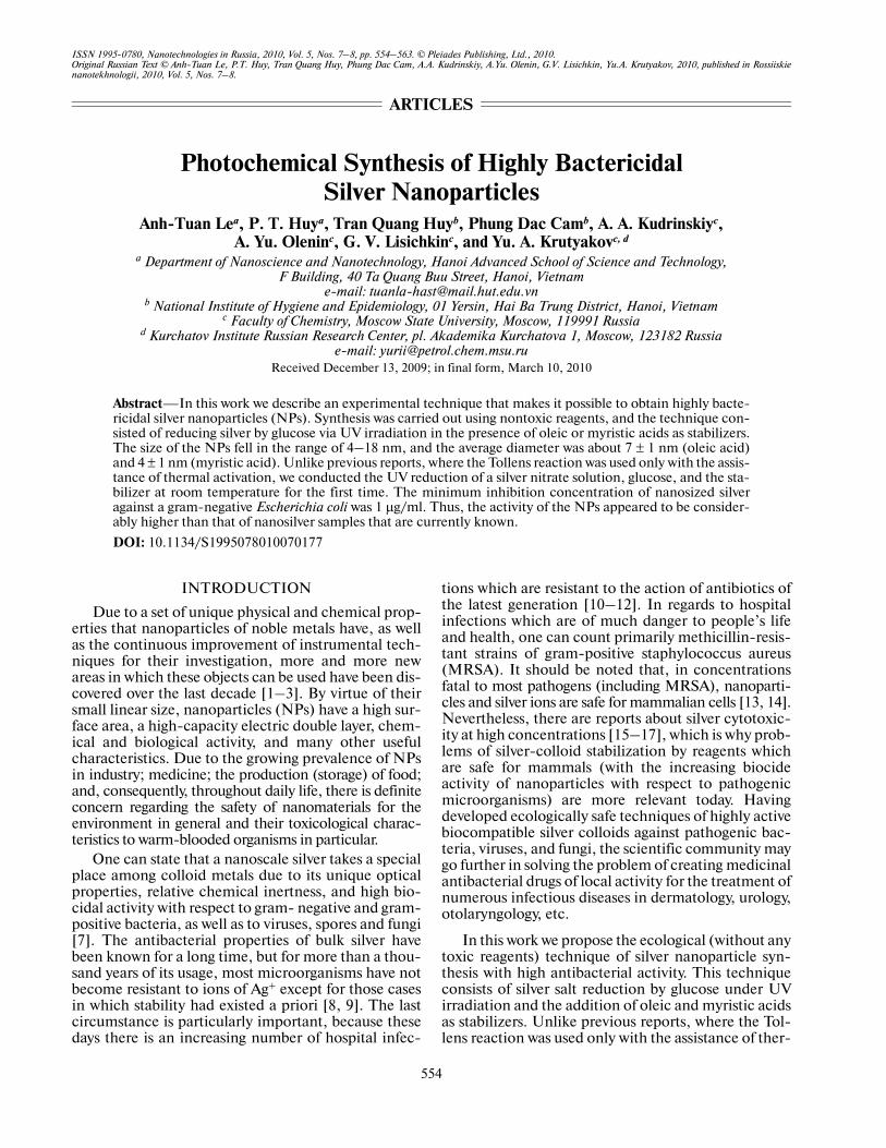
where RCHO is an aldehyde or a carbohydrate. Thedispersions obtained by Tollens reactions maintainstability for a long time without any addition of surfac�tants or other stabilizers [21]. The mean diameter, par�ticle size distribution, and aggregate stability dependon the conditions of synthesis (the temperature,ammonia concentration, and the change in the pH ofthe medium during the reduction process). The addi�tion of surfactants into the reaction medium mayaffect the key characteristics of NPs [23]. The colloi�dal solutions of silver obtained by traditional methodswith the thermal reduction of salt solutions in the pres�ence of carbohydrates usually contain NPs 20–100 nm in diameter and with a rather broad size distri�bution [18, 23].
In order to provide further possibilities for control�ling NP characteristics in the current work, we usedthe UV irradiation of the reagent mixture at the stageof reducing the silver ammonium complex with glu�cose. Activating the NP formation under the UV treat�ment with a wavelength of 365 nm made it possible tocarry out the process at room temperature whileobtaining the stable dispersion of NPs with a diameterin the range of 7 ± 1 nm when using oleic acid as a sta�bilizer and of 4 ± 1 nm when using myristic acid. Silverreduction under the same conditions (Fig. 1) butheated and without UV treatment results in the forma�tion of NPs with a broad size distribution (4–100 nm).
By controlling the reduction time, one can operatethe complete flow of nanosilver formation. It was dis�covered in previous experiments that, under the con�ditions described above (see Experimental), for 7 hunder UV irradiation, the output of the reaction prod�ucts was maximum and did not increase subsequently.This gave us a reason to assume that the reduction wascomplete in 7 h.
The mechanism and kinetics of silver NP growthformed by the UV irradiation was considered in thereports of other authors [7, 24]. Noble metals (espe�cially silver) in a nanoscale condition are extremelyphotoactive, and under UV treatment they stronglypolarize. This leads to the positive charging of metallicaggregates consisting of small clusters, and, as a con�sequence, to their instability and decay into compo�nents [25]. When using AgNO3 (as in our case), freeions Ag+ are present in the solution in considerable
] aq( )
+
concentrations, which leads to the formation of a largeamount of small clusters at the starting stage whichrapidly coagulate into huge agglomerates. One shouldexpect that embryos with sizes less than critical, due totheir thermodynamic instability, undergo dissolutionand promote the growth of larger particles. In its turn,large aggregates are photochemically unstable anddecay under UV irradiation [25]. Thus, the mecha�nism of silver NP formation under UV irradiation maybe considered quasi�equilibrium [24], which is fol�lowed by parallel processes of growth and fragmenta�tion, which finally leads to particles with sizes corre�sponding to thermodynamically stable objects and arelatively narrow size distribution.
On the basis of reports and the results of earlierresearches [7, 24], one can suppose that the quasi�equilibrium of the silver NPs obtaining a reactionunder the UV treatment is achieved due to the com�petitive stages of the photofragmentation of largemetal agglomerates and the growth of smaller parti�cles. Thus, as opposed to traditionally used techniquesof silver reduction via the Tollens reaction, where theformation process of NPs proceeds at high tempera�tures and has a nonequilibrium character, the UVtreatment during the time of synthesis leads to the for�mation of steady colloids of silver with relatively nar�row size distributions of NPs.
Optical Study of Silver Soles
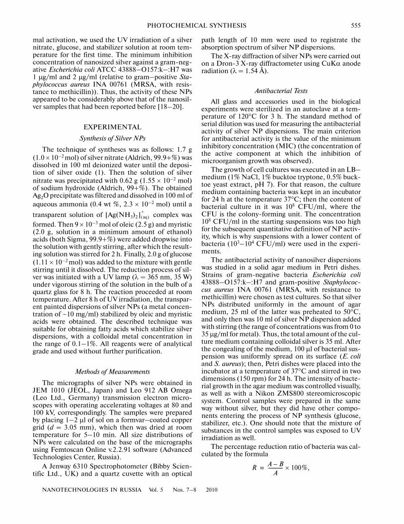
The extinction spectra of silver hydrosols obtainedunder conditions of UV treatment showed the pres�ence of characteristic lines of surface plasmon reso�nance (SPR) around 409–413 nm, which have a sym�metric form and little width at the half�height (Fig. 2,spectra 1, 3). The registration of silver colloids spectra24 h from the moment of synthesis did not revealchanges in the structure and intensity of SPR bands.This testifies to the absence of noticeable (fast) aggre�gative processes in dispersions. When the silver nitratethermal reduction with glucose was carried out with�out UV treatment, a decrease in the intensity of theresulting hydrosol at the maximum absorption band ofsilver NPs was observed, as well as a slight increase inthe bandwidth at the half�height, the appearance of acharacteristic arm at the red zone, and a change in theline symmetry (Fig. 2, spectrum 2) compared to thesoles of silver obtained under the UV irradiation atroom temperature. In the case of silver thermal reduc�tion, a growth in the line intensity in the long�wave�length part of spectrum, as well as a considerablebroadening of plasmon absorption peak links with anincreasing contribution of the dispersion componentto the extinction of the solution as a consequence of alarge amount of huge NPs and their aggregates beingpresent.
NANOTECHNOLOGIES IN RUSSIA Vol. 5 Nos. 7–8 2010
PHOTOCHEMICAL SYNTHESIS 557
Microscopic and Structural Research
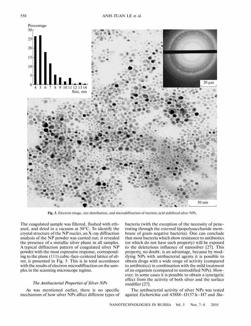
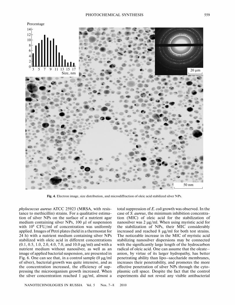
The data from transmission electron microscopyfor recently obtained samples indicated that myristicand oleic acids that stabilize silver NPs are spheres witha maximum size distribution in the range of 4–5 nm(Fig. 3) and 7–8 nm (Fig. 4), correspondingly. Oneshould note that, when using myristic acid as a stabi�lizer, the obtained dispersions contained silver parti�cles of smaller diameters, which could be explained bythe higher density of packing myristat–anions on theNPs surface [26]. The diffraction of electrons revealedthe presence of defect crystals with the most expressedresponse corresponding to the plate of the cubic�face�centered lattice of silver.
For X�ray diffraction analysis, it was necessary tohave purified dried nanosilver samples. The deposi�tion of silver NPs was carried out at a temperature of–30°C from a blend of ethanol–water (60 : 40 v/v).
20 μm
200 nm
Fig. 1. TEM images and the electron microdiffraction of silver NPs obtained by the thermo reduction of silver nitrate with glucosevia traditional Tollens reaction.
1.6
1.4
1.2
1.0
0.8
0.6
0.4
0.2
0
Absorbance, a. u.
420320 520 620 720 820Wavelength, nm
1
2
3
Fig. 2. Electron extinction spectra of nanosilver dispersionsobtained by (1) UV treatment with oleic acid, (2) thermalactivation with oleic acid, and (3) UV treatment withmyristic acid.
558
NANOTECHNOLOGIES IN RUSSIA Vol. 5 Nos. 7–8 2010
ANH�TUAN LE et al.
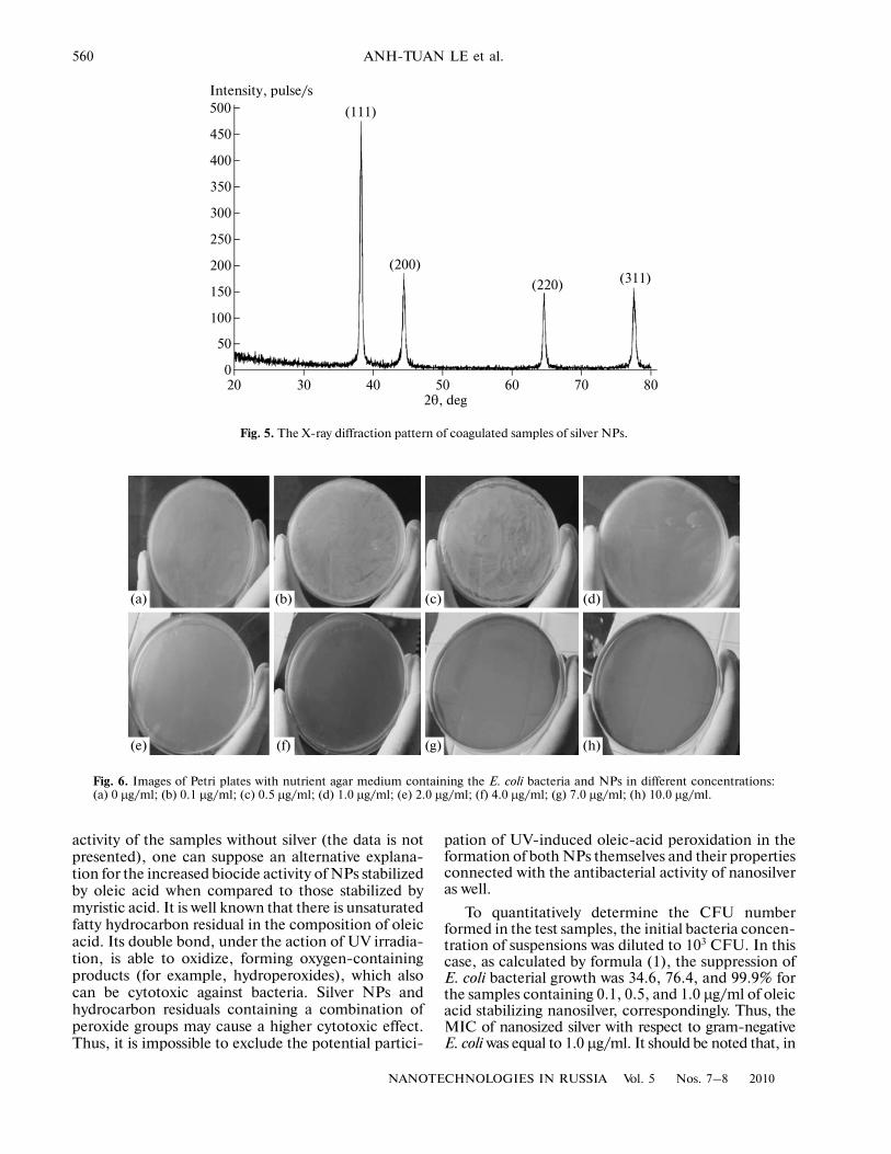
The coagulated sample was filtered, flushed with eth�anol, and dried in a vacuum at 50°C. To identify thecrystal structure of the NP nuclei, an X�ray diffractionanalysis of the NP powder was carried out; it revealedthe presence of a metallic silver phase in all samples.A typical diffraction pattern of coagulated silver NPpowder with the most expressive response, correspond�ing to the plane (111) cubic�face�centered lattice of sil�ver, is presented in Fig. 5. This is in total accordancewith the results of electron microdiffraction on the sam�ples in the scanning microscope regime.
The Antibacterial Properties of Silver NPs
As was mentioned earlier, there is no specificmechanism of how silver NPs affect different types of
bacteria (with the exception of the necessity of pene�trating through the external lipopolysaccharide mem�brane of gram�negative bacteria). One can concludethat most bacteria which show resistance to antibiotics(or which do not have such property) will be exposedto the deleterious influence of nanosilver [27]. Thisproperty, no doubt, is an advantage, because by mod�ifying NPs with antibacterial agents it is possible toobtain drugs with a wide range of activity (comparedto antibiotics) in combination with the mild treatmentof an organism (compared to unmodified NPs). How�ever, in some cases it is possible to obtain a synergeticeffect from the activity of both silver and the surfacemodifier [27].
The antibacterial activity of silver NPs was testedagainst Escherichia coli 43888–O157:k–H7 and Sta�
4
5
0
10
15
20
25
30
5 6 7 8 9 10 11 12 13 14
Percentage
Size, nm
50 nm
20 μm
Fig. 3. Electron image, size distribution, and microdiffraction of myristic acid stabilized silver NPs.
NANOTECHNOLOGIES IN RUSSIA Vol. 5 Nos. 7–8 2010
PHOTOCHEMICAL SYNTHESIS 559
phylococcus aureus ATCC 25923 (MRSA, with resis�tance to methicillin) strains. For a qualitative estima�tion of silver NPs on the surface of a nutrient agarmedium containing silver NPs, 100 μl of suspensionwith 108 CFU/ml of concentration was uniformlyapplied. Images of Petri plates (held in a thermostat for24 h) with a nutrient medium containing silver NPsstabilized with oleic acid in different concentrations(0.1, 0.5, 1.0, 2.0, 4.0, 7.0, and 10.0 μg/ml) and with anutrient medium without nanosilver, as well as animage of applied bacterial suspension, are presented inFig. 6. One can see that, in a control sample (0 μg/mlof silver), bacterial growth was quite intensive, and asthe concentration increased, the efficiency of sup�pressing the microorganism growth increased. Whenthe silver concentration reached 1 μg/ml, almost a
total suppression of E. coli growth was observed. In thecase of S. aureus, the minimum inhibition concentra�tion (MIC) of oleic acid for the stabilization ofnanosilver was 2 μg/ml. When using myristic acid forthe stabilization of NPs, their MIC considerablyincreased and reached 8 μg/ml for both test strains.The noticeable increase in the MIC of myristic acidstabilizing nanosilver dispersions may be connectedwith the significantly large length of the hydrocarbonradical of oleic acid. One can assume that the oleate–anion, by virtue of its larger hydropathy, has betterpenetrating ability than lipo–saccharide membranes,increases their penetrability, and promotes the moreeffective penetration of silver NPs through the cyto�plasmic cell space. Despite the fact that the controlexperiments did not reveal any visible antibacterial
3
2
0
4
6
8
10
12
Percentage
Size, nm
50 nm
20 μm5 7 9 11 13 15 17
14
Fig. 4. Electron image, size distribution, and microdiffraction of oleic acid stabilized silver NPs.
560
NANOTECHNOLOGIES IN RUSSIA Vol. 5 Nos. 7–8 2010
ANH�TUAN LE et al.
activity of the samples without silver (the data is notpresented), one can suppose an alternative explana�tion for the increased biocide activity of NPs stabilizedby oleic acid when compared to those stabilized bymyristic acid. It is well known that there is unsaturatedfatty hydrocarbon residual in the composition of oleicacid. Its double bond, under the action of UV irradia�tion, is able to oxidize, forming oxygen�containingproducts (for example, hydroperoxides), which alsocan be cytotoxic against bacteria. Silver NPs andhydrocarbon residuals containing a combination ofperoxide groups may cause a higher cytotoxic effect.Thus, it is impossible to exclude the potential partici�
pation of UV�induced oleic�acid peroxidation in theformation of both NPs themselves and their propertiesconnected with the antibacterial activity of nanosilveras well.
To quantitatively determine the CFU numberformed in the test samples, the initial bacteria concen�tration of suspensions was diluted to 103 CFU. In thiscase, as calculated by formula (1), the suppression ofE. coli bacterial growth was 34.6, 76.4, and 99.9% forthe samples containing 0.1, 0.5, and 1.0 μg/ml of oleicacid stabilizing nanosilver, correspondingly. Thus, theMIC of nanosized silver with respect to gram�negativeE. coli was equal to 1.0 μg/ml. It should be noted that, in
50
100
150
200
250
300
350
400
450
500Intensity, pulse/s
(111)
020 30 40 50 60 70 80
2θ, deg
(200)
(220) (311)
Fig. 5. The X�ray diffraction pattern of coagulated samples of silver NPs.
(a) (b) (c) (d)
(e) (f) (g) (h)
Fig. 6. Images of Petri plates with nutrient agar medium containing the E. coli bacteria and NPs in different concentrations:(a) 0 µg/ml; (b) 0.1 µg/ml; (c) 0.5 µg/ml; (d) 1.0 µg/ml; (e) 2.0 µg/ml; (f) 4.0 µg/ml; (g) 7.0 µg/ml; (h) 10.0 µg/ml.
NANOTECHNOLOGIES IN RUSSIA Vol. 5 Nos. 7–8 2010
PHOTOCHEMICAL SYNTHESIS 561
previous reports, the MIC of nanosilver was higher andwas in the range 3–10 μg/ml [18–20]. An increase inthe antibacterial activity of silver NPs in this case maybe explained by the significantly expressed aggregativeprocesses absence; the larger stability of silver hydro�sols; and the synergistic effect, which is caused by thestabilizer of the surface.
Mechanism of the Antibacterial Activity of Silver NPs
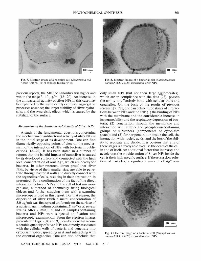
A study of the fundamental questions concerningthe mechanism of antibacterial activity of silver NPs isin the initial stage of its development. One can finddiametrically opposing points of view on the mecha�nism of the interaction of NPs with bacteria in publi�cations [18–20]. It has been stated in a number ofreports that the baleful impact of nanosilver is causedby its developed surface and connected with the highlocal concentration of ions Ag+, which are deadly forbacteria. In other research, direct proof that silverNPs, by virtue of their smaller size, are able to pene�trate through bacterial walls and directly connect withthe organelles of cells, resulting in their destruction, ispresented. For a confirmation of the fact of the directinteraction between NPs and the cell of test microor�ganisms, a method of chemically fixing biologicalobjects and further studying them with a scanningmicroscope is used in this report. For that reason, thedispersion of silver (with a metal concentration of5.0 μg/ml) was first spread uniformly on the surface ofa nutrient agar medium containing E. coli or S. aureusstrains. After 30 min, 1 h, and 2 h, samples containingbacteria and NPs were subjected to fixation andmicroscopic examination. From the electron imagespresented in Figs. 7, 8, and 9, it can be seen that a con�siderable quantity of silver NPs are directly associatedwith the cellular walls of bacteria and penetrate intocytoplasm space, spreading in it and interacting withthe essential organelles. One can also conclude that
only small NPs (but not their large agglomerates),which are in compliance with the data [28], possessthe ability to effectively bond with cellular walls andorganelles. On the basis of the results of previousresearch [7, 28], one can define three stages of interac�tions between NPs and the cell: (1) the binding of NPswith the membrane and the considerable increase inits permeability and the respiratory depression of bac�teria; (2) penetration through the membrane andinteraction with sulfur� and phosphorus�containinggroups of substances (components of cytoplasmspace); and (3) further penetration inside the cell, theinteraction with nucleic acids, and the loss of the abil�ity to replicate and divide. It is obvious that any ofthese stages is already able to cause the death of the cellin and of itself. An additional factor that increases andaccelerates the biocide action of Silver NPs inside thecell is their high specific surface. If there is a slow solu�tion of particles, a significant amount of Ag+ ions
100 nm
Fig. 7. Electron image of a bacterial cell (Escherichia coli43888�O157:k–:H7) exposed to silver NPs.
100 nm
Fig. 8. Electron image of a bacterial cell (Staphylococcusaureus ATCC 25923) exposed to silver NPs.
100 nm
Fig. 9 Electron image of a bacterial cell (Staphylococcusaureus ATCC 25923) exposed to silver NPs.
562
NANOTECHNOLOGIES IN RUSSIA Vol. 5 Nos. 7–8 2010
ANH�TUAN LE et al.
(which in turn negatively affect life activity of bacteria)escapes into the environment.
Thus, one can conclude that these experimentalobservations make it possible to go further in under�standing the antibacterial activity of nanosized silver.The high biocide activity of silver dispersions stabi�lized by oleic acid can be explained by the high mem�brane�acting activity of such NPs when compared tothe activity of particles stabilized by a less hydrophobiccompound (myristic acid).
CONCLUSIONS
In this report we present a technique for synthesiz�ing silver dispersions exhibiting high antibacterialactivity. One advantage of the described experimentaltechnique is the quasi�equilibrium reduction process.It is determined by UV treatment on a reactionarymedium instead of the traditionally used thermal heat�ing of reagents blend. Silver NPs obtained by the thistechnique are highly active against gram�positive andgram�negative bacteria, which is caused by the stabil�ity of dispersions, the small average diameter of parti�cles, and the synergetic effect of silver and the stabi�lizer (oleic acid); these factors increase the penetratingability of NPs with respect to the cell membranes. Itwas shown with the microscopic research of test sam�ples that silver NPs are able to act against bacteria asindependent biocide agents.
One can say that this technique for synthesizingnanosilver dispersion without the use of toxic reagents,as well as the high biological activity of the colloidalsolutions under an extremely low content of metal,can make the preparation of nanosilver a basis for thecreation of effective antiseptic and disinfectant drugsof the next generation for medicine, epidemiology,and sanitation.
ACKNOWLEDGMENTS
This research was supported by the Board of Edu�cation of Vietnam within the project B2008�01�155 atHanoi technical University (2008–2009).
REFERENCES
1. M.�C. Daniel and D. Astruc, “Gold Nanoparticles:Assembly, Supramolecular Chemistry, Quantum�Size�Related Properties, and Applications toward Biology,Catalysis, and Nanotechnology,” Chem. Rev. (Wa�shington) 104 (1), 293–346 (2004).
2. N. Toshima and T. Yonezawa, “Bimetallic Nanoparti�cles—Novel Materials for Chemical and PhysicalApplications,” New J. Chem. 22, 1179–1201 (1998).
3. D. L. Huber, Synthesis, “Properties, and Applicationsof Iron Nanoparticles,” Small 1 (5), 482–501 (2005).
4. G. Oberdörster, E. Oberdörster, and J. Oberdörster,“Nanotoxicology: An Emerging Discipline Evolvingfrom Studies of Ultrafine Particles,” Environ. HealthPerspec. 113 (7), 823–839 (2005).
5. A. El�Ansary and S. Al�Daihan, “On the Toxicity ofTherapeutically Used Nanoparticles: An Overview,”J. Toxicol. Article ID 754 810�1–754 810�9 (2009).
6. G. Oberdörster, V. Stone, and K. Donaldson, “Toxicol�ogy of Nanoparticles: A Historical Perspective,” Nano�toxicology 1 (1), 2–25 (2007).
7. Yu. A. Krutyakov, A. A. Kudrinskii, A. Yu. Olenin, andG. V. Lisichkin, “Synthesis and Properties of SilverNanoparticles: Advances and Prospects,” Russ. Chem.Rev. 77 (3), 233 (2008).
8. S. Silver, “Bacterial Silver Resistance: Molecular Biol�ogy and Uses and Misuses of Silver Compounds,”FEMS Microbiol. Rev. 27, 341–353 (2003).
9. A. Gupta, K. Matsui, J. F. Lo, and S. Silver, “MolecularBasis for Resistance to Silver Cations in Salmonella,”Nat. Med. 5, 183–188 (1999).
10. M. T. Valenzuela and C. de Quadros, “Antibiotic Resis�tance in Latin America: A Cause for Alarm,” Vaccine27 (Suppl. 3), C25–C28 (2009).
11. A. Voss, D. Milatovic, C. Wallrauch�Schwarz, V. T. Ros�dahl, and I. Braveny, “Methicillin�Resistant Staphylo�coccus aureus in Europe,” Eur. J. Clin. Microbiol.Infect. Dis. 13 (1), 50–55 (1994).
12. S. B. Levy, “Antibiotic Resistance—The ProblemIntensifies,” Adv. Drug Delivery Rev. 57, 1446–1450(2005).
13. S. Lu, W. Gao, and H. Y. Gu, “Construction, Applica�tion, and Biosafety of Silver Nanocrystalline ChitosanWound Dressing,” Burns 34, 623–628 (2008).
14. M. Rai, A. Yadav, and A. Gade, “Silver Nanoparticlesas a New Generation of Antimicrobials,” Biotechnol.Adv. 27, 76–83 (2009).
15. X. Chen and H. J. Schluesener, “Nanosilver: A Nano�product in Medical Application,” Toxicol. Lett. 176,1–12 (2008).
16. S. Arora, J. Jain, J. M. Rajwade, and K. M. Paknikar,“Cellular Responses Induced by Silver Nanoparticles:In vitro Studies,” Toxicol. Lett. 179, 93–100 (2008).
17. Y.�H. Hsin, C.�F. Chen, S. Huang, T.�S. Shih, P.�S. Lai,and P. J. Chueh, “The Apoptotic Effect of Nanosilver IsMediated by a ROS� and JNK�Dependent MechanismInvolving the Mitochondrial Pathway in NIH3T3Cells,” Toxicol. Lett. 179, 130–139 (2008).
18. A. Pana ek, L. Kvitek, R. Prucek, M. Kola � ,R. Ve e ova �, N. Pizu’rova �, V. K. Sharma, T. Nev na �,and R. Zbo il, “Silver Colloid Nanoparticles: Synthe�sis, Characterization, and Their Antibacterial Activity,”J. Phys. Chem. B 110, 16 248–16 253 (2006).
19. I. Sondi and B. Salopek�Sondi, “Silver Nanoparticlesas an Antimicrobial Agent: A Case Study of E. coli as aModel for Gram�Negative Bacteria,” J. Colloid Inter�face Sci. 275, 177–182 (2004).
20. M. Raffi, F. Hussain, T. M. Bhatti, J. I. Akhter,A. Hameed, and M. M. Hasan, “Antibacterial Char�acterization of Silver Nanoparticles against E. coliATCC�15224,” J. Mater. Sci. Technol. 24 (2), 192–196(2008).
21. Y. Yin, Z. Y. Li, B. Gates, Y. Xia, and S. Venkateswaran,“Synthesis and Characterization of Stable AqueousDispersions of Silver Nanoparticles though the TollensProcess,” J. Mater. Chem. 12, 522–527 (2002).
c
›
r
›
c
›
r
›
ec
› ›
r
›
NANOTECHNOLOGIES IN RUSSIA Vol. 5 Nos. 7–8 2010
PHOTOCHEMICAL SYNTHESIS 563
22. L. Qu and L. Dai, “Novel Silver Nanostructures fromSilver Mirror Reaction on Reactive Substrates,”J. Phys. Chem. B 109, 13 985–13 990 (2005).
23. L. Kvítek, A. Paná ek, J. Soukupova �, M. Kola � ,R. Ve e ova �, R. Prucek, M. Holecova �, and R. Zbo il,“Effect of Surfactants and Polymers on Stability andAntibacterial Activity of Silver Nanoparticles,” J. Phys.Chem. C 112, 5825–5834 (2008).
24. S. K. Ghosh, S. Kundu, M. Mandal, S. Nath, andT. Pal, “Studies on the Evolution of Silver Nanoparti�cles in Micelle by UV�Photoactivation,” J. Nanopart.Res. 5, 577–587 (2003).
25. P. V. Kamat, in Semiconductor Nanoclusters: Physical,Chemical and Catalytic Aspects, Ed. by P. V. Kamat andD. Miesel (Elsevier, Amsterdam, 1997), pp. 237–259.
26. G. V. Lisichkin, A. Yu. Fadeev, A. A. Serdan, P. N. Nes�terenko, P. G. Mingalev, and D. B. Furman, Chemistryof Grafted Surface Compounds (Fizmatlit, Moscow,2003) [in Russian].
27. G. K. Vertelov, Yu. A. Krutyakov, O. V. Efremenkova,A. Yu. Olenin, and G. V. Lisichkin, “A Versatile Synthe�sis of Highly Bactericidal Myramistin® Stabilized SilverNanoparticles,” Nanotechnology 19 (35), ArticleID 355 707 (2008).
28. J. R. Morones, J. L. Elechiguerra, A. Camacho,K. Holt, J. B. Kouri, J. T. Ramíres, and M. J. Yacaman,“The Bactericidal Effect of Silver Nanoparticles,”Nanotechnology 16, 2346–2353 (2005).
c
›
r
›
c
›
r
›
r
›