Embed Size (px)
Citation preview

233© Springer Nature Switzerland AG 2019B. G. Shapero et al. (eds.), The Massachusetts General Hospital Guide to Depression, Current Clinical Psychiatry, https://doi.org/10.1007/978-3-319-97241-1_18
Photobiomodulation
Marco Antonio Caldieraro and Paolo Cassano
M. A. Caldieraro Hospital de Clínicas de Porto Alegre, Department of Psychiatry, Porto Alegre, Brazile-mail: [email protected]
P. Cassano (*) Depression Clinical and Research Program and Center for Anxiety and Traumatic Stress Disorders, Department of Psychiatry, Massachusetts General Hospital, Harvard Medical School, Boston, MA, USAe-mail: [email protected]
18
Case Vignette
Ten years after losing her mother, a 44-year-old mar-ried woman, mother of two preadolescent children, came to see a psychiatrist for a consultation quite dis-satisfied with her pharmacological antidepressant treatment, prescribed for her 5-month recurrence of major depressive disorder. Although a very successful professional, she struggled at work to keep up with her administrative tasks; she did not feel motivated and expressed a painful and recurrent longing for her mother – diagnosed as a prolonged grief. At times, she wished to be dead, and yet the very idea of leaving her own children behind deepened her sorrow and brought more tears. Her interest in non-pharmacological inter-ventions for depression was driven by her dissatisfac-tion with her antidepressant medication, venlafaxine. She had been treated with venlafaxine 75 mg every day for 6 weeks, and – despite the low dose – venlafaxine had caused both decreased libido and anorgasmia. Of note, this woman had no impairment with her sexual function before starting venlafaxine, despite being depressed, but her libido had dropped significantly with the antidepressant, and she was also experiencing decreased lubrication and anorgasmia. Transcranial
photobiomodulation (t-PBM) with near-infrared light – twice a week for 8 weeks – was added to venla-faxine for the treatment of her depression. At each ses-sion, two Omnilux New-U (LED) devices were used simultaneously for 25 min, after bilateral positioning on her forehead (on the F3 and F4 points, correspond-ing to the dorsolateral prefrontal cortex – see also sec-tion on “Clinical Studies on t-PBM” (Sect. 3.3) for treatment parameters in the ELATED-2 study). At baseline her Hamilton Depression Rating Scale (HAM-D17) total score for depression severity was 21, while after 8 weeks and 16 sessions of t-PBM, the same total score was 0. Quite unexpectedly, despite continuing her venlafaxine, her severe loss of sexual interest, her mild problems with sexual lubrication, and her moder-ately delayed orgasm had completely resolved after ten t-PBM sessions. Also unexpectedly, the painful longing for her deceased mother had passed. After the acute phase of 8-week in-office t-PBM, the patient acquired her own device and started self-administering t-PBM, twice a week for 20 min, on her right forehead (F4). Her remission from depression was maintained for 12 months, even after discontinuation of venlafaxine, which occurred 6 months into remission. Of note, occasional brief bursts of depression occurred with family stressors and with related lapses in t-PBM home sessions (less than one session per week). At the 12-month mark from remission, a full-blown recur-rence of depression with passive suicidal ideation occurred, after family stressors and after 3 weeks of poor adherence to t-PBM. After self-discontinuation of t-PBM, increasingly frequent, vivid, illusionary phe-nomena occurred – such as the déjà vu and impression of seeing familiar faces in strangers. She had indeed experienced one 30-min episode of illusions immedi-
234
Introduction
Photobiomodulation (PBM), also called “low-level light therapy” or “low-level laser therapy,” is a novel device-based treatment under development for major depressive disorder (MDD) and other neuropsychiatric disorders [1]. PBM uses low-power lasers (LPL) or light-emitting diodes (LEDs) to deliver near-infrared (NIR) or red light aiming to modulate metabolism and functioning of different tissues and organs, including the brain [2]. Biological activity (e.g., stimulation of osteoblastic differentiation of human adipose-derived stem cells) has also been reported for other wavelengths in vitro [3]; however, the research on PBM has focused on NIR because light in this range has better tissue penetration and a well-known photoacceptor, the mitochondrial enzyme cytochrome c oxidase (CCO) [4].
PBM is noninvasive, and NIR is a nonionizing electro-magnetic irradiation that is absorbed by endogenous chro-mophores and is minimally dissipated as thermal energy [5]. The suggested primary mechanism of action of PBM using NIR is the enhancement of mitochondrial bioenergetics metabolism via the delivery of energy to the CCO and increased adenosine triphosphate (ATP) production. This action of NIR can improve brain hypometabolism and mito-chondrial dysfunction associated with depression [6–10]. In fact, there is evidence supporting benefits of NIR in other pathways relevant for MDD such as neurogenesis, inflamma-tion, and oxidative stress [2].
Most of the research on PBM for MDD focuses on tran-scranial PBM (t-PBM). This modality delivers NIR to the scalp, aiming to directly modulate areas of the brain cortex subjacent to the stimulation spot [11]. However, an antide-
pressant effect of indirect or systemic PBM (s-PBM) has also been postulated [12]. In the s-PBM modality, the light can be delivered intranasally or transcutaneously to other parts of the body (i.e., not necessarily to the scalp). In this case, the effect on the brain would be mediated by compo-nents of peripheral tissues, such as blood cells [13].
Except for laser devices (class 3–4) for which accidental light exposure of the retina might result in severe injury to the macula, devices used for PBM are typically categorized as nonsignificant risk devices by the FDA and are available over the counter, but not for psychiatric indications per se. Also, there is a low risk of interaction with medication, except for light-activated drugs such as cancer photosensi-tive chemotherapy (photodynamic therapy). Compared to FDA-approved devices for the treatment of MDD, such as repetitive transcranial magnetic stimulation (rTMS) and electroconvulsive therapy (ECT), PBM is easier to adminis-ter in the office setting and significantly less expensive. Differently from rTMS or ECT, which require frequent visits to the clinical facility, PBM can be administered at home after appropriate training of the patient or family member, provided that there is adequate clinical follow-up.
History
In 1965, McGuff and collaborators reported an antitumor effect of ruby lasers on human tumors implanted in hamsters and in cancer patients [14]. Two years later, Endre Mester – at the Semmelweis Medical University (Hungary) – discov-ered PBM while trying to replicate the same finding in mice. Inadvertently, Mester used lasers with much lower power, when compared to those of the McGuff’s study, and failed to replicate the antitumor effect. However, Mester observed a faster rate of hair growth in the mice treated with his low-power laser [15]. In a later study, he also reported that the same laser stimulated wound healing in rats [16]. After these initial observations on PBM, extensive research on the bio-logical effects of NIR and red light has been performed. Currently, NIR and red light are used in the medical field for the treatment of a variety of conditions such as muscle pain [17], wounds [18], neuropathic pain [19], headache [20], periorbital wrinkles [21, 22], and alopecia [23].
The pioneer in the use of PBM to treat central nervous system (CNS) injury was Shimon Rochkind. His group dem-onstrated that transcutaneous NIR irradiation enhanced axo-nal sprouting and spinal cord repair in animal models of spinal cord injury [24–26]. Later studies showed that light delivered transcutaneously penetrates to the spine and that penetration is highest for the NIR wavelengths [27]. The use of transcranial phototherapy for treating brain disorders started with its application to acute stroke. Numerous pre-clinical animal studies [28–30] suggested that the applica-
ately after her first treatment in the office; however, since then, illusions had only been sporadic, mild, and unrelated to the treatment administration. Sertraline was started and increased to 150 mg daily; the regi-men was well tolerated and led to remission; the illu-sions dissipated within 6 weeks from the switch in antidepressant treatment (or 9 weeks from self-discon-tinuation of t-PBM). With sertraline, the patient did not experience sexual dysfunction; however, her grief resurfaced with episodes of painful yearning for her mother, only “minutes” triggered by occasional prompts. This clinical case suggests an antidepressant response with t-PBM, a dose-dependent remission, as well as challenges in tolerability and potential with-drawal syndromes. The beneficial effects of t-PBM on sexual dysfunction and complicated grief remain speculative.
M. A. Caldieraro and P. Cassano

235
tion of NIR laser (810 nm) to the head after induction of an acute stroke had beneficial effects on subsequent neurologi-cal performance and reduced lesion size. These promising animal studies led to the conduction of a series of clinical trials called NeuroThera Effectiveness and Safety Trials (NEST). Altogether, three large studies were conducted in a total of 1410 acute stroke patients [NEST-1 (n = 120), NEST-2 (n = 660), NEST-3 (n = 630)] [31–33]. NEST-1 was designed to demonstrate safety and effectiveness of a single session of t-PBM in the first 24 h after a stroke. The group receiving the active treatment had a significantly greater improvement than the control condition (assessed by the NIH Stroke Severity Scale) at 5 and 90 days post-stroke. NEST-2 reported benefit of active t-PBM over sham treat-ment for moderate and moderate-to-severe stroke, but not for severe stroke. Despite promising results in the initial trials, the NEST-3 failed to replicate any clinical benefits for one session of PBM after acute stroke. There is no consensus on the literature to explain the discrepancies between the results of NEST-3 and the previous two trials. However, suggestions for future trials emerged, such as using higher-power, multi-ple sessions and initiating the treatment in the first hours after the stroke [1]. Of note, the NEST trials demonstrated that NIR light (810 nm) delivered transcranially with a class 4 laser was safe, with no significant differences in rates of adverse events when compared to sham (simulated) expo-sure. Other preclinical studies and clinical trials have sug-gested that transcranial PBM is safe and effective in reducing brain lesion volume, reducing inflammation, stimulating neurogenesis, and enhancing learning, memory, and execu-tive functioning in acute [34–39] and chronic [40–42] trau-matic brain injury (TBI) and has beneficial effects on neurodegenerative diseases (Alzheimer’s and Parkinson’s) [43, 44]. Studies on MDD will be discussed below.
New Advances and Research Support
Preclinical Studies on MDD
Studies in animal models indicate the potential of PBM as an antidepressant treatment and provide guidance on the opti-mal stimulation parameters. A study on depression following TBI reported that a single session of PBM using NIR laser (810 nm) was effective on reducing depressive-like behav-iors in mice. In the same study, a pulsed wave (PW) with a frequency of 10 Hz was superior to continuous wave (CW) and to PW with a frequency of 100 Hz, in terms of antide-pressant and neuroprotective effects [34].
A study testing one versus ten daily sessions of t-PBM on rats using a broad range of red and infrared light (600–1600 nm) suggests that multiple sessions are more effective than one, since an antidepressant effect in the forced swim
test (FST) – an animal model used as an initial test for anti-depressant treatments – was observed only in the group receiving repeated stimulation (ten sessions) [45]. The ben-efit of repeated sessions (seven daily sessions) of t-PBM using NIR (wavelength 804 nm) was also reported in a model of reserpine-induced depression in rats [46]. The same study compared three radiation power levels (80, 200, and 400 mW) and reported the greatest benefit for the lowest power: 80 mW. This result is consistent with a bell-shaped dose-response curve proposed for PBM, which indicates that further increasing the energy dose after an optimal level results in decrease of therapeutic effects and a possible increase of adverse effects [35]. Twenty-eight daily sessions of t-PBM with NIR (wavelength 808 nm) resulted in an anti-depressant effect on the FST and on the tail suspension test (TST) and also in increased ATP biosynthesis and expression and activity of the mitochondrial complex IV in the prefron-tal cortex (PFC) [47].
Two studies using the chronic mild stress model of depres-sion compared 3 weeks of PW laser t-PBM with the same duration of treatment with antidepressant medications (fluoxetine and citalopram). The benefit of t-PBM was simi-lar to the antidepressant drugs, as assessed by both the behavioral test (FST) and laboratory parameters. In both studies, t-PBM was superior to the drugs in terms of recover-ing normal weight, an indicator of well-being for the animal [48, 49].
Clinical Studies on MDD
Clinical investigations on PBM for MDD are preliminary in nature. Most studies include small samples and have signifi-cant methodological limitations. Moreover, optimal stimula-tion parameters are still to be established. Therefore, current studies are highly heterogeneous in terms of treatment regi-mens, limiting the combination or comparison of results from different studies. These limitations are even more sig-nificant for studies on s-PBM.
Clinical Studies on Transcranial PBM
In an open study, ten patients with treatment-resistant depres-sion (nine with a comorbid anxiety disorder) were treated with a single session of NIR t-PBM on the forehead, using an LED instrument (Marubeni America Corp.) [50]. The NIR was delivered on EEG sites F3 and F4, which cover the dor-solateral prefrontal cortex (DLPFC) bilaterally. The stimula-tion parameters were wavelength 810 nm, irradiance 250 mW/cm2, fluence 60 Joules (J)/cm2, and time 4 min per site. At weeks 2 and 4 posttreatment, a significant decrease was observed in depressive and anxious symptoms. The
18 Photobiomodulation
236
remission rate of MDD at week 2 (operationalized as a Hamilton Depression Rating Scale-21 items [HAM-D21] score <10) was 60%. At 4 weeks posttreatment, depressive symptoms were lower than pretreatment, but significantly higher than after 2 weeks, suggesting a limited duration of the effect of a single treatment.
Our group at MGH performed a pilot study of six sessions (two sessions a week for 3 weeks) of t-PBM with NIR (808 nm) in four participants with moderate-to-severe MDD (non-treatment resistant) [51]. At each treatment session, NIR was administered to the forehead bilaterally at four sites (2 min per site, 12.56 cm2 window each), using a class 4 laser (PhotoThera, Inc.) and the following treatment parameters: NIR irradiance 700 mW/cm2 and fluence 84 J/cm2, for a total NIR energy of 2.40 kJ per session. The depression severity assessed by the HAM-D 17 items (HAM-D17) decreased from 19.8 ± 4.35 at baseline to 13.0 ± 5.35 at the endpoint (5 weeks after the end of the treatment). This difference was large enough to achieve statistical significance even on this small sample.
Attention bias modification (ABM) is a cognitive inter-vention designed to improve symptoms by decreasing nega-tive attentional bias, but to date its efficacy in depression could not be demonstrated [52]. A randomized clinical trial (RCT) assessed if t-PBM could enhance the effects of ABM on individuals with elevated depressive symptoms [53]. The participants (n = 51) were randomized to receive two ses-sions of right forehead, left forehead, or sham t-PBM. The interval between the treatments was 48 h, and all participants received one session of ABM before and after each session of t-PBM. The light treatment used NIR (1064 nm) delivered by a laser device (Cell Gen Therapeutics) at two sites (13.6 cm2 window each) for 4 min per site. The treatment parameters were power 3.4 W, irradiance 250 mW/cm2, and a fluence of 60 J/cm2 per site, with a total energy of 1.63 kJ per session. In subjects who responded to the ABM, the improvement was enhanced by the right t-PBM, while no significant effect was observed for the left and sham t-PBM.
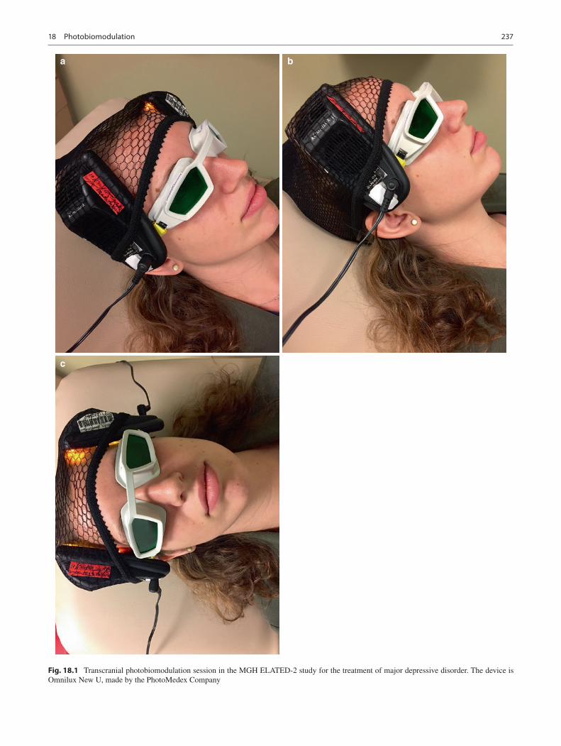
Our group at MGH also performed an RCT designed to access the efficacy of t-PBM as a primary treatment for MDD. Participants (n = 21) were randomized to receive bilateral stimulation on the prefrontal cortex (EEG sites F3 and F4) twice a week for 8 weeks or sham treatment in the same sites and with the same frequency (Fig. 18.1) [54]. Treatment was delivered by an LED device (PhotoMedex, Inc.) emitting NIR (830 nm). The duration of the initial ses-sion was 20 min, and subsequent sessions could be extended up to 30 min based on clinical judgment. Treatment param-eters were power 1 W, CW, irradiance 33.2 mW/cm2, and a fluence of 40–60 J/cm2 per site, with a total energy of 3.4 kJ per session and 45.6 kJ for the entire treatment. Remission (HAM-D17 ≤7) was observed in 50% of subjects who received the active treatment and in 18% of those in the sham
group. These findings showed a trend toward significance (p = 0.12); however, due to limited power, it did not detect group differences. The high remission rates are promising preliminary data suggesting the need for more well-powered studies.
Little is known about the long-term antidepressant effects of PBM. Our group reported the case of a patient receiving PBM for 31 months, as an add-on to antidepressant medica-tion to treat depression with anxious distress [55]. The treat-ment was started with intranasal PBM, and t-PBM was added in the last 9 months. The treatment was well tolerated, and a continuous improvement in the anxious symptoms was observed during the overall treatment follow-up, with a more pronounced improvement in depression observed after the addition of the t-PBM.
Clinical Studies on Systemic PBM
Although most studies on MDD are focused on t-PBM, there are studies indicating an antidepressant effect of light deliv-ered to peripheral tissues (i.e., systemic PBM). A Russian group reported two open studies using a combination of red (630 nm) and NIR (890 nm) delivered intravenously (IV) and transcutaneously by optic fibers. In the first study, 93 participants with a treatment-resistant depressive episode received 14–20 sessions of s-PBM divided in two cycles, as an add-on to ongoing pharmacological treatment. The authors reported a significant decrease in depression (HAM-D) and anxiety scores (Hamilton Anxiety Rating Scale – HAM-A) and an overall improvement of health, activity, and mood. Treated patients experienced better outcomes on these measures when compared to a non-randomized and non-blinded control group receiving only pharmacological treat-ment [56]. In the second study, the same group used a similar protocol to assess the prophylactic effect of s-PBM after remission from a depressive disorder (MDD, bipolar depres-sion, or mixed anxious and depressive disorder). One cycle of PBM was delivered once remission was achieved and a second cycle 3–4 months after the first one. One year after the initial remission, relapses or recurrences occurred in 23% of the treated patients. In a non-randomized and non-blinded control group with similar characteristics, the rate of relapse/recurrence in the same period was 50% [57]. Information about tolerability was not reported in the first of these two studies. Despite the potential risks and discomfort associated with the IV delivery, no PBM-related adverse effects or com-plications were observed in the second study.
An Australian group has also used NIR laser (808 nm) light delivered to acupuncture points on limbs and trunk to treat major depressive disorder. This group published two clinical trials (n = 30 and n = 47) of laser acupuncture focused on five primary depression acupoints [58, 59]. In both trials,
M. A. Caldieraro and P. Cassano
237
a
c
b
Fig. 18.1 Transcranial photobiomodulation session in the MGH ELATED-2 study for the treatment of major depressive disorder. The device is Omnilux New U, made by the PhotoMedex Company
18 Photobiomodulation
238
the total amount of energy delivered during each session ranged from 3 to 5 J. Although this dose of light was appar-ently low, the total energy delivered over the skin per session was sufficient to produce significant reduction of depressive symptoms in the two studies using up to 12 sessions over 8 weeks. These two studies were performed under the para-digm of acupuncture instead of PBM, and the stimulation occurred only on acupoints. Therefore, it is not possible to conclude if the results are specific to the stimulation of these points or if similar outcomes would be observed by a similar stimulation in non-specific spots.
These studies of both t- and s-PBM provide preliminary evidence of the efficacy of this treatment for MDD. However, well-powered studies are necessary to confirm the overall efficacy of PBM, the best treatment modality, the optimal stimulation parameters, and who will have the greatest ben-efit from this treatment.
Safety
The safety of one session of t-PBM was evaluated in the three large NEST studies on stroke with a pooled sample of 1410 participants [31, 33, 60]. No significant differences in the rates of adverse effects were observed between the group receiving NIR laser (808 nm) and the sham (simulated) stim-ulation group. Additionally, none of the clinical studies on MDD, already summarized, reported any serious adverse events.
The risk of thermal injury from PBM – delivered with the parameters reported in this chapter – is considered minimal and limited to the skin, where most of the light is indeed absorbed. Skin burn or other thermal effects were not reported in the literature reviewed in MDD patients. In ten individuals treated for TBI with 10–15 W lasers – much higher power than that used in most studies of PBM – the skin temperature increased no more than 3 °C with rapid cooling after the end of the stimulation. Patients reported slight warming of the skin, but no discomfort [61].
Inherent to the use of any laser device is the potential risk of retinal lesions resulting from improper use of the laser, from the shedding of the light beams straight through lens, and from their convergence on the macula. Protective eye wear is recommended when using class 3 and 4 lasers.
Proposed Treatment Mechanisms
Cellular Mechanisms of Action
PBM acts on different cellular pathways relevant to MDD. A mitochondrial enzyme, cytochrome c oxidase (CCO), is considered the primary chromophore for the
therapeutic effects of NIR and red light [4]. The peak absorption of light energy by the CCO occurs at four differ-ent wavelengths, and one of these peaks occurs with wave-lengths between 812 and 846 nm [62], which coincides with the wavelengths with best brain penetration, as described below.
The brain is a high-energy consumer organ and is suscep-tible to the deleterious effects of reduced energy production [9]. Depression is more frequent in individuals with mito-chondrial diseases or mitochondrial DNA mutations than in the general population [63]. A study using phosphorus mag-netic resonance spectroscopy in individuals with MDD demonstrated a correlation between treatment response to triiodothyronine (T3) and restoration of total nucleoside tri-phosphate (NTP), which primarily represents ATP [8]. In peripheral blood, deficits in mitochondrial activity were observed in platelets of MDD patients [13], and the reduc-tion in mitochondrial respiration in mononuclear cells was correlated with the severity of depressive symptoms [64]. These results suggest an important role of mitochondrial energetic metabolism dysfunction in the pathophysiology of MDD.
NIR delivers energy to the CCO and stimulates the mito-chondrial respiratory chain leading to increased ATP produc-tion [5, 65, 66]. In addition to the electronic excitation, the NIR improves mitochondrial activity by promoting a disso-ciation of nitric oxide (NO) from the CCO, thereby releasing the binding site for oxygen and restoring oxidative phos-phorylation [4]. The released NO might increase brain blood flow by acting as a local vasodilator [67]. A study of isolated mitochondria also reported an increase in mitochondrial RNA and protein synthesis after irradiation with a low-level laser [68].
Depression is associated with increased oxidative stress and decreased antioxidant defenses [69–74]. PBM also has beneficial effects on this pathophysiological mechanism. NIR induces short bursts of reactive oxygen species (ROS), leading to the activation of secondary antioxidant mecha-nisms and to the activation of the transcription factor, nuclear factor κB (NF-κB), resulting in a net reduction of the oxida-tive stress [67]. In a rat model of traumatized muscle, NIR (904 nm) blocked the release of harmful ROS and reduced the activation of NF-κB as well as the associated overexpres-sion of the inducible form of nitric oxide synthase (iNOS) [75]. An in vitro study found a protective effect of red light and NIR (700–2000 nm) against membrane oxidation on human red blood cells (RBCs) [76]. This result suggests that the antioxidant effects of PBM are not only related to CCO and oxidative phosphorylation, given that mitochondria are not present in RBCs. Proposed mechanisms for this effect on RBCs are the photochemical dissociation oxyhemoglobin to deoxyhemoglobin leading to phosphorylation of membrane proteins and the weakening of hydrogen bonds on the sur-
M. A. Caldieraro and P. Cassano

239
face of erythrocyte membranes, which moderates the surface charge, lowering the accessibility of charged free radicals into cells [76].
Recent literature supports inflammatory processes as depressogenic. Cytokines, which are inflammatory signal-ing molecules, provoke the dysregulation of several growth factors, including brain-derived neurotrophic factor (BDNF). The result of such dysregulation favors the devel-opment of depressive disorders [77]. Higher levels of dif-ferent proinflammatory cytokines were observed in depressed subjects, with increased levels of IL-6 and TNF-α consistently reported in three different meta-analyses [78–80], and are correlated with severity of depression and sui-cide attempts [81]. In addition, elevated peripheral IL-6 is associated with non-response to antidepressant treatment [82]. NIR light and red light (600–1600 nm) decreased synovial IL-6 gene expression in a rat model of rheumatoid arthritis [83]. NIR (810 nm) used as a treatment for pain in patients with rheumatoid arthritis decreased production of TNF-α, IL-1β, and IL-8 [84]. Irradiation with NIR in a rat model of spinal cord injury decreased invasion of cells involved in secondary damage to the spinal cord, including macrophages/activated microglia and T lymphocytes, sup-pressed the expression of proinflammatory cytokines, and altered the expression of genes involved in the immune response [27]. Transcranial NIR reduced neuroinflamma-tion in brain sections from mice that had suffered a trau-matic brain injury (TBI) while also improving cognitive function [85]. However, clinical studies assessing the impact of t-PBM on inflammatory process are necessary to understand how these results in animal models translate to humans.
The neurotrophin hypothesis of MDD proposes that decreased BDNF in key cortical and limbic brain regions contributes to depression, and antidepressants may work by increasing BDNF [86, 87]. In support to this hypothesis, it has been shown that acutely depressed individuals have lower serum levels of BDNF [88] that recover after remis-sion [89]. BDNF promotes neurogenesis, and neurogenesis, particularly on hippocampal structures, may be necessary for the symptoms improvement promoted by antidepressant treatments [90].
Animal research has shown that PBM stimulates neu-rogenesis and protects against cell death. Red light, close to the NIR spectrum (670 nm), protects the viability of cell culture after oxidative stress [91]. NIR also stimu-lates neurite outgrowth mediated by nerve growth factor and promotes axonal protection [91]. Neuroprotective effects of red light (670 nm) were documented in in vivo models of mitochondrial optic neuropathy [92]. Red light close to NIR spectrum (670 nm) has also been shown to protect neuronal cells against cyanide toxicity [93]. An increase in hippocampal neurogenesis was observed in
rats treated with NIR t-PBM [45]. In animal models of TBI, NIR (810 nm) appears to be an effective treatment and improves neurogenesis and synaptogenesis, via increased BDNF [34, 37, 38, 94, 95]. Other neurotrophic mechanisms proposed for PBM are the inhibition of GSK-3β and of pro-apoptotic molecules and the stimula-tion of the Akt/mTOR/cyclin D1 pathway [67]. Interestingly, in animal models of stroke and TBI, neuron proliferation and migration persisted after cessation of NIR exposure, indicating that a neurotrophic cascade may be triggered by the PBM [61].
Research in MDD implicates several pathophysiological mechanisms with the disorder: mitochondrial dysfunction, oxidative stress, disrupted neurogenesis, and inflammation. The primary effect of PBM on CCO can improve mitochon-drial function and start a cascade of other pathways to even-tually be beneficial in all these mechanisms. These effects were observed in preclinical studies on MDD and clinical studies on other disorders. More studies are necessary to understand how these processes are affected by PBM in humans with MDD.
Neurophysiological Mechanisms of Action
Additional research indicates that the effects of PBM go beyond the cellular level and promote neurophysiological changes in the brain. Red light (660 nm) delivered transcra-nially to rats – using an LED device (9 mW/cm2) – was asso-ciated with enhanced oxygen consumption in the frontal cortex [96]. An increase in cerebral blood flow – mediated by nitric oxide (NO) and by glutamate effect on NMDA – was reported in mice receiving NIR (808 nm) irradiation, deliv-ered by a laser device (1.6 W/cm2) [97]. Transcranial NIR (804 nm), also delivered by a laser device (640 mW/cm2), was associated with changes in electrocorticography param-eters [46].
Depression is associated with brain hypometabolism, decreased blood flow, and decreased glucose consumption [6, 7, 98, 99]. An increase in cerebral oxygenation (increase in oxygenated hemoglobin concentration) was observed in healthy volunteers, after t-PBM with a 3.4 W laser (1064 nm) [100]. T-PBM with a 0.2 W LED (627 nm) device was asso-ciated with increased blood flow in the middle cerebral and basilar arteries, in elderly women [101]. A modulatory effect on cortical excitability was also demonstrated in a study reporting that t-PBM (905 nm) using a laser device (50 mW/cm2) reduced the motor-evoked potentials elicited by single-pulse transcranial magnetic stimulation [102]. In patients with chronic aphasia, left t-PBM with a 0.5 W LED device combining red light (633 nm) and NIR (870 nm) was associ-ated with an increase in functional connectivity of the default mode network [103].
18 Photobiomodulation
240
Similar results were observed in s-PBM. In humans, a study on laser acupuncture, which is the stimulus of acu-points with low-level lasers, reported that NIR (808 nm), delivered on different points located on limbs and trunk, resulted in modulation of cerebral activity assessed by func-tional magnetic resonance imaging (fMRI) [104].
As a general conclusion, these results confirm that PBM delivered transcranially or systemically can affect brain functioning beyond the effect at the cellular level. More spe-cifically, some of these effects, such as an improvement in hypometabolism, may be relevant for the therapeutic effects on MDD.
Technical Issues
NIR Penetration
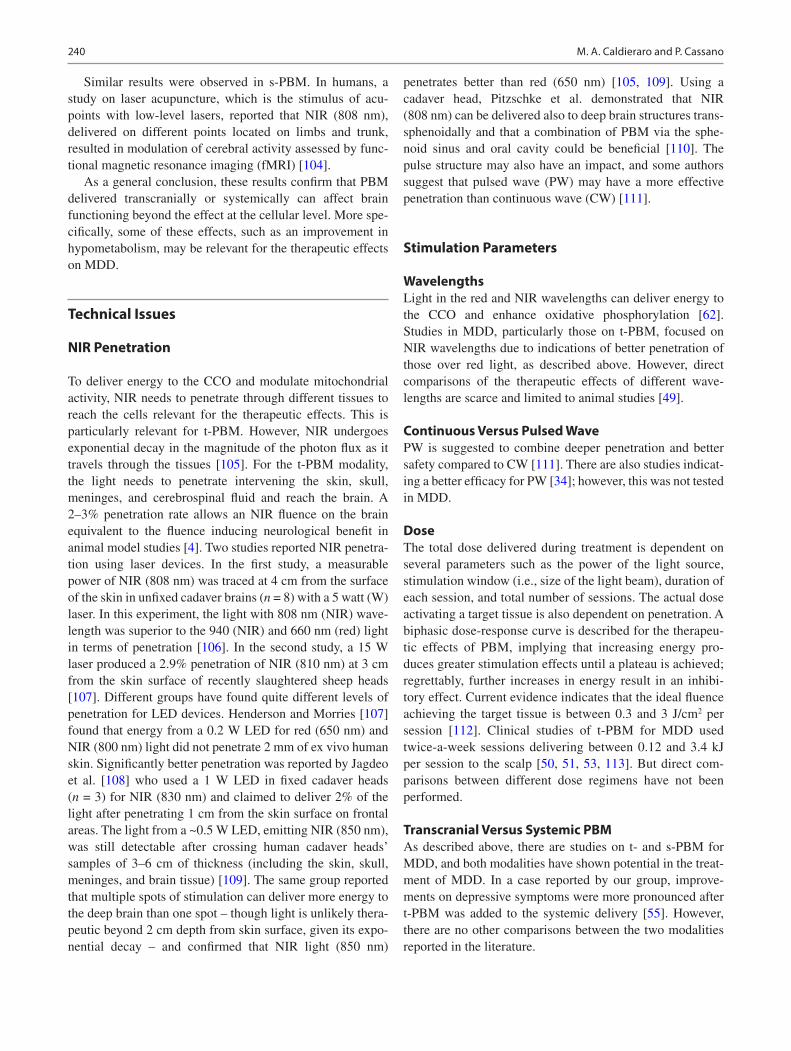
To deliver energy to the CCO and modulate mitochondrial activity, NIR needs to penetrate through different tissues to reach the cells relevant for the therapeutic effects. This is particularly relevant for t-PBM. However, NIR undergoes exponential decay in the magnitude of the photon flux as it travels through the tissues [105]. For the t-PBM modality, the light needs to penetrate intervening the skin, skull, meninges, and cerebrospinal fluid and reach the brain. A 2–3% penetration rate allows an NIR fluence on the brain equivalent to the fluence inducing neurological benefit in animal model studies [4]. Two studies reported NIR penetra-tion using laser devices. In the first study, a measurable power of NIR (808 nm) was traced at 4 cm from the surface of the skin in unfixed cadaver brains (n = 8) with a 5 watt (W) laser. In this experiment, the light with 808 nm (NIR) wave-length was superior to the 940 (NIR) and 660 nm (red) light in terms of penetration [106]. In the second study, a 15 W laser produced a 2.9% penetration of NIR (810 nm) at 3 cm from the skin surface of recently slaughtered sheep heads [107]. Different groups have found quite different levels of penetration for LED devices. Henderson and Morries [107] found that energy from a 0.2 W LED for red (650 nm) and NIR (800 nm) light did not penetrate 2 mm of ex vivo human skin. Significantly better penetration was reported by Jagdeo et al. [108] who used a 1 W LED in fixed cadaver heads (n = 3) for NIR (830 nm) and claimed to deliver 2% of the light after penetrating 1 cm from the skin surface on frontal areas. The light from a ~0.5 W LED, emitting NIR (850 nm), was still detectable after crossing human cadaver heads’ samples of 3–6 cm of thickness (including the skin, skull, meninges, and brain tissue) [109]. The same group reported that multiple spots of stimulation can deliver more energy to the deep brain than one spot – though light is unlikely thera-peutic beyond 2 cm depth from skin surface, given its expo-nential decay – and confirmed that NIR light (850 nm)
penetrates better than red (650 nm) [105, 109]. Using a cadaver head, Pitzschke et al. demonstrated that NIR (808 nm) can be delivered also to deep brain structures trans-sphenoidally and that a combination of PBM via the sphe-noid sinus and oral cavity could be beneficial [110]. The pulse structure may also have an impact, and some authors suggest that pulsed wave (PW) may have a more effective penetration than continuous wave (CW) [111].
Stimulation Parameters
WavelengthsLight in the red and NIR wavelengths can deliver energy to the CCO and enhance oxidative phosphorylation [62]. Studies in MDD, particularly those on t-PBM, focused on NIR wavelengths due to indications of better penetration of those over red light, as described above. However, direct comparisons of the therapeutic effects of different wave-lengths are scarce and limited to animal studies [49].
Continuous Versus Pulsed WavePW is suggested to combine deeper penetration and better safety compared to CW [111]. There are also studies indicat-ing a better efficacy for PW [34]; however, this was not tested in MDD.
DoseThe total dose delivered during treatment is dependent on several parameters such as the power of the light source, stimulation window (i.e., size of the light beam), duration of each session, and total number of sessions. The actual dose activating a target tissue is also dependent on penetration. A biphasic dose-response curve is described for the therapeu-tic effects of PBM, implying that increasing energy pro-duces greater stimulation effects until a plateau is achieved; regrettably, further increases in energy result in an inhibi-tory effect. Current evidence indicates that the ideal fluence achieving the target tissue is between 0.3 and 3 J/cm2 per session [112]. Clinical studies of t-PBM for MDD used twice-a-week sessions delivering between 0.12 and 3.4 kJ per session to the scalp [50, 51, 53, 113]. But direct com-parisons between different dose regimens have not been performed.
Transcranial Versus Systemic PBMAs described above, there are studies on t- and s-PBM for MDD, and both modalities have shown potential in the treat-ment of MDD. In a case reported by our group, improve-ments on depressive symptoms were more pronounced after t-PBM was added to the systemic delivery [55]. However, there are no other comparisons between the two modalities reported in the literature.
M. A. Caldieraro and P. Cassano
241
Clinical Application and Recommendation for Practitioners
PBM has the potential to benefit depressed patients who do not respond to other therapies. Its proposed mechanisms of action, while still under elucidation, appear consistent with antidepressant effects and are different from other treatments available for MDD. However, PBM does not have FDA approval for depression, and no large clinical trials have been performed on its use for depression. Therefore, PBM should not be used as a first-line treatment for MDD. PBM could be considered for patients who do not respond to, do not toler-ate, or simply decline antidepressant medications and evi-dence-based psychotherapies for depression. Given the limited scientific evidence in support of PBM for depression, depressed patients interested in PBM can safely be offered participation in PBM research protocols where they would receive close monitoring and could contribute to a greater scientific understanding of PBM. If research protocols are not available or the patient declines participation in research, the clinical decision on whether PBM is indeed indicated as well as the choice of the treatment parameters should be made under consultation with an expert in the field of PBM. This is particularly important because the modality
can be obtained by the patient for home use, and proper guid-ance is therefore important.
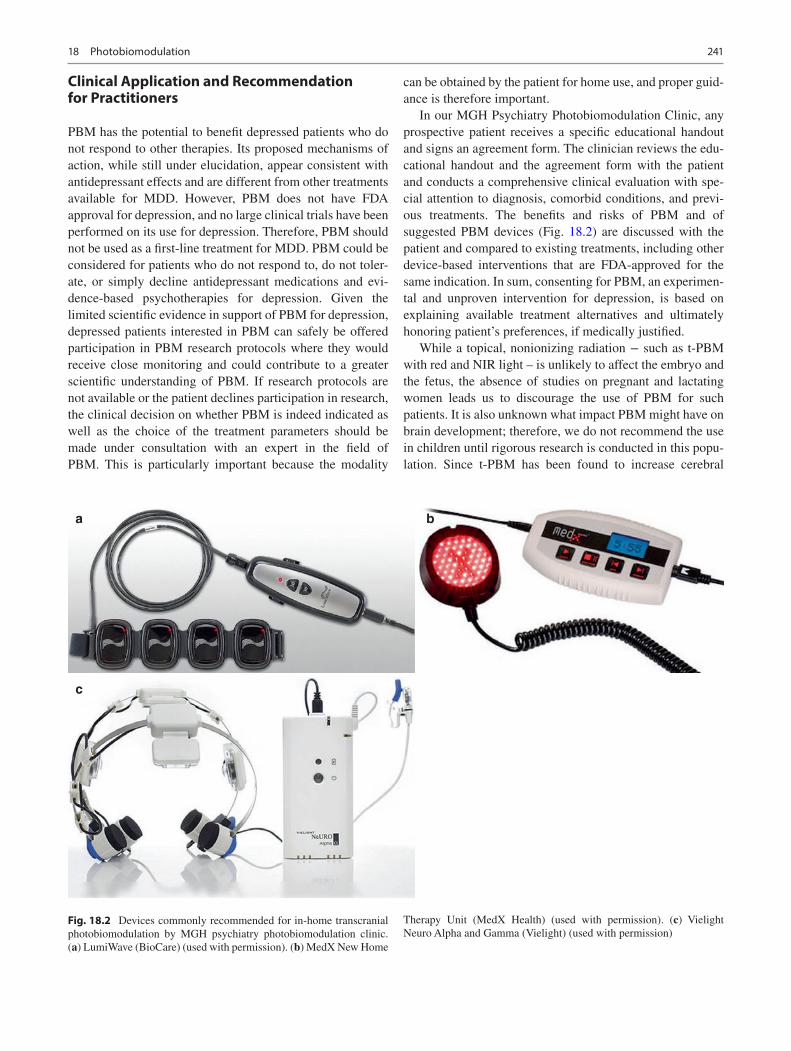
In our MGH Psychiatry Photobiomodulation Clinic, any prospective patient receives a specific educational handout and signs an agreement form. The clinician reviews the edu-cational handout and the agreement form with the patient and conducts a comprehensive clinical evaluation with spe-cial attention to diagnosis, comorbid conditions, and previ-ous treatments. The benefits and risks of PBM and of suggested PBM devices (Fig. 18.2) are discussed with the patient and compared to existing treatments, including other device-based interventions that are FDA-approved for the same indication. In sum, consenting for PBM, an experimen-tal and unproven intervention for depression, is based on explaining available treatment alternatives and ultimately honoring patient’s preferences, if medically justified.
While a topical, nonionizing radiation − such as t-PBM with red and NIR light – is unlikely to affect the embryo and the fetus, the absence of studies on pregnant and lactating women leads us to discourage the use of PBM for such patients. It is also unknown what impact PBM might have on brain development; therefore, we do not recommend the use in children until rigorous research is conducted in this popu-lation. Since t-PBM has been found to increase cerebral
a
c
b
Fig. 18.2 Devices commonly recommended for in-home transcranial photobiomodulation by MGH psychiatry photobiomodulation clinic. (a) LumiWave (BioCare) (used with permission). (b) MedX New Home
Therapy Unit (MedX Health) (used with permission). (c) Vielight Neuro Alpha and Gamma (Vielight) (used with permission)
18 Photobiomodulation
242
blood flow, there is a theoretical risk of dislodging implants in the brain such as stents, clips on aneurisms, or implantable shunts (e.g., Hakim valves) or of rupturing embolized vascu-lar malformations; therefore, the aforementioned are contra-indication to the use of t-PBM. Similarly, it is relatively contraindicated to use t-PBM in patients with a recent hem-orrhagic stroke (past 3 months), due to the potential recur-rence of bleeding with vasodilation and due to the risk of cerebral warming associated with light absorption by deep hematomas.
Conclusion
The proposed mechanism of action, safety profile, low cost, and positive results on preclinical and clinical studies stimu-lates research on PBM for the treatment of MDD. However, it is important to remark that studies on the field are still preliminary. Preclinical studies used different stimulation parameters, making it difficult to compare or combine results from different studies. The same is true for studies on humans. Moreover, clinical studies reported at this time are open or small clinical trials and, therefore, cannot be consid-ered enough evidence on the efficacy of PBM for MDD. More consistent evidence of efficacy will require more studies to define the best stimulation parameters and to guide the design of well-powered randomized clinical trials.
FAQs: Common Questions and Answers
Q1. How is PBM different from other device-based treat-ments for MDD?
A1. Other device-based treatments used to treat MDD modu-late the electric potential of neuron membranes using electric currents (e.g., ECT, TDCS) or magnetic pulses (e.g., rTMS). The exact mechanism of action of PBM is not completely understood, but it seems to work by enhancing mitochondrial metabolism leading to improved neuronal functioning and secondary improve-ments on oxidative stress, inflammation, and neurogenesis.
Q2. How is PBM different from other light-based treatments?
A2. PBM is noninvasive, and NIR and red light are nonion-izing electromagnetic irradiation, which are absorbed by specific endogenous chromophores and are minimally dissipated as thermal energy [5]. High-power lasers are used for ablative treatments and produce heating. Photodynamic therapy, such as for cancer chemother-apy, uses light to excite exogenously delivered chromo-phores (e.g., photosensitive anticancer drugs) to produce
therapeutic reactive oxygen species (ROS) [67]. Bright light therapy uses light in the visible, broad spectra to stimulate the retina and suppress the release of melato-nin and lengthen the photoperiod [114].
Q3. Is PBM safe during pregnancy and breast-feeding?A3. Considering the mechanism of action of PBM and the
limited tissue penetration of NIR, PBM has the potential to become a safe treatment for MDD during pregnancy and breast-feeding. However, given the lack of evidence regarding safety in this special population, including the risks for the embryo and fetus, we do not recommend PBM for women who are pregnant or lactating.
Q4. Can PBM be combined with other antidepressant treatments?
A4. Published studies included cases of patients receiving PBM in combination with psychotherapy and antide-pressant medications, and no serious adverse events were reported for these combinations. There are no reports on the combination of PBM with rTMS, TDCS, or ECT. There is no rationale for avoiding the simultane-ous use of PBM with other device-based treatments: potentiation of the antidepressant effects might occur, and some side effects – such as memory impairment with ECT – might be mitigated.
Q5. How long should PBM be used for the treatment of MDD?
A5. One single session of t-PBM may produce a decrease of depressive symptoms, but this effect seems to be tempo-rary [50]. More consistent improvement was observed in studies that used multiple sessions during 3–8 weeks for the treatment of an acute depressive episode [51, 113]. The safety profile of PBM suggests it could be used as maintenance treatment for responders. However, the only evidence for long-term use comes from a case report [55].
Q6. What is the difference between laser and LED light sources?
A6. Laser devices deliver a single wavelength wave, while LEDs also deliver light in a small range of different wavelengths close to the nominal wave (typically a 30 nm band) [1]. More recent studies are focusing on LED devices because they are less expensive than lasers, and the clinical efficacy does not seem to be determined by the kind of light source.
Q7. How does PBM cost compare to other treatments?A7. PBM is currently not reimbursed by US insurance carri-
ers. The in-office administration of PBM is therefore expensive and entirely out-of-pocket, unlike for FDA-approved MDD treatments such as rTMS, ECT, and antidepressant medications. At-home self-administra-tion of PBM still requires an upfront out-of-pocket expense, which averages between $400 and $2000 USD, depending on the chosen transcranial LED device. Some
M. A. Caldieraro and P. Cassano
243
manufacturers have a 6-month return policy: during this period the full cost is reimbursed after the device is returned, based on lack of efficacy.
References
1. Hamblin MR. Shining light on the head: photobiomodulation for brain disorders. BBA Clin. 2016;6:113–24.
2. Cassano P, Petrie SR, Hamblin MR, Henderson TA, Iosifescu DV. Review of transcranial photobiomodulation for major depres-sive disorder: targeting brain metabolism, inflammation, oxidative stress, and neurogenesis. Neurophotonics. 2016;3(3):031404.
3. Wang Y, Huang Y-Y, Wang Y, Lyu P, Hamblin MR. Photobiomodulation (blue and green light) encourages osteo-blastic-differentiation of human adipose-derived stem cells: role of intracellular calcium and light-gated ion channels. Nat Publ Group. 2016;6(1):33719.
4. Chung H, Dai T, Sharma SK, Huang Y-Y, Carroll JD, Hamblin MR. The nuts and bolts of low-level laser (light) therapy. Ann Biomed Eng. 2012;40(2):516–33.
5. Mochizuki-Oda N, Kataoka Y, Cui Y, Yamada H, Heya M, Awazu K. Effects of near-infra-red laser irradiation on adenosine tri-phosphate and adenosine diphosphate contents of rat brain tissue. Neurosci Lett. 2002;323(3):207–10.
6. Kennedy SH, Konarski JZ, Segal ZV, Lau MA, Bieling PJ, McIntyre RS, et al. Differences in brain glucose metabolism between responders to CBT and venlafaxine in a 16-week ran-domized controlled trial. Am J Psychiatry. 2007;164(5):778–88.
7. Mayberg HS, Brannan SK, Tekell JL, Silva JA, Mahurin RK, McGinnis S, et al. Regional metabolic effects of fluoxetine in major depression: serial changes and relationship to clinical response. Biol Psychiatry. 2000;48(8):830–43.
8. Iosifescu DV, Bolo NR, Nierenberg AA, Jensen JE, Fava M, Renshaw PF. Brain bioenergetics and response to triiodothyro-nine augmentation in major depressive disorder. Biol Psychiatry. 2008;63(12):1127–34.
9. Bansal Y, Kuhad A. Mitochondrial dysfunction in depression. Curr Neuropharmacol. 2016;14(6):610–8.
10. Klinedinst NJ, Regenold WT. A mitochondrial bioenergetic basis of depression. J Bioenerg Biomembr. 5 ed. Springer US. 2015;47(1–2):155–71.
11. Salehpour F, Rasta SH. The potential of transcranial photobio-modulation therapy for treatment of major depressive disorder. Rev Neurosci. 2017;28(4):441–53.
12. Sommer AP, Trelles MA. Light pumping energy into blood mito-chondria: a new trend against depression? Photomed Laser Surg. 2014;32(2):59–60.
13. Hroudová J, Fišar Z, Kitzlerová E, Zvěřová M, Raboch J. Mitochondrial respiration in blood platelets of depressive patients. Mitochondrion. 2013;13(6):795–800.
14. McGuff PE, Deterling RA, Gottlieb LS. Tumoricidal effect of laser energy on experimental and human malignant tumors. N Engl J Med. 1965;273(9):490–2.
15. Mester E, Szende B, Gärtner P. The effect of laser beams on the growth of hair in mice. Radiobiol Radiother (Berl). 1968;9(5):621–6.
16. Kovács IB, Mester E, Görög P. Stimulation of wound healing with laser beam in the rat. Experientia. 1974;30(11):1275–6.
17. Ferraresi C, Hamblin MR, Parizotto NA. Low-level laser (light) therapy (LLLT) on muscle tissue: performance, fatigue and repair benefited by the power of light. Photon Lasers Med. 2012;1(4):267–86.
18. Chaves ME de A, Araújo AR, de Piancastelli ACC, Pinotti M. Effects of low-power light therapy on wound healing: LASER x LED. An Bras Dermatol. 2014;89(4):616–23.
19. Harkless LB, DeLellis S, Carnegie DH, Burke TJ. Improved foot sensitivity and pain reduction in patients with peripheral neuropathy after treatment with monochromatic infrared photo energy – MIRE. J Diabetes Complicat. 2006;20(2):81–7.
20. Allais G, De Lorenzo C, Quirico PE, Lupi G, Airola G, Mana O, et al. Non-pharmacological approaches to chronic head-aches: transcutaneous electrical nerve stimulation, laser therapy and acupuncture in transformed migraine treatment. Neurol Sci. 2003;24(Suppl 2):S138–42.
21. Hersant B, SidAhmed-Mezi M, Bosc R, Meningaud JP. Current indications of low-level laser therapy in plastic surgery: a review. Photomed Laser Surg. 2015;33(5):283–97.
22. Russell BA, Kellett N, Reilly LR. A study to determine the efficacy of combination LED light therapy (633 nm and 830 nm) in facial skin rejuvenation. J Cosmet Laser Ther. 2005;7(3–4):196–200.
23. Zarei M, Wikramanayake TC, Falto-Aizpurua L, Schachner LA, Jimenez JJ. Low level laser therapy and hair regrowth: an evidence-based review. Lasers Med Sci. Springer London. 2016;31(2):363–71.
24. Rochkind S, Barr-Nea L, Bartal A, Nissan M, Lubart R, Razon N. New methods of treatment of severely injured sciatic nerve and spinal cord. An experimental study. Acta Neurochir Suppl (Wien). 1988;43:91–3.
25. Rochkind S, Ouaknine GE. New trend in neuroscience: low-power laser effect on peripheral and central nervous system (basic science, preclinical and clinical studies). Neurol Res. 1992;14(1):2–11.
26. Rochkind S, Vogler I, Barr-Nea L. Spinal cord response to laser treatment of injured peripheral nerve. Spine. 1990;15(1):6–10.
27. Anders JJ. The potential of light therapy for central nervous sys-tem injury and disease. Photomed Laser Surg. 2009;27(3):379–80.
28. Lapchak PA, Salgado KF, Chao CH, Zivin JA. Transcranial near-infrared light therapy improves motor function following embolic strokes in rabbits: an extended therapeutic window study using continuous and pulse frequency delivery modes. Neuroscience. 2007;148(4):907–14.
29. Lapchak PA, Wei J, Zivin JA. Transcranial infrared laser therapy improves clinical rating scores after embolic strokes in rabbits. Stroke. 2004;35(8):1985–8.
30. Oron A, Oron U, Chen J, Eilam A, Zhang C, Sadeh M, et al. Low-level laser therapy applied transcranially to rats after induction of stroke significantly reduces long-term neurological deficits. Stroke. 2006 Oct;37(10):2620–4.
31. Hacke W, Schellinger PD, Albers GW, Bornstein NM, Dahlof BL, Fulton R, et al. Transcranial laser therapy in acute stroke treatment: results of neurothera effectiveness and safety trial 3, a phase III clinical end point device trial. Stroke. American Heart Association, Inc. 2014;45(11):3187–93.
32. Zivin JA, Albers GW, Bornstein N, Chippendale T, Dahlof B, Devlin T, et al. Effectiveness and safety of transcranial laser therapy for acute ischemic stroke. Stroke. Lippincott Williams & Wilkins. 2009;40(4):1359–64.
33. Lampl Y, Zivin JA, Fisher M, Lew R, Welin L, Dahlof B, et al. Infrared laser therapy for ischemic stroke: a new treatment strat-egy: results of the NeuroThera Effectiveness and Safety Trial-1 (NEST-1). Stroke. 2007;38(6):1843–9.
34. Ando T, Xuan W, Xu T, Dai T, Sharma SK, Kharkwal GB, et al. Comparison of therapeutic effects between pulsed and continu-ous wave 810-nm wavelength laser irradiation for traumatic brain injury in mice. PLoS One. 2011;6(10):e26212.
35. Huang Y-Y, Sharma SK, Carroll J, Hamblin MR. Biphasic dose response in low level light therapy – an update. Dose Response. 2011;9(4):602–18.
18 Photobiomodulation
244
36. Huang Y-Y, Gupta A, Vecchio D, de Arce VJB, Huang S-F, Xuan W, et al. Transcranial low level laser (light) therapy for traumatic brain injury. Kirillin M, Sokolov K, Shakhova N, Steiner R, edi-tors. J Biophotonics. 2012;5(11–12):827–37.
37. Xuan W, Agrawal T, Huang L, Gupta GK, Hamblin MR. Low-level laser therapy for traumatic brain injury in mice increases brain derived neurotrophic factor (BDNF) and synaptogenesis. J Biophotonics. 2015;8(6):502–11.
38. Xuan W, Vatansever F, Huang L, Wu Q, Xuan Y, Dai T, et al. Transcranial low-level laser therapy improves neurological performance in traumatic brain injury in mice: effect of treat-ment repetition regimen. Borlongan CV, editor. PLoS One. 2013;8(1):e53454.
39. Xuan W, Vatansever F, Huang L, Hamblin MR. Transcranial low-level laser therapy enhances learning, memory, and neuropro-genitor cells after traumatic brain injury in mice. J Biomed Opt. 2014;19(10):108003.
40. Naeser MA, Martin PI, Ho MD, Krengel MH, Bogdanova Y, Knight JA, et al. Transcranial, red/near-infrared light-emitting diode therapy to improve cognition in chronic traumatic brain injury. Photomed Laser Surg. 2016;34(12):610–26.
41. Naeser MA, Zafonte R, Krengel MH, Martin PI, Frazier J, Hamblin MR, et al. Significant improvements in cognitive per-formance post-transcranial, red/near-infrared light-emitting diode treatments in chronic, mild traumatic brain injury: open-protocol study. J Neurotrauma. 2014;31(11):1008–17.
42. Naeser MA, Saltmarche A, Krengel MH, Hamblin MR, Knight JA. Improved cognitive function after transcranial, light-emitting diode treatments in chronic, traumatic brain injury: two case reports. Photomed Laser Surg. 2011;29(5):351–8.
43. Purushothuman S, Johnstone DM, Nandasena C, Mitrofanis J, Stone J. Photobiomodulation with near infrared light mitigates Alzheimer’s disease-related pathology in cerebral cortex – evi-dence from two transgenic mouse models. Alzheimers Res Ther. BioMed Central. 2014;6(1):2.
44. De Taboada L, Yu J, El-Amouri S, Gattoni-Celli S, Richieri S, McCarthy T, et al. Transcranial laser therapy attenuates amyloid-β peptide neuropathology in amyloid-β protein precursor transgenic mice. J Alzheimers Dis. IOS Press. 2011;23(3):521–35.
45. Tanaka Y, Akiyoshi J, Kawahara Y, Ishitobi Y, Hatano K, Hoaki N, et al. Infrared radiation has potential antidepressant and anxiolytic effects in animal model of depression and anxiety. Brain Stimul. 2011;4(2):71–6.
46. Mohammed HS. Transcranial low-level infrared laser irradiation ameliorates depression induced by reserpine in rats. Lasers Med Sci. Springer London. 2016;31(8):1651–6.
47. Xu Z, Guo X, Yang Y, Tucker D, Lu Y, Xin N, et al. Low-level laser irradiation improves depression-like behaviors in mice. Mol Neurobiol. 2016;34(1):13.
48. Wu X, Alberico SL, Moges H, De Taboada L, Tedford CE, Anders JJ. Pulsed light irradiation improves behavioral outcome in a rat model of chronic mild stress. Lasers Surg Med. Wiley Subscription Services, Inc., A Wiley Company. 2012;44(3):227–32.
49. Salehpour F, Rasta SH, Mohaddes G, Sadigh-Eteghad S, Salarirad S. Therapeutic effects of 10-HzPulsed wave lasers in rat depres-sion model: a comparison between near-infrared and red wave-lengths. Lasers Surg Med. 2016;48(7):695–705.
50. Schiffer F, Johnston AL, Ravichandran C, Polcari A, Teicher MH, Webb RH, et al. Psychological benefits 2 and 4 weeks after a sin-gle treatment with near infrared light to the forehead: a pilot study of 10 patients with major depression and anxiety. Behav Brain Funct. 2009;5(1):46.
51. Cassano P, Cusin C, Mischoulon D, Hamblin MR, De Taboada L, Pisoni A, et al. Near-infrared transcranial radiation for major depressive disorder: proof of concept study. Psychiatry J. 2015;2015:352979.
52. Mogoaşe C, David D, Koster EHW. Clinical efficacy of attentional bias modification procedures: an updated meta-analysis. J Clin Psychol. Wiley-Blackwell. 2014;70(12):1133–57.
53. Disner SG, Beevers CG, Gonzalez-Lima F. Transcranial laser stimulation as neuroenhancement for attention bias modification in adults with elevated depression symptoms. Brain Stimul. 2016;9(5):780–7.
54. Joffe RT, Uhde TW, Post RM, Minichiello MD. Motor activity in depressed patients treated with carbamazepine. Biol Psychiatry. 1987;22(8):941–6.
55. Caldieraro MA, Sani G, Bui E, Cassano P. Long-term near-infrared photobiomodulation for anxious depression compli-cated by Takotsubo Cardiomyopathy. J Clin Psychopharmacol. 2018;38(3):268–70.
56. Kartelishev AV, Kolupaev GP, Vernekina NS, Chebotkov AA, Lakosina ND, Ushakov AA. Laser technologies used in the com-plex treatment of psychopharmacotherapy resistant endogenic depression. Voen Med Zh. 2004;325(11):37–42.
57. Kolupaev GP, Kartelishev AV, Vernekina NS, Chebotkov AA, Lakosina ND. Technologies of laser prophylaxis of depressive disorder relapses. Voen Med Zh. 2007;328(2):31–4.
58. Quah-Smith I, Smith C, Crawford JD, Russell J. Laser acu-puncture for depression: a randomised double blind controlled trial using low intensity laser intervention. J Affect Disord. 2013;148(2–3):179–87.
59. Quah-Smith JI, Tang WM, Russell J. Laser acupuncture for mild to moderate depression in a primary care setting – a randomised controlled trial. Acupunct Med. 2005;23(3):103–11.
60. Huisa BN, Stemer AB, Walker MG, Rapp K, Meyer BC, Zivin JA, et al. Transcranial laser therapy for acute ischemic stroke: a pooled analysis of NEST-1 and NEST-2. Int J Stroke. 2013;8(5):315–20.
61. Morries LD, Cassano P, Henderson TA. Treatments for trau-matic brain injury with emphasis on transcranial near-infrared laser phototherapy. Neuropsychiatr Dis Treat. Dove Press. 2015;11:2159–75.
62. Karu TI, Kolyakov SF. Exact action spectra for cellular responses relevant to phototherapy. Photomed Laser Surg. 2005;23(4):355–61.
63. Morava E, Kozicz T. Mitochondria and the economy of stress (mal)adaptation. Neurosci Biobehav Rev. 2013;37(4):668–80.
64. Karabatsiakis A, Böck C, Salinas-Manrique J, Kolassa S, Calzia E, Dietrich DE, et al. Mitochondrial respiration in peripheral blood mononuclear cells correlates with depressive subsymp-toms and severity of major depression. Transl Psychiatry. 2014;4(6):e397.
65. Yu W, Naim JO, McGowan M, Ippolito K, Lanzafame RJ. Photomodulation of oxidative metabolism and electron chain enzymes in rat liver mitochondria. Photochem Photobiol. 1997;66(6):866–71.
66. Oron U, Ilic S, De Taboada L, Streeter J. Ga-As (808 nm) laser irradiation enhances ATP production in human neuronal cells in culture. Photomed Laser Surg. 2007;25(3):180–2.
67. de Freitas LF, Hamblin MR. Proposed mechanisms of photobio-modulation or low-level light therapy. IEEE J Sel Top Quantum Electron. 2016;22(3):348–64.
68. Greco M, Guida G, Perlino E, Marra E, Quagliariello E. Increase in RNA and protein synthesis by mitochondria irra-diated with helium-neon laser. Biochem Biophys Res Commun. 1989;163(3):1428–34.
69. Black CN, Bot M, Scheffer PG, Cuijpers P, Penninx BWJH. Is depression associated with increased oxidative stress? A sys-tematic review and meta-analysis. Psychoneuroendocrinology. 2015;51:164–75.
70. Sarandol A, Sarandol E, Eker SS, Erdinc S, Vatansever E, Kirli S. Major depressive disorder is accompanied with oxidative stress: short-term antidepressant treatment does not alter oxidative-anti-
M. A. Caldieraro and P. Cassano
245
oxidative systems. Hum Psychopharmacol. John Wiley & Sons, Ltd. 2007;22(2):67–73.
71. Eren I, Naziroğlu M, Demirdaş A. Protective effects of lamotrig-ine, aripiprazole and escitalopram on depression-induced oxida-tive stress in rat brain. Neurochem Res 3rd ed. Kluwer Academic Publishers-Plenum Publishers. 2007;32(7):1188–95.
72. Shungu DC, Weiduschat N, Murrough JW, Mao X, Pillemer S, Dyke JP, et al. Increased ventricular lactate in chronic fatigue syndrome. III. Relationships to cortical glutathione and clinical symptoms implicate oxidative stress in disorder pathophysiology. NMR Biomed John Wiley & Sons, Ltd. 2012;25(9):1073–87.
73. Spanemberg L, Caldieraro M, Arrua Vares E, Wollenhaupt de Aguiar B, Yuri Kawamoto S, Parker G, et al. Biological differences between melancholic and nonmelancholic depression subtyped by the CORE measure. Neuropsychiatr Dis Treat. 2014;10:1523.
74. Ozcan ME, Gulec M, Ozerol E, Polat R, Akyol O. Antioxidant enzyme activities and oxidative stress in affective disorders. Int Clin Psychopharmacol. 2004;19(2):89–95.
75. Rizzi CF, Mauriz JL, Freitas Corrêa DS, Moreira AJ, Zettler CG, Filippin LI, et al. Effects of low-level laser therapy (LLLT) on the nuclear factor (NF)-kappaB signaling pathway in traumatized muscle. Lasers Surg Med. Wiley Subscription Services, Inc., A Wiley Company. 2006;38(7):704–13.
76. Chludzińska L, Ananicz E, Jarosławska A, Komorowska M. Near-infrared radiation protects the red cell membrane against oxida-tion. Blood Cells Mol Dis. 2005;35(1):74–9.
77. Anisman H, Hayley S. Inflammatory factors contribute to depres-sion and its comorbid conditions. Sci Signal. 2012;5(244):pe45–5.
78. Dowlati Y, Herrmann N, Swardfager W, Liu H, Sham L, Reim EK, et al. A meta-analysis of cytokines in major depression. Biol Psychiatry. 2010;67(5):446–57.
79. Liu Y, Ho RC-M, Mak A. Interleukin (IL)-6, tumour necrosis fac-tor alpha (TNF-α) and soluble interleukin-2 receptors (sIL-2R) are elevated in patients with major depressive disorder: a meta-analy-sis and meta-regression. J Affect Disord. 2012;139(3):230–9.
80. Köhler CA, Freitas TH, Maes M, de Andrade NQ, Liu CS, Fernandes BS, et al. Peripheral cytokine and chemokine altera-tions in depression: a meta-analysis of 82 studies. Acta Psychiatr Scand. 2017;135(5):373–87.
81. Lindqvist D, Janelidze S, Hagell P, Erhardt S, Samuelsson M, Minthon L, et al. Interleukin-6 is elevated in the cerebrospinal fluid of suicide attempters and related to symptom severity. Biol Psychiatry. 2009;66(3):287–92.
82. Yoshimura R, Hori H, Ikenouchi-Sugita A, Umene-Nakano W, Ueda N, Nakamura J. Higher plasma interleukin-6 (IL-6) level is associated with SSRI- or SNRI-refractory depression. Prog Neuropsychopharmacol Biol Psychiatry. 2009;33(4):722–6.
83. Araki H, Imaoka A, Kuboyama N, Abiko Y. Reduction of interleu-kin-6 expression in human synoviocytes and rheumatoid arthritis rat joints by linear polarized near infrared light (Superlizer) irra-diation. Laser Ther. 2011;20(4):293–300.
84. Yamaura M, Yao M, Yaroslavsky I, Cohen R, Smotrich M, Kochevar IE. Low level light effects on inflammatory cytokine production by rheumatoid arthritis synoviocytes. Lasers Surg Med Wiley Subscription Services, Inc., A Wiley Company. 2009;41(4):282–90.
85. Khuman J, Zhang J, Park J, Carroll JD, Donahue C, Whalen MJ. Low-level laser light therapy improves cognitive deficits and inhibits microglial activation after controlled cortical impact in mice. J Neurotrauma. 2012;29(2):408–17.
86. Duman RS. Pathophysiology of depression and innovative treat-ments: remodeling glutamatergic synaptic connections. Dialogues Clin Neurosci. 2014;16(1):11–27.
87. Autry AE, Monteggia LM. Brain-derived neurotrophic factor and neuropsychiatric disorders. Daws LC, editor. Pharmacol Rev. 2012;64(2):238–58.
88. Molendijk ML, Spinhoven P, Polak M, Bus BAA, Penninx BWJH, Elzinga BM. Serum BDNF concentrations as peripheral mani-festations of depression: evidence from a systematic review and meta-analyses on 179 associations (N=9484). Mol Psychiatry. 2014;19(7):791–800.
89. Molendijk ML, Bus BAA, Spinhoven P, Penninx BWJH, Kenis G, Prickaerts J, et al. Serum levels of brain-derived neuro-trophic factor in major depressive disorder: state-trait issues, clinical features and pharmacological treatment. Mol Psychiatry. 2011;16(11):1088–95.
90. Neto FL, Borges G, Torres-Sanchez S, Mico JA, Berrocoso E. Neurotrophins role in depression neurobiology: a review of basic and clinical evidence. Curr Neuropharmacol. 2011;9(4):530–52.
91. Giuliani A, Lorenzini L, Gallamini M, Massella A, Giardino L, Calzà L. Low infra red laser light irradiation on cultured neural cells: effects on mitochondria and cell viability after oxidative stress. BMC Complement Altern Med. 2009;9(1):8.
92. Rojas JC, Lee J, John JM, Gonzalez-Lima F. Neuroprotective effects of near-infrared light in an in vivo model of mitochondrial optic neuropathy. J Neurosci. 2008;28(50):13511–21.
93. Wong-Riley MTT, Liang HL, Eells JT, Chance B, Henry MM, Buchmann E, et al. Photobiomodulation directly benefits primary neurons functionally inactivated by toxins: role of cytochrome c oxidase. J Biol Chem. 2005;280(6):4761–71.
94. Wu Q, Xuan W, Ando T, Xu T, Huang L, Huang Y-Y, et al. Low-level laser therapy for closed-head traumatic brain injury in mice: effect of different wavelengths. Lasers Surg Med Wiley Subscription Services, Inc., A Wiley Company. 2012;44(3):218–26.
95. Oron A, Oron U, Streeter J, De Taboada L, Alexandrovich A, Trembovler V, et al. Low-level laser therapy applied tran-scranially to mice following traumatic brain injury signifi-cantly reduces long-term neurological deficits. J Neurotrauma. 2007;24(4):651–6.
96. Rojas JC, Bruchey AK, Gonzalez-Lima F. Low-level light therapy improves cortical metabolic capacity and memory retention. J Alzheimers Dis. IOS Press. 2012;32(3):741–52.
97. Uozumi Y, Nawashiro H, Sato S, Kawauchi S, Shima K, Kikuchi M. Targeted increase in cerebral blood flow by transcranial near-infrared laser irradiation. Lasers Surg Med. Wiley Subscription Services, Inc., A Wiley Company. 2010;42(6):566–76.
98. Videbech P. PET measurements of brain glucose metabolism and blood flow in major depressive disorder: a critical review. Acta Psychiatr Scand. 2000;101(1):11–20.
99. Drevets WC, Bogers W, Raichle ME. Functional anatomical cor-relates of antidepressant drug treatment assessed using PET mea-sures of regional glucose metabolism. Eur Neuropsychopharmacol. 2002;12(6):527–44.
100. Tian F, Hase SN, Gonzalez-Lima F, Liu H. Transcranial laser stimulation improves human cerebral oxygenation. Lasers Surg Med. 2016;48(4):343–9.
101. Salgado ASI, Zângaro RA, Parreira RB, Kerppers II. The effects of transcranial LED therapy (TCLT) on cerebral blood flow in the elderly women. Lasers Med Sci. 2015;30(1):339–46.
102. Konstantinović LM, Jelić MB, Jeremić A, Stevanović VB, Milanović SD, Filipović SR. Transcranial application of near-infrared low-level laser can modulate cortical excitability. Lasers Surg Med. 2013;45(10):648–53.
103. Ho MD, Martin PI, Yee MK, Koo B, Baker E, Hamblin MR, et al. Increased functional connectivity in default mode network associated with application of transcranial, light-emitting diodes to treat chronic aphasia: case series. J Int Neuropsychol Soc. 2016;22(S1):229.
104. Quah-Smith I, Wen W, Chen X, Williams MA, Sachdev PS. The brain effects of laser acupuncture in depressed individuals: an fMRI investigation. Med Acupunct. 2012;24(3):161–71.
18 Photobiomodulation
246
105. Yue L, Humayun MS. Monte Carlo analysis of the enhanced tran-scranial penetration using distributed near-infrared emitter array. J Biomed Opt. 2015;20(8):88001.
106. Tedford CE, DeLapp S, Jacques S, Anders J. Quantitative analysis of transcranial and intraparenchymal light penetration in human cadaver brain tissue. Lasers Surg Med. 2015;47(4):312–22.
107. Henderson TA, Morries LD. Near-infrared photonic energy pen-etration: can infrared phototherapy effectively reach the human brain? Neuropsychiatr Dis Treat. Dove Press. 2015;11:2191–208.
108. Jagdeo JR, Adams LE, Brody NI, Siegel DM. Transcranial red and near infrared light transmission in a cadaveric model. Hamblin M, editor. PLoS One. 2012;7(10):e47460.
109. Yue L, Monge M, Ozgur MH, Murphy K, Louie S, Miller CA, et al. Simulation and measurement of transcranial near infra-red light penetration. San Francisco, California. SPIE BiOS. 2015;9321:93210S1–6.
110. Pitzschke A, Lovisa B, Seydoux O, Zellweger M, Pfleiderer M, Tardy Y, et al. Red and NIR light dosimetry in the human deep brain. Phys Med Biol. 2015;60(7):2921–37.
111. Hashmi JT, Huang Y-Y, Sharma SK, Kurup DB, De Taboada L, Carroll JD, et al. Effect of pulsing in low-level light therapy. Lasers Surg Med. 2010;42(6):450–66.
112. Sharma SK, Kharkwal GB, Sajo M, Huang Y-Y, De Taboada L, McCarthy T, et al. Dose response effects of 810 nm laser light on mouse primary cortical neurons. Lasers Surg Med. 2011;43(8):851–9.
113. Cassano P, Petrie SR, Mischoulon D, Ionescu DF, Cusin C, Katnani H, et al. Transcranial photobiomodulation for the treat-ment of major depressive disorder: the ELATED-2 clinical trial. Neuropsychopharmacology. 2016;41:S354–5.
114. Golden RN, Gaynes BN, Ekstrom RD, Hamer RM, Jacobsen FM, Suppes T, et al. The efficacy of light therapy in the treatment of mood disorders: a review and meta-analysis of the evidence. Am J Psychiatry. 2005;162(4):656–62.
M. A. Caldieraro and P. Cassano