Embed Size (px)
Citation preview

Clemson UniversityTigerPrints
All Theses Theses
5-2016
Phosphorylation of Eukaryotic Initiation Factor 2Alpha Regulates Stress in the Human ProtozoanParasite Entamoeba HistolyticaHolland M. HendrickClemson University, [email protected]
Follow this and additional works at: https://tigerprints.clemson.edu/all_theses
This Thesis is brought to you for free and open access by the Theses at TigerPrints. It has been accepted for inclusion in All Theses by an authorizedadministrator of TigerPrints. For more information, please contact [email protected].
Recommended CitationHendrick, Holland M., "Phosphorylation of Eukaryotic Initiation Factor 2 Alpha Regulates Stress in the Human Protozoan ParasiteEntamoeba Histolytica" (2016). All Theses. 2340.https://tigerprints.clemson.edu/all_theses/2340

i
PHOSPHORYLATION OF EUKARYOTIC INITIATION FACTOR 2 ALPHA REGULATES STRESS IN THE HUMAN PROTOZOAN PARASITE
ENTAMOEBA HISTOLYTICA
A Thesis
Presented to the Graduate School of
Clemson University
In Partial Fulfillment of the Requirements for the Degree
Master of Science Biological Sciences
by Holland M. Hendrick
May 2016
Accepted by: Dr. Lesly Temesvari, Committee Chair
Dr. Lisa Bain Dr. Lukasz Kozubowski

ii
ABSTRACT
Entamoeba histolytica is a food- and water-borne intestinal parasite responsible for
amoebic dysentery and amoebic liver abscess. The life cycle of E. histolytica alternates
between the host-restricted trophozoite form and the highly infective latent cyst stage that
is able to persist in the environment. Throughout its life cycle, which may include
invasion of tissues in the human host, the parasite is subjected to a variety of stressful
conditions. In other systems, stress can trigger the activation of kinases that
phosphorylate a serine residue on eukaryotic translation initiation factor-2α (eIF2α). This
modification inhibits the activity of eIF2 resulting in a general decline in protein
synthesis, and, paradoxically, an up-regulation of the expression of certain genes that
permit the cell to counter the stress. Genomic data reveal that E. histolytica possesses
eIF2α with a conserved phosphorylatable serine at position 59. Thus, this pathogen may
have the machinery for stress-induced translational control. To test this, we exposed E.
histolytica trophozoites to six different stress conditions and assessed viability, as well as
the level of total and phospho-EheIF2α via Western blot of cell lysates. Long term serum
starvation induced an increase in the level of phospho-EheIF2α, but no other stress
condition caused a significant change. Long term serum starvation also showed a
decrease in polyribosome abundance as observed through sucrose gradient
ultracentrifugation; this is consistent with the observation that this condition also induces
phosphorylation of EheIF2α. This suggests that the eIF2α-dependent stress response
system is operational in E. histolytica and that the system may be activated only by
certain stresses. To further examine the role of phosphorylation of EheIF2α during stress,

iii
three transgenic cell lines were created. EheIF2α-S59 over-expresses wild type eIF2α
protein. EheIF2α-S59A expresses eIF2α with the serine-59 residue mutated to an
alanine, creating a non-phosphorylatable subunit. EheIF2α-S59D expresses eIF2α with
the serine-59 residue mutated to an aspartic acid to mimic a phosphorylated residue.
EheIF2α-S59 exhibited a high level of phosphorylation of the exogenous protein, leading
to a decreased growth and polyribosome abundance when compared to the control cell
line. EheIF2α-S59A had the highest growth rate and retained a high abundance of
polyribosome. EheIF2α-S59D exhibited the slowest growth rate and had a decrease in
polyribosome when compared to control; however, EheIF2α-S59D did exhibit the highest
survival rate in over half the stress conditions tested. This may indicate the protective
nature of phosphorylation of EheIF2α during times of stress.

iv
DEDICATION
This body of work would not have been possible without the incredible support of
my parents, James and Marcia, my sisters, Hannah and Helena, and my partner in life,
Roddey. You have supported me throughout my life, but especially during this most
strenuous time. I am always thankful for having you in my life.

v
ACKNOWLEDGMENTS
I am eternally thankful for Dr. Lesly Temesvari and the support she has provided
during my studies. Her passion for science is a constant encouragement. I would also
like to thank Dr. Lisa Bain and Dr. Lukasz Kozubowski for being excellent teachers and
committee members. Their discussion and comments have been instrumental in this
project.
Thanks to the Temesvari lab, past and present, for their assistance, helpfulness,
and friendship. This would not have been possible without some of the stress relieving
conversations we had. Brenda Welter, thank you for always reminding us that, if science
was easy, it would have been done already. Your insight and guidance made each new
experiment a little bit easier.
A special thanks goes out to the faculty and students of EPIC, who have assisted
in troubleshooting and keeping me lighthearted even in the worst weeks. To you, my
friends, I thank you for making sure I had a life outside the Life Sciences Facility.
Thanks to my family, who helped and supported me for the past three years.
Your prayers and encouragement helped more than you will ever realize.
Financial assistance for this work was provided by a grant to Dr. Temesvari by
the National Institute of Health (R03AI107950) and by the Department of Biological
Sciences who provided funding for my studies here at Clemson University. This material
is based upon work supported by CSREES/USDA under project number SC-1700312.
The funding agencies had no role in study design, data collection and analysis, decision
to publish, or preparation of the manuscript. The content is solely the responsibility of the

vi
authors and does not necessarily represent the views of the National Institute of Allergy
and Infectious Diseases, the National Institutes of Health, or the USDA.

vii
TABLE OF CONTENTS
Page
TITLE PAGE .................................................................................................................... i ABSTRACT ..................................................................................................................... ii DEDICATION ................................................................................................................ iv ACKNOWLEDGMENTS ............................................................................................... v LIST OF FIGURES ...................................................................................................... viii CHAPTER 1. LITERATURE REVIEW .............................................................................. 1
I. Introduction ........................................................................................... 1 II. The stress response in Entamoeba histolytica ...................................... 4 III. Overview of translation control of stress ............................................ 12 IV. Stress-induced control of protein translation in eukaryotes ................ 19 V. Summary ............................................................................................. 29 VI. Literature Cited ................................................................................... 31
2. PHOSPHORYLATION OF EUKARYOTIC INITIATION FACTOR 2 α
REGULATES STRESS IN THE HUMAN PROTOZOAN PARASITE, ENTAMOEBA HSITOLYTICA ..................................................................... 36
I. Abstract ............................................................................................... 37 II. Authors Summary ............................................................................... 38 III. Introduction ......................................................................................... 40 IV. Results ................................................................................................. 42 V. Discussion ........................................................................................... 58 VI. Materials and Methods ........................................................................ 61 VII. Supplemental Figure ........................................................................... 68 VIII. Acknowledgements ............................................................................. 69 IX. Literature Cited ................................................................................... 69
viii
LIST OF FIGURES
Figure Page 1.1 Life cycle of Entamoeba histolytica .............................................................. 2 1.2 Phosphorylation of eIF2 blocks translation initiation ................................. 14 1.3 Activation of eIF2α kinases by specific stresses ......................................... 16 1.4 Life cycle of Plasmodium and the life stages of known eIF2α kinases ........................................................................................ 21 1.5 Life cycle of Toxoplasma gondii and the life stages of known eIF2α kinases ............................................................................. 24 1.6 Life cycle of Leishmania major and the life stages of known eIF2α kinases ............................................................................. 27 2.1 Alignment of the eukaryotic translation initiation factor 2, alpha subunit ...................................................................................... 43 2.2 Viability of trophozoites during stress ......................................................... 45 2.3 Western blot analysis of phospho-eIF2α and total eIF2α in control and stressed cells ................................................................... 46 2.4 Polyribosome abundance in control and stressed cells ................................ 48 2.5 Western blot analysis confirming exogenous protein expression in mutant cell lines ............................................................... 50 2.6 Polyribosome profile for control and mutant eIF2α cell lines after 72 h induction........................................................................ 51 2.7 SUnSET analysis of active protein translation in control and mutant cell lines .............................................................................. 54 2.8 Growth curves of control and mutant cell lines ........................................... 55 2.9 Control and mutant eIF2α cell line viabilities during stress conditions ..................................................................................... 57

ix
List of Figures (Continued) Figure Page
2.S1 Polyribosome profile for control and mutant eIF2α cell lines after 24 hour induction ............................................................................................... 68

1
CHAPTER ONE
LITERATURE REVIEW
I. Introduction
Entamoeba histolytica is the protozoan parasite responsible for amoebic dysentery
and liver abscess in humans and non-human primates. The life cycle of E. histolytica does
not require an intermediate host, as it is transmitted from human to human, often through
fecally contaminated food or water (Fig 1). This mode of transmission makes amoebic
dysentery a disease of importance to underdeveloped areas with no water filtration or
treatment systems. In the late 1990s, the WHO estimated that 500 million people around
the world were infected with Entamoeba, with 40,000-100,000 deaths annually [1].
Because of the ease of transmission in water, Entamoeba histolytica is classified as a Class
B bioterrorism agent, further highlighting the need for information about this parasite.
E. histolytica cysts are formed in the large intestine and exit the host with the fecal
matter. Cysts are typically rounded, quadrinucleated and stable (desiccation-tolerant, acid-
tolerant, heat-tolerant; and detergent-resistant) [reviewed in 2]. These latent cysts readily
survive the extreme conditions in the external environment. To continue the life cycle, the
cyst must be ingested where it can pass unharmed through the acidic condition of the
stomach into the small intestine. Once there, unknown triggers cause the cyst to undergo
excystation, resulting in eight active trophozoites for every cyst. These amoeboid
trophozoites continue down the intestinal tract until reaching the large intestine. This is
the location of intraintestinal infections of E. histolytica, where cells replicate by binary

2
Figure 1.1: Life cycle of Entamobea histolytica The infective stage, a latent cyst, is engested, usually due to fecally contaiminated food or water. This cyst can pass through the stomach unharmed and enter the small intestine. Here, unknown factors trigger an excystation event and 8 trophozoites emerge for every one cyst. These active trophozoites migrate through the small intestine and into the large intestine, where E. histolytica interintestinal infections occur. A small percentage of these trophozoites will encyst, though the molecular mechanism that triggers this process is unknown. These cysts can survive outside the host and be passed to another individual. Intestinal disease can occur when the trophozoites begin degrading the intestinal cells. If the trophozoites leave the large intestine and enter the bloodstream, extraintestinal complications can occur, often in the liver, lung, and brain [3]. Image modified from the Center of Disease Control [4].

3
fission and feed on bacteria and intestinal cells while replicating through binary fission.
The parasite is able to internalize host cells through the process of phagocytosis or
internalizing pieces of living cells through the process of trogocytosis. Trophozoites can
also invade through the intestinal wall to cause extraintestinal infections in the liver, lungs,
or brain [3].
For E. histolytica to establish infection in the colon, adhesion to host mucosal layer
and cells is necessary. In the amoebae, one important cell surface adhesion complex is the
Gal/GalNAc lectin. This lectin complex is composed of a heavy (Hgl), intermediate, (Igl)
and light (Lgl) subunit and is named for its affinity for galactose (Gal) and N-acetyl-D-
galactosamine (GalNAc) on host cells and mucin. Along with a large family of
transmembrane kinases (TMKs), the Gal/GalNAc lectin plays a role in phagocytosis,
contact-dependent cytoxicity of host cells, and host cell death [reviewed in 5]. Secreted
proteases and hydrolases assist the parasite in disruption of host cell layers, and are also
involved in the proteolytic inactivation of host antibodies and complement. E. histolytica
also secretes pore forming proteins, known as amoebapores. These also disrupts host cell
membrane integrity which facilitates their uptake by phagocytosis and the invasion of the
parasite into internal cell layers [reviewed in 6].
Within the large intestine, some trophozoites will undergo encystation in order to
exit the host and continue the life cycle. To date, complete encystation of E. histolytica
has never been achieved in in vitro culture. Therefore, to examine the pathways that
regulate encystation, another Entamobea species has been used: Entamobea invadens [2].
E. invadens infects reptilian hosts in the same manner as Entamboea histolytica and is often

4
used as a model organism, as encystation can be induced in vitro by simultaneous
application of osmotic stress, glucose starvation, and serum deprivation.
The current treatment for Entamobea infection is metronidazole, a drug developed
in the 1960s to treat bacterial infections as well as protozoan infections [7]. Metronidazole
is absorbed very well in the upper gastrointestinal (GI) tract and can be found in most body
fluids with few side effects [8]. However, given the site of infection (lower GI), higher
doses of metronidazole are required to treat amoebiasis. This can result in systemic
toxicity. However, clinical studies have shown less than 50% parasite clearance after
treatment with metronidazole and reoccurrence of infection after an initial relief of
symptoms [9]. In countries where infections are prevalent, dose compliance can also be
problematic if the patient stops taking or cannot access the medication. Given these
toxicity, efficacy, and compliance issues, it is clear that novel drugs are necessary to reduce
or even eliminate the risk of acquiring Entamoeba histolytica.
II. The Stress Response in Entamobea histolytica
At different points in the life cycle, E. histolytica must combat stress. Local
inflammatory responses can cause temperature increases, as well as oxidative and
nitrosative stresses. Nutrients can become scarce during periods of infection, and the
overgrowth of enteric bacteria can overcome small populations of the parasite in the
intestine. E. histolytica must possess a cellular response to survive and overcome these
extreme environmental conditions. Since simultaneous application of osmotic stress,
glucose starvation, and serum deprivation induces encystation in E. invadens, stage-

5
conversion is also presumed to be a response to stress. Considering that the stress response
is so important to survival and encystation in Entamoeba, it is conceivable that the
molecular components of the stress response system may serve as targets for new drugs.
Such drugs could cause death of the trophozoites by hindering their ability to counter stress
and/or to undergo encystation. The importance of the stress response system is E.
histolytca has provided the impetus for many in vitro studies, which are summarized in this
section.
Glucose Deprivation
Entamoeba histolytica does not possess a functional tricarboxylic acid cycle or a
mitochondrial electron transport chain, forcing the parasite to rely on glycolysis and
fermentation for energy production [3]. Within the human host, E. histolytica will
experiences different concentrations of extracellular glucose in a site-specific manner.
During the initial invasion in the colon, glucose levels are severely reduced due to efficient
glucose absorption in the small intestine. However, within the liver, glucose levels are
higher, allowing for glucose uptake by the parasite [reviewed in 10].
E. histolytica is routinely cultured in vitro in TYI-S-33 media that contains a
glucose concentration of 12 mM [11]. Reduction of glucose can induce phenotypic
changes. For example, short term glucose starvation (STGS; 12 hours) increases virulence
by enhancing hemolytic activity, cytopathic activity, and adhesion to mammalian cells
[12]. Although the mechanism by which STGS enhances virulence is unknown, proteomic
analysis of these stressed cells by mass spectrometry revealed 49 proteins that exhibited at

6
least a two-fold change in expression. While decreases in metabolic proteins and increases
in protein synthesis enzymes were discovered, an unexpected result also showed a decrease
in the proteins associated with virulence, such as amoebapore proteins and a cysteine
proteinase, during STGS. This is inconsistent with the observation the STGS increases
virulence.
Long term glucose starvation (LTGS; over 1 month) results in an overall reduction
in ATP levels. Microarray analysis revealed differential expression of 56 proteins in
LTGS, with several virulence factors being upregulated, such as the Gal/GalNAc lectin,
cysteine proteinase 4, and pore-forming peptides. Recovery in a high glucose media
resulted in further changes in expression, such as the upregulation of cysteine proteinases,
tyrosine kinases, cyst-wall specific proteins, and a multitude of other proteins [13]. It is
important to note that glucose starvation, both short and long term, does not stimulate the
expression of heat shock proteins, known for assisting in overcoming stress and in protein
folding.
Iron Deprivation
Iron is available to in vitro E. histolytica cultures by the addition of ferric
ammonium citrate to the medium [11]. An iron chelator, such as 2,2’-dipyridyl, can be
used to remove iron from the media [14]. This reduction in iron results in a growth defect
that correlates with the amount of iron in the media. Transcriptional analysis of iron-
deprived trophozoites revealed an increase in cysteine proteinases, ribosomal proteins, and
elongation factor-1 alpha [14].

7
With no functional mitochondria, the enzymes necessary for glycolysis are
essential in parasite survival. One enzyme in the glycolytic pathway is EhADH2, a duel
function enzyme responsible for converting acetyl-CoA to an intermediate acetaldehyde,
and then to the final product of ethanol. This enzyme is iron-dependent and the activity of
both enzymatic steps of EhADH2 can be enhanced with increasing amounts of Fe2+.
However, the addition of zinc or phenanthroline into the reaction decreases enzyme
activity, as these chemicals acts as chelators of iron. Like 2,2’-pyridyl, when zinc and
phenanthroline are added into the culture medium, there is a reduction in trophozoite
growth [15].
Serum Deprivation
In the E. histolytica culture media, TYI-S-33, adult bovine serum is used as a lipid
source and is usually supplied as a 10-15% v/v additive [11]. Growing trophozoites in
reduced serum alters the cell division cycle and synchronizes approximately 95% of the
trophozoites to the G0/G1 phase [16]. It has also been demonstrated that serum starvation
alters the expression of transmembrane kinases (TMKs). There are 90 putative TMKs in
E. histolytica that have been grouped into six families, named A through F [17]. Serum
starvation can affect the expression of EhTMKBs. For example, during serum starvation
(0.5% v/v), the expression of EhTMKB1-9 decreases and the expression of EhTMKB1-18
increass. The predominant EhTMKB1 gene expressed in proliferating cells is EhTMKB1-
9. Downregulation of EhTMKB1-9 slows proliferation, and decreases adhesion and
cytopathic activity. Specifically, EhTMKB1-9 expression is reduced from 95% to 47% of
8
the total EhTMKB1 transcripts identified and sequenced after starvation. Inversely, the
transcript of EhTMKB1-18 exhibited an increase in expression during serum starvation,
from 4% to 80% of the EhTMKB1 transcripts analyzed, even though this gene is predicted
to have no protein product [18]. While individual proteins have been analyzed, no “omics”
study has been performed on serum-deprived trophozoites, so global protein or mRNA
changes have not been reported.
Heat Shock
Entamoeba histolytica trophozoites are found in the host’s colon, where
temperatures are approximately 37°C. Upon invasion into the intestinal lining and
infection of the liver, the host’s inflammatory response may lead to an increase in
temperatures, creating a stressful environment for the parasite. The heat shock response is
a highly conserved pathway in many systems [19]. This response includes the upregulation
of heat shock proteins, which are used to solubilize, unfolded, and ubiquitinated proteins
within the cell. A microarray analysis of E. histolytica trophozoites exposed to 42°C for 4
hours revealed a massive down regulation of gene expression, with a subset of proteins
being up regulated [20]. These upregulated proteins included heat shock proteins and
cysteine proteinases.
Additional studies have examined individual proteins and their expression pattern
before, during, and after heat shock. One such protein examined was EhHsp100. This
protein is expressed during heat shock or the addition of the drugs 5-Azacytidine, an
inhibitor of DNA methyltransferase, and Trichostatin A, an inhibitor of histone deacetylase

9
[21]. Heat shock proteins are not the only differentially expressed genes during heat shock.
EhMLBP, E. histolytica methyl-binding protein, has heat shock protein-like features,
including a heat shock element in its promoter region. After 20 minutes of heat shock, the
expression level of EhMLBP is significantly higher than that in control cultures. Heat
shock also changes the localization of EhMLBP from the perinuclear area to a uniform
distribution in the nucleus [22] and to cytoplasmic granules [23]. Furthermore, EhMLBP
interacted with polyubiquitinated proteins within stress granules, an mRNA-containing
cytoplasmic granule that assembles during stress (see section III) [23].
The expression or activation of non-heat shock protein-like genes can also be
altered during heat shock in E. histolytica. Ehssp1, E. histolytica stress-sensitive protein,
family one, is expressed by multiple genes in a polymorphic manner. During normal
growth conditions, a single copy of the Ehssp1 gene is produced; however, during heat
shock, multiple polymorphic copies are expressed in as little as 20 minutes [24]. Another
protein that is altered during heat shock is EhMAPK, a mitogen-activated protein kinase.
While the mRNA levles of this kinase are unaltered during heat shock, an increase in kinase
activation and activity, as measured by phosphorylation, was observed. The activation of
EhMAPK was measured by Western blot and showed a 2.6 fold increase in phosphorylated
kinase when compared to that in control cells [25].
Oxidative and Nitrosative Stress
One method a host may utilize to kill a pathogen is the release of reactive oxygen
species (ROS) and reactive nitrogen species (RNS). ROS and RNS disrupt the structure

10
and function of proteins, nucleic acids, and lipids, and can lead to cell death. However, E.
histolytica has adapted pathways and systems to combat the stress brought about by ROS
and RNS.
Proteins can be post-translationally modified during ROS or RNS. In response to
oxidative stress, a specific subset of proteins becomes oxidized to protect the majority of
the cell from damage. After incubation with hydrogen peroxide, 154 proteins in E.
histolytica were identified through mass spectrometry to be oxidized. Many different
protein classes were represented in these 154 proteins, including transporters, chaperones,
oxidoreductases, kinases, and cytoskeletal proteins, to name a few. One protein group that
was found to be significantly oxidized involved in translation such as ribosomal proteins
and elongation factors. Global protein synthesis was also decreased during oxidative
stress, which may have been the results of changes in the expression of protein translation
machinery [26]. Nitrosative stress has been used to investigate the role of S-nitrosylated
proteins in E. histolytica. Treatment with 500 µM S-nitrosocysteine resulted in 142
proteins being S-nitrosylated. These proteins were categorized into protein translation,
protein transport, adhesion, and cell metabolism functions [27].
As during heat shock, EhMAPK’s mRNA levels remained unchanged during
oxidative stress. However, different concentrations of hydrogen peroxide resulted in
phosphorylation and activation of EhMAPK. A low concentration of H2O2 (0.5 mM)
resulted in a 1.8-fold increase in phosphorylated EhMAPK. Conversely, treatment with a
higher concentration of H2O2 (2 mM) resulted in a 4-fold decrease in phosphorylated

11
EhMAPK. The viability of E. histolytica grown in 2 mM H2O2 was severely reduced [25].
This suggests that there is a threshold of radical oxygen that results in a stress response;
however, beyond that threshold, E. histolytica cannot overcome the stress and dies.
Cells can also change gene expression in response to ROS or RNS stress. As
discussed earlier, Ehssp1 is a protein that exhibits polymorphic expression during heat
shock. A similar expression pattern was observed during exposure to excess oxygen [24].
Microarray analysis of E. histolytica cells exposed to 1mM H2O2 for one hour displayed a
total of 284 differentially expressed genes: 185 upregulated and 102 downregulated [28].
In the same study, the authors used 200 µM DPTA-NONOate to induce RNS formation.
This resulted in a greater number of genes (1,036 total) differentially expressed: 443
upregulated and 593 downregulated. Of the known proteins upregulated in both stress
conditions, most were categorized as DNA, protein and lipid repair proteins as well as those
involved in signaling and regulatory pathways [28].
An additional microarray analysis of adherent E. histolytica cells incubated with
the nitric oxide donor sodium nitroprusside (SNP) showed that 365 genes were upregulated
and 103 genes were downregulated. Proteins involved in macromolecule binding,
oxidoreduction, and glycolytic and hydrolytic reactions were the most differentially
expressed genes. During incubation with SNP, the unfolded protein response was not
noted, but the endoplasmic reticulum became fragmented and appeared as a vesicle-like
structure [29].

12
Several specific proteins have been analyzed further for their role in virulence
during ROS and RNS exposure. EhSIAF (E. histolytica stress-induced adhesion factor)
and EhPTPA (E. histolytica phospholipid transporting P-type ATPase/flippase) were both
upregulated during ROS and RNS stress conditions, as expression of these proteins was
absent during normal growth conditions. Overexpression of either of these two proteins
results in an increased resistance to oxidative stress and an increase in adherence to Chinese
hamster ovarian (CHO) cells [30].
Oxidative stress, in combination with the addition of trace amounts of cations to
the medium, induces the formation of cyst-like structures (CLS) in vitro. These CLSs are
phenotypically similar to clinical cysts. For example, CLSs are resistance to detergents
such as SDS, Sarkosyl and Triton. They exhibit a rounded morphology and 55% of these
structures display an increase in the number of nuclei compared to 10% of control cells not
exposed to oxidative and cation stress [31]. This lends support to the notion that
encystation is a stress response.
III. Overview of translational control of stress
During stress, it is important for cells to prioritize protein synthesis, so as to not
waste energy needed to overcome the stress. One response that leads to an overall decrease
in protein synthesis is the phosphorylation of the alpha subunit of the eukaryotic translation
initiation factor-2 (eIF2). eIF2 is a multimeric protein composed of three subunits (alpha,
beta, and gamma) and is responsible for delivering initiator methionyl-tRNA (Met-tRNAi)
13
to the 40S ribosomal complex for translation initiation. In its inactive form, eIF2 is bound
to GDP. To activate eIF2, this GDP must be exchanged for GTP, a reaction catalyzed by
eIF2B. Once bound to GTP, eIF2 is able to bind Met-tRNAi and associate with the 40S
ribosomal subunit. This association stimulates eIF5 to bind to the complex. eIF5
stimultates an unknown GTPase which hydrosizes the eIF2-bound GTP [32]. This inactive
eIF2 disassociates from the ribosomal complex due to a reduction in affinity, and is ready
to begin the process again (Fig 2) [33].
To conserve energy during suboptimal growth conditions, the activity of eIF2α is
downregulated by phosphorylation. Stress activates eIF2α kinases, which phosphorylate a
serine (Ser51 in mammalian systems) on eIF2α. This phosphorylation changes eIF2 from
a substrate for eIF2B to a competitive inhibitor, keeping eIF2-eIF2B in an inactive
complex. This decreases the active eIF2-GTP complexes in the cell, slowing down the
initiation of protein translation [32]. The level of phospho-eIF2α required to halt
translation can vary from as low as 15% to as high as 60% [34]. The ratio of eIF2α:eIF2B
can also alter the overall effect of this phosphorylation of eIF2α, depending on the type of
cell, with some cells expressing a 10:1 ratio of eIF2 to eIF2B, but some cells can express
much higher levels of eIF2, resulting in a 2:1 ratio [34]. By balancing these ratios, cell can
maintain a balance of protein translation and energy conservation. While phosphorylation
of eIF2α halts protein translation, it does not directly alter transcription. Newly transcribed
mRNAs can become abundant in the cell. If the mRNA is targeted for degradation, it can
accumulate in a cytoplasmic granule, known as a P body. Here, the 5’ cap structure on the

14
Figure 1.2: Phosphorylation of eIF2 blocks translation initiation
In normal conditions, the trimer eIF2 has two forms: GDP bound (inactive) or GTP bound (active). When bound to GTP, eIF2 binds to Met- tRNAi and delivers it to the 43S ribosomal complex for translation initiation. Once Met- tRNAi is unbound to eIF2, the GTP is hydrolyzed by an unknown GTPase; this activity is stimulated by the binding of eIF5 to the complex. To become active again, an exchange factor, eIF2B, must facilitate the exchange of GDP for GTP, beginning the cycle again. During stress, eIF2α kinases become activation, phosphorylating a key serine residue on the eIF2α subunit. This changes the binding properties of the eIF2-GDP complex and becomes a competitive inhibitor for eIF2B. This sequesters the exchange factor, keeping active eIF2-GTP complex levels low. In turn, overall protein synthesis slows as translation cannot be initiated [Reviewed in 32].

15
mRNA can be removed and the mRNA denatures. Stress granules can also appear during
times of low translation rates. These cytoplasmic granules are made of untranslated
mRNAs, as well as subset of translation initiation factors, including eIF2. The appearance
of stress granules is promoted by eIF2 phosphorylation, indicating their role in sequestering
mRNAs during times of low translation rates [35].
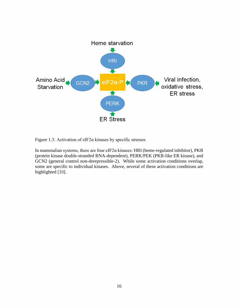
To date, 4 families of eIF2α kinases (EIF2AK1-4) have been described: HRI
(heme-regulated inhibitor), PKR (protein kinase double-stranded RNA-dependent),
PERK/PEK (PKR-like ER kinase), and GCN2 (general control non-derepressible-2). Each
family is activated by different stress conditions (Fig 3). While mammalian systems
encode for all four families of kinases, other systems may only express a subset of these
kinases.
Heme-regulated Inhibitor (HRI)
HRI is also known as EIF2AK1 and is expressed within erythrocyte precursors, liver cells,
and macrophages. Within the erythrocyte precursors, the kinase works to balance the
production of ɑ and β globin to iron levels and inhibit toxic levels of these components.
HRI is used to sense intracellular levels of heme through its heme-binding domain; when
heme levels are high, this bound heme keeps HRI from forming a catalytically active dimer.
During low iron levels, HRI can dimerize and become active, halting protein translation
through the phosphorylation of eIF2α [36]. Within the liver, HRI is most notably involved
16
Figure 1.3: Activation of eIF2α kinases by specific stresses In mammalian systems, there are four eIF2α kinases: HRI (heme-regulated inhibitor), PKR (protein kinase double-stranded RNA-dependent), PERK/PEK (PKR-like ER kinase), and GCN2 (general control non-derepressible-2). While some activation conditions overlap, some are specific to individual kinases. Above, several of these activation conditions are highlighted [33].

17
in regulating liver cell translation during heme deficient periods. Within macrophages,
HRI assists in forming an inflammatory response, as well as macrophage maturation [33].
Protein Kinase Double-stranded RNA-Dependent (PKR)
PKR is also known as EIF2AK2, and was found to be activated during viral
infection. By phosphorylating eIF2α, the cell inhibits the translation of viral mRNA,
slowing viral infection and replication. This is due to a double-stranded RNA binding
domain found on the N-terminus of the kinase, which can bind to the nucleic acid of
invading viruses. PKR expression is induced by interferon, which is usually released by
the host in response to a viral invasion. PKR can also become activated during oxidative
and ER stress independently of viral infection [33].
Found in the cytosol and the nucleus, PKR is activated after dimerization and
autophosphorylation. eIF2α is not the only target for PKR; it can also phosphorylate p53
and activate STAT and NF-Kβ. These additional substrates can change the final fate of the
cell, due to their ability to promote apoptosis. The loss or knockdown of PKR within mice
does not result in a detectable phenotype during reproduction and normal growth
conditions. However, due to the impact viral infections can cause on a system, it’s
important to note the possible redundancies in the anti-viral reaction [33].

18
PKR-like ER Kinase (PERK/PEK)
PERK is also known as PEK or EIF2AK3 and is a transmembrane endoplasmic
reticulum protein. The N-terminus is localized to the ER lumen and the C-terminus is
found in the cytoplasmic space. During normal growth conditions, immunoglobulin
binding protein (BiP) binds to PERK, keeping it in a monomer form within the ER lumen.
However, when misfolded proteins begin accumulating within the ER, BiP disassociates
from PERK. PERK can then form a homodimer during this unfolded protein response
(UPR) and become autophorphorylated to become an active kinase. To halt the synthesis
of new proteins during this buildup of non-functional proteins, PERK phosphorylates
eIF2α to inhibit translation initiation. While PERK activation allows time for the cell to
battle ER stress, constitutive activity by PERK is a pro-death signal, suggesting that long-
term ER stress is fatal to a cell. The loss of PERK in humans results in Wolcott-Rallison
syndrome (WRS). WRS is an extremely rare autosomal recessive disorder bought on by a
mutation the catylatic domain of the PERK gene. Though there are different types of
mutations, all resulting PERK proteins are non-functional kinases. General characteristiss
of WRS is the development of insulin-dependent diabetes before birth or during infancy,
bone dysplasia, and hepatic dysfunction [37]. The molecular mechanisms resulting in the
anomalies are unknown [33].

19
General Control Non-derepressible-2 (GCN2)
GCN2 (or EIF2AK4) acts as a sensor for amino acid and glucose levels. Activation
is carried out through the binding of uncharged tRNA to a histidyl tRNA synthetase
(HisRs) -related domain within the kinase. Like previously described kinases, dimerization
is also required for activation, and autophosphorylation helps stabilize and optimize the
kinase’s activity. Currently, the only known substrate of GCN2 is eIF2α. GCN2 can also
be activated during viral infection and can bind viral genomic RNA directly through its
HisRs domain [33]. In humans, this kinase is predominantly expressed within the brain,
where it is hypothesized to protect neural tissue during periods of amino acid starvation.
Through unknown feedback mechanisms, GNC2 activation can alter the diet of tested
mice, resulting in a selective feeding behavior to alleviate the stress by ingesting more
essential amino-acid rich foods [38].
IV. Stress-induced control of protein translation in eukaryotes
Plasmodium spp.:
Plasmodium is the apcomplexian responsible for the malaria. As of 2015, there are
an estimated 214 million cases of malaria worldwide, with the majority of cases occurring
in Africa and Southeast Asia. The annual death toll associated with malaria is currently
estimated to be 438,000. It is estimated the global cost of controlling malaria infections is
roughly $2.5 billion, highlighting the economic burden of this parasite. There are currently
20
5 species of Plasmodium that causes disease in humans: P. falciparium, P. vivax, P.
malariae, P. ovale, and P. knowlesii. The onset of symptoms, including chills and fevers,
is usually associated with the lysis of host blood cell. The interval of symptoms, as well
as molecular diagnostic tests, can determine the species responsible for individual
infections; however, co-infections with multiple species is possible. In the past,
chloroquine was used as a treatment for malaria infections; contemporary treatments
include drugs from the artemisin class, often in combination with other medications [39].
Plasmodium utilizes two hosts during its complex life cycle in order to reproduce and
spread: the definitive female Anopheles mosquito host and the vertebrate intermediate host.
Within the human host, Plasmodium cells have two different cycles: the exo-erythrocytic
cycle within liver cells and the erythrocytic cycle within red blood cells (Fig 4). In these
two cycles, sporozoites will invade liver cells, or schizonts will invade erythroid cells.
These stages grow as immature and mature trophozoites, then develop into mature
schizonts, that can continue the erythrocytic cycle of cellular invasion. However, in the
mosquito host, the dormant stage of the sporozoites is also important in elimination of this
parasite, as this stage is what initially enters the blood stream of the human host [40].
Plasmodium possesses three putative eIF2α kinases. Several different species of
Plasmodium were used to assess the functions of these kinases. PfIK1 is a GCN2-like
kinase found in the Plasmodium asexual human blood stage that becomes activated during
amino acid starvation. Plasmodium cell lines lacking PfIK1 showed no increase in
phospho-eIF2α after amino acid starvation. However, deletion of this kinase did not alter

21
Figure 1.4: Life cycle of Plasmodium and the life stage of known eIF2α kinases
Within the human host, the cells have two different cycles: the exo-erythrocytic cycle within liver cells and the erythrocytic cycle within red blood cells. During this blood stage, the cell can undergo asexual reproduction or develop into gametocytes for sexual reproduction. These gametocytes are taken up by an Anopheles mosquito during a blood meal. Sexual reproduction occurs within the gut of the mosquito to begin the sporogonic cycle. This ends with the sporozoite stage entering the salivary glands. Here, the parasite remains dormant until the mosquito takes a blood meal. The sporozoites are injected into the human host and must survive in the blood stream until they can invade liver cells and continue the life cycle. The dormant stage of the sporozoites allows the parasite to halt protein translation and store energy, as well as mRNA transcripts. Once protein translation begins again within the schizonts in the liver cell, these stored mRNAs will be transcribed to assist with cell development and survival [40]. The eIF2α kinases, highlighted in green, are shown with the life cycle where they are most prominently expressed. Figure is modified from the Center of Disease Control [42]
22
any developmental stages [41]. The second Plasmodium kinase, PK4, is required for
completion of the erythrocytic cycle of the parasite, PK4- cell lines were less able to infect
mouse models [40].
PfIK2, the final eIF2 kinase, is expressed predominantly in sporozoites found in the
mosquito’s salivary gland and is thought to be life cycle dependent, rather than stress
dependent. In PfIK2 knockout strains, eIF2α was not phosphorylated in the sporozoites
and the infectivity of this life stage was significantly decreased. Phosphorylation is thought
to be necessary to keep transcribed mRNAs from being translated at the wrong life stage.
By keeping these mRNAs in stress granules and decreasing global protein translation,
Plasmodium is prepared for the next life stage while in the salivary gland of the mosquito
[43]. This control of latency is key to Plasmodium’s initial infection of the human host, as
it keeps energy stores high should the parasite have to remain in this life stage for longer
periods of time.
Taken together, these studies highlight the importance of eIF2α phosphorylation in
Plasmodium during its multiple life stages and the unique conditions required for eIF2
kinase activation.
Toxoplasma gondii
Toxoplasma gondii is the protozoan parasite responsible for toxoplasmosis in
humans. Estimates of infected individuals have been as high as 25-30% of the world

23
population. Most infections occur after the consumption of infected meat, but can also
occur after the ingestion of any fecal oocysts (Fig 5). Occasionally, blood transfusions or
organ transplantation can introduce T. gondii into the human host. The highest disease
burden comes from transmission from a mother to a developing fetus. Outcomes can be
as severe as miscarriages and abnormalities occurring in fetuses infected during early
developmental stages [44].
T. gondii has three major life stages that have been studied: the active tachyzoites
and the latent tissue bradyzoites, and the latent fecal oocysts (Fig 5). It is the cycling of
tachyzoites to bradyzoites in the human host that is of particular interest. T. gondii
possesses four authentic eIF kinases, named TgIF2K-A-D. TgIF2K-A localizes to the
parasite’s endoplasmc reticulum around the nucleus. It is known that inducers of ER stress,
such as sodium arsenite and calcium ionophore induce phosphorylation of eIF2α. It is
highly likely the kinase responsible for this phosphorylation is TgIF2K-A [47] because of
the kinase’s location in the ER. TgIF2K-B is expressed in the cytosol of the parasite;
however, the conditions necessary to activate this kinase remains unknown [47].
Two of the T. gondii kinases are characterizes as being GCN2-like. TgIF2K-C is
expressed in the cytosol of tachyzoites. TgIF2K-C knockout lines were not deficient in
progressing through the lytic cycle and had similar virulence as wildtype cells. However,
if grown in glutamine-free media, knockout lines had decreased levels of phospho-eIF2α
[48]. This is consistent with the known function of GCN2 kinases in amino acid sensing.

24
Figure 1.5: Life cycle of Toxoplasma gondii and the life stages of known eIF2α kinases
While the normal life cycle of T. gondii includes the definitive feline host and an intermediate rodent or bird host, humans can become infected when the latent oocyst is ingested. These oocyst further develop into tachyzoites inside the host, and can transition into a latent tissue cyst form, called bradyzoites, during stress. Bradyzoties can excyst after the stress has subsided. Tachyzoites can invade macrophages and subsequently lyse the cell, forming the lytic cycle of the parasite. This constant cycling can continue until the death of the host, or the clearance of the parasite by the immune system [45]. eIF2 kinases, in green, are shown with the life cycle where they are most prominently expressed; although, it is currently unknown if TgIF2K-A and TgIF2K-B have a life stage-specific expression pattern. Figure is modified from the Center of Disease Control [46]

25
TgIFK2-D, also expressed in the cytosol, is involved in tachyzoite survival outside
of host cells. Once outside the macrophage, the cell must keep translation low to save
energy until another macrophage can be invaded. By phosphorylating eIF2α, translation
decreases by 90%. TgIF2K-D knockout lines could not compete against wildtype lines
and were outgrown after extracellular co-incubation in vitro, as determined by PCR
analysis using cell-line specific PCR primers [49].
During the cycling between active tachyzoites and latent bradyzoites in the human
host, protein synthesis must be altered to save any available energy. Previous studies have
shown that phosphorylation of eIF2α is important for controlling latency. For example,
latent bradyzoite formation is initiated when dephosphorylation of eIF2α is inhibited with
the phosphatase inhibitor, salubrinol [47]. While the eIF2α kinase responsible for this
phosphorylation of eIF2α during stage conversion is unknown, these findings indicate the
importance of the eIF2α phosphorylation pathway in overall parasite development.
Leishmania spp.
Leishmania is the causative agent of leishmaniasis, which can occur in three forms:
visceral, cutaneous, and mucocutaneous. Roughly 1.3 million cases of leishmaniasis occur
annually, with up to 50,000 deaths each year attributed to the visceral form. There are 21
morphologically identical species of Leishmania that infect mammals. While the
promastigote stage is initially injected into the human host by the sandfly vector, within
the macrophages, promastigotes develop into amastigotes, where the only known

26
Leishmania eIF2α is activated. Leishmania major and Leishmania infantum both have a
PERK homologue with approximately 30% identity to the human PERK. This kinase is
expressed in both the promastigote and amastigote stages of the Leishmania life cycle, but
is thought to be responsible for the increased phospho-eIF2α levels in the amastigote stage.
Localized to the ER, Leishmania PERK has been shown to become activated during the
unfolded protein response. To study PERK further, a truncated form of PERK was
expressed in cells cultured in vitro, acting as a dominant negative. The lack of
phosphorylation by PERK caused a significant decrease in differentiation to amastigotes.
Amastigote-specific genes had a marked delay in expression, indicating a delay in stage
transition. These findings were the same in both axenic cultures and within human and
mouse macrophage models. While most eIF2α sequences have a key serine residue
thatbecomes phosphorylated under stress, the eIF2α sequence of Leishmania has a
threonine at the key phosphorylation residue at position 166. This indicates the
conservation of the eIF2α pathway and the kinases associated with it [51].
Non-pathogenic eukaryotes
The eIF2-based stress pathway is found in other lower eukaryotic organisms. In
yeast, the only known eIF2 kinase is Gcn2. In Saccharomyces cerevisiae, Gcn2 is used for
sensing and responding to amino acid starvation, specifically tryptophan and arginine.
Activation of Gcn2 occurs when uncharged tRNAbinds to the histidyl-tRNA synthetase-
like domain on Gcn2 [52]. Due to the single eIF2 kinase, S. cerevisiae is often used to
authentice eIF2 kinase activity in proteins from other organisms. For example, the Gcn2p

27
Figure 1.6: Life cycle of Leishmania major and the life stages of known eIF2α kinases Transmission of Leishmania occurs after the bite of a female sandfly, when promastigotes are injected into the blood stream. This free-living stage is phagocytized by the host’s macrophages and allows for differentiation into amastigotes. These amastiogtes divide within the cell and can infect other cells. During a blood meal, the intermediate host, a sandfly, takes up macrophages infected with amastiogtes. Within the sandfly’s gut, amastiogtes transition back to a promastigote stage that multiply and migrate to the salivary gland of the sandfly. Here, the parasite is ready to be injected into a mammalian host to continue the cycle (Fig 6). The only known eIF2 kinase, PERK is activated during amistogote development and is indicated in green. Figure is adapted from the Center of Disease Control [50].

28
gene can be disrupted and replaced with cDNAs that encode entire eIF2α kinases [53] or
just active sites [40]. If the exogenous kinase is authentic, expression will induce
phenotypic changes, such as reduced growth [53].
Dictyostelium discoideumis a free living amoeba that feeds on bacteria in the soil
but can be grown axenically in the laboratory. During times of starvation, these amoebae
will aggregate and form mounds of approximately 100,000 cells. The mounds eventually
differentiate to form fruiting bodies consisting of dormant spores supported by stalk cells.
Dictyostelium has two eIF2 kinases: IfkA and IfkB. During normal development, the level
of phospho-eIF2α increase from a slight basal level slight for several hours, and then return
back to the initial basal level. IfkA null lines did not exhibit increased phospho-eIF2α
during any stage of development. IfkA knockout lines also had a slight growth defect in
axenic conditions, but were able to complete the entire life cycle. This suggests that this
kinase, and phosphorylation of eIF2α are not essential to development in this system.
Interestingly, IfkA knockout lines formed mounds earlier than the parental strain, with
mound size much larger than the parental strain due to the misregulation of countin.
Countin is a polypeptide secreted by Dictyostelium to regulate the number of cells within
a particular mound. All attempts to disrupt the IfkB gene were not successfully, indicating
that IfkB, a gene found in all life stages, is essential to development and growth. While
these two kinases have domains similar to the mammalian GCN2 kinase, neither were
found to be responsible for detecting amino acid starvation in the amoeba [54].

29
V. Summary
The ability to phosphorylation of eIF2α is utilized by many organisms during times
of stress or development. This phosphorylation is catalyzed by the eFI2 kinases in each
organism. To date, there has not been a study into the role of eIF2α in Entamoeba
histolytica, despite the many studies on the effects of stress on the parasite (reviewed
above). E. histolytica and E. invadens each possess eIF2α (EHI_005100 and EIN_242170,
respectively) and two presumptive eIF2α kinases (eIF2K) (Entamoeba histolytica-
EHI_109700, EHI_035950; Entamoeba invadens- EIN_059080, EIN_0333330) [55, 56].
The phosphorylated serine of eIF2α (see chapter 2) and critical active site lysines of the
kinases (data not shown) are conserved. In E. invadens expression of one of the kinases is
developmentally regulated (EIN_0333330) [55]. Thus, we hypothesize that this genus
uses eIF2α-based machinery to control translation during stage conversion. This
hypothesis is supported by the pioneering work of Dr. Gordon Bailey showing that
encystation was accompanied by the aggregation of ribosomes into structures known as a
chromatoid bodies and by a decrease in the incorporation of exogenous amino acids [57,
58]. Chromatoid bodies are an RNA- and ribosome-containing cytoplasmic structure that
assembles during encystation. They are reminiscent of stress granules or P bodies, but the
fate of RNAs within chromatoid bodies is unknown. Currently, these Entamoeba eIF2α
kinases have not been authenticated, nor have the conditions that lead to their activation
been discerned.

30
The percent identity of the catalytic domains of the two E. histolytica kinases to the
human host orthologs is considerably low at approximately 32% for each [56]. Because of
this low identity, it may be possible to use these kinases as potential drug targets. This
would prove to be especially useful if either kinase was shown to be necessary for parasite
growth or cyst development. If cyst production could be halted, spread of infection could
be halted, leading to the decline in E. histolytica infections. Toward this end, a better
understanding of the eIF2-based stress response system in E. histolytica is necessary.
Therefore, the aims of this study were to:
1. To define the role of the phosphorylation of the alpha subunit of the eukaryotic
initiation factor-2 (eIF2α) in Entamobea histolytica
2. To determine if phosphorylation of eIF2α is necessary to counter stress in the E.
histolytica system

31
VI. Literature Cited [1] Morf L, Singh U. (2012) Entamoeba histolytica: a snapshot of current research and
methods for genetic analysis. Curr Opin Microbiol 15: 469-475.
[2] Eichinger D. (2001) Encystation in parasitic protoza. Curr Opin Microbiol 4: 421-426.
[3] Stanley SLJ. (2003) Amoebiasis. Lancet 361: 1025-1034.
[4] Amebiasis Life Cyle. Digital image. Amebiasis DPDx-Laboratory identification of parasite diseases of public health concern, 29 Nov. 2013. Web.
[5] Marie C, Petri WJ. (2014) Regulation of virulence of Entamobea histolytica. Annu Rev Microbiol 68: 493-520.
[6] Laughlin RC, Temesvari LA. (2005) Cellular and molecular mechanisms that underlie Entamoeba histolytica pathogenesis: Prospects for intervention. Expert Rev Mol Med 7: 1-19.
[7] Samsuelson J. (1999) Why metronidazole is active against both bacteria and parasites. Amtimicrob Agents Chemother 43: 1533-1541.
[8] Lamp KC, Freeman CD, Klutman NE, Lacy MK. (1999) Pharmacokinetics and pharmacodynamics of the nitroimidazole antimicrobials. Clin Pharmacokinet 36: 353-373.
[9] Irusen EM, Jackson TFHG, Simjee AE. (1992) Asymptomatic intestinal colonization by pathogenic Entamoeba histolytica in amebic liver abscess: Prevalence, response to therapy, and pathogenic potential. Clin Infect Dis 14: 889-893.
[10] Baumel-Alterzon S, Ankri S. (2014) Entamoeba histolytica adaptation to glucose starvation: A matter of life and death. Curr Opin Microbiol 20: 139-145.
[11] Diamond LS, Harlow DR, Cunnick CC. (1978) A new medium for the axenic cultivation of Entamoeba histolytica and other Entamoeba. Trans R Soc Trop Med Hyg 72: 431-432.
[12] Tovy A, Hertz R, Siman-Tov R, Syan S, Faust D, Guillen, N, Ankri, S (2011) Glucose starvation boosts Entamoeba histolytica virulence. PLOS Neglect Trop D 5: e1247.
[13] Baumel-Alterzon S, Weber C, Guillen N, Ankri S. (2013) Identification of a dihydropyrimidine dehydrogenase as a virulence factor essential for the survival of Entamoeba histolytica in glucose-poor environments. Cell Microbiol 15: 130-144.
[14] Park SJ, Lee SM, Lee J, Yong TS. (2001) Differential gene expression by iron-limitation in Entamoeba histolytica. Mol Biochem Parasit 114: 257-260.

32
[15] Espinosa A, Perdrizet G, Paz-y-Miño C. G, Lanfranchi R, Phay M. (2009) Effects of iron depletion on Entamobea histolytica alcohol dehydrogenase 2 (EhADH2) and trophozoite growth: Implications for antiamoebic therapy. J Antimicrob Chemother 63(4): 675-678.
[16] Vohra H, Mahajan RC, Ganguly NK. (1998) Role of serum in regulating the Entamoeba histolytica cell cycle: A flowcytometric analysis. Parasitol Res 84: 835-838.
[17] Beck DL, Boettner DR, Dragulev B, Ready K, Nozaki T, Petri WA Jr. (2005) Identification and gene expression analysis of a large family of transmembrane kinases related to the Gal/GalNAc lectin in Entamoeba histolytica. Eukaryot Cell 4: 722-732.
[18] Shrimal S, Bhattacharya S, Bhattacharya A. (2010) Serum-dependent selective expression of EhTMKB1-9, a member of Entamoeba histolytica B1 family of transmembrane kinases. PLOS Pathog 6: e1000929.
[19] Pérez-Morales D, Espinoza B. (2015) The role of small heat shock proteins in parasites. Cell Stress Chaperon 20: 767-780.
[20] Weber C, Guigon G, Bouchier C, Frangeul L, Moreira S, Sismeiro, O, Gouyette, C, Mirelman, D, Coppee JY, Guillen, N (2006) Stress by heat shock induces massive down regulation of genes and allows differential allelic expression of the Gal/GalNAc lectin in Entamoeba histolytica. Eukaryot Cell 5: 871-875.
[21] Bernes S, Siman-Tov R, Ankri S. (2005) Epigenetic and classical activation of Entamoeba histolytica heat shock protein 100 (EHsp100) expression. FEBS Lett 579: 6395-6402.
[22] Katz S, Kushnir O, Tovy A, Tov RS, Ankri S. (2012) The Entamoeba histolytica methylated LINE-binding protein EhMLBP provides protection against heat shock. Cell Microbiol 14: 58-70.
[23] Katz S, Trebicz-Geffen M, Ankri S. (2014) Stress granule formation in Entamoeba histolytica: Cross-talk between EhMLBP, EhRLE3 reverse transcriptase and polyubiquitinated proteins. Cell Microbiol 16: 1211-1223.
[24] Satish S, Bakre AA, Bhattacharya S, Bhattacharya A. (2003) Stress-dependent expression of a polymorphic, charged antigen in the protozoan parasite Entamoeba histolytica. Infect Immun 71: 4472-4486.
[25] Ghosh AS, Ray D, Dutta S, Raha S. (2010) EhMAPK, the mitogen-activated protein kinase from Entamoeba histolytica is associated with cell survival. PLOS One 5: e13291.

33
[26] Shahi P, Trebicz-Geffen M, Nagaraja S, Alterzon-Baumel S, Hertz R, Methling, K, Lalk, M, Ankri, S (2016) Proteomic identification of oxidized proteins in Entamoeba histolytica by resin-assisted capture: Insights into the role of arginase in resistance to oxidative stress. PLoS Neglect Trop D 10: e0004340.
[27] Hertz R, Lulu SB, Shahi P, Trebicz-Geffen M, Benhar M, Ankri, S (2014) Proteomic identification of S-nitrosylated proteins in the parasite Entamoeba histolytica by resin-assisted catpure: Insights into the regulation of the Gal/GalNAc lectin by nitric oxide. PLOS One 9: e91518.
[28] Vicente JB, Ehrenkaufer GM, Saraiva LM, Teixeira M, Singh U. (2009) Entamoeba histolytica modulates a complex repertoire of novel genes in response to oxidative and nitrosative stresses: Implications for amebic pathogenesis. Cell Microbiol 11: 51-69.
[29] Santi-Rocca J, Smith S, Weber C, Pineda E, Hon CC Saavedra, E, Olivos-Garcia, A, Rousseau, S, Dillies, MA, Coppee, JY, Gullien, N (2012) Endoplasmic reticulum stress-sensing mechanism is activated in Entamoeba histolytica upon treatment with nitric oxide. PLOS One 7: e31777.
[30] Rastew E, Vicente JB, Singh U. (2012) Oxidative stress resistance genes contribute to the pathogenic potential of the anaerobic protozan parasite Entamoeba histolytica. Int J Parasitol 42: 1007-1015.
[31] Aguilar-Diaz H, Diaz-Gallardo M, Laclette JP, Carrero JC. (2010) In vitro induction of Entamoeba histolytica cyst-like structures from trophozoites. PLOS Neglect Trop D 4: e607.
[32] Kimball SR. (1999) Eukaryotic initiation factor eIF2. Int J Biochem Cell B 31: 25-29.
[33] Donnelly N, Gorman AM, Gupta S, Samali A. (2013) The eIF2 (alpha) kinases: Their structures and functions. Cell Mol Life Sci 70: 3493-3511.
[34] Dever TE, Yang W, Aström S, Byström AS, Hinnebusch AG. (1995) Modulation of tRNA(iMet), eIF-2, and eIF-2B expression shows that GCN4 translation is inversely coupled to the level of eIF-2.GTP.met-tRNA(iMet) ternary complexes. Mol Cell Biol 15: 6351-6363.
[35] Balagopal V, Parker R. (2009) Polysomes, P bodies, and stress granules: States and fates of eukaryotic mRNAs. Curr Opin Cell Biol 21: 403-408.
[36] Chen JJ. (2007) Regulation of protein synthesis by the heme-regulated eIF2α kinase: Relevance to anemias. Blood 109: 2693-2699.
[37] Julier C, Nicolino M. (2010) Wolcott-Rallison syndrome. Orphanet J Rare Dis 5.
[38] Dever TE, Hinnebusch AG. (2005) GCN2 whets the appetite for amino acids. Mol Cell 18: 141-142.
[39] WHO. (2015) World malaria report 2015.

34
[40] Zhang M, Mishra S, Sakthivel R, Rojas M, Ranjan R, Sullivan, WJ Jr, Fontoura, BMA, Menard, R, Dever, TE, Nussenzweig, V (2012) PK4, a eukaryotic initiation factor 2(alpha) (eIF2alpha) kinase, is essential for the development of the erythrocytic cycle of Plasmodium. PNAS 109: 3956-3961.
[41] Malaria Life Cyle. Digital image. Malaria DPDx-Laboratory identification of parasite diseases of public health concern, 29 Nov. 2013. Web.
[42] Fennell C, Babbitt S, Russo I, Wilkes J, Ranford-Cartwright L, Goldberg, D, Doerig, C (2009) PfeIK1, a eukaryotic initiation factor 2α kinase of the human malaria parasite Plasmodium falciparum, regulates stress-response to amino-acid starvation. Malaria J 8: 99-144.
[43] Zhang M, Fennell C, Ranford-Cartwright L, Sakthivel R, Gueirard P, Meister, S, Caspi, A, Doerig, C, Nussenzwig, RS, Tuteja, R, Sullivan WJ Jr, Roos, DS, Fontoura, BMA, Ménard, R, Winzeler, EA, Nuzzenzweig, V (2010) The Plasmodium eukaryotic initiation factor-2α kinase IK2 controls the latency of sporozoites in the mosquito salivary glands. J Exp Med 207: 1465-1474
[44] Robert-Gangneux F, Dardé ML. (2012) Epidemiology of and diagnostic strategies for toxoplasmosis. Clin Microbiol Rev 25: 264-296.
[45] Montoya JG, Liesenfeld O. (2004) Toxoplasmosis. Lancet 363: 1965-1976.
[46] Toxoplasma Life Cyle. Digital image. Toxoplasmosis DPDx-Laboratory identification of parasite diseases of public health concern, 6 July 2015. Web.
[47] Konrad C, Wek RC, Sullivan WJ Jr. (2014) GCN2-like eIF2α kinase manages the amino acid starvation response in Toxoplasma gondii. Int J Parasitol 44: 139-146.
[48] Narasimhan J, Joyce B, Naguleswaran A, Smith A, Livingston M, Dixon, S, Coppens, I, Wek, R, Sullivan, W Jr (2008) Translational regulation by eukaryotic initiation factor-2 kinases in the development of latent cysts in Toxoplasma gondii. J Biol Chem 283: 16591-16601.
[49] Konrad C, Wek RC, Sullivan WJJ. (2011) A GCN2-like eukaryotic initiation factor 2 kinase increases the viability of extracellular Toxoplasma gondii parasites. Eukaryot Cell 10: 1403-1412.
[50] Leishmania Life Cyle. Digital image. Leishmaniasis DPDx-Laboratory identification of parasite diseases of public health concern, 2 Nov. 2015. Web.
[51] Chow C, Cloutier S, Dumas C, Chou MN, Papadopoulou B. (2011) Promastigote to amastigote differentiation of Leishmania is markedly delayed in the absence of PERK eIF2alpha kinase-dependent eIF2alpha phosphorylation. Cell Microbiol 13: 1059-1077.

35
[52] Zaborske JM, Wu X, Wek RC, Pan T. (2010) Selective control of amino acid metabolism by the GCN2 eIF2 kinase pathway in Saccharomyces cerevisiae. BMC Biochem 11
[53] Dever TE, Chen JJ, Barber GN, Cigan AM, Feng L, Donahue, TF, London, IM, Katze, MG, Hinnebusch, AG (1993) Mammalian eukaryotic initiation factor 2α kinases functionally substitute for GNC2 protein kinase in the GCN4 translational control mechanism of yeast. Proc Natl Acad Sci USA 90: 4616-4620.
[54] Fang R, Xiong Y, Singleton CK. (2003) IfkA, a presumptive eIF2 alpha kinase of Dictyostelium, is required for proper timing of aggregation and regulation of mound size. BMC Dev Biol 3.
[55] Ehrenkaufer GM, Weedall GD, Williams D, Lorenzi HA, Caler E, Hall, N, Singh, U (2013) The genome and transciptome of the enteric parasite Entamobea invadens, a model for encystation. 14: R77.
[56] Vonlaufen N, Kanzok SM, Wek RC, Sullivan W.J. Jr (2008) Stress response pathways in protozoan parasites. Cell Microbiol 10: 2387-2399.
[57] Kusamrarn T, Vinijchaikul K, Bailey GB. (1975) Comparison of the structure and function of polysomal and helical ribosomes from Entamoeba invadens. J Cell Biol 65: 540-548.
[58] Kusamrarn T, Sobhon P, Bailey GB. (1975) The mechanism of formation of inhibitor-induced ribosome helieces in Entamoeba invadens. J Cell Biol 65: 529-539.

36
CHAPTER TWO
PHOSPHORYLATION OF EUKARYOTIC INITIATION FACTOR-2 α REGULATES
STRESS IN THE HUMAN PROTOZOAN PARASITE
ENTAMOEBA HISTOLYTICA
Holland M. Hendrick1,2, Matthew A. Hapstack1,2, Brenda H. Welter1,2, Lesly A.
Temesvari1,2*
1 Department of Biological Sciences
2Eukaryotic Pathogens Innovations Center
Clemson University
Clemson, South Carolina, United States of America 29634
*Corresponding author
Email: [email protected] (LAT)

37
I. Abstract
Entamoeba histolytica is a food- and water-borne intestinal parasite responsible for
amoebic dysentery and amoebic liver abscess. The life cycle of E. histolytica alternates
between the host-restricted trophozoite form and the highly infective latent cyst stage that
is able to persist in the environment. Throughout its life cycle, which may include invasion
of tissues in the human host, the parasite is subjected to a variety of stressful conditions.
In other systems, stress can trigger the activation of kinases that phosphorylate a serine
residue on eukaryotic translation initiation factor-2α (eIF2α). This modification inhibits
the activity of eIF2 resulting in a general decline in protein synthesis, and, paradoxically,
an up-regulation of the expression of certain genes that permit the cell to counter the stress.
Genomic data reveal that E. histolytica possesses eIF2α with a conserved phosphorylatable
serine at position 59. Thus, this pathogen may have the machinery for stress-induced
translational control. To test this, we exposed E. histolytica trophozoites to six different
stress conditions and assessed viability, as well as the level of total and phospho-EheIF2α
via Western blot of cell lysates. Long term serum starvation induced an increase in the
level of phospho-EheIF2α, but no other stress condition caused a significant change. Long
term serum starvation also showed a decrease in polyribosome abundance as observed
through sucrose gradient ultracentrifugation; this is consistent with the observation that this
condition also induces phosphorylation of EheIF2α. This suggests that the eIF2α-
dependent stress response system is operational in E. histolytica and that the system may
be activated only by certain stresses. To further examine the role of phosphorylation of
EheIF2α during stress, three transgenic cell lines were created. EheIF2α-S59 over-

38
expresses wild type eIF2α protein. EheIF2α-S59A expresses eIF2α with the serine-59
residue mutated to an alanine, creating a non-phosphorylatable subunit. EheIF2α-S59D
expresses eIF2α with the serine-59 residue mutated to an aspartic acid to mimic a
phosphorylated residue. EheIF2α-S59 exhibited a high level of phosphorylation of the
exogenous protein, leading to a decreased growth and polyribosome abundance when
compared to the control cell line. EheIF2α-S59A had the highest growth rate and retained
a high abundance of polyribosome. EheIF2α-S59D exhibited the slowest growth rate and
had a decrease in polyribosome when compared to control; however, EheIF2α-S59D did
exhibit the highest survival rate in over half the stress conditions tested. This may indicate
the protective nature of phosphorylation of EheIF2α during times of stress.
II. Author Summary
Entamoeba histolytica is a parasitic pathogen usually found in underdeveloped countries
that lack proper water filtration and treatment plants. During the E. histoltycia life cycle,
active trophozoites can reside in the large intestine, or invade the intestinal wall to cause
extraintestinal infection. The parasite encounters demanding growth conditions in the host
and must overcome these to survive. In other organisms, stress induces phosphorylation
of the alpha subunit of the eukaryotic translation initiation factor 2 (eIF2α). This, in turn,
inhibits protein translation allowing the cell to conserve energy. To determine the role of a
putative eIF2α in E. histolytica, we have applied 6 different stress conditions to active
trophozoites. One of these stress conditions, long term serum starvation, causes a
significant increase in phosho-EheIF2α levels. Consistent with this observation, long term
serum starvation also reduces the abundance of polyribosomes, and important component
39
of the translational machinery. We have also created three transgenic cell lines to test
mutant forms of EheIF2α: EheIF2α-S59, EheIF2α-S59A, and EheIF2α-S59D. These cell
lines overexpress the wildtype protein, a non-phosphorylatable protein, and a
phosphomimetic protein, respectively. Due to the phosphorylation/dephosphroyation
nature of the exogenous protein in EheIF2α-S59, both an increase in protein synthesis and
a decrease in growth was observed. EheIF2α-S59A exhibited the highest growth, but
additional studies are required to fully assess the translation machinery for this cell line.
EheIF2α-S59D had the slowest growth and a decreased level of translation. However,
EheIF2α-S59D had the highest survival in the majority of test stress conditins, indicating
that phosphorylation of EheIF2α can be used to alleviate the cellular pressures attributed
to stress.

40
III. Introduction
Entamoeba histolytica is an intestinal parasite that is the causative agent of amebic
dysentery and amoebic liver abscesses. It is transmitted by the cyst form of the pathogen
in fecally-contaminated food and water, making it prevalent in the developing world where
sanitation practices are substandard. There are 173 million people living in regions with
untreated water sources and one billion people carry out open defecation practices [1].
Thus, there is considerable risk for transmission of E. histolytica. E. histolytica is also
considered a Class B bioterrorism agent as a water safety threat. These factors make the
pursuit of knowledge regarding this parasite significant.
E. histolytica is passed from human to human without the utilization of an
intermediate host during its life cycle. The parasite’s latent stage, a cyst, is able to withstand
the extreme conditions in the external environment as well as the acidic pH of the host
stomach. The cyst exits the stomach and enters the small intestine, where unknown triggers
cause excystation. The emerging active trophozoites continue down the digestive system
until they reach the large intestine, where the parasites divide by binary fission.
Trophozoites are also responsible for extraintestinal complications of E. histolytica
infections, including liver abscess. During infection, which includes invasion of the host
intestinal tissue, the parasite may experience stress, in part due to immune pressure from
the host. This stress can include heat shock, osmotic shock, nutrient deprivation, and/or
exposure to reactive oxygen or nitrogen species, and high oxygen levels. The parasite must
elicit a cellular response to counter these stresses and survive.

41
In many systems, stress is controlled, in part, by the phosphorylation of the alpha
subunit of the eukaryotic initiation factor (eIF2α) [2]. Under normal conditions, eIF2α
forms a protein complex that, when bound to GTP, delivers Met-tRNAi to the ribosome to
initiate translation. Once Met-tRNAi is delivered, the bound GTP is hydrolyzed to GDP.
To become reactivated, eIF2-GDP binds to the guanine exchange factor, eIF2B, and the
GDP is released, allowing for the binding of GTP to start the cycle again. This is
considered the rate-limiting step of translation initiation [reviewed in 2]. During stress,
eIF2 kinases become activated and phosphorylate a key serine residue on the eIF2α subunit
to generate a phosphorylated form of the protein (phospho-eIF2α). This phosphorylation
induces a conformational change in eIF2, causing it to become the competitive inhibitor of
eIF2B. This leads to a general decrease in protein translation; however, paradoxically, the
expression of a subset of genes is up-regulated. This subset of genes assists the cell in
surviving the affront.
In other eukaryotic pathogens, one possible outcome of stress is stage conversion
to a latent form. For example, under stress, Toxoplasma gondii will convert from an active
tachyzoites form to a latent bradyzoite form. Phosphorylation of eIF2α is necessary for
this stage transition [3]. Phospho-eIF2α also regulates the formation of latent sporozoites
in Plasmodium spp. [4] and the transition of promastigotes to amastigotes in Leishmania
[5]. In non-parasitic organisms, such as yeast [6] and Dictyostelium [7], phosphorylation
of eIF2α stimulates the formation of latent spores. Genomic data suggest that E. histolytica
and E. invadens, a related reptilian intestinal parasite that can undergo encystation in vitro,

42
possess the components of this stress-response system [8]. However, the role of eIF2α
phosphorylation in the E. histolytica stress response has never been characterized.
In this study, we show that phosphorylation of EheIF2α occurs in E. histolytica in
response to one stress condition, namely long term serum-starvation. This is accompanied
by a reduction in global protein translation. We also demonstrate that expression of non-
phosphorylatable or phosphomimetic forms of EheIF2α influences growth, protein
translation, and the ability to counter stress. Together, these data support the hypothesis
that E. histolytica possesses an eIF2α-based stress response system that controls protein
translation.
IV. Results
eIF2α possesses conserved amino acid residues around the key phosphorylated serine
residue
An alignment of the E. histolytica eIF2α (EheIF2α) amino acid sequence with that
of six different organisms showed that it shared low sequence identity and moderate
sequence similarity across the entire protein, even when compared to the factor from other
eukaryotic pathogens (Fig 1A and B). The eIF2α with the highest homology to the EheIF2α
was that from E. invadens. Though overall shared homology was low, there was strong
sequence identity surrounding the key phosphorylated serine residue, which occurs at
amino acid position 59 in E. histolytica, (Fig 1C). Thus, this serine residue is likely to be

43
Figure 2.1: Alignment of the eukaryotic translation initiation factor 2, alpha subunit
Protein identity (A) and similarity (B) matrices were generated using BLOSUM 62 algorithm and Protein Blast. (C) The amino acids around the key serine residue, occurring at position 59 in E. histolytica were aligned using a Standard Protein BLAST. The key serine residue that becomes phosphorylated is indicated by shading. Fully conserved resides are noted by an asterisk (*) below the residues. Residues showing strongly similar properties are indicated by a colon (:). Aminio acid sequences were identified using UniProtKB; UniProtKB accession number identified. Eh, Entamoeba histolytica (accession no. C4M0A4); Ei, E. invadens (accession no. S0AZW3); Tg, Toxoplasma gondii (accession no. S8GC56); Pf, Plasmodium falciparum (accession no. Q8IBH7); Sc, Saccharomyces cerevisiae (accession no. P20459); Dm, Drosophila melanogaster (accession no. P41374); Hs, Homo sapiens (accession no. P05198)
44
the residue phosphorylated during stress. Conservation around this residue also suggests
that the machinery for an eIF2α-based stress-response system is present in E. histolytica.
Serum-starvation elicits an increase in the level of phospho-eIF2α
To determine if the level of phospho-eIF2α changes during stress, cells were
exposed to a variety of stress conditions: short and long term serum starvation [9], short
and long term heat shock [10], glucose deprivation [11], and oxidative stress [12]. Viability
was assessed and the level of total and phospho-eIF2α was tracked using Western blotting.
Long term serum starvation and oxidative stress resulted in significant cell death (Fig 2).
While the ratio of total eIF2α to actin showed some changes, none were singinficant (Fig
3A). There was a basal level of phosphorylated EheIF2α in control unstressed
trophozoites; however, only trophozoites that were serum-starved for 24 h exhibited
enhanced phosphorylation of EheIF2α compared to unstressed control cells (Fig 3B). This
was not simply due to cell death, as oxidative stress, which was accompanied by a similar
reduction in viability, did not result in a significant increase in phospho-EheIF2α.
Although cells that experienced long-term heat shock also exhibited an increase in
phospho-EheIF2α, the increase was not statistically significant. These data suggest that an
eIF2α-based stress-specific response system exists in E. histolytica and is activated in a
stress-specific manner.

45
Figure 2.2: Viability of trophozoites during stress
Log-phase trophozoites were incubated in a variety of stress conditions as described in the text. Cells were collected by centrifugation and live/dead cells were enumerated via microscopy and Trypan blue exclusion. Percent viability was determined using the ratio of live cells to the total cell population counted. The data represent the mean (± standard error) for at least three separate trials. *** indicates a highly significant result (P<0.001)

46
Figure 2.3: Western blot analysis of phospho-eIF2α and total eIF2α levels in control and stressed cells
Control or stressed cells were subjected to Western blot analysis using antibodies specific for actin, total eIF2α, phospho-eIF2α. The proportion of (A) total eIF2α to actin or (B) phospho-eIF2α to total eIF2α was determined by scanning densitometry and image analysis (Image J, NCBI). The data represent the mean (± standard error) for at least three separate trials. * indicates a significant result (P < 0.05). Only long term serum starvation induced a statistically significant (>2-fold) increase in the level of phospho-eIF2α.

47
Reduced protein translation accompanies phosphorylation of eIF2α
In other systems, eIF2α-based control of stress is accompanied by a reduction in
global protein translation [3]. Therefore, we examined global protein translation by
characterizing the abundance of polyribosomes using sucrose gradient ultracentrifugation
after long term serum-starvation, or after glucose starvation. Serum-starvation, which
induces phosphorylation of EheIF2α, resulted in a significant reduction in dense
polyribosomes and an increase in free ribosomes and monosomes (Fig 4A and B). On the
other hand, parasites subjected to glucose starvation, which does not induce
phosphorylation of EheIF2α, did not exhibit a decrease in dense polyribosome-bound
transcripts (Fig 4C). These results support the premise that E. histolytica possesses an
eIF2α-based stress response system that reduces protein translation.
Expression of mutant eIF2α alters global protein translation and growth
To further examine the function of phospho-EheIF2α, we generated cell lines that
conditionally overexpress non-phosphorylatable or phosphomimetic forms of EheIF2α.
The cDNA encoding EheIF2α was mutagenized in two ways using PCR. The codon for
serine (S) at position 59 was changed to that of alanine (A) or aspartic acid (D) to produce
non-phosphorylatable (EheIF2α-S59A) or phosphomimetic (EheIF2α-S59D) forms of
EheIF2α, respectively [13]. To distinguish exogenous EheIF2α from the endogenous form,
the PCR product was modified to include a FLAG epitope peptide sequence
(DYKDDDDK) [14] followed by a 5-glycine flexible region at the N-terminus.

48
Figure 2.4: Polyribosome abundance in control and stressed cells
Total RNA from control (A), serum-starved (B), or glucose-starved (C) cells were resolved by sucrose gradient (15-45%) ultracentrifugation, separating free ribosomes and monosomes into the lightest of fractions, from the polysomes in the densest fractions. Fractions were taken and analyzed by UV spectrometry (254 nm). Representative profiles of at least 3 separate trials are shown. Only long term serum starvation led to a decrease in large polysome abundance.

49
Modified PCR products were inserted into the E. histolytica expression vector
pGIR209, which confers G418 (neomycin) resistance and allows for tetracycline-inducible
expression of exogenous genes when introduced into trophozoites [15]. A standard
electroporation protocol [16] was utilized to introduce the expression vector into
trophozoites which had been previously transfected with an additional plasmid, pGIR308.
This partner plasmid encodes the tetracycline repressor protein, which is necessary for
tetracycline inducibility. Authentic transfection was confirmed by purification and
sequencing of the episomal expression plasmids from stably transfected cell lines [17].
A previously established cell line that conditionally expresses an irrelevant protein,
luciferase, was used as a control [15]. Expression of exogenous proteins was induced by
the addition of 5 µg mL-1 tetracycline to the culture medium for a minimum of 24 h.
Western blot analysis using anti-FLAG and anti-EheIF2α showed successful induction of
the protein with little to no expression of the exogenous proteins prior to the addition of
tetracycline (Fig 5). Interestingly, the EheIF2α-S59 exhibited a high level of
phosphorylation of the exogenous protein. This result suggests that the FLAG-tagged
variant is functional.
To confirm that the exogenously expressed EheIF2α variants were functional, we
monitored polyribosome abundance in the transgenic cell lines after 24 or 72 h of
tetracycline induction. After 24 h of induction, there was no change in polyribosome
abundance (Fig S1). However, after 72 h of induction, polyribosome abundance remained
high in the control 209-Luc cell line (Fig 6A). While high density polysomes were present

50
Figure 2.5: Western blot analysis confirming exogenous protein expression in mutant cell lines
Trophozoites were transfected with plasmids encoding luciferase (209-Luc), wildtype eIF2α (EheIF2α-S59), the non-phosphorylatable variant (EheIF2α-S59A), and the phosphomimetic variant (EheIF2α-S59D). Protein expression was induced using 5 µg mL-
1 tetracycline for 24 hours. Western blots of cell lysates were performed using antibodies specific for total, phospho-EheIF2α, the FLAG tag, or luciferase.

51
Figure 2.6: Polyribosome profiles for control and mutant eIF2α cell lines after 72 h of protein induction
RNA was isolated from the four transgenetic cell line after incubation in 5µg/mL tetracycline for 72 h: the control cell line 209-Luc (A), the cell line overexpressing EheIF2α (B), the cell expressing the non-phosphorylable form of EheIF2α (C), and the cell line expressing the phosphomimetic form of EheIF2α (D). The RNA was resolved by sucrose gradient (15-45%) ultracentrifugation, separating free ribosomes and monosomes into the lightest of fractions, with the polysomes in the densest fractions. Fractions were taken and analyzed by UV spectrometry (254 nm). Representative profiles that show the general data trend of at least 3 separate trials are shown for 209-Luc, EheIF2α-S59, and EheIF2α-S59D. n=1 for EheIF2α-S59A

52
in the cells expression EheIF2α-S59, the peks were not as well defined as those in the
control cell line (Fig 6B). This may indicate a reduction in the abundance of polysomes,
or a greater variety of different messages in the process of being translated. The former
would be consistent with the higher level of phosphorylation of this cell line. This would
be consistent with the high level of phosphorylation of the exogenous protein (Fig 5).
EheIF2α-S59A retained a high level of polysomes after 72 h of induction. However, this
result requires verification, due to the low sample number (n=1; Fig 6C). EheIF2α-S59D
did not have any defined polysome peaks after 72 h induction (Fig 6D). Given that the
phosphorylation of eIF2α down-regulates translation, it was not surprising to see a decrease
in polyribosome abundance in the EheIF2α-S59D cell line. Together, these data suggest
that the exogenous proteins are functional. Interestingly, the data also show that although
expression of exogenous protein is detectible at 24 h, it takes longer to detect any protein
translation phenotype.
Since polyribosome profiling provides a snapshot of protein translation at the
mRNA level, we wanted to confirm the alterations in translation in the mutants at the
protein level. Therefore, we used a second method known as SUrface SEnsing of
Translation (SUnSET). This non-isotopic technique uses anti-puromycin antibody for the
immunological detection of puromycin-labelled proteins [18, 19, 20]. When added to live
cell cultures, puromycin, a tyrosoyl-tRNA analog, becomes incorporated into actively
translating proteins. Subsequent Western blot analysis of whole cell lysates with anti-
puromycin antibody reveals the extent of active protein translation in the cell.

53
To determine if SUnSET could be used to assess protein translation in E. histolytica
in our hands, wildtype cells were incubated with puromycin before or after incubation with
cycloheximide, a protein translation inhibitor. Western blotting using anti-puromycin
antibody revealed that puromycin was readily incorporated into proteins in E. histolytica
(Fig 7A). The incorporation was specific since there was minimal background staining of
lysates from cells that were not treated with puromycin or from cells that were first treated
with cycloheximide (Fig 7A).
Protein expression was induced in the transgenic cell lines and then they were
subjected to SUnSET analysis. The overexpressing EheIF2α-S59 cell line exhibited the
highest incorporation of puromycin among the tested cell lines (Fig 7B). Since the
EheIF2α variant expressed in this cell line is wildtype in nature and can be phosphorylated
or dephosphorylated, the increased translation may indicate a decreased pressure on the
rate limiting step of protein translation. An increase in this initiation factor could, in theory,
enhance protein translation. Consistent with the polyribosome profile (Fig 6), the
incorporation of puromycin into the cell line expressing EheIF2α-S59A was similar to that
of the control cell line. Transgenic cells expressing EheIF2α-S59D exhibited the lowest
incorporation of puromycin. This supports the predicted function of the EheIF2α-S59D as
a phosphomimetic form of EheIF2α, as both the polysome profile and the SUnSET analysis
show a low level of protein translation.
To assess growth and viability in the transgenic cell lines, we generated a growth
curve after inducing protein synthesis. Cells expressing luciferase (control) and EheIF2α-
S59A exhibited a similar rate of growth (Fig 8). This was not surprising since protein

54
Figure 2.7: SUnSET analysis of active protein translation in control and mutant cell lines
(A) Wildtype cells were incubated in normal growth medium with 100 µg mL-1 cycloheximide (Cyclo), 10 µg mL-1 puromycin (Puro), or both (Cyclo + Puro). Cell lysates were subjected to SDS-PAGE and Western blotting using antibody specific for puromycin. Trophozoites readily incorporated puromycin into proteins (Puro). The incorporation was authentic given that it was blocked by cycloheximide treatment (Cyclo + Puro). This suggests that SUnSET can be used to assess protein translation in E. histolytica. (B) Protein expression was induced in the four transgenic cell lines by tetracycline for 72 h and SUnSET was performed. Cell overexpressing the wildtype EheIF2α showed the highest level of protein translaton over the incubation period. As expected, cells expressing the phosphomimetic protein (EheIF2α-S59D) had the lowest amount of proteins synthesized, corresponding to the proposed shutdown in translation machinery. Data represent at least 3 separate trials. Protein loads were verified by total protein staining samples with Coomassie Blue (lower panels).

55
Figure 2.8: Growth curves of control and mutant cell lines
Exogenous proteins expression was induced by 24 hour incubation with 5µg ml-1 prior. An original inoculum of 5x10⁴ cells was seeded into T-75 flasks in the presence of 5 µg ml-1 tetracycline. Cells were counted every 24 hours until the experiment was terminated after 72 hours. (A) Average cell count for each cell line, showing general trends. (B) Average cell counts and standard error bars for each individual cell line. Data represents the mean (± standard error) for at least three separate trials.

56
translation was seemingly unchanged in the EheIF2α-S59A-expressing cell line. Cells
expressing the phosphomimetic form of eIF2α, EheIF2α-S59D exhibited a dramatic
decrease in doubling time compared to the other cell lines (Fig 8). Cells overexpressing
wildtype eIF2α, EheIF2α-S59, also grew more slowly compared to control (Fig 8).
Increased protein translation could account for this slower growth due to the depletion of
ATP and oxidative stress. Indeed, this is the case in mammalian cells that have increased
protein translation [21].
Expression of mutant eIF2α alters viability during stress
The mutant cell lines were then assessed for their ability to survive the various
stress conditions applied previously. After 24 h of induction, there was no statistical
difference in survivability among the cell lines (Fig 9A). This is not surprising given that
there was no protein translation phenotype at 24 hours (Fig S1). However, when serum
starvation was carried out to 48 h, a distinct difference in viability was observed (Fig 9B).
Cells expressing EheIF2α-S59D were better than the other cell lines at surviving this stress
condition. Given that this variant is the phosphomimetic, it is possible that this cell line
was pre-conditioned to handle stress.

57
Figure 2.9: Control and mutant eIF2α cell line viabilities during stress conditions
Cell lines were exposed to various stress condition for the determined time (see methods). (A) Cell viability after an initial 24 hours of induction with 5 µg ml-1 tetracycline, followed by the incubation in the control or stress condition (see Materials and Methods). The cell line expressing EheIF2αS59D exhibited slightly higher viability after serum starvation, short term heat shock, and long term heat shock but these increases in viability were not statistically significant. (B) Protein expression was induced in the transgenic cells for 24 hours and then the cells were exposed to hour serum starvation for 48 hours. The cell line expressing EheIF2αS59D exhibited the highest viability. Data represents the mean (± standard error) for at least three separate trials. While no condition resulted in a significant difference, in four of the seven total tested conditions, the cell line expressing the phosphomimetic form of EheIF2α had the highest survival rate (EheIF2α-S59D).

58
V. Discussion
This study is the first to demonstrate that stress can induce phosphorylation of
eIF2α in E. histolytica. Specifically, eIF2α was phosphorylated and protein translation was
down-regulated as a result of long-term serum starvation. Transgenic cell lines expressing
a phosphomimetic form of eIF2α exhibited higher viability during long-term serum-
starvation than cell lines expressing wildtype eIF2α, a non-phosphorylatable mutant of
eIF2α, or an irrelevant control protein, luciferase. This suggests that phosphorylation of
eIF2α is beneficial to E. histolytica survival during stress.
The eIF2α response to stress appears to be specific to the condition applied since
phospho-eIF2α did not increase in other presumptive states of stress including short-term
serum starvation, heat shock, glucose deprivation or oxidative stress. We also cannot rule
out the possibility that combinations of stresses could fully activate the eIF2α kinases as in
other systems; for example, Leishmania requires the application of both heat shock and
alkaline stress to significantly phosphorylate eIF2α [5].
Analysis of E. histolytica genome data suggests that this pathogen possess other
components of this stress response system. There are putative homologs for eIF2β
(EHI_153480) and eIF2γ (EHI_132880). Furthermore, E. histolytica possesses two
presumptive eIF2α kinases (eIF2K) (EHI_109700, EHI_035950) [8]. Currently, these E.
histolyltica eIF2α kinases have not been authenticated, nor have the conditions that lead to
their activation been discerned. Nonetheless, the occurrence of each of the three subunits
of eIF2 as well as kinases in genome sequences indicates that this translation factor has a

59
conserved role in this pathogen in delivering Met-tRNAis to the translation machinery. In
support of this, cells expressing the phosphomimetic variant of EheIF2α exhibited reduced
polyribosme abundance. Furthermore, long-term serum starvation, a condition that
induces phosphorylation of eIF2α, also reduced polyribosome abundance. To date, there
have been no proteomic or microarray analyses in serum-starved E. histolytica cells.
Therefore, the types of proteins that are down regulated during the stress remain to be seen.
We did not observe an increase in the level of phospho-eIF2α as a result of oxidative
stress. However, others have shown that protein translation is inhibited in this condition
in E. histolytica. Using SUnSET, Shahi et al. (2016) demonstrated that exposure to 2.5
mM H2O2 for 15 minutes was sufficient to strongly inhibit incorporation of puromycin.
This observations suggests that down-regulation of protein synthesis during oxidative
stress must occur in an eIF2α-independent fashion. Interestingly, Shahi et al. also observed
that the components of the parasite’s translation machinery, such as ribosomal proteins and
elongation factors, were oxidized as a result of H2O2 exposure [19]. Although, this could
have contributed to the stress-induced inhibition of protein synthesis, a recent report
suggests that a more likely mechanism is global, enzymatic down-regulation of almost all
tRNA species [22]. Importantly, the concentration of H2O2 use by Shahi et al. was 5-fold
higher than the concentration used in the current study. Thus, we cannot rule out the
possibility that eIF2α could become phosphorylated in higher concentrations of H2O2.
In the current study, the expression of wild-type and mutant forms of eIF2α was
exogenous. Due to polyploidy, methods for homologous recombination and gene
replacement are not yet available for E. histolytica. As such, endogenous eIF2α was

60
present. Despite this limitation, we observed phenotypes in the transgenic cell lines. For
example, in cells expressing the phosphomimetic version of eIF2α, we observed a
reduction in polyribosome abundance. This phenotype is expected given the known
function of phospho-eIF2α in reduction of translation initiation. This suggests that the
exogenous proteins are functional and studies of these transgenic cell lines are valuable.
Currently, it is unknown if the level of phospho-eIF2α increases during encystation
of E. histolytica. It is also unknown if phosphorylation of eIF2α is sufficient to induce
stage conversion to the cyst form. However, eIF2-based systems are widely used in
eukaryotes for the conversion to latent or dormant forms. Phosphorylation of eIF2α is
responsible, in part, for stage conversion in Toxoplasma gondii [3], Plasmodium spp. [4]
Leishmania spp. [23], yeast [6], and Dictyostelium discoideum [7].
Several lines of evidence suggest that an eIF2-based system may play a role in stage
conversion in E. histolytica. First, encystation is believed to be a stress-response. Second,
encystation is accompanied by the aggregation of ribosomes into structures known as a
chromatoid bodies and by a decrease in the incorporation of exogenous amino acids [24,
25]. Chromotoid bodies are RNA- and ribosomal-containing cytoplasmic granules that
arise during stress. They are reminiscent of stress granules which accumulate in an eIF2-
dependent manner in other systems [26]. Third, E. invadens, the encystation model,
possesses a single eIF2α (EIN_242170) and two kinases (EIN_059080, EIN_0333330).
One of the kinases (EIN_0333330) is developmentally regulated [27]. Specifically, there
is a statistically significant increase in transcript at 24 h of encystation making it a candidate
for regulating the levels of phospho-eIF2α during stage conversion. Further studies with

61
the model organism may shed light on the role of eIF2-based control of protein translation
during encystation in E. histolytica.
In summary, this study demonstrates that an eIF2-based stress response pathway of
phospho-eIF2α is functional in E. histolytica. E. histolytica continues to infect thousands
of individuals worldwide every year and there is a desperate need for new drugs. Given
the importance of the stress response system in E. histolytca survival, components of this
pathway may one day serve as targets for novel therapies.
VI. Materials and Methods
Alignment of eIF2a protein sequences
Amino acid sequences of eIF2α for 7 model species were aligned individually to
the Entamoeba histolytica eIF2α to examine sequences surrounding the key serine reside.
The sequences were also analyzed using a Standard Protein BLAST v 2.3.1
(http://blast.ncbi.nlm.nih.gov/Blast.cgi?PAGE=Proteins) and the BIOSUM 62 algorithm
for a “positive” or similarity score, as well as an identity score to form a similarity and an
identity matrix.
Cell culture and stress
Entamoeba histolytica (strain HM-1:IMSS) was cultured axenically in TYI-S-33
medium in 15 mL glass screw cap tubes at 37°C [28]. Cells were passaged into fresh media
every 72 to 96 h.

62
Log-growth trophozoites were incubated on ice for 10 min to release the cells from
the glass surface. Centrifugation was performed at 500 x g for 5 min to pellet cells. The
cell pellet was resuspended in the appropriate stress medium as follows and incubated at
37°C unless otherwise noted. To induce short term serum starvation, cells were cultured
in TYI-S-33 medium without the addition of adult bovine serum, penicillin-streptomycin,
and Diamond’s Vitamins for 1 h prior to analysis. To induce long term serum starvation,
cells were incubated in the same medium for 24 h prior to analysis [9]. Short term heat
shock was induced by incubating trophozoites in complete growth medium in a 42°C water
bath for 4 h [10]. To assess long term heat shock, cells were incubated at 39°C for 24 h.
To induce glucose deprivation, trophozoites were incubated for 12 h in TYI-S-33 medium
without glucose [11]. To induce oxidative stress, 500 µM hydrogen peroxide (Fisher
Scientific) was added to the normal TYI-S-33 medium and incubated for 45 min [12].
Viability was determined with microscopy using Trypan Blue exclusion (VWR).
Antibody development
Antibodies were developed in rabbits against the synthetic phosphorylated
polypeptide- ILMSEL(pS)KRRFRS and against the unphosphorylated polypeptide
EMGTYVALKEYDDIQGMIP targeting the phosphorylated and total eIF2α levels,
respectively (Pierce Biotechnology, Inc., Rockford, IL, USA). These antibodies purified
by ELISA and confirmed against the synthetic polypeptide used in the initial immunization.

63
Western Blot of total and phospho-eIF2α
SDS-PAGE and Western blot analysis were performed as described previously
[29]. Stressed and unstressed trophozoites (5x10⁴) were collected by centrifugation and
resuspended in NuPage LDS buffer (Life Technologies, Carlsbad, CA, USA) according to
the manufacturer's instructions. Samples were then heated for 10 minutes at 70°C and
loaded onto a precast 12% Bis-Tris polyacrylamide gel (Life Technologies, Carlsbad, CA,
USA). The gel was electophoresed at 200V and the separated proteins were transferred to
a polyvinylidene difluoride membrane (PVDF; Invitrogen, CA, USA) for 1.5 hours at 12V
in Towbin buffer. The membrane was then blotted with 5% w/v powdered milk in
TBS/0.5% Tween 20 solution (50mM Tris, 150mM NaCl) for 30 minutes at 37°C. Total
and phospho-eIF2α antibodies were used at a dilution of 1:1000. To control for loading,
mouse anti-actin commercial antibodies were used at a dilution of 1:5000 (Abcam,
Cambridge, MA, USA). The membrane was incubated in primary antibody overnight at
4°C, followed by extensive washes in TBS/0.5% Tween 20. The blots were then incubated
for 1 hour at 20-22°C with commercially available secondary antibody: peroxidase-
conjugated goat anti-rabbit (dilution factor 1:10,000) or anti-mouse (1:5000) (Fisher
Scientific, Fair Lawn, NJ, USA). After washing with a TS/0.05% Tween 20 the membrane
was developed using the Enhanced ChemiLuminescence Western blotting detection
system (Thermo Scientific, Hercules, CA, USA) according to the manufacturer's
instructions. Protein was quantified by scanning densitometry (ImageJ, version 1.47,
National Institute of Health, USA) and normalized to the actin load control.

64
Polyribosome analysis of stress conditions
To halt protein translation, stressed and unstressed E. histolytica trophozoites were
treated with cycloheximide (100 µg mL-1) for 10 min at 37°C. Cells were collected by
centrifugation (500 x g for 5 min at 4°C). The cell pellet was suspended with cold 1X PBS
buffer, washed and resuspended in Breaking/Polysome Buffer (BPB) (10mM Tris-HCl (pH
7.4), 300 mM KCl, 10 mM MgCl2, 1% (v/v) Triton-X-100, 2 mM DTT, 1 mg/ml heparin,
50 μg/ml cycloheximide, and 0.04 units/μl RNase Out) in the presence of protease
inhibitors. Lysis was achieved by passing cells twice through a 27 gauge syringe needle.
Lysates were clarified by centrifugation at 14,000 × g for 5 min. Samples were loaded onto
a 15–45% sucrose gradient in BPB without RNase Out, heparin, DTT or Triton as
described previously [30, 31]. Ultracentrifugation was performed at 230,000 x g for 2 h.
Gradients were fractionated and the fractions were analyzed for polyribosome abundance
by spectrophotometry (254 nm) and the absorbance was corrected for cell count.
Mutagenesis of eIF2α and transfection of E. histolytica trophozoites
The E. histolytica eIF2α gene is predicted to be intron-less. Therefore, genomic
DNA was purified (Wizard Genomic DNA Purification Kit, Promega) and the EheIF2α
gene was isolated by PCR. During PCR, nucleotides encoding a BglII restriction site, a
FLAG tag [14], and a 5-glycine flexible region were added to the 5’ end of the gene and
nucleotides encoding SalI were added to the 3’ end of the gene.
Site-directed mutagenesis of the codon for serine (S) (TCA; amino acid position
59) to the codon for alanine (A) (GCA) or aspartic acid (D) (GAT) was carried out using a

65
PCR-based protocol using the QuikChange Kit (Stratagene) according to manufacturer’s
instructions. Successful mutagenesis was confirmed by sequencing. Wildtype and mutated
EheIF2α coding sequences were digested with BglII and SalI and ligated into the
Entamoeba histolytica expression vector, pGIR209 [15] (gift of Dr. W. A. Petri, University
of Virginia, Charlottesville, VA), which had been digested with BglII and SalI. This vector
allows for the inducible expression of exogenous proteins via the addition of tetracycline
to the medium and is co-transfected with a second vector, pGIR308, which encodes the
tetracycline repressor.
Exponentially growing trophozoites of E. histolytica, harboring pGIR308, were
transfected with the engineered pGIR209 vector as described [16]. As a control, amoebae
were also transfected with pGIR209 containing the gene encoding luciferase [15].
Transfected amoebae were maintained by adding 6 µg mL-1 G418 (pGIR209) and 15 µg
mL-1 hygromycin (pGIR308) selection agents to the medium.
Mutant eIF2α expression was induced by the addition of 5 µg mL-1 tetracycline to
the culture medium for 24 to 72 h prior to all studies and confirmed by Western blotting
was performed as described [29] with rabbit anti-luciferase (Invitrogen), anti-FLAG
(Sigma), anti-eIF2α and anti-phospho-eIF2α at dilutions of 1:2500, 1:5000, 1:1000, or
1:1000, respectively.
Polyribosome analysis, as described above was used to assess the polysome profile
of the mutant cell lines after 24 h or 72 h post induction of exogenous protein translation.

66
SUnSET
SUnSET analysis has been previously used to assess translational machinery in E.
histolytica [19, 20]. To determine if SUnSET could be used to assess protein translation
in E. histolytica in our hands, we assessed the incorporation of puromycin into wildtype
trophozoites. Cells (2x10⁶) were incubated with 10 µg mL-1 puromycin (Sigma-Aldrich)
for 15 min before or after incubation with 100 µg mL-1 cycloheximide for 10 min. All
incubations were held at 37°C. Cells were then pelleted and proteins were precipitated
using 20% (v/v) TCA and incubating on ice for 10 min. Proteins were isolated via
centrifugation at 2200 x g for 5 min and washed with 5% (v/v) TCA. The protein pellet
was resuspended in 2X SDS running buffer and incubated in boiling water for 10 min (Ref
Works 118). The lysate was frozen at -80°C until analyzed via Western blot as described
above. Mouse anti-puromycin monoclonal antibodies (Sigma-Aldrich) were used at a
1:2500 dilution. As a loadinng control, samples were stained with Bio-safe Coomassie
(Bio-Rad Laboratories). The protocol was repeated in the transgenic cell lines after a 72 h
induction period.
Growth assays and viability studies of mutant cell lines
Cells (5x10⁴) expressing luciferase, EheIF2α-S59, EheIF2α-S59A, or EheIF2α-
S59D were seeded into 15 mL glass screw cap tubes with TYI-S-33 media with selection
agents and tetracycline. At 24 hour intervals, cells were enumerated by microscopy with
Trypan exclusion.

67
The viability of the four mutant cell lines was also assessed as described above. An
additional stress of 48 h incubation without Diamond’s Vitamins, PenStrep, or adult bovine
serum, was also used to assess any viability differences.
Statistical analysis
All values are given as means ± standard error of at least 3 trials. To compare
means, statistical analyses were performed using GraphPad Prism v.6.05 software with a
one-way analysis of variance (ANOVA) and a Tukey-Kramer multiple-comparison test. In
all cases, P values of less than 0.001 were considered highly statistically significant, while
P values of less than 0.01 or 0.05 were considered statistically significant.

68
VII. Supplemental Figure
Supplemental Fig 2.S1: Polyribosome profiles for mutant eIF2α cell lines after 24 h induction
Ribosomes were isolated from four different cell line after incubating with 5µg/mL tetracycline for 24 h: the control cell line 209-Luc (A), the cell line overexpressing EheIF2α (B), the cell expressing the non-phosphorylable form of EheIF2α (C), and the cell line expressing the phosphomimetic form of EheIF2α (D). These isolates were purified using sucrose gradient (15-45%) ultracentrifugation, separating free ribosomes and monosomes into the lightest of fractions, with the polysomes in the densest fractions. Fractions were taken and analyzed by spectrometry (254 nm). Representative profiles of at least 3 separate trials are shown. All cell lines exhibited a high level of free ribosomes and monosomes. However, all cells still had multiple ribosomes attached to mRNAs, as indicated by the increased absorbance in higher sucrose fractions. As no growth phenotype was observed after a 24 h induction, the induction time was increased to 72 h (see Fig 6).

69
VIII. Acknowledgements
The authors would like to thank the Temesvari lab for support and assistance. We also
acknowledge the Eukaryotic Pathogens Innovations Center and its members for helpful
assistance and discussion. A special thanks to Dr. W. A. Petri at the University of Virginia,
Charlottesville, VA for the tetracycline inducible vectors pGIR308/pGIR209.
IX. Literature Cited
[1] WHO. Progress on sanitation and drinking water: 2010 update. Programme for water supply and sanitation, Geneva, Switzerland.
[2] Wek RC, Cavener DR. (2007) Translational control and the unfolded protein response. Antioxid Redox Sign 9: 2357-2371.
[3] Narasimhan J, Joyce B, Naguleswaran A, Smith A, Livingston M, Dizon, S, Coppens, I, Wek, R, Sullivan, W Jr (2008) Translational regulation by eukaryotic initiation factor-2 kinases in the development of latent cysts in Toxoplasma gondii. J Biol Chem 283: 16591-16601.
[4] Zhang M, Mishra S, Sakthivel R, Rojas M, Ranjan, R.Sullivan, WJ Jr, Fontoura, BMA, Menard, R, Dever, TE, Nussenzwig, V (2012) PK4, a eukaryotic initiation factor 2(alpha) (eIF2alpha) kinase, is essential for the development of the erythrocytic cycle of Plasmodium. Pnas 109: 3956-3961.
[5] Cloutier S, Laverdiere M, Chou M, Boilard N, Chow C, Papadopoulou, B (2012) Translational control through eIF2alpha phosphorylation during the Leishmania differentiation process. PLOS One 7: e35085.
[6] Zhan K, Vattem KM, Bauer BN, Dever TE, Chen J, Wek, RC (2002) Phosphorylation of eukaryotic initiation factor 2 by heme-regulated inhibitor kinase-regulated protein kinases in Schizosaccharomyces pombe is important for resistance to environmental stresses. Mol Cell Biol 22: 7134-7146.
[7] Fang R, Ziong Y, Singleton CK. (2003) IfkA, a presumptive eFI2a kinase of Dictyostelium, is required for proper timing of aggregation and regulation of mound size. BMC Dev Biol 3.

70
[8] Vonlaufen N, Kanzok SM, Wek RC, Sullivan J, W.J. (2008) Stress response pathways in protozoan parasites. Cell Microbiol 10: 2387-2399.
[9] Shrimal S, Bhattacharya S, Bhattacharya A. (2010) Serum-dependent selective expression of EhTMKB1-9, a member of Entamoeba histolytica B1 family of transmembrane kinases. PLOS Pathog 6: e1000929.
[10] Weber C, Guigon G, Bouchier C, Frangeul L, Moreira S, Sismeiro, O, Gouyette, C, Mirelman, D, Coppee, JY, Guillen, N (2006) Stress by heat shock induces massive down regulation of genes and allows differential allelic expression of the Gal/GalNAc lectin in Entamoeba histolytica. Eukaryot Cell 5: 871-875.
[11] Tovy A, Hertz R, Siman-Tov R, Syan S, Faust D, Guillen, N, Ankri, S (2011) Glucose starvation boosts Entamoeba histolytica virulence. PLOS Neglect Trop D 5: e1247.
[12] Rastew E, Vicente JB, Singh U. (2012) Oxidative stress resistance genes contribute to the pathogenic potential of the anaerobic protozoan parasite Entamoeba histolytica. Int J Parasitol 42: 1007-1015.
[13] Joyce BR, Queener SF, Wek RC, Sullivan J, W.J. (2010) Phosphorylation of eukaryotic initiation factor-2α promotes the extracellular survival of obligate intracellular parasite Toxoplasma gondii. PNAS 107: 17200-17205.
[14] Perkins DJ, Barber GN. (2004) Defects in translational regulation mediated by the alpha subunit of eukaryotic initiation factor 2 inhibit antiviral activity and facilitate the malignant transformation of human fibroblasts. Mol Cell Biol 24: 2025-2040.
[15] Ramakrishnan G, Vines RR, Mann BJ, Petri J, W.A. (1997) A tetracycline-inducible gene expression system in Entamoeba histolytica. Mol Biochem Parasit 84: 93-100.
[16] Welter BH, Temesvari LA. (2009) Overexpression of a mutant form of EhRabA, a unique Rab GTPase of Entamoeba histolytica, alters endoplasmic reticulum morphology and localization of the Gal/GalNAc adherence lectin. Eukaryot Cell 8: 1014-1026.
[17] Vines RR, Purdy JE, Ragland BD, Samuelson J, Mann BJ, Petri, WJ Jr (1995) Stable episomal transfection of Entamoeba histolytica. Mol Biochem Parasit 71: 265-627.
[18] Goodman CA, Hornberger TA. (2013) Measuring protein synthesis with SUnSET: A valid alternative to traditional techniques? Exerc Sport Sci Rev 41: 107-115.

71
[19] Shahi P, Trebicz-Geffen M, Nagaraja S, Alterzon-Baumel S, Hertz R, Methling, K, Lalk, M, Ankri, S (2016) Proteomic identification of oxidized proteins in Entamoeba histolytica by resin-assisted capture: Insights into the role of arginase in resistance to oxidative stress. PLoS Neglect Trop D 10: e0004340.
[20] Hertz R, Tovy A, Kirschenbaum M, Geffen M, Nozaki T, Adir, N, Ankri, S (2014) The Entamoeba histolytica Dnm2 homolog (Ehmeth) confers resistance to nitrosative stress. Eukaryot Cell 13: 494-503.
[21] Han J, Back SH, Hur J, Lin Y, Gildersleeve R, Shan, J, Yuan, CL, Krokowski, D, Want, S, Hatzoglou, M, Kiilberg, MS, Sartor, MA, Kaufman, RJ (2013) ER-stress-induced transcriptional regulation increases protein synthesis leading to cell death. Nat Cell Biol 15: 481-490.
[22] Zhong J, Xiao C, Gu W, Du G, Sun X, He, Q, Zhang, G (2015) Transfer RNAs mediate the rapid adaption of Escherichia coli to oxidative stress. PLoS Genet 11: e1005302.
[23] Chow C, Cloutier S, Dumas C, Chou MN, Papadopoulou B. (2011) Promastigote to amastigote differentiation of Leishmania is markedly delayed in the absence of PERK eIF2alpha kinase-dependent eIF2alpha phosphorylation. Cell Microbiol 13: 1059-1077.
[24] Kusamrarn T, Vinijchaikul K, Bailey GB. (1975) Comparison of the structure and function of polysomal and helical ribosomes from Entamoeba invadens. J Cell Biol 65: 540-548.
[25] Kusamrarn T, Sobhon P, Bailey GB. (1975) The mechanism of formation of inhibitor-induced ribosome helieces in Entamoeba invadens. J Cell Biol 65: 529-539.
[26] Balagopal V, Parker R. (2009) Polysomes, P bodies, and stress granules: States and fates of eukaryotic mRNAs. Curr Opin Cell Biol 21: 403-408.
[27] Ehrenkaufer GM, Weedall GD, Williams D, Lorenzi HA, Caler E, Hall, N, Singh, U (2013) The genome and transciptome of the enteric parasite Entamoeba invadens, a model for encystation. 14: R77.
[28] Diamond LS, Harlow DR, Cunnick CC. (1978) A new medium for the axenic cultivation of Entamoeba histolytica and other Entamoeba.. Trans R Soc Trop Med Hyg 72: 431-432.
[29] Welter B, Laughlin R, Temesvari L. (2002) Characterization of a Rab7-like GTPase, EhRab7: A marker for the early stages of endocytosis in Entamoeba histolytica. Mol Biochem Parasit 121: 254-264.

72
[30] Esposito AM, Mateyak M, He D, Lewis M, Sasikumar AN, Hutton, J, Copeland, PR, Kinzy, TG (2010) Eukaryotic polyribosome profile analysis. Jove 40.
[31] Brecht M, Parsons M. (1998) Changes in polysome profiles accompany trypansome development. Mol Biochem Parasit 97: 189-198.

![The Structure of Eukaryotic Translation Initiation · The Structure of Eukaryotic Translation Initiation Factor-4E from Wheat Reveals a Novel Disulfide Bond1[OA] Arthur F. Monzingo,](https://img.pdfslide.us/doc/110x75/5f0acf037e708231d42d71dd/the-structure-of-eukaryotic-translation-the-structure-of-eukaryotic-translation.jpg)


![Combination Therapy Targeting Ribosome Biogenesis and mRNA ...€¦ · 12/2/2015 · translation initiation factors [e.g., eukaryotic initiation factor 4E (eIF4E); refs. 2, 9–11]](https://img.pdfslide.us/doc/110x75/6020e2ec87f3114eb05a02e1/combination-therapy-targeting-ribosome-biogenesis-and-mrna-1222015-translation.jpg)