Embed Size (px)
Citation preview
1
Phospholipid flipping facilitates annexin translocation across
membranes
Sarah E Stewart1*, Avraham Ashkenazi2*, Athena Williamson1, David C Rubinsztein2,3† and Kevin Moreau1†
1University of Cambridge, Metabolic Research Laboratories, Wellcome Trust-Medical Research Council Institute of Metabolic Science, Cambridge, CB2 0QQ, U.K
2University of Cambridge, Department of Medical Genetics, Cambridge Institute for Medical Research, Wellcome/MRC Building, Addenbrooke's Hospital, Hills Road, Cambridge CB2 0XY, UK
3UK Dementia Research Institute, Cambridge Biomedical Campus, Cambridge Biomedical
Campus, Hills Road, Cambridge, UK.
*Co-first authors †
Co-senior authors
For correspondence: KM: [email protected], DCR: [email protected]
Abstract
Annexins are phospholipid binding proteins that somehow translocate from the inner
leaflet of the plasma membrane to the outer leaflet 1-4. For example, Annexin A2 is known to
localise to the outer leaflet of the plasma membrane (cell surface) where it is involved in
plasminogen activation leading to fibrinolysis and cell migration, among other functions 1,5-9.
Despite having well described extracellular functions, the mechanism of annexin transport
from the cytoplasmic inner leaflet to the extracellular outer leaflet of the plasma membrane
remains unclear. As annexin A2 and A5 bind to negatively charged lipid head groups of the
inner and outer leaflets of the plasma membrane in a calcium-dependent manner 4,10, we
hypothesised that lipid remodelling may be involved in their translocation to the cell surface.
Here, we show that phospholipid flipping activity is crucial for the transport of annexins A2
.CC-BY 4.0 International licensenot peer-reviewed) is the author/funder. It is made available under aThe copyright holder for this preprint (which was. http://dx.doi.org/10.1101/241976doi: bioRxiv preprint first posted online Jan. 2, 2018;
2
and A5 across membranes in cells and in liposomes. The lantibiotic, cinnamycin, which flips
lipids 11,12, facilitates the transport of annexin A2 and A5 to the cell surface in many types of
human cells. Furthermore, we identified TMEM16F (anoctamin-6) as a lipid scramblase
required for transport of these annexins to the outer leaflet of the plasma membrane. This work
reveals a mechanism for annexin translocation across membranes which depends on plasma
membrane phospholipid flipping. This translocation process may be relevant to many other
phospholipid binding proteins in diverse membrane compartments.
Main
To assess the role of lipid remodelling in annexin transport across membranes, we
studied the effect of the lipid flipping toxin, cinnamycin, in mammalian cells 11,12. Cinnamycin
is a 19-amino acid lantibiotic that flips phosphatidylethanolamine (PE) and phosphatidylserine
(PS) from one leaflet of the lipid bilayer to the other, both in liposomes and cell membranes 11,12. We confirmed that cinnamycin can flip lipids in HeLa cells, as measured by the amount
of PE and PS on the cell surface determined by flow cytometry (Fig. 1a). We next treated HeLa
cells with cinnamycin for 30 min in serum-free medium, washed the cells with serum-free
medium and incubated the cells with EDTA to dissociate all available cell surface annexin A2
and A5 13. Cinnamycin treatment dramatically increased the amount of annexin A2 and A5 in
the EDTA eluate without impacting cell morphology or viability, as shown by microscopy and
lactate dehydrogenase activity in the eluate fraction, respectively (Fig. 1b and Extended Data
Fig. 1a-b). As a negative control, no annexin A2 and A5 was detected in the eluate when cells
were incubated in serum-free medium without EDTA (Fig. 1b). The increase in annexin A2
and A5 on the cell surface in cinnamycin-treated cells was specific, as no cytosolic proteins or
transmembrane proteins such as actin, Arf1, Arf6 and transferrin receptor were detected in the
eluate fractions (Fig. 1b and Extended Data Fig. 1c). When cinnamycin was used at
concentrations that compromised cell membrane integrity, we observed actin release in the
eluate fraction (Extended Data Fig. 1d), suggesting that actin in the eluate correlated with cell
lysis. Mass spectrometry confirmed that cinnamycin stimulated translocation of annexin A1,
A2, A3, A4 and A5 to the cell surface (Fig. 1c). This phenomenon was not limited to HeLa
cells and could be demonstrated in several other lines (Extended Data Fig. 1e). Furthermore,
mastoparan X, another lipid-flipping toxin 14, caused a similar increase in annexin A2
detectable on the cell surface (Extended Data Fig. 1f).
.CC-BY 4.0 International licensenot peer-reviewed) is the author/funder. It is made available under aThe copyright holder for this preprint (which was. http://dx.doi.org/10.1101/241976doi: bioRxiv preprint first posted online Jan. 2, 2018;
3
To assess the requirement for lipid flipping activity in annexin translocation to the cell
surface, we conducted the assay at 4°C. At this temperature, cinnamycin binds to the membrane
but lipid flipping is abrogated 11. Cinnamycin-mediated annexin A2 translocation to the cell
surface was inhibited at 4°C (Extended Data Fig. 1g). To evaluate the importance of annexin
A2 lipid binding in the transport process, we analysed the translocation of an annexin A2
mutant (Y23A) which is defective in lipid binding and has a defect in cell surface localisation 1. Cinnamycin was unable to facilitate the translocation of the annexin A2 lipid binding mutant
(Fig. 1d). Together, these data support a mechanism whereby annexin A2 and A5 are
transported to the cell surface by first binding to lipids on the inner leaflet of the membrane
before being translocated across to the cell surface during lipid remodelling.
To evaluate whether lipid flipping is the minimal requirement for annexin transport
across membranes, we developed an in-vitro liposome system using recombinant annexin A5
and cinnamycin. We first confirmed that cinnamycin could flip lipids in liposomes using an
assay based on the quenching of NBD-PE by dithionite (Extended Data Fig. 2). Next, liposome
binding and sedimentation experiments showed that annexin A5 binds to
phosphatidylcholine:phosphatidylethanolamine (PC:PE) liposomes in the presence of calcium
and can be removed from the membrane by the calcium chelator EGTA (Fig. 2a, lane 1 vs 3).
Interestingly, pre-treatment with cinnamycin increased the EGTA-resistant annexin A5
fraction (Fig. 2a, lane 4). This suggests that annexin A5 was translocated from the outer leaflet
of the liposome membrane (surface of the liposome) to the inner leaflet or lumen of the
liposome where it was protected from removal by EGTA. To confirm this phenomenon, we
used a proteinase K protection assay. Proteinase K is very efficient in cleaving exposed proteins
from membrane surfaces 15. PC:PE liposomes were pre-incubated with annexin A5 before
incubation with proteinase K, which resulted in digestion of free and surface-bound annexin
A5 detected by a dramatic decrease in amount of full-length annexin A5 seen by western blot
(Fig. 2b, lane 1 vs 3). A small fraction of full-length annexin A5 and a near full-length cleavage
product was also detected; this corresponded to fully-protected and partially membrane-
inserted annexin A5, respectively (Fig. 2b, lane 3). Protease protection was membrane-
dependent, as all available annexin A5 was degraded in the presence of membrane-solubilising
detergent (triton) (Fig. 2b, lane 4). Importantly, cinnamycin pre-treatment increased the levels
of the full-length annexin A5 and the near full-length cleavage product detectable after
proteinase K digestion (Fig. 2b, lane 5). Therefore, cinnamycin increased the proportion of
annexin A5 protected from proteinase K degradation, in agreement with the EGTA assay.
.CC-BY 4.0 International licensenot peer-reviewed) is the author/funder. It is made available under aThe copyright holder for this preprint (which was. http://dx.doi.org/10.1101/241976doi: bioRxiv preprint first posted online Jan. 2, 2018;
4
A similar protection assay was used in combination with a fluorescence quenching
assay, allowing a real-time readout for degradation. In this assay, annexin A5 was labelled with
fluorescein isothiocyanate (FITC) molecules. At high density, the fluorescence of the FITC
molecules is quenched due to their close proximity on annexin A5. When proteinase K is added,
accessible FITC-annexin A5 is cleaved, allowing the FITC molecules to be spatially separated,
triggering an increase in their intrinsic fluorescence (dequenching). Proteinase K-induced
FITC-annexin A5 dequenching was unaffected when incubated with cinnamycin then triton
without liposomes, or when incubated with PC-only liposomes (Fig. 2c (i-ii)), as annexin A5
does not bind PC alone 16. By contrast, proteinase K-induced FITC-annexin A5 dequenching
was reduced when PE-containing liposomes were used (Fig. 2c(iii)). This is consistent with
results from the previous protection assay, indicating that there is a fraction of annexin A5
protected from protease cleavage (Fig. 2c(iii)). Furthermore, in agreement with previous
results, PE-containing liposomes pre-treated with cinnamycin exhibited further attenuated
FITC dequenching, compared to control/DMSO (Fig. 2c(iii)). Finally, the addition of triton
completely recovered FITC fluorescence in both DMSO and cinnamycin conditions (Fig. 2c
(iii)). Taken together, these three approaches suggest that lipid flipping induced by cinnamycin
facilitates annexin A5 translocation across liposome membranes.
Given that cinnamycin lipid flipping activity is sufficient to translocate annexin across
membranes in cells and liposomes, we looked for a mammalian protein that could drive this
process in cells. Plasma membrane lipid asymmetry is maintained by transmembrane proteins
that flip lipids from the inner leaflet to the outer leaflet and vice versa 17,18,19. One family of
proteins with lipid flipping activity are the scramblases 20. Scramblase activity is calcium-
dependent and energy-independent 19-21. Scramblase dysfunction results in Scott’s syndrome,
a mild bleeding disorder resulting from a lack of phosphatidylserine externalisation 22. Scott’s
syndrome has been attributed to mutations in the phospholipid scramblase known as
TMEM16F (also called anotamin-6) 21. Due to the importance of TMEM16F lipid flipping
activity in vivo, we investigated a role for TMEM16F in the translocation of annexin A2 and
A5 to the cell surface. Clonal TMEM16F HeLa knockout cell lines were generated using
CRISPR/Cas9 and matched wild-type controls with no targeting were also isolated (Extended
Data Fig. 3a, b). We confirmed gene targeting using sequencing and qPCR (Extended Data
Fig. 3c). We were unable to measure the protein level of TMEM16F as the few antibodies that
we tried did not show specific signals on western blot (data not shown). However, we
confirmed a functional defect in lipid flipping in the TMEM16F knockout cell lines by
.CC-BY 4.0 International licensenot peer-reviewed) is the author/funder. It is made available under aThe copyright holder for this preprint (which was. http://dx.doi.org/10.1101/241976doi: bioRxiv preprint first posted online Jan. 2, 2018;
5
measuring the level of PS on cell surface in cells challenged with ionomycin (Fig. 3a(i)). We
treated TMEM16F wild-type or -deficient cells with ionomycin for 10 min at 37°C in the
presence of annexin A5-Cy5 and propidium iodide. Cell surface PS (and PE) were analysed by
measuring the amount of annexin A5-Cy5 bound to live cells by flow cytometry. TMEM16F-
deficient cells were unable to externalise PS in response to an increase in intracellular calcium,
whereas wild-type cells and positive matched non-targeted controls showed an increase in the
amount of PS on the cell surface (Fig. 3a(i) and Extended Data Fig. 4a). This confirms that
TMEM16F activity is abolished in TMEM16F knockout cells. Interestingly, the level of PS
(and PE) on the cell surface of TMEM16F deficient cells under unstimulated conditions was
also slightly reduced compared to the wild-type and untargeted controls (Fig. 3a(ii) and
Extended Data Fig. 4b), showing for the first time that TMEM16F is constitutively active.
To investigate the role of TMEM16F in translocation of annexins, we assessed the level
of annexin A2 and A5 on the cell surface of TMEM16F deficient cells. Cells were treated with
EDTA to release annexin A2 and A5 and the eluate was assessed by western blot, as described
earlier. Strikingly, in both TMEM16F-deficient clones, annexin A2 and A5 were severely
reduced in the EDTA eluate (Fig. 3b). Annexin A2 and A5 were not detected in the serum-free
medium (SFM) eluate, and actin and LAMP-2 were absent from all eluates (SFM or EDTA)
indicating this was a specific process (Fig. 3b). To ensure that the lack of annexin on the cell
surface was due to reduced translocation rather than reduced retention at the cell surface, we
assessed the level of annexin released into the supernatant over 24 hours. No annexin A2 and
A5 were detected in the supernatant from wild-type or TMEM16F-deficient cells (Extended
Data Fig. 4c). This demonstrated that annexin A2 and A5 were not translocated and thus present
in the medium in TMEM16F-deficient cells (Extended Data Fig. 4c), whereas annexin A2 and
A5 are translocated across the membrane in wild-type cells and detectable on the cell surface
(Fig. 3b). The lack of annexin A2 and A5 on the cell surface in TMEM16F-deficient cells is
not due to off-target effects as the phenotype is consistent across both clones targeted with
different sgRNAs. Furthermore, the phenotype was normalised when the TMEM16F-deficient
cells were reconstituted with mouse, mCherry-tagged TMEM16F (mCherry-mTMEM16F) via
lentiviral transduction that restored lipid flipping activity (Fig. 3c, d and Extended Data 5a, b
and c). This established that TMEM16F is required for transport of annexin to the cell surface.
Our data indicate that cinnamycin stimulates lipid flipping in a manner comparable to
TMEM16F, therefore, we set out to determine if its activity could substitute for TMEM16F.
Cells were treated with cinnamycin and the level of PS and PE externalisation was assessed by
.CC-BY 4.0 International licensenot peer-reviewed) is the author/funder. It is made available under aThe copyright holder for this preprint (which was. http://dx.doi.org/10.1101/241976doi: bioRxiv preprint first posted online Jan. 2, 2018;
6
annexin A5-Cy5 binding to live cells by flow cytometry. Cinnamycin stimulated lipid flipping
in both wild-type and TMEM16F-deficient cells (Fig. 4a and Extended Data Fig. 6a, b).
Therefore, cinnamycin can be used as a surrogate for TMEM16F lipid flipping activity.
TMEM16F-deficient cells treated with DMSO showed reduced annexin A2 and A5 in the
EDTA eluate (Fig. 4b), however when treated with cinnamycin there was a significant increase
in the amounts of annexin A2 and A5 detectable in the EDTA eluate in these cells (Fig. 4b).
Cinnamycin also increased the level of annexin A2 and A5 on the cell surface in wild-type
HeLa, as expected (Fig. 4b). This demonstrates that lipid flipping activity is sufficient for
translocation of annexin A2 and A5 from the cytosol to the cell surface and was not due to a
lack of annexin retention at the cell surface as annexin A2 was not present in the medium during
cinnamycin treatment as described earlier (Extended Data Fig. 6c).
In summary, we have identified a mechanism for membrane translocation of annexins
which is mediated by lipid flipping/remodelling. This process provides an explanation for the
longstanding mystery of how some cytoplasmic proteins reach the cell surface independent of
conventional secretion or described modes of unconventional secretion 23-26. In cells, this lipid
flipping-dependent translocation is primarily mediated by TMEM16F and enables transport of
a protein from the inner intracytoplasmic domain of the plasma membrane to the exterior outer
leaflet. This transport is ATP-independent and calcium-dependent and does not require the
complex translocon-type machinery as seen for conventional trafficking of proteins into the
ER or mitochondria.
.CC-BY 4.0 International licensenot peer-reviewed) is the author/funder. It is made available under aThe copyright holder for this preprint (which was. http://dx.doi.org/10.1101/241976doi: bioRxiv preprint first posted online Jan. 2, 2018;
7
Methods
Cell culture
HeLa cells (from ATCC) were cultured in DMEM D6546 (Molecular Probes)
containing 10% fetal bovine serum, supplemented with 2 mM L-glutamine and 100 U ml−1
penicillin/streptomycin in 5% CO2 at 37°C. PANC-1 and AsPC-1 (a gift from Frances
Richards, Cancer Research UK Cambridge Institute) were cultured in DMEM D6546
(Molecular Probes) containing 10% fetal bovine serum, supplemented with 2 mM L-glutamine
and 100 U ml−1 penicillin/streptomycin in 5% CO2 at 37°C, or RPMI-1640 (Molecular Probes)
containing 10% fetal bovine serum, supplemented with 2 mM L-glutamine and 100 U ml−1
penicillin/streptomycin in 5% CO2 at 37°C, respectively. Human embryonic kidney cells 293
(HEK) were cultured in DMEM D6546 (Molecular Probes) containing 10% fetal bovine serum,
supplemented with 2 mM L-glutamine and 100 U ml−1 penicillin/streptomycin in 5% CO2 at
37°C. All cells were tested negative for mycoplasma contamination.
Antibodies
Antibodies include: mouse monoclonal anti-annexin A2 (BD Biosciences; 610071;
1/1,000), mouse monoclonal anti-annexin A5 (Abcam; ab54775), mouse monoclonal anti-
transferrin receptor (Zymed; H68.4; 1/1,000), rabbit polyclonal anti-actin (Sigma; A2066;
1/2,000), mouse monoclonal anti-tubulin (Sigma; T9026; 1/4,000), mouse monoclonal anti-
FLAG (Sigma-Aldrich; clone M2; 1/4,000), mouse monoclonal anti-Arf1 (Santa Cruz
Biotechnology; sc-53168), mouse monoclonal anti-Arf6 (Santa Cruz Biotechnology; clone 3A-
1; 1/500), mouse monoclonal anti-LAMP2 (Biolegend; 354302; WB: 1/1,000) and rabbit
polyclonal anti-mCherry antibody (Genetex; GTX128508-S).
Reagents
Reagents include: cinnamycin (Santa Cruz Biotechnology; sc-391464), mastoparan X
(Alfa Aesar; J61173), annexin A5-FITC (Abcam; ab14085), 1,2-Dioleoyl-sn-glycero-3-
phosphoethanolamine (DOPE, Sigma-Aldrich; 54008), 2-Oleoyl-1-palmitoyl-sn-glycero-3-
phosphocholine (POPC, Sigma-Aldrich; P3017), 1,2-dioleoyl-sn-glycero-3-
phosphoethanolamine-N-(7-nitro-2-1,3-benzoxadiazol-4-yl) (NBD-PE, Avanti Polar Lipids,
.CC-BY 4.0 International licensenot peer-reviewed) is the author/funder. It is made available under aThe copyright holder for this preprint (which was. http://dx.doi.org/10.1101/241976doi: bioRxiv preprint first posted online Jan. 2, 2018;
8
810145) and proteinase K (Molecular Biology; BP1700-100), EGTA (Sigma-Aldrich; E3889),
Sodium dithionite (Sigma-Aldrich; 71699), versene solution containing
ethylenediaminetetraacetic acid (EDTA) (Gibco; 15040-033), propidium iodide solution
(Biolegend; 421301), QuickExtract DNA extraction solution (Epicenter; QE0905T), Herculase
II fusion DNA polymerase (Agilent; 600675), annexin V-FITC and annexin V-Cy5 Apoptosis
Staining / Detection Kit (ab14085, ab14150) and ionomycin (Cayman Chemical Company;
10004974). Oligonucleotides for TMEM16F CRISPR targeting and sequencing were
synthesized from Sigma-Aldrich (Extended Table 1).
Plasmids
Annexin A2-FLAG and Annexin A2 Y23A-FLAG were a gift from Lei Zheng (Johns
Hopkins Technology Ventures) 1, pSpCas9(BB)-2A-Puro (PX459) was a gift from Feng Zhang
(Addgene plasmid # 48139). ANO6-Plvx-mCherry-c1 was a gift from Renzhi Han (Addgene
plasmid # 62554). psPAX2 and pMD2.G were a gift from Didier Trono (Addgene plasmid #
12260 and # 12259, respectively).
Preparation of liposomes
Thin films were generated following dissolution of the lipids in a 2:1 (v/v)
chloroform/methanol mixture and then dried under a stream of argon gas while they were
rotated. The final compositions in mole percentage of the different liposome population were
PE-containing liposomes (50% DOPE and 50% POPC) and PC-containing liposomes (100%
POPC). The films were lyophilized overnight, and the containers were sealed with argon gas
to prevent oxidation and stored at −20°C. Multilamellar vesicles (MLV) were generated by
solubilising the lipid films with physiologic salt buffer (PSB), composed of 100 mM KOAc, 2
mM Mg(OAc)2, 50 mM HEPES, pH 7.4, using vigorous vortexing. For generation of large
unilamellar vesicle (LUV), the films were suspended in PSB and vortexed for 1.5 min. The
lipid suspension underwent five cycles of freezing and thawing followed by extrusion through
polycarbonate membranes with 1 and 0.1 µm diameter pores (from Avanti Polar Lipids) to
create LUV.
.CC-BY 4.0 International licensenot peer-reviewed) is the author/funder. It is made available under aThe copyright holder for this preprint (which was. http://dx.doi.org/10.1101/241976doi: bioRxiv preprint first posted online Jan. 2, 2018;
9
Lipid flipping assay in liposomes
The following lipid flipping assay in liposomes is well established, see ref 27. Briefly,
fluorescent measurements were performed using SpectraMax M5 (Molecular Devices) in 96-
well plates with total reaction volumes of 100 µl at a constant temperature of 37°C. Excitation
was set on 480nm, and emission was set on 530 nm (NBD fluorescence) with low PMT
sensitivity. LUV (PC:PE 1:1, and 0.6% NBD-PE) at 500 µM in PSB were pre-incubated with
cinnamycin (10 µM) or DMSO, as a control, for 30 min and changes in NBD fluorescence
during the experimental period were recorded and the effects of dithionite (3 mM) were
assessed. Dithionite reduces the NBD molecules on the head group of the lipids. Because only
NBD in the outer membrane leaflet is accessible to react with dithionite, a partial decrease in
fluorescence is observed. When the fluorescent system reached a steady-state, a membrane-
solubilising detergent (triton 0.5%) was added, which exposes the NBD in the inner leaflet and
the decrease in NBD fluorescence was assessed. If cinnamycin induces lipid flipping, then
there would be a difference in the level of fluorescence (compared to DMSO control) after
dithionite treatment, which correlates to the different level of NBD-PE in the outer leaflet.
Annexin A5 liposome binding experiments
MLV suspension (1 mM) was supplemented with CaCl2 (5 mM) and was incubated
with FITC-Annexin A5 (1:7 dilution from stock solution) for 30 min in room temperature to
allow Annexin A5 binding to liposomes. Cinnamycin (10 µM) or DMSO was added for a
further 40 min incubation at 37°C. Some of the samples were also treated with EGTA (10 mM)
for 20 min in in room temperature before all samples were centrifuged (16,000g, 30 min at
4°C). Liposome pellets were mixed with boiling SDS-sample buffer for 1 min, separated by
SDS-PAGE, transferred onto PVDF membranes and subjected to western blot analysis with
anti-Annexin A5 antibodies for the detection of the membrane-bound fraction of Annexin A5.
Protease protection assay - SDS-PAGE analysis
LUV suspension (100 µM) was supplemented with CaCl2 (5 mM) and was incubated
with FITC-Annexin A5 (1:4.5 dilution from stock solution) for 30 min in room temperature.
Cinnamycin (10 µM) or DMSO was added for further 40 min incubation at 37°C. For
proteinase K experiments, a stock solution of the protease was freshly prepared by in PSB (10
.CC-BY 4.0 International licensenot peer-reviewed) is the author/funder. It is made available under aThe copyright holder for this preprint (which was. http://dx.doi.org/10.1101/241976doi: bioRxiv preprint first posted online Jan. 2, 2018;
10
mg/ml) prior to the experiment and was kept on ice. Proteinase K was added to liposome
samples (1:20 dilution from stock solution) and incubation was carried out for further 1 h at
37°C to allow full digestion. For some samples, triton X-100 (0.5%) was added together with
Proteinase K. We then inactivated the protease by adding a small amount of PMSF (from 0.25
M stock dissolved in DMSO) to the liposome samples, and put this on ice. The proteolysis
samples were transferred into boiling SDS sample buffer and immediately pipetted several
times. The boiled protein samples were resolved by SDS-PAGE and the proteinase K-protected
fraction of Annexin A5 was detected by western blot analysis with anti-Annexin A5 antibodies.
Protease protection assay – fluorescent measurements.
Fluorescent measurements were performed using SpectraMax M5 (Molecular Devices)
in 96-well plates with total reaction volume of 100 µl in constant temperature of 37°C. All
measurements were performed in duplicate. Excitation was set on 480nm, and emission was
set on 530 nm (detection of FITC fluorescence) with low PMT sensitivity. FITC-Annexin A5
(1:25 dilution from stock solution) was added to LUV suspension (500 µM) supplemented with
CaCl2 (5 mM) and divided in 100 µl samples in a 96-well plate. Cinnamycin (10 µM) or DMSO
was added to the plate and FITC fluorescence was recorded every min using the kinetic
measurement mode of the instrument. After 40 min, proteinase k was added to the wells for
further 50 min incubation and FITC florescence was recorded. Then, triton X-100 (0.5%) was
added to the proteolysis samples for further 25 min.
Cinnamycin treatment and EDTA assay in cells
Cells were treated from 30 min to 1 h at 37°C with 1 µm cinnamycin (diluted in serum-
free medium, SFM) or DMSO used as control. Cells were then washed twice with SFM and
incubated with versene (EDTA solution) or SFM used as control for 10 min at 37°C. The
supernatant was collected, floating cells pelleted at 300 g for 5 min before filtering through a
0.2 µm syringe filter. The a sample of clarified supernatant was then mixed with 4X sample
buffer (50 mM Tris-HCl pH 6.8,2% SDS (w/v),0.1% Bromophenol Blue, 10% Glycerol and
100 mM DTT) and boiled for 5 min. Cells were lysed in lysis buffer (20 mM Tris-HCl pH 6.8,
137 mM NaCl, 1 mM EDTA, 1% triton X-100 and 10% glycerol) at 4°C for 10 min and
insoluble material removed by centrifugation at 10,000 g for 10 min 4°C. Sample buffer was
.CC-BY 4.0 International licensenot peer-reviewed) is the author/funder. It is made available under aThe copyright holder for this preprint (which was. http://dx.doi.org/10.1101/241976doi: bioRxiv preprint first posted online Jan. 2, 2018;
11
added and cell lysate were samples boiled added (as above). Cell lysates and cell supernatants
were then subjected to SDS-PAGE.
Western blotting
All samples were resolved by 12% SDS-polyacrylamide gel electrophoresis (PAGE)
and transferred to polyvinylidene difluoride membranes for blotting. Membranes were blocked
with 0.05% (w/v) skim milk powder in PBS containing 0.1% Tween-20 (PBS-Tween) for 30
min at room temperature. Membranes were then probed with an appropriate dilution of primary
antibody overnight at 4°C. Membranes were washed three times in PBS-Tween before
incubation in diluted secondary antibody for 1 h at room temperature. Membranes were washed
as before and developed with ECL (Amersham ECL Western Blotting Detection Reagent
RPN2106 for the detection of proteins in the cell lysates or Cyanagen, Westar XLS100 for the
detection of proteins in the eluate fractions) using a Bio Rad Chemi Doc XRS system.
Membranes were stripped with Restore plus (ThermoFisher Scientific, 46430) as per
manufactures instructions.
Lentiviral transfection
HEK293FT packaging cells growing in 10-cm dishes were transfected with a mix of
11.68 µg packaging vector (psPAX2), 5.84 µg envelope vector (pMD2.G) and 18.25 µg ANO6-
Plvx-mCherry-c1 vector. PEI (polyethylenimine) was used as transfection reagent. 48h after
transfection, cell culture medium was collected and replaced by fresh medium; this step was
repeated 2 times at intervals of 24 h. Virus preparations were then combined. Viral particles
were added to cells, spin at 1,000g for 30 min and incubated overnight. After 24 h, medium
was replaced by full medium and cells were incubated for 5 more days. Transduced cells were
selected with puromycin and sorted to enrich mCherry-expressing cells.
LDH assay
Lactate dehydrogenase (LDH) assay was performed according to manufacturer
instructions (Thermo Fischer, 88953).
.CC-BY 4.0 International licensenot peer-reviewed) is the author/funder. It is made available under aThe copyright holder for this preprint (which was. http://dx.doi.org/10.1101/241976doi: bioRxiv preprint first posted online Jan. 2, 2018;
12
Mass spectrometry
Samples were submitted to the Cambridge Institute for Medical Research–Institute of
Metabolic Science proteomics facility where they were analyzed using Thermo Orbitrap Q
Exactive with EASY-spray source and Dionex RSLC 3000 UPLC.
TMEM16F CRISPR-mediated gene disruption
TMEM16F was targeted in either exon 2 or exon 3, both of which are conserved across
splice variants. TMEM16F specific oligonucleotides (Sigma-Aldrich; table S1) were designed
and top and bottom strands were annealed, and then cloned into the Cas9 expression vector
pSpCas9(BB)-2A-Puro (PX459) (Addgene plasmid # 48139) as previously described 28.
Transfected cells were selected with 2.5 µg/ml puromycin for 24 hours. Once recovered, cells
were single cell sorted into 96 well plates by FACS. TMEM16F targeting was verified by
collecting genomic DNA from clonal lines using QuickExtract DNA extraction solution and
the CRISPR/Cas9 targeted region amplified with primers flanking at least 200 base pairs either
side of the expected cut site (table S1). PCR products were sequenced by Sanger sequencing
and insertions and deletions analysed by using the Tracking of Indels by DEcomposition
(TIDE) web tool 29. Additionally, to analyse insertions and deletions larger than 50 base pairs,
the R code was kindly provided by Prof van Steensel. TIDE analysis showed that the expected
region had been targeted and each knockout clone was devoid of wild type TMEM16F DNA
(Fig. S3A and B). Clonal wild type control lines that had been through transfection, selection
and single cell cloning steps, but had not efficiently targeted TMEM16F, were used as matched
positive controls for each exon (Fig. S3A and B). Due to the lack of antibodies specific for
TMEM16F we were unable to analyse expression at the protein level, therefore, we assessed
the mRNA levels which were reduced in TMEM16F knockout cells (Fig. S3C).
Ionomycin and cinnamycin PS flow cytometry assay
Approximately 1x10⁶ HeLa cells in 6 well plates were washed once in serum-free
medium, incubated in versene (EDTA solution) at 37°C until detached and collected in to an
excess volume of complete DMEM. Cells were pelleted at 300 g and resuspended in 500 µl
annexin V binding buffer (Abcam). Cells were then transferred into FACS tubes containing 5
µl annexin V-Cy5 and 1 µl propidium iodide and ether 1 µl ionomycin or ethanol. Cells were
.CC-BY 4.0 International licensenot peer-reviewed) is the author/funder. It is made available under aThe copyright holder for this preprint (which was. http://dx.doi.org/10.1101/241976doi: bioRxiv preprint first posted online Jan. 2, 2018;
13
carefully mixed and incubated at 37°C for 10 min only. Cells were immediately analysed on a
FACSCalibur (BD) equipped with lasers providing 488nm and 633nm excitation sources.
Annexin-Cy5 fluorescence was detected in FL4 detector (661/16 BP) and propidium iodide
was detected in FL2 (585/42 BP). For analysis of cinnamycin, cells were collected into annexin
V binding buffer as above and incubated with 1 µM cinnamycin or the equivalent volume of
DMSO at 37°C for 50 min. Annexin V-Cy5 and propidium iodide were then added and cells
incubated at 37°C for a further 10 min. Cells were analysed by FACS immediately, as described
above.
Cell sorting
For sorting, cells were collected with trypsin/EDTA, washed and FACS was carried on
an Influx cell sorter (BD) or Aria-Fusions (BD) equipped with lasers providing 488 nm and
640 nm excitation sources. mCherry fluorescence was detected in 610/20 BP detector on Influx
and the Aria Fusion.
Statistical analysis
Significance levels for comparisons between groups were determined with t- tests.
.CC-BY 4.0 International licensenot peer-reviewed) is the author/funder. It is made available under aThe copyright holder for this preprint (which was. http://dx.doi.org/10.1101/241976doi: bioRxiv preprint first posted online Jan. 2, 2018;
14
Bibliography
1 Zheng,L.etal.Tyrosine23phosphorylation-dependentcell-surfacelocalizationofannexinA2is required for invasion and metastases of pancreatic cancer. PloS one 6, e19390,doi:10.1371/journal.pone.0019390(2011).
2 Siever,D.A.&Erickson,H.P.ExtracellularannexinII.Theinternationaljournalofbiochemistry&cellbiology29,1219-1223(1997).
3 Gerke,V.,Creutz,C.E.&Moss,S.E.Annexins:linkingCa2+signallingtomembranedynamics.Naturereviews.Molecularcellbiology6,449-461,doi:10.1038/nrm1661(2005).
4 Gerke,V.&Moss,S.E.Annexins:fromstructuretofunction.Physiologicalreviews82,331-371,doi:10.1152/physrev.00030.2001(2002).
5 Valapala,M.,Maji,S.,Borejdo,J.&Vishwanatha,J.K.CellsurfacetranslocationofannexinA2facilitates glutamate-induced extracellular proteolysis. The Journal of biological chemistry289,15915-15926,doi:10.1074/jbc.M113.511550(2014).
6 Madureira,P.A.etal.TheroleoftheannexinA2heterotetramerinvascularfibrinolysis.Blood118,4789-4797,doi:10.1182/blood-2011-06-334672(2011).
7 Kim,J.&Hajjar,K.A.AnnexinII:aplasminogen-plasminogenactivatorco-receptor.Frontiersinbioscience:ajournalandvirtuallibrary7,d341-348(2002).
8 Luo,M.&Hajjar,K.A.AnnexinA2systeminhumanbiology:cellsurfaceandbeyond.SeminThrombHemost39,338-346,doi:10.1055/s-0033-1334143(2013).
9 Hajjar,K.A.TheBiologyofAnnexinA2:FromVascularFibrinolysistoInnateImmunity.TransAmClinClimatolAssoc126,144-155(2015).
10 Kirsch,T.etal.AnnexinV-mediatedcalciumfluxacrossmembranesisdependentonthelipidcomposition: implications for cartilage mineralization. Biochemistry 36, 3359-3367,doi:10.1021/bi9626867(1997).
11 Makino, A. et al. Cinnamycin (Ro 09-0198) promotes cell binding and toxicity by inducingtransbilayer lipid movement. The Journal of biological chemistry 278, 3204-3209,doi:10.1074/jbc.M210347200(2003).
12 Iwamoto, K. et al. Curvature-dependent recognition of ethanolamine phospholipids byduramycin and cinnamycin. Biophys J 93, 1608-1619, doi:10.1529/biophysj.106.101584(2007).
13 Deora,A.B.,Kreitzer,G.,Jacovina,A.T.&Hajjar,K.A.Anannexin2phosphorylationswitchmediates p11-dependent translocation of annexin 2 to the cell surface. The Journal ofbiologicalchemistry279,43411-43418,doi:10.1074/jbc.M408078200(2004).
14 Matsuzaki,K., Yoneyama,S.,Murase,O.&Miyajima,K.Transbilayer transportof ionsandlipids coupled with mastoparan X translocation. Biochemistry 35, 8450-8456,doi:10.1021/bi960342a(1996).
15 Wu,C.C.,MacCoss,M.J.,Howell,K.E.&Yates,J.R.,3rd.Amethodforthecomprehensiveproteomicanalysisofmembraneproteins.NatBiotechnol21,532-538,doi:10.1038/nbt819(2003).
16 Choung, S. Y., Kobayashi, T., Takemoto, K., Ishitsuka,H.& Inoue, K. Interactionof a cyclicpeptide, Ro09-0198, with phosphatidylethanolamine in liposomal membranes. BiochimBiophysActa940,180-187(1988).
17 Segawa, K., Kurata, S. & Nagata, S. Human Type IV P-type ATPases ThatWork as PlasmaMembranePhospholipidFlippasesandTheirRegulationbyCaspaseandCalcium.TheJournalofbiologicalchemistry291,762-772,doi:10.1074/jbc.M115.690727(2016).
18 Daleke,D.L.Regulationoftransbilayerplasmamembranephospholipidasymmetry.Journaloflipidresearch44,233-242,doi:10.1194/jlr.R200019-JLR200(2003).
19 Pomorski, T.G.&Menon,A.K. Lipid somersaults:Uncovering themechanismsofprotein-mediatedlipidflipping.ProgLipidRes64,69-84,doi:10.1016/j.plipres.2016.08.003(2016).
.CC-BY 4.0 International licensenot peer-reviewed) is the author/funder. It is made available under aThe copyright holder for this preprint (which was. http://dx.doi.org/10.1101/241976doi: bioRxiv preprint first posted online Jan. 2, 2018;
15
20 Sahu, S. K., Gummadi, S. N., Manoj, N. & Aradhyam, G. K. Phospholipid scramblases: anoverview. Archives of biochemistry and biophysics 462, 103-114,doi:10.1016/j.abb.2007.04.002(2007).
21 Suzuki,J.,Umeda,M.,Sims,P.J.&Nagata,S.Calcium-dependentphospholipidscramblingbyTMEM16F.Nature468,834-838,doi:10.1038/nature09583(2010).
22 Zwaal,R. F., Comfurius,P.&Bevers, E.M. Scott syndrome,ableedingdisorder causedbydefective scrambling of membrane phospholipids. Biochim Biophys Acta 1636, 119-128,doi:10.1016/j.bbalip.2003.07.003(2004).
23 Rabouille,C.,Malhotra,V.&Nickel,W.Diversityinunconventionalproteinsecretion.Journalofcellscience125,5251-5255,doi:10.1242/jcs.103630(2012).
24 Rabouille,C.PathwaysofUnconventionalProteinSecretion.Trends incellbiology27,230-240,doi:10.1016/j.tcb.2016.11.007(2017).
25 Nickel,W.&Seedorf,M.Unconventionalmechanismsofproteintransporttothecellsurfaceof eukaryotic cells. Annu Rev Cell Dev Biol 24, 287-308,doi:10.1146/annurev.cellbio.24.110707.175320(2008).
26 Nickel,W.&Rabouille,C.Mechanismsofregulatedunconventionalproteinsecretion.Naturereviews.Molecularcellbiology10,148-155,doi:10.1038/nrm2617(2009).
27 Menon, I. et al. Opsin is a phospholipid flippase. Curr Biol 21, 149-153,doi:10.1016/j.cub.2010.12.031(2011).
28 Ran,F.A.etal.GenomeengineeringusingtheCRISPR-Cas9system.NatProtoc8,2281-2308,doi:10.1038/nprot.2013.143(2013).
29 Brinkman,E.K.,Chen,T.,Amendola,M.&vanSteensel,B.Easyquantitativeassessmentofgenome editing by sequence trace decomposition. Nucleic Acids Res 42, e168,doi:10.1093/nar/gku936(2014).
.CC-BY 4.0 International licensenot peer-reviewed) is the author/funder. It is made available under aThe copyright holder for this preprint (which was. http://dx.doi.org/10.1101/241976doi: bioRxiv preprint first posted online Jan. 2, 2018;
16
Acknowledgements
We thank Frances Richards (Cancer Research UK Cambridge Institute) for the PANC1
and AsPC1 cells, Lei Zheng (Johns Hopkins Technology Ventures) for the Annexin A2 vectors.
We also thank CIMR/IMS Proteomics Facility for the mass spectrometry analysis and the
NIHR Cambridge BRC Cell Phenotyping Hub for cell sorting.
Funding Statement
This work was supported by Wellcome Trust Strategic Award [100574/Z/12/Z], MRC
Metabolic Diseases Unit [MRC_MC_UU_12012/5], BBSRC Future Leader Fellowship
(BB/P010911/1) and the Isaac Newton Trust/Wellcome Trust ISSF/University of Cambridge
joint research grant for SES and KM, Wellcome Trust (Principal Research Fellowship to DCR
(095317/Z/11/Z)), Strategic Grant to Cambridge Institute for Medical Research
(100140/Z/12/Z), UK Dementia Research Institute (funded by the MRC, Alzheimer’s Research
UK and the Alzheimer’s Society) (DCR), and by the Federation of European Biochemical
Societies (FEBS Long-Term Fellowship) for AA.
Author contributions
AA, KM, and DCR designed liposome experiments, SES and KM designed cell line
experiments. AA conducted liposome experiments. SES and KM conducted cell line
experiments. AW conducted some of the experiments in cell. SES generated, validated and
conducted TMEM16F experiments with support from KM. SES, AA, KM and DCR wrote the
manuscript. KM and DCR supervised the research.
Competing financial interests
The authors declare no competing financial interests.
.CC-BY 4.0 International licensenot peer-reviewed) is the author/funder. It is made available under aThe copyright holder for this preprint (which was. http://dx.doi.org/10.1101/241976doi: bioRxiv preprint first posted online Jan. 2, 2018;
17
Figure legends
Figure 1. Cinnamycin facilitates annexin translocation across membranes in cells.
(a) Cinnamycin lipid flipping activity. HeLa cells were treated with 1 µM cinnamycin for 50
min at 37°C. Then, annexin A5-Cy5 (as a probe for PS) and propidium iodide (PI) (to exclude
PI-containing dead cells) were added and incubated for a further 10 min at 37°C. Annexin A5-
Cy5 binding and PI accumulation were analysed by FACS. Representative histograms of
annexin A5-Cy5 binding to live cells are shown (n=3). (b) Western blotting analysis of cell
lysates and eluates of HeLa cells treated with cinnamycin (30 min at 37°C) and then with
EDTA (10 min at 37°C) as indicated. Quantification of cell surface annexin A2 and annexin
A5 (fold change measured as band intensity [cinnamycin(eluate/lysate)/DMSO(eluate/lysate)])
is shown. Error bars represent ±s.e.m. from biological replicates (n = 3); * p<0.05. (c) Mass
spectrometry analysis of cell surface annexin from the samples in (E). Data are fold change of
the number of peptides identified measured as
[cinnamycin(eluate/lysate)/DMSO(eluate/lysate)]. (d) Left, western blotting analysis of cell
lysates and eluates of HeLa cells transfected for 24 h with annexin A2-FLAG or annexin A2
Y23A-FLAG and then treated with cinnamycin (30 min at 37°C) and with EDTA (10 min at
37°C) as indicated. Right, quantification of cell surface annexin A2 (fold change measured as
band intensity [cinnamycin(eluate/lysate)/DMSO(eluate/lysate)]). Error bars represent ±s.e.m.
from biological replicates (n = 3); * p<0.05, NS: not significant.
Figure 2. Cinnamycin facilitates annexin translocation across membranes in liposomes.
(a) Cinnamycin increases EGTA-resistant membrane-bound fraction of Annexin A5. PC:PE
liposomes (MLV) in CaCl2-containing buffer were incubated with Annexin A5. Cinnamycin
or DMSO was added for a further 40 min incubation at 37°C. Some of the samples were also
treated with EGTA before all samples were centrifuged. Liposome pellets were mixed with
boiling SDS-sample buffer, separated by SDS-PAGE and analysed by western blot with anti-
annexin A5 antibodies for the detection of the membrane-bound fraction of Annexin A5.
Results are mean ± s.d. comparing lane 3 vs. lane 4, n=3 independent experiments, * P ˂ 0.05
two-tailed paired t-test. (b) Protease protection assay (western blot). PC:PE liposomes (LUV)
in CaCl2 containing buffer were incubated with FITC-Annexin A5. Cinnamycin or DMSO was
added for further 40 min incubation at 37°C. Proteinase K alone or together with triton X-100
.CC-BY 4.0 International licensenot peer-reviewed) is the author/funder. It is made available under aThe copyright holder for this preprint (which was. http://dx.doi.org/10.1101/241976doi: bioRxiv preprint first posted online Jan. 2, 2018;
18
(0.5%) was added to liposome samples for further 1 hr at 37°C. After protease inactivation and
boiling with SDS sample buffer, samples were resolved by SDS-PAGE and the proteinase K-
protected fraction of Annexin A5 was detected by western blot analysis with anti-annexin A5
antibodies. Results are mean ± s.d. comparing lane 3 vs. lane 5, n=4 independent experiments,
* P ˂ 0.05 two-tailed paired t-test.. SE= short exposure; LE = long exposure (c) Protease
protection assay analysing proteinase K-induced FITC-annexin A5 dequenching. FITC-
annexin A5 was added to buffer alone (i), PC alone LUV suspension (ii) and PC:PE LUV
suspension (iii). Cinnamycin or DMSO was added to the samples and changes in FITC
fluorescence during the experiment time were recorded. After 40 min, proteinase K was added
to the samples for further 50 min at 37°C. Then, triton X-100 (0.5%) was added to the
proteolysis samples for further 25 min. Results are average of duplicate measurements. Similar
effects were detected in two independent experiments.
Figure 3. TMEM16F lipid flipping activity is required for annexin A2 and A5 cell surface
localisation.
(a) (i) TMEM16F knockout cells do not externalise PS in response to ionomycin stimulation.
Wild type, positive control and TMEM16F knockout cells were treated with 10 mM ionomycin
for 10 min at 37°C in the presence of annexin A5-Cy5 and PI. Annexin A5-Cy5 binding and
PI accumulation were analysed by FACS. Representative histograms of annexin A5-Cy5
binding to live cells are shown (n=4). (i) Basal cell surface PS is reduced in TMEM16F
knockout cells. Cells were incubated with annexin A5-Cy5 and PI for 10 min at 37°C before
FACS analysis. Representative histograms displayed (n=5). (b) TMEM16F knockout cells
have severely reduced annexin A2 and A5 on their surface. Wild-type, positive control and
TMEM knockout HeLa cells were incubated in versene (EDTA solution) for 10 min at 37°C
before the eluate collected and analysed for annexin A2 and A5 by western blotting.
Representative western blot shown (n=4). (c) Expression of mCherry-mTMEM16F rescues
lipid flipping in TMEM16F knockout cells. Wild-type and TMEM16F-knockout Hela cells
alone or expressing mCherry-mTMEM16F were treated with ionomycin and analysed for
annexin A5-cy5 binding as described in (a). Representative experiment shown (n=3). (d)
Expression of mCherry-mTMEM16F rescues annexin A2 and A5 expression at the cell surface.
Annexin A2 and A5 on the cell surface were evaluated with EDTA and western blotting as
described in (C). Representative experiment shown (n=4).
.CC-BY 4.0 International licensenot peer-reviewed) is the author/funder. It is made available under aThe copyright holder for this preprint (which was. http://dx.doi.org/10.1101/241976doi: bioRxiv preprint first posted online Jan. 2, 2018;
19
Figure 4. Cinnamycin restores annexin A2 and A5 cell surface localisation in TMEM16F-
deficient cells.
(a) Cinnamycin lipid flipping activity externalises PS in TMEM16F-knockout cells. Wild type,
positive control and TMEM16F-knockout cells were treated with 1 µM cinnamycin for 50 min
at 37°C, annexin A5-Cy5 and PI were then added and incubated for a further 10 min at 37°C.
Annexin A5-Cy5 binding and PI accumulation were analysed by FACS. Representative
histograms of annexin A5-Cy5 binding to live cells are shown (n=3). (b) Annexin A2 and A5
cell surface localisation is restored after cinnamycin treatment. Wild-type, positive control and
TMEM-knockout HeLa cells were incubated with DMSO or cinnamycin at 37°C for 1 hour.
Cells were washed and annexin A2 and A5 released with versene (EDTA solution) for 10 min
at 37°C before the eluate collected and analysed by western blotting. Representative western
blot shown (n=4).
.CC-BY 4.0 International licensenot peer-reviewed) is the author/funder. It is made available under aThe copyright holder for this preprint (which was. http://dx.doi.org/10.1101/241976doi: bioRxiv preprint first posted online Jan. 2, 2018;
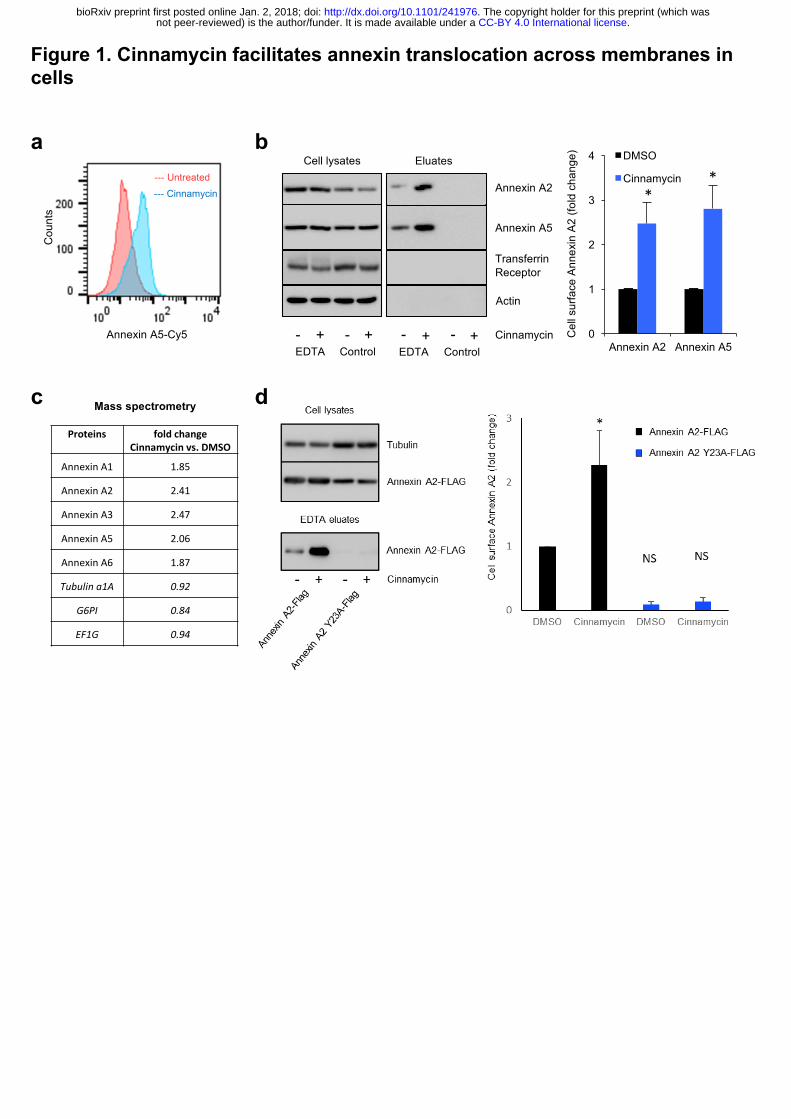
Figure 1. Cinnamycin facilitates annexin translocation across membranes in cells
a
d
b
Annexin A5-Cy5
Cou
nts
--- Untreated--- Cinnamycin
cProteins foldchange
Cinnamycinvs.DMSO
AnnexinA1 1.85
AnnexinA2 2.41
AnnexinA3 2.47
AnnexinA5 2.06
AnnexinA6 1.87
Tubulina1A 0.92
G6PI 0.84
EF1G 0.94
Mass spectrometry
Cell lysates Eluates
Annexin A2
Annexin A5
CinnamycinEDTA Control- + - +
EDTA Control- + - + 0
1
2
3
4
Annexin A2 Annexin A5
DMSO
Cinnamycin
Cel
l sur
face
Ann
exin
A2
(fold
cha
nge)
**
Actin
Transferrin Receptor
.CC-BY 4.0 International licensenot peer-reviewed) is the author/funder. It is made available under aThe copyright holder for this preprint (which was. http://dx.doi.org/10.1101/241976doi: bioRxiv preprint first posted online Jan. 2, 2018;
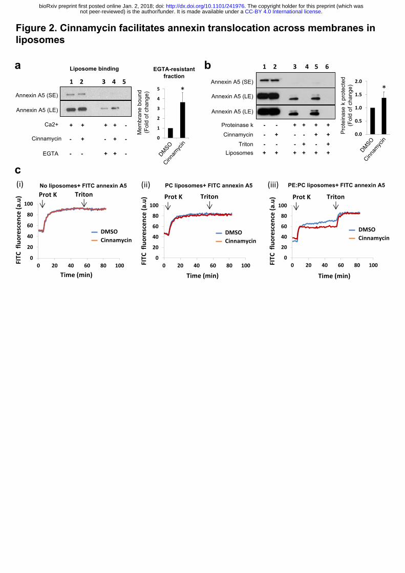
Figure 2. Cinnamycin facilitates annexin translocation across membranes in liposomes
0.0
0.5
1.0
1.5
2.0
1 2
Prot
eina
se k
pro
tect
ed
(Fol
d of
cha
nge)
*
bAnnexin A5 (SE)
Annexin A5 (LE)
Proteinase kCinnamycin
Triton
+ + + +--- - + ++-- + - +--
Liposomes + + + +++
Annexin A5 (LE)
1 2 3 4 5 6
0
1
2
3
4
5
1 2
Annexin A5 (SE)
Annexin A5 (LE)
Ca2+
EGTA
+ + + + -
- - + + -
- + - + -Cinnamycin
1 2 3 4 5
Liposome bindinga
Mem
bran
e bo
und
(Fol
d of
cha
nge)
EGTA-resistant fraction
*
c(i) (ii) (iii)
0
20
40
60
80
100
0 20 40 60 80 100FITC
fluorescence(a.u)
Time(min)
Prot K TritonNo liposomes+ FITC annexin A5
DMSOCinnamycin
0
20
40
60
80
100
0 20 40 60 80 100FITC
fluorescence(a.u)
Time(min)
Prot K TritonPC liposomes+ FITC annexin A5
DMSOCinnamycin
0
20
40
60
80
100
0 20 40 60 80 100FITC
fluorescence(a.u)
Time(min)
Prot K Triton
PE:PC liposomes+ FITC annexin A5
DMSOCinnamycin
.CC-BY 4.0 International licensenot peer-reviewed) is the author/funder. It is made available under aThe copyright holder for this preprint (which was. http://dx.doi.org/10.1101/241976doi: bioRxiv preprint first posted online Jan. 2, 2018;
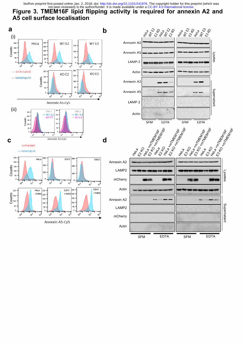
Figure 3. TMEM16F lipid flipping activity is required for annexin A2 and A5 cell surface localisation
a b
dc
(i)
(ii)
EDTASFM
LysatesSupernatant
Annexin A2
LAMP2
mCherry
Actin
Annexin A2
LAMP2
mCherry
Actin
EDTASFM
.CC-BY 4.0 International licensenot peer-reviewed) is the author/funder. It is made available under aThe copyright holder for this preprint (which was. http://dx.doi.org/10.1101/241976doi: bioRxiv preprint first posted online Jan. 2, 2018;
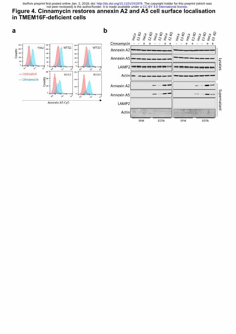
Figure 4. Cinnamycin restores annexin A2 and A5 cell surface localisation in TMEM16F-deficient cells
a b
.CC-BY 4.0 International licensenot peer-reviewed) is the author/funder. It is made available under aThe copyright holder for this preprint (which was. http://dx.doi.org/10.1101/241976doi: bioRxiv preprint first posted online Jan. 2, 2018;
1
Extended Data Figure Legends:
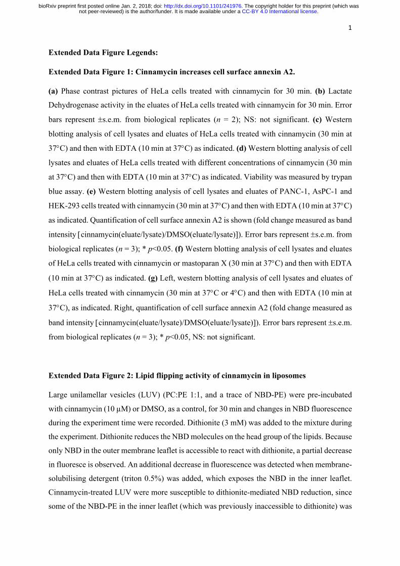
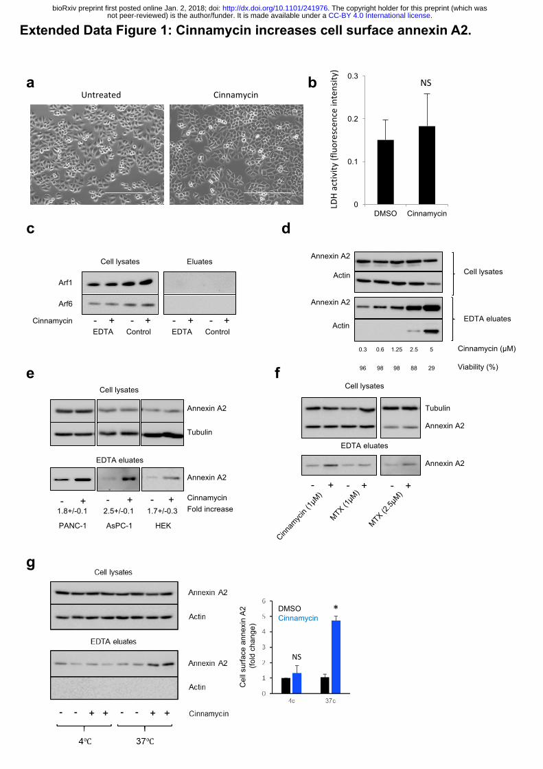
Extended Data Figure 1: Cinnamycin increases cell surface annexin A2.
(a) Phase contrast pictures of HeLa cells treated with cinnamycin for 30 min. (b) Lactate
Dehydrogenase activity in the eluates of HeLa cells treated with cinnamycin for 30 min. Error
bars represent ±s.e.m. from biological replicates (n = 2); NS: not significant. (c) Western
blotting analysis of cell lysates and eluates of HeLa cells treated with cinnamycin (30 min at
37°C) and then with EDTA (10 min at 37°C) as indicated. (d) Western blotting analysis of cell
lysates and eluates of HeLa cells treated with different concentrations of cinnamycin (30 min
at 37°C) and then with EDTA (10 min at 37°C) as indicated. Viability was measured by trypan
blue assay. (e) Western blotting analysis of cell lysates and eluates of PANC-1, AsPC-1 and
HEK-293 cells treated with cinnamycin (30 min at 37°C) and then with EDTA (10 min at 37°C)
as indicated. Quantification of cell surface annexin A2 is shown (fold change measured as band
intensity [cinnamycin(eluate/lysate)/DMSO(eluate/lysate)]). Error bars represent ±s.e.m. from
biological replicates (n = 3); * p<0.05. (f) Western blotting analysis of cell lysates and eluates
of HeLa cells treated with cinnamycin or mastoparan X (30 min at 37°C) and then with EDTA
(10 min at 37°C) as indicated. (g) Left, western blotting analysis of cell lysates and eluates of
HeLa cells treated with cinnamycin (30 min at 37°C or 4°C) and then with EDTA (10 min at
37°C), as indicated. Right, quantification of cell surface annexin A2 (fold change measured as
band intensity [cinnamycin(eluate/lysate)/DMSO(eluate/lysate)]). Error bars represent ±s.e.m.
from biological replicates (n = 3); * p<0.05, NS: not significant.
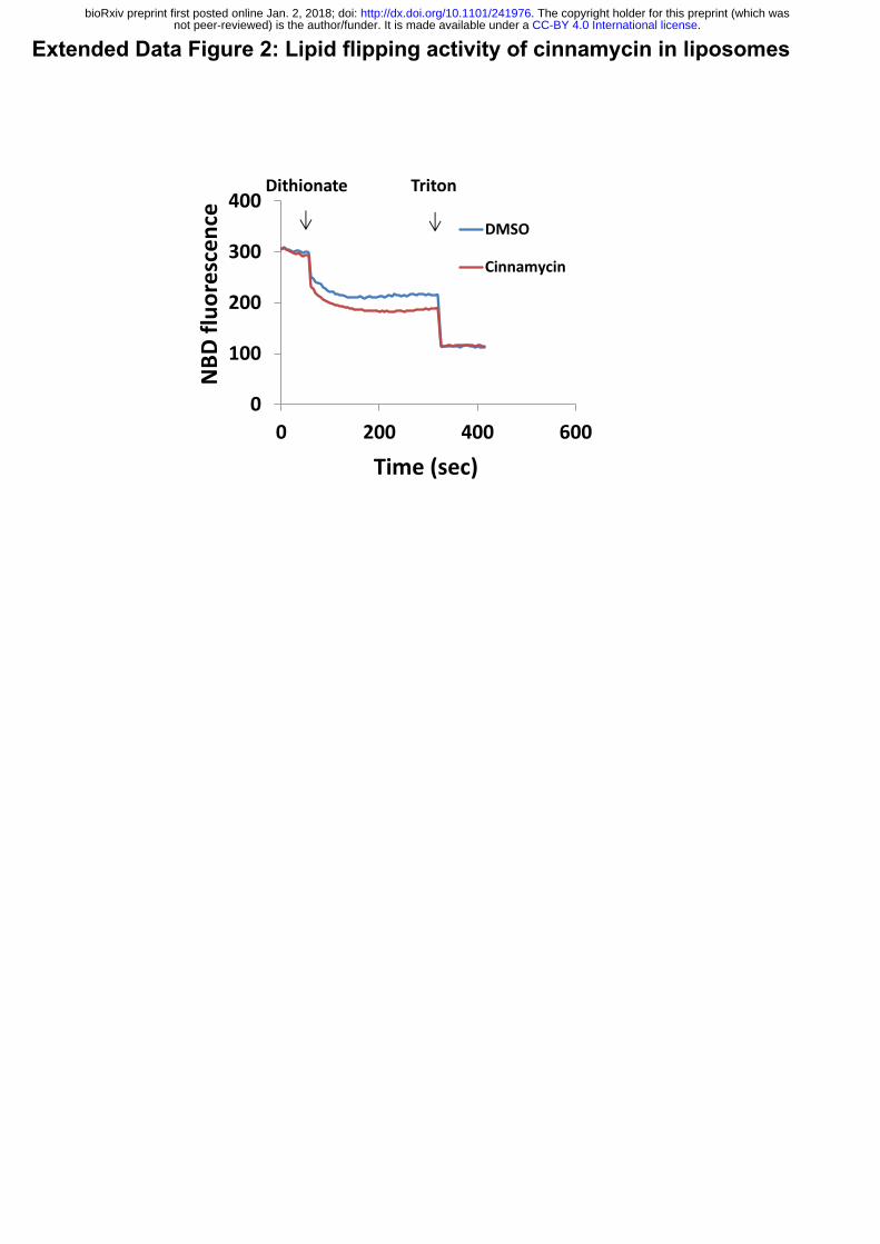
Extended Data Figure 2: Lipid flipping activity of cinnamycin in liposomes
Large unilamellar vesicles (LUV) (PC:PE 1:1, and a trace of NBD-PE) were pre-incubated
with cinnamycin (10 µM) or DMSO, as a control, for 30 min and changes in NBD fluorescence
during the experiment time were recorded. Dithionite (3 mM) was added to the mixture during
the experiment. Dithionite reduces the NBD molecules on the head group of the lipids. Because
only NBD in the outer membrane leaflet is accessible to react with dithionite, a partial decrease
in fluoresce is observed. An additional decrease in fluorescence was detected when membrane-
solubilising detergent (triton 0.5%) was added, which exposes the NBD in the inner leaflet.
Cinnamycin-treated LUV were more susceptible to dithionite-mediated NBD reduction, since
some of the NBD-PE in the inner leaflet (which was previously inaccessible to dithionite) was
.CC-BY 4.0 International licensenot peer-reviewed) is the author/funder. It is made available under aThe copyright holder for this preprint (which was. http://dx.doi.org/10.1101/241976doi: bioRxiv preprint first posted online Jan. 2, 2018;
2
flipped to the outer leaflet by cinnamycin and this fluorescence (which is detectable on both
inner and outer leaflets) would have been quenched by the dithionate after flipping to the outer
leaflet. Thus, the decrease in fluorescence caused by cinnamycin versus DMSO reflects the
amount to NBD-PE that has flipped.
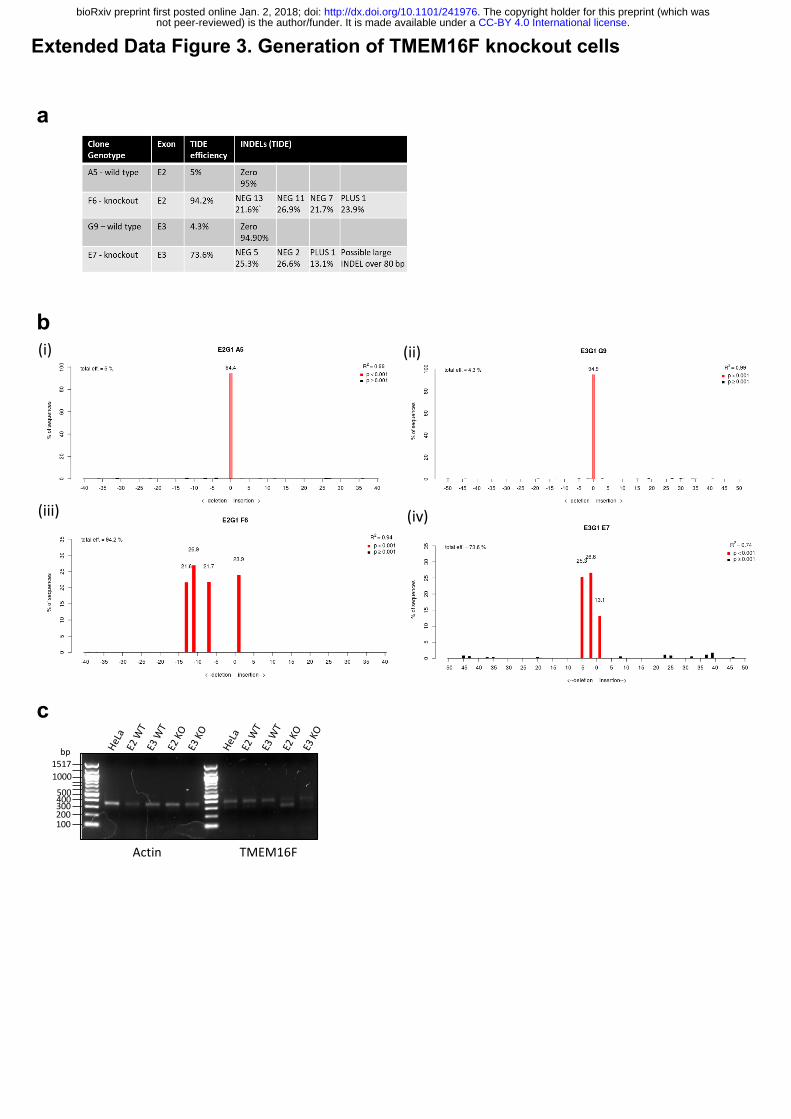
Extended Data Figure 3: Generation of TMEM16F knockout cells.
(a) Tracking of Indels by Decomposition (TIDE) analysis of TMEM16F CRISPR targeted
clone summary. (b) Tracking of Indels by Decomposition (TIDE) analysis (i) sgRNA targeting
TMEM16F exon 2, 94.4% sequence wild type; (ii) sgRNA targeting TMEM16F exon 3, 94.9%
sequence wild type; (iii) sgRNA targeting TMEM16F exon 2, 0% sequence wild type; (iv)
sgRNA targeting TMEM16F exon 3, 0 % sequence wild type. (c) Reverse transcription PCR
for TMEM16F expression. RNA from TMEM16F clones analysed for TMEM16F mRNA.
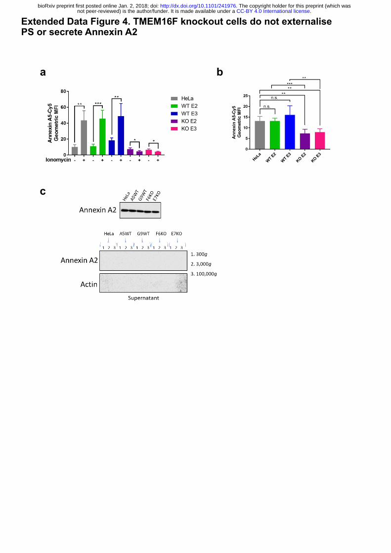
Extended Data Figure 4. TMEM16F knockout cells do not externalise PS or secrete
Annexin A2.
(a) TMEM16F-knockout cells do not externalise PS in response to ionomycin stimulation.
Wild-type, positive control and TMEM16F knockout cells were treated with 10 mM ionomycin
for 10 min at 37°C in the presence of annexin A5-Cy5 and PI. Annexin A5-Cy5 binding and
PI accumulation were analysed by FACS. Quantification of the geometric mean fluorescence
intensity of annexin A5-Cy5 binding from four separate experiments are shown. * p<0.05, **
p<0.01, *** p<0.001, n.s.: not significant. (b) Basal cell surface PS is reduced in TMEM16F
knockout cells. Cells were incubated with annexin A5-Cy5 and PI for 10 min at 37⁰ C before
FACS analysis. The geometric mean fluorescence intensity of annexin A5-Cy5 is plotted from
five separate experiments. * p<0.05, ** p<0.01, *** p<0.001, n.s.: not significant. (c) Annexin
A2 and A5 are not secreted from TMEM16F-deficient cells. Cells were seeded at 70%
confluence and incubated in SFM for 24h at 37°C. The medium was collected, cells and debris
removed by centrifugation and analysed for annexin A2 and A5 by western blotting (n=3).
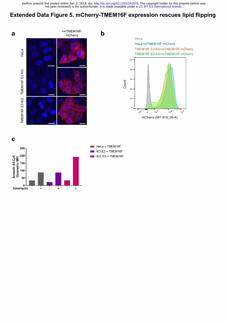
Extended Data Figure 5. mCherry-TMEM16F expression rescues lipid flipping.
.CC-BY 4.0 International licensenot peer-reviewed) is the author/funder. It is made available under aThe copyright holder for this preprint (which was. http://dx.doi.org/10.1101/241976doi: bioRxiv preprint first posted online Jan. 2, 2018;
3
(a) Subcellular localisation of mCherry-mTMEM16F. In both wild-type and TMEM16F-
deficient cells mCherry-TMEM16F localises to the perinuclear location and to the plasma
membrane as seen by confocal microscopy analysis of mCherry expression. (b) Flow
cytometry analysis of mCherry-mTMEM16F expression in wild-type and TMEM16F-deficient
cells. (c) Expression of mCherry-mTMEM16F rescues lipid flipping in TMEM16F-knockout
cells. Wild-type and TMEM16F-knockout Hela cells expressing mCherry-mTMEM16F were
treated with ionomycin and analysed for annexin A5-cy5 binding. Representative results shown
as the expression level of mCherry-mTMEM16F varied in 3 separate experiments.
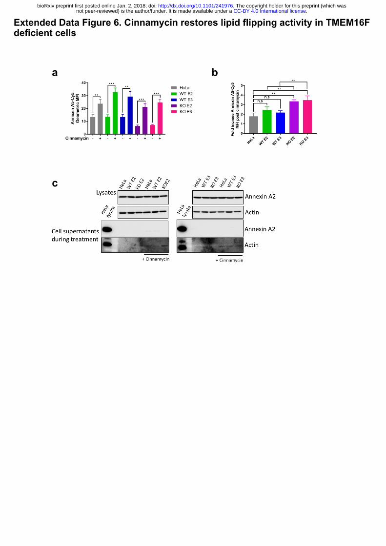
Extended Data Figure 6. Cinnamycin restores lipid flipping activity in TMEM16F
deficient cells.
(a) Cinnamycin lipid flipping activity externalises PS in TMEM16F-knockout cells. Wild-type,
positive control and TMEM16F-knockout cells were treated with 1 µM cinnamycin for 50 min
at 37°C, annexin A5-Cy5 and PI were then added and incubated for a further 10 min at 37°C.
Annexin A5-Cy5 binding and PI accumulation were analysed by FACS. The geometric mean
fluorescence intensity of of annexin A5-Cy5 binding to live cells are shown from 3 separate
experiments. * p<0.05, ** p<0.01, *** p<0.001, n.s.: not significant. (b) Fold increase in
annexin A5-Cy5 fluorescence intensity post cinnamycin treatment as described in (a). (c)
Annexin A2 and A5 are retained on the cell surface and not secreted in the presence of
cinnamycin. The medium was collected after 1 hour cinnamycin treatment and analysed for
annexin A2 and A5 free in the medium. Actin is used as a positive control.
Extended Data Table 1. Oligonucleotides used for TMEM16F CRISPR/Cas9 targeting,
amplification of targeted area and sequencing.
.CC-BY 4.0 International licensenot peer-reviewed) is the author/funder. It is made available under aThe copyright holder for this preprint (which was. http://dx.doi.org/10.1101/241976doi: bioRxiv preprint first posted online Jan. 2, 2018;
b
PANC-1 AsPC-1 HEK
Annexin A2
Tubulin
Cinnamycin- + - + - +
Annexin A2
Cell lysates
EDTA eluates
Fold increase1.8+/-0.1 2.5+/-0.1 1.7+/-0.3
a
Extended Data Figure 1: Cinnamycin increases cell surface annexin A2.
Annexin A2
Tubulin
Annexin A2
Cell lysates
EDTA eluates
- + - + - +
f
c
g
d
e
0
0.1
0.2
0.3
DMSO Cinnamycin
LDHactiv
ity(fluorescenceintensity
)
NS
Arf1
Cinnamycin
Arf6
EDTA Control- + - +
EDTA Control- + - +
Cell lysates EluatesAnnexin A2
Cell lysates
EDTA eluates
Annexin A2
0.3 0.6 1.25 2.5 5
Viability (%)
Actin
Actin
96 98 98 88 29
Untreated Cinnamycin
Cel
l sur
face
ann
exin
A2
(fold
cha
nge)
DMSOCinnamycin
*
NS
Cinnamycin (µM)
.CC-BY 4.0 International licensenot peer-reviewed) is the author/funder. It is made available under aThe copyright holder for this preprint (which was. http://dx.doi.org/10.1101/241976doi: bioRxiv preprint first posted online Jan. 2, 2018;
Extended Data Figure 2: Lipid flipping activity of cinnamycin in liposomes
NBDflu
orescence
0
100
200
300
400
0 200 400 600
DMSO
Cinnamycin
Time(sec)
Dithionate Triton
.CC-BY 4.0 International licensenot peer-reviewed) is the author/funder. It is made available under aThe copyright holder for this preprint (which was. http://dx.doi.org/10.1101/241976doi: bioRxiv preprint first posted online Jan. 2, 2018;
a
c
Extended Data Figure 3. Generation of TMEM16F knockout cells
b
(iii)
(ii)
(iv)
(i)
Actin TMEM16F
100200300400500
10001517bp
.CC-BY 4.0 International licensenot peer-reviewed) is the author/funder. It is made available under aThe copyright holder for this preprint (which was. http://dx.doi.org/10.1101/241976doi: bioRxiv preprint first posted online Jan. 2, 2018;
Extended Data Figure 4. TMEM16F knockout cells do not externalise PS or secrete Annexin A2
ba
c
.CC-BY 4.0 International licensenot peer-reviewed) is the author/funder. It is made available under aThe copyright holder for this preprint (which was. http://dx.doi.org/10.1101/241976doi: bioRxiv preprint first posted online Jan. 2, 2018;
Extended Data Figure 5. mCherry-TMEM16F expression rescues lipid flipping
a
c
HeL
aTM
EM16
F E2
KO
TMEM
16F
E3 K
O
+mTMEM16F-mCherry b
HeLa+mTMEM16F-mCherryTMEM16F E2 KO+mTMEM16F-mCherryTMEM16F E3 KO+mTMEM16F-mCherry
HeLa
Cou
nt
mCherry (561 610_20-A)
.CC-BY 4.0 International licensenot peer-reviewed) is the author/funder. It is made available under aThe copyright holder for this preprint (which was. http://dx.doi.org/10.1101/241976doi: bioRxiv preprint first posted online Jan. 2, 2018;
Extended Data Figure 6. Cinnamycin restores lipid flipping activity in TMEM16F deficient cells
a b
C.c
.CC-BY 4.0 International licensenot peer-reviewed) is the author/funder. It is made available under aThe copyright holder for this preprint (which was. http://dx.doi.org/10.1101/241976doi: bioRxiv preprint first posted online Jan. 2, 2018;
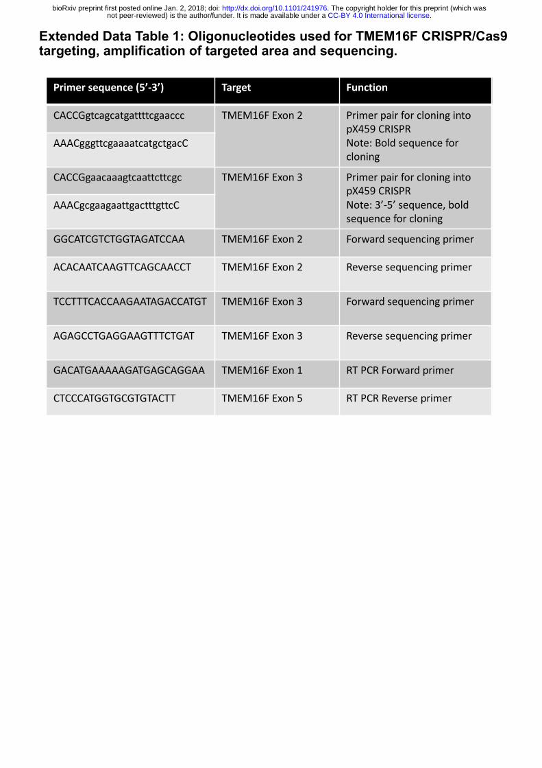
Extended Data Table 1: Oligonucleotides used for TMEM16F CRISPR/Cas9 targeting, amplification of targeted area and sequencing.
Primersequence(5’-3’) Target Function
CACCGgtcagcatgattttcgaaccc TMEM16FExon2 PrimerpairforcloningintopX459CRISPRNote:Boldsequenceforcloning
AAACgggttcgaaaatcatgctgacC
CACCGgaacaaagtcaattcttcgc TMEM16FExon3 PrimerpairforcloningintopX459CRISPRNote:3’-5’sequence,boldsequenceforcloning
AAACgcgaagaattgactttgttcC
GGCATCGTCTGGTAGATCCAA TMEM16FExon2 Forwardsequencingprimer
ACACAATCAAGTTCAGCAACCT TMEM16FExon2 Reversesequencingprimer
TCCTTTCACCAAGAATAGACCATGT TMEM16FExon3 Forwardsequencingprimer
AGAGCCTGAGGAAGTTTCTGAT TMEM16FExon3 Reversesequencingprimer
GACATGAAAAAGATGAGCAGGAA TMEM16FExon1 RTPCRForwardprimer
CTCCCATGGTGCGTGTACTT TMEM16FExon5 RTPCRReverseprimer
.CC-BY 4.0 International licensenot peer-reviewed) is the author/funder. It is made available under aThe copyright holder for this preprint (which was. http://dx.doi.org/10.1101/241976doi: bioRxiv preprint first posted online Jan. 2, 2018;