Embed Size (px)
Citation preview

Phosphodiesterases coordinate cAMP propagationinduced by two stimulatory G protein-coupledreceptors in heartsShubai Liua,1, Ying Lib,1, Sungjin Kima, Qin Fua, Dippal Parikha, Bharat Sridhara, Qian Shia, Xiaoying Zhangb,Yinzheng Guanb, Xiongwen Chenb,2, and Yang K. Xianga,2,3
aDepartment of Molecular and Integrative Physiology, University of Illinois at Urbana Champaign, Urbana, IL 61801; and bDepartment of Physiology, TempleUniversity Medical Center, Philadelphia, PA 19140
Edited by Robert J. Lefkowitz, Duke University Medical Center/Howard Hughes Medical Institute, Durham, NC, and approved March 15, 2012 (received forreview October 29, 2011)
Inflammation is a significant player in the progression of heartfailure and has detrimental effects on cardiac function. Prostaglan-din (PG)E2, a major proinflammatory prostanoid in the cardiovas-cular system, is a potent stimulus in inducing intracellular cAMP butminimally affects cardiac contractile function. Here, we show thatthe PGE2 stimulation attenuates the adrenergic-induced cardiaccontractile response in animal hearts. Stimulation with PGE2 leadsto stimulatory G protein (Gs)-dependent production of cAMP. How-ever, the induced cAMP is spatially restricted because of its deg-radation by phosphodiesterase (PDE)4 and cannot access theintracellular sarcoplasmic reticulum (SR) for increasing calcium sig-naling and myocyte contraction. Moreover, pretreatment withPGE2 significantly inhibits PKA activities at the SR induced by aβ-adrenergic agonist, isoproterenol, and subsequently blocks iso-proterenol-induced PKA phosphorylation of phospholamban andcontractile responses in myocytes. Further analysis reveals that thePGE2-induced cAMP/PKA is sufficient to phosphorylate and acti-vate PDE4D isoforms, which, in turn, spatially inhibits the diffusionof adrenergic-induced cAMP from the plasma membrane to the SR.Inhibition of PDE4 rescues the adrenergic-induced increase incAMP/PKA activities at the SR, PKA phosphorylation of phospho-lamban, and contractile responses in PGE2-pretreated myocytes.Thus, this offers an example that one Gs-coupled receptor is ableto inhibit the intracellular signaling transduction initiated by an-other Gs-coupled receptor via controlling the diffusion of cAMP,presenting a paradigm for G protein-coupled receptor (GPCR) sig-nal transduction. It also provides a mechanism for the integrationof signaling initiated by different neurohormonal stimuli, as wellas long-term effects of chronically circulating proinflammatory fac-tors in myocardium.
FRET | subcellular diffusion | cross-talk
Inflammation plays a significant role in the progression of heartfailure (1, 2). The expression of prostaglandin (PG)E2, a major
proinflammatory prostanoid in cardiovascular system, is elevatedin the hearts of patients and animals with myocardial infarctionand heart failure (3, 4). Clinically, heart failure is characterizedwith gradual development of cardiac hypertrophy and myocytedeath attributable to elevated β-adrenergic stimulation and car-diac dysfunction (5, 6). However, cardiac function under E seriesprostaglandin (EP) receptor signaling is not clear, and the im-pact on cardiac β-adrenergic signaling and function during thedevelopment of heart diseases remains to be addressed.In human and animal hearts, PGE2 and catecholamines ac-
tivate divergent signaling cascades for distinct cellular andphysiological function. EP4 is the most abundantly expressedEP subtype in the myocardium (7). Biochemically, activation ofEP4 leads to an accumulation of cAMP through activation ofstimulatory G (Gs) protein. However, the induced cAMP signalappears to be confined along the plasma membrane (PM) andhas limited effect on myocyte contractile response (7). Thisconfinement of cAMP is primarily regulated through cAMP
hydrolysis by PDE4 isoforms (8, 9). Consequently, PGE2 stimu-lation fails to promote phosphorylation of proteins such as phos-pholamban (PLB), a critical player on the intracellular calciumstorage compartment sarcoplasmic reticulum (SR) that releasescalcium for contractile response. Catecholamines stimulate β ad-renergic receptors (βARs) to induce Gs protein activation andcAMP production. In contrast to PGE2 stimulation, the adren-ergic-induced cAMP signaling leads to robust PKA phosphoryla-tion of substrates on the PM, the SR, and the myofibril, andpromotes cardiomyocyte contractility (10, 11). Previous studieshave also characterized the association of different PDE4D iso-forms with βAR subtypes, which regulate subcellular distributionof the adrenergic signaling-induced cAMP activities (12, 13).Under agonist stimulation, these PDE4D isoforms dissociate fromthe activated βARs to allow cAMP diffusion from the PM to theintracellular compartments (12, 13). Interestingly, early studieshave indicated that PGE2 treatment impairs the sympatheticregulation of cardiac contractile function in animal hearts (14). Itis, thus, likely that the signaling induced by PGE2 and β-adren-ergic stimuli are integrated in the myocardium. We hypothesizethat PGE2 exerts inhibitory effects on βAR signaling cascades inthe myocardium via a cross-talk by activating the βAR-associatedPDE4D isoforms.Here, we show that the PGE2-induced cAMP signal is con-
fined within the PM domain and has minimal effects on PKAphosphorylation of PLB at the SR and contractile function inanimal hearts. However, PGE2 stimulation inhibits intracellularβAR signaling transduction by blocking propagation of cAMPfrom the PM to the SR, which blocks PKA phosphorylation ofPLB and inhibits calcium cycling regulation in myocytes. Furtherdissection reveals that PGE2 stimulation induces PKA phos-phorylation of PDE4D isoforms, which prevents the βAR-in-duced cAMP diffusion to the SR. Consequently, the PGE2stimulation blocks the βAR-induced contractile responses incardiomyocytes and in animal hearts. The elegant cross-talkbetween two Gs-coupled receptors in shaping subcellular distri-bution of second messenger cAMP presents a paradigm on Gprotein-coupled receptor (GPCR) signaling transduction for in-tegration of multiple extracellular stimuli.
Author contributions: S.L., Y.L., Q.F., X.C., and Y.K.X. designed research; S.L., Y.L., S.K., Q.F.,D.P., B.S., Q.S., X.Z., and Y.G. performed research; S.L., Q.F., D.P., Q.S., X.C., and Y.K.X.analyzed data; and X.C. and Y.K.X. wrote the paper.
The authors declare no conflict of interest.
This article is a PNAS Direct Submission.1S.L. and Y.L. contributed equally to this work.2To whom correspondence may be addressed. E-mail: [email protected] or [email protected].
3Present address: Department of Pharmacology, University of California, Davis, CA 95616.
This article contains supporting information online at www.pnas.org/lookup/suppl/doi:10.1073/pnas.1117862109/-/DCSupplemental.
6578–6583 | PNAS | April 24, 2012 | vol. 109 | no. 17 www.pnas.org/cgi/doi/10.1073/pnas.1117862109
Dow
nloa
ded
by g
uest
on
Apr
il 5,
202
0

ResultsProstaglandin Impairs β-Adrenergic Stimulation on Contractile Responsesin Animal Hearts. Proinflammatory factor PGE2 is a potent stimulusin inducing intracellular cAMP signaling but fails to promote car-diac contractile response (7). We envisioned that prostaglandin-induced PKA phosphorylation and activation of phosphodiesterasecould impair signaling transduction induced by β-adrenergic stim-ulation in themyocardium and block adrenergic-induced contractileresponses, offering a mechanism on how inflammation contributesto cardiac dysfunction. Using Langendorff perfusion of mousehearts, we first tested different doses of PGE2 on cardiac con-tractility and found that PGE2 exerted a positive inotropic effecton the heart at minimal concentration of 1μM (Fig. 1A). Incomparison, β-adrenergic stimulation with isoproterenol (ISO)induced a sensitive dose-dependent contractile response (Fig. 1B)with EC50 at 1.99 nM and maximal response at the concentrations>10 nM. ISO was able to increase peak left ventricular developedpressure (LVDP) from 71.4 ± 13.0 to 115.3 ± 12.0 mmHg(61.8%; EC50, −8.708), maximal dp/dt from 2,343.7 ± 159.7 to5,984.4 ± 622.8 mmHg (150.6%; EC50, −8.675), and minimum –dp/dt from −1,787.8,± 325.2 to 3,492.2 ± 142.03 mmHg/s (95.4%;EC50, −8.729). In contrast, when the heart was pretreated with 0.1μM PGE2, a concentration that was noneffective on cardiacfunction by itself, the ISO dose–response curve was shiftedrightward, and the EC50 of ISO was changed to ∼199 nM, 100times higher than the EC50 of ISO alone. Furthermore, themaximal responses of LVDP (from 63.8 ± 3.9 to 81.5 ± 10.9mmHg; 27.8%; EC50, −6.891), dp/dt (from 2,229.5 ± 54.3 to4,009.6 ± 504.3 mmHg/s; 77.1%; EC50, −6.78) and –dp/dt (from−1,840.8 ± 179.0 to −3,127.7 ± 750.9 mmHg; 70.0%; EC50,−6.577) were all substantially decreased. These data suggest thatPGE2 pretreatment blunts β-adrenergic responsiveness ofcardiomyocytes.To validate the contractile response, we examined phosphor-
ylation level of a PKA substrate PLB. PLB is located at the SR
and plays a critical role in calcium cycling by regulating SERCAactivity. Adrenergic stimulation, but not prostaglandin stimula-tion, significantly promotes both PKA and Ca2+/calmodulin-dependent protein kinase (CaMK)II phosphorylation of PLB inanimal hearts. Pretreatment with 0.1 μM PGE2 abolishedphosphorylation of PLB induced by ISO (Fig. 1C). These datasuggest that although PGE2 stimulation fails to induce cAMPand PKA activities at the SR, it inhibits the adrenergic-inducedPKA activities at the SR for cardiac contractile response inanimal hearts.
Prostaglandin and Adrenergic Stimulation Induces Distinct SubcellularcAMP-PKA Activities in Cardiomyocytes. We sought to directly mea-sure PKA activities in different intracellular compartments withbiosensors under the signaling cross-talk between EP and βAR incardiomyocytes. To achieve this, we used the recently developedPKAbiosensors that are targeted to the PM, the cytoplasm, and theSR (PM-AKAR3, cyto-AKAR3, and SR-AKAR3, respectively)(Fig. 2A and Fig. S1) (15, 16). In neonatal cardiomyocytes, directstimulation of adenylyl cyclases with forskolin (FSK) inducedstrong PKA activities at the PM, less robust PKA activities in thecytoplasm, and much lower PKA activities at the SR (Fig. 2 B–D).The pattern follows a diffusion of cAMP from the PMwhere cAMPis produced. In comparison, stimulation with ISO or PGE2 inducedlower increases in PKA activities at the PM than FSK (Fig. 2 B–D).Interestingly, under ISOor PGE2 stimulation, the PKA activities inthe cytoplasm were much more robust than those at the PM, re-spectively (Fig. 2 A–C). Furthermore, adrenergic stimulation in-duced significantly higher PKA activities at the SR than FSKstimulation, whereas PGE2 failed to generate significant increase(Fig. 2 D and E). The data indicate that the adrenergic-inducedcAMP preferentially activates PKA at the intracellular compart-ments like the SR for myocyte contractile response, whereas PGE2stimulation completely fails to do so. In addition, dopamine in-duced modest PKA activities at the SR, whereas adenosine failed
80
100
120
140
160
180
Nor
mal
ized
dp/
dt
100
120
140
160
180
200
Log [PGE2]
Nor
mal
ized
- dp
/dt
-10 -9 -8 -7 -6 -5 -480
100
120
140
160
180
200
Log [PGE2]
Nor
mal
ized
LV
DP
(mm
Hg)
Log [PGE2]
80
100
120
140
160
180 ISO
PGE2+ISO
Nor
mal
ized
LV
DP
(%)
100
150
200
250
300 ISO
PGE2+ ISO
Nor
mal
ized
dp/
dt (%
)
-14 -12 -10 -8 -6 -450
100
150
200
250 ISO
PGE2+ ISO
Nor
mal
ized
- dp
/dt (
%)
Log [ISO]-14 -12 -10 -8 -6 -4
Log [ISO]-14 -12 -10 -8 -6 -4
Log [ISO]
PLB pS16
PLB
Tubulin
PLB pT17
control ISO 0.1µM PGE2 0.1µM ISO + PGE2
A
B
C
1 2 3 1 2 3 1 2 3 1 2 3mice
-10 -9 -8 -7 -6 -5 -4 -10 -9 -8 -7 -6 -5 -4
Fig. 1. PGE2 inhibits adrenergic-inducedphosphorylation of PLB and contractile re-sponse in animal hearts. Mouse hearts werecannulated for Langerdorff perfusion withprostaglandin or ISO or with prostaglandin for5 min before addition of ISO at concentrationsindicated. The LVDP, maximal dp/dt, and min-imal −dp/dt were analyzed and normalizedagainst basal levels. (A) Cardiac functionalresponses to different concentrations of PGE2.(B) Cardiac functional responses to differentconcentrations of ISO after PGE2 (0.1 μM)pretreatment. (C) Phosphorylation of PLB atthe Ser16 site (PKA site) and Thr17 site (CaMKIIsite) after perfusion of the hearts with drugsindicated.
Liu et al. PNAS | April 24, 2012 | vol. 109 | no. 17 | 6579
CELL
BIOLO
GY
Dow
nloa
ded
by g
uest
on
Apr
il 5,
202
0
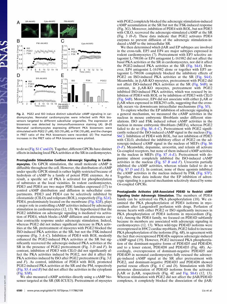
to do so (Fig. S1C andD). Together, differentGPCRs have distincteffects in inducing local PKA activities at the SR in cardiomyocytes.
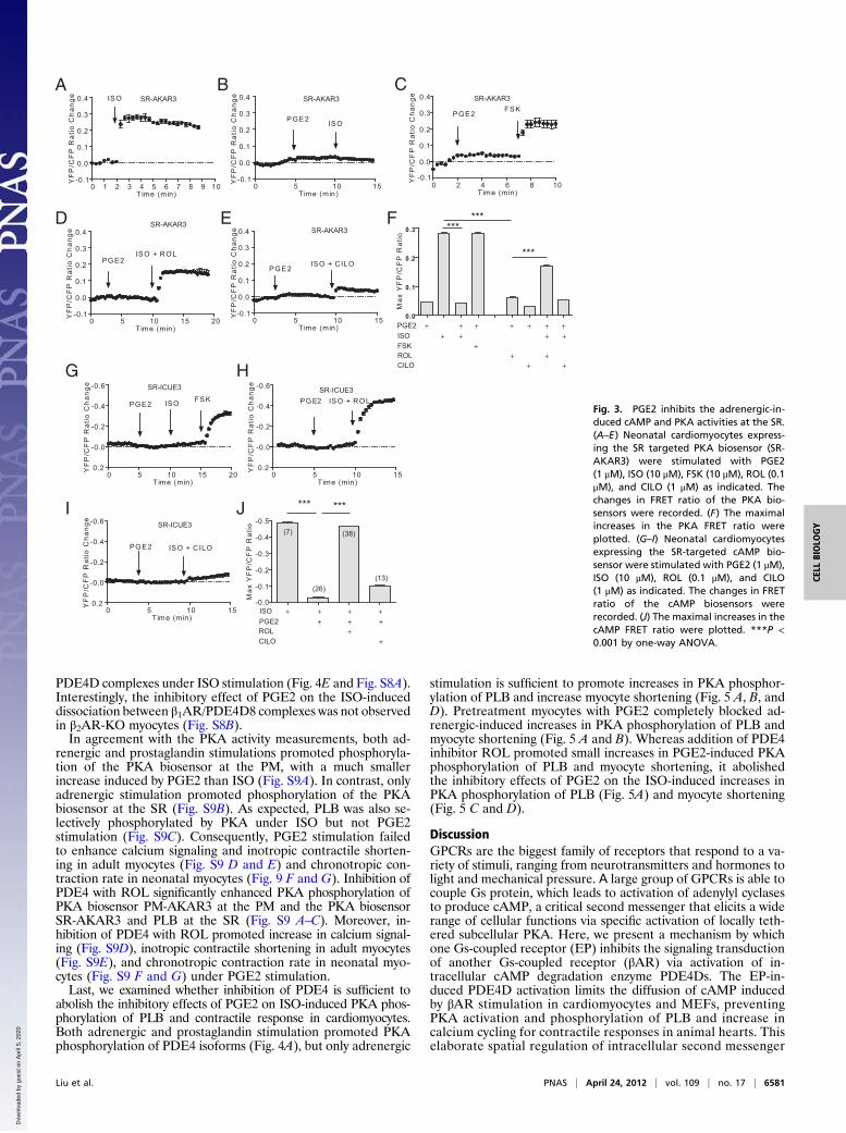
Prostaglandin Stimulation Confines Adrenergic Signaling in Cardio-myocytes. On GPCR stimulation, the small molecule cAMP isdiffusible throughout the cell. However, the distribution of cAMPunder specific GPCR stimuli is rather highly restricted because ofhydrolysis of cAMP by a family of potent PDE enzymes. As aresult, a specific set of PKA is activated for phosphorylationof substrates at the local vicinities. In rodent cardiomyocytes,PDE3 and PDE4 are two major PDE families expressed (17) tocontrol cAMP distribution and diffusion in subcellular com-partments. PDE3 and PDE4 can be selectively inhibited bycilostamide (CILO) and rolipram (ROL), respectively (Fig. S2A).PDE4, predominately located on the membrane (Fig. S2B), playsa major role in controlling cAMP activities induced by adrenergicstimulation in cardiomyocytes (12, 13). We hypothesized that thePGE2 inhibition on adrenergic signaling is mediated via activa-tion of PDE4, which blocks cAMP diffusion and attenuates car-diac contractile response under adrenergic stimulation. Indeed,whereas PGE2 did not induce significant increase in PKA activ-ities at the SR, pretreatment of myocytes with PGE2 blocked theISO-induced PKA activities at the SR, but not the FSK-inducedresponse (Fig. 3 A–C). Inhibition of PDE4 with ROL modestlyenhanced the PGE2-induced PKA activities at the SR but sig-nificantly recovered the adrenergic-induced PKA activities at theSR in the presence of PGE2 pretreatment (Fig. 3 D and F). Incontrast, inhibition of PDE3 with CILO did not significantly af-fect the PKA activities induced by PGE2, nor did it affect thePKA activities induced by ISO after PGE2 pretreatment (Fig. 3 Eand F). As control, inhibition of PDE4 with ROL promotedPGE2-induced PKA activities at the SR and the PM, respectively(Fig. S3 A and B) but did not affect the activities in the cytoplasm(Fig. S3B).We also measured cAMP activities directly using a cAMP bio-
sensor targeted at the SR (SR-ICUE3). Pretreatment of myocytes
with PGE2 completely blocked the adrenergic stimulation-inducedcAMP accumulation at the SR but not the FSK-induced response(Fig. 3G). Moreover, inhibition of PDE4 with ROL, but not PDE3with CILO, recovered the adrenergic-stimulated cAMP at the SR(Fig. 3 H–J). These data indicate that PGE2 activates PDE4enzymes to prevent diffusion of the adrenergic stimulation-in-duced cAMP to the intracellular SR.We then determined which βAR and EP subtypes are involved
in the cross-talk. EP3 and EP4 are major subtypes expressed inrodent cardiomyocytes (7). Pretreatment with EP3 selective an-tagonist L-798106 or EP4 antagonist L-161982 did not affect thebasal PKA activities at the SR in cardiomyocytes, nor did it affectthe PGE2-induced PKA activities at the SR (Fig. S4A). How-ever, EP4 antagonist L-161982 alone or together with EP3 an-tagonist L-798106 completely blocked the inhibitory effects ofPGE2 on ISO-induced PKA activities at the SR (Fig. S4A).Meanwhile, in β2AR-KO myocytes, pretreatment with PGE2 didnot affect ISO-induced PKA activities at the SR (Fig. S4B); incontrast, in β1AR-KO myocytes, pretreatment with PGE2inhibited ISO-induced PKA activities, which was rescued by in-hibition of PDE4 with ROL or by inhibition of PDE3 with CILO(Fig. S4B). Moreover, EP4 did not associate with either β1AR orβ2AR when expressed in HEK293 cells, suggesting that the cross-talk occurs via downstream intracellular mechanisms (Fig. S5).To explore whether the EP inhibition of adrenergic signaling is
a general mechanism, we measured the cAMP activities in thenucleus in mouse embryonic fibroblasts under different stim-ulations. ISO and FSK induced robust cAMP activities in thenucleus in mouse embryonic fibroblasts (MEFs), whereas PGE2failed to do so (Fig. S6 A–C). Pretreatment with PGE2 signifi-cantly reduced the ISO-induced cAMP signal in the nucleus (Fig.S6C). Inhibition of PDE4 with ROL, but not inhibition of PDE3with CILO, abolished the inhibitory effect of PGE2 on the ad-renergic-induced cAMP signal in the nucleus of MEFs (Fig. S6D–F). Meanwhile, dopamine, urocortin, and relaxin all activateGs-coupled receptors, but none of them induced cAMP activitiesin the nucleus in MEFs (Fig. S7 A–E). Pretreatment with do-pamine almost completely inhibited the ISO-induced cAMPactivities in the nucleus (Fig. S7 B and F). Urocortin partiallyinhibited the cAMP activities, whereas relaxin failed to do so(Fig. S7 D and E). In contrast, none of these agonists inhibitedthe cAMP activities in the nucleus induced by FSK (Fig. S7F).Together, these data indicate that the EP inhibition of adren-ergic signaling is a general mechanism selectively shared by someGs-coupled GPCRs.
Prostaglandin Activates βAR-Associated PDE4D to Restrict cAMPSignaling Under Adrenergic Stimulation. The members of PDE4family can be activated via PKA phosphorylation (18). We ex-amined the PKA phosphorylation of PDE4 isoforms in myo-cardium after Langendorff perfusion with drugs. Perfusion ofmouse hearts with either PGE2 or ISO significantly increases inPKA phosphorylation of PDE4 isoforms in myocardium (Fig.4A). Among the PDE4 family, we focused on PDE4D subfamilybecause its members are associated with adrenergic signaling incardiomyocytes (12, 13). When wild-type PDE4D isoforms wereoverexpressed inH9C2 cardiacmyoblasts, PGE2 failed to increasePKA phosphorylation of the isoforms (Fig. 4B), in agreement withthe fact that overexpressed PDE4Ds suppress adrenergic-inducedcAMP signal (19). However, PGE2 increased PKA phosphoryla-tion of the dominant-negative forms of PDE4D5 and PDE4D9,and to a lesser extent, PDE4D8 and PDE4D3 (Fig. 4B). Ac-cordingly, overexpression of dominant-negative PDE4D5 andPDE4D9 in neonatal cardiomyocytes fully rescued the adrener-gic-induced cAMP signal at the SR after pretreatment withPGE2, and dominant-negative PDE4D3 and PDE4D8 showedpartial rescue effects (Fig. 4 C and D). Stimulation with ISOpromotes dissociation of PDE4D isoforms from the activatedβ1AR or β2AR, respectively (Fig. 4E and Fig. S8A) (12, 13).Whereas stimulation with PGE2 did not affect the βAR/PDE4Dcomplexes, it completely blocked the dissociation of the βAR/
E
0.0
0.2
0.4
0.6
0.8
1.0 FSKISO PGE2
Max
imal
YF
P/C
FP
Rat
io
PM-AKAR3
SR-AKAR3Cyto-AKAR3
+ + ++ + +
+ + +
B C
D
SR-AKAR3 Cyto-AKAR3 PM-AKAR3
PM -AKAR3
0 2 4 6 8 10 12 14 160.0
0.2
0.4
0.6
0.8
1.0
Time (min)
YF
P/C
FP
Rat
io C
hang
e
Cyto-AKAR3
0 2 4 6 8 10 12 14 160.0
0.2
0.4
0.6
0.8
1.0
ISOPGE2FSK
Time (min)
YF
P/C
FP
Rat
io C
hang
e
SR-AKAR3
0 2 4 6 8 10 12 14 160.0
0.1
0.2
0.3
0.4 ISOPGE2FSK
Time (min)
YF
P/C
FP
Rat
io C
hang
e
A
ISOPGE2FSK
Fig. 2. PGE2 and ISO induce distinct subcellular cAMP signaling in car-diomyocytes. Neonatal cardiomyocytes were infected with PKA bio-sensors targeted to different subcellular organelles. The expression ofbiosensors was detected by immunofluorescence staining (A). (B–D)Neonatal cardiomyocytes expressing different PKA biosensors werestimulated with PGE2 (1 μM), ISO (10 μM), or FSK (10 μM), and the changesin FRET ratio of the PKA biosensors were recorded. (E ) The maximalincreases in the FRET ratio of PKA biosensors were plotted.
6580 | www.pnas.org/cgi/doi/10.1073/pnas.1117862109 Liu et al.
Dow
nloa
ded
by g
uest
on
Apr
il 5,
202
0
PDE4D complexes under ISO stimulation (Fig. 4E and Fig. S8A).Interestingly, the inhibitory effect of PGE2 on the ISO-induceddissociation between β1AR/PDE4D8 complexes was not observedin β2AR-KO myocytes (Fig. S8B).In agreement with the PKA activity measurements, both ad-
renergic and prostaglandin stimulations promoted phosphoryla-tion of the PKA biosensor at the PM, with a much smallerincrease induced by PGE2 than ISO (Fig. S9A). In contrast, onlyadrenergic stimulation promoted phosphorylation of the PKAbiosensor at the SR (Fig. S9B). As expected, PLB was also se-lectively phosphorylated by PKA under ISO but not PGE2stimulation (Fig. S9C). Consequently, PGE2 stimulation failedto enhance calcium signaling and inotropic contractile shorten-ing in adult myocytes (Fig. S9 D and E) and chronotropic con-traction rate in neonatal myocytes (Fig. 9 F and G). Inhibition ofPDE4 with ROL significantly enhanced PKA phosphorylation ofPKA biosensor PM-AKAR3 at the PM and the PKA biosensorSR-AKAR3 and PLB at the SR (Fig. S9 A–C). Moreover, in-hibition of PDE4 with ROL promoted increase in calcium signal-ing (Fig. S9D), inotropic contractile shortening in adult myocytes(Fig. S9E), and chronotropic contraction rate in neonatal myo-cytes (Fig. S9 F and G) under PGE2 stimulation.Last, we examined whether inhibition of PDE4 is sufficient to
abolish the inhibitory effects of PGE2 on ISO-induced PKA phos-phorylation of PLB and contractile response in cardiomyocytes.Both adrenergic and prostaglandin stimulation promoted PKAphosphorylation of PDE4 isoforms (Fig. 4A), but only adrenergic
stimulation is sufficient to promote increases in PKA phosphor-ylation of PLB and increase myocyte shortening (Fig. 5 A, B, andD). Pretreatment myocytes with PGE2 completely blocked ad-renergic-induced increases in PKA phosphorylation of PLB andmyocyte shortening (Fig. 5 A and B). Whereas addition of PDE4inhibitor ROL promoted small increases in PGE2-induced PKAphosphorylation of PLB and myocyte shortening, it abolishedthe inhibitory effects of PGE2 on the ISO-induced increases inPKA phosphorylation of PLB (Fig. 5A) and myocyte shortening(Fig. 5 C and D).
DiscussionGPCRs are the biggest family of receptors that respond to a va-riety of stimuli, ranging from neurotransmitters and hormones tolight and mechanical pressure. Α large group of GPCRs is able tocouple Gs protein, which leads to activation of adenylyl cyclasesto produce cAMP, a critical second messenger that elicits a widerange of cellular functions via specific activation of locally teth-ered subcellular PKA. Here, we present a mechanism by whichone Gs-coupled receptor (EP) inhibits the signaling transductionof another Gs-coupled receptor (βAR) via activation of in-tracellular cAMP degradation enzyme PDE4Ds. The EP-in-duced PDE4D activation limits the diffusion of cAMP inducedby βAR stimulation in cardiomyocytes and MEFs, preventingPKA activation and phosphorylation of PLB and increase incalcium cycling for contractile responses in animal hearts. Thiselaborate spatial regulation of intracellular second messenger
C
F
A
H
0 5 10 15-0.1
0.0
0.1
0.2
0.3
0.4
PGE2ISO + C ILO
Time (min)
YF
P/C
FP
Rat
io C
hang
e
SR-AKAR3
SR-AKAR3
0 5 10 15 20-0.1
0.0
0.1
0.2
0.3
0.4
PGE2ISO + ROL
Time (min)
YF
P/C
FP
Rat
io C
hang
e
SR-AKAR3B
G
E
SR-ICUE3
0 5 10 15 20
-0.6
-0.4
-0.2
-0.0
0.2
PGE2 ISO FSK
Time (min)
YF
P/C
FP
Rat
io C
hang
e
0 5 10 15
-0.6
-0.4
-0.2
-0.0
0.2
PGE2 ISO + ROL
Time (min)
YF
P/C
FP
Rat
io C
hang
e
D
SR-ICUE3
0 5 10 15
-0.6
-0.4
-0.2
-0.0
0.2
Time (min)
YF
P/C
FP
Rat
io C
hang
e
SR-ICUE3 -0.5
-0.4
-0.3
-0.2
-0.1
-0.0Max
YF
P/C
FP
Rat
io
ISOPGE2ROLCILO
+ + + +
+ + ++
+
PGE2 ISO + C ILO
(26)
(7) (38)
(13)
*** ***
0 2 4 6 8 10
0.0
0.1
0.2
0.3
0.4
PGE2FSK
Time (min)
YF
P/C
FP
Rat
io C
hang
e
0 1 2 3 4 5 6 7 8 9 10-0.1
0.0
0.1
0.2
0.3
0.4
Time (min)
YF
P/C
FP
Rat
io C
hang
e
0 5 10 15-0.1
0.0
0.1
0.2
0.3
0.4
PGE2 ISO
Time (min)
YF
P/C
FP
Rat
io C
hang
e
0.0
0.1
0.2
0.3
ISOPGE2
ROLCILO
+ + + +
+ + +
++
+
FSK
+
+
+
+
+
+
I J
ISO
SR-AKAR3
SR-AKAR3
-0.1
***
***
***
Max
YF
P/C
FP
Rat
io
Fig. 3. PGE2 inhibits the adrenergic-in-duced cAMP and PKA activities at the SR.(A–E) Neonatal cardiomyocytes express-ing the SR targeted PKA biosensor (SR-AKAR3) were stimulated with PGE2(1 μM), ISO (10 μM), FSK (10 μM), ROL (0.1μM), and CILO (1 μM) as indicated. Thechanges in FRET ratio of the PKA bio-sensors were recorded. (F) The maximalincreases in the PKA FRET ratio wereplotted. (G–I) Neonatal cardiomyocytesexpressing the SR-targeted cAMP bio-sensor were stimulated with PGE2 (1 μM),ISO (10 μM), ROL (0.1 μM), and CILO(1 μM) as indicated. The changes in FRETratio of the cAMP biosensors wererecorded. (J) The maximal increases in thecAMP FRET ratio were plotted. ***P <0.001 by one-way ANOVA.
Liu et al. PNAS | April 24, 2012 | vol. 109 | no. 17 | 6581
CELL
BIOLO
GY
Dow
nloa
ded
by g
uest
on
Apr
il 5,
202
0

by two Gs-coupled receptors presents a paradigm in GPCRsignaling.EPs and βARs represent two distinct families of GPCRs that
are coexpressed in many cells such as cardiomyocytes, neurons,and astrocytes. Despite the activation of both receptors trans-duces extracellular stimuli to the cAMP and PKA signalingpathway, these stimuli can lead to divergent and sometimes evenopposing cellular processes in cells. As proinflammatory factors,the prostaglandin-induced cAMP and PKA activities play im-portant roles in diversified inflammatory responses in fibroblastsin the hearts (20) and in microglia cells in the brain (21) but havelimited roles in promoting muscle contraction (7) and possiblyneuron excitation (21). In contrast, the βAR-induced cAMP hasa broad reach into different intracellular compartments. Con-sequently, activation of PKA leads to a wide range of responses,including muscle contraction and relaxation and neuron excita-tion. Traditionally, it is thought that these two signaling ma-chineries are physically segregated via enrichment in differentlipid raft microdomains on the PM (22), through which thecAMP is confined at distinct subcellular locations by PDE4enzymes (8, 9). Thus, the stimulated signaling exerts differentcellular responses via targeting selective sets of substrates.However, it is also well known that EPs and βARs act re-
ciprocally to blunt the function of one another in vivo. In themyocardium, early studies show that PGEs have a negative effecton inotropy under sympathetic regulation (14). In microglia cells,EP signaling promotes activation of glia cells, whereas βAR acti-vation inhibits the inflammatory response. These studies suggestthat these two signaling systems converge and interact in-tracellularly.Here, we show that PGE2 stimulation of EP4 inducescAMP, which activates PKA to phosphorylate PDE4 isoforms(Fig. 4). This phosphorylation enhances the activities of PDE4isoforms (18) that not only play a role in attenuating the EP sig-naling (9) but also blocks the diffusion of cAMP induced by βARtothe intracellular SRandnucleus. Theobservation is also consistentwith the functional association between these PDE4 isoforms andβAR subtypes (12, 13); however, PGE2 stimulation also blocks thedissociation of PDE4 isoforms from both βAR subtypes (Fig. 4Eand Fig. S8A). As a result, PGE2 treatment attenuates the ad-renergic-induced contractile response in myocytes, as well as inanimal hearts. The elegant and elaborate cross-talk between thesetwo Gs-coupled receptors in shaping subcellular distribution ofsecondmessenger, which is shared by some but not all Gs-coupledreceptors. This study provides a mechanism for understandingcoordination and integration of extracellular stimuli for diversified
cellular functions under stress. It also offers an aspect in un-derstanding long-term effect of chronically circulating proin-flammatory factors in myocardium.
A BCon ISO 0.1µM PGE2 0.1µM
C D-0.4
-0.3
-0.2
-0.1
-0.0
Max
YF
P/C
FP
Rat
io C
hang
e
YF
P/C
FP
Rat
io C
hang
e
0 5 10
-0.4
-0.3
-0.2
-0.1
-0.0
0.1
dn-4D9 (51)dn-4D8 (24)
Con (35)
Time (min)Con dn-4D9dn-4D8dn-4D5dn-4D3
PGE2ISO
(35)
(40)
(52)
(24)
(51)***
***
***
***
##
##
pPDE4D
424 387466495 462 470 383 384 467
PDE4
PGE2 - + - + - + - + - + - +- + - +4D3 4D5 4D8 4D9 dn-4D3 dn-4D5 dn-4D8 dn-4D9
pPDE4D
Tubulin
PDE4
IB: PDE4
IP : HA ( β1AR)
Lysate
IB: PDE4
IB: HA
IB: Tubulin
IB: HA
E
Tubulin
Fig. 4. PGE2 induces PKA phos-phorylation of phosphodiesterase4D isoforms to attenuate cAMPsignaling. (A) Animal hearts un-dergoing Langendorff perfusionwith PGE2 (0.1 μM)or ISO (0.1 μM)were harvested. The tissueslysates were blotted with anti-body against the PKA phosphor-ylated PDE4D. (B) H9C2 cardiacmyoblasts overexpressing differ-ent wild-type or dominant-nega-tive (dn) PDE4D isoforms werestimulated with PGE2 before be-ing lysated. The cell lysates wereblotted with antibody againstthe PKA phosphorylated PDE4D.(C) Neonatal cardiomyocytes ex-pressing the SR-targeted cAMPbiosensors together with differ-ent dominant-negative PDE4D isoforms were pretreated with PGE2 (1 μM) before stimulation with ISO (10 μM). The changes in the cAMP FRET ratio wererecorded. (D) The maximal increases in cAMP FRET ratio were plotted. (E) Neonatal cardiomyocytes expressing hemagglutinin (HA)-β1AR and PDE4D8 weretreated with PGE2 (0.1 μM) or ISO (0.1 μM) for 5 min, or ISO (0.1 μM) for 5 min after pretreatment with PGE2 (0.1 μM) for 5 min. Cell lysates were incubated withanti-HA beads to immunoprecipitate HA-β1AR. The pull-down receptor and associated PDE4D8 were blotted with antibodies indicated. ***P < 0.001 by one-way ANOVA in comparison with control; ##P < 0.01 by one-way ANOVA in comparison with PDE4D5 group.
+++
+
++
A
C
0 100 200
0
10
20
Time (sec)0 100 200
0
10
20
30
Time (sec)
PGE2 PGE2ISO
ISO + ROL
Frac
tiona
l sh
orte
ning
(a.u
)B
D
PGE2ISOROL
+ ++
+FSK
0
3
6
9
12
Frac
tiona
l Sho
rteni
ng (a
.u.)
Frac
tiona
l sho
rteni
ng (a
.u)
(7) (10)
(14)(6)
***
Tubulin
P LB
P LB pS16
IS O P GE 2 ROL
+ + ++ + +
+++
*****
(7)
(8)(10)
***
Fig. 5. Inhibition of PDE4 abolishes the inhibitory effect of PGE2 on theadrenergic signaling for myocyte contractile response. Adult cardiomyocytewere stimulated with different PGE2 (1 μM), ISO (10 μM), FSK (10 μM), or ROL(1 μM) as indicated. The phosphorylation of PDE4D and PLB at the PKA sitewere blotted with anti-phospho PLB antibody (A), the increases in iontropiccontractile response were recorded (B and C), and the maximal increaseswere plotted (D). **P < 0.05 and ***P < 0.001 by one-way ANOVA.
6582 | www.pnas.org/cgi/doi/10.1073/pnas.1117862109 Liu et al.
Dow
nloa
ded
by g
uest
on
Apr
il 5,
202
0

Materials and MethodsAdenovirus Construction. Adenoviruses expressing individual wild-type anddominant-negative forms of PDE4D-mcherry that lack catalytic activity be-cause of point mutation (e.g., D556A in PDE4D5 isoform) were describedpreviously (13). The regular cytoplasmic, PM- and SR-specific PKA activitybiosensors cyto-AKAR3, PM-AKAR3, and SR-AKAR3 were published pre-viously (16, 23). The SR-specific cAMP biosensor SR-ICUE3 was generatedfrom ICUE3 with the same strategy used for SR-AKAR3 (16), and the nuclear-specific cAMP biosensor NLS-ICUE3 was generated with the same strategyused for NLS-AKAR3 (16, 23). These reporters were subcloned into theshuttle vector of the AdEasy system for virus production according to theinstructions of the manufacturer (MP Biomedical).
Langendorff Perfusion Heart Preparation. Animal experiments were per-formed following the National Institutes of Health Guide for the Care andUse of Laboratory Animals, and all procedures were approved by the In-stitutional Animal Care and Use Committee at the University of Illinois atUrbana and Temple University. The isolated heart perfusion technique wasdescribed previously (24). See SI Materials and Methods for more details.
Cardiomyocyte Culture and Adenoviral Infection. Ventricular neonatal andadult cardiomyocytes were isolated from wild-type FVB animals and culturedas described previously (25). For expressing the PKA or cAMP activity bio-sensors, neonatal myocytes were infected with adenoviruses at the multi-plicity of infection (MOI) of 100 in serum-free Dulbecco modified Eagle/F12media. After 24 h, myocytes were fixed with 4% paraformaldehyde andblocking with 5% goat serum. The fluorescent images were captured byLeica SP2 Vis Laser confocal microscope. Other experiments were carried outafter expression for 24h.
Fluorescent Resonance Energy Transfer Measurement. Myocytes expressingPKA or cAMP biosensors were rinsed and maintained in PBS for fluorescentresonance energy transfer (FRET) recording as described previously (25). Cellswere imaged on a Zeiss Axiovert 200M microscope with a 40×/1.3NA oil-immersion objective lens and cooled CCD camera. Dual emission ratio
imaging was acquired with a 420DF20 excitation filter, a 450DRLP dichroicmirror, and two emission filters (475DF40 for cyan and 535DF25 for yellow).The acquisition was set with 200-ms exposure in both channels and 20-selapses. Images in both channels were subjected to background subtraction,and ratios of yellow-to-cyan color were calculated at different time points.
Neonatal Myocyte Contraction Rate Assay and Adult Myocyte-ShorteningAssay. Measurement of spontaneous contraction rate and adult myocyteshortening was carried out as described previously (25). Responses in myocytebeating rate and contractile shortening after drug treatments were ana-lyzed by Metamorph software. Myocytes were stimulated with FSK (Sigma),ISO (Sigma), or PGE2 (Sigma) at indicated doses and times. Cells were alsotreated with ROL (Calbiochem) or CILO (Calbiochem) at indicated doses andtimes.
Western Blotting. Drug-treated myocytes were lysed in lysis buffer containing25 mM Hepes (pH 7.5), 2.5 mM EDTA, 50 mM NaCl, 30 mM sodium pyro-phosphate, 10% (vol/vol) glycerol, and 1% (vol/vol) Triton X-100 and proteaseinhibitor mixture tablets (Pierce) after washing twice with ice-cold PBS. Celllysates were separated by SDS/PAGE for Western blotting with antibodiesagainst phosphorylated serine 16 and threonine 17 of PLB and PLB (Badrilla),PDE4 and phosphorylated PDE4D S190 (Fabgennix), γ-tubulin, GFP, andphosphorylated PKA substrate (SCBT). Primary antibodies were visualizedwith IRDye 680CW goat-anti mouse or with IRDye 800CW goat-anti rabbitsecondary antibodies using an Odyssey scanner (Li-cor biosciences). Quanti-fication of phosphoproteins was performed using densitometry softwareQuantity One. Arbitrary phosphorylation units were calculated, and resultswere plotted against controls.
Statistical Analysis. One-way ANOVA and Student t test were performedusing Prism (GraphPad Software, CA).
ACKNOWLEDGMENTS. This study was supported by National Institutes ofHealth Grants HL082646 (to Y.K.X.) and HL088243 and American HeartAssociation Scientist Development Grant 0730347N (to X.C.).
1. Nian M, Lee P, Khaper N, Liu P (2004) Inflammatory cytokines and postmyocardialinfarction remodeling. Circ Res 94:1543–1553.
2. Mann DL (2002) Inflammatory mediators and the failing heart: Past, present, and theforeseeable future. Circ Res 91:988–998.
3. Yuhki K, et al. (2011) Roles of prostanoids in the pathogenesis of cardiovascular dis-eases: Novel insights from knockout mouse studies. Pharmacol Ther 129:195–205.
4. Rabinowitz B, Schollmayer E, Weiss M (1997) Prostaglandin E1 in heart disease: Re-view and perspective. Am J Ther 4:353–358.
5. Lohse MJ, Engelhardt S, Eschenhagen T (2003) What is the role of beta-adrenergicsignaling in heart failure? Circ Res 93:896–906.
6. Lefkowitz RJ, Rockman HA, Koch WJ (2000) Catecholamines, cardiac beta-adrenergicreceptors, and heart failure. Circulation 101:1634–1637.
7. Di Benedetto G, et al. (2008) Protein kinase A type I and type II define distinct in-tracellular signaling compartments. Circ Res 103:836–844.
8. Terrin A, et al. (2006) PGE(1) stimulation of HEK293 cells generates multiple contig-uous domains with different [cAMP]: Role of compartmentalized phosphodiesterases.J Cell Biol 175:441–451.
9. Christian F, et al. (2011) Small molecule AKAP-protein kinase A (PKA) interactiondisruptors that activate PKA interfere with compartmentalized cAMP signaling incardiac myocytes. J Biol Chem 286:9079–9096.
10. Xiang Y, Kobilka BK (2003) Myocyte adrenoceptor signaling pathways. Science 300:1530–1532.
11. Xiao RP, et al. (2006) Subtype-specific alpha1- and beta-adrenoceptor signaling in theheart. Trends Pharmacol Sci 27:330–337.
12. Richter W, et al. (2008) Signaling from beta1- and beta2-adrenergic receptors is de-fined by differential interactions with PDE4. EMBO J 27:384–393.
13. De Arcangelis V, Liu R, Soto D, Xiang Y (2009) Differential association of phospho-diesterase 4D isoforms with beta2-adrenoceptor in cardiac myocytes. J Biol Chem 284:33824–33832.
14. Endoh M (1976) Effects of prostaglandin E1 on the positive inotropic actions ofnoradrenaline, nerve stimulation and calcium in the isolated blood-perfused papillarymuscle of the dog. Eur J Pharmacol 39:259–265.
15. Allen MD, et al. (2006) Reading dynamic kinase activity in living cells for high-throughput screening. ACS Chem Biol 1:371–376.
16. Liu S, Zhang J, Xiang YK (2011) FRET-based direct detection of dynamic protein kinaseA activity on the sarcoplasmic reticulum in cardiomyocytes. Biochem Biophys ResCommun 404:581–586.
17. Richter W, Jin SL, Conti M (2005) Splice variants of the cyclic nucleotide phosphodi-esterase PDE4D are differentially expressed and regulated in rat tissue. Biochem J388:803–811.
18. Baillie G, MacKenzie SJ, Houslay MD (2001) Phorbol 12-myristate 13-acetate triggersthe protein kinase A-mediated phosphorylation and activation of the PDE4D5 cAMPphosphodiesterase in human aortic smooth muscle cells through a route involvingextracellular signal regulated kinase (ERK). Mol Pharmacol 60:1100–1111.
19. De Arcangelis V, Liu S, Zhang D, Soto D, Xiang YK (2010) Equilibrium between ad-enylyl cyclase and phosphodiesterase patterns adrenergic agonist dose-dependentspatiotemporal cAMP/protein kinase A activities in cardiomyocytes. Mol Pharmacol78:340–349.
20. Brilla CG, Zhou G, Rupp H, Maisch B, Weber KT (1995) Role of angiotensin II andprostaglandin E2 in regulating cardiac fibroblast collagen turnover. Am J Cardiol 76:8D–13D.
21. Furuyashiki T, Narumiya S (2011) Stress responses: The contribution of prostaglandin E(2) and its receptors. Nat Rev Endocrinol 7:163–175.
22. Agarwal SR, et al. (2011) Effects of cholesterol depletion on compartmentalized cAMPresponses in adult cardiac myocytes. J Mol Cell Cardiol 50:500–509.
23. Allen MD, Zhang J (2006) Subcellular dynamics of protein kinase A activity visualizedby FRET-based reporters. Biochem Biophys Res Commun 348:716–721.
24. MacDonnell SM, et al. (2008) Adrenergic regulation of cardiac contractility does notinvolve phosphorylation of the cardiac ryanodine receptor at serine 2808. Circ Res102:e65–e72.
25. Soto D, De Arcangelis V, Zhang J, Xiang Y (2009) Dynamic protein kinase a activitiesinduced by beta-adrenoceptors dictate signaling propagation for substrate phos-phorylation and myocyte contraction. Circ Res 104:770–779.
Liu et al. PNAS | April 24, 2012 | vol. 109 | no. 17 | 6583
CELL
BIOLO
GY
Dow
nloa
ded
by g
uest
on
Apr
il 5,
202
0