Embed Size (px)
Citation preview
PHILIPPINE CLINICAL PRACTICE GUIDELINES ON THE DIAGNOSIS AND MANAGEMENT OF OBSTRUCTIVE SLEEP APNEA IN ADULTS
A Project of the
Philippine Society of Sleep Medicine (PSSM)
Philippine College of Chest Physicians Council on Sleep Medicine (PCCP)
Philippine Academy of Sleep Surgeons (PASS)
______________
2015
Published: April 2016
Technical Review Committee Members: Dr. Virginia S. de los Reyes – Head Dr. Emelie B. Ojascastro Dr. Richmond B. Ceniza Dr. Rodolfo V. Dizon, Jr. Dr. Aileen G. Banzon
Dr. Maria Cecilia I. Jocson Dr. Manuel C. Jorge II Dr. Agnes T. Remulla Dr. Abigail C. Zaraspe
Facilitator and Adviser (Methodologic expert): Prof. Cecilia A. Jimeno, MD UP College of Medicine Department of Pharmacology and Toxicology and Department of Medicine; UPCM Philippine General Hospital Administrative Panel: Dr. Albert L. Rafanan (PSSM) Dr. Keith A. Aguilera (PASS) Dr. Patrick L. Moral (PCCP) Panel of Experts:
Associations /Agencies Representative
Philippine Society of Sleep Medicine Dr. Albert L. Rafanan
Philippine College of Chest Physicians Dr. Patrick L. Moral
PCCP Council on Sleep Medicine Dr. Emelie B. Ojascastro
Philippine Academy of Sleep Surgeons Dr. Keith A. Aguilera
Philippine Society of Otorhinology-HNS Dr. Gil Vicente Dr. Pio Pajarillo
Philippine College of Physicians Dr. Maria Encarnita B. Limpin
Philippine Heart Association (PHA) Dr. Eduardo O. Yambao Jr.
Philippine Academy of Family Physicians (PAFP) Dr. Charles Florendo
Philippine Dental Association Dr. Herminia Chavez
Philippine College of Occupational Medicine Dr. Maria Antonia Yamamoto
Philippine Society of Endocrinology, Diabetes and Metabolism (PSEDM)
Dr. Bien J. Matawaran
Representatives of Persons with OSA (Non-voting)
Dr. Marie Charisma L. de la Trinidad
PhilHealth (Non-voting) Dr. Jonathan Michael Ele
Objectives of the Clinical Practice Guidelines To develop clinical practice guidelines on the screening, diagnosis and management of Obstructive Sleep Apnea (OSA) among adults which reflect the current best evidence and which incorporate local data into the recommendations, in view of aiding clinical decision making for the benefit of the Filipino patient.
Scope of the Problem: Epidemiology of Obstructive Sleep Apnea in the Philippines OSA is a common but under-recognized medical disorder. It is associated with increased morbidity and mortality from cardiovascular causes, or vehicular accidents due to excessive daytime sleepiness (EDS). Obstructive sleep apnea syndrome (OSAS), which is characterized by abnormal apnea-hypopnea index (AHI) and symptoms of EDS, is present in 2% of women and 4% of men living in Western communities.1 If these prevalence rates from the US are applied to our local adult population, the extrapolated prevalence of OSA in the Philippines is approximately 3,804,780 (3.8M).2
Direct data from population-based studies regarding the prevalence of OSA in Asians is lacking. A systematic review regarding OSA in Asia revealed only a few studies that provide an estimate of its burden in various countries in the region.3 In Hong Kong for example, the prevalence of OSA and OSAS is around 7% and 3.5% respectively4,5. In India, the prevalence is 13.74% for OSA and 3.57% for OSAS.6 Male gender, older age, greater BMI, neck circumference and waist to hip ratio, increased blood pressure, smoking, snoring, longer time to fall asleep and a higher Epworth Sleepiness Scale score were associated with OSA in the aforementioned studies. In the Philippines there is still no prevalence data for OSA. A cross sectional study of 344 Filipino patients with clinical suspicion of OSA and who all underwent nocturnal polysomnography (PSG) was done in a sleep disorders laboratory of a tertiary medical center in 2003. The within-laboratory prevalence of OSA was 62%. Body mass index, snoring affecting others and daytime sleepiness were found to be significant predictors for obstructive sleep apnea.7 Community studies however are more likely to portray epidemiology with better accuracy than single center hospital studies since the latter usually enroll patients with a high pre-test probability of diagnosis. This is true for studies using questionnaires/symptomatology as well as polysomnogram (PSG) done in hospitals or sleep laboratories that are thus likely to over-estimate prevalence. Therefore, community-based epidemiologic studies investigating the prevalence of OSA are needed to improve our knowledge on the burden of OSA in the Philippines. References:
1. Sleep-related breathing disorders in adults: recommendations for syndrome definition and measurement techniques in clinical research. The report of an American Academy of Sleep Medicine Task force. Sleep 1999, 22:667–689.
2. www.rightdiagnosis.com 3. Mirrakhimov et al. Prevalence of obstructive sleep apnea in Asian adults: a systematic review of the
literature. BMC Pulmonary Medicine 2013, 13:10 4. Ip MS, Lam B, Lauder IJ, Tsang KW, Chung KF, Mok YW, Lam WK: A community study of sleep-
disordered breathing in middle-aged Chinese men in Hong Kong. Chest 2001, 119:62–69. 5. Ip MS, Lam B, Tang LC, Lauder IJ, Ip TY, Lam WK: A community study of sleep-disordered breathing in
middle-aged Chinese women in Hong Kong: prevalence and gender differences. Chest 2004, 125:127–134.
6. Reddy EV, Kadhiravan T, Mishra HK, Sreenivas V, Handa KK, Sinha S, Sharma SK: Prevalence and risk factors of obstructive sleep apnea among middle-aged urban Indians: a community-based study. Sleep Med 2009, 10:913–918.
7. Ian Homer Y Cua, Loreto J Codamos, Mercy Antoine Gappi. Validation of the St. Lukes Medical Center-
obstructive sleep apnea clinical scoring system. Philippine Journal of Internal Medicine. July 2003; Vol.
41 ( 4 ) : p. 175-178
Scope of the Guidelines The main focus of these guidelines is the diagnosis and management of adult patients with OSA. The guideline statements will cover three general areas: 1. Screening 2. Diagnosis 3. Treatment (Pharmacologic and Non-pharmacologic, Surgical) of OSA Intended Users These guidelines are intended for all physicians who are caring for patients with OSA including general practitioners, family physicians and general internists, as well as for medical students, resident trainees of internal medicine or family medicine, and surgeons. Anatomy of Guidelines Each of the guideline statements will follow this structure: • Question or Issue • Statement of the Guideline Recommendation • Summary of Evidence • Strength of Recommendation Keywords: Clinical practice guidelines, obstructive sleep apnea, Philippines
Executive Summary Clinical practice guidelines are easy-to-use statements that bring together the best external evidence (research) and clinical experience for rational decision-making about a specific health problem. These evidence-based guidelines should ideally be cost-effective, adapted to the local setting, incorporate patient’s values in decision making, and in a developing country like the Philippines, consider issues of equity. This CPG used two main methods for guideline development: (1) guideline adaptation using the ADAPTE process (ADAPTE 2007); and (2) de novo development of guideline statements whenever there are no guidelines on certain issues from published literature or for issues that are unique to local practice. The latter is the strategy used for developing statements regarding behavioral and lifestyle modifications such as weight reduction and positional therapy for the treatment of OSA. The rationale for the ADAPTE process is to take advantage of existing guidelines and reduce duplication of effort, thereby shortening the amount of time needed for guideline generation. “The ADAPTE process provides a systematic approach to adapting guidelines produced in one setting for use in a different cultural and organizational context. The process has been designed to ensure that the adapted guideline not only addresses specific health questions relevant to the context of use but also is suited to the needs, priorities, legislation, policies, and resources in the targeted setting. The ADAPTE process has been developed to meet the needs of different user groups, including guideline developers, health care providers, and policy makers at the local, national, and international level, as well as groups with lesser or greater resources interested in developing or implementing guidelines. The process is designed to be flexible, depending on the application. The transparent and explicit reporting of the adaptation process if followed will enhance the quality and validity of the adapted guideline.” (ADAPTE, 2007) (Appendix A)
Local researches on epidemiology, screening, diagnosis and interventions on OSA will be included in the review of evidence whenever available. Sources for local literature are the list of abstracts of researches of the Philippine Journal of Chest Diseases; the Philippine Council for Health Research and Development (PCHRD) HERDIN database; and the local journal of the Philippine College of Physicians, the Philippine Journal of Internal Medicine. At the end of
this CPG development process, gaps in research and opportunities for improvement in the way we care for OSA patients were also identified. The following are the steps that were followed in the development of these clinical practice guidelines: Step 1: Research Question Generation The technical and administrative groups, and other members of the Philippine Society of Sleep Medicine, Philippine College of Chest Physicians Council on Sleep Medicine, and the Philippine Academy of Sleep Surgeons held a meeting to define the scope of the CPG. Questions were developed covering two general areas: Summary of Research Questions Addressed by this Guideline
1. Screening and Diagnosis of OSA a. When should OSA be suspected? b. What is the utility of clinical prediction rules/questionnaires for the diagnosis
of OSA? c. In what clinical settings should we screen for OSA? d. What is the gold standard for the diagnosis of OSA? e. What other tests are used for the diagnosis of OSA?
2. Management of OSA
a. When should OSA be managed? b. Why should OSA be managed? (Goals of management) c. What is the primary treatment of OSA in adults? d. What is the role of auto-CPAP in the management of OSA? e. What is the role of the following interventions for the management of OSA?
i. Behavioral and Lifestyle modifications ii. Oral appliance iii. Pharmacologic agents iv. Oxygen therapy v. Other treatment approaches (unproven therapies)
f. When is surgery indicated? g. Which patients require urgent treatment for OSA?
Guideline development began by searching MEDLINE in PUBMED (www.ncbi.nlm.nih.gov) in January 2015. From MEDLINE using the key terms “obstructive sleep apnea (20,832 articles)” and “practice guidelines (725,305),” in adults 19+ years, 23 articles dealing with clinical practice guidelines on OSA were identified. These search results were merged and unified to eliminate duplicate publications. References that were not guidelines were eliminated. These guidelines were then assessed using these criteria:
Inclusion Criteria:
a. Guideline must be about OSA in the clinic or hospital-based setting b. Published (in print or online) since the details of the review must be available c. Written in English or with English translation d. Published in the last ten years (2005- onwards) to ensure that the evidence base
is relatively current. In case that the guideline has an update, then both the original guideline and the update will be retrieved and reviewed.
e. Only evidence-based guidelines will be included (guideline must include a report on systematic literature searches and explicit links between individual recommendations and their supporting evidence)
f. Only national and/or international guidelines will be included (see exclusion b)
Exclusion Criteria:

a. For duplicate guidelines (e.g., update or revision of previous guidelines) reviewers will only consider the most current
b. Guidelines commissioned by or published by HMO’s will not be included since the intent and the use of these guidelines is different from the intended users of this present guideline
c. Guidelines for special situations which may be unique to the western population will not be included e.g., care of institutionalized patients, homeless, nursing homes, etc.
d. Guidelines written by a single author not on behalf of an organization; in order to be valid and comprehensive, a guideline ideally requires multidisciplinary input
e. Guidelines published without references – as the panel needs to know whether a thorough literature review was conducted and whether current evidence was used in the preparation of the recommendations.
Of the 23 initial articles, 8 were non-English (French, Finnish, German) while 15 articles were in English. Of the 15 articles, 3 are not “general” articles (1 on portable monitoring and 2 on the use of auto-titrating CPAP’s), 4 were published before 2005 (2004, 2003, 1994, 1983). Excluding the articles published before 2005, we are left with 11 articles. After applying the inclusion and exclusion criteria, and removiing the 3 articles on CPAP/portable monitoring, we are left with 5 articles. The 5 clinical practice guidelines which dealt with the diagnosis and management of obstructive sleep apnea included:
1. Clinical Guideline for the Evaluation, Management and Long-Term Care of Obstructive Sleep Apnea in Adults from the American Academy of Sleep Medicine (AASM) 2009
2. Diagnosis of Obstructive Sleep Apnea in Adults: A Clinical Practice Guideline from the American College of Physicians (ACP) 2014
3. Management of Obstructive Sleep Apnea in Adults: A Clinical Practice Guideline From the American College of Physicians (ACP) 2013
4. Canadian Thoracic Society (CTS) 2011 Guideline Update: Diagnosis and Treatment of Sleep Disordered Breathing
5. Diagnosis and treatment of sleep apnea-hypopnea syndrome of the Spanish Society of Pulmonology and Thoracic Surgery (SEPAR)
As the guideline development process progressed, updates of some of the international guidelines were completed and published. These updates were retrieved and are incorporated into the local CPG whenever applicable. For example, the AASM had a 2015 update regarding oral appliance therapy. Since this is only one of the issues addressed in this guideline, it is not mentioned in the 5 main CPG’s but it was certainly used as a reference. Step 2: Assessment of Guidelines Using the AGREE II Tool for Critical Appraisal (focusing on Rigour of Guideline Development) The Appraisal of Guidelines Research & Evaluation (AGREE II) instrument provides a framework for assessing the quality of clinical practice guidelines. The AGREE tool is the method that is recommended by the ADAPTE process for assessing the quality of the clinical practice guidelines that were retrieved. This checklist consists of 23 items that are used to assess the methods used for developing the guideline and the quality of the reporting. (Appendix C) Each guideline was assessed by at least 2 members of the Technical Review Committee (TRC) using the AGREE II tool (Appendix C). Each of the 23 items was evaluated and then an overall assessment was made. The following aspects of the guidelines were assessed using the AGREE II tool:
1. Scope and Purpose – 3 items
2. Stakeholder Involvement – 3 items 3. Methodology (Rigour of Guideline Development) – 8 items 4. Clarity and Presentation – 3 items 5. Applicability – 4 items 6. Methodology (Funding and Conflicts of Interest) – 2 items
After appraising the articles using the 23-item criteria, an overall recommendation was made. This overall assessment item allows appraisers to make a judgment on the quality of the guideline as a whole, as to whether they would ‘strongly recommend,’ ‘recommend with alterations,’ ‘would not recommend,’ or are ‘unsure’ about recommending the guideline. A training resource toolkit is available on the AGREE web site, www.agreetrust.org. Step 3: Selection of Guidelines for Inclusion At the onset of the project, the TRC members decided on the following criteria for inclusion of studies based on the outcome of the appraisal process using AGREE II: 1. Should obtain a grade of 3 in at least 4 of the 7 categories of rigour 2. Should also obtain an overall rating of at least 60% 3. Obtain an overall assessment of strongly recommend or recommend with alterations. A guideline will be included if all 3 criteria are fulfilled. All the 5 guidelines that were identified fulfilled all 3 of the criteria. The final list of guidelines included the:
1. Clinical Guideline for the Evaluation, Management and Long-Term Care of Obstructive Sleep Apnea in Adults from the American Academy of Sleep Medicine (AASM) 2009
2. Diagnosis of Obstructive Sleep Apnea in Adults: A Clinical Practice Guideline from the American College of Physicians (ACP) 2014
3. Management of Obstructive Sleep Apnea in Adults: A Clinical Practice Guideline From the American College of Physicians (ACP) 2013
4. Canadian Thoracic Society (CTS) 2011 Guideline Update: Diagnosis and Treatment of Sleep Disordered Breathing
5. Diagnosis and treatment of sleep apnea-hypopnea syndrome of the Spanish Society of Pulmonology and Thoracic Surgery (SEPAR)
Step 4: Draft Guideline Report The research questions were then answered by obtaining the guideline statements from the 5 CPGs which were tabulated and summarized, noting both the actual content (the statement giving the recommendation), and the levels of evidence and strengths of the recommendation. Subsequently, a draft statement for each question was made with a corresponding strength of recommendation based on the levels of evidence. The original evidence or references used as the basis for the statements were also retrieved by the TRC to ensure that the grade of the evidence given in the original guidelines were correct. We used the Oxford Centre for Evidence-Based Medicine Levels of Evidence (March 2009) for grading the levels of the evidence and the strength of recommendations (Appendix D: CEBM Levels of Evidence and Strength of Recommendation). Briefly, the levels of the evidence are graded according to Arabic numerals 1-5, considering the hierarchy of literature (e.g., for questions of therapeutic efficacy, randomized controlled trials are ranked higher than non-blinded or non-randomized trials or observational studies). The strength of the guideline recommendation is indicated as follows:
• STRONGLY RECOMMEND- is the strongest recommendation based on consistent level 1 studies to use an intervention or test
• RECOMMEND - is derived from consistent level 2 or 3 studies or extrapolations from level 1 studies
• DO NOT RECOMMEND- is the strongest recommendation based on consistent level 1 studies not to use an intervention or test
• RECOMMEND (CONSENSUS) - from level 4 studies or extrapolations from level 2 or 3 studies
• NO RECOMMENDATION DUE TO INSUFFICIENT DATA - based on level 5 evidence or troublingly inconsistent or inconclusive studies of any level
Recommendations of the Philippine Clinical Practice Guidelines on the Diagnosis and Management of Obstructive Sleep Apnea in Adults
Section I. Questions On Screening (Q1-3) Question 1: When should OSA be suspected? [This question defines the profile of persons who should be screened for OSA] Answer:
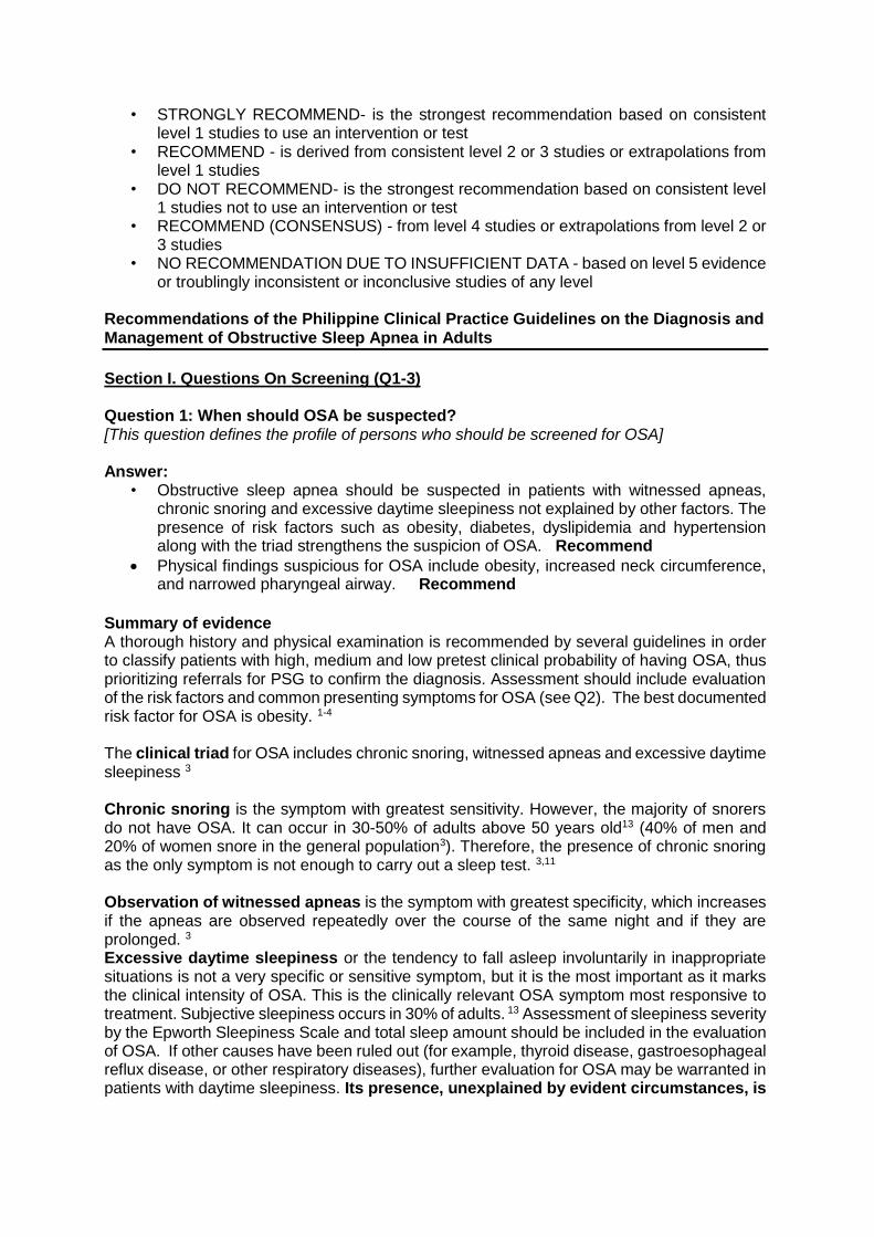
• Obstructive sleep apnea should be suspected in patients with witnessed apneas, chronic snoring and excessive daytime sleepiness not explained by other factors. The presence of risk factors such as obesity, diabetes, dyslipidemia and hypertension along with the triad strengthens the suspicion of OSA. Recommend
Physical findings suspicious for OSA include obesity, increased neck circumference, and narrowed pharyngeal airway. Recommend
Summary of evidence A thorough history and physical examination is recommended by several guidelines in order to classify patients with high, medium and low pretest clinical probability of having OSA, thus prioritizing referrals for PSG to confirm the diagnosis. Assessment should include evaluation of the risk factors and common presenting symptoms for OSA (see Q2). The best documented risk factor for OSA is obesity. 1-4 The clinical triad for OSA includes chronic snoring, witnessed apneas and excessive daytime sleepiness 3
Chronic snoring is the symptom with greatest sensitivity. However, the majority of snorers do not have OSA. It can occur in 30-50% of adults above 50 years old13 (40% of men and 20% of women snore in the general population3). Therefore, the presence of chronic snoring as the only symptom is not enough to carry out a sleep test. 3,11 Observation of witnessed apneas is the symptom with greatest specificity, which increases if the apneas are observed repeatedly over the course of the same night and if they are prolonged. 3
Excessive daytime sleepiness or the tendency to fall asleep involuntarily in inappropriate situations is not a very specific or sensitive symptom, but it is the most important as it marks the clinical intensity of OSA. This is the clinically relevant OSA symptom most responsive to treatment. Subjective sleepiness occurs in 30% of adults. 13 Assessment of sleepiness severity by the Epworth Sleepiness Scale and total sleep amount should be included in the evaluation of OSA. If other causes have been ruled out (for example, thyroid disease, gastroesophageal reflux disease, or other respiratory diseases), further evaluation for OSA may be warranted in patients with daytime sleepiness. Its presence, unexplained by evident circumstances, is
sufficient even in the absence of other symptoms or signs to carry out a sleep study for diagnosis. 2-4,11
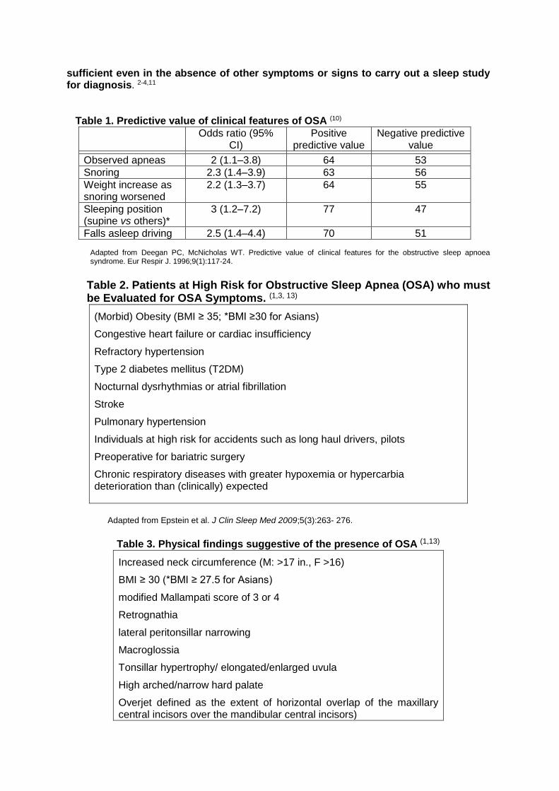
Table 1. Predictive value of clinical features of OSA (10)
Odds ratio (95% CI)
Positive predictive value
Negative predictive value
Observed apneas 2 (1.1–3.8) 64 53
Snoring 2.3 (1.4–3.9) 63 56
Weight increase as snoring worsened
2.2 (1.3–3.7) 64 55
Sleeping position (supine vs others)*
3 (1.2–7.2) 77 47
Falls asleep driving 2.5 (1.4–4.4) 70 51 Adapted from Deegan PC, McNicholas WT. Predictive value of clinical features for the obstructive sleep apnoea syndrome. Eur Respir J. 1996;9(1):117-24.
Table 2. Patients at High Risk for Obstructive Sleep Apnea (OSA) who must be Evaluated for OSA Symptoms. (1,3, 13)
(Morbid) Obesity (BMI ≥ 35; *BMI ≥30 for Asians)
Congestive heart failure or cardiac insufficiency
Refractory hypertension
Type 2 diabetes mellitus (T2DM)
Nocturnal dysrhythmias or atrial fibrillation
Stroke
Pulmonary hypertension
Individuals at high risk for accidents such as long haul drivers, pilots
Preoperative for bariatric surgery
Chronic respiratory diseases with greater hypoxemia or hypercarbia deterioration than (clinically) expected
Adapted from Epstein et al. J Clin Sleep Med 2009;5(3):263- 276.
Table 3. Physical findings suggestive of the presence of OSA (1,13)
Increased neck circumference (M: >17 in., F >16)
BMI ≥ 30 (*BMI ≥ 27.5 for Asians)
modified Mallampati score of 3 or 4
Retrognathia
lateral peritonsillar narrowing
Macroglossia
Tonsillar hypertrophy/ elongated/enlarged uvula
High arched/narrow hard palate
Overjet defined as the extent of horizontal overlap of the maxillary central incisors over the mandibular central incisors)
Nasal abnormalities such as septal deviation, nasal polyps, congestion or enlargement of turbinates
Adapted from Epstein et al. J Clin Sleep Med 2009;5(3):263- 276.
A local study of 100 adult Filipino subjects diagnosed to have OSA by PSG showed that majority were middle aged, obese, male, hypertensive, with an increased tonsillar grade and a family history of snoring8. Complaints included snoring, abnormal breathing pattern during sleep and waking up with dry mouth or sore throat, and need for daytime naps. Those presenting with an abnormal breathing pattern during sleep, obesity, smoking history, and enlarged tonsils have an increased likelihood of severe OSA. Epworth sleepiness scale score though was poorly correlated with the severity of OSA. 8
Another local study evaluated the relationship between neck circumference and body mass index (BMI), and polysomnographic parameters in 149 male patients seen at a sleep disorders laboratory suspected to have OSA12. For the OSA group, the mean neck circumference was 42.03 cm with a mean BMI of 29.14 while the mean neck circumference for the normal group was 39.05 cm with a mean BMI of 25.36. A significant difference was noted in both the neck circumference and BMI between the OSA group and the normal group (p<0.005). Neck circumference and BMI measurements were also correlated with increasing severity of sleep apnea in the OSA group. The >40 cm neck circumference among male adults with symptoms of OSA was 80% sensitive and 67% specific with a positive predictive value of 94%. References:
1. Epstein LJ; Kristo D; Strollo PJ; Friedman N; Malhotra A; Patil SP; Ramar K; Rogers R; Schwab RJ; Weaver EM; Weinstein MD. Clinical guideline for the evaluation, management and long-term care of obstructive sleep apnea in adults. J Clin Sleep Med 2009; 5 (3):263- 276.
2. Qaseem A, Dallas P, Owens DK, Starkey M, Holty JC, Shekelle P, et al. Diagnosis of Obstructive Sleep Apnea in Adults: A Clinical Practice Guideline From the American College of Physicians. Ann Intern Med. 2014;161:210-220.
3. Lloberes P et al. Diagnosis and treatment of sleep apnea-hypopnea syndrome. Arch Bronconeumol. 2011; 47(3):143-156.
4. Capote F, Masa JF, Jiménez A, Peces-Barba G, Amilibia J, Rubio R, Manifestaciones clínicas del SAHS. Métodos diagnósticos. Síndrome de resistencia aumentada de la vía aérea superior. Arch Bronconeumol. 2002; 38 (Supl 3):21-7.
5. Myers KA, Mrkobrada M, Simel DL. Does this patient have obstructive sleep apnea? The Rational Clinical Examination systematic review. JAMA 2013; 310:731.
6. McNicholas WT. Diagnosis of obstructive sleep apnea in adults. Proc Am Thorac Soc. 2008; 5:154-60. 7. Johns MW. Daytime sleepiness, snoring, and obstructive sleep apnea. The Epworth Sleepiness Scale.
Chest. 1993;103:30-6. 8. dela Trinidad MC, de los Reyes VS. 3-year review of patients diagnosed with obstructive sleep apnea in the
Lung Center of the Philippines. Philippine Journal of Chest Diseases. 2015.16(2)3-9. 9. Hessel NS, de Vries N. Diagnostic work-up of socially unacceptable snoring. II. Sleep endoscopy. Eur Arch
Otorhinolaryngol. 2002; 259 (3):158-61. 10. Deegan PC, McNicholas WT. Predictive value of clinical features for the obstructive sleep apnoea syndrome.
Eur Respir J. 1996; 9 (1):117-24. 11. Kushida CA; Littner MR; Morgenthaler T et al. Practice parameters for the indications for polysomnography
and related procedures: An update for 2005. SLEEP 2005; 28 (4):499-521. 12. Veloro LV, Sarte AA, Castañeda SS. Collar size as predictor of obstructive sleep apnea. Philippine Journal
of Otolaryngology Head and Neck Surgery. July 2008; 23 (2): 14-16. 13. WHO expert consultation. Appropriate body-mass index for Asian populations and its implications for policy
and intervention strategies. The Lancet, 2004; 157-163.
Question 2: When should we screen for OSA? [This question defines the settings or clinical situations when screening is recommended]
Answer: Screening for OSA should be done
1. During routine health maintenance evaluation
2. Routinely, among patients for pre-operative evaluation 3. In populations where OSA poses a public health hazard (e.g. Public utility
drivers, long haul drivers, pilots) Recommend As part of the initial sleep evaluation, and prior to objective testing, patients should receive education regarding diagnosis, diagnostic steps and procedure involved in any testing. Those patients with symptoms of OSA and deemed high risk for OSA should have the diagnosis confirmed and severity determined with polysomnography in an expedited manner in order to initiate treatment. Summary of Evidence: The AASM is the only guideline that directly addressed this question and gave consensus recommendations on the 3 settings at which physicians should screen for OSA1. 1. Evaluation of individuals with symptoms of OSA. A comprehensive sleep history in a
patient suspected of OSA should include an evaluation for snoring, witnessed apneas,
gasping/choking episodes, excessive sleepiness not explained by other factors, as well
as an assessment of sleepiness severity by the Epworth Sleepiness Scale, total sleep
amount, nocturia, morning headaches, sleep fragmentation/sleep maintenance insomnia,
and decreased concentration and memory. (Table 4) An evaluation of secondary
conditions that may occur as a result of OSA, including hypertension, stroke, myocardial
infarction, cor pulmonale, decreased daytime alertness, and motor vehicle accidents,
should also be obtained.
2. Evaluation of patients at high risk of OSA – These include individuals who are
(morbidly) obese; with congestive heart failure, atrial fibrillation, treatment refractory
hypertension, type 2 diabetes, stroke, nocturnal dysrhythmias, and pulmonary
hypertension; high-risk driving populations (such as commercial truck drivers or pilots),
and those being evaluated for bariatric surgery.1,2 (Table 2)
3. Routine health maintenance evaluation - Questions to be asked during a routine health
maintenance evaluation should include a history of snoring and daytime sleepiness and
an evaluation for the presence of obesity, retrognathia, or hypertension (Table 5). Positive
findings in this OSA screen should lead to a more comprehensive sleep history and
physical examination.
Table 4. OSA symptoms that should be evaluated during a comprehensive sleep evaluation 1,2, 3
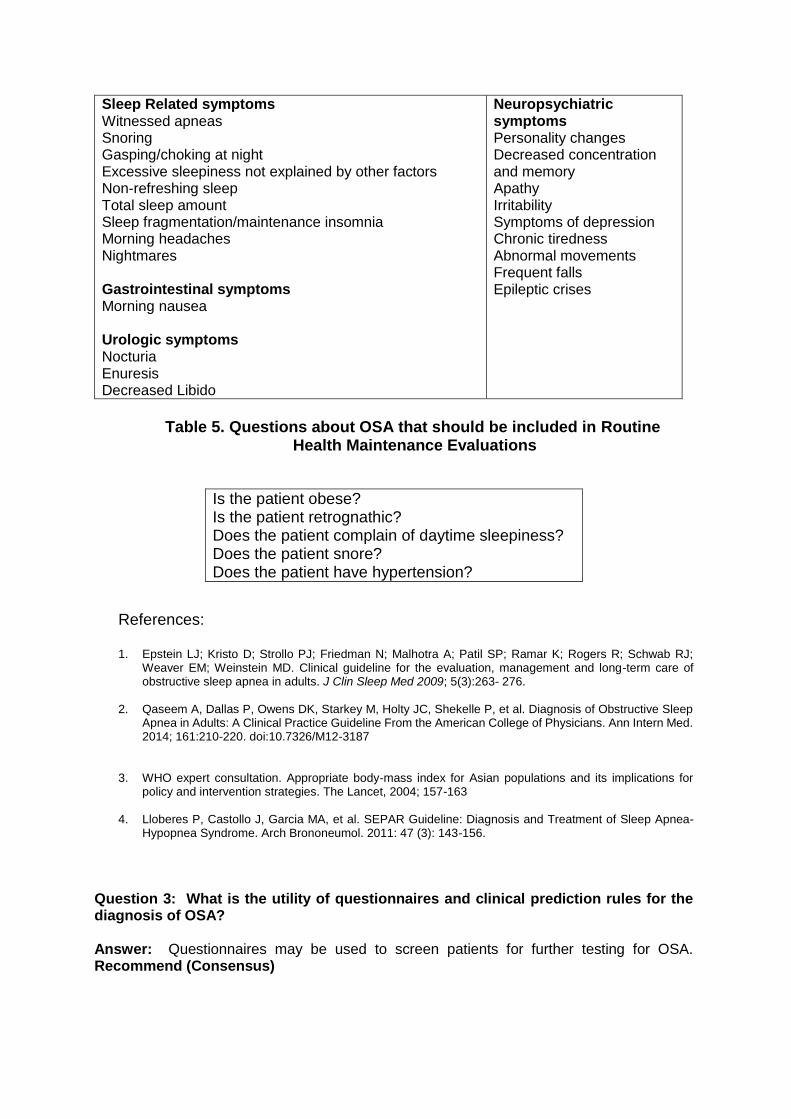
Sleep Related symptoms Witnessed apneas Snoring Gasping/choking at night Excessive sleepiness not explained by other factors Non-refreshing sleep Total sleep amount Sleep fragmentation/maintenance insomnia Morning headaches Nightmares Gastrointestinal symptoms Morning nausea Urologic symptoms Nocturia Enuresis Decreased Libido
Neuropsychiatric symptoms Personality changes Decreased concentration and memory Apathy Irritability Symptoms of depression Chronic tiredness Abnormal movements Frequent falls Epileptic crises
Table 5. Questions about OSA that should be included in Routine
Health Maintenance Evaluations
References: 1. Epstein LJ; Kristo D; Strollo PJ; Friedman N; Malhotra A; Patil SP; Ramar K; Rogers R; Schwab RJ;
Weaver EM; Weinstein MD. Clinical guideline for the evaluation, management and long-term care of obstructive sleep apnea in adults. J Clin Sleep Med 2009; 5(3):263- 276.
2. Qaseem A, Dallas P, Owens DK, Starkey M, Holty JC, Shekelle P, et al. Diagnosis of Obstructive Sleep Apnea in Adults: A Clinical Practice Guideline From the American College of Physicians. Ann Intern Med. 2014; 161:210-220. doi:10.7326/M12-3187
3. WHO expert consultation. Appropriate body-mass index for Asian populations and its implications for policy and intervention strategies. The Lancet, 2004; 157-163
4. Lloberes P, Castollo J, Garcia MA, et al. SEPAR Guideline: Diagnosis and Treatment of Sleep Apnea-
Hypopnea Syndrome. Arch Brononeumol. 2011: 47 (3): 143-156.
Question 3: What is the utility of questionnaires and clinical prediction rules for the diagnosis of OSA? Answer: Questionnaires may be used to screen patients for further testing for OSA. Recommend (Consensus)
Is the patient obese? Is the patient retrognathic? Does the patient complain of daytime sleepiness? Does the patient snore? Does the patient have hypertension?
No one questionnaire or physical finding can be used to diagnose OSA. Hence, aggregations of signs and symptoms using clinical questionnaires or prediction rules may be helpful in screening those suspected with OSA. For the most part, clinical questionnaires are more useful for ruling out OSA (in the presence of a low score) than for ruling in the diagnosis. They can be used to identify those with low-likelihood in whom PSG should be avoided or those with high likelihood of disease who will require full nocturnal polysomnography (PSG) for definitive diagnosis. Different questionnaires have different intents and uses:
The Berlin Questionnaire may be used in predicting risk for OSA
The STOP questionnaire and its extended version, the STOP-Bang may be used for OSA screening in surgical patients
The Epworth Sleepiness Scale (ESS) may be used for monitoring symptoms of excessive daytime sleepiness.
Summary of Evidence The gold standard for the diagnosis of OSA is laboratory polysomnography (PSG); however, due to the high cost and limited availability of PSG in our country, physicians should determine which patients would need further sleep evaluation during clinic visits. Most individual signs and symptoms have limited utility in determining the likelihood of OSA, and thus, no single clinical feature is sufficiently sensitive or specific to effectively rule in or rule out the diagnosis.1 These signs and symptoms have been aggregated into several questionnaires and clinical prediction rules to quickly identify patients at risk for OSA. Questionnaires The ACP Clinical Practice Guidelines evaluated a total of 47 studies comparing the accuracy of various questionnaires for the diagnosis of OSA using the PSG as the gold standard.2 The sensitivity and specificity of most questionnaires in these studies is probably overestimated compared to unselected community-based populations since most patients came from sleep centers or preoperative referral based populations.3
Low-quality evidence from 22 studies using the Epworth Sleepiness Scale (ESS), 5 on the STOP-BANG Questionnaire, 3 on the Multivariate Apnea Prediction Index, and 3 on the Pittsburgh Sleep Quality Index, showed that these questionnaires had low accuracy for diagnosis of OSA. Evidence was insufficient to determine the diagnostic accuracy of the other questionnaires.2 Included in this current summary are questionnaires with either the best performance as screening tools or the most frequently used in various clinical settings.
The Berlin questionnaire (Appendix E) consists of 10 items on snoring, non-restorative sleep, sleepiness while driving, apneas during sleep, hypertension, and body mass index. The questionnaire consists of 3 categories related to the risk of having sleep apnea. Patients can be classified into high risk (if 2 or more categories are positive) or low risk based on their responses to the individual items and their overall scores in the symptom categories. 4,5 It is well studied and has been used in different populations including the general population, the elderly, surgical patients, sleep clinic patients, those with kidney disease and cardiac patients 4-17. A large study on a Caucasian general population (N= 16,302) detected 518 subjects suspected to have OSA based on the questionnaire who underwent in-hospital PSG for confirmation. It showed that the questionnaire had low sensitivity of 37.2% with good specificity of 84% at a cutoff of AHI ≥ 5. This suggests that subjects without OSA among the general population are most likely to be true negatives18. A large study was also done among a general population of Koreans (N=1,305) showing that it had a sensitivity of 69% and a specificity of 83% at a cutoff of AHI ≥ 5 19. A Filipino version of the Berlin questionnaire (BQ)
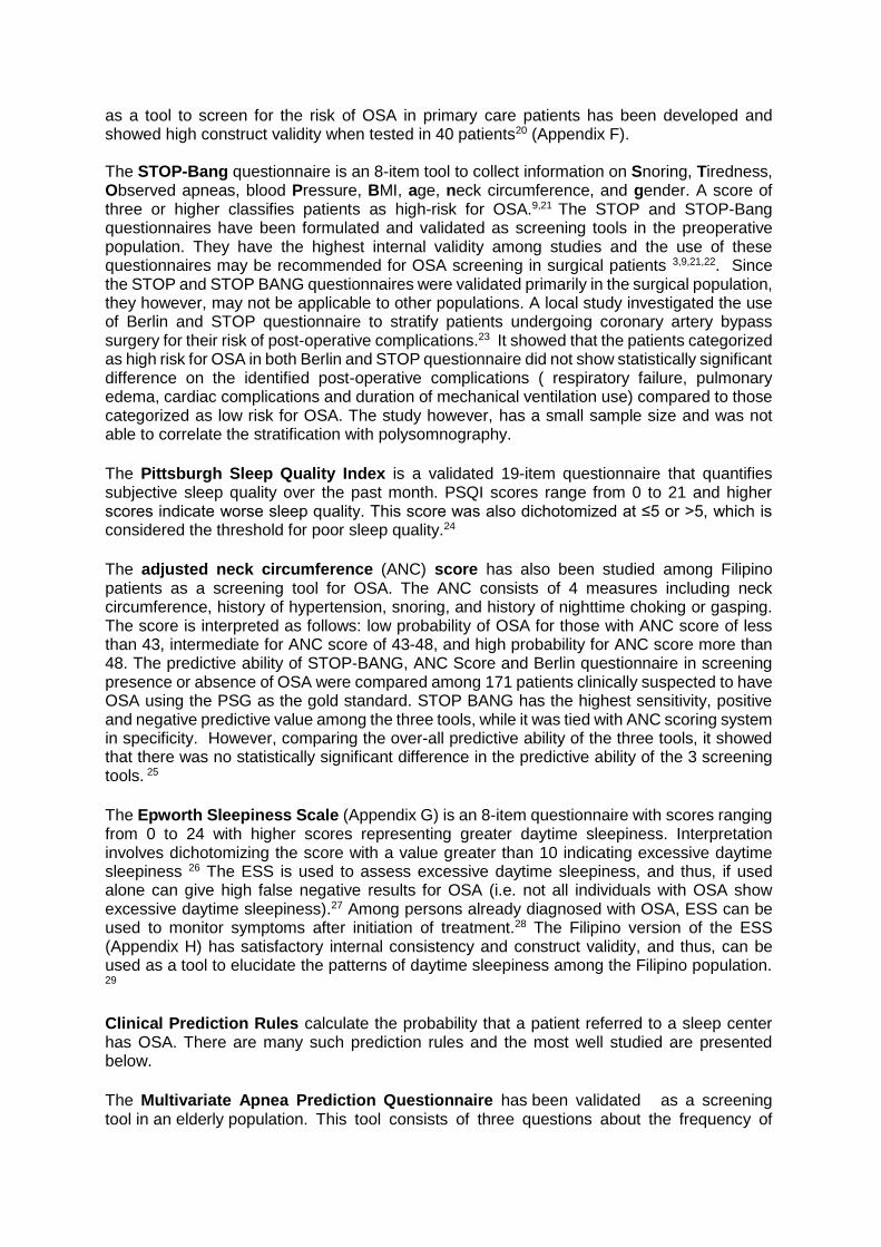
as a tool to screen for the risk of OSA in primary care patients has been developed and showed high construct validity when tested in 40 patients20 (Appendix F). The STOPBang questionnaire is an 8item tool to collect information on Snoring, Tiredness, Observed apneas, blood Pressure, BMI, age, neck circumference, and gender. A score of three or higher classifies patients as high-risk for OSA.9,21 The STOP and STOP-Bang questionnaires have been formulated and validated as screening tools in the preoperative population. They have the highest internal validity among studies and the use of these questionnaires may be recommended for OSA screening in surgical patients 3,9,21,22. Since the STOP and STOP BANG questionnaires were validated primarily in the surgical population, they however, may not be applicable to other populations. A local study investigated the use of Berlin and STOP questionnaire to stratify patients undergoing coronary artery bypass surgery for their risk of post-operative complications.23 It showed that the patients categorized as high risk for OSA in both Berlin and STOP questionnaire did not show statistically significant difference on the identified post-operative complications ( respiratory failure, pulmonary edema, cardiac complications and duration of mechanical ventilation use) compared to those categorized as low risk for OSA. The study however, has a small sample size and was not able to correlate the stratification with polysomnography.
The Pittsburgh Sleep Quality Index is a validated 19-item questionnaire that quantifies subjective sleep quality over the past month. PSQI scores range from 0 to 21 and higher scores indicate worse sleep quality. This score was also dichotomized at ≤5 or >5, which is considered the threshold for poor sleep quality.24
The adjusted neck circumference (ANC) score has also been studied among Filipino patients as a screening tool for OSA. The ANC consists of 4 measures including neck circumference, history of hypertension, snoring, and history of nighttime choking or gasping. The score is interpreted as follows: low probability of OSA for those with ANC score of less than 43, intermediate for ANC score of 43-48, and high probability for ANC score more than 48. The predictive ability of STOP-BANG, ANC Score and Berlin questionnaire in screening presence or absence of OSA were compared among 171 patients clinically suspected to have OSA using the PSG as the gold standard. STOP BANG has the highest sensitivity, positive and negative predictive value among the three tools, while it was tied with ANC scoring system in specificity. However, comparing the over-all predictive ability of the three tools, it showed that there was no statistically significant difference in the predictive ability of the 3 screening tools. 25
The Epworth Sleepiness Scale (Appendix G) is an 8-item questionnaire with scores ranging from 0 to 24 with higher scores representing greater daytime sleepiness. Interpretation involves dichotomizing the score with a value greater than 10 indicating excessive daytime sleepiness 26 The ESS is used to assess excessive daytime sleepiness, and thus, if used alone can give high false negative results for OSA (i.e. not all individuals with OSA show excessive daytime sleepiness).27 Among persons already diagnosed with OSA, ESS can be used to monitor symptoms after initiation of treatment.28 The Filipino version of the ESS (Appendix H) has satisfactory internal consistency and construct validity, and thus, can be used as a tool to elucidate the patterns of daytime sleepiness among the Filipino population. 29
Clinical Prediction Rules calculate the probability that a patient referred to a sleep center has OSA. There are many such prediction rules and the most well studied are presented below.
The Multivariate Apnea Prediction Questionnaire has been validated as a screening tool in an elderly population. This tool consists of three questions about the frequency of
symptoms of sleep apnea (snorting, gasping, loud snoring, and breathing stops, choking, or struggling for breath) during the past month. 30
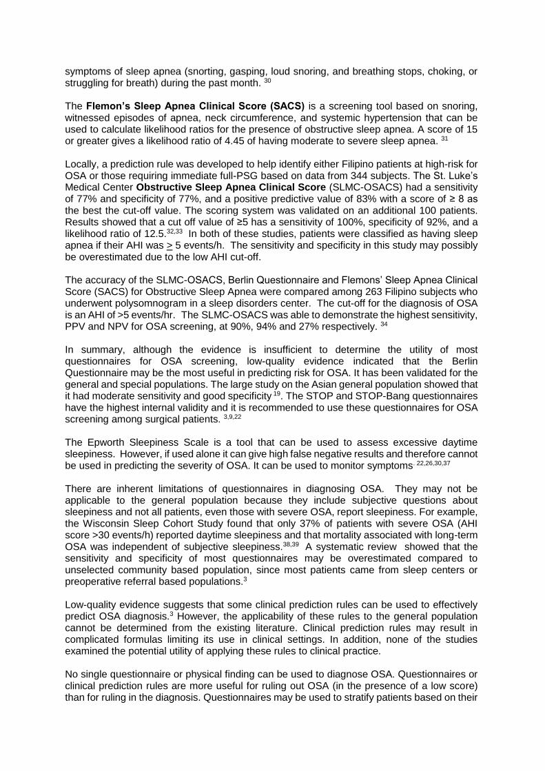
The Flemon’s Sleep Apnea Clinical Score (SACS) is a screening tool based on snoring, witnessed episodes of apnea, neck circumference, and systemic hypertension that can be used to calculate likelihood ratios for the presence of obstructive sleep apnea. A score of 15 or greater gives a likelihood ratio of 4.45 of having moderate to severe sleep apnea. 31 Locally, a prediction rule was developed to help identify either Filipino patients at high-risk for OSA or those requiring immediate full-PSG based on data from 344 subjects. The St. Luke’s Medical Center Obstructive Sleep Apnea Clinical Score (SLMC-OSACS) had a sensitivity of 77% and specificity of 77%, and a positive predictive value of 83% with a score of ≥ 8 as the best the cut-off value. The scoring system was validated on an additional 100 patients. Results showed that a cut off value of ≥5 has a sensitivity of 100%, specificity of 92%, and a likelihood ratio of 12.5.32,33 In both of these studies, patients were classified as having sleep apnea if their AHI was > 5 events/h. The sensitivity and specificity in this study may possibly be overestimated due to the low AHI cut-off. The accuracy of the SLMC-OSACS, Berlin Questionnaire and Flemons’ Sleep Apnea Clinical Score (SACS) for Obstructive Sleep Apnea were compared among 263 Filipino subjects who underwent polysomnogram in a sleep disorders center. The cut-off for the diagnosis of OSA is an AHI of >5 events/hr. The SLMC-OSACS was able to demonstrate the highest sensitivity, PPV and NPV for OSA screening, at 90%, 94% and 27% respectively. 34 In summary, although the evidence is insufficient to determine the utility of most questionnaires for OSA screening, low-quality evidence indicated that the Berlin Questionnaire may be the most useful in predicting risk for OSA. It has been validated for the general and special populations. The large study on the Asian general population showed that it had moderate sensitivity and good specificity 19. The STOP and STOP-Bang questionnaires have the highest internal validity and it is recommended to use these questionnaires for OSA screening among surgical patients. 3,9,22
The Epworth Sleepiness Scale is a tool that can be used to assess excessive daytime sleepiness. However, if used alone it can give high false negative results and therefore cannot be used in predicting the severity of OSA. It can be used to monitor symptoms. 22,26,30,37 There are inherent limitations of questionnaires in diagnosing OSA. They may not be applicable to the general population because they include subjective questions about sleepiness and not all patients, even those with severe OSA, report sleepiness. For example, the Wisconsin Sleep Cohort Study found that only 37% of patients with severe OSA (AHI score >30 events/h) reported daytime sleepiness and that mortality associated with long-term OSA was independent of subjective sleepiness.38,39 A systematic review showed that the sensitivity and specificity of most questionnaires may be overestimated compared to unselected community based population, since most patients came from sleep centers or preoperative referral based populations.3
Low-quality evidence suggests that some clinical prediction rules can be used to effectively predict OSA diagnosis.3 However, the applicability of these rules to the general population cannot be determined from the existing literature. Clinical prediction rules may result in complicated formulas limiting its use in clinical settings. In addition, none of the studies examined the potential utility of applying these rules to clinical practice. No single questionnaire or physical finding can be used to diagnose OSA. Questionnaires or clinical prediction rules are more useful for ruling out OSA (in the presence of a low score) than for ruling in the diagnosis. Questionnaires may be used to stratify patients based on their
clinical symptoms, their physical examinations, and their risk factors, in order to ascertain patients at high risk and in urgent need for PSG and/or further treatment and patients at low risk who may not need PSG. References:
1. Myers KA, Mrkobrada M, Simel DL. Does this patient have obstructive sleep apnea? The Rational Clinical Examination systematic review. JAMA 2013; 310:731.
2. Qaseem A, Dallas P, Owens DK, Starkey M, Holty JC, Shekelle P, et al. Diagnosis of Obstructive Sleep Apnea in Adults: A Clinical Practice Guideline From the American College of Physicians. Ann Intern Med. 2014; 161:210-220. doi:10.7326/M12-3187.
3. Abrishami A, Khajehdehi A, Chung F. A systematic review of screening questionnaires for obstructive sleep apnea. Can J Anaesth 2010; 57:423.
4. Netzer NC, Stoohs RA, Netzer CM, et al. Using the Berlin Questionnaire to identify patients at risk for the sleep apnea syndrome. Ann Intern Med 1999; 131:485–491.
5. Netzer NC, Hoegel, JJ, Loube D, Netzer CM, Hay B, Alvarez-Sala R, Strohl KP. Prevalence of Symptoms and Risk of Sleep Apnea in Primary Care. Chest 2003; 124:1406–1414.
6. Koyama RG, Esteves AM, Oliveira e Silva L, Lira FS, Bittencourt LR, Tufik S, et al. Prevalence of and risk factors for obstructive sleep apnea syndrome in Brazilian railroad workers. Sleep Med. 2012; 13:1028-32. [PMID: 22841037].
7. Sharma SK, Vasudev C, Sinha S, Banga A, Pandey RM, Handa KK. Validation of the modified Berlin Questionnaire to identify patients at risk for the obstructive sleep apnoea syndrome. Indian J Med Res. 2006; 124:281-90. [PMID: 17085831].
8. Sforza E, Chouchou F, Pichot V, Herrmann F, Barthe ́le ́my JC, Roche F. Is the Berlin Questionnaire a useful tool to diagnose obstructive sleep apnea in the elderly? Sleep Med. 2011; 12:142-6. [PMID: 21227749].
9. Chung F, Yegneswaran B, Liao P, Chung SA, Vairavanathan S, Islam S, et al. Validation of the Berlin Questionnaire and American Society of Anesthesiologists checklist as screening tools for obstructive sleep apnea in surgical patients. Anesthesiology. 2008; 108:822-30. [PMID: 18431117].
10. Vaz AP, Drummond M, Mota PC, Severo M, Almeida J, Winck JC. Translation of Berlin Questionnaire to Portuguese language and its application in OSA identification in a sleep disordered breathing clinic. Rev Port Pneumol. 2011; 17:59-65. [PMID: 21477567].
11. Nicholl DD, Ahmed SB, Loewen AH, Hemmelgarn BR, Sola DY, Beecroft JM, et al. Clinical presentation of obstructive sleep apnea in patients with chronic kidney disease. J Clin Sleep Med. 2012; 8:381-7. [PMID: 22893768].
12. Drager LF, Genta PR, Pedrosa RP, Nerbass FB, Gonzaga CC, Krieger EM, et al. Characteristics and predictors of obstructive sleep apnea in patients with systemic hypertension. Am J Cardiol. 2010; 105:1135-9. [PMID: 20381666].
13. Laporta R, Anandam A, El-Solh AA. Screening for obstructive sleep apnea in veterans with ischemic heart disease using a computer-based clinical decision- support system. Clin Res Cardiol. 2012; 101:737-44. [PMID: 22476823].
14. Sert Kuniyoshi FH, Zellmer MR, Calvin AD, Lopez-Jimenez F, Albuquer- que FN, van der Walt C, et al. Diagnostic accuracy of the Berlin Questionnaire in detecting sleep-disordered breathing in patients with a recent myocardial infarction. Chest. 2011; 140:1192-7. [PMID: 21596794].
15. Thurtell MJ, Bruce BB, Rye DB, Newman NJ, Biousse V. The Berlin Questionnaire screens for obstructive sleep apnea in idiopathic intracranial hypertension. J Neuroophthalmol. 2011; 31:316-9.
16. Danzi-Soares NJ, Genta PR, Nerbass FB, Pedrosa RP, Soares FS, Ce ́sar LA, et al. Obstructive sleep apnea is common among patients referred for coronary artery bypass grafting and can be diagnosed by portable monitoring. Coron Artery Dis. 2012; 23:31-8. [PMID: 22107804].
17. Martinez D, da Silva RP, Klein C, Fiori CZ, Massierer D, Cassol CM, et al. High risk for sleep apnea in the Berlin Questionnaire and coronary artery disease. Sleep Breath. 2012; 16:89-94.
18. Hrubos-Strøm H, Randby A, Namtvedt SK, Kristiansen HA, Einvik G, Benth J, et al. A Norwegian population-based study on the risk and prevalence of obstructive sleep apnea. The Akershus Sleep Apnea Project (ASAP). J Sleep Res.2011; 20:162-70. [PMID: 20561172].
19. Kang K, Park KS, Kim JE, Kim SW, Kim YT, Kim JS, Lee HW. Usefulness of the Berlin Questionnaire to identify patients at high risk for obstructive sleep apnea: a population-based door-to-door study. Sleep Breath. 2013 May; 17(2):803-10. doi: 10.1007/s11325-012-0767-2. Epub 2012 Sep 29.
20. Jorge MC, Nomorosa KMP, David-Ona DIA.Validation of the Filipino version of the Berlin questionnaire to identify population at risk of sleep apnea syndrome. Acta Medica Philippina. July 2012; Vol. 46 (3):p. 59-61.
21. Chung F, Yegneswaran B, Liao P, Chung SA, Vairavanathan S, Islam S, et al. STOP questionnaire: a tool to screen patients for obstructive sleep apnea. Anesthesiology. 2008; 108:812-21.
22. Tanaka S, Shima M. Assessment of screening tests for sleep apnea syndrome in the workplace. J Occup Health. 2010; 52:99-105. [PMID: 20110621].
23. Banate MY. The Use of Berlin Questionnaire Versus Stop Questionnaire As Screening Tool Among Filipino Patients Undergoing Coronary Artery Bypass Surgery At Risk For Obstructive Sleep Apnea (unpublished).
24. Buysse DJ; Yu L; Moul DE; Germain A; Stover A; Dodds NE; Johnston KL; Shablesky-Cade MA; Pilkonis PA. Development and validation of patient-reported outcome measures for sleep disturbance and sleep-related impairments. SLEEP 2010; 33(6):781-792.
25. Armas CM and Moral PGL. Adjusted Neck Circumference Score, Stop-Bang and Berlin Questionnaire as Screening Tools for Obstructive Sleep Apnea. Philippine Journal of Chest Diseases.2015;.16(2); 27-3.
26. Johns MW. A new method for measuring daytime sleepiness: the Epworth Sleepiness scale. Sleep 1991; 14:540-3.
27. Knutson KL, Zhao X, Mattingly M, Galli G, Cizza G. Predictors of sleep-disordered breathing in obese adults who are chronic short sleepers. Sleep Med. 2012; 13:484-9. [PMID: 22326831]
28. Martinez D, Breitenbach TC, Lumertz MS, Alcaˆntara DL, da Rocha NS, Cassol CM, et al. Repeating administration of Epworth Sleepiness Scale is clinically useful. Sleep Breath. 2011; 15:763-73.
29. Albay AB, Sison CM, Jorge MC II. Validation of the Filipino Version of the Epworth Sleepiness Scale. Chest. 2007; 132(4_MeetingAbstracts):649a. doi:10.1378/chest.132.4_MeetingAbstracts.649a
30. Morales CR, Hurley S, Wick LC, Staley B, Pack FM, Gooneratne NS, et al. In-home, self-assembled sleep studies are useful in diagnosing sleep apnea in the elderly. Sleep. 2012; 35:1491-501.
31. Flemons WW, Whitelaw WA, Brant R, Remmers JE. Likelihood ratios for a sleep apnea clinical prediction rule. Am J Respir Crit Care Med 1994; 150:1279-85.
32. Cua IHY, Codamos LJ, Gappi MAS. St. Luke's Medical Center-Obstructive Sleep Apnea Clinical Scoring System. PJIM vol.41 issue 4 July-aug 2003 pp 169-174.
33. Cua IHY, Codamos LJ, Gappi MAS. Validation of the St. Luke's Medical Center-Obstructive Sleep Apnea Clinical Scoring System. PJIM vol.41 issue 4 july-aug 2003 pp 175-178.
34. Noscal K., Palec J., Lopez, A., Esposo E., Gappi MAS. Comparison of the different screening tools for Obstructive Sleep Apnea among Filipino patients in St. Luke’s Medical Center. Presented in 2013 PCP 13th Annual Convention (unpublished).
35. Hoffstein V, Szalai J. Predictive value of clinical features in diagnosing obstructive sleep apnea. Sleep 1993; 16:118–122.
36. dela Trinidad MCL, delos Reyes VS. 3-year review of Patients Diagnosed with Obstructive Sleep Apnea in the Lung Center of the Philippines. PJCD.2015; 6(2):3-9.
37. Patt BT, Jarjoura D, Lambert L, Roy S, Gordillo G, Schlanger R, et al. Prevalence of obstructive sleep apnea in patients with chronic wounds. J Clin Sleep Med. 2010; 6:541-4. [PMID: 21206743].
38. Young T, Shahar E, Nieto FJ, et al. Predictors of sleep-disordered breathing in community-dwelling adults: the Sleep Heart Health Study. Arch Intern Med. 2002; 162(8):893-900.
39. Albuquerque FN, Calvin AD, Sert Kuniyoshi FH, Konecny T, Lopez- Jimenez F, Pressman GS, et al. Sleep-disordered breathing and excessive daytime sleepiness in patients with atrial fibrillation. Chest. 2012; 141:967-73. [PMID: 21903736].
Section II. QUESTIONS ON DIAGNOSIS (Q 4-7)
Question 4: What is the gold standard for the diagnosis of OSA?
Answer: Attended, in-laboratory polysomnogram is the gold standard to diagnose OSA.
Strongly Recommended
Summary of Evidence The presence of OSA must be confirmed and its severity determined before initiating treatment in order to identify those patients at risk of developing the complications of sleep apnea, to guide selection of appropriate treatment and to provide a baseline to establish the effectiveness of subsequent treatment1. The diagnosis of OSA cannot be made based alone on compatible clinical signs or symptoms identified during sleep oriented history and physical examination. The diagnosis can only be made with certainty using polysomnography. 2 Sleep testing or polysomnography is a test to evaluate various types of sleep disorders and not just OSA. Ideally, PSG should be done either at night (overnight) or during the subject’s usual sleep schedule, with a recording of no less than 6.5 hours, including at least 3 hours of sleep. There may be less than the ideal hours of recording for as long as it is interpreted by a sleep specialist who can make the appropriate clinical correlation. Polysomnography generally includes monitoring of the following: electroencephalogram (EEG), electro-oculogram (EOG), chin electromyogram, airflow, oxygen saturation, respiratory effort, electrocardiogram (ECG), and limb movements.3 An attended study
requires the constant presence of a trained individual who can monitor for technical adequacy, patient compliance, and relevant patient behavior.4 On the other hand, a portable monitor (PM) consists of at least 2 respiratory channels such nasal airflow, oxygen saturation and respiratory effort but generally do not have an EEG, EOG or EMG. Portable monitors cannot reliably distinguish between awake and asleep states, and cannot measure a type of obstructive event found in OSA called respiratory event related arousals (see also Question 7).
Full-night, attended, in-laboratory PSG is thus, considered the reference standard diagnostic test for OSA (consistent recommendation, high quality of evidence) 5,6,7,8 The PSG (compared to PM) provides the most comprehensive information needed to make the diagnosis of OSA to reliably distinguish between the various sleep stages; to compute for the frequency of respiratory events during sleep (the so-called AHI or apnea-hypopnea index); to assess the quality and continuity of sleep; and also to rule out the presence of other sleep disorders.
References:
1. American Academy of Sleep Medicine. International classification of sleep disorders, 2nd edition: diagnostic and coding manual. Westchester, Il: American Academy of Sleep Medicine; 2005.
2. Kushida CA, Morgenthaler TI, Littner MR, et al. Practice parameters for the treatment of snoring and Obstructive Sleep Apnea with oral appliances: an update for 2005. Sleep 2006; 29:240-3.
3. Iber C, Ancoli-Israel S, Chesson AL, Quan SF. The AASM manual for the scoring of sleep and associated events: rules, terminology and technical specifications. Westchester, IL: American Academy of Sleep Medicine; 2007.
4. Kushida CA, Littner MR, Morgenthaler T, et al. Practice parameters for the indications for polysomnography and related procedures: an update for 2005. Sleep 2005; 28:499-521.
5. Comparative Effectiveness review no 32. AHRQ publication no 11-m EHC052 – EF (Prepared by Tufts Evidence –based Practice Center under contract 290-2007-100551) Rockville MD. Agency for Healthcare Research and Quality, 201.
6. Epstein LJ, Kristo D, Strollo PJ Jr et al. Clinical Guideline for the Evaluation, Management, Long- Term Care of Obstructive Sleep Apnea in Adults. J Clin Sleep Medicine 2009, 5:263-276.
7. Qaseem, Dallas P, Owens D, et al. Diagnosis of Obstructive Sleep Apnea in Adults: A Clinical Practice Guideline From The American College of Physicians. Ann Intern Med 2014; 161: 210-220.
8. Llobere P, Castollo J, Garcia MA, et al SEPAR Guideline: Diagnosis and Treatment of Sleep Apnea-Hypopnea Syndrome Arch. Brononeumol. 2011 47 (3):143-156.
Question 5: Can portable monitors or other diagnostic tests be used as an alternative to PSG in the diagnosis of OSA? Answer: The use of Portable Monitors (at least type 3) is RECOMMENDED as an
alternative to Polysomnography for diagnostic testing in patients suspected of OSA provided
all of the following conditions are met:
• High risk for moderate to severe OSA • Do not have serious co-morbidities • Other sleep disorders are not a consideration, and • With a prior comprehensive sleep evaluation by a sleep specialist.
The following tools are NOT RECOMMENDED to diagnose OSA: • Type 4 Portable Monitors • Overnight oximetry • Auto-titrating Positive Airway Pressure (APAP) • Multiple Sleep Latency Testing (MSLT), and • Actigraphy
Summary of Evidence:
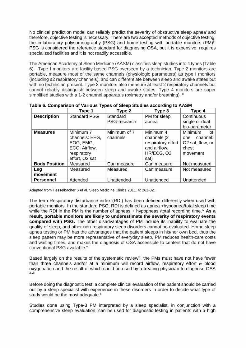
No clinical prediction model can reliably predict the severity of obstructive sleep apneai and therefore, objective testing is necessary. There are two accepted methods of objective testing; the in-laboratory polysomnography (PSG) and home testing with portable monitors (PM)ii. PSG is considered the reference standard for diagnosing OSA, but it is expensive, requires specialized facilities and it is not readily accessible. The American Academy of Sleep Medicine (AASM) classifies sleep studies into 4 types (Table 6). Type I monitors are facility-based PSG overseen by a technician. Type 2 monitors are portable, measure most of the same channels (physiologic parameters) as type I monitors (including ≥2 respiratory channels), and can differentiate between sleep and awake states but with no technician present. Type 3 monitors also measure at least 2 respiratory channels but cannot reliably distinguish between sleep and awake states. Type 4 monitors are super simplified studies with a 1-2 channel apparatus (oximetry and/or breathing). iii Table 6. Comparison of Various Types of Sleep Studies according to AASM
Type 1 Type 2 Type 3 Type 4
Description Standard PSG Standard PSG-research
PM for sleep apnea
Continuous single or dual bio-parameter
Measures Minimum 7 channels: EEG, EOG, EMG, ECG, Airflow, respiratory effort, O2 sat
Minimum of 7 channels
Minimum 4 channels (2 respiratory effort and airflow, HR/ECG, O2 sat)
Minimum of one channel: O2 sat, flow, or chest movement
Body Position Measured Can measure Can measure Not measured
Leg movement
Measured Measured Can measure Not measured
Personnel Attended Unattended Unattended Unattended
Adapted from Hesselbacher S et al. Sleep Medicine Clinics 2011. 6: 261-82.
The term Respiratory disturbance index (RDI) has been defined differently when used with portable monitors. In the standard PSG, RDI is defined as apnea +hypopnea/total sleep time while the RDI in the PM is the number of apneas + hypopneas /total recording time.iv As a result, portable monitors are likely to underestimate the severity of respiratory events compared with PSG. The other disadvantages of PM include its inability to evaluate the quality of sleep, and other non-respiratory sleep disorders cannot be evaluated. Home sleep apnea testing or PM has the advantages that the patient sleeps in his/her own bed, thus the sleep pattern may be more representative of everyday sleep. PM reduces health-care costs and waiting times, and makes the diagnosis of OSA accessible to centers that do not have conventional PSG available.v Based largely on the results of the systematic reviewvi, the PMs must have not have fewer than three channels and/or at a minimum will record airflow, respiratory effort & blood oxygenation and the result of which could be used by a treating physician to diagnose OSA 2,vii
Before doing the diagnostic test, a complete clinical evaluation of the patient should be carried out by a sleep specialist with experience in these disorders in order to decide what type of study would be the most adequate.5 Studies done using Type-3 PM interpreted by a sleep specialist, in conjunction with a comprehensive sleep evaluation, can be used for diagnostic testing in patients with a high
pre-test probability for moderate to severe OSA 5,8,9 who do not have comorbid cardiopulmonary or neuromuscular disorders, or in whom other sleep disorders are not a consideration 2,7,9,10 PM testing may also be used for the diagnosis of OSA in patients for whom in-laboratory PSG is not possible due to immobility, safety or critical illness and to monitor response to non-CPAP therapies (Consensus).9
The Australasian Sleep Association & Thoracic Society of Australia and New Zealand (ASA/TSANZ) states that “…that if portable, limited channel sleep studies are to be used, this should only be under the supervision of an accredited sleep physician who is familiar with the strengths and weaknesses of these types of studies and who is knowledgeable about the specific device to be used.11,12
The utility of PM as a diagnostic test in those cases with low probability of OSA is not validated and thus its use in this group of patients is uncertain.5 Regarding the usefulness of PM for the assessment in patients with co-morbid medical condition, the ASSM 2009, SEPAR 2010, CTS 2011 and the ACP 2014 shared the same statement that it is not recommended to be used for diagnosing OSA patients with serious medical condition such as COPD, CHF, or neurologic disorders since this group of patients were often excluded in most studies, and thus its utility is unknown. 2,5 This recommendation is however based only on moderate quality evidence.7,13 Type 4 Portable Monitors for the diagnosis of OSA: The ACP, SEPAR and the CTS, consider the limitation of the super-simplified system (type 4 monitors) to distinguish between central and obstructive apneas. There are no validation studies recommending the use of these type 4 monitors (weak recommendation, low quality of evidence). Oximetry for the diagnosis of OSA: Nocturnal oximetry can demonstrate the presence of apnea or hypopnea but it neither distinguishes the central obstructive disorders nor does it detect events without desaturation, thus the SEPAR does not recommend the use of overnight oximetry as a diagnostic method to diagnose OSA.5
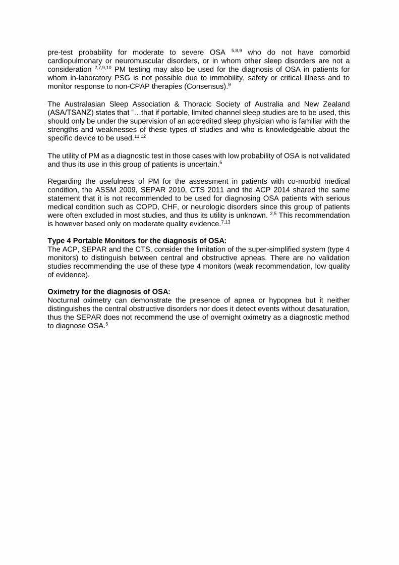
Figure 1. Decision Tree for Portable Monitoring. Flow chart depicting recommended pathway of patients Considered for Portable Monitoring. Adapted from JCSM Journal of Clinical Sleep Medicine Vol 3, no.7, 2007
Multiple Sleep Latency Testing (MSLT), Actigraphy and Auto-titrating Positive Airway Pressure (APAP) for the diagnosis of OSA. The AASM 2009 issued these statements regarding the use of these modalities for the diagnosis of OSA. (1) “The MSLT is not routinely indicated in the initial evaluation and diagnosis of OSA or in an assessment of change following treatment with nasal CPAP. However, if excessive sleepiness continues despite optimal treatment, the patient may require an evaluation for possible narcolepsy, including the MSLT. (2) Actigraphy alone is not indicated for the routine diagnosis of OSA but may be useful adjunct to PMs when determining the rest-activity pattern during the testing period (option). (3) Autotitrating positive airway pressure (APAP) is not recommended to diagnose OSA”. The ACP, SEPAR and the CTS did not address these issues. Automated Sleep Scoring: There is a rapidly growing body of literature supporting various schemes for the automated scoring of sleep and associated events14 There are a number of conflicting views about the use of automated scoring 9,15,16 PM devices must allow for the display of raw data for manual scoring or editing of automated scoring by specialists with expertise in respiratory sleep disorders and PSG with automatic analysis is not reliable. (Consistent recommendation, high quality of evidence).17 Summary: Unobserved (at home) registers of at least type-3 Portable monitors in conjunction with a comprehensive sleep evaluation by a sleep specialist, could be used as an alternative to PSG for diagnostic testing in patients with high probability of having moderate to severe OSA and those who do not have serious comorbidities.
MSLT and actigraphy are not routinely included in the initial evaluation and diagnosis of OSA. Autotitrating positive airway pressure (APAP) is not recommended to diagnose OSA. Overnight oximetry is not a recommended method for diagnosing OSA but it is useful if utilized within the appropriate clinical context. The tracing generated by the digital equipment should be reviewed and/or analyzed manually by a sleep specialist since automated analysis is not reliable.
References:
1. Kushida CA, Littner MR, Morgenthaler T, et al. Practice parameters for the indications for polysomnography and related procedures: an update 2005. Sleep 2005;28:499-521
2. Epstein E, Kristo D, Strollo P, Clincal Guideline for the Evaluation, Management and Long-Term Care of Obstructive Apnea in Adults, Journal of Clinical Sleep Medicine, Vol 5, No 3, 2009
3. Hesselbacher, S, Mattewal A, Hirshkowitz M, Sharafkhaneh A, Classification, technical specifications, and types of home sleep testing devices for sleep-disordered breathing. Sleep Medicine Clinics 2011, 6: 261-82
4. Trikalinos TA, Lau J. Obstructive Sleep Apnea-Hypopnea Syndrome: modeling different strategies. Rockville, MD: AHRQ Technology Assessment Program; US Dept Health Human Services; Agency for Healthcare Research and Quality, 2007.
5. Lloberes P, Castollo J, Garcia MA, et al. SEPAR Guideline: Diagnosis and Treatment of Sleep Apnea-Hypopnea Syndrome. Arch Brononeumol. 2011: 47 (3): 143-156
6. Trikalinos TA, Ip S, Raman G, et al. Technology assessment: Home diagnosis of obstructive sleep apnea hypopnea syndrome. Bethesda: Agency for Healthcare Research and Quality, Department of Health and Human Services, 2007.
7. John Fleetham MD, Najib Ayas MD, Douglas Bradley MD, et al. Canadian Thoracic Society 2011 guideline update: Diagnosis and treatment of sleep disordered breathing. Can Respir J 2011; 18 (1): 25-47.
8. Centers for Medicare & Medicaid Services. Decision Memo for Continuous Positive Airway Pressure (CPAP) Therapy for Obstructive Sleep Apnea (OSA) (CAG-00093R2). March 13, 2008.
9. Collop NA, Anderson WM, Boehlecke B, et al. Clinical guidelines for the use of unattended portable monitors in the diagnosis of obstructive sleep apnea in adult patient. Portable Monitoring Task Force of the American Academy of Sleep Medicine. J. Clin Sleep Med 2007; 3: 737-47
10. Timothy I. Morgenthaler, American Academy of Sleep Medicine Response to the ACP Clinical Practice Guideline for the Diagnosis of Obstructive Sleep Apnea in Adults September 2014
11. Hensley MJ, Hillman DR, McEvoy RD (Chair), Neill AM, Solin P, Teichtahl H, Thompson BR, Tolhurst S, Thornton AT, Worsnop CJ. Guidelines For Sleep Studies In Adults. Prepared for the Australasian Sleep Association & Thoracic Society of Australia and New Zealand, October 2005
12. ASTA/ASA Commentary on AASM Manual for the Scoring of Sleep and Associated Events Version 1.7 December 2010
13. Qaseem A, Dallas P, Owens D, et al. Diagnosis of Obstructive Sleep Apnea in Adults: A Clinical Practice Guideline from The American College of Physicians. Ann Intern Med 2014; 161: 210-220
14. Mulgrew AT, Fox N, Ayas NT, Ryan CF. Diagnosis and initial management of obstructive sleep apnea without polysomnography: A randomized validation study. Ann Intern Med 2007;146:157-66.
15. Antic NA, Buchan C, Esterman A, et al. A randomized controlled trial of nurse-led care for symptomatic moderate-severe obstructive sleep apnea. Am J Respir Crit Care Med 2009;179:501-8.
16. Penzel T, Hirshkowitz M, Harsh J, et al. Digital analysis and technical specifications. J Clin Sleep Med 2007; 3:109–120.
17. Svetnik V, Ma J, Soper KA, Doran S, Renger JJ, Deacon S, Koblan KS. Evaluation of Automated and Semi-Automated Scoring of Polysomnographic Recordings from a Clinical Trial Using Zolpidem in the Treatment of Insomnia. Sleep. 2007; 30(11): 1562–74.
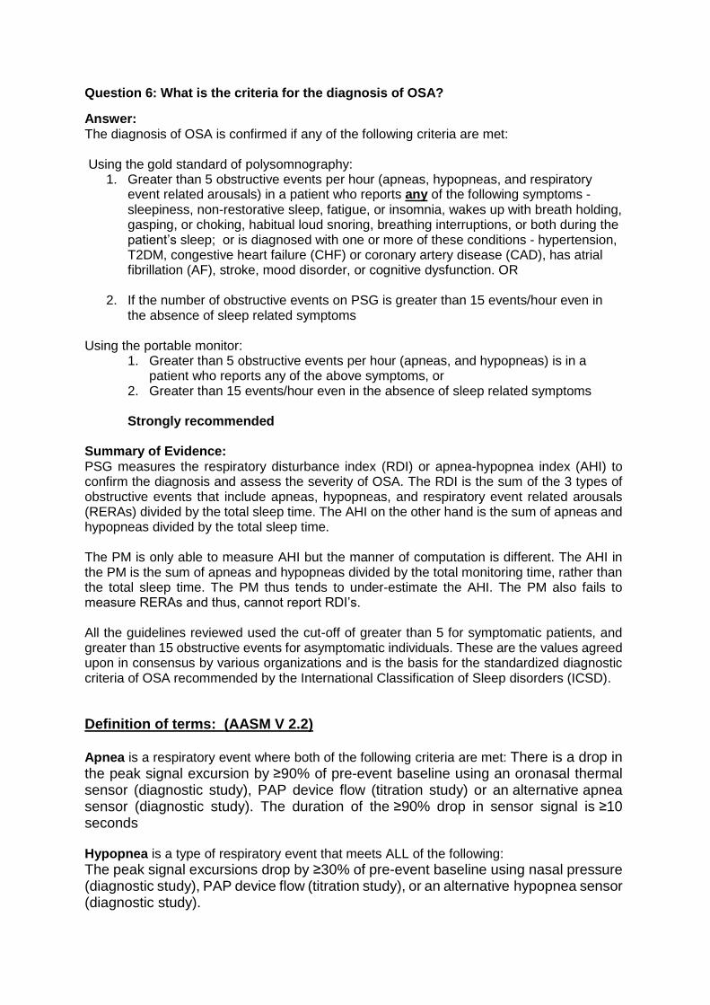
Question 6: What is the criteria for the diagnosis of OSA?
Answer: The diagnosis of OSA is confirmed if any of the following criteria are met: Using the gold standard of polysomnography:
1. Greater than 5 obstructive events per hour (apneas, hypopneas, and respiratory event related arousals) in a patient who reports any of the following symptoms - sleepiness, non-restorative sleep, fatigue, or insomnia, wakes up with breath holding, gasping, or choking, habitual loud snoring, breathing interruptions, or both during the patient’s sleep; or is diagnosed with one or more of these conditions - hypertension, T2DM, congestive heart failure (CHF) or coronary artery disease (CAD), has atrial fibrillation (AF), stroke, mood disorder, or cognitive dysfunction. OR
2. If the number of obstructive events on PSG is greater than 15 events/hour even in the absence of sleep related symptoms
Using the portable monitor: 1. Greater than 5 obstructive events per hour (apneas, and hypopneas) is in a
patient who reports any of the above symptoms, or 2. Greater than 15 events/hour even in the absence of sleep related symptoms
Strongly recommended
Summary of Evidence: PSG measures the respiratory disturbance index (RDI) or apnea-hypopnea index (AHI) to confirm the diagnosis and assess the severity of OSA. The RDI is the sum of the 3 types of obstructive events that include apneas, hypopneas, and respiratory event related arousals (RERAs) divided by the total sleep time. The AHI on the other hand is the sum of apneas and hypopneas divided by the total sleep time. The PM is only able to measure AHI but the manner of computation is different. The AHI in the PM is the sum of apneas and hypopneas divided by the total monitoring time, rather than the total sleep time. The PM thus tends to under-estimate the AHI. The PM also fails to measure RERAs and thus, cannot report RDI’s. All the guidelines reviewed used the cut-off of greater than 5 for symptomatic patients, and greater than 15 obstructive events for asymptomatic individuals. These are the values agreed upon in consensus by various organizations and is the basis for the standardized diagnostic criteria of OSA recommended by the International Classification of Sleep disorders (ICSD).
Definition of terms: (AASM V 2.2) Apnea is a respiratory event where both of the following criteria are met: There is a drop in the peak signal excursion by ≥90% of pre-event baseline using an oronasal thermal sensor (diagnostic study), PAP device flow (titration study) or an alternative apnea sensor (diagnostic study). The duration of the ≥90% drop in sensor signal is ≥10 seconds Hypopnea is a type of respiratory event that meets ALL of the following:
The peak signal excursions drop by ≥30% of pre-event baseline using nasal pressure (diagnostic study), PAP device flow (titration study), or an alternative hypopnea sensor (diagnostic study).
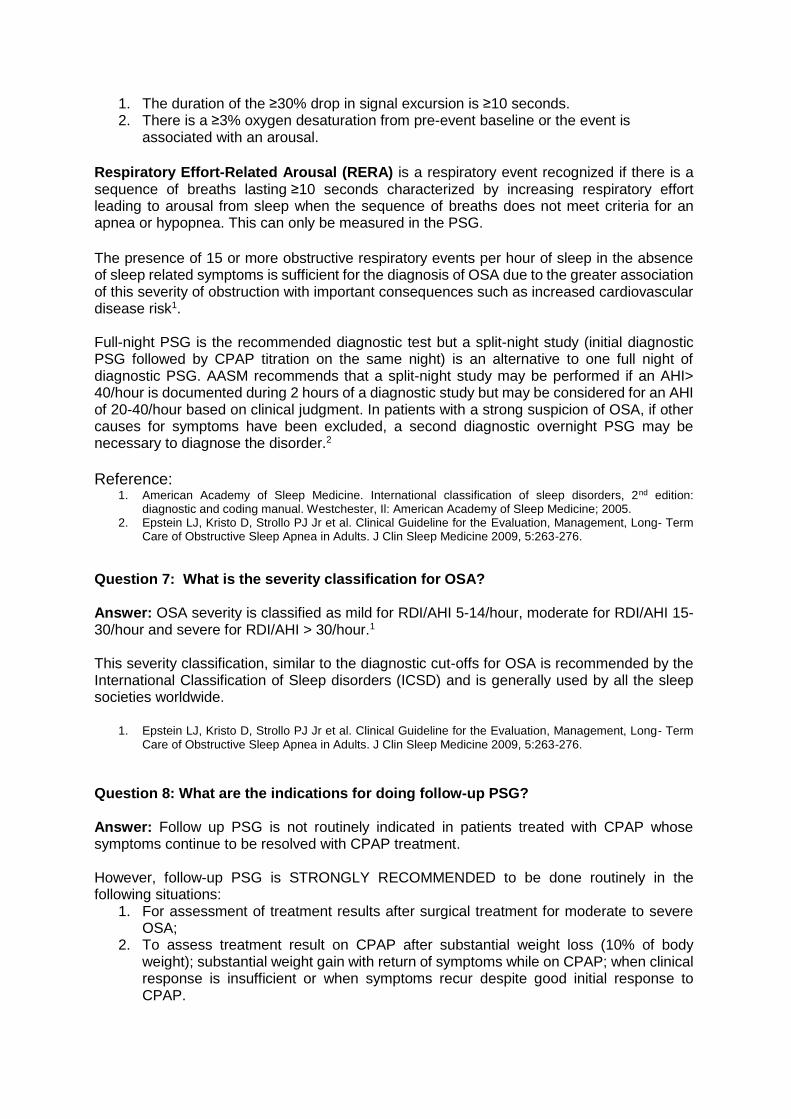
1. The duration of the ≥30% drop in signal excursion is ≥10 seconds. 2. There is a ≥3% oxygen desaturation from pre-event baseline or the event is
associated with an arousal.
Respiratory Effort-Related Arousal (RERA) is a respiratory event recognized if there is a sequence of breaths lasting ≥10 seconds characterized by increasing respiratory effort leading to arousal from sleep when the sequence of breaths does not meet criteria for an apnea or hypopnea. This can only be measured in the PSG.
The presence of 15 or more obstructive respiratory events per hour of sleep in the absence of sleep related symptoms is sufficient for the diagnosis of OSA due to the greater association of this severity of obstruction with important consequences such as increased cardiovascular disease risk1. Full-night PSG is the recommended diagnostic test but a split-night study (initial diagnostic PSG followed by CPAP titration on the same night) is an alternative to one full night of diagnostic PSG. AASM recommends that a split-night study may be performed if an AHI> 40/hour is documented during 2 hours of a diagnostic study but may be considered for an AHI of 20-40/hour based on clinical judgment. In patients with a strong suspicion of OSA, if other causes for symptoms have been excluded, a second diagnostic overnight PSG may be necessary to diagnose the disorder.2
Reference:
1. American Academy of Sleep Medicine. International classification of sleep disorders, 2nd edition: diagnostic and coding manual. Westchester, Il: American Academy of Sleep Medicine; 2005.
2. Epstein LJ, Kristo D, Strollo PJ Jr et al. Clinical Guideline for the Evaluation, Management, Long- Term Care of Obstructive Sleep Apnea in Adults. J Clin Sleep Medicine 2009, 5:263-276.
Question 7: What is the severity classification for OSA? Answer: OSA severity is classified as mild for RDI/AHI 5-14/hour, moderate for RDI/AHI 15-30/hour and severe for RDI/AHI > 30/hour.1
This severity classification, similar to the diagnostic cut-offs for OSA is recommended by the International Classification of Sleep disorders (ICSD) and is generally used by all the sleep societies worldwide.
1. Epstein LJ, Kristo D, Strollo PJ Jr et al. Clinical Guideline for the Evaluation, Management, Long- Term
Care of Obstructive Sleep Apnea in Adults. J Clin Sleep Medicine 2009, 5:263-276.
Question 8: What are the indications for doing follow-up PSG? Answer: Follow up PSG is not routinely indicated in patients treated with CPAP whose symptoms continue to be resolved with CPAP treatment. However, follow-up PSG is STRONGLY RECOMMENDED to be done routinely in the following situations:
1. For assessment of treatment results after surgical treatment for moderate to severe OSA;
2. To assess treatment result on CPAP after substantial weight loss (10% of body weight); substantial weight gain with return of symptoms while on CPAP; when clinical response is insufficient or when symptoms recur despite good initial response to CPAP.
References:
1. American Academy of Sleep Medicine. International classification of sleep disorders, 2nd edition: diagnostic and coding manual. Westchester, Il: American Academy of Sleep Medicine; 2005.
2. Kushida CA, Morgenthaler TI, Littner MR, et al. Practice parameters for the treatment of snoring and Obstructive Sleep Apnea with oral appliances: an update for 2005. Sleep 2006; 29:240-3.
3. Iber C, Ancoli-Israel S, Chesson AL, Quan SF. The AASM manual for the scoring of sleep and associated events: rules, terminology and technical specifications. Westchester, IL: American Academy of Sleep Medicine; 2007.
4. Kushida CA, Littner MR, Morgenthaler T, et al. Practice parameters for the indications for polysomnography and related procedures: an update for 2005. Sleep 2005; 28:499-521.
5. Comparative Effectiveness review no 32. AHRQ publication no 11-m EHC052 – EF (Prepared by Tufts Evidence –based Practice Center under contract 290-2007-100551) Rockville MD. Agency for Healthcare Research and Quality, 201
6. Epstein LJ, Kristo D, Strollo PJ Jr et al. Clinical Guideline for the Evaluation, Management, Long- Term Care of Obstructive Sleep Apnea in Adults. J Clin Sleep Medicine 2009, 5:263-276.
7. Qaseem , Dallas P, Owens D, et al. Diagnosis of Obstructive Sleep Apnea in Adults: A Clinical Practice Guideline From The American College of Physicians. Ann Intern Med 2014; 161: 210-220
8. Llobere P, Castollo J, Garcia MA, et al SEPAR Guideline: Diagnosis and Treatment of Sleep Apnea-Hypopnea Syndrome Arch. Brononeumol. 2011 47 (3):143-156
Section III: Questions on the Management of OSA Question 9: When should OSA be managed? Answer: Management using a multidisciplinary approach should commence once the
diagnosis and severity classification of OSA has been established. Strongly Recommend
(Consensus)
Summary of Evidence The presence or absence of OSA, as well as its severity must be determined before initiating treatment. Once the diagnosis and severity classification is established, management can commence. The individual with OSA must be at the center of decision-making for the most appropriate treatment strategy, which oftentimes requires a multidisciplinary approach. A multidisciplinary team that consists of a sleep specialist, various allied healthcare providers relevant to the management of OSA (dentist, nursing personnel, respiratory therapist and/or sleep technologist) and the referring physician is needed to adequately address the behavioral, medical and surgical options in managing OSA. Having a multidisciplinary team will ensure that all treatment options and adjunctive therapies can be discussed and provided to the patient. There is very little evidence in literature on this subject matter; however, since the immediate management of OSA provides numerous benefits and absence of harm, it was voted upon that the statement be given a strong recommendation. References:
1. American Academy of Sleep Medicine, Clinical Guideline for the Diagnosis, Management and Long Term Care of OSA in adults, 2009.
2. The International Classification of Sleep Disorders, 3rd Edition: Diagnostic and Coding Manual. Westchester, IL: American Academy of Sleep Medicine; 2014.
3. Epstein LJ, Kristo D, Strollo PJ Jr, Friedman N, Malhotra A, Patil SP, et al; Adult Obstructive Sleep Apnea Task Force of the American Academy of Sleep Medicine. Clinical guideline for the evaluation management and long-term care of obstructive sleep apnea in adults. J Clin Sleep Med. 2009; 5:263-76.
4. Centers for Medicare & Medicaid Services. National Coverage Determination (NCD) for Continuous Positive Airway Pressure (CPAP) Therapy for Obstructive Sleep Apnea (OSA) (240.4). Baltimore: Centers for Medicare & Medicaid Services; 2008.
5. Canadian Thoracic Society 2011 guideline update: Diagnosis and treatment of sleep disordered breathing
Question 10: What are the goals of therapy for OSA? Answer: The goals of therapy for OSA are:
1. To improve symptoms (excessive sleepiness, concentration, snoring), quality of life and sexual intimacy.
2. To decrease AHI to <5, events/hour with no desaturations nor arousals 3. Improvement of associated comorbidities such as hypertension, arrhythmia, heart
failure, stroke, and hyperglycemia. 4. To prevent or minimize the risk for cardiovascular events and traffic accidents.
Strongly recommend
Summary of the Evidence: Persons with OSA may experience loud snoring, low oxygen saturation, frequent arousals and disruption of sleep. Disrupted sleep can result in hypersomnolence and impaired concentration during the day.1,2 CPAP use shows improvement of self-reported sleepiness (10 RCTs) and improved quality of life (7 RCTs).3 PAP treatment for OSA is associated with modest but significant reductions in diurnal and nocturnal SBP and DBP. 28 RCTs involving 1,948 subjects showed a weighted mean difference in diurnal SBP (−2.58 mm Hg, 95% CI −3.57 to −1.59 mm Hg) and DBP (−2.01 mm Hg, 95% CI −2.84 to −1.18 mm Hg) favoring PAP treatment over control, with similar results seen in nocturnal readings. Statistically significant reductions in BP were seen in studies whose patients were younger, sleepier, had more severe OSA, and exhibited greater PAP adherence.4
CPAP also improves left ventricular ejection fraction among patients with OSA and heart failure as seen in 10 RCTs involving 259 subjects. A significant improvement in the LVEF was observed after CPAP treatment (weighted mean difference (WMD) = 3.59, 95% CI=1.74–5.44; P,0.001).5 The use of CPAP is associated with significant reductions (42%) in recurrence of atrial fibrillation (AF) in patients with OSA. Seven prospective cohort studies (N=1,087) showed that
the use of CPAP was associated with a significant reduction in AF recurrence (relative risk:
0.58, 95% CI 0.51 to 0.67; heterogeneity chi-square p = 0.91, I2 = 0%). The beneficial effect
of CPAP use was statistically significant in both groups of patients: those who underwent
catheter ablation with pulmonary vein isolation and those who did not undergo ablation and
were managed medically.6
Improvement of insulin sensitivity in patients with OSA when using CPAP was noted in six RCT’s involving 128 subjects. Although CPAP treatment did not alter HbA1c levels, its use significantly improved insulin sensitivity, indicating that treating OSA can positively impact on the management of type 2 diabetes.7
The adverse impact of OSA on intimate and sexual relationships due to sleepiness also improved with CPAP treatment as seen in a pre-posttest, quasi-experimental study involving 123 male patients. Compared to normal males, those with OSA were significantly sleepier and had more impairment in intimate and sexual relationships. Following treatment, patients were significantly more alert and had reported improved intimate and sexual relationships, with the greatest change occurring in those with the most severe disease.8
Treatment of OSA with CPAP reduces the risk of fatal and non-fatal cardiovascular events. There is no data from RCT’s but an observational study involving 1,651 men with severe OSA showed that those with untreated severe disease had a higher incidence of fatal cardiovascular events (1.06 per 100 person-years) and non-fatal cardiovascular events (2.13 per 100 person-years) than did untreated patients with mild-moderate disease (0.55, p=0.02 and 0.89, p<0.0001), simple snorers (0.34, p=0.0006 and 0.58, p<0.0001), patients treated with CPAP (0.35, p=0.0008 and 0.64, p<0.0001), and healthy participants (0.3, p=0.0012 and 0.45, p<0.0001). Multivariate analysis, adjusted for potential confounders, showed that untreated severe OSA significantly increased the risk of fatal (odds ratio 2.87, 95%CI 1.17-7.51) and non-fatal (3.17, 1.12-7.51) cardiovascular events compared with healthy participants. 9
Treatment of sleep apnea consistently improved driver performance including decreased crashes in a systematic review of 40 studies with 17,291 subjects. Since noncommercial drivers with sleep apnea have 2 to 3 times increased risk for vehicular crashes, clinicians should educate their patients with sleep apnea about the importance of treatment adherence for driving safety.10
References:
1. Victor LD. Obstructive sleep apnea. Am Fam Physician. 1999; 60:2279-86. 2. Young T, Palta M, Dempsey J, Skatrud J, Weber S, Badr S. The occurrence of sleep-disordered
breathing among middle-aged adults. N Engl J Med. 1993; 328:1230-5. 3. Practice Parameters for the use of continuous and bilevel positive airway pressure devices to treat adult
patients with sleep-related breathing disorders. SLEEP 2006; 29:375-80. 4. Montesi SB; Edwards BA; Malhotra A; Bakker JP. The effect of continuous positive airway pressure
treatment on blood pressure: a systematic review and meta-analysis of randomized controlled trials. J Clin Sleep Med 2012; 8(5):587-596.
5. Sun H, Shi J, Li M, Chen X (2013) Impact of Continuous Positive Airway Pressure Treatment on Left Ventricular Ejection Fraction in Patients with Obstructive Sleep Apnea: A Meta-Analysis of Randomized Controlled Trials. PLoS ONE 2015 8(5): e62298. doi:10.1371/journal.pone.0062298
6. Shukla A. Aizer A. Holmes D. et. al. Effect of Obstructive Sleep Apnea Treatment on Atrial Fibrillation Recurrence A Meta-Analysis. J Am Coll Cardiol EP 2015; 1-2:41–51.
7. Liang Chen, Jian-Hao Pei, Hong-Mei Chen. Effects of continuous positive airway pressure treatment on glycaemic control and insulin sensitivity in patients with obstructive sleep apnoea and type 2 diabetes: a meta-analysis.Arch Med Sci 2014; 10, 4: 637–642.
8. Reishtein JL, Maislin G, Weaver TE. Multisite Study group. Outcome of CPAP treatment on intimate and sexual relationships in men with obstructive sleep apnea. J Clin Sleep Med 2010; 6:221-226.
9. Marin JM, Carrizo SJ, Vicente E, Agusti AG. Long-term cardiovascular outcomes in men with obstructive sleep apnoea-hypopnoea with or without treatment with continuous positive airway pressure: an observational study. Lancet. 2005; 365:1046-53.
10. Ellen RL, Marshall SC, Palayew M, Molnar FJ, Wilson KG, Man-Son-Hing M. Systematic review of motor vehicle crash risk in persons with sleep apnea. J Clin Sleep Med. 2006; 2:193-200.
Question 11: What is the primary treatment for Obstructive Sleep Apnea in Adults?
Answer: CPAP at a fixed pressure is the standard initial treatment of choice for OSA in adults.
It is strongly recommended for moderate to severe OSA and recommended only for mild
OSA.
CPAP should be used for at least 4 hours during sleep daily for optimal benefit. Strongly
Recommend
Summary of evidence:
OSA is a chronic disease that requires long-term multidisciplinary management. Positive airway (PAP) is the treatment of choice for mild, moderate, and severe OSA and should be offered as an option to all patients. CPAP improves self-reported sleepiness, improves quality of life, and can be used as an adjunctive therapy to lower blood pressure in hypertensive patients with OSA. Alternative therapies may be offered depending on the severity of the OSA and the patient’s anatomy, risk factors, and preferences and should be discussed in detail. The rationale for using PAP in OSA is that it provides pneumatic splinting of the upper airway and is effective in reducing AHI.1 PAP may be delivered in continuous (CPAP), bilevel (BPAP), or autotitrating (APAP) modes. Fixed and auto-CPAP, as well as C-Flex (pressure relief), have similar adherence and efficacy. However it is only fixed CPAP that has been extensively studied. All of the CPGs reviewed have the same recommendation that CPAP is the primary treatment for OSA. ASSM and ACP specifically recommended the use of CPAP for moderate to severe OSA and ASSM recommended CPAP as only an option for treatment of mild OSA due to mixed results of 2 Level I 2,3 and 3 Level II4-6 outcome studies. The evidence for the benefit of using CPAP have already been elucidated in the previous recommendation. These benefits are especially true for those who have moderate to severe OSA. For mild OSA, the following are the alternatives to CPAP: conservative or medical therapy, dental appliance or surgery. Conservative medical therapy consists of weight loss, positional therapy in patients with OSA in the supine position, and nasal corticosteroids in patients with allergic rhinitis.7 When CPAP was compared to a dental appliance (such as mandibular advancement device, mandibular advancement splint, or mandibular position appliance) in the population of mild OSA, CPAP was superior, particularly with respect to AHI.8 On the other hand, dental appliance when compared to surgery (uvulopalatopharyngoplasty) was found to be superior.9 Thus, these studies suggest that the hierarchy of effectiveness of interventions in mild OSA are CPAP > dental appliance > surgery. What interface should be used with the CPAP? There is no general consensus across the different guidelines as to what is the best interface that should be used with CPAP i.e. nasal, oral or oronasal as there are insufficient studies that have tested the comparative efficacy of oronasal versus nasal CPAP. Insufficient studies are available to determine the comparative efficacy of oronasal CPAP versus nasal CPAP. 10 Thus, this recommendation from ASSM might be the most reasonable. Patients, in conjunction with their care team (sleep specialist, referring physician, nursing personnel, respiratory therapist, and sleep technologist), should work together to select the most appropriate PAP interface. The nasal airway is the preferred delivery route, however, alternatives may be tried to accommodate for comfort or difficulties (Consensus).11 References: 1. Gay P, Weaver T, Loube D, Iber C. Evaluation of positive airway pressure treatment of sleep related breathing
disorders in adults. Sleep 2006; 29:381-401. 2. Monasterio C, Vidal S, Duran J, et al. Effectiveness of continuous positive airway pressure in mild sleep apnea-
hypopnea syndrome. Am J Respir Crit Care Med. Sep 15 2001; 164(6):939-943. 3. Redline S, Adams N, Strauss ME, Roebuck T, Winters M, Rosenberg C. Improvement of mild sleep-disordered
breathing with CPAP compared with conservative therapy. Am J Respir Crit Care Med. Mar 1998; 157(3 Pt 1):858- 865.
4. Barnes M, Houston D, Worsnop CJ, et al. A randomized controlled trial of continuous positive airway pressure in mild obstructive sleep apnea. Am J Respir Crit Care Med. Mar 15 2002;1 65(6):773-780.
5. Engleman HM, Kingshott RN, Wraith PK, Mackay TW, Deary IJ, Douglas NJ. Randomized placebo-controlled crossover trial of continuous positive airway pressure for mild sleep Apnea/Hypopnea syndrome. Am J Respir Crit Care Med. Feb 1999; 159(2):461-467.
6. Engleman HM, Martin SE, Deary IJ, Douglas NJ. Effect of continuous positive airway pressure treatment on daytime function in sleep apnoea/hypopnoea syndrome. Thorax. 1997; 52(2):114-119.
7. Morgenthaler TI, Kapen S, Lee-Chiong T, et al. Practice parameters for the medical therapy of obstructive sleep apnea. Standards of Practice Committee of the American Academy of Sleep Medicine. Sleep 2006; 29:1031-5.
8. Barnes M, McEvoy RD, Banks S, et al. Efficacy of positive airway pressure and oral appliance in mild to moderate obstructive sleep apnea. Am J RespirCrit Care Med 2004; 170: 656-64.
9. Walker-Engstrom ML, Tegelberg A, Wilhelmsson B, Ringqvist I. 4-year follow-up of treatment with dental appliance or uvulopalatopharyngoplasty in patients with obstructive sleep apnea: a randomized study. Chest 2002; 121:739-46.
10. Qaseem A, Holty, JE, Owens DK, Dallas P, Starkey MS, Shekelle P, American College of Physicians Clinical Practice Guideline on Management of OSA in Adults, Ann Intern Med. 2013;159:471–483.
11. Adult OSA Task Force of ASSM.Clinical Guideline for the evaluation, management and long-term care of obstructive sleep apnea in adults. JCSM 2009.
Question 12: What measures can be used to increase CPAP compliance? Answer: Because compliance or adherence is a major determinant to the success of CPAP therapy in the long term, the following interventions can be used be used to improve compliance or adherence to its use:
• The addition of heated humidification. Recommend • Use of BiPAP is an option in CPAP-intolerant patients. Recommend • Pressure waveform modification technologies (i.e. pressure relief).
Recommend-consensus Trained health care providers should closely follow-up patients with OSA for compliance to PAP usage and to identify problems with its use in order to establish effective utilization patterns and remediate problems especially during the first few weeks of use. Strongly recommend
Summary of the evidence: To obtain optimal benefit in OSA, CPAP must be used during all of a patient's sleep phases. However, as in all chronic treatments, there is a constant problem of noncompliance. Generally, most studies have considered good adherence as use of the device for a minimum number of hours per night (~4 h in the literature) for 70% of the nights of the week. When a minimum use of 4 h/night is established as the cutoff point, 29–83% of patients may not be compliant.1,2,3 Since the effectiveness of the device depends on its regular use and noncompliance has been associated with reduced quality of life and greater prevalence of cardiovascular events, measures should be undertaken to improve compliance. The following are the most important interventions to address compliance to CPAP use: the addition of heated humidification to relieve nasal discomfort or irritation; use of pressure waveform modification technologies (i.e. pressure relief) to improve patient comfort; and the use of BiPAP as an option in CPAP-intolerant patients. Table 7 summarizes the various problems that are seen among CPAP users and the possible options or solutions. 20% of OSA patients discontinued CPAP after a period of use primarily because of a lack of perceived benefit.4 but the more common reasons are not due to lack of efficacy but are due to adverse experiences as well as apparatus discomfort, sense of claustrophobia, anxiety, inconvenience, frequent nocturnal awakening, partner complaints and nasal discomfort. 5, 6
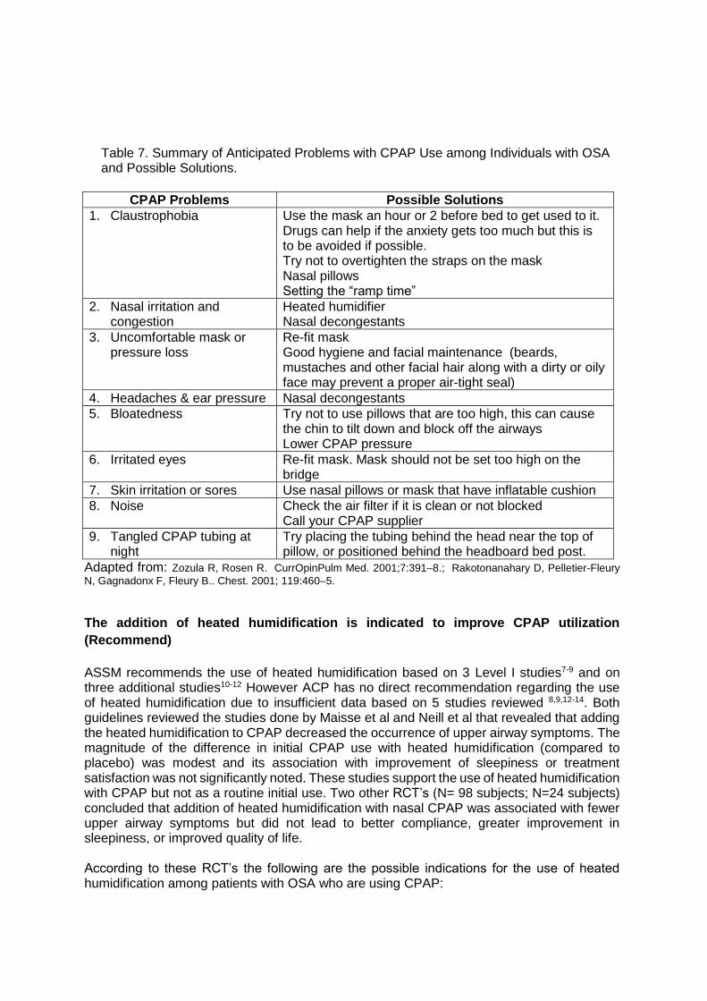
Table 7. Summary of Anticipated Problems with CPAP Use among Individuals with OSA and Possible Solutions.
CPAP Problems Possible Solutions
1. Claustrophobia Use the mask an hour or 2 before bed to get used to it. Drugs can help if the anxiety gets too much but this is to be avoided if possible. Try not to overtighten the straps on the mask Nasal pillows Setting the “ramp time”
2. Nasal irritation and congestion
Heated humidifier Nasal decongestants
3. Uncomfortable mask or pressure loss
Re-fit mask Good hygiene and facial maintenance (beards, mustaches and other facial hair along with a dirty or oily face may prevent a proper air-tight seal)
4. Headaches & ear pressure Nasal decongestants
5. Bloatedness Try not to use pillows that are too high, this can cause the chin to tilt down and block off the airways Lower CPAP pressure
6. Irritated eyes Re-fit mask. Mask should not be set too high on the bridge
7. Skin irritation or sores Use nasal pillows or mask that have inflatable cushion
8. Noise Check the air filter if it is clean or not blocked Call your CPAP supplier
9. Tangled CPAP tubing at night
Try placing the tubing behind the head near the top of pillow, or positioned behind the headboard bed post.
Adapted from: Zozula R, Rosen R. CurrOpinPulm Med. 2001;7:391–8.; Rakotonanahary D, Pelletier-Fleury
N, Gagnadonx F, Fleury B.. Chest. 2001; 119:460–5.
The addition of heated humidification is indicated to improve CPAP utilization
(Recommend)
ASSM recommends the use of heated humidification based on 3 Level I studies7-9 and on three additional studies10-12 However ACP has no direct recommendation regarding the use of heated humidification due to insufficient data based on 5 studies reviewed 8,9,12-14. Both guidelines reviewed the studies done by Maisse et al and Neill et al that revealed that adding the heated humidification to CPAP decreased the occurrence of upper airway symptoms. The magnitude of the difference in initial CPAP use with heated humidification (compared to placebo) was modest and its association with improvement of sleepiness or treatment satisfaction was not significantly noted. These studies support the use of heated humidification with CPAP but not as a routine initial use. Two other RCT’s (N= 98 subjects; N=24 subjects) concluded that addition of heated humidification with nasal CPAP was associated with fewer upper airway symptoms but did not lead to better compliance, greater improvement in sleepiness, or improved quality of life. According to these RCT’s the following are the possible indications for the use of heated humidification among patients with OSA who are using CPAP:
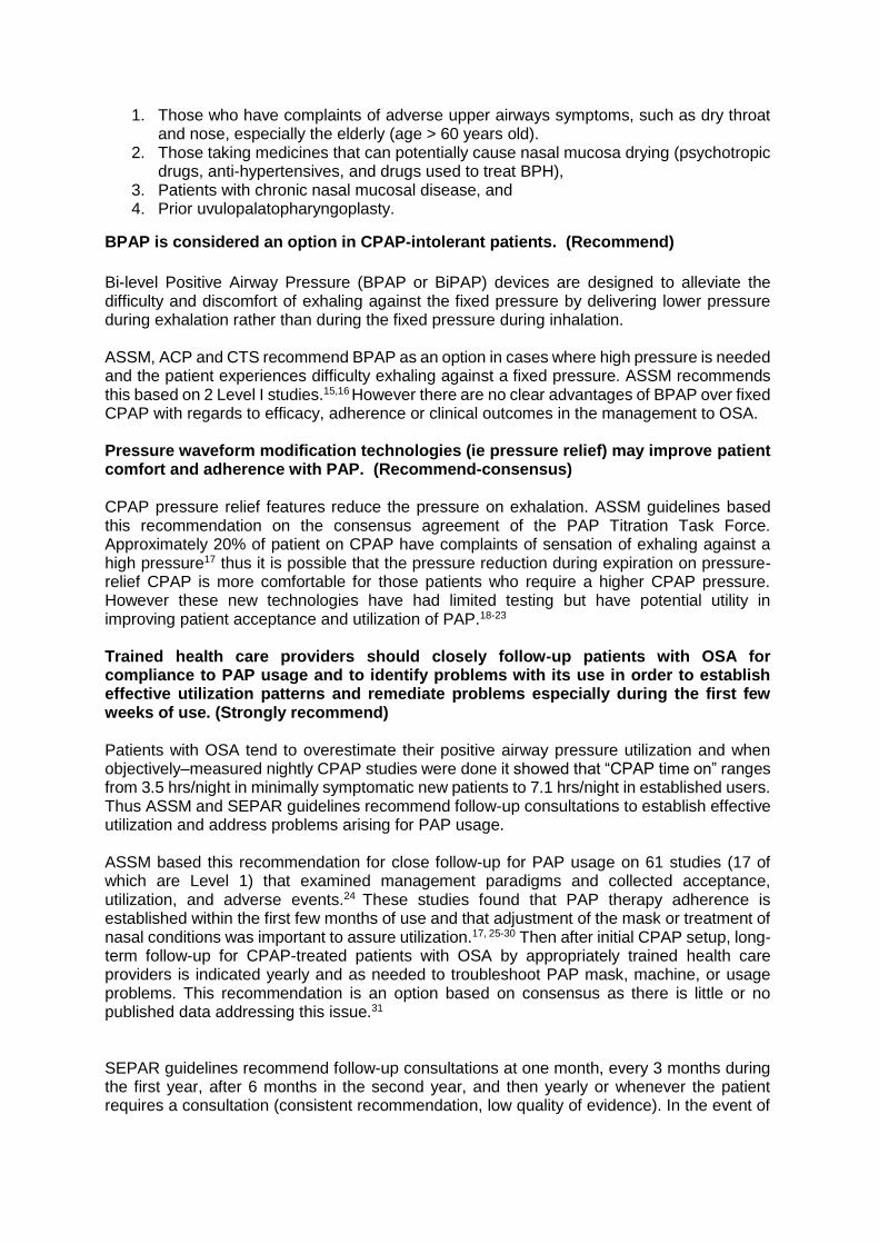
1. Those who have complaints of adverse upper airways symptoms, such as dry throat and nose, especially the elderly (age > 60 years old).
2. Those taking medicines that can potentially cause nasal mucosa drying (psychotropic drugs, anti-hypertensives, and drugs used to treat BPH),
3. Patients with chronic nasal mucosal disease, and 4. Prior uvulopalatopharyngoplasty.
BPAP is considered an option in CPAP-intolerant patients. (Recommend)
Bi-level Positive Airway Pressure (BPAP or BiPAP) devices are designed to alleviate the difficulty and discomfort of exhaling against the fixed pressure by delivering lower pressure during exhalation rather than during the fixed pressure during inhalation. ASSM, ACP and CTS recommend BPAP as an option in cases where high pressure is needed and the patient experiences difficulty exhaling against a fixed pressure. ASSM recommends this based on 2 Level I studies.15,16 However there are no clear advantages of BPAP over fixed CPAP with regards to efficacy, adherence or clinical outcomes in the management to OSA. Pressure waveform modification technologies (ie pressure relief) may improve patient comfort and adherence with PAP. (Recommend-consensus) CPAP pressure relief features reduce the pressure on exhalation. ASSM guidelines based this recommendation on the consensus agreement of the PAP Titration Task Force. Approximately 20% of patient on CPAP have complaints of sensation of exhaling against a high pressure17 thus it is possible that the pressure reduction during expiration on pressure-relief CPAP is more comfortable for those patients who require a higher CPAP pressure. However these new technologies have had limited testing but have potential utility in improving patient acceptance and utilization of PAP.18-23
Trained health care providers should closely follow-up patients with OSA for compliance to PAP usage and to identify problems with its use in order to establish effective utilization patterns and remediate problems especially during the first few weeks of use. (Strongly recommend) Patients with OSA tend to overestimate their positive airway pressure utilization and when objectively–measured nightly CPAP studies were done it showed that “CPAP time on” ranges from 3.5 hrs/night in minimally symptomatic new patients to 7.1 hrs/night in established users. Thus ASSM and SEPAR guidelines recommend follow-up consultations to establish effective utilization and address problems arising for PAP usage. ASSM based this recommendation for close follow-up for PAP usage on 61 studies (17 of which are Level 1) that examined management paradigms and collected acceptance, utilization, and adverse events.24 These studies found that PAP therapy adherence is established within the first few months of use and that adjustment of the mask or treatment of nasal conditions was important to assure utilization.17, 25-30 Then after initial CPAP setup, long-term follow-up for CPAP-treated patients with OSA by appropriately trained health care providers is indicated yearly and as needed to troubleshoot PAP mask, machine, or usage problems. This recommendation is an option based on consensus as there is little or no published data addressing this issue.31
SEPAR guidelines recommend follow-up consultations at one month, every 3 months during the first year, after 6 months in the second year, and then yearly or whenever the patient requires a consultation (consistent recommendation, low quality of evidence). In the event of
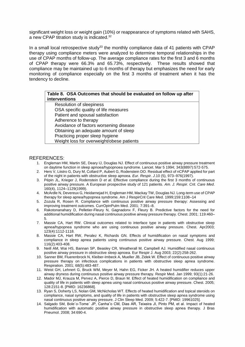
significant weight loss or weight gain (10%) or reappearance of symptoms related with SAHS, a new CPAP titration study is indicated.32
In a small local retrospective study33 the monthly compliance data of 41 patients with CPAP therapy using compliance meters were analyzed to determine temporal relationships in the use of CPAP months of follow-up. The average compliance rates for the first 3 and 6 months of CPAP therapy were 66.3% and 65.73%, respectively. These results showed that compliance may be maintained up to 6 months of therapy but emphasizes the need for early monitoring of compliance especially on the first 3 months of treatment when it has the tendency to decline.
REFERENCES:
1. Engleman HM, Martin SE, Deary IJ, Douglas NJ. Effect of continuous positive airway pressure treatment on daytime function in sleep apnoea/hypopnoea syndrome. Lancet. Mar 5 1994; 343(8897):572-575.
2. Hers V, Liistro G, Dury M, Collard P, Aubert G, Rodenstein DO. Residual effect of nCPAP applied for part of the night in patients with obstructive sleep apnoea. Eur. Respir. J.10 (5), 973–976(1997).
3. Pépin JL, Krieger J, Rodenstein D et al. Effective compliance during the first 3 months of continuous positive airway pressure. A European prospective study of 121 patients. Am. J. Respir. Crit. Care Med. 160(4), 1124–1129(1999).
4. McArdle N, Devereux G, Heidarnejad H, Engleman HM, Mackay TW, Douglas NJ. Long-term use of CPAP therapy for sleep apnea/hypopnea syndrome. Am J RespirCrit Care Med. 1999;159:1108–14
5. Zozula R, Rosen R. Compliance with continuous positive airway pressure therapy: Assessing and improving treatment outcomes. CurrOpinPulm Med. 2001; 7:391–8.
6. Rakotonanahary D, Pelletier-Fleury N, Gagnadonx F, Fleury B. Predictive factors for the need for additional humidification during nasal continuous positive airway pressure therapy. Chest. 2001; 119:460–5.
7. Massie CA, Hart RW. Clinical outcomes related to interface type in patients with obstructive sleep apnea/hypopnea syndrome who are using continuous positive airway pressure. Chest. Apr2003; 123(4):1112-1118.
8. Massie CA, Hart RW, Peralez K, Richards GN. Effects of humidification on nasal symptoms and compliance in sleep apnea patients using continuous positive airway pressure. Chest. Aug 1999; 116(2):403-408.
9. Neill AM, Wai HS, Bannan SP, Beasley CR, Weatherall M, Campbell AJ. Humidified nasal continuous positive airway pressure in obstructive sleep apnoea. Eur Respir J. Aug 2003; 22(2):258-262.
10. Sanner BM, Fluerenbrock N, Kleiber-Imbeck A, Mueller JB, Zidek W. Effect of continuous positive airway pressure therapy on infectious complications in patients with obstructive sleep apnea syndrome. Respiration. 2001; 68(5):483-487.
11. Weist GH, Lehnert G, Bruck WM, Meyer M, Hahn EG, Ficker JH. A heated humidifier reduces upper airway dryness during continuous positive airway pressure therapy. Respir Med. Jan 1999; 93(1):21-26.
12. Mador MJ, Krauza M, Pervez A, Pierce D, Braun M. Effect of heated humidification on compliance and quality of life in patients with sleep apnea using nasal continuous positive airway pressure. Chest. 2005; 128:2151-8. [PMID: 16236868].
13. Ryan S, Doherty LS, Nolan GM, McNicholas WT. Effects of heated humidification and topical steroids on compliance, nasal symptoms, and quality of life in patients with obstructive sleep apnea syndrome using nasal continuous positive airway pressure. J Clin Sleep Med. 2009; 5:422-7. [PMID: 19961025].
14. Salgado SM, Bole´o-Tome´ JP, Canha˜o CM, Dias AR, Teixeira JI, Pinto PM, et al. Impact of heated humidification with automatic positive airway pressure in obstructive sleep apnea therapy. J Bras Pneumol. 2008; 34:690-4.
Table 8. OSA Outcomes that should be evaluated on follow up after interventions
Resolution of sleepiness OSA specific quality of life measures Patient and spousal satisfaction Adherence to therapy Avoidance of factors worsening disease Obtaining an adequate amount of sleep Practicing proper sleep hygiene Weight loss for overweight/obese patients
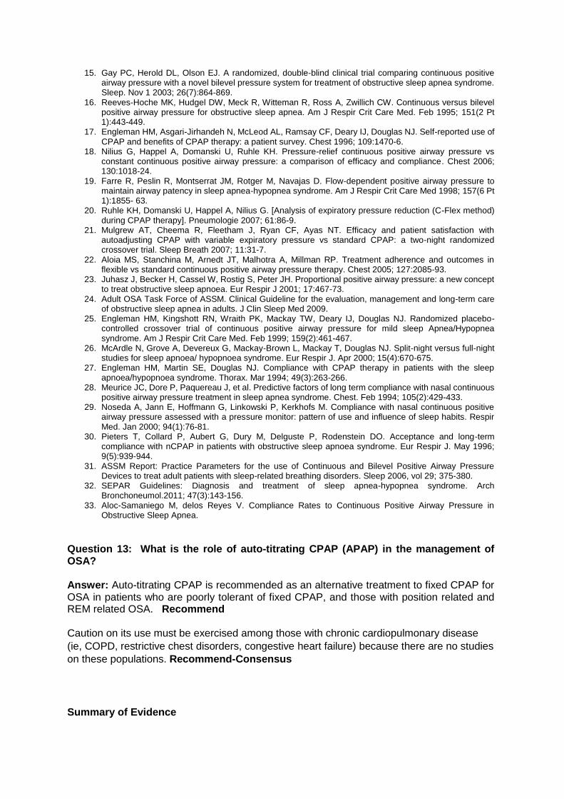
15. Gay PC, Herold DL, Olson EJ. A randomized, double-blind clinical trial comparing continuous positive airway pressure with a novel bilevel pressure system for treatment of obstructive sleep apnea syndrome. Sleep. Nov 1 2003; 26(7):864-869.
16. Reeves-Hoche MK, Hudgel DW, Meck R, Witteman R, Ross A, Zwillich CW. Continuous versus bilevel positive airway pressure for obstructive sleep apnea. Am J Respir Crit Care Med. Feb 1995; 151(2 Pt 1):443-449.
17. Engleman HM, Asgari-Jirhandeh N, McLeod AL, Ramsay CF, Deary IJ, Douglas NJ. Self-reported use of CPAP and benefits of CPAP therapy: a patient survey. Chest 1996; 109:1470-6.
18. Nilius G, Happel A, Domanski U, Ruhle KH. Pressure-relief continuous positive airway pressure vs constant continuous positive airway pressure: a comparison of efficacy and compliance. Chest 2006; 130:1018-24.
19. Farre R, Peslin R, Montserrat JM, Rotger M, Navajas D. Flow-dependent positive airway pressure to maintain airway patency in sleep apnea-hypopnea syndrome. Am J Respir Crit Care Med 1998; 157(6 Pt 1):1855- 63.
20. Ruhle KH, Domanski U, Happel A, Nilius G. [Analysis of expiratory pressure reduction (C-Flex method) during CPAP therapy]. Pneumologie 2007; 61:86-9.
21. Mulgrew AT, Cheema R, Fleetham J, Ryan CF, Ayas NT. Efficacy and patient satisfaction with autoadjusting CPAP with variable expiratory pressure vs standard CPAP: a two-night randomized crossover trial. Sleep Breath 2007; 11:31-7.
22. Aloia MS, Stanchina M, Arnedt JT, Malhotra A, Millman RP. Treatment adherence and outcomes in flexible vs standard continuous positive airway pressure therapy. Chest 2005; 127:2085-93.
23. Juhasz J, Becker H, Cassel W, Rostig S, Peter JH. Proportional positive airway pressure: a new concept to treat obstructive sleep apnoea. Eur Respir J 2001; 17:467-73.
24. Adult OSA Task Force of ASSM. Clinical Guideline for the evaluation, management and long-term care of obstructive sleep apnea in adults. J Clin Sleep Med 2009.
25. Engleman HM, Kingshott RN, Wraith PK, Mackay TW, Deary IJ, Douglas NJ. Randomized placebo-controlled crossover trial of continuous positive airway pressure for mild sleep Apnea/Hypopnea syndrome. Am J Respir Crit Care Med. Feb 1999; 159(2):461-467.
26. McArdle N, Grove A, Devereux G, Mackay-Brown L, Mackay T, Douglas NJ. Split-night versus full-night studies for sleep apnoea/ hypopnoea syndrome. Eur Respir J. Apr 2000; 15(4):670-675.
27. Engleman HM, Martin SE, Douglas NJ. Compliance with CPAP therapy in patients with the sleep apnoea/hypopnoea syndrome. Thorax. Mar 1994; 49(3):263-266.
28. Meurice JC, Dore P, Paquereau J, et al. Predictive factors of long term compliance with nasal continuous positive airway pressure treatment in sleep apnea syndrome. Chest. Feb 1994; 105(2):429-433.
29. Noseda A, Jann E, Hoffmann G, Linkowski P, Kerkhofs M. Compliance with nasal continuous positive airway pressure assessed with a pressure monitor: pattern of use and influence of sleep habits. Respir Med. Jan 2000; 94(1):76-81.
30. Pieters T, Collard P, Aubert G, Dury M, Delguste P, Rodenstein DO. Acceptance and long-term compliance with nCPAP in patients with obstructive sleep apnoea syndrome. Eur Respir J. May 1996; 9(5):939-944.
31. ASSM Report: Practice Parameters for the use of Continuous and Bilevel Positive Airway Pressure Devices to treat adult patients with sleep-related breathing disorders. Sleep 2006, vol 29; 375-380.
32. SEPAR Guidelines: Diagnosis and treatment of sleep apnea-hypopnea syndrome. Arch Bronchoneumol.2011; 47(3):143-156.
33. Aloc-Samaniego M, delos Reyes V. Compliance Rates to Continuous Positive Airway Pressure in Obstructive Sleep Apnea.
Question 13: What is the role of auto-titrating CPAP (APAP) in the management of OSA? Answer: Auto-titrating CPAP is recommended as an alternative treatment to fixed CPAP for OSA in patients who are poorly tolerant of fixed CPAP, and those with position related and REM related OSA. Recommend Caution on its use must be exercised among those with chronic cardiopulmonary disease
(ie, COPD, restrictive chest disorders, congestive heart failure) because there are no studies
on these populations. Recommend-Consensus
Summary of Evidence
Treatment for OSA must be based on its prior diagnosis by an established method, typically by an attended, in-laboratory polysomnogram. APAP devices are not intended for diagnostic purposes.1 Therefore, we do not recommend using auto-titrating CPAP for the diagnosis of OSA. CPAP is the treatment of choice for OSA as it significantly reduces sleep-related respiratory events and sleepiness in patients with this condition. The optimal pressure level is ideally determined manually during in-laboratory sleep recording and after obtaining this pressure during the overnight sleep study (polysomnography); afterwards, this value is then set in the fixed CPAP device. APAP devices automatically change the treatment pressure based on feedback from various patient measures such as airflow, pressure fluctuations, or measures of airway resistance. Thus, the positive pressure in an APAP device varies versus the CPAP which delivers a fixed pressure. The theoretical ability of these machines to continuously adapt pressure settings to ventilatory needs led to the concept that it could not only replace conventional CPAP (pre-venting the need for formal CPAP titration) but also improve treatment adherence and reduce side effects. The evidence on this concept at best is inconsistent. APAP devices are also generally more expensive usually costing twice more than conventional CPAP. On the other hand, APAP is not recommended for use in patients with cardiopulmonary or neuromuscular disease, or when sleep disordered breathing is not exclusively obstructive (eg, central apnea and hypoventilation). This is because most studies evaluating APAP, regardless of the technology used, exclude such patients because the sensors and algorithms identifying respiratory events may not be sensitive or specific under these circumstances.1 The CTS strongly recommends conventional CPAP at a fixed pressure as the primary treatment for patients with OSAS.2 Both the CTS and ACP state that generally, the improvement in the measured outcomes of fixed CPAP and APAP is identical.2,3 The reduction in positive pressure level with APAP therapy is still inconsistently reported, and adherence to treatment between conventional CPAP and APAP rarely differ. The CTS guidelines state that APAP can be considered as an alternative effective treatment to fixed CPAP for OSAS in the absence of comorbid diseases and conditions. (Grade of recommendation: 1B).2 The evidence shows that overall, fixed and auto-CPAP have similar efficacy and adherence to use.3 A meta-analysis of 24 RCTs4 (2 high, 12 moderate, and 10 low-quality) compared auto-CPAP with fixed CPAP in patients with moderate-severe OSA (AHI scores greater than 15 events per hour) and those who are obese with BMI greater than 30 kg/m2. Follow-up ranged from 3 weeks to 9 months. Despite some small differences in Epworth Sleepiness Scale (0.5 point), minimum oxygen saturation (1%), and adherence (11 minutes), these differences are not likely to be clinically significant. Overall, moderate-quality evidence showed that auto-CPAP and fixed CPAP have similar adherence and treatment effects for patients with OSA. The CTS guidelines state that limited data is available regarding the identification of subjects in whom APAP therapy would improve treatment outcomes.2 It also noted that the reduction in positive pressure level that is occasionally observed with APAP is not associated with an improvement in treatment adherence nor with a preference for APAP devices2. One study5 evaluated the impact of treatment mode on cardiometabolic risk and found that APAP may be less effective than CPAP in preventing this risk. In this study, significant reductions in SBP, DBP, and HOMA-IR were observed in the CPAP group but not in the APAP group, while CRP plasma levels were similarly reduced. The general findings of these studies were that CPAP and APAP are equivalent in their ability to normalize breathing at night, improve daytime sleepiness and treatment adherence.
However, pending a formal cost effectiveness analysis, APAP is only an alternative since it is generally more expensive than a fixed CPAP and there are findings that cardiometabolic outcomes may not be reduced as effectively. For patients compliant with constant CPAP, the data confirm that there is probably little or no advantage for using an APAP device. If the preference data are explained at least in part by the prolonged periods of low pressure, patients with the greatest variability in pressure would be the best candidates for such a device. Hence, auto-titrating CPAP may be an alternative treatment to fixed CPAP for OSA in patients poorly tolerant of fixed CPAP, and those with position related and REM related OSA.
The diagnosis of postural OSA is made when the apnea hypopnea index (AHI) in the supine position is at least twice that in the lateral position.6,7 Others have employed more stringent criteria for the diagnosis of positional OSA by adding an additional criterion – that the AHI in the lateral position (L-AHI) fall in the “mild” range (AASM 1999) 4 and be less than 15 per hour.8 Patients with positional OSA tend to be younger and thinner with fewer and less severe breathing abnormalities than “non-positional” patients.9,10
The optimal pressure may be higher than necessary for much of the night as the needed CPAP pressure can vary considerably with sleeping posture and sleep stage.11 For example, a pressure of 16 cm H2O may be needed for supine REM sleep while a pressure of 10 cm H2O may work well during NREM sleep in the lateral sleeping position.8 The use of a single higher pressure for the entire night could potentially increase mask leaks, mouth leaks, pressure intolerance, and theoretically reduce acceptance and adherence with CPAP treatment in some patients. The optimal pressure could also change with time, secondary to multiple factors including weight gain and nasal congestion. It is important to remember that the majority of studies completed with APAP included patients with severe OSAS and used strict exclusion criteria. Inclusion criteria varied widely from one study to another, but chronic cardiopulmonary disease (ie, COPD, restrictive chest disorders, congestive HF) and hypnotics/narcotics intake represented exclusion criteria in the majority of them. Hence, according to the AASM guidelines, patients with congestive heart failure, significant lung disease such as chronic obstructive pulmonary disease, patients expected to have nocturnal arterial oxyhemoglobin hypoventilation syndrome), patients who do not snore (either naturally or as a result of palate surgery), and patients who have central sleep apnea syndromes are not currently candidates for APAP titration or treatment.12
Is there harm from the use of Auto-CPAP for those with co-morbidities? Potentially, there may be harm among patients with co-morbidities but the data is limited. Safety issues are of concern, especially if APAP titration is to be performed as an unattended study. The literature search identified only two studies specifically addressing safety issues.13,14 One is a case report describing the appearance of central apneas as pressure was increased during titration.13 However, central apneas could conceivably result in APAP devices delivering a progressive increase in pressure. This action may not be effective in inducing a resolution of these events. If excessive pressure triggers arousals, this action could cause further increase in central apnea in some patients.15
In patients demonstrating a significant number of central events, increases in pressure are often inadequate and typically these are usually not good candidates for APAP.16,17 Patients with lung disease and OSA, or obesity hypoventilation syndrome might also potentially have problems during unattended APAP titrations. Subjects with respiratory insufficiency are at risk of prolonged REM hypoventilation, which is not usually detected by APAP devices16. These patients can desaturate during sleep in the absence of apnea or
hypopnea, especially during REM sleep. Treatment with supplemental oxygen in addition to positive pressure or switch to bi-level pressure may be needed. This would not be available during an unattended APAP titration. Thus, it seems reasonable to expect that patients with significant heart or lung disease as well as OSA may have problems with automated titrations. A study on attended but automated CPAP titration in 21 patients randomly selected from a group of 162 diagnosed as having OSA showed that in 6 patients, complications developed during the titration including central apnea with arrhythmia and hypoxemia despite continued airflow (presumed hypoventilation) 14. The patients with these complications had congestive heart failure or lung disease. Until evidence is published to the contrary, it seems prudent to exclude patients with significant lung disease, daytime hypoxemia or hypoventilation, and congestive heart failure from unattended APAP titrations. Reference:
1. Littner M, Hirshkowitz M, Davila D, et al. Practice parameters for the use of auto-titrating continuous positive airway pressure devices for titrating pressures and treating adult patients with obstructive sleep apnea syndrome. An American Academy of Sleep Medicine report. Sleep 2002; 25:143-7.
2. J Fleetham, n Ayas, D Bradley, et al; The Canadian Thoracic Society Sleep Disordered Breathing Committee. Thoracic 2011 guideline update: Diagnosis and treatment of sleep disordered breathing. Can Respir J 2011; 18(1):25-47.
3. Amir Qaseem, Jon-Erik C. Holty, Douglas K. Owens, Paul Dallas, Melissa Starkey, Paul Shekelle. Management of Obstructive Sleep Apnea in Adults: A Clinical Practice Guideline From the American College of Physicians. Ann Intern Med. 2013; 159:471-483.
4. Ip S, D’Ambrosio C, Patel K, Obadan N, Kitsios GD, Chung M, et al. Auto-titrating versus fixed continuous positive airway pressure for the treatment of obstructive sleep apnea: a systematic review with meta-analyses. Syst Rev. 2012; 1:20. [PMID: 22587875].
5. Patruno V, Aiolfi S, Costantino G, et al. Fixed and autoadjusting continuous positive airway pressure treatments are not similar in reducing cardiovascular risk factors in patients with obstructive sleep apnea. Chest 2007; 131:1393-9.
6. Cartwright RD. Effect of sleep position on sleep apnea severity. Sleep 1984; 7:110–4. 7. Cartwright R, Ristoanovic R, Diaz F, Caldarelli D, Alder G. A comparative study of treatments for positional
sleep apnea. Sleep 1991; 14:546–552. 8. Jokic R, Klimaszewski A, Crossley M, Sridhar G, Fitzpatrick MF. Positional treatment vs. continuous
positive airway pressure in patients with positional obstructive sleep apnea syndrome. Chest 1999; 115:771–781.
9. Richard W, Kox D, den Herder C, Laman M, van Tinteren H, de Vries N. The role of sleep position in obstructive sleep apnea syndrome. Sleep Diagnosis and Therapy 2006-7;
10. Oksenberg A, Silverberg DS, Arons E, Radwan H. Positional vs non-positional obstructive sleep apnea patients. Anthropomorphic, nocturnal polysomnographic, and multiple sleep latency test data. Chest 1997; 112:629–639.
11. Oksenberg A, Silverberg DS, Arons E, Radwan H. The sleep supine position has a major effect on optimal nasal continuous positive airway pressure: relationship with rapid eye movements and non-rapid eye movements sleep, body mass index, respiratory disturbance index, and age. Chest 1999; 116:1000-6.
12. Epstein LJ; Kristo D; Strollo PJ; Friedman N; Malhotra A; Patil SP; Ramar K; Rogers R; Schwab RJ; Weaver EM; Weinstein MD. Clinical guideline for the evaluation, management and long-term care of obstructive sleep apnea in adults. J Clin Sleep Med 2009; 5(3):263-276.
13. Boudewyns A, Van de Heyning P, De Backer W. Appearance of central apnoea in a patient treated with auto-CPAP for obstructive sleep apnea. Respir Medicine 1998; 92:891-3.
14. Juhasz J, Schillen J, Urbigkeit A, Ploch T, Penzel T, Peter JH. Unattended continuous positive airway pressure titration. Clinical relevance and cardiorespiratory hazards of the method. Am J Resp Crit Care Med 1996; 154:359-65.
15. Xie A, Wong B, Phillipson EA, Slutsky AS, Bradley TD. Interaction of hyperventilation and arousals in the pathogenesis of idiopathic central sleep apnea. Am J Respir Crit Care Med 1994; 50:489-95.
16. Teschler H, Berthon-Jones M. Intelligent CPAP systems: clinical experience. Thorax 1998; 53:S49–S54.2.
17. Beninati W, Sanders MH. Optimal continuous positive airway pressure for the treatment of obstructive sleep apnea/hypopnea. Sleep Med Rev 2001; 5:7–23.
Question 14: Can auto-titrating CPAP (APAP) be used in determining fixed CPAP pressure in lieu of formal CPAP titration?
Answer: APAP use during an ambulatory titration procedure to determine a fixed CPAP treatment pressure for patients with OSA in lieu of formal CPAP titration, is currently NOT RECOMMENDED since studies are few and have not consistently shown that APAP is equivalent to the standard titration. Summary of the evidence: AASM recommends that certain APAP devices may be used during attended titration with polysomnography to identify a single pressure for use with standard CPAP for treatment of moderate to severe OSA.1 According to CTS, APAP titration was generally found to be as effective as in-laboratory CPAP titration in normalizing AHI, and in improving diurnal symptoms and QoL. This was based on 7 RCTs that assessed APAP used as a titration tool.4-
8 Sample size for the 7 RCTS varied from 12-360 patients and two of these RCTS had a cross-over design and 5 had a parallel-group design. APAP as a method of titration was not associated with significant differences compared with in-laboratory titration in terms of mean positive pressure level, treatment adherence, treatment preference and side effects as well as pressure recommendation. The cost-effectiveness of APAP titration was demonstrated in one well-designed, well-powered study.9 Further studies are however, needed to evaluate the cost-effectiveness of auto-CPAP initiated at home and to compare it to other cost-saving and simplified strategies, such as the split-night strategy in the local setting. The effectiveness of APAP devices to identify a single pressure to reduce sleep disordered breathing events may however, be device specific. APAP devices using methods that monitor snoring, apnea or hypopnea by airflow, flow contour, and/or impedance by the forced oscillation technique are those found to effectively determine a pressure to reduce sleep dis-ordered breathing events to the same extent as standard CPAP.10 CTS also stated that differences in recommendations for pressure setting are observed among APAP machines depending on their algorithm of pressure response to reduce sleep disordered breathing events. Evidence for APAP titration is specific to each device, including the particular version of software and type of device. A recent study11 showed large differences in treatment efficacy and in the accuracy of reports between APAP devices used for the treatment of OSA. Since different APAP technologies are used, at times with variable results, further research is needed to determine which technologies are most appropriate for specific patient groups. The optimal way to derive the effective pressure from attended and unattended APAP titrations also needs to be standardized. AASM further recommends that certain APAP devices may be used in an unattended way to determine a fixed CPAP treatment pressure for patients with moderate to severe OSA without significant comorbidities (CHF, COPD, central sleep apnea syndromes, or hypoventilation syndromes).1 However this was only graded as an option, meaning, it is a patient care strategy that reflects uncertain clinical use and implies inconclusive or conflicting evidence or conflicting expert opinion. At present, sleep laboratories are not easily accessible for some regions in our country but there are vendors that may offer auto-CPAP titration. In our setting, this may be an option when attended CPAP titration cannot be done. However, patients being treated with fixed CPAP on the basis of APAP titration or being treated with APAP must have close clinical follow up to determine treatment effectiveness and safety. This is especially important during the first few weeks of PAP use.1 A reevaluation and, if necessary, a standard attended CPAP titration should be performed if symptoms do not resolve or if the APAP treatment otherwise appears to lack efficacy.1 References:
1. Epstein LJ; Kristo D; Strollo PJ; Friedman N; Malhotra A; Patil SP; Ramar K; Rogers R; Schwab RJ; Weaver EM; Weinstein MD. AASM Clinical guideline for the evaluation, management and long-term care of obstructive sleep apnea in adults. J Clin Sleep Med 2009; 5(3):263-276.
2. Masa JF, Jimenez A, Duran J, et al. Alternative methods of titrating continuous positive airway pressure: A large multicenter study. Am J Respir Crit Care Med 2004; 170:1218-24.
3. Lloberes P, Rodriguez B, Roca A, et al. Comparison of conventional nighttime with automatic or manual daytime CPAP titration in unselected sleep apnea patients: Study of the usefulness of daytime titration studies. Respir Med 2004; 98:619-25.
4. Stammnitz A, Jerrentrup A, Penzel T, Peter JH, Vogelmeier C, Becker HF. Automatic CPAP titration with different self-setting devices in patients with obstructive sleep apnoea. Eur Respir J 2004; 24:273-8.
5. Cross MD, Vennelle M, Engleman HM, et al. Comparison of CPAP titration at home or the sleep laboratory in the sleep apnea hypopnea syndrome. Sleep 2006; 29:1451-5.
6. Mulgrew AT, Cheema R, Fleetham J, Ryan CF, Ayas NT. Efficacy and patient satisfaction with auto adjusting CPAP with variable expiratory pressure vs standard CPAP: A two-night randomized crossover trial. Sleep Breath 2007; 11:31-7.
7. Berry RB, Hill G, Thompson L, McLaurin V. Portable monitoring and autotitration versus polysomnography for the diagnosis and treatment of sleep apnea. Sleep 2008; 31:1423-31.
8. Series F, Plante J, Lacasse Y. Reliability of home CPAP titration with different automatic CPAP devices. Respir Res 2008; 956.
9. Cross MD, Vennelle M, Engleman HM, et al. Comparison of CPAP titration at home or the sleep laboratory in the sleep apnea hypopnea syndrome. Sleep 2006; 29:1451-5.
10. Littner M, Hirshkowitz M, Davila D, et al. Practice parameters for the use of auto-titrating continuous positive airway pressure devices for titrating pressures and treating adult patients with obstructive sleep apnea syndrome. An American Academy of Sleep Medicine report. Sleep 2002; 25:143-7.
11. Zhu K, Roisman G, Aouf S, Escourrou P. All APAPs are not equivalent for the treatment of sleep disordered breathing: a bench evaluation of eleven commercially available devices. J Clin Sleep Med 2015; 11(7):725–734.
Question 15: What is the role of the following interventions for the management of OSA? Answers: 15.1. Behavioral and Lifestyle Modifications All overweight and obese patients diagnosed with OSA should be encouraged to lose weight as medically supervised weight loss may improve the AHI. However it should be combined with primary treatment because of the low success rates for weight loss alone. Strongly recommend Alcohol intake and routine use of sedatives among patients with OSA is discouraged. Recommend Summary of Evidence: Body weight and neck circumference are important factors in the pathogenesis of obstructive sleep apnea 1 Obstructive sleep-related breathing disorders are caused by pharyngeal and/or laryngeal collapse. Pharyngeal fat deposits lead to a decrease in pharyngeal patency and underline the risk factor of obesity 2, 3. Weight loss through dieting is associated with a significant increase in the volume of the retroglossal and retropalatal airway lumen 4. Weight reduction will lead to a decrease in critical closing pressure (Pcrit), and consequently decreases the severity of OSA5. In the Sleep Heart Health Study, a weight loss of 10% translated into a 26% decrease in AHI; conversely a weight gain of 10% led to an increase in AHI of 32%6. These findings emphasize the importance of weight loss in the management of OSA in overweight or obese individuals. 5 A study from Sweden showed that a nine-week inpatient program, where patients with severe OSA were placed on a liquid, very low energy diet resulted in a successful weight loss of 20kg or more in patients in the intervention group; these patients had a mean drop in AHI
of 25 events/hour and 67% of the intervention group had an AHI <15 at the end of the nine weeks 7. Even more impressively, at follow-up one year later, patients were found to have maintained this improvement; however, these patients had sustained follow-up through the year to help them maintain their weight loss8. Very low calorie diet combined with active lifestyle counseling resulting in marked weight reduction is a feasible and effective treatment for the majority of patients with mild OSA, and the achieved beneficial outcomes are maintained at 1-year follow-up.9 Most studies indicate improvement in measures of OSA in patients with obesity but few were cured by dietary approach alone. Furthermore, PAP, dental devices, and surgery have an immediate effect whereas the response to diet is delayed. In a study of 216 overweight patients (mean BMI 32.8) on a weight-reduction program involving a low-calorie diet, exercise program and regular visits to an outpatient clinic for compliance reinforcement, weight reduction sufficient to cure the OSA was achieved in only 24 (11%) patients. The mean BMI in “cured” patients decreased to 27 and AHI decreased from a mean of 44 to just 3. Weight loss was maintained successfully in only 13 patients on reassessment after 94 months; six of 13 patients (46%) who maintained weight loss and 11 of 13 patients (85%) who regained the lost weight reported a recurrence of OSAS symptoms with a corresponding increase in their AHI back to >40. Overall, only 3% of patients had long-term relief of OSA with conservative weight- loss measures alone 10. The fact that some patients had recurrent symptoms despite maintaining weight loss highlights that although weight is certainly an important risk factor for OSA, it is not the only reason why patients develop OSA. Therefore, while dietary weight loss is recommended as a component of therapy for obese patients with OSA, this approach should be combined with a proven treatment11. In exploratory analyses, the combination of CPAP and the weight-loss intervention was associated with a larger reduction in blood pressure than was either intervention alone among participants who adhered to the therapeutic regimen.12 Alcohol, sedatives and Muscle relaxants. Patients with CPAP should be advised to refrain from using alcohol, narcotic agents, sedatives, and muscle relaxants that might decrease the activity of the upper airway dilating muscles and, thus, worsen sleep-related breathing disorders. Alcohol has CNS-depressant effects. Its use is associated with sleep disordered breathing (SDB) in healthy men who snore and do not have sleep apnea.13 Alcohol use is associated with increased nocturnal oxygen desaturation in men more than in women and with exacerbation of SDB.14 Bedtime use of alcohol is associated with increased upper airway resistance particularly after the first 2 hours of ingestion.15 Even moderate doses of alcohol worsen respiratory events in patients with mild to moderate OSA.16 Alcohol suppresses the arousal response during airway occlusion in sleep as well as the magnitude of inspiratory effort.17 Alcohol-related SDB is also associated with reduced processing of sensory respiratory neural information and nasal congestion.18 Use of benzodiazepines has muscle-relaxing effects on the upper airway musculature, causing a reduction of the posterior pharyngeal airway. The result is increased risk for hypoventilation, hypercapnia, and hypoxemia necessitating monitoring of oxygenation and ventilation.
Narcotics can induce respiratory depression by actions on the brainstem respiratory center, central and peripheral chemoreceptors, and actions on decreasing respiratory effort in response to airway resistance.19,20 Besides hypoxemia and hypoventilation, narcotics are associated with central and obstructive apneas.
Reference:
1. White DP. Pathogenesis of obstructive and central sleep apnea. Am J Respir Crit Care Med 2005; 172:1363-70.
2. Shelton KE, Woodson H, Gay S, et al. Pharyngeal fat in obstructive sleep apnea. Am Rev Respir Dis 1993; 148: 462-466.
3. Oliven A, Aspandiarov E, Gankin I, et al. Collapsibility of the relaxed pharynx and risk of sleep apnoea. Eur Respir J. 2008; 32: 1309-315.
4. Welch KC, Foster GD, Ritter CT, et al. A novel volumetric magnetic resonance imaging paradigm to study upper airway anatomy. Sleep 2002; 25:532-42.
5. Schwartz AR, Gold AR, Schubert N, et al. Effect of weight loss on upper airway collapsibility in obstructive sleep apnea. Am Rev Respir Dis 1991; 144: 494-498.
6. Peppard PE, Young T, Palta M, Dempsey J, Skatrud J. Longitudinal study of moderate weight change and sleep disordered breathing. JAMA 2000; 284:3015-21.
7. Johansson K, Neovius M, Lagerros YT, Harlid R, Rössner S, Granath F, et al. Effect of a very low energy diet on moderate and severe obstructive sleep apnea in obese men: a randomized controlled trial. BMJ. 2009; 339:b4609.
8. Johansson K, Hemmingsson E, Harlid R, Trolle Lagerros Y, Granath F, Rössner S et al. Longer term effects of very low energy diet on obstructive sleep apnea in cohort derived from randomized controlled trial: Prospective observational follow-up study. BMJ. 2011; 342: d3017.
9. Tuomilehto H., Seppa J., Parinen M., et.al. Lifestyle Intervention with Weight Reduction First-line Treatment in Mild Obstructive Sleep Apnea. Am J Respir Crit Care Med 2009 Vol 179: 320–327
10. Sampol G, Muñoz X, Sagalés MT, Martí S, Roca A, Dolors de la Calzada M, et al. Long-term efficacy of dietary weight loss in sleep apnea/hypopnea syndrome. Eur Respir J. 1998; 12(5):1156-9.
11. Timothy I. Morgenthaler, MD1; Sheldon Kapen, MD2; Teofilo Lee-Chiong, MD,et al Practice Parameters for the Medical Therapy of Obstructive Sleep Apnea Standards of Practice Committee of the American Academy of Sleep Medicine
12. Chirinos J, Gurubhagavatula I, Teff K., et al., CPAP, Weight Loss, or Both for Obstructive Sleep Apnea N Engl J Med 2014; 370:2265-2275
13. Taasan VC, Block AJ,Boysen PG, et al. Alcohol increases sleep apnea and oxygen desaturation in asymptomatic men. AM J Med 1981; 71(2)240-5
14. Peppard PE,AUstin D, Brown RL, Association of alcohol consumption and sleep disordered breathing in men and women. J Clin Sleep Med 2007;3(3) 265-70
15. Mitler MM,Dawson A, Henriksen SJ et al. Bedtime ethanol increases resisrance of upper airways and produces sleep apneas in asymptomatic snorers. Alcohol Clin Exp Res 1988;12(6):801-5
16. Scanian MF, Roebuck T, Little PJ et al. Effect of Moderate alcohol upon obstructive sleep apnea. Eur Respir J 2000; 16(5):909-13.
17. Berry RB, Bonnet MH, Light RW. Effect of ethanol on the arousal response to airway occlusion during sleep in normal subjects. Am Rev Respir Dis 1992; 145(2 Pt 1):445-52.
18. Eckert DJ, Elgar NJ, McEvoy RD,et al. Alcohol alters sensory processing to respiratory stimuli in healthy men and women during wakefulness. Sleep 2010;33(10);1389-95
19. Yue HJ, Guilleminault C. Opiod medication and sleep disordered breathing. Med Clin North Am 2010.94 (3) 435-46.
20. Drummond GB.Comparison of decreases in ventilation caused by enflurane and fentanyl during anaesthesia. Br J Anaesth 1984,55 825-35
15.2 Positional Therapy Strategies that keep the patient in a non-supine position can be used as adjunctive treatment for positional OSA. Recommend Summary of the Evidence: The severity of OSA has been shown to vary with posture in a significant subset of patients with OSA. The cross-section and the closing pressures of the pharynx differ according to body position and stage of sleep 1-3. Ventilatory drive is dependent on body position. Thus, there is ample evidence suggesting a positive effect of a lateral position during sleep. Using the definition of positional OSA as a supine AHI of at least twice that in the lateral position, a prevalence of 50% is reported 4. In a review of 326 polysomnograms, positional sleep apnea (using the above criteria with the additional caveat that the non-supine AHI must be <5) was seen in 49 of 99 patients (49.5%) with mild sleep apnea (AHI 5 to 15/h), 14 of 72 patients (19.4%) with moderate sleep apnea
(AHI 15 to 30/h), and 5 of 77 patients (6.5%) with severe sleep apnea (AHI > 30/h) 5. Clinical experience and observational studies suggest that the patients exhibiting a large decrease in AHI in the lateral position compared with the supine position tend to have a lower AHI, to be younger and to be less obese 6,7. It is not possible to extract from the data whether AHI, age or obesity is the best predictor of treatment success. It is more likely that these parameters are mutually interrelated. Different devices such as tennis balls, vests, positional alarms, verbal instruction and (orthopedic) pillows are used to avoid the supine position 8-14. There are no data comparing the different devices, with the exception that verbal instructions seem to be less effective than a positional alarm15,16. In a study, the tennis ball method was used where patients were asked to sew a pocket containing a tennis ball to the back of their pajamas which caused discomfort in the supine position and thus, caused them to turn to their side. In a six-month follow-up of 50 patients who were thus advised, 38% reported that they were still compliant at six months and a further 24% said they were no longer using the tennis ball method but were able to avoid the supine sleeping posture by other means. These patients had a significant improvement in sleep quality, decrease in snoring and daytime sleepiness compared to those who were not able to avoid the supine posture. Individuals who were not able to comply with the tennis ball method were generally younger and were unable to comply because of discomfort17. A number of short-term studies demonstrate significant but moderate effects on AHI. However, most studies were uncontrolled and small. 8-10, 12,13,18 More importantly, even in a subset of patients with clear positional sleep apnea, effectiveness was limited. Two uncontrolled studies suggested some improvement of sleep stages or daytime symptoms with positional therapy.13,14. Another small, randomized single-blind trial of 13 patients with mild-moderate sleep apnea (mean AHI 17) found that CPAP was superior to positional therapy using the tennis-ball method in decreasing AHI and improving minimum oxygen saturation, but there was no difference in Epworth, maintenance of wakefulness, sleep latency or mood and quality- of-life measurements.11 The largest study done to date, to assess effectiveness and long-term compliance of positional therapy as a primary treatment option in patients with different severities of positional OSA showed that on the short-term, positional therapy (both self-made construction and commercial band), was an easy and effective method in most patients with positional OSA. However, as long-term compliance is low, close follow-up of patients with regard to their compliance was necessary, especially in patients with moderate-to-severe OSA, as these patients are more prone to stop using positional therapy 19. Positional therapy can yield moderate reductions in AHI but is clearly inferior to CPAP. Long-term compliance with positional therapy is poor. If positional therapy is used, sleep studies are recommended to document individual success. Long-term compliance has to be determined by follow-up studies 20.
References:
1. Issa FG, Sullivan CE. Upper airway closing pressures in obstructive sleep apnea. J Appl Physiol 1984; 57: 520–527.
2. Pevernagie DA, Stanson AW, Sheedy PF 2nd, et al. Effects of body position on the upper airway of patients with obstructive sleep apnea. Am J Respir Crit Care Med 1995; 152: 179 -185.
3. Shepard JW Jr, Burger CD. Nasal and oral flow-volume loops in normal subjects and patients with obstructive sleep apnea. Am Rev Respir Dis 1990; 142: 1288-1293.
4. Chan AS, Lee RW, Cistulli PA. Non-positive airway pressure modalities: mandibular advancement devices/ positional therapy. Proc Am Thorac Soc 2008; 5: 179–184.
5. Mador MJ, Kufel TJ, Magalang UJ, Rajesh SK, Watwe V, Grant BJB. Prevalence of positional sleep apnea in patients undergoing polysomnography. Chest. 2005; 128(4):2130-7.
6. McEvoy RD, Sharp DJ, Thornton AT. The effects of posture on obstructive sleep apnea. Am Rev Respir Dis 1986; 133: 662-666.
7. Oksenberg A, Silverberg DS, Arons E, et al. Positional vs non positional obstructive sleep apnea patients: anthropomorphic, nocturnal polysomnographic, and multiple sleep latency test data. Chest 1997; 112: 629-639.
8. Maurer JT, Stuck BA, Hein G, et al. Treatment of obstructive sleep apnea with a new vest preventing the supine position.] Dtsch Med Wochenschr 2003; 128: 71–75.
9. Zuberi NA, Rekab K, Nguyen HV. Sleep apnea avoidance pillow effects on obstructive sleep apnea syndrome and snoring. Sleep Breath 2004; 8: 201–207.
10. Cartwright RD, Lloyd S, Lilie J, et al. Sleep position training as treatment for sleep apnea syndrome: a preliminary study. Sleep 1985; 8: 87–94.
11. Jokic R, Klimaszewski A, Crossley M, et al. Positional treatment vs continuous positive airway pressure in patients with positional obstructive sleep apnea syndrome. Chest 1999; 115: 771–781.
12. Wenzel S, Smith E, Leiacker R, et al. Effektivita ̈t und Langzeit. Efficacy and longterm compliance of the vest preventing the supine position in patients with obstructive sleep apnea. Laryngorhinootologie 2007; 86: 579–583.
13. Loord H, Hultcrantz E. Positioner—a method for preventing sleep apnea. Acta Otolaryngol 2007; 127: 861–868.
14. Oksenberg A, Silverberg D, Offenbach D, et al. Positional therapy for obstructive sleep apnea patients: a 6-month follow-up study. Laryngoscope 2006; 116: 1995–2000.
15. Cartwright RD. Effect of sleep position on sleep apnea severity. Sleep 1984; 7: 110–114. 16. Cartwright R, Ristanovic R, Diaz F, et al. A comparative study of treatments for positional sleep apnea.
Sleep 1991; 14: 546–552. 17. Oksenberg A, Silverberg D, Offenbach D, Arons E. Positional therapy for obstructive sleep apnea
patients: A 6-month follow-up study. Laryngoscope. 2006; 116(11):1995-2000. 18. Berger M, Oksenberg A, Silverberg DS, et al. Avoiding the supine position during sleep lowers 24 h blood
pressure in obstructive sleep apnea (OSA) patients. J Hum Hypertens 1997; 11: 657–664. 19. de Vries GE, Hoekema A, Doff MH, Kerstjens HA, Meijer PM, van der Hoeven JH, Wijkstra PJ. Usage of
positional therapy in adults with obstructive sleep apnea. J Clin Sleep Med 2015; 11(2):131–137. 20. Randerath W.J., Verbraecken J, Andreas S, et al. Non-CPAP therapies in obstructive sleep apnea ERS
Task Force Report Eur Respir J 2011; 37: 1000–1028
15.3. Oxygen therapy Oxygen supplementation is NOT RECOMMENDED as a sole treatment for OSA. Summary of the Evidence: The role of oxygen therapy in OSA treatment is controversial. The administration of nocturnal oxygen leads to the improvement of intermittent hypoxemia in patients with OSA. The long-term consequences of chronic nocturnal administration of oxygen are unknown in patients with OSA and has been shown to prolong apnea duration in patients with OSA, perhaps as a result of the suppression of the hypoxic respiratory drive.1-3 In an observational study, the rise in blood pressure following each apneic episode was primarily linked to apnea duration and was not linked to hypoxemia.4 Prolonged apnea duration may also increase the severity of hypercarbia and acidosis in patients with OSA.1,2,5 This potential risk mandates careful monitoring for arrhythmias and other consequences of hypercarbia, especially in those with comorbid lung disease. In randomized trials of CPAP versus nocturnal oxygen, it was shown that while CPAP treats sleep disruptions, arousals and abolishes apneas and hypopneas, supplemental oxygen was able only to improve desaturations during sleep with no effect on other sleep parameters or blood pressure. 7,8 The evidence from the controlled trials does support the preferential use of CPAP over oxygen in patients with OSA since CPAP significantly improves the oxyhemoglobin saturation and reduces AHI and systemic blood pressure with improvement in daytime sleepiness. On the other hand, oxygen therapy is a double-edged sword, which not only lengthens the apnea duration but potentially increases the risk of hypercarbia with minimal to no effect on blood pressure and daytime sleepiness. 6
The American Academy of Sleep Medicine does not recommend oxygen supplementation as the primary treatment of OSA. 9 References:
1. Gold AR, Schwartz AR, Bleecker ER, et al. The effect of chronic nocturnal oxygen administration upon sleep apnea. Am Rev Respir Dis 1987; 134:925-9.
2. Alford NJ, Fletcher EC, Nickeson D. Acute oxygen in patients with sleep apnea and COPD. Chest 1986; 89:30-8.
3. Block AJ, Hellard DW, Cicale MJ. Snoring, nocturnal hypoxemia, and the effect of oxygen inhalation. Chest 1987; 92:411-7.
4. Ali NJ Davies RJ, Fleetham JA, Stradling JR. The acute effects of continuous positive airway pressure and oxygen administration on blood pressure during obstructive sleep apnea. Chest 1992; 101:1526-32.
5. Motta J, Guilleminault C. Effects of oxygen administration in sleep-induced apneas. In Sleep apnea syndromes. New York 1978; 137-44.
6. Vanita M, Taiender V, Barbara P, Frances C. Obstructive Sleep Apnea and Oxygen Therapy: A Systematic Review of the Literature and Meta-analysis. Journal of Clinical Sleep Medicine 2013
7. Norman D, Loredo JS, Nelesen RA, Ancoli-Israel S, Mills PJ, Ziegler MG, et al. Effects of continuous positive airway pressure versus supplemental oxygen on 24-hour ambulatory blood pressure. Hypertension. 2006; 47(5):840-5.
8. Loredo JS, Ancoli-Israel S, Kim EJ, Lim WJ, Dimsdale JE. Effect of continuous positive airway pressure versus supplemental oxygen on sleep quality in obstructive sleep apnea: A placebo-CPAP-controlled study. Sleep. 2006; 29(4):564-71.
9. Morgenthaler TI, Kapen S, Lee-Chiong T, Alessi C, Boehlecke B, Brown T, et al. Practice parameters for the medical therapy of obstructive sleep apnea. Sleep. 2006; 29(8):1031-5.
15.4 Pharmacologic Therapy
There is no accepted pharmacological treatment for OSA. Strongly recommend Summary of Evidence:
No pharmacologic agent is entirely effective in treating OSA 1. Evidence from 7 RCTs 2-8 showing that drug therapy, including mirtazapine, xylometazoline, fluticasone, paroxetine, pantoprazole, steroid plus CPAP (vs. CPAP alone), acetazolamide, and protriptyline, is superior to control treatment of OSA was insufficient. Each study reported on a different pharmacologic intervention, and outcomes were inconsistent across the studies. The only instances in which pharmacological therapy has been shown to be effective in the treatment of OSA is when OSA is related to hypothyroidism and to acromegaly.1 In a study of newly diagnosed hypothyroid patients, 15 of 50 patients (30%) were found to have AHI >5. Twelve of these were studied again after achieving biochemical euthyroidism with thyroxine replacement; 10 of 12 patients were found to have normalized their AHI 9. In acromegalic patients, up to 50% were found to have sleep-disordered breathing with an AHI >20; after six month’s therapy with ocreotide, there was a mean decrease in AHI of 28%+10% together with a decrease in tongue volume on MRI 10.
Addressing other co-morbidities such as allergic rhinitis, gastroesophageal reflux disease or COPD with pharmacologic therapy may also help to improve some of the symptoms associated with OSA.
References:
1. Veasey, S.,Guilleminault C,Strohl P., et al. Medical Therapy for Obstructive Sleep Apnea: A Review by the Medical Therapy for Obstructive Sleep Apnea Task Force of the Standards of Practice Committee of the American Academy of Sleep Medicine. Sleep 2006; 29(8): 1036-1044.

2. Ryan S, Doherty LS, Nolan GM, McNicholas WT. Effects of heated humidification and topical steroids on compliance, nasal symptoms, and quality of life in patients with obstructive sleep apnea syndrome using nasal continuous positive airway pressure. J Clin Sleep Med. 2009; 5:422-7.
3. Carley DW, Olopade C, Ruigt GS, Radulovacki M. Efficacy of mirtazapine in obstructive sleep apnea syndrome. Sleep. 2007; 30:35-41.
4. Clarenbach CF, Kohler M, Senn O, Thurnheer R, Bloch KE. Does nasal decongestion improve obstructive sleep apnea? J Sleep Res. 2008; 17:444-9.
5. Kiely JL, Nolan P, McNicholas WT. Intranasal corticosteroid therapy for obstructive sleep apnea in patients with co-existing rhinitis. Thorax. 2004; 59: 50-5.
6. Kraiczi H, Hedner J, Dahlo f̈ P, Ejnell H, Carlson J. Effect of serotonin uptake inhibition on breathing during sleep and daytime symptoms in obstructive sleep apnea. Sleep. 1999; 22:61-7.
7. Suurna MV, Welge J, Surdulescu V, Kushner J, Steward DL. Randomized placebo-controlled trial of pantoprazole for daytime sleepiness in GERD and obstructive sleep disordered breathing. Otolaryngol Head Neck Surg. 2008; 139: 286-90.
8. Whyte KF, Gould GA, Airlie MA, Shapiro CM, Douglas NJ. Role of protriptyline and acetazolamide in the sleep apnea/hypopnea syndrome. Sleep. 1988; 11:463-72.
9. Jha A, Sharma SK, Tandon N, Lakshmy R, Kadhiravan T, Handa KK, et al. Thyroxine replacement therapy reverses sleep-disordered breathing in patients with primary hypothyroidism. Sleep Med. 2006; 7(1):55-61.
10. Herrmann BL, Wessendorf TE, Ajaj W, Kahlke S, Teschler H, Mann K. Effects of octreotide on sleep apnea and tongue volume (magnetic resonance imaging) in patients with acromegaly. Eur J Endocrinol. 2004; 151(3):309-15.
15.5.1. What is the role of oral appliance therapy in OSA?
Answer: The use of prefabricated non-custom, non-titratable oral appliance for OSA is NOT RECOMMENDED for OSA.
The decision to use custom fitted titratable oral appliances must be made by a sleep specialist in conjunction with a dentist trained in sleep medicine. Recommend Review of Evidence: An oral appliance (OA) is a custom fabricated device using digital or physical impressions and models of an individual’s oral structures whose function is to protrude and help stabilize the mandible in order to maintain a patent upper airway during sleep. As such, it is not a primarily prefabricated item that is trimmed, bent, relined or otherwise modified. It is made of biocompatible materials and engages both the maxillary and mandibular arches. It maintains a stable retentive relationship to the teeth, implants or edentulous ridge and retains the prescribed setting during use. It is typically easy to place and remove, and maintains its structural integrity over a minimum of 3 years. The purpose of an oral appliance is to treat obstructive sleep apnea (OSA), primary snoring, and associated symptoms. 1 Oral appliances are most effective in the treatment of mild to moderate sleep apnea, and are treatment alternatives for patients with severe OSA who cannot or will not tolerate positive airway pressure therapy. Oral devices to treat OSA must be prescribed and fitted by a dentist who has sleep medicine experience. The efficacy and effectiveness of oral appliance therapy have been confirmed by several high quality studies, including RCT’s, systematic reviews, and meta- analyses.2-17 These studies have validated, via overnight polysomnography (PSG), the utility of mandibular advancement oral appliances in decreasing the frequency and/or duration of apneas, hypopneas, RERAs and/or snoring events, as well as improving nocturnal oxygenation. The ESS score, as a measure of daytime sleepiness, has been shown to normalize or improve by 2-4 points.11 The 2015 update for the clinical practice guideline for the treatment of obstructive sleep apnea and snoring with oral appliance therapy suggests that a qualified dentist must prescribe a custom, titratable appliance over non-custom oral devices .18 The overall grade for the body of evidence exploring the impact of custom vs. non-custom OAs to treat OSA varies between low and moderate depending on the physiologic sleep outcome measures. A systematic review of the evidence has shown that custom, titratable OAs reduce the AHI, arousal index, and oxygen desaturation index (ODI), and increase oxygen saturation to a greater extent than do non-custom OAs. There are 2 types of custom-fitted oral appliances- the titratable and non-titratable types. There appears to be no significant difference between these 2 types in terms of the mentioned outcomes but the titratable devices had a narrower confidence interval. Both types of custom appliances are more effective than non-custom OAs. Neither custom nor non-custom OAs have been shown to significantly affect sleep architecture and sleep efficiency. However, the use of custom titratable OAs improves daytime sleepiness as measured by the mean change in Epworth Sleepiness Scale (ESS) with the reduction in subjective daytime sleepiness being non-inferior to that reported with CPAP therapy. Custom titratable OAs also improve quality of life (QOL). There is on the other hand only very limited data regarding the impact of non-custom, and custom non-titratable oral appliances on sleepiness and QOL. Therefore, their use cannot be recommended. 18
Several studies have revealed that custom-titratable oral appliances showed greater patient acceptance than non-custom oral appliances.19,20,21 Non-custom oral appliances tend to be bulky and ill-fitting, resulting in difficulties retaining the device on the oral structures.22 This diminishes the ability of the appliance to maintain a stable mandibular protrusive position during sleep and may increase patient discomfort.19 A direct comparison study of a non-custom (pre-fabricated) thermoplastic oral appliance and custom-fabricated oral appliance looked at differences in AHI and reduction of snoring. The non-custom oral appliance failed to reduce the AHI and had limited success in reducing snoring since the pre-fabricated device may not have been retained adequately by the teeth or allowed sufficient mandibular protrusion. Additionally, the pre-fabricated appliance had decreased patient acceptance due to discomfort associated with the lack of retention during sleep.21 A custom oral appliance was associated with greater patient comfort, had greater range of protrusive movement, and was more effective. In a retrospective study of 497 OSA patients with all levels of OSA severity, oral appliance therapy reduced the mean AHI from 30.0 to 8.4, and the ESS improved significantly. 23 A comparison of PSG parameters between oral appliance and CPAP therapy was available for 397 subjects. Looking at the efficacy outcome of reduction of AHI to < 5, oral appliance therapy had equivalent efficacy to CPAP among the mild subjects at 62% versus 76% respectively (p=0.15). In the moderate and severe groups, CPAP was more effective than oral appliances with 71% vs. 51% in the moderate group, and 63% vs. 40% in the severe group. However, when the magnitude of reduction in AHI was compared between treatments, the decrease in AHI was significant only for the severe group where CPAP decreased AHI by an additional 5.9 events/hour (p < 0.001). The amount of reduction in AHI by both treatments in the mild and moderate groups differed by less than 2 events/hour and was not statistically significant.23 In addition to improvements in respiratory variables and daytime sleepiness, other health sequelae related to sleep disordered breathing that improved with oral appliance therapy included hypertension 24-30 and cardiovascular function. Changes in both systolic and diastolic pressure (ambulatory daytime and sleep BP) that were maintained at one year follow-up, and which were similar to those seen with CPAP use.31,32
An RCT of 126 individuals with moderate to severe demonstrated that while CPAP was more efficacious than oral appliance therapy in reducing AHI, no difference was detected when evaluating other health outcomes.29 Outcomes assessed included subjective sleepiness, driving simulator performance, and quality of life. Neurobehavioral outcomes improved similarly in ESS and quality of life with both treatments. Oral appliances are not recommended as a first line treatment in patients with severe OSA. However, these patients might consider an OA if they have failed CPAP or upper airway surgery, recognizing that the results of OA therapy in severe OSA are unpredictable. The literature now provides better evidence for the efficacy of OAs and indications for use. An evidence-based systematic review regarding the use of oral appliances for treating OSA concluded that overall, patients with mild to severe OSA have a 52% chance of being able to control their sleep apnea using an appliance. Success rates ranged between 14 and 61% among patients with severe OSA (AHI defined as greater than 30 in some studies and greater than 40 in others). Better success rates were seen in patients with lower AHI. OAs are on the whole less effective than CPAP but may be better accepted by patients than nasal CPAP in studies where subjects used both treatments. 3 What are the qualifications and roles of the dentist in prescribing oral appliances for the treatment of OSA?
The decision to use custom fitted titratable OA must be made by a sleep specialist in conjunction with a dentist trained in sleep medicine. The dentist who provides therapy with OAs for the management of sleep related breathing disorders should thus, have adequate knowledge and skills to provide safe and effective treatment. Qualified practitioners are those who have undertaken comprehensive training in sleep medicine and/or sleep-related breathing disorders with an emphasis on the scientific literature and the use of an appropriate protocol for diagnosis, treatment and follow-up. Treatment provided by individuals who have little or no training and education in this unique multi-disciplinary area should be discouraged. 33 In addition, the dental practitioner should be proficient in understanding various diagnostic and follow-up testing modalities including, but not limited to, the polysomnographic evaluation, and be adept at interacting with medical sleep specialists and other attending physicians for the purposes of proper diagnosis, treatment and follow-up. The dental practitioner should also understand the functional characteristics and design variations of many different OAs and must be able to recognize and manage the side effects and complications, especially concerning occlusal changes, tooth movement and temporomandibular joint symptoms. In this regard, the prudent practitioner understands the implications of life-long therapy and the importance of regular, periodic, follow-up examinations. After the prescription of an oral appliance, the dentist should observe appliance usage, side effects, complications and the degree of advancement of the appliance at follow-up visits, initially at 1 to 2 week intervals. He or she should also monitor the subjective changes in the patient’s symptoms of OSA. The appliance may need repairs, adjustments, further advancement or even replacement with a different device if side effects develop or if there is an inadequate subjective or objective improvement. No studies have reported on the ideal frequency of follow-up visits, but regular assessment in the early weeks and months of therapy is important to manage side effects, promote compliance and reduce the potential for early discontinuation due to any difficulties the patient may have using the appliance. Following initial adaptation to the OA, regular dental assessment becomes even more critical to evaluate and manage possible complications such as tooth movement, skeletal change or occlusal alteration. It is suggested that for long-term follow-up, the dentist should evaluate each patient every 6 months for the first several years and annually thereafter to ensure the integrity of the oral structures. 33 Additionally, knowledge of dental materials and a variety of dental devices including the knowledge of the patients’ dental status will likely ensure fewer side effects. A qualified dentist will be able to screen for many problems and choose and/or build the OA with features to minimize the side effects of the therapy. A qualified dentist will have the skills to choose the proper OA and make necessary modifications to accommodate patients who, among other things, may have allergies to metals or acrylics, are strong teeth grinders, or have anatomical deviations. The patient’s history and exam, appliance preference, and review of any side effects should be taken into account to avoid device breakage, allergic reactions, or discomfort that leads to frustration or discontinuation of the therapy.34
Reference:
1. Scherr S., Dort, L, Almeida F., Definition of an Effective Oral Appliance for the Treatment of Obstructive Sleep Apnea and Snoring: A Report of the American Academy of Dental Sleep Medicine. Journal of Dental Sleep Medicine Vol. 1, No. 1, 2014.
2. Epstein LJ, Kristo D, Strollo PJ, Jr., et al. Clinical guideline for the evaluation, management and long-term care of obstructive sleep apnea in adults. J Clin Sleep Med 2009; 5: 263-76.
3. Ferguson KA, Cartwright R, Rogers R, Schmidt-Nowara W. Oral appliances for snoring and obstructive sleep apnea: a review. Sleep 2006; 29:244-62.
4. Kushida CA, Morgenthaler TI, Littner MR, et al. Practice parameters for the treatment of snoring and obstructive sleep apnea with oral appliances: an update for 2005. Sleep 2006; 29:240-3.
5. Oral appliances for obstructive sleep apnea: an evidence-based analysis. Ontario health technology assessment series 2009; 9:1-51.
6. Oral appliances for treatment of snoring and obstructive sleep apnea: a review of clinical effectiveness. CADTH technology overviews 2010; 1:e0107.
7. Aarab G, Lobbezoo F, Hamburger HL, Naeije M. Oral appliance therapy versus nasal continuous positive airway pressure in obstructive sleep apnea: a randomized, placebo-controlled trial. Respiration 2011; 81:411-9.
8. Bennett LS, Davies RJ, Stradling JR. Oral appliances for the management of snoring and obstructive sleep apnoea. Thorax 1998; 53 Suppl 2:S58-64.
9. Ferguson KA, Ono T, Lowe AA, Keenan SP, Fleetham JA. A randomized crossover study of an oral appliance vs nasal-continuous positive airway pressure in the treatment of mild-moderate obstructive sleep apnea. Chest 1996; 109:1269-75.
10. Giles TL, Lasserson TJ, Smith BJ, White J, Wright J, Cates CJ. Continuous positive airways pressure for obstructive sleep apnoea in adults. Cochrane Database Syst Rev 2006:CD001106.
11. Hensley M, Ray C. Sleep apnoea. Clinical Evid 2009. 12. Hoekema A. Efficacy and comorbidity of oral appliances in the treatment of obstructive sleep apnea-
hypopnea: a systematic review and preliminary results of a randomized trial. Sleep Breath 2006; 10:102-3.
13. Hoekema A, Stegenga B, De Bont LG. Efficacy and co-morbidity of oral appliances in the treatment of obstructive sleep apnea-hypopnea: a systematic review. Crit Rev Oral Biol Med 2004; 15:137-55.
14. Lettieri CJ, PaolinoN, Eliasson AH,Shah AA,Holley AB.Comparison of adjustable and fixed oral appliances for the treatment of obstructive sleep apnea. J Clin Sleep Med 2011; 7:439-45.
15. Lim J, Lasserson TJ, Fleetham J, Wright J. Oral appliances for obstructive sleep apnoea. The Cochrane Database Syst Rev 2006:CD004435.
16. Marklund M, Verbraecken J, Randerath W. Non-CPAP therapies in obstructive sleep apnoea: mandibularadvancement device therapy. Eur Respir J 2012; 39:1241-7.
17. Mehta A, Qian J, Petocz P, Darendeliler MA, Cistulli PA. A randomized, controlled study of a mandibularadvancement splint for obstructive sleep apnea. Am J Respir Critical Care Med 2001; 163:1457-61.
18. Ramar K, Dort LC, Katz SG, Lettieri CJ, Harrod CG, Thomas SM, Chervin RD. Clinical practice guideline for the treatment of obstructive sleep apnea and snoring with oral appliance therapy: an update for 2015. J Clin Sleep Med 2015;11(7):773–827.
19. Marklund M, Verbraecken J, Randerath W. Non-CPAP therapies in obstructive sleep apnoea: mandibular advancement device therapy. Eur Respir J 2012; 39:1241-7.
20. Ahrens A, McGrath C, Hagg U. Subjective efficacy of oral appliance design features in the management of obstructive sleep apnea: a systematic review. Am J Orthod Dentofacial Orthop 2010; 138:559-76.
21. Vanderveken OM, Devolder A, Marklund M, et al. Comparison of a custom-made and a thermoplastic oral appliance for the treatment of mild sleep apnea. Am J Respir Critical Care Med 2008; 178:197-202.
22. Cooke ME, Battagel JM. A thermoplastic mandibular advancement device for the management of non-apnoeic snoring: a randomized controlled trial. Eur J Orthod 2006; 28:327-38.
23. Holley AB, Lettieri CJ, Shah AA. Efficacy of an adjustable oral appliance and comparison with continuous positive airway pressure for the treatment of obstructive sleep apnea syndrome. Chest 2011; 140:1511-6.
24. Phillips CL, Grunstein RR, Darendeliler MA, et al. Health outcomes of continuous positive airway pressure versus oral appliance treatment for obstructive sleep apnea: a randomized controlled trial. Am J Respir Critical Care Med 2013; 187:879-87.
25. Barnes M, McEvoy RD, Banks S, et al. Efficacy of positive airway pressure and oral appliance in mild to moderate obstructive sleep apnea. Am J Respir Critical Care Med 2004; 170:656-64.
26. Gotsopoulos H, Kelly JJ, Cistulli PA. Oral appliance therapy reduces blood pressure in obstructive sleep apnea: a randomized, controlled trial. Sleep 2004; 27:934-41.
27. Iftikhar IH, Hays ER, Iverson MA, Magalang UJ, Maas AK. Effect of oral appliances on blood pressure in obstructive sleep apnea: a systematic review and meta-analysis. J Clin Sleep Med 2013; 9:165-74.
28. Lam B, Sam K, Lam JC, Lai AY, Lam CL, Ip MS. The efficacy of oral appliances in the treatment of
severe obstructive sleep apnea. Sleep Breath 2011; 15:195-201. 29. Otsuka R, Ribeiro de Almeida F, Lowe AA, Linden W, Ryan F. The effect of oral appliance therapy on
blood pressure in patients with obstructive sleep apnea. Sleep Breath 2006; 10:29-36. 30. Yoshida K. Effect on blood pressure of oral appliance therapy for sleep apnea syndrome. Int J
Prosthodont 2006; 19:61-6. 31. Hoekema A, Voors AA, Wijkstra PJ, et al. Effects of oral appliances and CPAP on the left ventricle and
natriuretic peptides. Int J Cardiol 2008; 128:232-9.
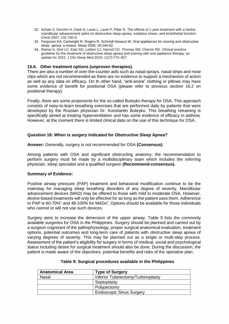
32. Itzhaki S, Dorchin H, Clark G, Lavie L, Lavie P, Pillar G. The effects of 1-year treatment with a herbst mandibular advancement splint on obstructive sleep apnea, oxidative stress, and endothelial function. Chest 2007; 131:740-9.
33. Ferguson KA, Cartwright R, Rogers R, Schmidt-Nowara W. Oral appliances for snoring and obstructive sleep apnea: a review. Sleep 2006; 29:244-62.
34. Ramar K, Dort LC, Katz SG, Lettieri CJ, Harrod CG, Thomas SM, Chervin RD. Clinical practice guideline for the treatment of obstructive sleep apnea and snoring with oral appliance therapy: an update for 2015. J Clin Sleep Med 2015; 11(7):773–827.
15.6. Other treatment options (unproven therapies). There are also a number of over-the-counter aids such as nasal sprays, nasal strips and nose clips which are not recommended as there are no evidence to support a mechanism of action as well as any data on efficacy. On th other hand, “anti-snore” clothing or pillows may have some evidence of benefit for positional OSA (please refer to previous section 16.2 on positional therapy). Finally, there are some proponents for the so-called Buteyko therapy for OSA. This approach consists of easy-to-learn breathing exercises that are performed daily by patients that were developed by the Russian physician Dr. Konstantin Buteyko. This breathing retraining is specifically aimed at treating hyperventilation and has some evidence of efficacy in asthma. However, at the moment there is limited clinical data on the use of this technique for OSA. Question 16: When is surgery indicated for Obstructive Sleep Apnea?
Answer: Generally, surgery is not recommended for OSA (Consensus) Among patients with OSA and significant obstructing anatomy, the recommendation to perform surgery must be made by a multidisciplinary team which includes the referring physician, sleep specialist and a qualified surgeon (Recommend-consensus).
Summary of Evidence: Positive airway pressure (PAP) treatment and behavioral modification continue to be the mainstay for managing sleep breathing disorders of any degree of severity. Mandibular advancement devices (MAD) may be offered to those with mild to moderate OSA. However, device-based treatments will only be effective for as long as the patient uses them. Adherence to PAP is 60-70%1 and 48-100% for MADs2. Options should be available for those individuals who cannot or will not use such devices. Surgery aims to increase the dimension of the upper airway. Table 9 lists the commonly available surgeries for OSA in the Philippines. Surgery should be planned and carried out by a surgeon cognizant of the pathophysiology, proper surgical anatomical evaluation, treatment options, potential outcomes and long-term care of patients with obstructive sleep apnea of varying degrees of severity. This may be planned out as a single or multi-step process. Assessment of the patient’s eligibility for surgery in terms of medical, social and psychological status including desire for surgical treatment should also be done. During the discussion, the patient is made aware of the objectives, potential benefits and risks of the operative plan.
Table 9: Surgical procedures available in the Philippines
Anatomical Area Type of Surgery
Nasal Inferior Tubinectomy/Turbinoplasty
Septoplasty
Polypectomy
Endoscopic Sinus Surgery
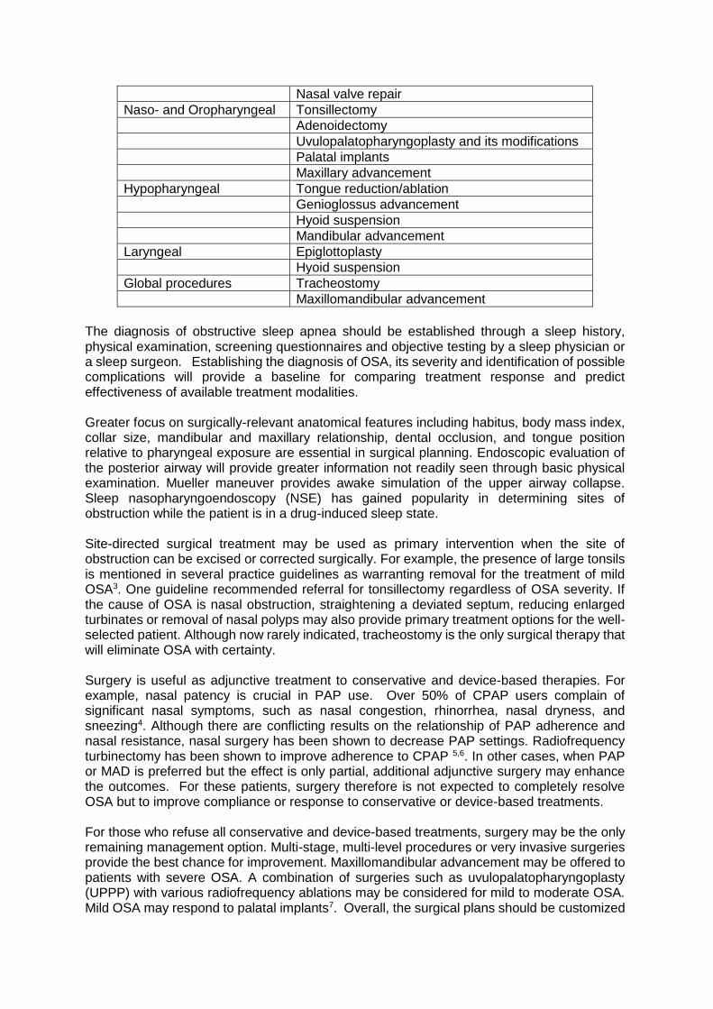
Nasal valve repair
Naso- and Oropharyngeal Tonsillectomy
Adenoidectomy
Uvulopalatopharyngoplasty and its modifications
Palatal implants
Maxillary advancement
Hypopharyngeal Tongue reduction/ablation
Genioglossus advancement
Hyoid suspension
Mandibular advancement
Laryngeal Epiglottoplasty
Hyoid suspension
Global procedures Tracheostomy
Maxillomandibular advancement
The diagnosis of obstructive sleep apnea should be established through a sleep history, physical examination, screening questionnaires and objective testing by a sleep physician or a sleep surgeon. Establishing the diagnosis of OSA, its severity and identification of possible complications will provide a baseline for comparing treatment response and predict effectiveness of available treatment modalities. Greater focus on surgically-relevant anatomical features including habitus, body mass index, collar size, mandibular and maxillary relationship, dental occlusion, and tongue position relative to pharyngeal exposure are essential in surgical planning. Endoscopic evaluation of the posterior airway will provide greater information not readily seen through basic physical examination. Mueller maneuver provides awake simulation of the upper airway collapse. Sleep nasopharyngoendoscopy (NSE) has gained popularity in determining sites of obstruction while the patient is in a drug-induced sleep state. Site-directed surgical treatment may be used as primary intervention when the site of obstruction can be excised or corrected surgically. For example, the presence of large tonsils is mentioned in several practice guidelines as warranting removal for the treatment of mild OSA3. One guideline recommended referral for tonsillectomy regardless of OSA severity. If the cause of OSA is nasal obstruction, straightening a deviated septum, reducing enlarged turbinates or removal of nasal polyps may also provide primary treatment options for the well-selected patient. Although now rarely indicated, tracheostomy is the only surgical therapy that will eliminate OSA with certainty. Surgery is useful as adjunctive treatment to conservative and device-based therapies. For example, nasal patency is crucial in PAP use. Over 50% of CPAP users complain of significant nasal symptoms, such as nasal congestion, rhinorrhea, nasal dryness, and sneezing4. Although there are conflicting results on the relationship of PAP adherence and nasal resistance, nasal surgery has been shown to decrease PAP settings. Radiofrequency turbinectomy has been shown to improve adherence to CPAP 5,6. In other cases, when PAP or MAD is preferred but the effect is only partial, additional adjunctive surgery may enhance the outcomes. For these patients, surgery therefore is not expected to completely resolve OSA but to improve compliance or response to conservative or device-based treatments.
For those who refuse all conservative and device-based treatments, surgery may be the only remaining management option. Multi-stage, multi-level procedures or very invasive surgeries provide the best chance for improvement. Maxillomandibular advancement may be offered to patients with severe OSA. A combination of surgeries such as uvulopalatopharyngoplasty (UPPP) with various radiofrequency ablations may be considered for mild to moderate OSA. Mild OSA may respond to palatal implants7. Overall, the surgical plans should be customized
for the specific anatomy and needs of each patient, keeping in mind the expertise/skill of the surgeon. Laser Assisted Uvuloplasty (LAUP) is not recommended for the management of OSA8. Uvulopalato-pharyngoplasty (UPPP) has inconsitent results and does not reliably improve respiratory measures. Moreover, severe complications have been reported7. Therefore, much consideration of the risks versus benefits of UPPP should be deliberated. Meticulous patient selection with possible additional staged procedures are crucial when considering UPPP. Electrical upper airway stimulation for the treatment of moderate to severe OSA was approved by the US Food and Drug Administration (USFDA) in 2014.10 The system is composed of implanted components with the purpose of stimulating the hypoglossal nerve in response to inhalation. This causes anterior movement of the ipsilateral base of tongue with subsequent enlargement of the hypopharyngeal airway. The results are very promising. However, the technology is very expensive and is not available in the Philippines. References:
1. Weaver TE, Mancini C, Maislin G, Cater J, Staley B, Landis JR, Ferguson KA, George CF, Schulman DA, Greenberg H, Rapoport DM, Walsleben JA, Lee-Chiong T, Gurubhagavatula I, Kuna ST. Continuous positive airway pressure treatment of sleepy patients with milder obstructive sleep apnea: results of the CPAP Apnea Trial North American Program (CATNAP) randomized clinical trial.Am J RespirCrit Care Med. 2012 Oct 1; 186(7):677-83.
2. Ferguson KA; Cartwright R; Rogers R et al. Oral Appliances for Snoring and Obstructive Sleep Apnea: A Review. SLEEP 2006; 29(2): 244-262.
3. Epstein LJ, Kristo D, Strollo PJ Jr, Friedman N, Malhotra A, Patil SP, RamarK, Rogers R, Schwab RJ, Weaver EM, Weinstein MD; Adult Obstructive Sleep ApneaTask Force of the American Academy of Sleep Medicine. Clinical guideline for the evaluation, management and long-term care of obstructive sleep apnea in adults. J Clin Sleep Med. 2009 Jun 15; 5(3):263-76.
4. Hoffstein V1, Viner S, Mateika S, Conway JTreatment of obstructive sleep apnea with nasal continuous positive airway pressure. Patient compliance, perception of benefits, and side effects. Am Rev Respir Dis. 1992 Apr; 145(4 Pt 1):841-5.
5. Powell NB, Zonato AI, Weaver EM, Li K, Troell R, Riley RW, Guilleminault C. Radiofrequency treatment of turbinate hypertrophy in subjects using continuous positive airway pressure: a randomized, double-blind, placebo-controlled clinical pilot trial.Laryngoscope. 2001 Oct; 111(10):1783-90.
6. Friedman M, Tanyeri H, Lim JW, Landsberg R, Vaidyanathan K, Caldarelli D. Effect of improved nasal breathing on obstructive sleep apnea. Otolaryngol Head Neck Surg. 2000 Jan; 122(1):71-4.
7. Aurora RN; Casey KR; Kristo D; Auerbach S; Bista SR; Chowdhuri S; Karippot A; Lamm C; Ramar K; Zak R; Morgenthaler TI. Practice parameters for the surgical modifications of the upper airway for obstructive sleep apnea in adults. Sleep 2010; 33(10):1408-1413.
8. Littner M, Kushida CA, Hartse K, Anderson WM, Davila D, Johnson SF, Wise MS, Hirshkowitz M, Woodson BT. Practice parameters for the use of laser-assisted uvulopalatoplasty: an update for 2000. Sleep. 2001 Aug 1; 24(5):603-19.
9. http://www.fda.gov/MedicalDevices/ProductsandMedicalProcedures/DeviceApprovalsandClearances/Recently-ApprovedDevices/ucm398321.htm
Question 17: Which patients require urgent treatment for OSA? Answer:
• Any patient with known or suspected OSA with severe/unstable co-morbid conditions may benefit from a referral to a sleep specialist for evaluation and/or possible initiation of CPAP or non-invasive ventilation
• Among patients with suspected OSA, a definitive PSG is recommended after stabilization of co-morbid condition to confirm the diagnosis of OSA.
Summary of the Evidence: Urgent OSA treatment is warranted in the following conditions if there is a: 1. Risk of mortality from a sudden catastrophic medical event or accident; and
2. Limited window of opportunity to prevent long-term consequences of OSA. Previously published guidelines addressed the question of maximum wait times and criteria for prioritizing patient access to a sleep lab. The CTS guidelines recommend laboratory evaluation within 4 weeks for patients with unstable ischemic heart disease, recent cerebrovascular disease, congestive heart failure, refractory systemic hypertension, obstructive/restrictive lung disease, pulmonary hypertension, hypercapneic respiratory failure, pregnancy and those in safety-critical occupations1. The SIGN guideline identified OSA patients with EDS who operate vehicles or those with respiratory failure be considered for urgent referral to a sleep lab but no wait time was mentioned2. These qualifications for urgency presuppose treatment will be readily available once a diagnosis is made. In the Philippines, wait times for a PSG is not as protracted; however, getting to treatment may well be the source of delay. Observational studies show significant improvement in cardiovascular outcomes among OSA patients with decrease in mortality rates 3,4,5,6. In a recent review of the available RCTs on the vascular effect of CPAP therapy on OSA, there was a significant decrease in blood pressure for moderate to severe OSA with associated daytime sleepiness with CPAP treatment; however, no clear effect on mortality was demonstrated7. Despite the lack of conclusive evidence that OSA treatment can reduce mortality rates, efforts should be exerted in facilitating treatments for OSA patients with severe or unstable medical co-morbidities. The prevalence of OSA in pregnant women is unknown. Hormonal, physiologic and physical changes to support a growing fetus result in alterations that either protect from or promote the development of OSA. Weight gain, elevation of the diaphragm, reduction of functional reserve capacity, nasal congestion/rhinitis, hyperventilation, increased stage 1 of non-REM sleep, and sleep fragmentation promote the development of OSA. On the other hand, natural avoidance of supine position, increased minute ventilation, increased dilating actions of pharyngeal muscles, and reduction of REM sleep are protective adaptations8. Untreated sleep breathing disorders in pregnant women is associated with the development of gestational hypertension, preeclampsia and diabetes9. Gestational hypertension and preeclampsia are known risk factors for long-term cardiovascular disease specifically hypertension, ischemic heart disease and stroke10. When patients with preeclampsia and SDB were given CPAP therapy, significant decrease in arterial blood pressure was achieved 11, 12. Intermittent maternal hypoxia is postulated to cause placental ischemia with adverse effects on fetal growth including low birth weight and intrauterine growth retardation 13. Although evidence for the relationship of OSA in pregnancy requires further investigation, the potential risk to mother and child warrant urgent investigation and immediate management. Control of weight gain, lateral sleeping position, elevation of the head and avoidance of alcohol and sedating medications should be done. If there is evidence of sleep breathing disorder on PSG, positive airway pressure therapy is the treatment of choice.10,14 Motor vehicle drivers with excessive sleepiness threaten road safety. The risk of a motor vehicle accident (MVA) is 2.4 times higher in drivers with OSA 15. Drivers diagnosed with OSA who remain untreated are 2 to 10 times more likely to be involved in an MVA compared with controls 16-20. CPAP therapy reduces accident risk in patients with moderate to severe OSA and daytime sleepiness is relieved after 1 day of use 21. In the US, strict guidelines for the evaluation and clearance of commercial truck drivers are enforced. Evaluations must be done every 2 years and treatment initiated within 2 weeks of diagnosis. The patient must show evidence of compliance to maintain the license to drive.22 The Philippines does not have policies on drivers with OSA. Nevertheless, physicians must maintain a high index of suspicion and screen all adult, driving patients for OSA and EDS. Once diagnosed, treatment and compliance monitoring is essential in maintaining driver and road safety.
Patients may be scheduled for surgery without a prior diagnosis of OSA or OSA may first be recognized intraoperatively23. Unfortunately, a large number of surgeons and anesthesiologists fail to recognize OSA prior to surgery24. Patients undergoing surgery, particularly if under sedation or general anesthesia pose a greater surgical risk for upper airway obstruction. There is evidence that OSA is an independent risk factor for adverse postoperative outcomes25. According to the updated guidelines published by the American Society of Anesthesiologists 201426, if preoperative evaluation points to possible OSA, the anesthesiologist and surgeon should jointly decide whether to (1) manage the patient peri-operatively based on clinical criteria alone or (2) obtain sleep studies, conduct a more extensive airway examination, and initiate indicated OSA treatment in advance of surgery. If there is suspicion of OSA evaluated on the day of surgery, presumptive management may be done with the consideration of more aggressive treatment than needed had a sleep test been available. This would include, among others, post-operative use of PAP despite the patient being unfamiliar with the treatment, use of a nasal or oral airway, and considering admission to close-monitoring units. The severity of the patient’s OSA, the invasiveness of the diagnostic or therapeutic procedure, and the requirement for postoperative analgesics should be taken into account in determining whether a patient is at increased perioperative risk from OSA. The risk and benefits of the particular procedure with OSA in the background must be explained and understood by the patient and relatives26.
References: 1. J Fleetham, N Ayas, D Bradley, et al; The Canadian Thoracic Society sleep disordered breathing committee.
Canadian Thoracic Society 2011 guideline update: diagnosis and treatment of sleep disordered breathing. Can Respir J 2011; 18(1):25-47.
2. Scottish Intercollegiate Guidelines Network: Management of Obstructive Sleep Apnoea/Hypopnoea Syndrome in Adults, A national clinical guideline. 2003.
3. Marin JM, Carrizo SJ, Vicente E, Agusti AG. Long-term cardiovascular outcomes in men with obstructive sleep apnoea–hypopnoea with or without treatment with continuous positive airway pressure: an observational study. Lancet 2005; 365(9464):1046–53.
4. Martínez-García MA, Campos-Rodríguez F, Catalán-Serra P, Soler-Cataluña JJ, Almeida-Gonzalez C, De la Cruz Morón I, Durán-Cantolla J, Montserrat JM. Cardiovascular mortality in obstructive sleep apnea in the elderly: role of long-term continuous positive airway pressure treatment: a prospective observational study. Am J Respir Crit Care Med. 2012 Nov 1; 186(9):909-16.
5. Campos-Rodriguez F, Martinez-Garcia MA, de la Cruz-Moron I, Almeida-Gonzalez C, Catalan-Serra P, Montserrat JM. Cardiovascular mortality in women with obstructive sleep apnea with or without continuous positive airway pressure treatment: a cohort study. Ann Intern Med. 2012 Jan 17; 156(2):115-22.
6. Campos-Rodriguez F, Peña-Griñan N, Reyes-Nuñez N, De la Cruz-Moron I, Perez-Ronchel J, De la Vega-Gallardo F, Fernandez-Palacin A. Mortality in obstructive sleep apnea-hypopnea patients treated with positive airway pressure. Chest. 2005 Aug; 128(2):624-33.
7. Wons AM, Kohler M. Established vascular effects of continuous positive airway pressure therapy in patients with obstructive sleep apnoea—an update J Thorac Dis. 2015 May; 7(5): 912–919.
8. Kapsimalis F, Kryger M. Obstructive sleep apnea in pregnancy. Sleep Med Clin 2 (2007) 603–613.
9. Pamidi S, Pinto LM, Marc I, Benedetti A, Schwartzman K, Kimoff RJ. Maternal sleep-disordered breathing and adverse pregnancy outcomes: a systematic review and metaanalysis. Am J Obstet Gynecol. 2014 Jan; 210(1):52.e1-52.e14.
10. Dunietz GL, Chervin RD, O'Brien LM. Sleep-disordered breathing during pregnancy: future implications for cardiovascular health. Obstet Gynecol Surv. 2014 Mar; 69(3):164-76.
11. Edwards N, Blyton DM, Kirjavainen T, et al. Nasal pressure positive airway pressure reduces sleep-induced blood pressure increments in preeclampsia. Am J Respir Crit Care Med 2000; 162:252–7.
12. Connolly G, Razak ARA, Hayanga A, et al. Inspiratory flow limitation during sleep in preeclampsia: comparison with normal pregnant and nonpregnant women. Eur Respir J 2001; 18:672–6.
13. Schoenfeld A, Ovadia Y, Neri A, et al. Obstructive sleep apnea (OSA)-implications in maternal-fetal medicine: a hypothesis. Med Hypotheses 1989; 30(1):51–4.
14. Feinsilver S, Hertz G. Respiration during pregnancy. Clin Chest Med 1992; 13:637–44. 15. Tregear S, Reston J, Schoelles K, et al. Obstructive sleep apnea and risk of motor vehicle crash: systematic
review and meta-analysis. J Clin Sleep Med 2009; 5(6):573–81. 16. Horstmann S, Hess CW, Bassetti C, et al. Sleepiness-related accidents in sleep apnea patients. Sleep 2000;
23(3):383–9. 17. Haraldsson PO, Carenfelt C, Diderichsen F, et al. Clinical symptoms of sleep apnea syndrome and automobile
accidents. ORL J Otorhinolaryngol Relat Spec 1990; 52(1):57–62.
18. Haraldsson PO, Akerstedt T. Drowsiness–greater traffic hazard than alcohol. Causes, risks and treat- ment. Lakartidningen 2001; 98(25):3018–23.
19. George CF. Reduction in motor vehicle collisions following treatment of sleep apnoea with nasal CPAP. Thorax 2001; 56(7):508–12.
20. Philip P, Sagaspe P, Lagarde E, et al. Sleep disorders and accidental risk in a large group of regular registered highway drivers. Sleep Med 2010; 11(10):973–9.
21. Tregear S, Reston J, Schoelles K, et al. Continuous positive airway pressure reduces risk of motor vehicle crash among drivers with obstructive sleep apnea: systematic review and meta-analysis. Sleep 2010; 33(10):1373–80.
22. Hartenbaum N, Collop N, Rosen IM, et al. Sleep apnea and commercial motor vehicle operators: statement from the joint task force of the American College of Chest Physicians, the American College of Occupational and Environmental Medicine, and the National Sleep Foundation. Chest 2006; 130(3):902–5.
23. Singh M, Liao P, Kobah S, Wijeysundera DN, Shapiro C, Chung F. Proportion of surgical patients with undiagnosed obstructive sleep apnoea. Br J Anaesth 2013; 110:629–36.
24. Kaw R, Michota F, Jaffer A, Ghamande S, Auckley D, Golish J. Unrecognized sleep apnea in the surgical patient: implications for the perioperative setting. Chest. 2006 Jan; 129(1):198-205.
25. Gupta RM, Parvizi J, Hanssen AD, Gay PC. Postoperative complications in patients with obstructive sleep apnea syndrome undergoing hip or knee replacement: a case-control study. Mayo Clin Proc. 2001 Sep;76(9):897-905.
26. American Society of Anesthesiologists Task Force on Perioperative Management of patients with obstructive sleep apnea. Practice guidelines for the perioperative management of patients with obstructive sleep apnea: an updated report by the American Society of Anesthesiologists Task Force on Perioperative Management of patients with obstructive sleep apnea. Anesthesiology. 2014 Feb;120(2):268-86,
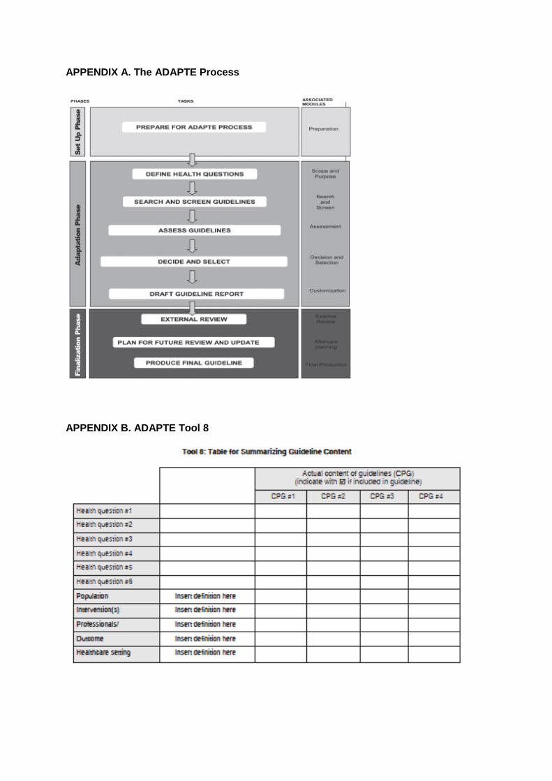
APPENDIX A. The ADAPTE Process
APPENDIX B. ADAPTE Tool 8
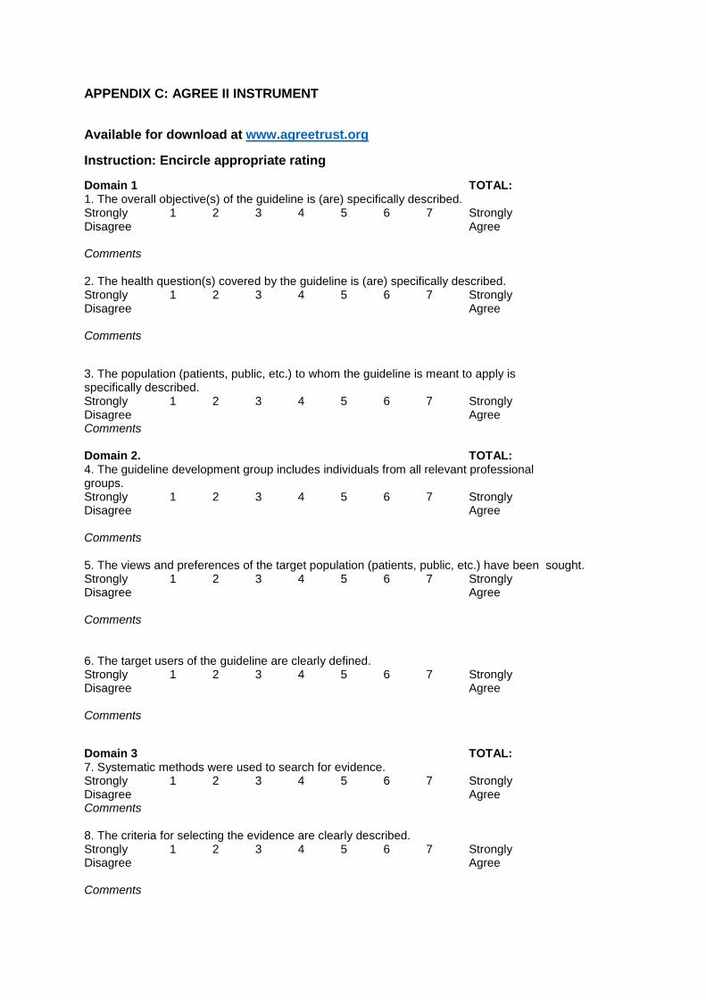
APPENDIX C: AGREE II INSTRUMENTDO
Available for download at www.agreetrust.org
Instruction: Encircle appropriate rating
Domain 1 TOTAL: 1. The overall objective(s) of the guideline is (are) specifically described. Strongly 1 2 3 4 5 6 7 Strongly Disagree Agree Comments 2. The health question(s) covered by the guideline is (are) specifically described. Strongly 1 2 3 4 5 6 7 Strongly Disagree Agree Comments
3. The population (patients, public, etc.) to whom the guideline is meant to apply is specifically described. Strongly 1 2 3 4 5 6 7 Strongly Disagree Agree Comments Domain 2. TOTAL: 4. The guideline development group includes individuals from all relevant professional groups. Strongly 1 2 3 4 5 6 7 Strongly Disagree Agree Comments 5. The views and preferences of the target population (patients, public, etc.) have been sought. Strongly 1 2 3 4 5 6 7 Strongly Disagree Agree Comments 6. The target users of the guideline are clearly defined. Strongly 1 2 3 4 5 6 7 Strongly Disagree Agree Comments
Domain 3 TOTAL: 7. Systematic methods were used to search for evidence. Strongly 1 2 3 4 5 6 7 Strongly Disagree Agree Comments 8. The criteria for selecting the evidence are clearly described. Strongly 1 2 3 4 5 6 7 Strongly Disagree Agree Comments
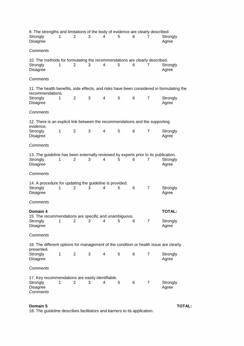
9. The strengths and limitations of the body of evidence are clearly described. Strongly 1 2 3 4 5 6 7 Strongly Disagree Agree Comments 10. The methods for formulating the recommendations are clearly described. Strongly 1 2 3 4 5 6 7 Strongly Disagree Agree Comments 11. The health benefits, side effects, and risks have been considered in formulating the recommendations. Strongly 1 2 3 4 5 6 7 Strongly Disagree Agree Comments 12. There is an explicit link between the recommendations and the supporting evidence. Strongly 1 2 3 4 5 6 7 Strongly Disagree Agree Comments DOMAIN 3. RIGOUR OF DEVELOPMENT continued 13. The guideline has been externally reviewed by experts prior to its publication. Strongly 1 2 3 4 5 6 7 Strongly Disagree Agree Comments 14. A procedure for updating the guideline is provided. Strongly 1 2 3 4 5 6 7 Strongly Disagree Agree Comments Domain 4 TOTAL: 15. The recommendations are specific and unambiguous. Strongly 1 2 3 4 5 6 7 Strongly Disagree Agree Comments 16. The different options for management of the condition or health issue are clearly presented. Strongly 1 2 3 4 5 6 7 Strongly Disagree Agree Comments 17. Key recommendations are easily identifiable. Strongly 1 2 3 4 5 6 7 Strongly Disagree Agree Comments Domain 5 TOTAL: 18. The guideline describes facilitators and barriers to its application.
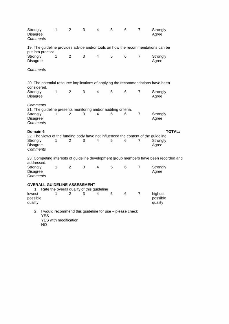
Strongly 1 2 3 4 5 6 7 Strongly Disagree Agree Comments 19. The guideline provides advice and/or tools on how the recommendations can be put into practice. Strongly 1 2 3 4 5 6 7 Strongly Disagree Agree Comments 20. The potential resource implications of applying the recommendations have been considered. Strongly 1 2 3 4 5 6 7 Strongly Disagree Agree Comments 21. The guideline presents monitoring and/or auditing criteria. Strongly 1 2 3 4 5 6 7 Strongly Disagree Agree Comments OMAIN 6. EDITORIAL INDEPENDENCE Domain 6 TOTAL: 22. The views of the funding body have not influenced the content of the guideline. Strongly 1 2 3 4 5 6 7 Strongly Disagree Agree Comments 23. Competing interests of guideline development group members have been recorded and addressed. Strongly 1 2 3 4 5 6 7 Strongly Disagree Agree Comments OVERALL GUIDELINE ASSESSMENT
1. Rate the overall quality of this guideline lowest 1 2 3 4 5 6 7 highest possible possible quality quality
2. I would recommend this guideline for use – please check
YES YES with modification NO
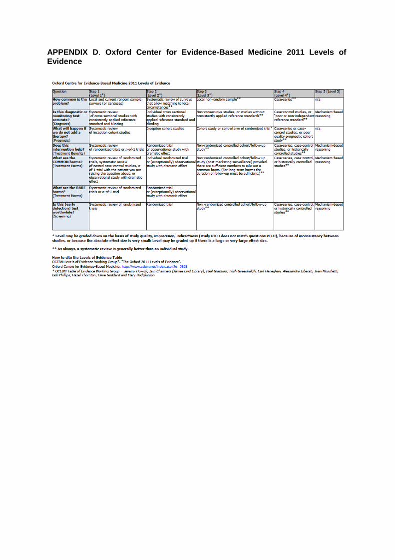
APPENDIX D. Oxford Center for Evidence-Based Medicine 2011 Levels of Evidence
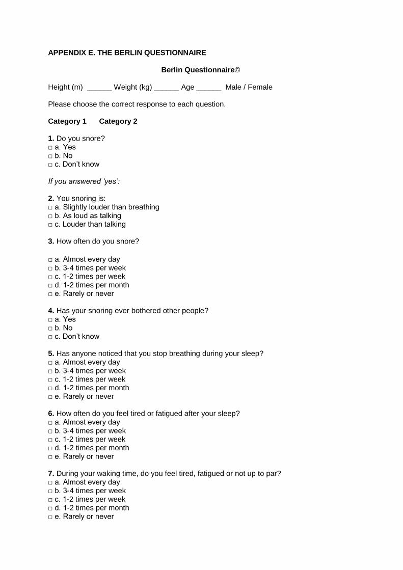
APPENDIX E. THE BERLIN QUESTIONNAIRE
Berlin Questionnaire© Height (m) ______ Weight (kg) ______ Age ______ Male / Female Please choose the correct response to each question. Category 1 Category 2 1. Do you snore? □ a. Yes □ b. No □ c. Don’t know If you answered ‘yes’: 2. You snoring is: □ a. Slightly louder than breathing □ b. As loud as talking □ c. Louder than talking 3. How often do you snore?
□ a. Almost every day □ b. 3-4 times per week □ c. 1-2 times per week □ d. 1-2 times per month □ e. Rarely or never 4. Has your snoring ever bothered other people? □ a. Yes □ b. No □ c. Don’t know 5. Has anyone noticed that you stop breathing during your sleep? □ a. Almost every day □ b. 3-4 times per week □ c. 1-2 times per week □ d. 1-2 times per month □ e. Rarely or never 6. How often do you feel tired or fatigued after your sleep? □ a. Almost every day □ b. 3-4 times per week □ c. 1-2 times per week □ d. 1-2 times per month □ e. Rarely or never 7. During your waking time, do you feel tired, fatigued or not up to par? □ a. Almost every day □ b. 3-4 times per week □ c. 1-2 times per week □ d. 1-2 times per month □ e. Rarely or never
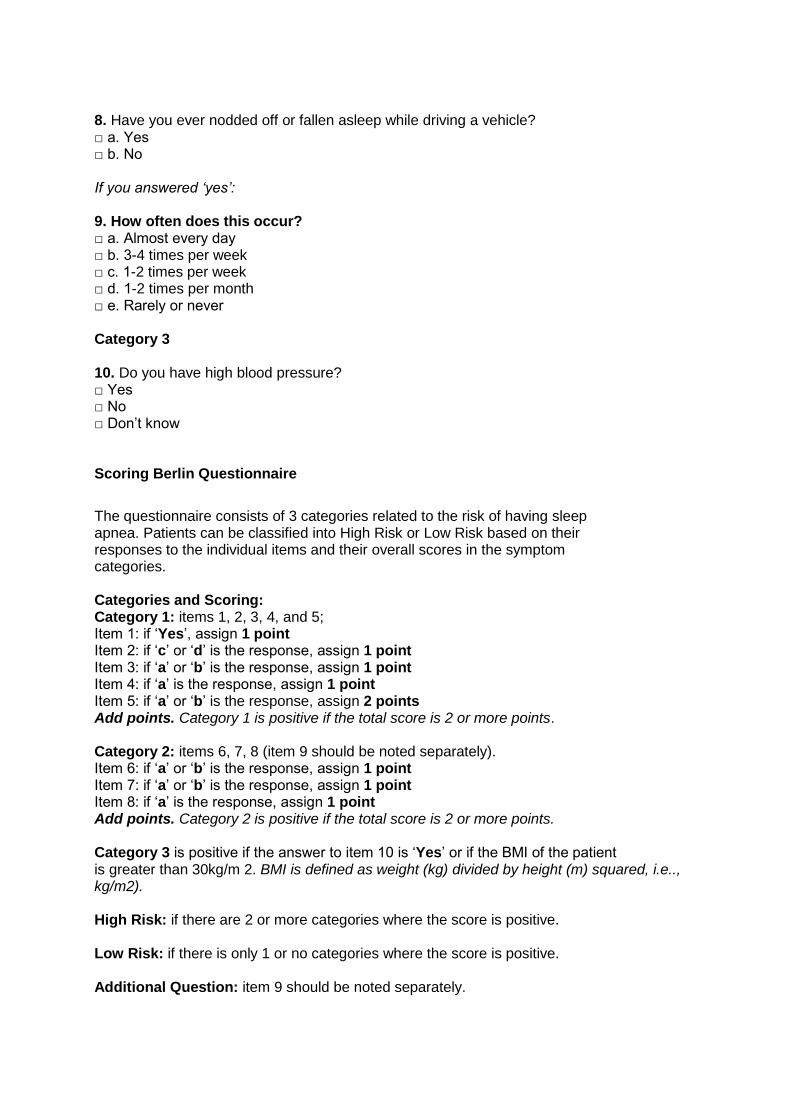
8. Have you ever nodded off or fallen asleep while driving a vehicle? □ a. Yes □ b. No If you answered ‘yes’: 9. How often does this occur? □ a. Almost every day □ b. 3-4 times per week □ c. 1-2 times per week □ d. 1-2 times per month □ e. Rarely or never Category 3 10. Do you have high blood pressure? □ Yes □ No □ Don’t know Scoring Berlin Questionnaire
The questionnaire consists of 3 categories related to the risk of having sleep apnea. Patients can be classified into High Risk or Low Risk based on their responses to the individual items and their overall scores in the symptom categories. Categories and Scoring: Category 1: items 1, 2, 3, 4, and 5; Item 1: if ‘Yes’, assign 1 point Item 2: if ‘c’ or ‘d’ is the response, assign 1 point Item 3: if ‘a’ or ‘b’ is the response, assign 1 point Item 4: if ‘a’ is the response, assign 1 point Item 5: if ‘a’ or ‘b’ is the response, assign 2 points Add points. Category 1 is positive if the total score is 2 or more points. Category 2: items 6, 7, 8 (item 9 should be noted separately). Item 6: if ‘a’ or ‘b’ is the response, assign 1 point Item 7: if ‘a’ or ‘b’ is the response, assign 1 point Item 8: if ‘a’ is the response, assign 1 point Add points. Category 2 is positive if the total score is 2 or more points. Category 3 is positive if the answer to item 10 is ‘Yes’ or if the BMI of the patient is greater than 30kg/m 2. BMI is defined as weight (kg) divided by height (m) squared, i.e.., kg/m2). High Risk: if there are 2 or more categories where the score is positive. Low Risk: if there is only 1 or no categories where the score is positive. Additional Question: item 9 should be noted separately.
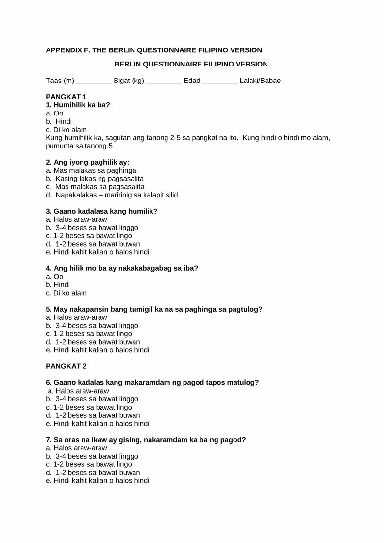
APPENDIX F. THE BERLIN QUESTIONNAIRE FILIPINO VERSION
BERLIN QUESTIONNAIRE FILIPINO VERSION Taas (m) _________ Bigat (kg) _________ Edad _________ Lalaki/Babae PANGKAT 1 1. Humihilik ka ba? a. Oo b. Hindi c. Di ko alam Kung humihilik ka, sagutan ang tanong 2-5 sa pangkat na ito. Kung hindi o hindi mo alam, pumunta sa tanong 5. 2. Ang iyong paghilik ay: a. Mas malakas sa paghinga b. Kasing lakas ng pagsasalita c. Mas malakas sa pagsasalita d. Napakalakas – maririnig sa kalapit silid 3. Gaano kadalasa kang humilik? a. Halos araw-araw b. 3-4 beses sa bawat linggo c. 1-2 beses sa bawat lingo d. 1-2 beses sa bawat buwan e. Hindi kahit kalian o halos hindi 4. Ang hilik mo ba ay nakakabagabag sa iba? a. Oo b. Hindi c. Di ko alam 5. May nakapansin bang tumigil ka na sa paghinga sa pagtulog? a. Halos araw-araw b. 3-4 beses sa bawat linggo c. 1-2 beses sa bawat lingo d. 1-2 beses sa bawat buwan e. Hindi kahit kalian o halos hindi PANGKAT 2 6. Gaano kadalas kang makaramdam ng pagod tapos matulog? a. Halos araw-araw b. 3-4 beses sa bawat linggo c. 1-2 beses sa bawat lingo d. 1-2 beses sa bawat buwan e. Hindi kahit kalian o halos hindi 7. Sa oras na ikaw ay gising, nakaramdam ka ba ng pagod? a. Halos araw-araw b. 3-4 beses sa bawat linggo c. 1-2 beses sa bawat lingo d. 1-2 beses sa bawat buwan e. Hindi kahit kalian o halos hindi
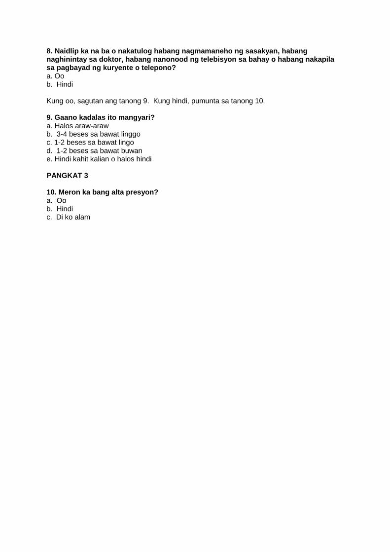
8. Naidlip ka na ba o nakatulog habang nagmamaneho ng sasakyan, habang naghinintay sa doktor, habang nanonood ng telebisyon sa bahay o habang nakapila sa pagbayad ng kuryente o telepono? a. Oo b. Hindi Kung oo, sagutan ang tanong 9. Kung hindi, pumunta sa tanong 10. 9. Gaano kadalas ito mangyari? a. Halos araw-araw b. 3-4 beses sa bawat linggo c. 1-2 beses sa bawat lingo d. 1-2 beses sa bawat buwan e. Hindi kahit kalian o halos hindi PANGKAT 3 10. Meron ka bang alta presyon? a. Oo b. Hindi c. Di ko alam
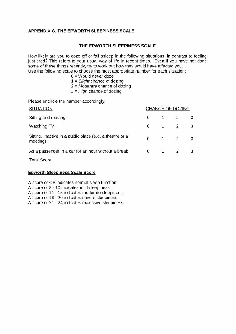
APPENDIX G. THE EPWORTH SLEEPINESS SCALE
THE EPWORTH SLEEPINESS SCALE How likely are you to doze off or fall asleep in the following situations, in contrast to feeling just tired? This refers to your usual way of life in recent times. Even if you have not done some of these things recently, try to work out how they would have affected you. Use the following scale to choose the most appropriate number for each situation: 0 = Would never doze 1 = Slight chance of dozing 2 = Moderate chance of dozing 3 = High chance of dozing Please encircle the number accordingly:
SITUATION CHANCE OF DOZING
Sitting and reading 0 1 2 3
Watching TV 0 1 2 3
Sitting, inactive in a public place (e.g. a theatre or a meeting)
0 1 2 3
As a passenger in a car for an hour without a break 0 1 2 3
Total Score:
Epworth Sleepiness Scale Score A score of < 8 indicates normal sleep function A score of 8 - 10 indicates mild sleepiness A score of 11 - 15 indicates moderate sleepiness A score of 16 - 20 indicates severe sleepiness A score of 21 - 24 indicates excessive sleepiness

APPENDIX H. THE FILIPINO VERSION OF EPWORTH SLEEPINESS SCALE
FILIPINO VERSION OF EPWORTH SLEEPINESS SCALE
Ito ay isang pagsusuri sa pagkaantok. Para sa mga sumusunod na sitwasyon, bilugan ang sagot na naaangkop sa iyo. Taliwas sa pakiramdam ng pagod, ikaw ba ay malamang na maiidlip o makakatulog sa mga sumusunod na kalagayan… 1) Nakaupo at nagbabasa
Hindi kailanman Bahagyang
pagkakataon Katamtamang pagkakataon
Palagi
2) Nanonood ng telebisyon
Hindi kailanman Bahagyang
pagkakataon Katamtamang pagkakataon
Palagi
3) Nakaupong hindi gumagalaw sa pampublikong lugar (gaya ng sinehan)
Hindi kailanman Bahagyang
pagkakataon Katamtamang pagkakataon
Palagi
4) Pasahero sa isang sasakyan sa loob ng isang oras na walang hinto
Hindi kailanman Bahagyang
pagkakataon Katamtamang pagkakataon
Palagi
5) Nakahiga para magpahinga sa hapon
Hindi kailanman Bahagyang
pagkakataon Katamtamang pagkakataon
Palagi
6) Nakaupo at nakikipag-usap sa isang tao
Hindi kailanman Bahagyang
pagkakataon Katamtamang pagkakataon
Palagi
7) Nakaupo na matahimik pagkatapos mag-tanghalian na walang ininum na alak
Hindi kailanman Bahagyang
pagkakataon Katamtamang pagkakataon
Palagi
8) Nasa isang sasakyan na huminto ng ilang minuto sa trapik?
Hindi kailanman Bahagyang
pagkakataon Katamtamang pagkakataon
Palagi
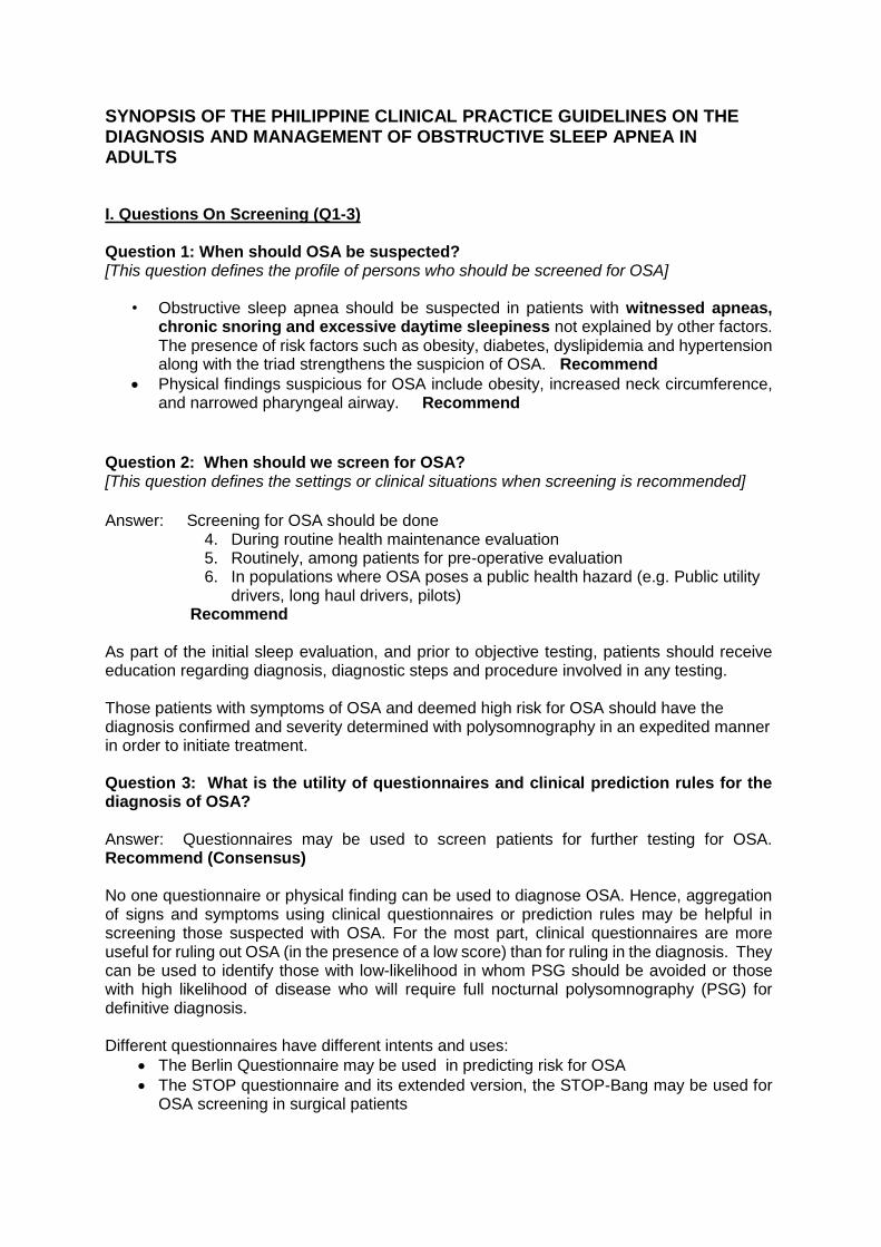
SYNOPSIS OF THE PHILIPPINE CLINICAL PRACTICE GUIDELINES ON THE DIAGNOSIS AND MANAGEMENT OF OBSTRUCTIVE SLEEP APNEA IN ADULTS I. Questions On Screening (Q1-3) Question 1: When should OSA be suspected? [This question defines the profile of persons who should be screened for OSA]
• Obstructive sleep apnea should be suspected in patients with witnessed apneas, chronic snoring and excessive daytime sleepiness not explained by other factors. The presence of risk factors such as obesity, diabetes, dyslipidemia and hypertension along with the triad strengthens the suspicion of OSA. Recommend
Physical findings suspicious for OSA include obesity, increased neck circumference, and narrowed pharyngeal airway. Recommend
Question 2: When should we screen for OSA? [This question defines the settings or clinical situations when screening is recommended]
Answer: Screening for OSA should be done
4. During routine health maintenance evaluation 5. Routinely, among patients for pre-operative evaluation 6. In populations where OSA poses a public health hazard (e.g. Public utility
drivers, long haul drivers, pilots) Recommend As part of the initial sleep evaluation, and prior to objective testing, patients should receive education regarding diagnosis, diagnostic steps and procedure involved in any testing. Those patients with symptoms of OSA and deemed high risk for OSA should have the diagnosis confirmed and severity determined with polysomnography in an expedited manner in order to initiate treatment. Question 3: What is the utility of questionnaires and clinical prediction rules for the diagnosis of OSA? Answer: Questionnaires may be used to screen patients for further testing for OSA. Recommend (Consensus) No one questionnaire or physical finding can be used to diagnose OSA. Hence, aggregation of signs and symptoms using clinical questionnaires or prediction rules may be helpful in screening those suspected with OSA. For the most part, clinical questionnaires are more useful for ruling out OSA (in the presence of a low score) than for ruling in the diagnosis. They can be used to identify those with low-likelihood in whom PSG should be avoided or those with high likelihood of disease who will require full nocturnal polysomnography (PSG) for definitive diagnosis. Different questionnaires have different intents and uses:
The Berlin Questionnaire may be used in predicting risk for OSA
The STOP questionnaire and its extended version, the STOP-Bang may be used for OSA screening in surgical patients
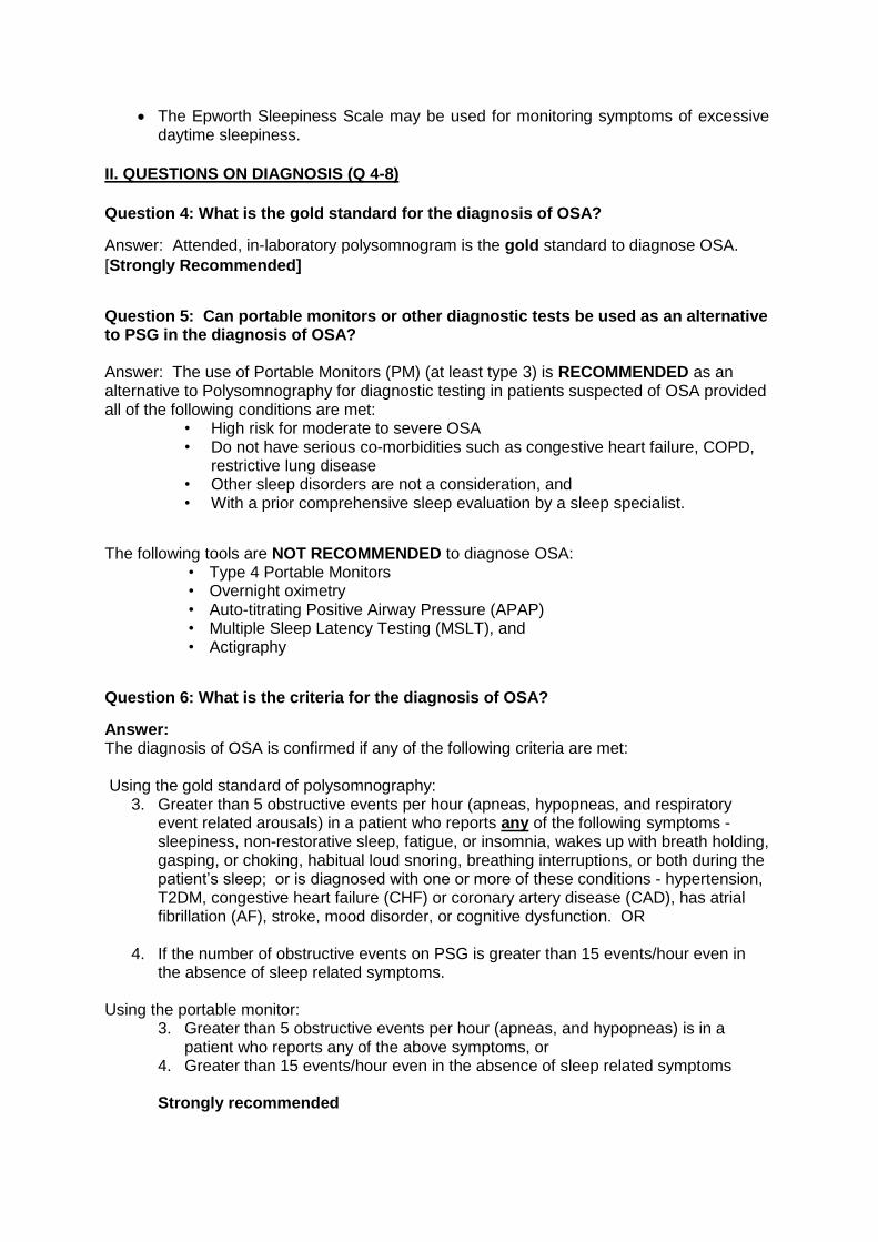
The Epworth Sleepiness Scale may be used for monitoring symptoms of excessive daytime sleepiness.
II. QUESTIONS ON DIAGNOSIS (Q 4-8)
Question 4: What is the gold standard for the diagnosis of OSA?
Answer: Attended, in-laboratory polysomnogram is the gold standard to diagnose OSA.
[Strongly Recommended]
Question 5: Can portable monitors or other diagnostic tests be used as an alternative to PSG in the diagnosis of OSA? Answer: The use of Portable Monitors (PM) (at least type 3) is RECOMMENDED as an alternative to Polysomnography for diagnostic testing in patients suspected of OSA provided all of the following conditions are met:
• High risk for moderate to severe OSA • Do not have serious co-morbidities such as congestive heart failure, COPD,
restrictive lung disease • Other sleep disorders are not a consideration, and • With a prior comprehensive sleep evaluation by a sleep specialist.
The following tools are NOT RECOMMENDED to diagnose OSA: • Type 4 Portable Monitors • Overnight oximetry • Auto-titrating Positive Airway Pressure (APAP) • Multiple Sleep Latency Testing (MSLT), and • Actigraphy
Question 6: What is the criteria for the diagnosis of OSA?
Answer: The diagnosis of OSA is confirmed if any of the following criteria are met: Using the gold standard of polysomnography:
3. Greater than 5 obstructive events per hour (apneas, hypopneas, and respiratory event related arousals) in a patient who reports any of the following symptoms - sleepiness, non-restorative sleep, fatigue, or insomnia, wakes up with breath holding, gasping, or choking, habitual loud snoring, breathing interruptions, or both during the patient’s sleep; or is diagnosed with one or more of these conditions - hypertension, T2DM, congestive heart failure (CHF) or coronary artery disease (CAD), has atrial fibrillation (AF), stroke, mood disorder, or cognitive dysfunction. OR
4. If the number of obstructive events on PSG is greater than 15 events/hour even in the absence of sleep related symptoms.
Using the portable monitor:
3. Greater than 5 obstructive events per hour (apneas, and hypopneas) is in a patient who reports any of the above symptoms, or
4. Greater than 15 events/hour even in the absence of sleep related symptoms
Strongly recommended

Question 7: What is the severity classification for OSA? Answer: OSA severity is classified as mild for RDI/AHI 5-14/hour, moderate for RDI/AHI 15-30/hour and severe for RDI/AHI > 30/hour.
This severity classification, similar to the diagnostic cut-offs for OSA is recommended by the International Classification of Sleep disorders (ICSD) and is generally used by all the sleep societies worldwide.
Question 8: What are the indications for doing follow-up PSG? Answer: Follow up PSG is not routinely indicated in patients treated with CPAP whose symptoms continue to be resolved with CPAP treatment. However, follow-up PSG is Strongly Recommended to be done routinely in the following situations:
3. For assessment of treatment results after surgical treatment for moderate to severe OSA; (routine)
4. To assess treatment result on CPAP after substantial weight loss (10% of body weight); substantial weight gain with return of symptoms while on CPAP; when clinical response is insufficient or when symptoms recur despite good initial response to CPAP. (routine)
III: Questions on the Management of OSA(Q 9-17)
Question 9: When should OSA be managed? Management using a multidisciplinary approach should commence once the diagnosis and
severity classification of OSA has been established. Strongly Recommend (Consensus)
Question 10: What are the goals of therapy for OSA? Answer: The goals of therapy for OSA are:
5. To improve symptoms (excessive sleepiness, concentration, snoring), quality of life and sexual intimacy.
6. To decrease AHI to <5, events/hour with no desaturations nor arousals 7. Improvement of associated comorbidities such as hypertension, arrhythmia, heart
failure, stroke, and hyperglycemia. 8. To prevent or minimize the risk for cardiovascular events and traffic accidents. Strongly recommend
Question 11: What is the primary treatment for Obstructive Sleep Apnea in Adults?
Answer: CPAP at a fixed pressure is the standard initial treatment of choice for OSA in adults.
It is strongly recommended for moderate to severe OSA and recommended only for mild
OSA.
CPAP should be used for at least 4 hours during sleep daily for optimal benefit. Strongly
Recommend
Question 12: What measures can be used to increase CPAP compliance?
Because compliance or adherence is a major determinant to the success of CPAP therapy in the long term, the following interventions can be used be used to improve compliance or adherence to its use:
• The addition of heated humidification (Recommend) • Use of BiPAP is an option in CPAP-intolerant patients (Recommend) • Pressure waveform modification technologies (i.e. pressure relief)
(Recommend-consensus) Trained health care providers should closely follow-up patients with OSA for compliance to PAP usage and to identify problems with its use in order to establish effective utilization patterns and remediate problems especially during the first few weeks of use. (Strongly recommend)
Generally, most studies have considered good adherence as use of the device for a minimum number of hours per night (~4 h in the literature) for 70% of the nights of the week. Question 13: What is the role of auto-titrating CPAP (APAP) in the management of OSA? Answer:
Auto-titrating CPAP is recommended as an alternative treatment to fixed CPAP for
OSA in patients who are poorly tolerant of fixed CPAP, and those with position
related and REM related OSA (Recommend)
Caution on its use must be exercised among those with chronic cardiopulmonary
disease (ie, COPD, restrictive chest disorders, congestive heart failure) because there are no studies on these populations. Recommend-Consensus
Question 14: Can auto-titrating CPAP (APAP) be used in determining fixed CPAP pressure in lieu of formal CPAP titration?
Answer: APAP use during an ambulatory titration procedure to determine a fixed
CPAP treatment pressure for patients with OSA in lieu of formal CPAP titration, is
currently NOT RECOMMENDED since studies are few and have not consistently
shown that APAP is equivalent to the standard titration.
Question 15: What is the role of the following interventions for the management of OSA?
15.1. Behavioral and Lifestyle Modifications.
All overweight and obese patients diagnosed with OSA should be encouraged to lose weight as medically supervised weight loss may improve the AHI. However it should be combined with primary treatment because of the low success rates for weight loss alone. Strongly recommend
Alcohol intake and routine use of sedatives among patients with OSA is discouraged. Recommend
15.2. Positional Therapy
Strategies that keep the patient in a non-supine position can be used as adjunctive treatment for positional OSA. Recommend.
15.3. Oxygen therapy
Oxygen supplementation is NOT RECOMMENDED as a sole treatment for OSA. 15.4. Pharmacologic Therapy
There is no accepted pharmacological treatment for OSA. Strongly recommend
15.5. What is the role of oral appliance therapy in OSA? The use of prefabricated non-custom, non-titratable oral appliance for OSA is NOT
RECOMMENDED for OSA.
The decision to use custom fitted titratable oral appliances must be made by a sleep specialist in conjunction with a dentist trained in sleep medicine. Recommend.
15.6 Other treatment options (unproven therapies).
The following treatment options are NOT recommended: nasal sprays, nasal strips and nose clips. There are also a number of “anti-snore” clothing or pillows that may have some evidence of benefit for positional OSA (please refer to previous section 16.2 on positional therapy).
The use of the Buteyko breathing exercise technique is currently not recommended due to limited clinical data on the use of this technique for OSA.
Question 16: When is surgery indicated for Obstructive Sleep Apnea?
Generally, surgery is not recommended for OSA (Consensus)
Among patients with OSA and significant obstructing anatomy (such as enlarged tonsils or deviated nasal septum), the recommendation to perform surgery must be made by a multidisciplinary team which includes the referring physician, sleep specialist and a qualified surgeon (Recommend-consensus).
Question 17: Which patients require urgent treatment for OSA?
• Any patient with known or suspected OSA with severe/unstable co-morbid conditions
may benefit from a referral to a sleep specialist for evaluation and/or possible initiation of CPAP or non-invasive ventilation
• Among patients with suspected OSA, a definitive PSG is recommended after stabilization of co-morbid condition to confirm the diagnosis of OSA.

Disclosures of potential conflicts of interest: Technical Review Committee members: Consultant, Sleep Laboratory: Dr. Virginia S. de los Reyes, Dr. Emelie B. Ojascastro, Dr. Dr. Rodolfo V. Dizon, Jr., Dr. Aileen G. Banzon, Dr. Maria Cecilia I. Jocson, Dr. Agnes T. Remulla; Owner sleep lab/stock holder: Dr. Rodolfo V. Dizon, Jr; Reader CPAP company: Dr. Virginia S. de los Reyes, Dr. Emelie B. Ojascastro, Dr. Rodolfo V. Dizon, Jr., Dr. Agnes T. Remulla; No conflict of interest: Dr. Richmond B. Ceniza, Dr. Manuel C. Jorge II, Dr. Abigail C. Zaraspe. Panelists: Consultant, Sleep Laboratory: Dr. Albert L. Rafanan, Dr. Keith A. Aguilera, Dr. Maria Encarnita B. Limpin; Owner sleep lab/ stock holder: Dr. Albert L. Rafanan; Reader CPAP company: Dr. Patrick L. Moral, No conflict of interest: Dr. Gil M. Vicente, Dr. Pio R. Pajarillo, Dr. Eduardo O. Yambao Jr., Dr. Charles Florendo, Dr. Herminia Chavez, Dr. Maria Antonia Yamamoto, Dr. Bien J. Matawaran, Dr. Marie Charisma L. de la Trinidad, Dr. Jonathan Michael Ele.