Embed Size (px)
Citation preview
clinical case report
Copy
right
© A
BE&
M to
dos o
s dire
itos r
eser
vado
s.
Arq Bras Endocrinol Metab. 2010;54/8738
Phenotypic variability in a family with x-linked adrenoleukodystrophy caused by the p.Trp132Ter mutationVariabilidade fenotípica em uma família com adrenoleucodistrofia ligada ao X causada pela mutação p.Trp132Ter
Fernanda Caroline Soardi1, Adriana Mangue Esquiaveto-Aun1,2, Gil Guerra-Júnior2, Sofia Helena Valente de Lemos-Marini2, Maricilda Palandi de Mello1
SUMMARYX-linked adrenoleukodystrophy (X-ALD) is an inherited disease with clinical heterogeneity varying from presymptomatic individuals to rapidly progressive cerebral ALD forms. This disea-se is characterized by increased concentration of very long chain fatty acids (VLCFAs) in plasma and in adrenal, testicular and nervous tissues. Affected individuals can be classified in different clinical settings, according to phenotypic expression and age at onset of initial symptoms. Mo-lecular defects in X-ALD individuals usually result from ABCD1 gene mutations. In the present report we describe clinical data and the ABCD1 gene study in two boys affected with the chil-dhood cerebral form that presented with different symptomatic manifestations at diagnosis. In addition, their maternal grandfather had been diagnosed with Addison’s disease indicating phe-notypic variation for X-ALD within this family. The mutation p.Trp132Ter was identified in both male patients; additionally, three females, out of eleven family members, were found to be he-terozygous after screening for this mutation. In the present report, the molecular analysis was especially important since one of the heterozygous females was in first stages of pregnancy. Therefore, depending on the fetus outcome, if male and p.Trp132Ter carrier, storage of the um-bilical cord blood should be recommended as hematopoietic stem cell transplantation could be considered as an option for treatment in the future. Arq Bras Endocrinol Metab. 2010;54(8):738-43
SUMÁRIOA adrenoleucodistrofia é uma doença genética com padrão de herança ligado ao X (X-ALD) que apresenta heterogeneidade clínica e varia desde a forma infantil cerebral severa até casos de indivíduos pré-sintomáticos. Essa doença é caracterizada pelo acúmulo de ácidos graxos de cadeia muito longa (VLCFA) no plasma, nas adrenais, nos testículos e no sistema nervoso. Indivíduos afetados podem apresentar diferentes formas clínicas, as quais são classificadas de acordo com a expressão fenotípica e a idade de aparecimento dos sintomas iniciais. Alterações moleculares em indivíduos com X-ALD são geralmente mutações no gene ABCD1. No presente trabalho, descrevemos os dados clínicos e a investigação molecular do gene ABCD1 em uma família com duas crianças do sexo masculino afetadas com a forma infantil cerebral, que apre-sentaram diferenças nas primeiras manifestações sintomáticas para o diagnóstico. Além disso, houve referência ao avô materno diagnosticado com doença de Addison’s, indicando a varia-bilidade fenotípica da X-ALD nessa família. A análise molecular indicou a mutação p.Trp132Ter nos dois pacientes masculinos, e três indivíduos do sexo feminino, entre os onze estudados, mostraram-se heterozigotos para mutação. O conhecimento molecular descrito no presente relato adquiriu maior importância uma vez que uma das portadoras da mutação apresentou-se nos primeiros estágios de gestação. Assim, poderá ser oferecida a possibilidade de armazena-mento de sangue de cordão umbilical para que se possa considerar, no futuro, o transplante de células-tronco hematopoiéticas como forma de tratamento, caso a criança seja do sexo mascu-lino e afetada. Arq Bras Endocrinol Metab. 2010;54(8):738-43
1 Center for Molecular Biology and Genetic Engineering (CBMEG), Universidade Estadual de Campinas (Unicamp), Campinas, SP, Brazil2 Pediatric Endocrinology, Department of Pediatrics, Faculdade de Ciências Médicas (FCM), Unicamp, Campinas, SP, Brazil
Correspondence to:Maricilda Palandi de MelloLaboratório de Genética Molecular Humana, Centro de Biologia Molecular e Engenharia Genética,Universidade Estadual de Campinas13083-875 − Campinas, SP, BrazilCaixa postal [email protected]
Received on Jul/30/2010Accepted on Nov/23/2010
Copy
right
© A
BE&
M to
dos o
s dire
itos r
eser
vado
s.
739Arq Bras Endocrinol Metab. 2010;54/8
Phenotypic variability for ABCD1 mutation
IntROdUctIOn
Adrenoleukodystrophy (X-ALD - MIM ID #300100) is an inherited X-linked disease charac-
terized by increased concentration of very long chain fatty acids (VLCFAs) in plasma that affects myelin, spi-nal cord, peripheral nerves, adrenal cortex and testis. The accumulation of VLCFA may also be observed in neonatal ALD, which is considered to be inherited as an autosomal recessive disorder. The incidence of X-ALD is estimated in 1:20.000 without differences among ethnic groups (1).
Affected individuals may present with different clini-cal forms that are classified according to phenotypic ex-pression and age at initial symptoms (Table 1). When symptoms are observed before 10 years of age it is char-acterized as the childhood cerebral form. It manifests as progressive neurological damage with rapid evolution, leading to a vegetative state within six months to two years after onset of symptoms, followed by death at vari-able ages. The second form is the adrenomyeloneuropa-thy (AMN), which presents initial symptoms in adult men with ages ranging from 20 years to middle-aged men. Affected individuals develop progressive weakness
in the legs, sphincter control abnormalities, and may also have serious cognitive and behavioral disturbances over the decades. A third variant is the cerebral AMN form that presents similar neurological findings in association with some other characteristics such as dementia or psy-chosis. These symptoms arise between 10 and 21 years of age denoting a delayed onset with slow evolution rates. The fourth form that can be distinguished is Ad-dison’s disease, which accounts for only 10% of affected individuals. In this case, initial signs appear at different ages between two years of age and adulthood (1,2). Ad-ditionally, asymptomatic forms have been identified in older men (3). Although it is an X-linked disease, 50% of female carriers may develop AMN signals manifesting as mild to moderate spastic paraparesis in middle-aged women or later with normal adrenal function (4).
X-ALD is caused by mutations in the ATP-binding cassette, subfamily D, member 1 gene (ABCD1 – MIM ID *300371). ABCD1 gene is located at Xq28 and encodes a peroxisomal adenosin triphosphate (ATP) – binding cassette transporter protein (ABCD1, ALDP) that plays a crucial role in VLCFA transport, or their coenyme A (CoA) derivatives into peroxisomes (5,6). If the protein function is impaired no beta oxidation of VL-CFAs occurs and they accumulate in body fluids and tis-sues leading either to neuroinflammation and demyelin-ation in the brain characterizing the cerebral childhood form or to axonal degeneration in spinal cord in AMN (7). The mechanisms of the disease pathogenesis remain unclear whereas it is possible that VLCFA cytotoxicity causes oxidative stress with subsequent neuroinflamma-tion followed by generalized peroximal dysfunction (8).
Neither VLCFA plasma concentration nor the type of mutation can usually predict the phenotype of X-ALD because a same ABCD1 gene mutation can be associated with different phenotypes (9-12).
The present study reports the phenotypic variabil-ity and the molecular investigation in a family with two affected male cousins with X-linked adrenoleu-kodystrophy. The ABCD1 gene sequencing indicated p.Trp132Ter mutation segregation in affected male individuals and in normal female carriers.
SUBJEctS And MEtHOdS
clinical data Case 1 is a previously healthy boy who presented the first symptoms at the age of 6 years. For the last eight
table 1. X-ALD phenotypes
Phenotypes clinical manifestations
Age at first symptoms Frequency
Males
Childhood cerebral form
Behavior changes, school failure,
dementia, psychoses, paralysis, epilepsy, loss of vision, loss of speech
Before 10 years ~ 35%
AMN Paraparesis, sphincter disturbances, sensory
changes, loss of coordination, pain,
impotence
Between 2nd and 4th decades
~ 40% to 45%
Addison’s disease only
Adrenocortical insufficiency
Between 2 years and adulthood
~ 10%
AMN cerebral Pure AMN plus dementia-like
behavioral disturbances, psychosis, epilepsy, aphasia, visual loss
Between 10 and 21 years
~ 5 to 10%
Asymptomatic None Older than 60 years
Increasing
Females
Asymptomatic None Any age ~ 50%
Symptomatic carriers
AMN (paraparesis) like with normal adrenal
function
Middle-aged or later
~ 50%
Copy
right
© A
BE&
M to
dos o
s dire
itos r
eser
vado
s.
740 Arq Bras Endocrinol Metab. 2010;54/8
Phenotypic variability for ABCD1 mutation
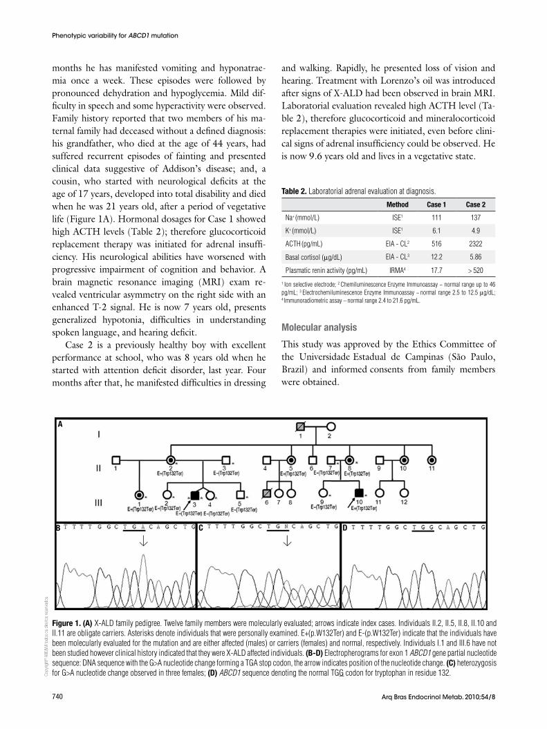
months he has manifested vomiting and hyponatrae-mia once a week. These episodes were followed by pronounced dehydration and hypoglycemia. Mild dif-ficulty in speech and some hyperactivity were observed. Family history reported that two members of his ma-ternal family had deceased without a defined diagnosis: his grandfather, who died at the age of 44 years, had suffered recurrent episodes of fainting and presented clinical data suggestive of Addison’s disease; and, a cousin, who started with neurological deficits at the age of 17 years, developed into total disability and died when he was 21 years old, after a period of vegetative life (Figure 1A). Hormonal dosages for Case 1 showed high ACTH levels (Table 2); therefore glucocorticoid replacement therapy was initiated for adrenal insuffi-ciency. His neurological abilities have worsened with progressive impairment of cognition and behavior. A brain magnetic resonance imaging (MRI) exam re-vealed ventricular asymmetry on the right side with an enhanced T-2 signal. He is now 7 years old, presents generalized hypotonia, difficulties in understanding spoken language, and hearing deficit.
Case 2 is a previously healthy boy with excellent performance at school, who was 8 years old when he started with attention deficit disorder, last year. Four months after that, he manifested difficulties in dressing
and walking. Rapidly, he presented loss of vision and hearing. Treatment with Lorenzo’s oil was introduced after signs of X-ALD had been observed in brain MRI. Laboratorial evaluation revealed high ACTH level (Ta-ble 2), therefore glucocorticoid and mineralocorticoid replacement therapies were initiated, even before clini-cal signs of adrenal insufficiency could be observed. He is now 9.6 years old and lives in a vegetative state.
table 2. Laboratorial adrenal evaluation at diagnosis.
Method case 1 case 2
Na+ (mmol/L) ISE1 111 137
K+ (mmol/L) ISE1 6.1 4.9
ACTH (pg/mL) EIA - CL2 516 2322
Basal cortisol (mg/dL) EIA - CL3 12.2 5.86
Plasmatic renin activity (pg/mL) IRMA4 17.7 > 520
1 Ion selective electrode; 2 Chemiluminescence Enzyme Immunoassay − normal range up to 46 pg/mL; 3 Electrochemiluminescence Enzyme Immunoassay − normal range 2.5 to 12.5 mg/dL; 4 Immunoradiometric assay – normal range 2.4 to 21.6 pg/mL.
Figure 1. (A) X-ALD family pedigree. Twelve family members were molecularly evaluated; arrows indicate index cases. Individuals II.2, II.5, II.8, II.10 and II.11 are obligate carriers. Asterisks denote individuals that were personally examined. E+(p.W132Ter) and E-(p.W132Ter) indicate that the individuals have been molecularly evaluated for the mutation and are either affected (males) or carriers (females) and normal, respectively. Individuals I.1 and III.6 have not been studied however clinical history indicated that they were X-ALD affected individuals. (B-d) Electropherograms for exon 1 ABCD1 gene partial nucleotide sequence: DNA sequence with the G>A nucleotide change forming a TGA stop codon, the arrow indicates position of the nucleotide change. (c) heterozygosis for G>A nucleotide change observed in three females; (d) ABCD1 sequence denoting the normal TGG codon for tryptophan in residue 132.
Molecular analysis
This study was approved by the Ethics Committee of the Universidade Estadual de Campinas (São Paulo, Brazil) and informed consents from family members were obtained.
A
B c d
Copy
right
© A
BE&
M to
dos o
s dire
itos r
eser
vado
s.
741Arq Bras Endocrinol Metab. 2010;54/8
Phenotypic variability for ABCD1 mutation
Genomic DNA from peripheral leukocytes was puri-fied by proteinase K lysis, phenol/chloroform extraction and ethanol precipitation using standard techniques (13). Each exon of the ABCD1 gene and its respective flanking regions were amplified by PCR from genomic DNA using primers designed with GeneRunner v3.1 free software (Table 3). PCR products were purified in 1% agarose gel electrophoresis with the Wizard SV Gel & PCR clean-up system (Promega, USA), and both sense and antisense strands were sequenced with the BigDye Terminator v3.1 Cycle Sequencing Kit (Ap-plied Biosystems, USA) using the same primers as for the PCR reactions. Chromas Lite and CLC Sequence Viewer v.5.0.1 free softwares were used to analyze and compare sequences with the reference ABCD1 gene se-quence (ENSG00000101986, www.ensembl.org).
exon 1 (Figure 1B, C, D). This change creates a stop codon at residue 132 and causes the nonsense mutation p.Trp132Ter. The screening for p.Trp132Ter mutation in the family revealed three heterozygous females out of eleven evaluated members (Figure 1A).
dIScUSSIOn
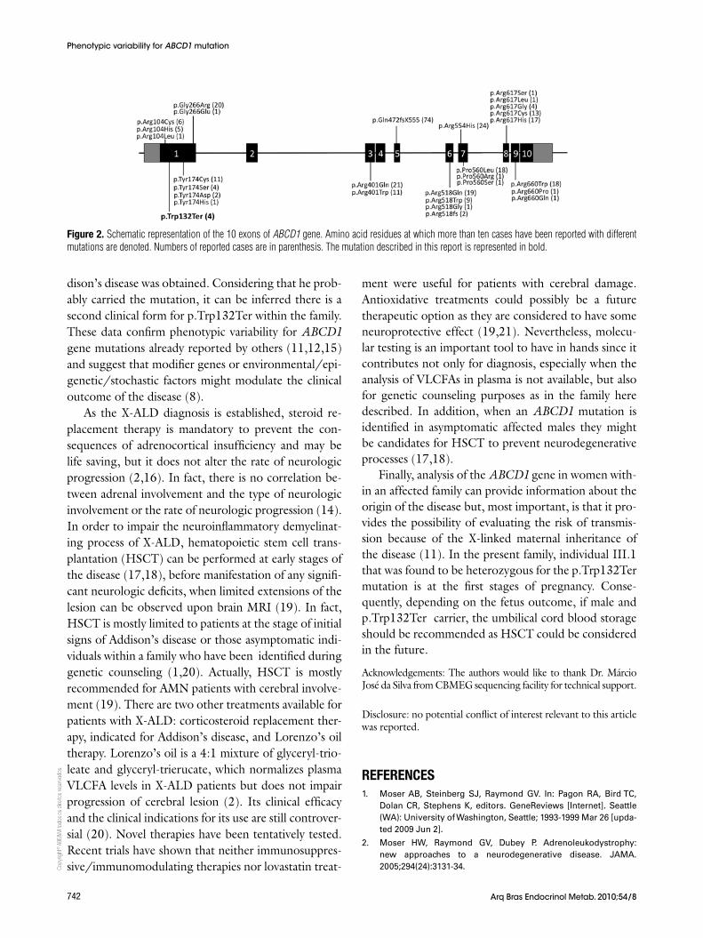
The present report describes a family with two boys af-fected by X-linked adrenoleukodystrophy. The study of the ABCD1 gene revealed mutation p.Trp132Ter as re-sponsible for the disease. Three female members of the family were found to carry the mutation including the pregnant sister of the index case. This nonsense muta-tion was previously described in a patient with child-hood cerebral adrenoleukodystrophy (11) additionally, the X-ALD Mutation Database (http://www.x-ald.nl/mutations-gene/mutations-in-abcd1) refers to three other unpublished cases with the p.Trp132Ter muta-tion. There are 1,133 mutations annotated for ABCD1 gene in the Mutation Database last updated on No-vember 12th, 2010 and almost 50% are recurrent. Ac-cording to database’s statistics 10% of total mutations are nonsense, 22% are frameshift, 61% are missense, and the other 7% correspond to insertions or dele-tions. There are some residues in ALDP that seem to be more susceptible to mutations since the same muta-tion or different mutations in the same amino acid have been described in different studies (Figure 2). How-ever, they do not represent mutation hotspots since they occur in almost every exon, except in exons 2, 4 and 10. Although expression and functional studies for p.Trp132Ter mutation have not been performed, it is putatively considered to result in the absence of ALDP likewise 74% of all mutations identified in ABCD1 gene.
The two patients described here as carrying the same mutation, present the childhood cerebral form of the disease although Case 1 has presented clini-cal signs of adrenal insufficiency more evidently than Case 2. It is well known that more than one clinical phenotype can manifest within a single X-ALD pedi-gree (9,10,12). Mild phenotypes have been described for some frameshift and nonsense mutations which are generally predicted to form a truncated transcript (12,14). This variability suggests that the X-linked ad-renoleukodystrophy phenotype may be influenced to a great extent by other genetic or environmental factors (8,11). As the family investigation progressed the in-formation that the maternal grandfather presented Ad-
table 3. Primers designed for amplification and sequencing of ABCD1 gene
Primer Sequence (5’-3’) Ta (°C)1 Size (bp)2
5’UTRs GGAGAAGGTGGAGAGGAAGAGA 62.2 1356
Int1as GGCCTGCCCACACCTTTG 62.9
Ex1s3 GGAGACGGGGCTGCTGGC 67.0
Ex1as3 CGACAGGAAGGTGCGGCTC 65.0
Int1s CAGCGTGTGTGAGTGGCA 57.2 717
Int2as GGCTTCCCACTCCTCTAAA 55.7
Int2s GCAGAAGAGCCTCGCCT 56.6 621
Int4as CAGAAGCACATGGAGGTCC 56.5
Int4s GACCTGGCTGTGTTCCCTAG 58.4 483
Int5as CCTTGGTCAATCCTGGTATCA 58.9
Int5s GAGATCAAGAATGGCCTGC 56.3 931
Int7as CCCTTCCCTAGAGCACCTG 58.4
Int7s GCACGATTCCAGTCCCCAC 61.7 1077
3’UTRas CGCCACCCTCCACATCTACT 61.2
Ex9s3 CTGCTCTCCATCACCCACCG 64.7
Int9as3 CAGGCAGGGAGACAGGGC 61.8
1 Anealing temperature used in PCR; 2 Size of amplified fragments; 3 Internal primers used for sequencing only.
RESULtS
Clinical and laboratorial data indicated that in Case 1, the initial presentation was adrenal insufficiency followed by mild to moderated neurological manifestations, whereas in Case 2 neurological damage was the first sign that evolved rapidly and drastically to a severe condition with-out clinical indicatives of adrenal insufficiency.
ABCD1 sequence analysis of the affected individu-als revealed the nucleotide change TGG>TGA within
Copy
right
© A
BE&
M to
dos o
s dire
itos r
eser
vado
s.
742 Arq Bras Endocrinol Metab. 2010;54/8
Figure 2. Schematic representation of the 10 exons of ABCD1 gene. Amino acid residues at which more than ten cases have been reported with different mutations are denoted. Numbers of reported cases are in parenthesis. The mutation described in this report is represented in bold.
dison’s disease was obtained. Considering that he prob-ably carried the mutation, it can be inferred there is a second clinical form for p.Trp132Ter within the family. These data confirm phenotypic variability for ABCD1 gene mutations already reported by others (11,12,15) and suggest that modifier genes or environmental/epi-genetic/stochastic factors might modulate the clinical outcome of the disease (8).
As the X-ALD diagnosis is established, steroid re-placement therapy is mandatory to prevent the con-sequences of adrenocortical insufficiency and may be life saving, but it does not alter the rate of neurologic progression (2,16). In fact, there is no correlation be-tween adrenal involvement and the type of neurologic involvement or the rate of neurologic progression (14). In order to impair the neuroinflammatory demyelinat-ing process of X-ALD, hematopoietic stem cell trans-plantation (HSCT) can be performed at early stages of the disease (17,18), before manifestation of any signifi-cant neurologic deficits, when limited extensions of the lesion can be observed upon brain MRI (19). In fact, HSCT is mostly limited to patients at the stage of initial signs of Addison’s disease or those asymptomatic indi-viduals within a family who have been identified during genetic counseling (1,20). Actually, HSCT is mostly recommended for AMN patients with cerebral involve-ment (19). There are two other treatments available for patients with X-ALD: corticosteroid replacement ther-apy, indicated for Addison’s disease, and Lorenzo’s oil therapy. Lorenzo’s oil is a 4:1 mixture of glyceryl-trio-leate and glyceryl-trierucate, which normalizes plasma VLCFA levels in X-ALD patients but does not impair progression of cerebral lesion (2). Its clinical efficacy and the clinical indications for its use are still controver-sial (20). Novel therapies have been tentatively tested. Recent trials have shown that neither immunosuppres-sive/immunomodulating therapies nor lovastatin treat-
ment were useful for patients with cerebral damage. Antioxidative treatments could possibly be a future therapeutic option as they are considered to have some neuroprotective effect (19,21). Nevertheless, molecu-lar testing is an important tool to have in hands since it contributes not only for diagnosis, especially when the analysis of VLCFAs in plasma is not available, but also for genetic counseling purposes as in the family here described. In addition, when an ABCD1 mutation is identified in asymptomatic affected males they might be candidates for HSCT to prevent neurodegenerative processes (17,18).
Finally, analysis of the ABCD1 gene in women with-in an affected family can provide information about the origin of the disease but, most important, is that it pro-vides the possibility of evaluating the risk of transmis-sion because of the X-linked maternal inheritance of the disease (11). In the present family, individual III.1 that was found to be heterozygous for the p.Trp132Ter mutation is at the first stages of pregnancy. Conse-quently, depending on the fetus outcome, if male and p.Trp132Ter carrier, the umbilical cord blood storage should be recommended as HSCT could be considered in the future.
Acknowledgements: The authors would like to thank Dr. Márcio José da Silva from CBMEG sequencing facility for technical support.
Disclosure: no potential conflict of interest relevant to this article was reported.
REFEREncES1. Moser AB, Steinberg SJ, Raymond GV. In: Pagon RA, Bird TC,
Dolan CR, Stephens K, editors. GeneReviews [Internet]. Seattle (WA): University of Washington, Seattle; 1993-1999 Mar 26 [upda-ted 2009 Jun 2].
2. Moser HW, Raymond GV, Dubey P. Adrenoleukodystrophy: new approaches to a neurodegenerative disease. JAMA. 2005;294(24):3131-34.
Phenotypic variability for ABCD1 mutation
Copy
right
© A
BE&
M to
dos o
s dire
itos r
eser
vado
s.
743Arq Bras Endocrinol Metab. 2010;54/8
3. Schönberger S, Roerig P, Schneider DT, Reifenberger G, Göbel U, Gär-tner J. Genotype and protein expression after bone marrow trans-plantation for adrenoleukodystrophy. Arch Neurol. 2007;64:651-7.
4. Moser HW, Smith KD, Watkins PA, Powers J, Moser AB. X-linked adrenoleukodystrophy. In: Scriver CR, Beaudet AL, Sly WS, Valle D, editors. The metabolic and molecular bases of inherited disea-se. New York: McGraw Hill; 2001. p. 3257-301.
5. Mosser J, Douar AM, Sarde CO, Kioschis P, Feil R, Moser H, et al. Putative X-linked adrenoleukodystrophy gene shares unexpected homology with ABC transporters. Nature. 1993;361:726-30.
6. Higgins CF. ABC transporters: from microorganisms to man. Ann Rev Cell Biol. 1992;8:67-113.
7. Kim JH, Kim HJ. Childhood X-linked adrenoleukodystrophy: clini-cal-pathologic overview and MR imaging manifestations at initial evaluation and follow-up. Radiographics. 2005;25(3):619-31.
8. Singh I, Pujol A. Pathomecanisms underlying X-adrenoleukodis-trophy: a three-hit hypothesis. Brain Pathol. 2010;20:838-44.
9. Power JM. Adreno-leukodystrophy (adreno-testiculo-leukomye-loneuropathiccomplex). Clin Neuropathol. 1985;4:191-99.
10. Power JM, Liu Y, Moser AB, Moser HW. The inflammatory myelino-pathy of adrenoleukodystrophy: cells, effector molecules, and pa-thogenetic implications. J Neuropathol Exp Neurol. 1992;51:630-43.
11. Pan H, Xiong H, Wu Y, Zhang YH, Bao XH, Jiang YW, et al. ABCD1 gene mutations in Chinese patients with X-linked adrenoleuko-dystrophy. Pediatr Neurol. 2005;33:114-20.
12. Takano H, Koike R, Onodera O, Sasaki R, Tsuji S. Mutational analysis and genotype-phenotype correlation of 29 unrelated Ja-panese patients with X-linked adrenoleukodystrophy. Arch Neu-rol. 1999;56:295-300.
13. Sambrook J, Fritsch EF, Maniatis TE. Molecular cloning, a labora-tory manual. Cold Spring Harbor, New York. 1989.
14. Cartier N, Aubourg P. Hematopoietic stem cell transplantation and hematopoietic stem cell gene therapy in X-linked adrenoleu-kodystrophy. Brain Pathol. 2010;20:857-62.
15. Krasemann EW, Meier V, Korenke GC, Hunneman DH, Hanefeld F. Identification of mutations in the ALD-gene of 20 families with adrenoleukodystrophy/ adrenomyeloneuropathy. Hum Genet. 1996;97:194-97.
16. Kemp S, Pujol A, Waterham HR, van Geel BM, Boehm CD, Ray-mond GV, et al. ABCD1 mutations and the X-linked adrenoleu-kodystrophy mutation database: role in diagnosis and clinical correlations. Hum Mut. 2001;18:499-515.
17. Mahmood A, Dubey P, Moser HW, Moser A. X-linked adrenoleu-kodystrophy: therapeutic approaches to distinct phenotypes. Pe-diatr Transplant. 2005;9(7):55-62.
18. Shapiro E, Krivit W, Lockman L, Jambaque I, Peters C, Cowan M, et al. Long-term effect of bone-marrow transplantation for childhood-on-set cerebral X-linked adrenoleukodystrophy. Lancet. 2000;356:713-8.
19. Peters C, Charnas LR, Tan Y, Ziegler RS, Shapiro EG, DeFor T, et al. Cerebral X-linked adrenoleukodystrophy: the international hematopoietic cell transplantation experience from 1982 to 1999. Blood. 2004;104:881-8.
20. Berger J, Pujol A, Aubourg P, Forss-Petter S. Current and future pharmacological treatment strategies in X-linked adrenoleuko-distrophy. Brain Pathol. 2010;20(4):845-56.
21. Halliwell B. Role of free radicals in the neurodegenerative dise-ases: therapeutic implications for antioxidant treatment. Drugs Aging. 2001;18:685-716.
Phenotypic variability for ABCD1 mutation


![Understanding gene expression variability in its ... · [16]Nathalie Balaban, Jack Merrin, Remy Chait, Lukasz Kowalik, and Stanislas Leibler. Bacterial persistence as a phenotypic](https://img.pdfslide.us/doc/110x75/5f0624117e708231d4167ee0/understanding-gene-expression-variability-in-its-16nathalie-balaban-jack.jpg)