Embed Size (px)
Citation preview
www.elsevier.com/locate/jphotobiol
Journal of Photochemistry and Photobiology B: Biology 86 (2007) 45–58
Review
Phenothiazinium derivatives for pathogeninactivation in blood products
Mark Wainwright a,*, Harald Mohr b, Wolfram H. Walker c
a School of Pharmacy and Chemistry, James Parsons Building, Liverpool John Moores University, Liverpool L3 3AF, UKb Blood Center for the German Red Cross Chapters of NStOB, Institute Springe, 31832 Springe, Germany
c MacoPharma International GmbH, Robert-Bosch-Str. 11, 63225 Langen, Germany
Received 6 May 2006; received in revised form 29 June 2006; accepted 4 July 2006Available online 18 September 2006
Abstract
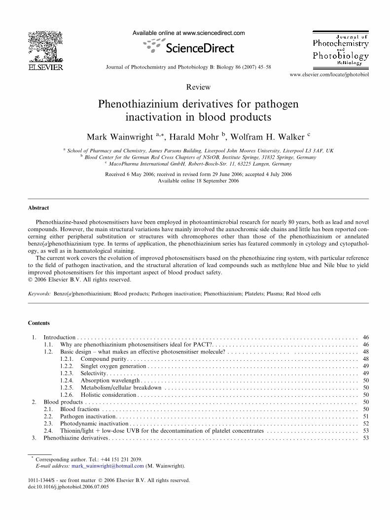
Phenothiazine-based photosensitisers have been employed in photoantimicrobial research for nearly 80 years, both as lead and novelcompounds. However, the main structural variations have mainly involved the auxochromic side chains and little has been reported con-cerning either peripheral substitution or structures with chromophores other than those of the phenothiazinium or annelatedbenzo[a]phenothiazinium type. In terms of application, the phenothiazinium series has featured commonly in cytology and cytopathol-ogy, as well as in haematological staining.
The current work covers the evolution of improved photosensitisers based on the phenothiazine ring system, with particular referenceto the field of pathogen inactivation, and the structural alteration of lead compounds such as methylene blue and Nile blue to yieldimproved photosensitisers for this important aspect of blood product safety.� 2006 Elsevier B.V. All rights reserved.
Keywords: Benzo[a]phenothiazinium; Blood products; Pathogen inactivation; Phenothiazinium; Platelets; Plasma; Red blood cells
Contents
1. Introduction . . . . . . . . . . . . . . . . . . . . . . . . . . . . . . . . . . . . . . . . . . . . . . . . . . . . . . . . . . . . . . . . . . . . . . . . . . . . . . . . . 46
1011-1
doi:10.
* CoE-m
1.1. Why are phenothiazinium photosensitisers ideal for PACT?. . . . . . . . . . . . . . . . . . . . . . . . . . . . . . . . . . . . . . . . . . . 461.2. Basic design – what makes an effective photosensitiser molecule? . . . . . . . . . . . . . . . . . . . . . . . . . . . . . . . . . . . . 48
1.2.1. Compound purity . . . . . . . . . . . . . . . . . . . . . . . . . . . . . . . . . . . . . . . . . . . . . . . . . . . . . . . . . . . . . . . . . . . . 481.2.2. Singlet oxygen generation . . . . . . . . . . . . . . . . . . . . . . . . . . . . . . . . . . . . . . . . . . . . . . . . . . . . . . . . . . . . . . 491.2.3. Selectivity. . . . . . . . . . . . . . . . . . . . . . . . . . . . . . . . . . . . . . . . . . . . . . . . . . . . . . . . . . . . . . . . . . . . . . . . . . 491.2.4. Absorption wavelength . . . . . . . . . . . . . . . . . . . . . . . . . . . . . . . . . . . . . . . . . . . . . . . . . . . . . . . . . . . . . . . . 501.2.5. Metabolism/cellular breakdown . . . . . . . . . . . . . . . . . . . . . . . . . . . . . . . . . . . . . . . . . . . . . . . . . . . . . . . . . 501.2.6. Holistic consideration . . . . . . . . . . . . . . . . . . . . . . . . . . . . . . . . . . . . . . . . . . . . . . . . . . . . . . . . . . . . . . . . . 50
2. Blood products . . . . . . . . . . . . . . . . . . . . . . . . . . . . . . . . . . . . . . . . . . . . . . . . . . . . . . . . . . . . . . . . . . . . . . . . . . . . . 50
2.1. Blood fractions . . . . . . . . . . . . . . . . . . . . . . . . . . . . . . . . . . . . . . . . . . . . . . . . . . . . . . . . . . . . . . . . . . . . . . . . . . . 502.2. Pathogen inactivation. . . . . . . . . . . . . . . . . . . . . . . . . . . . . . . . . . . . . . . . . . . . . . . . . . . . . . . . . . . . . . . . . . . . . . . 512.3. Photodynamic inactivation . . . . . . . . . . . . . . . . . . . . . . . . . . . . . . . . . . . . . . . . . . . . . . . . . . . . . . . . . . . . . . . . . . . 522.4. Thionin/light + low-dose UVB for the decontamination of platelet concentrates . . . . . . . . . . . . . . . . . . . . . . . . . . . 533. Phenothiazine derivatives. . . . . . . . . . . . . . . . . . . . . . . . . . . . . . . . . . . . . . . . . . . . . . . . . . . . . . . . . . . . . . . . . . . . . . . . 53
344/$ - see front matter � 2006 Elsevier B.V. All rights reserved.
1016/j.jphotobiol.2006.07.005
rresponding author. Tel.: +44 151 231 2039.ail address: [email protected] (M. Wainwright).
46 M. Wainwright et al. / Journal of Photochemistry and Photobiology B: Biology 86 (2007) 45–58
3.1. Phenothiazinium photosensitisers . . . . . . . . . . . . . . . . . . . . . . . . . . . . . . . . . . . . . . . . . . . . . . . . . . . . . . . . . . . . . . 53
3.1.1. Use in pathogen eradication in blood products . . . . . . . . . . . . . . . . . . . . . . . . . . . . . . . . . . . . . . . . . . . . . . 533.2. Phenothiazones . . . . . . . . . . . . . . . . . . . . . . . . . . . . . . . . . . . . . . . . . . . . . . . . . . . . . . . . . . . . . . . . . . . . . . . . . . . 54
3.2.1. Use in pathogen eradication. . . . . . . . . . . . . . . . . . . . . . . . . . . . . . . . . . . . . . . . . . . . . . . . . . . . . . . . . . 543.3. Benzo[a]phenothiazinium derivatives . . . . . . . . . . . . . . . . . . . . . . . . . . . . . . . . . . . . . . . . . . . . . . . . . . . . . . . . . . 553.4. Other annelated derivatives . . . . . . . . . . . . . . . . . . . . . . . . . . . . . . . . . . . . . . . . . . . . . . . . . . . . . . . . . . . . . . . . . . 56
4. Conclusion . . . . . . . . . . . . . . . . . . . . . . . . . . . . . . . . . . . . . . . . . . . . . . . . . . . . . . . . . . . . . . . . . . . . . . . . . . . . . . . . 565. Abbreviations . . . . . . . . . . . . . . . . . . . . . . . . . . . . . . . . . . . . . . . . . . . . . . . . . . . . . . . . . . . . . . . . . . . . . . . . . . . . . . 56
References . . . . . . . . . . . . . . . . . . . . . . . . . . . . . . . . . . . . . . . . . . . . . . . . . . . . . . . . . . . . . . . . . . . . . . . . . . . . . . . . . . . 56
1. Introduction
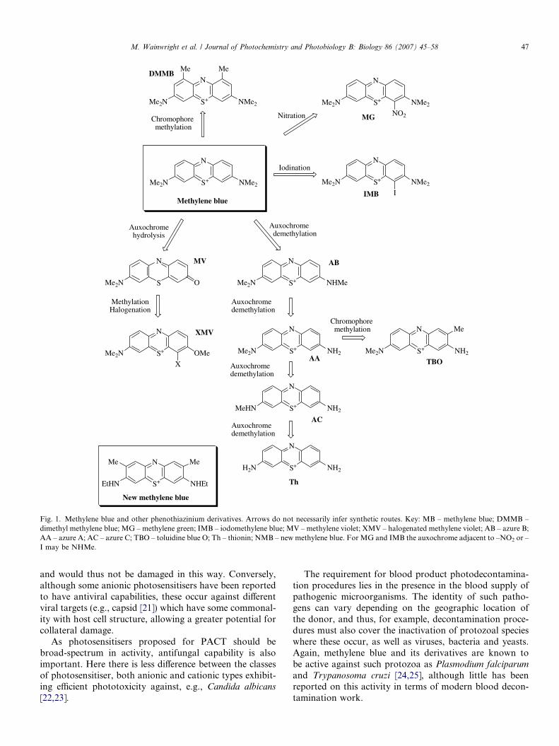
Methylene blue (Fig. 1) has been used as a lead com-pound in conventional antimicrobial research for over ahundred years. Since the first publication of its efficacy asan antimalarial compound in 1891, it has also beenemployed as an antibacterial, e.g., in local antisepsis andagainst tuberculosis [1–3] and its low toxicity in man isreflected in its current clinical use in methaemoglobinaemia[4]. While its long usage may be explained by the scarcity ofactive compounds available early in the last century, it con-tinues to be used as a lead antimicrobial compound, e.g., inrecent antimalarial research [5–7]. However, it is unlikelythat methylene blue is the optimum compond among phe-nothiazine derivatives for antimicrobial purposes in allcases. Unsurprisingly, there are few records covering therange of compounds examined before the Second WorldWar, but the number is likely to be limited, as in recentyears, to alteration of the auxochromic amino groups(Fig. 2). In addition, organised screening of compoundsfor photodynamic activity was not carried out at that time.
Phenothiazinium synthesis is well established, havingbeen reported as early as 1876. However, the reagentsrequired to furnish the oxidised character of the chromo-phore have often deterred approaches to more complexmolecules, e.g., having ring functionalisation/elaborationbeyond the 3,7-bis-auxochromic groups. Workers in thisarea have normally relied on functionalisation of theN-alkyl moieties for novelty, as this may be carried outafter chromophore construction (e.g., the anticancer bis-nitrogen mustard analogue of methylene blue, Fig. 3 [8]).
In addition many groups investigating photoantimicro-bial applications have used only standard photosensitiserssuch as methylene blue and toluidine blue (Fig. 1) withoutattempting to synthesise more active analogues. Logically,in terms of medicinal chemistry, there is little justificationfor the acceptance of either of these compounds as the sine
qua non among phenothiazinium derivatives. Indeed, a fewmore adventurous groups have synthesised both moreactive and more toxic analogues, as would be expected inmodern drug discovery programmes [9,10].
Since the renaissance in photodynamic therapy (PDT) asa novel approach to cancer treatment 20 years ago, manynew compounds have appeared in the literature, represent-ing a wide range of chromophoric types. Among these,photosensitisers based on the phenothiazinium nucleus rep-
resent a major contribution [11], with benzannelation fur-nishing new groups of compounds, although againusually within the limits of auxochromic variation(Fig. 2) [12]. However, in the field of photoantimicrobialchemotherapy (PACT [13]), it has become apparent thatcationic photosensitisers producing significant yields of sin-glet oxygen are of premier importance, particularly interms of the scope of useful activity, e.g., against both
Gram-positive and Gram-negative bacteria [14], as wellas viruses and yeasts [13]. Within this group, the phenothi-aziniums and their congeners are ideally placed to act aslead compounds in terms of drug discovery.
1.1. Why are phenothiazinium photosensitisers ideal for
PACT?
Among the aniline dyes invented in the nineteenth cen-tury, phenothiazine derivatives including methylene blue,the azures and toluidine blue were quickly adopted fortheir utility in biological staining. It is no surprise thatthese dyes were subsequently thoroughly investigated invarious areas of biomedicine [15].
Since methylene blue and its close congeners are selec-tively taken up by microbial cells (i.e., in the presence ofanimal cells), the fact that most reported phenothiaziniumsalts are also photosensitisers producing significant yieldsof singlet oxygen should be sufficient reason for their wide-spread examination in PACT. That this has not been thecase perhaps reflects the dominance of porphyrin-basedphotosensitisers from the related anti-cancer field of photo-dynamic therapy (PDT). There is, of course, far less ratio-nale for the use of anionic porphyrins in PACT,particularly where there is a need for broad-spectrum anti-microbial activity.
It has been shown recently that cationic photosensitisersare much more efficient as broad-spectrum antibacterialsthan are their anionic or neutral congeners [16,17]. Thisis due to the greater complexity of the Gram-negative cellwall, which is less permeable to anionic species, but is dis-rupted by cationics [18,19].
In terms of antiviral agents, the positive charge of phe-nothiazinium derivatives allows efficient binding to viralnucleic acid – thus the considerable use of methylene blueand its congeners as nucleic acid probes [20]. In the disin-fection of blood products this is given extra importancesince such products do not contain viable nucleic acid
N
S+Me2N NMe2
N
S+Me2N NH2
N
S+Me2N NMe2
N
S+EtHN NHEt
N
S+Me2N NHMe
N
S+Me2N NH2
N
S+MeHN NH2
N
S+H2N NH2
N
S+Me2N OMe
N
SMe2N O
Me
Me Me
MeMe
N
S+Me2N NMe2
NO2
N
S+Me2N NMe2
I
X
Methylene blue
Chromophore methylation
Nitration
Iodination
Auxochrome hydrolysis
Auxochrome demethylation
Chromophore methylation
MethylationHalogenation
Auxochromedemethylation
Auxochromedemethylation
Auxochromedemethylation
New methylene blue
DMMB
MG
IMB
MV
XMV
AB
AA TBO
AC
Th
Fig. 1. Methylene blue and other phenothiazinium derivatives. Arrows do not necessarily infer synthetic routes. Key: MB – methylene blue; DMMB –dimethyl methylene blue; MG – methylene green; IMB – iodomethylene blue; MV – methylene violet; XMV – halogenated methylene violet; AB – azure B;AA – azure A; AC – azure C; TBO – toluidine blue O; Th – thionin; NMB – new methylene blue. For MG and IMB the auxochrome adjacent to –NO2 or –I may be NHMe.
M. Wainwright et al. / Journal of Photochemistry and Photobiology B: Biology 86 (2007) 45–58 47
and would thus not be damaged in this way. Conversely,although some anionic photosensitisers have been reportedto have antiviral capabilities, these occur against differentviral targets (e.g., capsid [21]) which have some commonal-ity with host cell structure, allowing a greater potential forcollateral damage.
As photosensitisers proposed for PACT should bebroad-spectrum in activity, antifungal capability is alsoimportant. Here there is less difference between the classesof photosensitiser, both anionic and cationic types exhibit-ing efficient phototoxicity against, e.g., Candida albicans
[22,23].
The requirement for blood product photodecontamina-tion procedures lies in the presence in the blood supply ofpathogenic microorganisms. The identity of such patho-gens can vary depending on the geographic location ofthe donor, and thus, for example, decontamination proce-dures must also cover the inactivation of protozoal specieswhere these occur, as well as viruses, bacteria and yeasts.Again, methylene blue and its derivatives are known tobe active against such protozoa as Plasmodium falciparum
and Trypanosoma cruzi [24,25], although little has beenreported on this activity in terms of modern blood decon-tamination work.
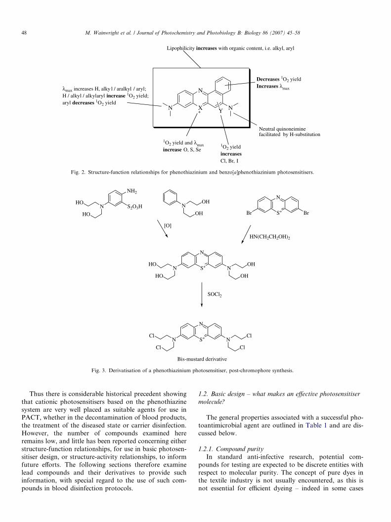
Decreases 1O2 yieldIncreases maxmax increases H, alky l / aralkyl / aryl;
H / alkyl / alkylaryl increase 1O2 yield;aryl decreases 1O2 yield
1O2 yield and maxincrease O, S, Se
Lipophilicity increases with organic content, i.e. alkyl, aryl
Neutral quinoneimine facilitated by H-substitution
1O2 yieldincreasesCl, Br, I
X+
N
N NY
Fig. 2. Structure-function relationships for phenothiazinium and benzo[a]phenothiazinium photosensitisers.
N
NH2
S2O3HHO
HO
NOH
OH S+
N
Br Br
HN(CH2CH2OH)2
[O]
S+
N
N NHO
HO
OH
OH
SOCl2
S+
N
N NCl
Cl
Cl
Cl
Bis-mustard derivative
Fig. 3. Derivatisation of a phenothiazinium photosensitiser, post-chromophore synthesis.
48 M. Wainwright et al. / Journal of Photochemistry and Photobiology B: Biology 86 (2007) 45–58
Thus there is considerable historical precedent showingthat cationic photosensitisers based on the phenothiazinesystem are very well placed as suitable agents for use inPACT, whether in the decontamination of blood products,the treatment of the diseased state or carrier disinfection.However, the number of compounds examined hereremains low, and little has been reported concerning eitherstructure-function relationships, for use in basic photosen-sitiser design, or structure-activity relationships, to informfuture efforts. The following sections therefore examinelead compounds and their derivatives to provide suchinformation, with special regard to the use of such com-pounds in blood disinfection protocols.
1.2. Basic design – what makes an effective photosensitiser
molecule?
The general properties associated with a successful pho-toantimicrobial agent are outlined in Table 1 and are dis-cussed below.
1.2.1. Compound purity
In standard anti-infective research, potential com-pounds for testing are expected to be discrete entities withrespect to molecular purity. The concept of pure dyes inthe textile industry is not usually encountered, as this isnot essential for efficient dyeing – indeed in some cases
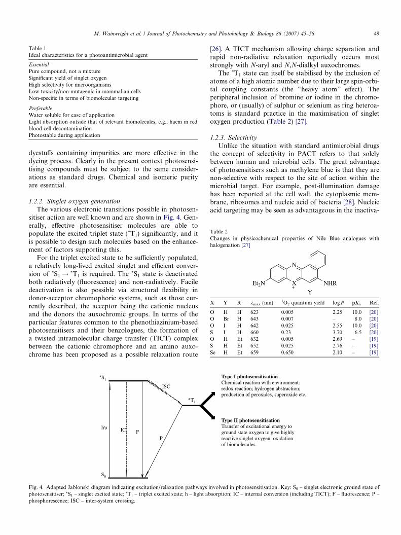
Table 1Ideal characteristics for a photoantimicrobial agent
Essential
Pure compound, not a mixtureSignificant yield of singlet oxygenHigh selectivity for microorganismsLow toxicity/non-mutagenic in mammalian cellsNon-specific in terms of biomolecular targeting
Preferable
Water soluble for ease of applicationLight absorption outside that of relevant biomolecules, e.g., haem in redblood cell decontaminationPhotostable during application
Table 2Changes in physicochemical properties of Nile Blue analogues withhalogenation [27]
X+
N
Et2N NHR
Y
X Y R kmax (nm) 1O2 quantum yield logP pKa Ref.
O H H 623 0.005 2.25 10.0 [20]O Br H 643 0.007 – 8.0 [20]O I H 642 0.025 2.55 10.0 [20]S I H 660 0.23 3.70 6.5 [20]O H Et 632 0.005 2.69 – [19]S H Et 652 0.025 2.76 – [19]Se H Et 659 0.650 2.10 – [19]
M. Wainwright et al. / Journal of Photochemistry and Photobiology B: Biology 86 (2007) 45–58 49
dyestuffs containing impurities are more effective in thedyeing process. Clearly in the present context photosensi-tising compounds must be subject to the same consider-ations as standard drugs. Chemical and isomeric purityare essential.
1.2.2. Singlet oxygen generation
The various electronic transitions possible in photosen-sitiser action are well known and are shown in Fig. 4. Gen-erally, effective photosensitiser molecules are able topopulate the excited triplet state (*T1) significantly, and itis possible to design such molecules based on the enhance-ment of factors supporting this.
For the triplet excited state to be sufficiently populated,a relatively long-lived excited singlet and efficient conver-sion of *S1! *T1 is required. The *S1 state is deactivatedboth radiatively (fluorescence) and non-radiatively. Faciledeactivation is also possible via structural flexibility indonor-acceptor chromophoric systems, such as those cur-rently described, the acceptor being the cationic nucleusand the donors the auxochromic groups. In terms of theparticular features common to the phenothiazinium-basedphotosensitisers and their benzologues, the formation ofa twisted intramolecular charge transfer (TICT) complexbetween the cationic chromophore and an amino auxo-chrome has been proposed as a possible relaxation route
h
S0
*S1
ISC
IC FP
*T1
Fig. 4. Adapted Jablonski diagram indicating excitation/relaxation pathwaysphotosensitiser; *S1 – singlet excited state; *T1 – triplet excited state; h – light abphosphorescence; ISC – inter-system crossing.
[26]. A TICT mechanism allowing charge separation andrapid non-radiative relaxation reportedly occurs moststrongly with N-aryl and N,N-dialkyl auxochromes.
The *T1 state can itself be stabilised by the inclusion ofatoms of a high atomic number due to their large spin-orbi-tal coupling constants (the ‘‘heavy atom’’ effect). Theperipheral inclusion of bromine or iodine in the chromo-phore, or (usually) of sulphur or selenium as ring heteroa-toms is standard practice in the maximisation of singletoxygen production (Table 2) [27].
1.2.3. Selectivity
Unlike the situation with standard antimicrobial drugsthe concept of selectivity in PACT refers to that solelybetween human and microbial cells. The great advantageof photosensitisers such as methylene blue is that they arenon-selective with respect to the site of action within themicrobial target. For example, post-illumination damagehas been reported at the cell wall, the cytoplasmic mem-brane, ribosomes and nucleic acid of bacteria [28]. Nucleicacid targeting may be seen as advantageous in the inactiva-
Type I photosensitisationChemical reaction with environment:redox reaction; hydrogen abstraction;production of peroxides, superoxide etc.
Type II photosensitisationTransfer of excitational energy toground state oxygen to give highlyreactive singlet oxygen: oxidationof biomolecules.
involved in photosensitisation. Key: S0 – singlet electronic ground state ofsorption; IC – internal conversion (including TICT); F – fluorescence; P –
50 M. Wainwright et al. / Journal of Photochemistry and Photobiology B: Biology 86 (2007) 45–58
tion of viruses in blood products, e.g., plasma, but this isnot essential. Indeed although the DNA-intercalative nat-ure of methylene blue is well-established, the use of thisphotosensitiser with HIV-1 produced post illuminationdamage to enzymes such as reverse transcriptase and theviral capsid as well as nucleic acid [29].
1.2.4. Absorption wavelength
Photosensitisers used for biological application mustabsorb light outside that of the operative environment.For the present case, the most important example is givenby haem absorption in red blood cells. Realistically here,photosensitisers must exhibit significant light absorption>630 nm in order to be effective. The requirement in plate-let concentrates and plasma, having little visible absorp-tion, is less stringent.
For a given photosensitiser, both benzannelation andthe inclusion of heavy atoms usually increase the maximumwavelength of absorption considerably. The modification/elaboration of auxochromic side chains has a lesser effect(Fig. 2).
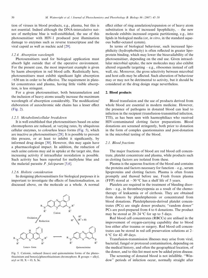
1.2.5. Metabolism/cellular breakdown
It is well established that photosensitisers based on azinechromophores are reduced, at varying rates, by ubiquitouscellular enzymes, to colourless leuco forms (Fig. 5), whichare inactive as photosensitisers [28]. It is possible to preventthis process, or at least to inhibit it significantly, byinformed drug design [30]. However, this may again havea pharmacological impact. In addition, the reduction ofsuch azine cations may aid in uptake at the target site, thusincreasing activity if intracellular reoxidation is possible.Such activity has been reported for methylene blue andthe malarial parasite P. falciparum [5,6].
1.2.6. Holistic consideration
In designing photosensitisers for biological purposes it isimportant to investigate the effects of functionalisation, asdiscussed above, on the molecule as a whole. A normal
X+
N
N NR
R1
R3
R2
X
HN
N NR
R1
R3
R2
X
N
N NR
R1 R2
ReductionOxidation -H+
(R3 = H)H+
Cation
Leuco- form Quinoneimine
Fig. 5. Cationic, reduced (leuco) and quinoneimine forms of the pheno-thiazinium and benzo[a]phenothiazinium chromophore. R groups = alkyl,aryl or H; X = O, S, Se.
effect either of ring annelation/expansion or of heavy atomsubstitution is that of increased lipophilicity, – the newmolecule exhibits increased organic partitioning, e.g., intolipids in biological media (or, in vitro, in the standard aque-ous buffer-octanol system).
In terms of biological behaviour, such increased lipo-philicity (hydrophobicity) is often reflected in greater lipo-protein binding, which may lower the bioavailability of thephotosensitiser, depending on the end use. Given intracel-lular microbial uptake, the new molecules may also exhibitaltered organelle targeting – e.g., ribosomes instead of cellwall, etc. Moreover, the gross selectivity between microbialand host cells may be affected. Such alteration of behaviourmay or may not be detrimental to activity, but it should beconsidered at the drug design stage nevertheless.
2. Blood products
Blood transfusion and the use of products derived fromwhole blood are essential in modern medicine. However,the presence of pathogens in donated blood can lead toinfection in the recipient (transfusion-transmitted infection,TTI), as has been seen with haemophiliacs who receivedHIV-contaminated clotting factor preparations. Blooddonations are screened stringently, both prior to donationin the form of complex questionnaires and post-donationin the microbial testing of the blood.
2.1. Blood fractions
The major fractions of blood are red blood cell concen-trate, platelet concentrate and plasma, while products suchas clotting factors are isolated from these.
Plasma is the aqueous fraction of the blood and containsthe proteins and factors necessary for function – e.g., serumlipoproteins and clotting factors. Plasma is often frozenpromptly and thawed before use. Fresh frozen plasma(FFP) stored at �30 �C has a shelf life of 5 years.
Platelets are required in the treatment of bleeding disor-ders – e.g., in thrombocytopenia as a result of the chemo-therapy of leukaemia or of cirrhosis. They are obtainedfrom donors by plateletpheresis or concentrated fromblood donations. Plateletpheresis-derived platelet concen-trates (PCs) are single donor products; ‘‘random donor’’PCs are pool-prepared from 4 to 6 donations. The productmay be stored at 20–24 �C for up to 5 days.
Red blood cell concentrates (RBCCs) are utilised in theimprovement of oxygen-carrying capability due to bloodloss either after trauma or surgery. Red blood cell concen-trates can be stored in red cell preservation solutions at 2–6 �C for 42–49 days.
Transfusion-transmission illnesses may arise from viral,bacterial, fungal or protozoal contamination, depending onthe medical history, and often the geographical location, ofthe donor, and to this list must now be added prion disease.
The screening of donated blood is not infallible. ‘‘Win-dow’’ periods of infection occur, normally straight after
M. Wainwright et al. / Journal of Photochemistry and Photobiology B: Biology 86 (2007) 45–58 51
colonisation, in which the microbe is undetectable by sero-logical testing [31]. Blood donation during such a periodcould obviously lead to transfusion-transmission of dis-ease. In addition, the large-scale manufacture of bloodproducts utilizing the principle of ‘‘pooling’’, i.e., combin-ing many blood fractions from different donors, has a con-comitant increase in the risk of resultant infection. In termsof the elimination of the disease threat, there are problemsin selectivity, the degree of complexity depending on theblood fraction concerned.
2.2. Pathogen inactivation
Current approaches to pathogenic inactivation inplasma include heating (e.g., pasteurization), UV treatmentor nanofiltration. Unfortunately, none of these is com-pletely effective, nor without the disadvantage of collateraldamage, e.g., to proteins.
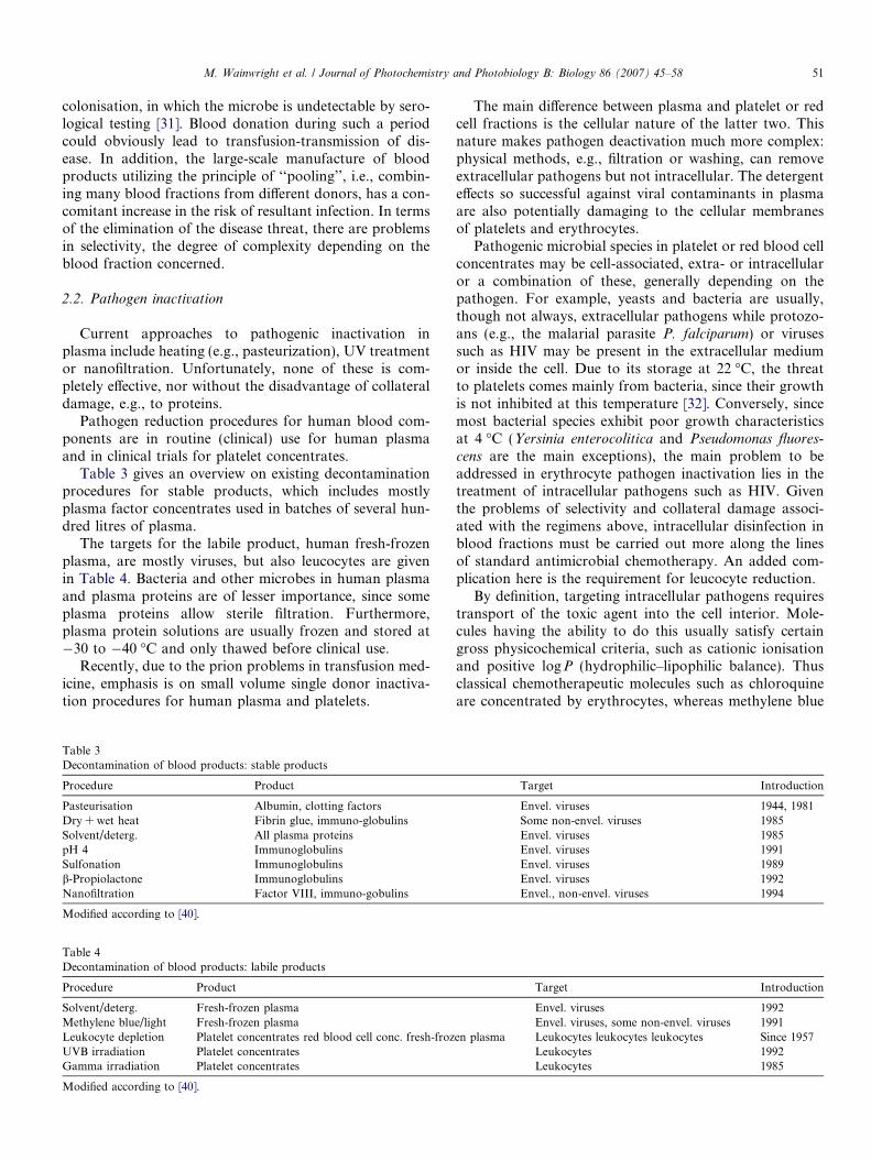
Pathogen reduction procedures for human blood com-ponents are in routine (clinical) use for human plasmaand in clinical trials for platelet concentrates.
Table 3 gives an overview on existing decontaminationprocedures for stable products, which includes mostlyplasma factor concentrates used in batches of several hun-dred litres of plasma.
The targets for the labile product, human fresh-frozenplasma, are mostly viruses, but also leucocytes are givenin Table 4. Bacteria and other microbes in human plasmaand plasma proteins are of lesser importance, since someplasma proteins allow sterile filtration. Furthermore,plasma protein solutions are usually frozen and stored at�30 to �40 �C and only thawed before clinical use.
Recently, due to the prion problems in transfusion med-icine, emphasis is on small volume single donor inactiva-tion procedures for human plasma and platelets.
Table 3Decontamination of blood products: stable products
Procedure Product
Pasteurisation Albumin, clotting factorsDry + wet heat Fibrin glue, immuno-globulinsSolvent/deterg. All plasma proteinspH 4 ImmunoglobulinsSulfonation Immunoglobulinsb-Propiolactone ImmunoglobulinsNanofiltration Factor VIII, immuno-gobulins
Modified according to [40].
Table 4Decontamination of blood products: labile products
Procedure Product
Solvent/deterg. Fresh-frozen plasmaMethylene blue/light Fresh-frozen plasmaLeukocyte depletion Platelet concentrates red blood cell conc. fresh-frozUVB irradiation Platelet concentratesGamma irradiation Platelet concentrates
Modified according to [40].
The main difference between plasma and platelet or redcell fractions is the cellular nature of the latter two. Thisnature makes pathogen deactivation much more complex:physical methods, e.g., filtration or washing, can removeextracellular pathogens but not intracellular. The detergenteffects so successful against viral contaminants in plasmaare also potentially damaging to the cellular membranesof platelets and erythrocytes.
Pathogenic microbial species in platelet or red blood cellconcentrates may be cell-associated, extra- or intracellularor a combination of these, generally depending on thepathogen. For example, yeasts and bacteria are usually,though not always, extracellular pathogens while protozo-ans (e.g., the malarial parasite P. falciparum) or virusessuch as HIV may be present in the extracellular mediumor inside the cell. Due to its storage at 22 �C, the threatto platelets comes mainly from bacteria, since their growthis not inhibited at this temperature [32]. Conversely, sincemost bacterial species exhibit poor growth characteristicsat 4 �C (Yersinia enterocolitica and Pseudomonas fluores-
cens are the main exceptions), the main problem to beaddressed in erythrocyte pathogen inactivation lies in thetreatment of intracellular pathogens such as HIV. Giventhe problems of selectivity and collateral damage associ-ated with the regimens above, intracellular disinfection inblood fractions must be carried out more along the linesof standard antimicrobial chemotherapy. An added com-plication here is the requirement for leucocyte reduction.
By definition, targeting intracellular pathogens requirestransport of the toxic agent into the cell interior. Mole-cules having the ability to do this usually satisfy certaingross physicochemical criteria, such as cationic ionisationand positive logP (hydrophilic–lipophilic balance). Thusclassical chemotherapeutic molecules such as chloroquineare concentrated by erythrocytes, whereas methylene blue
Target Introduction
Envel. viruses 1944, 1981Some non-envel. viruses 1985Envel. viruses 1985Envel. viruses 1991Envel. viruses 1989Envel. viruses 1992Envel., non-envel. viruses 1994
Target Introduction
Envel. viruses 1992Envel. viruses, some non-envel. viruses 1991
en plasma Leukocytes leukocytes leukocytes Since 1957Leukocytes 1992Leukocytes 1985
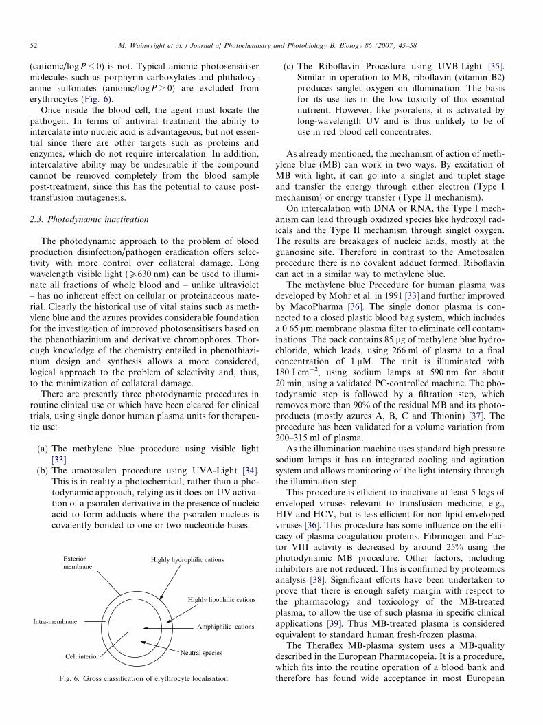
52 M. Wainwright et al. / Journal of Photochemistry and Photobiology B: Biology 86 (2007) 45–58
(cationic/logP < 0) is not. Typical anionic photosensitisermolecules such as porphyrin carboxylates and phthalocy-anine sulfonates (anionic/logP > 0) are excluded fromerythrocytes (Fig. 6).
Once inside the blood cell, the agent must locate thepathogen. In terms of antiviral treatment the ability tointercalate into nucleic acid is advantageous, but not essen-tial since there are other targets such as proteins andenzymes, which do not require intercalation. In addition,intercalative ability may be undesirable if the compoundcannot be removed completely from the blood samplepost-treatment, since this has the potential to cause post-transfusion mutagenesis.
2.3. Photodynamic inactivation
The photodynamic approach to the problem of bloodproduction disinfection/pathogen eradication offers selec-tivity with more control over collateral damage. Longwavelength visible light (P630 nm) can be used to illumi-nate all fractions of whole blood and – unlike ultraviolet– has no inherent effect on cellular or proteinaceous mate-rial. Clearly the historical use of vital stains such as meth-ylene blue and the azures provides considerable foundationfor the investigation of improved photosensitisers based onthe phenothiazinium and derivative chromophores. Thor-ough knowledge of the chemistry entailed in phenothiazi-nium design and synthesis allows a more considered,logical approach to the problem of selectivity and, thus,to the minimization of collateral damage.
There are presently three photodynamic procedures inroutine clinical use or which have been cleared for clinicaltrials, using single donor human plasma units for therapeu-tic use:
(a) The methylene blue procedure using visible light[33].
(b) The amotosalen procedure using UVA-Light [34].This is in reality a photochemical, rather than a pho-todynamic approach, relying as it does on UV activa-tion of a psoralen derivative in the presence of nucleicacid to form adducts where the psoralen nucleus iscovalently bonded to one or two nucleotide bases.
Amphiphilic cations
Highly hydrophilic cations
Highly lipophilic cations
Exteriormembrane
Intra-membrane
Cell interior Neutral species
Fig. 6. Gross classification of erythrocyte localisation.
(c) The Riboflavin Procedure using UVB-Light [35].Similar in operation to MB, riboflavin (vitamin B2)produces singlet oxygen on illumination. The basisfor its use lies in the low toxicity of this essentialnutrient. However, like psoralens, it is activated bylong-wavelength UV and is thus unlikely to be ofuse in red blood cell concentrates.
As already mentioned, the mechanism of action of meth-ylene blue (MB) can work in two ways. By excitation ofMB with light, it can go into a singlet and triplet stageand transfer the energy through either electron (Type Imechanism) or energy transfer (Type II mechanism).
On intercalation with DNA or RNA, the Type I mech-anism can lead through oxidized species like hydroxyl rad-icals and the Type II mechanism through singlet oxygen.The results are breakages of nucleic acids, mostly at theguanosine site. Therefore in contrast to the Amotosalenprocedure there is no covalent adduct formed. Riboflavincan act in a similar way to methylene blue.
The methylene blue Procedure for human plasma wasdeveloped by Mohr et al. in 1991 [33] and further improvedby MacoPharma [36]. The single donor plasma is con-nected to a closed plastic blood bag system, which includesa 0.65 lm membrane plasma filter to eliminate cell contam-inations. The pack contains 85 lg of methylene blue hydro-chloride, which leads, using 266 ml of plasma to a finalconcentration of 1 lM. The unit is illuminated with180 J cm�2, using sodium lamps at 590 nm for about20 min, using a validated PC-controlled machine. The pho-todynamic step is followed by a filtration step, whichremoves more than 90% of the residual MB and its photo-products (mostly azures A, B, C and Thionin) [37]. Theprocedure has been validated for a volume variation from200–315 ml of plasma.
As the illumination machine uses standard high pressuresodium lamps it has an integrated cooling and agitationsystem and allows monitoring of the light intensity throughthe illumination step.
This procedure is efficient to inactivate at least 5 logs ofenveloped viruses relevant to transfusion medicine, e.g.,HIV and HCV, but is less efficient for non lipid-envelopedviruses [36]. This procedure has some influence on the effi-cacy of plasma coagulation proteins. Fibrinogen and Fac-tor VIII activity is decreased by around 25% using thephotodynamic MB procedure. Other factors, includinginhibitors are not reduced. This is confirmed by proteomicsanalysis [38]. Significant efforts have been undertaken toprove that there is enough safety margin with respect tothe pharmacology and toxicology of the MB-treatedplasma, to allow the use of such plasma in specific clinicalapplications [39]. Thus MB-treated plasma is consideredequivalent to standard human fresh-frozen plasma.
The Theraflex MB-plasma system uses a MB-qualitydescribed in the European Pharmacopeia. It is a procedure,which fits into the routine operation of a blood bank andtherefore has found wide acceptance in most European
M. Wainwright et al. / Journal of Photochemistry and Photobiology B: Biology 86 (2007) 45–58 53
countries. More than four million units of MB-treatedplasma have been used clinically to date.
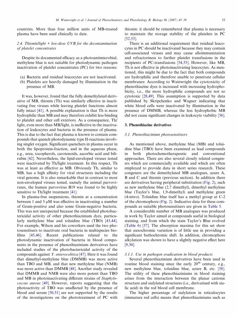
2.4. Thionin/light + low-dose UVB for the decontamination
of platelet concentrates
Despite its documented efficacy as a photoantimicrobial,methylene blue is not suitable for photodynamic pathogeninactivation of platelet concentrates (PC) for two reasons:
(a) Bacteria and residual leucocytes are not inactivated.(b) Platelets are heavily damaged by illumination in the
presence of MB.
It was, however, found that the fully demethylated deriv-ative of MB, thionin (Th) was similarly effective in inacti-vating free viruses while leaving platelet functions almostfully intact [41]. A possible explanation is that Th is morehydrophilic than MB and may therefore exhibit less bindingto platelet and other cell exteriors. As a consequence, Th/light, even more than MB/light, is ineffective in the inactiva-tion of leukocytes and bacteria in the presence of plasma.This is due to the fact that plasma is known to contain com-pounds that quench photodynamic type II-reactions involv-ing singlet oxygen. Significant quenchers in plasma occur inboth the lipoprotein-fraction, and in the aqueous phase,e.g., urea, tocopherols, carotenoids, ascorbic acid and bili-rubin [42]. Nevertheless, the lipid-enveloped viruses testedwere inactivated by Th/light treatment. In this respect, Thwas at least as effective as MB. Obviously Th, similar toMB, has a high affinity for viral structures including theviral genome. It is also remarkable that in contrast to mostnon-enveloped viruses tested, namely the animal parvovi-ruses, the human parvovirus B19 was found to be highlysensitive to Th/light treatment [41].
In plasma-free suspensions, thionin at a concentrationbetween 1 and 5 lM was effective in inactivating a numberof Gram-positive and also some Gram-negative bacteria.This was not unexpected because the established photobac-tericidal activity of other phenothiazinium dyes, particu-larly methylene blue and toluidine blue (TBO) [43,44].For example, Wilson and his coworkers used the two pho-tosensitisers to inactivate oral bacteria in multispecies bio-films [45,46]. Recent publications related to thephotodynamic inactivation of bacteria in blood compo-nents in the presence of phenothiazinium derivatives haveincluded studies of the photobactericidal activity of thecompounds against Y. enterocolitica [47]. Here it was foundthat dimethyl-methylene blue (DMMB) was more activethan TBO and MB, and that new methylene blue (NMB)was more active than DMMB [48]. Another study revealedthat DMMB and NMB were also more potent than TBOand MB in photoinactivating different strains of Staphylo-
coccus aureus [49]. However, reports suggesting that thephotoactivity of TBO was unaffected by the presence ofblood and serum [50,51] are not supported by the resultsof the investigations on the phototreatment of PC with
thionin: it should be remembered that plasma is necessaryto maintain the storage stability of the platelets in PC[52,53].
There is an additional requirement that residual leuco-cytes in PC should be inactivated because they may containcell-associated viruses and may cause alloimmunizationand refractoriness to further platelet transfusions in therecipients of PC-transfusions [54,55]. However, like MB,Th is not effective in photoinactivating leucocytes. As men-tioned, this might be due to the fact that both compoundsare hydrophilic and therefore unable to penetrate cellularmembranes: According to Wainwright the cytotoxicity ofphenothiazine dyes is increased with increasing hydropho-bicity, i.e., the more hydrophilic compounds are not socytotoxic [28,49]. This assumption is supported by datapublished by Skripchenko and Wagner indicating thatwhite blood cells were inactivated by illumination in thepresence of DMMB, whereas the less hydrophobic MBdid not cause significant changes in leukocyte viability [56].
3. Phenothiazine derivatives
3.1. Phenothiazinium photosensitisers
As mentioned above, methylene blue (MB) and tolui-dine blue (TBO) have been examined as lead compoundsin both photochemotherapeutic and conventionalapproaches. There are also several closely related congen-ers which are commercially available and which are oftenemployed to provide data in short series research. Thesecongeners are the demethylated MB analogues, azure A,B and C and thionin (previous section). In addition thereare derivatives having peripheral substitution (Fig. 2), suchas new methylene blue (2,7 dimethyl), dimethyl methyleneblue (Taylor’s blue, 1,9-dimethyl) and methylene green(4-nitro). Toluidine blue itself has a methyl group at C-2of the chromophore (Fig. 2). Indicative data for these com-pounds as suitable photosensitisers are given in Table 5.
A considerable number of MB analogues was producedin work by Taylor aimed at compounds useful in biologicalstaining, and from which the stain Taylor’s Blue evolved(Table 6) [57]. The absorption maxima for this set showthat auxochromic variation is of little use in providing asignificant bathochromic shift. In addition, chromophorealkylation was shown to have a slightly negative effect here[9,30].
3.1.1. Use in pathogen eradication in blood products
Several phenothiazinium derivatives have been used inroutine blood staining since the early 20th century, e.g.,new methylene blue, toluidine blue, azure B, etc. [58].The utility of these phenothiaziniums in blood stainingarises from the interaction between the planar cationicstructure and sialylated structures (i.e., derivatised with sia-lic acid) in the red blood cell membrane.
The higher percentage of sialylation in reticulocytes(immature red cells) means that phenothiaziniums such as
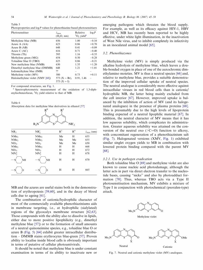
Table 6Absorption data for methylene blue derivatives in ethanol [57]
S+
N
R2N NR'2
R9 R1
NR2 NR02 R1 R9 kmax (nm)
NMe2 NMe2 Me H 655NMe2 NMe2 Me Me 655NEt2 NEt2 Me Me 650NMe2 NMe2 H H 668NEt2 NEt2 H H 672NPrn
2 NPrn2 H H 678
NHN NHN H H 670
ON ON H H 665
S
N
OMe2N
S
N
OMe2N
XS+
N
Me2N
X
OMe
Methylene violet
Neutral Cationic
Fig. 7. Neutral and cationic methylene violet (MV) analogues.
Table 5Photoproperties and logP values for phenothiazine-based photosensitisers
Photosensitiser kmax
(H2O, nm)Relative1O2 yielda
logP
Methylene blue (MB) 656 1.00 �0.10Azure A (AA) 623 0.86 +0.70Azure B (AB) 648 0.41 �0.09Azure C (AC) 616 0.71 �0.40Thionin (Th) 595 1.16 �0.35Methylene green (MG) 654 0.50 �0.28Toluidine blue O (TBO) 625 0.86 �0.21New methylene blue (NMB) 630 1.35 +1.20Dimethyl methylene blue (DMMB) 648 1.21 +1.01Iodomethylene blue (IMB) ?? – –Methylene violet (MV) 580 0.73 +0.11Halomethylene violet (XMV [68]) 573 (X = Br),
575 (X = I)0.91, 1.46 –
For compound structures, see Fig. 1.a Spectrophotometric measurement of the oxidation of 1,3-diph-
enylisobenzofuran, 1O2 yield relative to that of MB.
54 M. Wainwright et al. / Journal of Photochemistry and Photobiology B: Biology 86 (2007) 45–58
MB and the azures are useful stains both in the demonstra-tion of erythropoiesis [59,60], and in the decay of bloodcells due to ageing [61].
The combination of cationic/hydrophilic character ofmost of the commercially available phenothiaziniums aidsin membrane targeting, i.e., at hydrophilic (sialylated)regions of the glycocalyx membrane structure [62,63].Those compounds with the ability also to dissolve in lipids,either due to more positive lipophilicity (e.g., dimethylmethylene blue [57]) or to the formation of small amountsof a neutral quinoneimine species, e.g., toluidine blue O orazure B (Fig. 5) [64] exhibit greater intracellular distribu-tion – DMMB stains erythrocytes blue-green [57]. Provenability to localise inside blood cells is obviously importantin terms of putative of cellular photoantivirals.
It should be noted that methylene blue is under constantexamination in terms of its ability to inactivate new or
emerging pathogens which threaten the blood supply.For example, as well as its efficacy against HIV-1, HBVand HCV, MB has recently been reported to be highlyeffective, under white light illumination, in the inactivationof West Nile virus, and to inhibit completely its infectivityin an inoculated animal model [65].
3.2. Phenothiazones
Methylene violet (MV) is simply produced via thealkaline hydrolysis of methylene blue, which leaves a dou-ble-bonded oxygen in place of one of the auxochromic dim-ethylamino moieties. MV is thus a neutral species [66] and,relative to methylene blue, provides a suitable demonstra-tion of the improved cellular uptake of neutral species.The neutral analogue is considerably more effective againstintracellular viruses in red blood cells than is cationic/hydrophilic MB, the latter being mainly excluded fromthe cell interior [67]. However, improved uptake is bal-anced by the inhibition of action of MV (and its haloge-nated analogues) in the presence of plasma proteins [68].This is presumably due to the high levels of lipoproteinbinding expected of a neutral lipophilic material [67]. Inaddition, the neutral character of MV means that it haslow aqueous solubility, which complicates its administra-tion. Greater aqueous solubility was attained on the con-version of the neutral oxo (>C=O) function to alkoxy,with concomitant regeneration of a phenothiazinium salt(Fig. 7). Halogenated versions (XMV, Fig. 1) exhibitedsimilar singlet oxygen yields to MB in combination withlowered protein binding compared with the parent MV[68].
3.2.1. Use in pathogen eradication
Both toluidine blue O [69] and methylene violet are alsoknown to cause nucleic acid photodamage, although thelatter acts in part via direct electron transfer to the nucleo-tide bases, causing ‘‘nicks’’ and also by photoadduct for-mation [70]. Thus, whereas TBO acts via a Type IIphotosensitisation mechanism, MV exhibits a mixture ofType I in conjunction with photochemical (psoralen-type)attack.
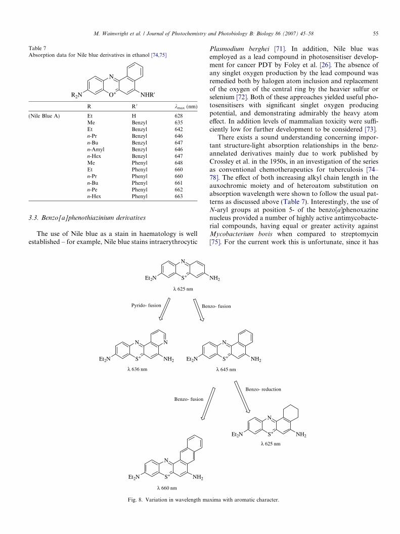
Table 7Absorption data for Nile blue derivatives in ethanol [74,75]
O+
N
R2N NHR'
R R 0 kmax (nm)
(Nile Blue A) Et H 628Me Benzyl 635Et Benzyl 642n-Pr Benzyl 646n-Bu Benzyl 647n-Amyl Benzyl 646n-Hex Benzyl 647Me Phenyl 648Et Phenyl 660n-Pr Phenyl 660n-Bu Phenyl 661n-Pe Phenyl 662n-Hex Phenyl 663
M. Wainwright et al. / Journal of Photochemistry and Photobiology B: Biology 86 (2007) 45–58 55
3.3. Benzo[a]phenothiazinium derivatives
The use of Nile blue as a stain in haematology is wellestablished – for example, Nile blue stains intraerythrocytic
λ 625 nm
λ 660 nm
λ 636 nm
Pyrido- fusion Ben
Benzo- fusion
S+
N
Et2N
S+
N
Et2N NH2
N
Et2N
S+
N
Et2N NH2
Fig. 8. Variation in wavelength ma
Plasmodium berghei [71]. In addition, Nile blue wasemployed as a lead compound in photosensitiser develop-ment for cancer PDT by Foley et al. [26]. The absence ofany singlet oxygen production by the lead compound wasremedied both by halogen atom inclusion and replacementof the oxygen of the central ring by the heavier sulfur orselenium [72]. Both of these approaches yielded useful pho-tosensitisers with significant singlet oxygen producingpotential, and demonstrating admirably the heavy atomeffect. In addition levels of mammalian toxicity were suffi-ciently low for further development to be considered [73].
There exists a sound understanding concerning impor-tant structure-light absorption relationships in the benz-annelated derivatives mainly due to work published byCrossley et al. in the 1950s, in an investigation of the seriesas conventional chemotherapeutics for tuberculosis [74–78]. The effect of both increasing alkyl chain length in theauxochromic moiety and of heteroatom substitution onabsorption wavelength were shown to follow the usual pat-terns as discussed above (Table 7). Interestingly, the use ofN-aryl groups at position 5- of the benzo[a]phenoxazinenucleus provided a number of highly active antimycobacte-rial compounds, having equal or greater activity againstMycobacterium bovis when compared to streptomycin[75]. For the current work this is unfortunate, since it has
λ 625 nm
λ 645 nm
zo- fusion
Benzo- reduction
NH2
S+
N
NH2
S+
N
Et2N NH2
xima with aromatic character.
56 M. Wainwright et al. / Journal of Photochemistry and Photobiology B: Biology 86 (2007) 45–58
also been shown that bis(N-arylamino)phenothiaziniumderivatives do not produce measurable quantities of singletoxygen [79]. However, the use of N-benzyl derivatives inCrossley’s study also realised highly active antitubercularactivity [74]. Although little mammalian toxicological datais available on any of these compounds they retain consid-erable potential in terms of blood product use.
3.4. Other annelated derivatives
Recent work by one of the authors (MW) has also shownthat other aryl fusions of the phenothiazinium nucleus arepossible, furnishing derivatives of similar type, in terms ofphotoproperties, but with the potential for future chromo-phore modification. These compounds include naphtho[2,3 a]- and pyrido[2,3 a]-fusions (Fig. 8) [80].
4. Conclusion
The current use of MB in plasma photodecontaminationencouraged the testing of similar compounds for plateletconcentrates and this has resulted in the present proposalof thionin/UV to this end. The situation with red cell decon-tamination is still some way from being resolved, largelydue to problems with haemolysis. However, a more rationalapproach to photosensitiser design, taking into account redcell membrane binding should provide dividends.
The synthetic chemistry associated with methylene blueand its congeners is, by now, well understood, if not widelyappreciated. The preparation of close MB analogues is thusrelatively straightforward, while that of more complexmolecular structures involving extended chromophores isat least achievable, if not yet in high yield. Consequently,the provision of full ranges of compounds for screeningin the search for, for example, potential red cell photoanti-microbials is feasible.
5. Abbreviations
CFU colony-forming unitsDMMB dimethyl methylene blueDNA deoxyribonucleic acidHBV hepatitis B virusHCV hepatitis C virusHIV human immunodeficiency virusMB methylene blueMV methylene violetNMB new methylene bluePACT photoantimicrobial chemotherapyPC Platelet concentratePDT photodynamic therapyRNA ribonucleic acidTBO toluidine blue OTh thioninTICT twisted intramolecular charge transferUV ultraviolet
References
[1] B. Zeina, J. Greenman, W.M. Purcell, B. Das, Killing of cutaneousmicrobial species by photodynamic therapy, Br. J. Dermatol. 144(2001) 274–278.
[2] F. DeEds, Antiseptic value of phenothiazines in urinary tractinfections, J. Pharmacol. 65 (1939) 353–371.
[3] L.M. DeWitt, Preliminary report of experiments in the vital stainingof tubercles, J. Infect. Dis. 12 (1913) 68–92.
[4] M. Wainwright, K.B. Crossley, Methylene Blue – a therapeutic dyefor all seasons? J. Chemother. 14 (2002) 431–443.
[5] J.L. Vennerstrom, M.T. Makler, C.K. Angerhofer, J.A. Williams,Antimalarial dyes revisited: xanthenes, azines, oxazines, and thia-zines, Antimicrob. Agents Chemother. 39 (1995) 2671–2677.
[6] M. Wainwright, L. Amaral, The phenothiazinium chromophore andthe evolution of antimalarial drugs, Trop. Med. Int. Health 10 (2005)501–511.
[7] R.H. Schirmer, B. Coulibaly, A. Stich, et al., Methylene blue as anantimalarial agent, Redox Rep. 8 (2003) 272–276.
[8] G. Csaba, J. Korosi, A new antitumour agent: phenazathionium-mustard salt, Neoplasma 15 (1968) 443–445.
[9] M. Wainwright, Phenothiazinium photosensitisers V. Photobacteri-cidal activities of chromophore-methylated phenothiazinium salts,Dyes Pigments 73 (2006) 7–12.
[10] S.J. Wagner, A. Skripchenko, D. Robinette, J.W. Foley, L. Cincotta,Factors affecting virus photoinactivation by a series of phenothiazinedyes, Photochem. Photobiol. 67 (1998) 343–349.
[11] M. Wainwright, R.M. Giddens, Phenothiazinium photosensitisers:choices in synthesis and application, Dyes Pigments 57 (2003) 245–257.
[12] J.W. Foley, L. Cincotta, A.H. Cincotta, Structure and properties ofnovel benzo[a]phenoxazinium photochemotherapeutic agents, Proc.SPIE 847 (1987) 90–95.
[13] M. Wainwright, Photodynamic antimicrobial chemotherapy (PACT),J. Antimicrob. Chemother. 42 (1998) 13–28.
[14] M. Wainwright, D.A. Phoenix, J. Marland, D.R.A. Wareing, F.J.Bolton, A study of photobactericidal activity in the phenothiaziniumseries, FEMS Immunol. Med. Microbiol. 19 (1997) 75–80.
[15] M. Wainwright, The use of dyes in modern biomedicine, Biotech.Histochem. 78 (2003) 147–155.
[16] J. O’Neill, M. Wilson, M. Wainwright, Comparative antistreptococ-cal activity of a range of photobactericidal agents, J. Chemother. 15(2003) 329–334.
[17] A. Segalla, C.D. Borsarelli, S.E. Braslavsky, et al., Photophysical,photochemical and antibacterial photosensitizing properties of anovel octationic Zn(II)-phtalocyanine, Photochem. Photobiol. Sci. 1(2002) 641–648.
[18] A. Minnock, D.I. Vernon, J. Schofield, J. Griffiths, J.H. Parish, S.B.Brown, Photoinactivation of bacteria. Use of a cationic water-solublezinc phthalocyanine to photoinactivate both Gram-negative andGram-positive bacteria, J. Photochem. Photobiol., B: Biol. 32 (1996)159–164.
[19] N.S. Soukos, L.A. Ximenez-Fyvie, M.R. Hamblin, S.S. Socransky, T.Hasan, Targeted antibacterial photochemotherapy, Antimicrob.Agents Chemother. 42 (1998) 2595–2601.
[20] E.M. Tuite, J.M. Kelly, New trends in photobiology: photochemicalinteractions of methylene blue and analogues with DNA and otherbiological substrates, J. Photochem. Photobiol., B: Biol. 21 (1993)103–124.
[21] L. Corash, Virus inactivation in cellular components, Vox Sang. 70(1996) 9–16.
[22] M. Wilson, N. Mia, Effect of environmental factors on the lethalphotosensitization of Candida albicans in vitro, Lasers Med. Sci. 9(1994) 105–109.
[23] S.C. de Souza, J.C. Junqueira, I. Balducci, C.Y. Koga-Ito, E.Munin, A.O.C. Jorge, Photosensitization of different Candida
species by low power laser light, J. Photochem. Photobiol., B:Biol. 83 (2006) 34–38.
M. Wainwright et al. / Journal of Photochemistry and Photobiology B: Biology 86 (2007) 45–58 57
[24] J.P. Thurston, The chemotherapy of Plasmodium berghei. I. Resis-tance to drugs, Parasitology 43 (1953) 246–252.
[25] C. Boda, B. Enanga, B. Courtioux, J.C. Breton, B. Bouteille,Trypanocidal activity of methylene blue evidence for in vitro efficacyand in vivo failure, Chemotherapy 52 (2006) 16–19.
[26] L. Cincotta, J.W. Foley, A.H. Cincotta, Novel phenothiaziniumphotosensitizers for photodynamic therapy, SPIE Adv. Photochemo-ther. 997 (1988) 145–153.
[27] L. Cincotta, J.W. Foley, A.H. Cincotta, Novel red absorbingbenzo[a]phenoxazinium and benzo[a]phenothiazinium photosensitiz-ers: in vitro evaluation, Photochem. Photobiol. 46 (1987) 751–758.
[28] M. Wainwright, Methylene blue derivatives – suitable photoantimi-crobials for blood product disinfection? Int. J. Antimicrob. Agents 16(2000) 381–394.
[29] B. Bachmann, J. Knuver-Hopf, B. Lambrecht, H. Mohr, Targetstructures for HIV-1 inactivation by methylene blue and light, J. Med.Virol. 47 (1995) 172–178.
[30] M. Wainwright, D.A. Phoenix, L. Rice, S.M. Burrow, J.J. Waring,Increased cytotoxicity and phototoxicity in the methylene blue seriesvia chromophore methylation, J. Photochem. Photobiol., B: Biol. 40(1997) 233–239.
[31] R.Y. Dodd, The risk of transfusion-transmitted infection, New Engl.J. Med. 327 (1992) 419–421.
[32] A.J. Blajchman, M. Goldman, F. Baeza, Improving the bacteriolog-ical safety of platelet transfusions, Tranfusion Med. Rev. 18 (2004)11–24.
[33] B. Lambrecht, H. Mohr, J. Knuever-Hopf, H. Schmitt, Photoinac-tivation of viruses in human fresh plasma by phenothiazine dyes incombination with visible light, Vox. Sang. 60 (1991) 207–213.
[34] E. Snyder, J. McCullough, S.J. Slichter, R.G. Strauss, I. Lopez-Plaza,J.-S. Lin, L. Corash, M.G. Conlan, Clinical safety of plateletsphotochemically treated with amotosalen HCl and ultraviolet A lightfor pathogen inactivation: the SPRINT trial, Transfusion 45 (2005)1864–1875.
[35] P.H. Ruane, R. Edrich, D. Gampp, S.D. Keil, R.L. Leonard, R.P.Goodrich, Photochemical inactivation of selected viruses and bacteriain platelet concentrates using riboflavin and light, Transfusion 44(2004) 877–885.
[36] L.M. Williamson, R.A. Cardigan, C.V. Prowse, Methylene blue-treated fresh-frozen plasma: what is its contribution to blood safety?Transfusion 43 (2003) 1322–1329.
[37] T. Verpoort, S. Chollet, C. Heron, et al., Filtration of Methylene Blueand photoproducts after photodynamic treatment of plasma usingBlueflex, ISBT-Congress, Istanbul, 2003; Abstr. P246.
[38] J.D. Tissot, D.F. Hochstrasser, B. Schneider, J.J. Morgenthaler, P.Schneider, No evidence for protein modification in fresh-frozenplasma after photochemical treatment: an analysis by high-resolutiontwo-dimensional electropheresis, Brit. J. Hematol. 86 (1994) 143–149.
[39] P. Pohler, W.H. Walker, S. Reichenberg, H. Mohr, U. Gravemann,T.H. Muller, Methylene blue treated plasma: pharmacokinetic andtoxicological profile of MB and photoproducts, Vox. Sang. 87 (2004)93. Abstr. A 10.2.
[40] Pathogen inactivation of Labile Blood Products. Council of EuropePublishing, February 2001; ISBN: 92-871-4560-1, Strasbourg, France.
[41] H. Mohr, A. Redecker-Klein, Inactivation of pathogens in plateletconcentrates by using a two-step procedure, Vox Sang. 84 (2002) 96–104.
[42] J.R. Karnofsky, Quenching of singlet oxygen by human plasma,Photochem. Photobiol. 51 (1990) 299–303.
[43] T. T’ung, Photodynamic action of methylene blue on bacteria, Proc.Soc. Exp. Biol. Med. 33 (1935) 328–330.
[44] T. T’ung, S.H. Zia, Photodynamic action of various dyes on bacteria,Proc. Soc. Exp. Biol. Med. 36 (1937) 326–330.
[45] M. Wilson, J. Dobson, W. Harvey, Sensitization of oral bacteria tokilling by low-power laser radiation, Curr. Microbiol. 25 (1992) 77–81.
[46] S. Sarkar, M. Wilson, Lethal photosensitization of bacteria insubgingival plaque from patient with chronic perodontitis, J.Periodont. Res. 28 (1993) 204–210.
[47] M. Wainwright, D.A. Phoenix, D.R.A. Wareing, T.E. Smillie,Photobactericidal activity of phenothiaziniums against Yersinia
enterocolitica, J. Chemother. 13 (2001) 503–509.[48] M. Wainwright, The emerging chemistry of blood disinfection, Chem.
Soc. Rev. 31 (2002) 126–136.[49] M. Wainwright, D.A. Phoenix, S.L. Laycock, D.R.A. Wareing, P.A.
Wright, Photobacterial activity of phenothiazinum dyes againstmethicillin-resistant strains of Staphylococcus aureus, FEMS Micro-biol. Lett. 160 (1998) 177–181.
[50] M. Wilson, S. Sarkar, J.S. Bulman, Effect of blood on lethalphotosensitization of bacteria in subgingival plaque from patientswith chronic periodontitis, Lasers Med. Sci. 8 (1993) 297–303.
[51] M. Wilson, J. Pratten, Lethal photosensitization of Staphylococcus
aureus in vitro: effect of growth phase, serum and pre-irradiation time,Lasers Surg. Med. 16 (1995) 272–276.
[52] H. Gulliksson, S. Larsson, G. Kumlien, A. Shanwell, Storage ofplatelets in additive solutions: effects of phosphate, Vox Sang. 78(2000) 176–184.
[53] S. Murphy, The efficacy of synthetic media in the storage of humanplatelets for transfusion, Transfus. Med. Rev. 13 (1999) 153–163.
[54] W. Dzik, Use of leukodepletion filters for the removal of bacteria,Immunol. Invest. 24 (1995) 95–115.
[55] J.G. Eernisse, A. Brand, Prevention of platelet refractoriness due toHLA antibodies by administration of leukocyte-poor blood compo-nents, Exp. Hematol. 9 (1981) 77–83.
[56] A.A. Skripchenko, S.J. Wagner, Inactivation of WBCs in RBCsuspensions by photoactive phenothiazine dyes: comparison ofdimethylmethylene blue and MB, Transfusion 40 (2000) 968–975.
[57] K.B. Taylor, G.M. Jeffree, A new basic metachromatic dye, 1:9-dimethyl methylene blue, Histochem. J. 1 (1969) 199–204.
[58] M. Wainwright, Pathogen inactivation in blood products, Curr. Med.Chem. 9 (2002) 127–143.
[59] B. Rudensky, Comparison of a semi-automated new Coulter meth-ylene blue method with fluorescence flow cytometry in reticulocytecounting, Scand. J. Clin. Lab. Invest. 57 (1997) 291–296.
[60] T. Inoue, N. Tatsumi, Evaluation of erythropoiesis by new methyleneblue staining to establish reticulocyte maturity in bone marrowaspirates and peripheral blood, Acta. Cytol. 35 (1991) 479–480.
[61] T.J. Greenwalt, E.A. Steane, F.O. Lau, K. Sweeney-Hammond,Aging of the human erythrocyte, Prog. Clin. Biol. Res. 43 (1980) 195–212.
[62] M. Gliesing, K.J. Halbhuber, Topo-optical studies of graduallydisintegrated erythrocyte membrane derivates: different kinds ofghosts, Acta. Histochem. 86 (1989) 117–121.
[63] J. Makovitzky, S. Bozsoky, E. Laszlo, Topo-optical reactions ofthe human blood platelet membrane, Histochemistry 79 (1983)281–287.
[64] P.N. Marshall, S.A. Bentley, S.M. Lewis, Purified azure B as areticulocyte stain, J. Clin. Pathol. 29 (1976) 1060–1063.
[65] J.F. Papin, R.A. Floyd, D.P. Dittmer, Methylene blue photoinacti-vation abolishes West Nile virus infectivity in vivo, Antiviral Res. 68(2005) 84–87.
[66] L. Adamcikova, K. Paylikova, P. Sevcik, The decay of methylene bluein alkaline solution, React. Kin. Catal. Lett. 69 (2000) 91–94.
[67] A. Skripchenko, D. Robinette, S.J. Wagner, Comparison of methy-lene blue and methylene violet for photoinactivation of intracellularand extracellular virus in red cell suspensions, Photochem. Photobiol.65 (1997) 451–455.
[68] M.A. Houghtaling, R. Perera, K.E. Owen, S. Wagner, R.J. Kuhn, H.Morrison, Photobiological properties of positively charged methyleneviolet analogs, Photochem. Photobiol. 71 (2000) 20–28.
[69] D. Smelt, P. Repanovici, A. Pascaru, R. Portocala, Photodynamiceffect of toluidine blue on MM virus, Rev. Roum. Med. Virol. 27(1976) 203–207.
[70] H. Morrison, T. Mohammad, R. Kurukulasuriya, Photobiologicalproperties of methylene violet, Photochem. Photobiol. 66 (1997) 245–252.
58 M. Wainwright et al. / Journal of Photochemistry and Photobiology B: Biology 86 (2007) 45–58
[71] L.K. Eveland, E.G. Allen, Nile blue stain: Plasmodium berghei anduninfected erythrocytes, Trans. Roy. Soc. Trop. Med. Hyg. 66 (1972)512–513.
[72] Photo-inactivation of cancer cells, US Patent 4962197, 09.10.90.[73] C.W. Lin, J.R. Shulok, S.D. Kirley, et al., Photodynamic destruction
of lysosomes mediated by Nile blue photosensitizers, Photochem.Photobiol. 58 (1993) 81–91.
[74] M.L. Crossley, P.F. Dreisbach, C.M. Hofmann, R.P. Parker,Chemotherapeutic dyes. I. 5-Aralkylamino-9-alkylaminobenzo[a]phe-noxazines, J. Am. Chem. Soc. 74 (1952) 573–578.
[75] M.L. Crossley, R.J. Turner, C.M. Hofmann, P.F. Dreisbach, R.P.Parker, Chemotherapeutic dyes. II. 5-Arylamino-9-dialkylamino-benzo[a]phenoxazines, J. Am. Chem. Soc. 74 (1952) 578–584.
[76] M.L. Crossley, C.M. Hofmann, P.F. Dreisbach, Chemotherapeuticdyes. III. 5-Heterocyclicamino-9-dialkylaminobenzo[a]phenoxazines,J. Am. Chem. Soc. 74 (1952) 584–586.
[77] R.C. Clapp, J.H. Clark, J.P. English, C.E. Fellows, R.E. Grotz, R.G.Shepherd, Chemotherapeutic dyes. IV. Phenoxazines andbenzo[a]phenoxazines, J. Am. Chem. Soc. 74 (1952) 1989–1993.
[78] R.C. Clapp, J.P. English, C.E. Fellows, J. Forsythe, R.E. Grotz, R.G.Shepherd, Chemotherapeutic dyes. V. Benzo[a]phenothiazines andbenzo[a]phenazines, J. Am. Chem. Soc. 74 (1952) 1994–1996.
[79] M. Wainwright, N.J. Grice, L.E.C. Pye, Phenothiazine Photosensi-tisers. Part II. 3,7-Bis(arylamino)phenothiazines, Dyes Pigments 42(1999) 45–51.
[80] M. Wainwright, Publication in preparation.