Embed Size (px)
Citation preview

PhD in“Experimental and Regenerative Medicine”
XXIX cicle
“Diaphragmatic dysfunction in critically ill pa-tients undergone mechanical ventilation”
Tutor PhD StudentProf. Cinnella Gilda Dr. Spadaro Savino
________________________________________________________________Final Dissertation Academic Year 2015-2016

1
1. Abstract (p. 2)
2. Background (p. 3)
3. Altered Diaphragmatic Structure and Biochemistry in Mechanically Ventilated Humans. (p. 6)
4. The impact of different modes of MV on diaphragm atrophy. Assisted mechanical ventilation: the role of new modalities “Neurally adjusted ventilatory assist versus pressure support ventilation” (p. 9)
5. Monitoring of diaphragm function (p. 12)
5.1 The role of diaphragm electrical activity (EAdi)Ultrasound evalua-
tion of diaphragm: Diaphragmatic displacement and thickening fraction (p. 15)
5.2 Neuroventilatory and neuromechanical efficiency. (p. 16)
6. Biochemical markers of muscular damage (p. 17)
7. Strategies to prevent diaphragm dysfunction (p. 25)
8. Materials and methods (p. 27)
9. Statistic (p. 34)
10. Results (p. 35)
11. Discussion (p. 40)
12. Conclusion (p. 43)
13. References (p. 44)
14. Tables and Figures
2
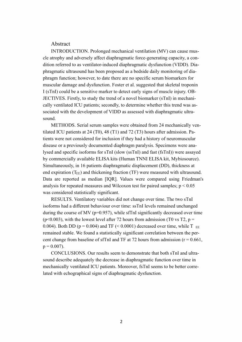
Abstract INTRODUCTION. Prolonged mechanical ventilation (MV) can cause mus-
cle atrophy and adversely affect diaphragmatic force-generating capacity, a con-dition referred to as ventilator-induced diaphragmatic dysfunction (VIDD). Dia-phragmatic ultrasound has been proposed as a bedside daily monitoring of dia-phragm function; however, to date there are no specific serum biomarkers for muscular damage and dysfunction. Foster et al. suggested that skeletal troponin I (sTnI) could be a sensitive marker to detect early signs of muscle injury. OB-JECTIVES. Firstly, to study the trend of a novel biomarker (sTnI) in mechani-cally ventilated ICU patients; secondly, to determine whether this trend was as-sociated with the development of VIDD as assessed with diaphragmatic ultra-sound.
METHODS. Serial serum samples were obtained from 24 mechanically ven-tilated ICU patients at 24 (T0), 48 (T1) and 72 (T3) hours after admission. Pa-tients were not considered for inclusion if they had a history of neuromuscular disease or a previously documented diaphragm paralysis. Specimens were ana-lysed and specific isoforms for sTnI (slow (ssTnI) and fast (fsTnI)) were assayed by commercially available ELISA kits (Human TNNI ELISA kit, Mybiosource). Simultaneously, in 16 patients diaphragmatic displacement (DD), thickness at end expiration (TEE) and thickening fraction (TF) were measured with ultrasound. Data are reported as median [IQR]. Values were compared using Friedman's analysis for repeated measures and Wilcoxon test for paired samples; p < 0.05 was considered statistically significant.
RESULTS. Ventilatory variables did not change over time. The two sTnI isoforms had a different behaviour over time: ssTnI levels remained unchanged during the course of MV (p=0.957), while sfTnI significantly decreased over time (p<0.003), with the lowest level after 72 hours from admission (T0 vs T2, p = 0.004). Both DD (p = 0.004) and TF (< 0.0001) decreased over time, while T EE remained stable. We found a statistically significant correlation between the per-cent change from baseline of sfTnI and TF at 72 hours from admission (r = 0.661, p = 0.007).
CONCLUSIONS. Our results seem to demonstrate that both sTnI and ultra-sound describe adequately the decrease in diaphragmatic function over time in mechanically ventilated ICU patients. Moreover, fsTnI seems to be better corre-lated with echographical signs of diaphragmatic dysfunction.

3
Background Mechanical ventilation (MV) is used clinically to achieve sufficient pulmo-
nary gas exchange in patients unable to sustain adequate alveolar ventilation on their own. Common indications for MV include respiratory failure due to chronic obstructive pulmonary disease, status asthmaticus, and/or heart failure.
In addition, MV is often an essential intervention in patients suffering from acute drug overdose, neuromuscular diseases, sepsis, and during surgery along with postsurgical recovery. Although MV can be a lifesaving measure, prolonged MV results in the rapid development of diaphragmatic weakness due to both at-rophy and contractile dysfunction. This detrimental impact of prolonged MV on the diaphragm has been termed ventilator-induced diaphragmatic dysfunction (VIDD) and VIDD is predicted to be a major contributor to problems in weaning patients from the ventilator. [1], [2]
It is now clear that MV itself contributes to atrophy and injury of muscle fibers, which further leads to decreased force-generating capacity of the dia-phragm. [1], [2]
Several animal models have shown that complete cessation of diaphragm ac-tivity with controlled MV results in atrophy and injury of diaphragmatic fibers. Muscle function alterations are time-dependent, becoming evident as early as 12 hours after initiation of MV and worsening as MV is prolonged. [3], [4], [5], [6] Under prolonged MV, both partial and full support ventilation result in diaphrag-matic atrophy and contractile dysfunction (i.e., VIDD) in both animals and hu-mans.
Current data support a complex underlying pathophysiology involving oxi-dative stress and the activation of severe intracellular proteolytic pathways in-volved in degradation of the contractile apparatus. [7], [8] Furthermore, in criti-cally ill patients, the reduced diaphragm perfusion results in a decrease in the ability of diaphragm to augment blood flow to match oxygen demand in response to contractile activity. The imbalance in oxygen supply demand will promote muscle fatigue. [9] Interestingly, animal studies showed a similar level of atro-phy and weakness with assisted (pressure support) ventilation. [10] These data support the existence of mechanisms other than disuse for VIDD.
One of the major complications associated with prolonged mechanical ven-tilation is the inability to separate the patient from the ventilator. Removing pa-tients from ventilator support is termed “weaning,” and the incidence of difficult weaning varies between different ICUs but can reach the level of 30% of patients

4
exposed to prolonged MV [11]; [12]. The inability to wean patients from the ventilator results in extended hospital stays and increases morbidity and mortal-ity. [11] In patients experiencing weaning difficulty, weaning procedures can ac-count for 40% of total ventilator time with even greater time (>60% of total ven-tilator time) invested in weaning of COPD patients. [11]
The causes of weaning failure can be multifactorial and include aspects such as medications (e.g., neuromuscular blockers), confounding morbidities (e.g., sepsis), psychological factors (i.e., anxiety), neurological disorders, and an
imbalance between respiratory load and the respiratory muscle capacity. [13], [8]; [12]; [14] Although controversy exists, it has been predicted that de-pressed strength and endurance of respiratory muscles can play a key role in weaning difficulties. [14]
However, direct evidence demonstrating cause and effect that VIDD is pri-marily responsible for weaning problems is lacking due to the difficulty of per-forming these experiments in humans. Nonetheless, several lines of indirect evi-dence support the belief that impaired inspiratory muscle function contributes to weaning difficulties. First, although debate exists, several studies reveal that in-spiratory muscle endurance is decreased in patients during prolonged MV and that maximal inspiratory pressure generation is lower in patients that experience difficult weaning compared with patients that are successfully weaned. [15]; [16]; [17] Furthermore, a recent study reveals that patients with diaphragmatic contractile dysfunction exhibit a high incidence of weaning failures compared with patients with normal diaphragm function. [18] Finally, a growing number of studies suggest that inspiratory muscle training, designed to increase dia-phragm strength and endurance, increases weaning success in patients who pre-viously failed repeated weaning attempts by conventional methods. [19]; [20] Collectively, these studies support the concept that VIDD contributes to weaning failure and therefore the prevention of VIDD is a potential therapeutic target to prevent weaning difficulties. A relevant point is also to determine the minimal level of diaphragm activity to be achieved to preserve its function. This is actually a crucial point that is also warranted for the next generation of ventilators (e.g. NAVA).
Respiratory muscle weakness in ventilated ICU patients is associated with prolonged duration of ventilator weaning [21] and increased risk of ICU and hospital readmission. [22] Therefore, it seems of crucial importance to limit the detrimental effects of critical illness, and in particular mechanical ventilation, on the respiratory muscles.
5
The primary endpoint of this project is to evaluate the ability of US in eval-uating diaphragmatic contractility in relation to a possible muscle cell injury as well as serum markers of muscle injury, during assisted mechanical ventilation. The qualitative and quantitative monitoring of diaphragmatic activity with Eadi signal might be helpful in prevention of ventilator- induced diaphragm dysfunc-tion, and could be used as an additional weaning parameter. Plasma markers of skeletal muscle injury have been extensively explored; however, they are limited in their diagnostic sensitivity and specificity. Foster et al [6] suggest that fast sTnI is a sensitive method capable of detecting early signs of low levels of respiratory muscle injury at 1 h. Furthermore, we will investigate if the serum level of skel-etal TnI (sTnI) may be a useful diagnostic marker of respiratory muscle dysfunc-tion, relating it to the timing and severity of the patient’s injury.
6
Altered Diaphragmatic Structure and Biochemistry in Mechani-
cally Ventilated Humans Obtaining diaphragm samples in mechanically ventilated critically ill pa-
tients is technically difficult and ethically questionable; some authors have stud-ied diaphragmatic tissue samples from brain-dead organ donors at the time of organ procurement. Numerous experiments have reported that prolonged MV promotes diaphragm contractile dysfunction. Less than a decade ago, Levine et al. were the first to describe a rapid loss of diaphragm muscle mass in patients on
controlled mechanical ventilation. [7] Biopsies were obtained from 14 brain-dead
organ donors on controlled mechanical ventilation for 18 to 69 h before organ
harvest. The cross -sectional area (CSA) of diaphragm fibers was significantly
lower (53 and 57% for fast - and slow -twitch fibers respectively) compared to
fibers obtained from patients referred for elective lung cancer surgery. Interest-ingly, the severity of atrophy was less pronounced in the pectoralis muscle, indi-cating that the diaphragm is much more sensitive to the effects of disuse. In sub-sequent studies, it was demonstrated that the decrease in diaphragm fiber CSA was proportional to the duration of mechanical ventilation. [23] Interestingly, both ultrastructural injury and atrophy of diaphragmatic fibers in the long-term MV group were significantly correlated with the total duration of MV. Patients mechanically ventilated exhibited an increased level in muscle proteolysis bio-logical markers such as caspase-3, the ubiquitine proteasome system, and the cal-pain system (calpain isoforms 1, 2, and 3). [8] There is good evidence that the calpain, caspase, and ubiquitin-proteasome proteolysis pathways all play signif-icant roles in the development of MV-induced atrophy. Therefore, these proteo-lytic systems are also logical targets for therapeutic intervention.
Oxidative stress in the mechanically ventilated diaphragm results from both an increased generation of reactive oxygen species (ROS) and a dimin-
ished antioxidant capacity in the diaphragm. Increased oxidative stress was also observed in the brain donor group, as suggested by decreased levels of glutathi-one, an antioxidant molecule. [7] The pathways responsible for controlled MV-induced ROS production in the diaphragm remain unclear.
Mitochondria could be the main source of excessive ROS production in the diaphragm during MV. [24] Mitochondria isolated from mechanically ventilated rats release significantly more ROS, exhibit biochemical evidence of oxidative damage, [24] and show abnormal diaphragmatic mitochondrial respiration. [25],
7
[24] Furthermore, the administration of a novel mitochondria-targeted antioxi-dant (SS-31) protects the diaphragm from VIDD, which supports the hypothesis that mitochondria are a primary source of ROS production in the diaphragm dur-ing prolonged MV.
A recent observational study using serial ultrasound measurements to assess diaphragmatic thickness revealed that diaphragm thinning occurs within 48 h af-ter the initiation of partial support MV. [26] This study confirmed that diaphrag-matic atrophy occurs linearly as a function of time on the ventilator with the rate of atrophy averaging a 6% loss of diaphragm thickness per day of MV. Together, these human studies demonstrate that prolonged MV results in rapid atrophy of diaphragm muscle fibers in humans. Although it is difficult to make a direct com-parison between the human and animal studies in the rates of MV-induced dia-phragmatic atrophy, it is clear that the temporal patterns of MV-induced dia-phragmatic atrophy are similar between rats and humans with diaphragmatic at-rophy occurring in both species within 24–48 h after the initiation of MV. [23] Furthermore, both human and animal studies revealed that MV-induced dia-phragmatic atrophy occurs during both partial [10] and full support MV. [27]; [26]; [23]; [7]
Key events in such process are diaphragm injury and atrophy, which are char-acterized by an increased protein degradation and decreased protein synthesis [28], supported by an increased production of reactive oxygen species (ROS) which damage proteins, lipids and DNA thus enhancing diaphragmatic proteol-ysis and contractile dysfunction. [29], [30] Indeed, it is widely reported that pro-longed muscle disuse results in an accumulation of oxidatively modified proteins and lipids. [31]
Collectively, these data demonstrate that inactivity of the diaphragm under controlled mechanical ventilation is associated with rapid posttranslational pro-tein modifications, activation of proteolytic pathways and muscle fiber atrophy.
However, these early studies used biopsies of the diaphragm of brain -dead pa-
tients. This might be an excellent model for diaphragm disuse associated with mechanical ventilation, but differences in treatment and underlying pathophysi-ology in more representative ICU patients should be recognized. Hooijman et al. were the first to study structural, biochemical and functional modifications of biopsies obtained from the diaphragm of ventilated ICU patients (n = 22, mean duration on the ventilator: 7 days, range 14 to 607 h). [32] These authors found
that both fast - and slow -twitch diaphragm fibers had a CSA that was approxi-
mately 25% smaller than that of fibers from the diaphragm of patients referred
8
for elective surgery. Biochemical analysis revealed activation of the proteolytic
ubiquitin-proteasome pathway. Histological analysis demonstrated that the num-
ber of inflammatory cells, including neutrophils and macrophages, was signifi-cantly increased in the diaphragm of ICU patients; this supports a role for inflam-matory mediators in the development of atrophy or injury. Interestingly, van Hees et al. demonstrated that plasma from septic shock patients, but not from healthy subjects, induced atrophy in (healthy) cultured skeletal muscle myotubes. [33] This indicates that plasma from septic shock patients contains molecules with catabolic properties. Additional experiments presented in that paper suggest
that interleukin (IL)-6 plays a role in the development of muscle atrophy in sep-
sis. There are also recent observations suggesting an upregulation of the au-tophagic system reported in the mechanically ventilated diaphragm, which is as-sociated with an upregulation of the transcription factor FOXO-1. [34]
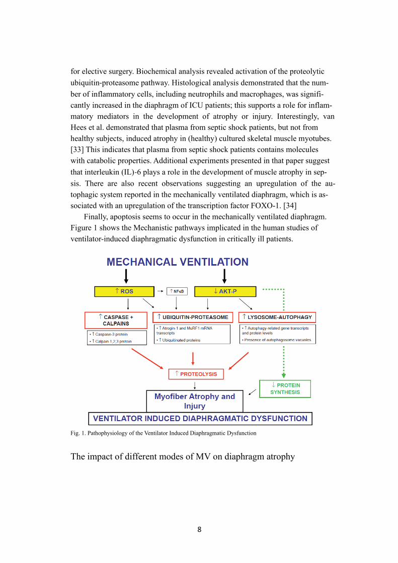
Finally, apoptosis seems to occur in the mechanically ventilated diaphragm. Figure 1 shows the Mechanistic pathways implicated in the human studies of ventilator-induced diaphragmatic dysfunction in critically ill patients.
Fig. 1. Pathophysiology of the Ventilator Induced Diaphragmatic Dysfunction
The impact of different modes of MV on diaphragm atrophy
9
The promotion of diaphragm activity seems to be a key factor in VIDD pre-vention. Maintaining a short period of diaphragm activity by allowing animals under controlled MV to breathe spontaneously for 5 min/h alleviates the decline in diaphragm force production, prevents diaphragm muscle fibers atrophy, and increases the expression of transcription factors myogenin and Myo. [35] As-sisted modes of ventilation, rather than intermittent ventilation, are a more real-istic alternative for clinical treatment. Specifically, ventilator support to patients is classified into two general categories or modes: 1) full ventilator support (often called controlled MV); or 2) partial ventilator support (numerous modes of partial support MV exist). When a patient is provided full ventilator support the venti-lator provides all of the ventilation to the lungs and the patient’s respiratory mus-cles remain inactive. In contrast, during partial ventilator support, the patient’s respiratory muscles (i.e., diaphragm) provide a portion of the work of breathing and the ventilator provides the remainder.
Pressure-support ventilation, a partial mode commonly used in ICU patients, can prevent the reduction of protein synthesis and accelerate the proteolysis as-sociated with controlled MV, but does not appear to completely ameliorate in-creased oxidative stress in the diaphragm. [36] Similar observations have been reported in piglets receiving adaptive ventilatory support. [37] It is worth noting that only partial modes with low level of assistance are beneficial, whereas high levels of assistance are as deleterious as controlled MV.
In recent years, there has been an increasing interest in the use of partial ven-tilator support modes not only as weaning techniques but also in the acute phases of respiratory failure. During assisted spontaneous breathing, a variable propor-tion of the work of breathing is provided by the ventilator, to unload the patient’s respiratory muscles. [38] Multiple ventilator modes are currently available for assisted spontaneous breathing: among these, neurally adjusted ventilator assist (NAVA). The latter is conceptually different from any other mode of ventilation, since the ventilator directly controlled by the neural activity of respiratory cen-ters, expressed by the diaphragm electromyogram (EAdi). [39] EAdi is recorded through a modified nasogastric tube equipped with multiple-array oesophageal electrodes positioned in the lower oesophagus, near the crural portion of the dia-phragm. Several studies have demonstrated that EAdi provide a reliable estimate of inspiratory timing and drive, and that electrical activity of the crural diaphragm is related to global inspiratory efforts in healthy subjects and in patients with acute respiratory failure.
10
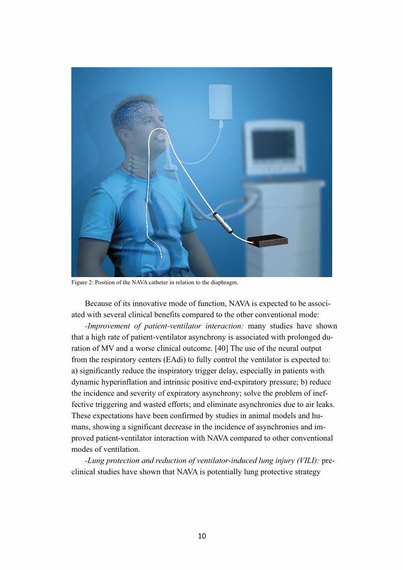
Figure 2: Position of the NAVA catheter in relation to the diaphragm.
Because of its innovative mode of function, NAVA is expected to be associ-
ated with several clinical benefits compared to the other conventional mode: -Improvement of patient-ventilator interaction: many studies have shown
that a high rate of patient-ventilator asynchrony is associated with prolonged du-ration of MV and a worse clinical outcome. [40] The use of the neural output from the respiratory centers (EAdi) to fully control the ventilator is expected to: a) significantly reduce the inspiratory trigger delay, especially in patients with dynamic hyperinflation and intrinsic positive end-expiratory pressure; b) reduce the incidence and severity of expiratory asynchrony; solve the problem of inef-fective triggering and wasted efforts; and eliminate asynchronies due to air leaks. These expectations have been confirmed by studies in animal models and hu-mans, showing a significant decrease in the incidence of asynchronies and im-proved patient-ventilator interaction with NAVA compared to other conventional modes of ventilation.
-Lung protection and reduction of ventilator-induced lung injury (VILI): pre-clinical studies have shown that NAVA is potentially lung protective strategy
11
because the delivery of excessive tidal volume and transpulmonary pressure is limited by the neural feedback from pulmonary stretch receptor. Studies in hu-mans have confirmed that increasing the NAVA level is not associated with an increase in Vt, because when the assist level is increased the EAdi is down-reg-ulated with the net result of reducing the risk of over-assistance and hyperinfla-tion.
-Prevention of diaphragmatic dysfunction: NAVA minimizes the risk of ven-tilator-induced diaphragmatic atrophy because it is based on the continuous cou-pling between ventilator assistance and patient’s neural output. During PSV, a progressive increase in support ultimately leads to disappearance of detectable EAdi signal, clearly indicating over-assistance. In contrast, increasing the NAVA level can efficiently unload the respiratory muscles without abolishing the EAdi. Preservation of adequate muscle function should be particularly important.
- Maintenance of the “physiological” variability of breathing pattern: stud-ies in humans have clearly demonstrated that NAVA is characterized by a signif-icant increase in breathing pattern variability and complexity compared to PSV, but the clinical relevance of this has to be assessed.

12
Monitoring of diaphragm function Despite growing evidence that respiratory muscle dysfunction develops in
critically ill patients and contributes to weaning, the respiratory muscles are poorly monitored in the ICU. Most information regarding diaphragm contractil-ity in MV patients arises from studies of very few subjects or that were not de-signed to study VIDD directly. One reason for the lack of large-scale studies in ICU patients is the difficulty to study diaphragm contractility in these patients. The American Thoracic and European Respiratory Societies have extensively re-viewed the methodologies for respiratory muscle evaluation in patients. [41] Measurements of maximum inspiratory pressure require a large degree of volun-tary coordination. They are not easy in ambulatory patients and their measure-ments in ICU patients are clearly a major issue. In addition, because they require the full participation of the subject, their use in a large scale ICU study is unreal-istic as ICU mechanically ventilated patients receive sedative agents, at least dur-ing the first day of MV. To overcome the various limitations of the maximum inspiratory pressure test, it is possible to perform bilateral phrenic nerve stimula-tion and record the pressure generated by the contracting diaphragm. Stimulation is usually magnetic, which offers a higher level of tolerance (as opposed to elec-trical stimulation) and requires a lower level of expertise. The contractile activity of the diaphragm in response to bilateral cervical magnetic stimulation (CMS) of the phrenic nerves was originally evaluated through the measure of transdi-aphragmatic pressure (Pdi), which requires simultaneous recordings of esopha-geal and gastric pressures using balloon-equipped probes, with Pdi determined as the pressure difference across the diaphragm. Probe placement may be chal-lenging in the ICU patients. However, this test is invasive and uncomfortable as it requires placement of esophageal and gastric probes. Moreover, it is insensitive in detecting unilateral diaphragmatic dysfunction; because of the contraction of the functioning hemidiaphragm, Pdi may not significantly drop in this condition
Electromyography may also be used to diagnose diaphragmatic dysfunction. To evaluate in vivo diaphragm contractile function in ventilated patients is used to assess the change in endotracheal tube pressure induced by magnetic stimula-tion of the phrenic nerves during airway occlusion (Ptr,magn). The major ad-vantage of this technique is that it can be performed at the bedside and does not require patient cooperation. Demoule et al. measured Ptr,magn within 24 h of mechanical ventilation in a population of 85 critically ill patients. [42] Of this group, 54 (64%) patients were diagnosed with diaphragm dysfunction defined as
13
a Ptr,magn < 11 cmH2O. More recently, the same investigators confirmed these earlier findings in 43 ventilated patients [43]; 23 (53%) of these patients exhibited diaphragm dysfunction at ICU admission. These are important findings, as they indicate that other factors besides disuse play a role in the pathophysiology of respiratory muscle weakness in ICU patients. In fact, in their earlier study [42], the authors reported that diaphragm dysfunction was independently associated with sepsis. Consistent with that study, Supinski et al. [44] found that measures for in vivo contractile force were affected more in ventilated patients with infec-tion than in ventilated patients without infection.
The recent study by Demoule et al. [43] also assessed Ptr,magn during the ICU stay. In that study, 61% of patients fulfilling criteria for diaphragm dys-function at admission had persistent respiratory muscle weakness while in the ICU. Of the patients with normal diaphragm function at ICU admission, 55% developed weakness while on the ventilator. This study demonstrates that 80% of all patients fulfilling their inclusion criteria (i. e., > 5 days on mechanical ven-tilation), develop respiratory muscle weakness at some time while on the venti-lator.
Chest radiography is reasonable sensitive in detecting unilateral diaphrag-matic paralysis (sensitivity, 90%), but its specificity is unacceptably low (44%), and it is often not sensitive when bilateral diaphragmatic dysfunction is present.
Pulmonary function testing is another non-invasive means of assessing pa-tients for diaphragmatic dysfunction. The accuracy and reproducibility of these tests are limited by their dependence on lung volumes, as well as patient effort and ability to cooperate. In addition, there is a wide degree of variability within the normal range and an age related decrease in normal values.
The fluoroscopic evaluation of diaphragm dome motion is perhaps the most commonly used non-invasive diagnostic technique. Descent of the diaphragm is visualized via the sniff test, which consist of a short, sharp inspiratory effort through the nostrils at functional residual capacity (FCR). Reduced or paradoxi-cal motion of the hemidiaphragm is indicative of diaphragm dysfunction. In the setting of bilateral diaphragmatic paralysis, apparently normal descent of dia-phragms may be seen during inspiration as a result of compensatory respiratory strategies, leading to a false-negative result. False positive results are also possi-ble.
In the last few decades, ultrasound devices have been studied in critical care medicine in many settings. Exploring diaphragmatic dysfunction with ultrasound

14
is feasible, reliable and accurate at bedside. Assessment of diaphragmatic anat-omy and function using echography was first described in the 1980s ( [45]; [46]; [47]. In a seminal study, Wait et al. measured diaphragm thickness with a ultra-sound at necroscopy, verifying the measure of the same segment by ruler. They found a good correlation between anatomic measurements and evaluation with ultrasound.
Recently, Goligher et al. described the evolution of diaphragmatic thickness during mechanical ventilation and its impact on diaphragm function, assessed with ultrasound [48].
Fig. 3 (A) A patient with normal right diaphragmatic excursion showing an inspiratory peak above
the baseline. (B) A patient with dysfunction of the right hemidiaphragm with a negative inspiratory peak below the baseline, indicating paradoxic movement of the diaphragm.
The diaphragm thickness is better measured in time motion mode, as the dis-
tance (in millimiters) between the outer edges of the echogenic lines at the end-expiration. Changes in diaphragm thickness were found in 56% of the study pop-ulation (n = 128). Both loss and gain of diaphragm thickness were observed dur-ing the first week of ventilation, in 44 and 12% of the patients, respectively. Con-tractile activity of the diaphragm was estimated as the diaphragm thickening frac-tion during maximal inspiratory effort. There was a significant correlation be-tween the mean diaphragm thickening fraction during the first 3 days of ventila-tion and changes in diaphragm thickness (p = 0.01): a loss of diaphragm muscle mass was associated with lower contractile activity, while higher contractile ac-tivity was found for patients exhibiting increases in diaphragm thickness. Both
15
increased and decreased diaphragm thickness seemed to be modulated by the in-tensity of respiratory muscle work performed by the patient, because the change in diaphragm thickness was inversely correlated with the driving pressure applied by the ventilator over the first 72 h of ventilation (p = 0.04, after removal of one outlier).
Furthermore, the ultrasound is useful in diagnosing and quantifying VIDD as a cause of difficult weaning and may guide adjustments of ventilator setting. In an observational study, Kim et al evaluated diaphragmatic dysfunction in 88 patients in a medical ICU who were ventilated for more than 48 hours. Diaphrag-matic dysfunction was diagnosed by a vertical excursion of < 10mm or paradoxic movements and a rapid shallow breathing index was simultaneously calculated at the bedside. Diaphragmatic dysfunction was identified in 29% of patients and there was a correlation with longer duration of MV and with weaning failure. The cut-off values of diaphragmatic excursion for predicting primary weaning failure were 14 mm for the right diaphragm and 12 mm for the left diaphragm. Interest-ingly, the area under the receiving weaning failure was similar to that of the rapid shallow breathing index. [18] A considerable amount of research regarding weaning from MV with assisted modes has been focused on the ability of these techniques to reduce the work of breathing.
The role of diaphragm electrical activity (EAdi) Recently, continuous monitoring of diaphragm electrical activity (EAdi) has
become available for ICU patients. Neurally- adjusted ventilator assist (NAVA) is a relatively new mode for partially assisted ventilation that uses EAdi to control the ventilator. EAdi is acquired using a dedicated nasogastric feeding tube with nine electrodes positioned at the level of the diaphragm muscle. The electrical activity is shown real - time on the ventilator screen (Servo - I/U, Maquet Swe-den), even when the patient is ventilated in another mode than NAVA. Therefore, EAdi may be an alternative to esophageal and gastric balloons to monitor dia-phragm activity (Figure). Indeed, absence of EAdi is consistent with inactivity of the diaphragm, when technical issues have been excluded. However, conversion of EAdi (in micro-volt) into pressure has not yet been well validated. Bellani et al. proposed a proportionality coefficient (pressure- electrical activity index, PEi) that allows calculation of pressure generated by the diaphragm from EAdi. [49] Another index derived from the EAdi is the patient - ventilator breath con-tribution (PVBC) index, which provides an estimation of the fraction of breathing effort that is generated by the patient compared to the total work of breathing
16
(ventilator + patient). Liu et al. demonstrated in 12 patients ventilated using the NAVA mode that PVBC predicts the contribution of the inspiratory muscles ver-sus the pressure delivered by the ventilator. [50], although both PEi and PVBC may be helpful to limit the risk of ventilator over- assist.
EAdi may be helpful in monitoring respiratory muscle loading, patient–ven-tilator synchrony, and efficiency of breathing in critically ill patients.
Currently, new indices are evaluated for respiratory muscle function in ven-tilated patients.
Neuroventilatory and neuromechanical efficiency. The ratio between VT and EAdi represents neuroventilatory efficiency (NVE)
of the diaphragm. An improved NVE indicates that a patient is able to generate the same VT with lower levels of EAdi, whereas a higher EAdi suggests the op-posite. NVE has been used to discriminate between extubation failure and suc-cess in patients weaning from mechanical ventilation. Evidently, NVE is sensi-tive to changes in diaphragm function (atrophy, fatigue, and hyperinflation) as well as a patient’s load of breathing (airway compliance and resistance). Moni-toring the ratio between Pdi and EAdi, representing neuro-mechanical efficiency (NME), excludes the influence of a patient’s load of breathing. A gradual de-crease in NME over days indicates the development of diaphragm weakness, whereas an increase suggests recovery. Biochemical markers of muscular damage
17
Studies in critically ill patients and ventilated animals revealed that respira-tory muscle weakness is associated with muscle fiber damage and loss of con-tractile proteins. Considering the important role of plasma markers in the diag-nosis of myocardial damage and rhabdomyolysis, it could be reasoned that the detection of muscle-specific proteins in plasma would be a valuable tool to mon-itor damage of the respiratory muscles.
Although potential candidates include classical markers, such as creatine ki-nase and myoglobin, the clinical use of these markers is probably hampered by their large range in healthy individuals and non-specificity for structural damage. [51] Recent experimental findings indicate that measuring serum levels of skel-etal muscle troponin I is more sensitive to detect structural injury of respiratory muscles. [6] However, the relation between plasma troponin I levels and func-tional measures in critically ill patients has not yet been investigated. Of note, respiratory muscle weakness in the critically ill is often part of generalized mus-cle weakness.
In those cases, evaluation of circulatory biomarkers of skeletal muscle dam-age may not specifically reflect the functioning of respiratory muscles but could rather represents overall skeletal muscle function.
Enzymes or proteins of skeletal muscles present in the serum are indicators of the functional state of the muscle tissue, and their levels change significantly in physiological or pathological conditions. Examples are creatine kinase, al-dolase, aspartic aminotransferase (AST) and skeletal troponins, which have been found increased in several pathological conditions. [52] Although not specific, their use might help in monitoring the muscle functionality also in clinical con-ditions: therefore, in the next pages I will provide a general introduction on cir-culating markers of muscle functionality and their usefulness to monitor clinical and pathological conditions, with a particular insight on diaphragmatic dysfunc-tion. Creatine kinase
Creatine kinase (CK) is an enzyme that plays a key role in energy metabolism of the cell for its ability to produce ATP from creatine phosphate and ADP and vice versa, resulting in a rapid storage or release of energy. [53]

18
Figure 4. Reaction catalysed by creatine kinase
CK is a dimeric globular protein resulting from the combination of two mon-omers, defined M (muscle) and B (brain), whit a molecular mass, respectively, of 43–45 kDa. There are five forms of CK, named on the basis of their tissue origin into MM-CK, BB-CK, MB-CK, Miu-CK and Mis-CK. [52]
In physiological conditions, the two isoenzymes are present in cytoplasm as homo-dimers (MM-CK and BB-CK). BB-CK is usually distributed in brain, heart, smooth muscle, nervous system and other tissues and MM-CK is the pre-dominant isoform in highly differentiated skeletal muscle tissue. Only the MM-CK is able to interact with the M-band region of a myofibrillar sarcomere: there-fore, it is functionally important in the skeletal muscle. In addition, a third form of creatine kinase composed by M and B monomers (MB-CK) is found in the cytoplasm of cardiomyocytes.
The two mitochondrial isoenzymes, the ubiquitous (Mi u-CK) and the sarco-meric (Mis-CK) forms, are octamers but can be dissociated into dimers.
The cytoplasmic isoenzymes can be used as markers of tissue damage on the basis of their distribution: for instance, the levels of CK and in particular the MM-CK isoenzyme is increased in case of muscle damage, whereas MB-CK isoform levels are found particularly high in the case of myocardial necrosis. [52]

19
Lactate dehydrogenase Lactate dehydrogenase (LDH) is an enzyme that catalyses the conversion of
pyruvate to lactate at the expense of a reduced nicotinamide adenine dinucleotide (NADH) by following the reaction in Figure 3. In addition, it can also convert back the lactate to pyruvate generating NADH.
Figure 5. Reaction catalysed by lactate dehydrogenase
LDH is a tetrameric enzyme composed of two different subunits:
1. M, converts pyruvate to lactate and it is mainly present in skeletal muscle and liver.
2. H, improves the aerobic oxidation of pyruvate and it is found in the heart, spleen, kidney, brain and erythrocytes.
The combination of these two monomers can generate five isoforms: two are homo-tetramers (LDH-1 (H4) and LDH-5 (M4)), whereas three are hybrid te-tramers, (LDH-2 (M1H3), LDH-3 (M2H2) and LDH-4 (M3H1)) ( [52]; [54]).
The quantitative distribution of the LDH isoenzymes in various tissues and fluids is different and characteristic for each isoform. The release of different isoenzymes from the tissues into the bloodstream due to an altered cellular me-tabolism, inflammation, degenerative process, toxicity or injury causes a change of the normal fluid pattern. The resulting pattern can give indications about the possible site of origin of the abnormality. [55]
Aldolase
Aldolase is an enzyme involved in several metabolic pathways, including gluconeogenesis and glycolysis. Its molecular weight is 160 kDa and catalyses the reversible cleavage of fructose 1-6-biphosphate to glyceraldehyde-3-phos-phate and dihydroxyacetone phosphate in the fourth reaction of glycolysis (Fig-ure 5).

20
Figure 6. Reaction catalyzed by Aldolase
Aldolase is located in the cytoplasm and in the nucleus within the hetero-
chromatin. [52] Aldolase is probably expressed in all cells, but there are three tissue-specific isozymes encoded by different genes, producing enzymes with similar molecular weights and catalytic mechanisms. Aldolase A is predomi-nantly expressed in muscle and red blood cells; aldolase B is expressed in liver, kidney, and small intestine; aldolase C is expressed in brain, smooth muscle, and neuronal tissue [56] Aldolase A regulates cell contraction by binding to actin-containing filaments of the cytoskeleton. In addition, increased circulating levels of this isoenzyme have been found in cases of myotonic muscle disease. [52]
Aminotransferases
Aminotransferases or transaminases are a group of enzymes that catalyse the interconversion of amino acids and oxoacids by transfer of amino group. The most important enzymes with a clinical application are aspartate aminotransfer-ase (AST) and alanine aminotransferase (ALT). [57] AST has a molecular weight of 90 kDa and catalysed the reaction:

21
Figure 7. Reaction catalysed by AST This reaction occurs between the mitochondria and cytosol, and provides energy to the cells. The enzyme is mainly located in skeletal and myocardial muscles, liver and erythrocytes. [52] ALT is a pyridoxal enzyme catalysing the transfer of an amino group from ala-nine to α-ketoglutarate, reversibly producing pyruvate and glutamate.
Figure 8. Reaction catalysed by ALT
ALT is widely distributed: human isozymes are found in the cytosol and mito-chondria of liver, kidney, and skeletal and cardiac muscles. When found in blood, AST and ALT are considered to be markers of liver dis-ease,[57] but in chronic muscle injury, AST and ALT levels were found both increased. [52]

22
Myoglobin
Myoglobin is a cytoplasmic hemoprotein expressed in cardiac myocytes and oxidative skeletal muscle fibers. It is a monomeric protein of 153 amino acids residues with a secondary structure consisting of eight α-helices connected through the turns with an oxygen binding site (Figure 6A). It contains a heme group composed by protoporphyrin and an iron ion located in its centre (Figure 6B), providing the ability to bind oxygen. The polypeptide chain is folded and cradles the heme prosthetic group, positioning it between two histidine residues, His64 and His93. The iron is coordinated by nitrogen belonging to four pyrrole rings (Figure 6B). In addition, the iron can form two additional bonds, one on each side of the heme plane. In myoglobin, the fifth coordination site is occupied by the imidazole ring from histidine 93 which stabilizes the heme group. The sixth coordination site is available to bind oxygen.
Figure 9.. Structure of myoglobin (A) X-ray crystallography (α-helices in blue and heme group in red). (B) The heme group is stabilized by histidine residues above (His64) and below (His93)
The main function of myoglobin is to store oxygen during hypoxic or anoxic conditions or transport it fromred blood cells to mitochondria during periods of increased metabolic activity. [58]

23
In addition, myoglobin has other roles including the regulation of nitric oxide (NO) at microvascular and tissue level. [52]
There are no specific tissue forms of myoglobin so its release into the blood-stream could indicate damage to the myocardium or skeletal muscle. However, due to the small size and location of the protein, it quickly released into the blood-stream after a cellular damage, where it is promptly detectable after 1-2 hours from the event, making myoglobin a very sensitive marker of necrosis. On the other hand, its way of elimination is through kidneys: thus, its concentration may be abnormally elevated in kidney failure conditions. [59]
Troponin
The troponin complex (Figure 7), associated to thin filaments with tropomy-osin, is essential in the regulation of muscle contraction. In fact, it makes the actin-myosin interaction sensitive to cytosolic calcium levels. Troponin is com-posed of three subunits:
1. Ca2+-binding troponin C (TnC), it binds calcium and regulates the thin filament activation during skeletal and heart muscles contraction.
2. Inhibitory troponin I (TnI), responsible for the inhibition of actin-activated myosin Mg2+-ATPase activity.
3. Tropomyosin-binding troponin T (TnT), has a binding site for tro-pomyosin permitting the correct positioning of the complex on the actin filament.
Figure 10. Troponin complex

24
The TnC, TnI and the C-terminal region of TnT molecule form a globular head; the remaining part of the TnT is structured in a rod-like tail interacting with the tropomyosin connected to actin (Figure 8). [60]
TnT and TnI have three different isoforms, each encoded by a different gene. These isoforms are found in cardiac muscle (cTn), in the fast skeletal muscle fibers (fsTn) and in the slow skeletal muscle fibers (ssTn).
The TnC is present as a single isoform in all types of muscles. [59] Several studies in humans have indicated a strong correlation between an
increase in sTnI and skeletal muscle injury from a variety of different etiologies. Furthermore, animal studies evidenced a direct correlation between increased fsTnI serum levels and skeletal muscle toxicity. [61]
Carbonic anhydrase III
Carbonic anhydrase is an enzyme that catalyses the rapid interconversion of carbon dioxide and water to bicarbonate and protons. It is involved in pH regu-lation, CO 2 and bicarbonate transport, ion transport, water and electrolyte bal-ance, ureagenesis, gluconeogenesis and lipogenesis. There are several isoenzyme differing in their structure, kinetics and distribution. In particular, carbonic anhy-drase III is predominantly expressed in skeletal muscle and is absent in the myo-cardium; therefore, it can be a good marker of muscle injury. [51]

25
Strategies to prevent diaphragm dysfunction
Although respiratory muscle weakness may already be present at ICU ad-mission, [42] many patients develop weakness while in the ICU. [43] Sepsis is a recognized risk factor for development of respiratory muscle weakness, [43] but apart from appropriate treatment of sepsis, no specific interventions can be ap-plied to protect the respiratory muscles. Certain drugs have been associated with the development of respiratory muscle weakness, in particular corticosteroids and neuromuscular blockers. [62] However, it should be noticed that short periods (48 h) of full neuromuscular blockade are not associated with the development of clinically relevant respiratory muscle weakness, because ARDS patients treated with a neuromuscular blocker were liberated faster from mechanical ven-tilation than patients in a placebo group. [63]
Inactivity appears to be an important risk factor for the development of res-piratory muscle dysfunction. No clinical studies have compared the effects of controlled and partially supported modes on the development of respiratory mus-cle dysfunction in humans. Sassoon et al. [64] investigated the effect of con-trolled mechanical ventilation or partially supported modes of mechanical venti-lation on diaphragm force - velocity relationships in vitro in rabbits. The loss of diaphragmatic force- generating capacity was significantly less in rabbits on par-tially supported modes compared to those receiving controlled mechanical ven-tilation. Despite the absence of evidence in humans, from a physiological per-spective it is reasonable to apply partially assisted ventilator modes when feasible and safe. Recently, it was demonstrated that assisted ventilation in patients with mild- to- moderate ARDS improved patient - ventilator interaction and pre-served respiratory variability, while maintaining lung - protective ventilation in terms of tidal volume and lung- distending pressure. [65]
Nevertheless, in some critically ill patients admitted to the ICU partially as-sisted modes are not feasible, for instance due to very high respiratory drive. In these patients, high levels of sedation may be required, resulting in full unloading of the respiratory muscles. An interesting hypothesis is that in these patients elec-trical activation of the muscles (‘pacing’) may limit development of dysfunction. Pavlovic and Wendt were the first to hypothesize that electrical pacing of the diaphragm may prevent disuse atrophy and contractile dysfunction in mechani-cally ventilated patients. [66] This hypothesis was based on evidence that elec-trical pacing of limb skeletal muscle is an effective therapy to maintain muscle activity without patient cooperation. For example, multiple studies have shown

26
that non- invasive pacing of the quadriceps in COPD patients improves muscle performance and exercise capacity. [ [67], [68], [69]] Based on the effectiveness of these studies, there is a growing interest for pacing the respiratory muscles in the critically ill ventilated patient.
Today, the major clinical application of non - invasive pacing of the phrenic nerve has been as a diagnostic tool, using brief transcutaneous stimuli at the neck for the evaluation of phrenic nerve conduction time, diaphragm contractility and fatigue. [ [70], [71]] However, this method is not feasible for therapeutic pur-poses, because prolonged transcutaneous pacing is uncomfortable for patients and correct positioning of the stimulator (coil) is cumbersome in an ICU setting because of its selectiveness: minor changes in the location of the stimulus can result in submaximal activation of the phrenic nerve and co - activation of other anatomically related nerves.
Although there is no clinical evidence for therapeutic pacing to reduce dia-phragm atrophy and dysfunction during mechanical ventilation, a few studies have been conducted.

27
Material and Methods
This is a prospective, observational study that would evaluate, in ventilator dependent ICU patients, the ability of US in evaluating diaphragmatic contractil-ity in relation to a possible diaphragmatic injury in critically ill patients under-gone MV.
Patients admitted over a period of two years (from May 2014 to December
2016) to the ICU of the University of Ferrara Academic Hospital were considered for enrollment in the study. The local ethics committee (Azienda Ospedaliero-Universitaria, Policlinico di Ferrara, Ethic Committee, protocol number: 257/C.E. March 2013) approved the investigative protocol, and written informed consent was obtained from each patient or next of kin. A physician not involved in the study was always present for patient care.
Patients were eligible for the study if they were older than 18 years, oro-
tracheally or naso-tracheally intubated, had been ventilated for acute respiratory failure with CMV (flow-limited, pressure-limited or volume-targeted pressure-limited) for at least 72 hours consecutively and were candidates for assisted ven-tilation. The criteria for defining the readiness to assisted ventilation were: a) improvement of the condition leading to acute respiratoryfailure; b) positive end-expiratory pressure (PEEP) lower than 10 cmH2O and inspiratory oxygen frac-tion (FiO2) lower than 0,5; c) Richmond agitation sedation scale (RASS) score between 0 and –1 obtained with no or moderate levels of sedation and, d) ability to trigger the ventilator, i.e., to decrease pressure airway opening (PAO) >3–4 cmH2O during a brief (5–10 s) end-expiratory occlusion test. Other criteria in-cluded hemodynamic stability without vasopressor or inotropes (excluding a do-butamine and dopamine infusion <5 gamma/Kg/min and 3 gamma/ Kg/min, re-spectively) and normothermia.
Exclusion criteria: history of diaphragm atrophy or paralysis, cervical spine
injury, or neuromuscular disease (myasthenia gravis, Guillain-Barre´ syndrome, amyotrophic lateral sclerosis); current thoracostomy; pneumothorax; or pneumo-mediastinum; hypoxiemia requiring FIO2 greater than 60%, presence of bron-chopleural air leaks, pregnancy.

28
Measurements Patients were studied in the semi-recumbent position. All patients were ven-
tilated with a Servo I ventilator (Maquet Critical Care, Solna, Sweden) equipped with the NAVA software. [72] At the beginning of the study the standard naso-gastric tube was replaced with a 16 Fr, 125 cm, EAdi catheter (Maquet Critical Care, Solna, Sweden). The EAdi catheter was first positioned according to the corrected nose-ear lobe-xyphoid distance formula. [73] Its position was subse-quently titrated through the EAdi catheter position tool (Servo i, NAVA soft-ware). [24] The digital PAO, Flow and EAdi signals obtained from the RS232 port of the ventilator were stored in a personal computer at a sampling rate of 100 Hz (NAVA tracker software, Maquet Critical Care, Solna, Sweden).
Subsequently the NAVA tracker files were converted and analyzed using the ICU Lab software package (Kleistek Engineering; Bari, Italy).
Peak airway opening pressure (PAO PEAK) and PEEP were measured from the PAO signal. Tidal volume (VT) was measured as the integral of the inspira-tory flow. Mechanical respiratory rate (RR MECH) was measured by the flow and PAO signals. Mechanical inspiratory and expiratory time (Ti, MECH and Te, MECH, respectively) were determined from the flow signal. Peak EAdi (EAdi PEAK) and neural inspiratory time (Ti,NEUR) were determined from the EAdi signal. [74]
The NVE was calculated as: VT/EAdiPEAK
the NME was calculated as the ratio between the peak negative value in airway pressure of a single inspiratory effort (recorded during a 2–3 s end-expiratory occlusion) and the corresponding EAdi PEAK. [74] [75] [76] The pressure gener-ated by the diaphragm (PDI) was calculated from the EAdi signal according to Bellani and co-workers. [49] Briefly, since the fall in PAO during a spontaneous inspiratory effort against the occluded airways is equal to the fall in esophageal pressure (PES), [77] the NME can be used as an index to convert the EAdi signal into a PDI signal:
PDI = EAdi * NME The inspiratory pressure–time product of the PDI per breath (PTP DI/b) was
calculated as the area under the PDI signal. The PTPDI per minute (PTP DI/min) was calculated as:
PTPDI/min = PTPDI/b * RR:

29
The coefficient of variation (CV) for breathing pattern and the EAdi param-
eters was calculated as: standard deviation/mean. Ultrasonography: The patients have been studied in the semi-recumbent po-
sition throughout the study. A single well trained expert used an Esaote ultra-sound machine. Probe was placed over one of the lower intercostal spaces in the right anterior axillary line for the right diaphragm and the left midaxillary line for the left diaphragm. We collected information about diaphragm thickness and diaphragm displacement for both emidiaphragms. In the M mode, the diaphrag-matic excursion (displacement, cm), the speed of diaphragmatic contraction (slope, cm/s), the inspiratory time (Tinsp, s) and the duration of the cycle (Ttot, s) can be measured. Ultrasounds recordings of diaphragm thickness will be per-formed as previously reported by Vivier et al. The diaphragm thickness will be recorded in time motion (TM) mode. The measurements will be diaphragm thick-ness at end-expiration (TEE) and at end-inspiration (TEI). TEE will be measured just before the thickening start and TEI will be measured at maximal thickening. Three measurements will be recorded and averaged. The thickening fraction (TF) is calculated as (TEI-TEE)/TEE and expressed as a percentage. Measurements will be recorded for each hemidiaphragm, on the left and the right side. Three measurements will be recorded and averaged for each side. Us collected data will be: diaphragm thickness at end-expiration (TEE); diaphragm thickness at end-inspiration (TEI); thickening fraction(TF); diaphragmatic excursion.
Three measurements will be recorded and averaged for each side.
Serum sampling Moreover, we will investigate if the impact of respiratory muscle injury with
ventilator weaning should consider using sTnI as a sensitive marker of skeletal muscle injury. Serum samples were obtained from the collection of venous blood in anticoagulant-free tubes at baseline, after 24 hours, and after 48 hours from the beginning of the mechanical ventilation, by centrifugation at 3000 rpm for 10 minutes after clotting. Serum samples were then aliquoted and stored at -80°C until assay.
30
Determination of circulating markers of skeletal muscle injury Creatin Kinase
Creatin Kinase was determined by routine analysis of the Laboratory analy-sys of Sant’ Anna hospital,Ferrara, Italy. All the other biochemical analyses were performed at the department of Biomedical and Specialist Surgical Sciences, sec-tion of Medical Biochemistry, Molecular Biology and Genetics. Aldolase
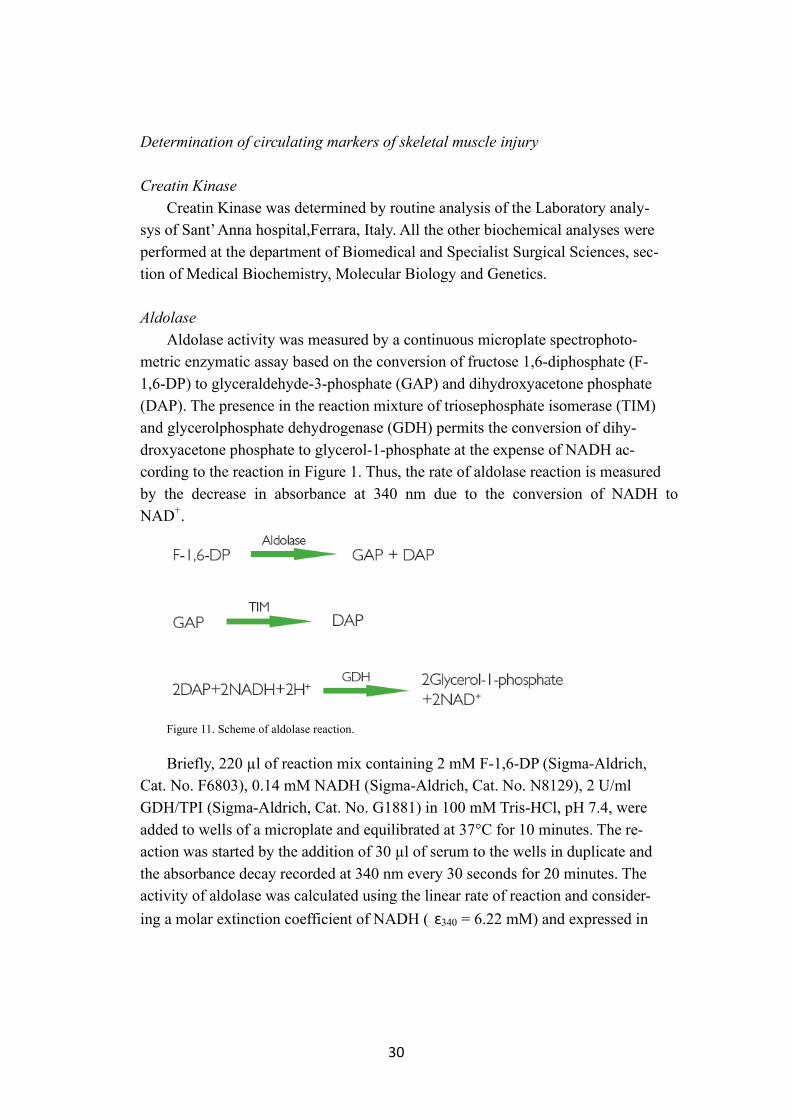
Aldolase activity was measured by a continuous microplate spectrophoto-metric enzymatic assay based on the conversion of fructose 1,6-diphosphate (F-1,6-DP) to glyceraldehyde-3-phosphate (GAP) and dihydroxyacetone phosphate (DAP). The presence in the reaction mixture of triosephosphate isomerase (TIM) and glycerolphosphate dehydrogenase (GDH) permits the conversion of dihy-droxyacetone phosphate to glycerol-1-phosphate at the expense of NADH ac-cording to the reaction in Figure 1. Thus, the rate of aldolase reaction is measured by the decrease in absorbance at 340 nm due to the conversion of NADH to NAD+.
Figure 11. Scheme of aldolase reaction.
Briefly, 220 µl of reaction mix containing 2 mM F-1,6-DP (Sigma-Aldrich,
Cat. No. F6803), 0.14 mM NADH (Sigma-Aldrich, Cat. No. N8129), 2 U/ml GDH/TPI (Sigma-Aldrich, Cat. No. G1881) in 100 mM Tris-HCl, pH 7.4, were added to wells of a microplate and equilibrated at 37°C for 10 minutes. The re-action was started by the addition of 30 µl of serum to the wells in duplicate and the absorbance decay recorded at 340 nm every 30 seconds for 20 minutes. The activity of aldolase was calculated using the linear rate of reaction and consider-
ing a molar extinction coefficient of NADH ( ε340 = 6.22 mM) and expressed in

31
U/L, were a unit is defined as the amount of enzyme which catalyzes the con-sumption of 1 µmole of NADH per minute at pH 7.4 and 37°C.
Aspartate aminotransferase (AST or GOT)
Aspartate aminotransferase (AST) catalyzes the transamination from L-as-partate to α-ketoglutarate (or α-oxoglutarate), forming L-glutamate and oxalo-acetate. The oxaloacetate formed is reduced to malate by malate dehydrogenase (MDH) with simultaneous oxidation of NADH (Figure 2). The change in absorb-ance with time due to the conversion of NADH to NAD is directly proportional to the AST activity and is measured evaluating the absorbance decay at 340 nm by a kinetic continuous method.
Figure 12. Reactions for the determination of AST. Briefly, 220 µl of reaction mix containing 200 mM L-aspartate (Sigma-Al-
drich, Cat. No. A6683), 12 mM α-oxoglutarate (Sigma-Aldrich, Cat. No. 75890), 0.18 mM NADH, 0.1 mM pyridoxal 5′-phosphate (Sigma-Aldrich, Cat. No. P9255), MDH/LDH 2/5 U/L (Sigma-Aldrich, Cat. No. M2634 and L7525, re-spectively), in 80 mM Tris-HCl, pH 7.4, were added to wells of a microplate and equilibrated at 37°C for 10 minutes. The reaction was started by the addition of 30 µl of serum to the wells in duplicate and the absorbance decay recorded at 340 nm every 30 seconds for 20 minutes. The activity of AST was calculated using the linear rate of reaction and considering a molar extinction coefficient of
NADH (ε340 = 6.22 mM) and expressed in U/L, were a unit is defined as the amount of enzyme which catalyzes the consumption of 1 µmole of NADH per minute at pH 7.4 and 37°C.
Alanine aminotransferase (ALT).
Alanine aminotransferase catalyzes the transfer of an amino group from L-alanine to α-oxoglutarate producing L-glutamate and pyruvate. Pyruvate is then used as a substrate together with NADH by Lactic Dehydrogenase (LDH) form-ing lactate and NAD+ (Figure 3). The decrease in absorbance due to NADH con-sumption is directly proportional to the amount of ALT present in the sample.
32
Figure 13. Scheme of ALT assay.
For the assay, 220 µl of reaction mix containing 0.8 M L-alanine (Sigma-
Aldrich, Cat. No. A7627), 18 mM α-oxoglutarate, 0.18 mM NADH, 5 U/L LDH, in 80 mM Sodium Phosphate Buffer, pH 7.4, were added to wells of a microplate and equilibrated at 37°C for 10 minutes. The reaction was started by the addition of 30 µl of serum to the wells in duplicate and the absorbance decay recorded at 340 nm every 30 seconds for 20 minutes.
The activity of ALT was calculated using the linear rate of reaction and con-
sidering a molar extinction coefficient of NADH (ε340 = 6.22 mM) and expressed in U/L, were a unit is defined as the amount of enzyme which catalyzes the con-sumption of 1 µmole of NADH per minute at pH 7.4 and 37°C.
Slow Skeletal Troponin I (TNNI1) assay
Slow (TNNI1) skeletal Troponin I (sTnI) was assayed by commercially available ELISA kits according to the manufacturer’s instructions (Human TNNI1 ELISA kit, Mybiosource, Cat. No. MBS2510383). All reagents and standards were included in the kits. Briefly, undiluted serum was analyzed in duplicate into 96-microwell microtiter plates pre-coated with anti-TNNI1 anti-bodies. Seven serial dilutions of TNNI1 standard within a range of 46.8-1500 pg/ml were dispensed on each plate in duplicate and incubated for 90 minutes at 37°C.
At the end of the incubation, the plate was emptied and 100 µl of Biotinylated detection antibody working solution were added to each wells and incubated for 1 hour at 37°C. After 3 washing cycles, 100 µl of Streptavidin-HRP conjugate working solution was added to each wells and the plate was incubated for further 30 minutes at 37°C and then washed 5 times.
Finally, 90 µl of substrate solution was added to each wells and the plate was incubated at 37°C for 30 minutes. The reaction was stopped by the addition of 50 µl of Stop Solution and the absorbance read at 450 nm. The amount of TNNI1 was determined by interpolation from the standard curve. The lower limit of quantification of the assay was 28.5 pg/mL, and both intra-assay and inter-assay coefficient of variations (CV) were below 10%.
33
Fast Skeletal Troponin I (TNNI2) assays Fast (TNNI2) skeletal Troponin I was assayed by commercially available
ELISA kits according to the manufacturer’s instructions (Human TNNI2 ELISA kit, Mybiosource, Cat. No. MBS927961). Briefly, undiluted serum was analyzed in duplicate into 96-microwell microtiter plates pre-coated with anti-TNNI2 an-tibodies. Seven serial dilutions of TNNI2 standard in a range of 6.25-400 pg/ml were dispensed on each plate in duplicate and incubated for two hours at 37°C.
At the end of the incubation, the plate was emptied and 100 µl of Biotinylated detection antibody working solution were added to each wells and incubated for 1 hour at 37°C. After 3 washing cycles, 100 µl of Streptavidin-HRP conjugate working solution was added to each wells and the plate was incubated for further 30 minutes at 37°C and then washed 5 times. Finally, 90 µl of substrate solution was added to each wells and the plate was incubated at 37°C for 30 minutes. The reaction was stopped by the addition of 50 µl of Stop Solution and the absorbance read at 450 nm. The amount of TNNI2 was determined by interpolation from the standard curve. The lower limit of quantification of the assay was 1.56 pg/mL. Intra-assay and inter-assay CVs were below 8% and 10%, respectively.
34
Statistical Analysis The normal distribution of quantitative data was evaluated through the
D’Agostino test. All the data approaching the normal distribution are summarized as mean
and standard deviation (SD). Non-normally distributed data are expressed as median (interquartile range). Since the variables were not normally distributed, the effect of time of intubation on the levels of the serum markers of muscle functionality was evaluated by Friedman test. Correlations between the varia-bles were performed by Spearman’s rank test.
Breathing pattern, gas exchange and EAdi-derived parameters at the three SBT time points (0, 24 and 48 hours) were normally distributed and differences were evaluated through an analysis of variance (ANOVA) model for repeated measure with interaction (time*method). Post hoc comparisons, between the two groups at each time point and within each group between the three time points, were carried out using Student’s t test, with Bonferroni correction.
All the analyses were performed by SPPS 21.0 for windows, with a p<0.05 considered significant.

35
Results
Of the 37 patients screened, 28 met the study inclusion criteria. The reasons for exclusion were thoracostomy, primary neuromuscular disease, rib fractures, and ventilation for less than 48 h. Four more patients were excluded because of hemidiaphragm paralysis detected during diaphragmatic ultrasonog-raphy. The clinical and physiological characteristics of the studied patients are presented in Table 1.
Table 1 – Clinical characteristics of the 24 ICU patients enrolled in the study. Total population n = 24 Age, years 70 ± 11 Male, n (%) 15 (63) BMI, kg/m2 27.4 ± 4.1 Smokers
Current smoker 8 (33) Former smoker 2 (8)
Comorbidities Pulmonary disease
COPD 4 (17) Asthma 1 (4) OSAS 2 (8)
Cardiovascular disease Hypertension 13 (54) Chronic ischemic disease 6 (25) Atrial fibrillation 5 (21) Chronic heart failure 5 (21) PAD 1 (4)
Chronic renal failure 2 (8) Neurological disease
Stroke 1 (4) Epilepsy 1 (4)
Metabolic disease Diabetes 6 (25) Liver cirrhosis 2 (8) Chronic pancreatitis 1 (4) Hypothyroidism 1 (4)
Cancer Hematological cancer 1 (4) Other 5 (21)
Type of admission Medical 12 (56)

36
Surgical 11 (40) Elective 2 (8) Emergency 9 (37)
Trauma 1 (4) SAPS II score on admission 45 [36 – 58] SOFA score on admission 7.50 [5.75 – 9.00] Hospital LOS, days 40 [19 – 53] ICU mortality, n (%) 4 (17)
Normally distributed data are shown as mean ± SD; percentage data are shown as n (%); not normally distributed data as median [IQR]. BMI = body mass index; COPD = chronic obstructive pulmonary disease; OSAS = obstructive sleep apnea syndrome; PAD = peripheral artery disease; SAPS = Simplified Acute Physiology Score; SOFA = Sepsis-related Organ Failure Assessment; ICU = Intensive Care Unit; LOS = Length of stay.
Table 2 shows the breathing pattern and EAdi parameters recorded during
the three SBTs and, in addition, the RASS score and the gas exchange parameters recorded immediately before each SBT. The EAdiPEAK was significantly higher at 48 H in patients undergone PSV (average 19.6 ± 9.2 µV at 48 H and 17.2 ± 6.7 µV at 24 H).
37
T0 T1 T2 p-value*
DD, mm 1.85 [1.28 – 2.01]
1.40 [1.05 – 1.75] 1.10 [0.80 – 1.55] 0.004
TF, % 0.45 [0.39 – 0.49]
0.39 [0.29 – 0.43] 0.30 [0.26 – 0.39] < 0.0001
TEE, mm 0.30 [0.26 – 0.39]
0.34 [0.32 – 0.31] 0.36 [0.31 – 0.39] 1.000
P0.1, cmH2O
1.5 [0.9 – 2.1] 0.95 [0.65 -1.60] 1.20 [0.58 – 2.18] 0.199
Table 3 – T0 (24 hours from ICU admission), T1(48 hours from ICU admission) and T2 (72 hours from ICU admission) ultrasounographic measurements of the diaphragm. Data are reported as median [IQR]. * Friedman test was used to compare medians at 3 different times.
Values of circulating markers of muscle function The circulating markers of muscle function were measured in the serum of
patients underwent mechanical ventilation as stated in the material and methods section. First, we evaluated the time-change of skeletal troponins (TNNIs): the results are presented in Figure 12.
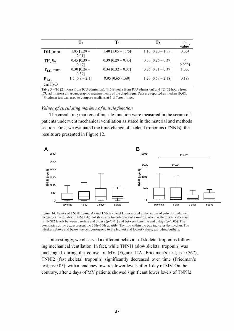
Figure 14. Values of TNNI1 (panel A) and TNNI2 (panel B) measured in the serum of patients underwent mechanical ventilation. TNNI1 did not show any time-dependent variation, whereas there was a decrease in TNNI2 levels between baseline and 2 days (p<0.01) and between baseline and 3 days (p<0.05). The boundaries of the box represent the 25th–75th quartile. The line within the box indicates the median. The whiskers above and below the box correspond to the highest and lowest values, excluding outliers.
Interestingly, we observed a different behavior of skeletal troponins follow-
ing mechanical ventilation. In fact, while TNNI1 (slow skeletal troponin) was unchanged during the course of MV (Figure 12A, Friedman’s test, p=0.767), TNNI2 (fast skeletal troponin) significantly decreased over time (Friedman’s test, p<0.05), with a tendency towards lower levels after 1 day of MV. On the contrary, after 2 days of MV patients showed significant lower levels of TNNI2

38
than baseline (Wilcoxon signed rank test corrected for multiple comparisons, p<0.01), reaching the minimum after 3 days of MV (Figure 12, panel B, Wil-coxon signed rank test corrected for multiple comparisons, p<0.05).
A similar behavior was observed for Creatine Kinase (CPK, Figure 13, panel
A), showing a significant decrease over time (Friedman’s test, p<0.01) with lower levels of the enzyme at 2 days with respect to baseline (p<0.05), reaching a minimum at 3 days of MV (p<0.05). On the contrary, both aldolase and AST were not different at any time point analyzed.
Figure 14. Values of CPK (panel A), aldolase (panel B) and AST (panel C) measured in the serum of pa-tients underwent mechanical ventilation. CPK decreased over time starting from 2 days post MV (p<0.05 vs. baseline), reaching the minimum after 3 days (p<0.05). Aldolase and AST were not different at any time point. The boundaries of the box represent the 25th–75th quartile. The line within the box indicates the median. The whiskers above and below the box correspond to the highest and lowest values, excluding outliers.
TNNI1 TNNI2 CPK Aldolase AST TNNI1 -0.095,
p=0.364 -0.052, p=0.619
-0.095, p=0.360
0.205, p=0.047
TNNI2 0.739, p=0.0001
0.425, p=0.0001
0.394, p=0.0001
CPK 0.355, p=0.0005
0.519, p=0.0001
Aldolase 0.287, p=0.005
Table 4. Correlations between the markers of muscle function measured in the serum as a whole.

39
Data are presented as Spearman’s rank correlation coefficients and statistical significance.
We found a statistically significant correlation between the percent change from baseline of sfTnI and TF at 72 hours from admission (r = 0.661, p = 0.007).
Figure 15. Correlation between percent change versus baseline of sfTnI and percent change versus base-line of TF at T2 (48 hours after admission).
-100
-50
0
50
100
150
200
250
300
350
400
0 20 40 60 80 100 120
TFT2
(%of
baseline)
sfTnI T2 (% of baseline)
p = 0.007r = 0.661
40
Discussion
In this study, we were able to detect ssTnI and fsTnI in serum from critically ill patients undergone mechanical ventilation. From our results, it seems that se-rum levels of fsTnI, but not ssTnI, could be a useful diagnostic marker of respir-atory muscle dysfunction. In fact, the decrease in the levels fsTnI levels corre-lated with TF and was mirrored by the decrease in diaphragm functionality as measured by ultrasonography. Indeed, at this time this technique is considered as the gold standard for the evaluation of the work load of the diaphragm, although it might have some culprits due to the variable operator experience or other source of variations. Therefore, the presence of something circulating in the blood might have a large impact for the evaluation of diaphragm functionality.
Previous studies evaluated Skeletal troponin I (sTnI), with its two distinct isoforms [fast (fsTnI) and slow (ssTnI)], TnI (sTnI), as a marker of skeletal mus-cle damage, indicating that it is more sensitive and specific than traditional mark-ers (creatine kinase and myoglobin) of skeletal muscle injury [51]; [78].
Our results showed that the level of fsTnI decreased significantly by day 3, while ssTnI remained unchanged during the treatment. Interestingly, the serum profile of total CPK, but not AST or Aldolase, resembled the time profile of fsTnI rather than that of ssTnI.Our data provide no information about the mecha-nism(s) underlying sTnI release. We included patients undergoing mechanical stress associated with the “self” ventilator induced diaphragmatic dysfunction (VIDD) determined by assisted ventilation. The latter impaired the local meta-bolic conditions of the inspiratory muscles, particularly the diaphragm, the most likely source of the sTnI due to its fatigue.
There are no previous studies using a similar protocol to which we can com-pare our data. However, Jiang et al. [79] observed injury of the diaphragm 3 days after Inspiratory resistive loading (IRL).
We do not know if these changes in fsTnI reflect specific patient’s condition because changes in fsTnI and/or ssTnI could be influenced by clinical conditions, directly or indirectly, associated with clinical condition of patients. Acute renal failure, compartmental syndrome, and cardiac abnormalities are common com-plications that can affect either the release or clearance of sTnI [51]. In addition, patients may experience muscle atrophy, regeneration, and/or adaptation over time; all of which could influence the results of serum sTnI analysis.
Diaphragm dysfunction is underdiagnosed, but should not be neglected, as it can negatively impact quality of life, can be a marker of disease severity and,

41
in some instances, such as in the intensive care unit, be a prognostic marker. (Dubè, JCM 2016). Determining if fatigue is present is complicated and usually involves detecting a deficit in respiratory muscle contractile function. Functional measurement includes inspiratory and expiratory pressure differences generated by patients breathing against a closed valve; however, this measurement is effort dependent and poorly reproducible, particularly in intubated, critically ill pa-tients. [26]
Esophageal and gastric balloons with pressure transducers can also be used to calculate transdiaphragmatic pressures by subtracting the esophageal pressure from the gastric pressure correlating with diaphragmatic strength. Another method is the maximal sniff transdiaphragmatic pressure. This is used as the vo-litional indicator of diaphragm strength. These measurements, however, depend on effort and are poorly reproducible. As such, there is currently no simple and reproducible method to measure diaphragm muscle strength (force-generating capacity).
Diaphragmatic dysfunction as assessed by M-mode is common practice in medical ICU. In this study using serial ultrasound measurements of the dia-phragm showed that diaphragmatic thickness can be measured easily using ultra-sonography shortly after endotracheal intubation. Furthermore, we observed that thinning begins within 48 h of the initiation of MV decrease, and occurs linearly over the subsequent time period. Our data agree with data from human [26] and animal studies, in which diaphragm inactivity with controlled MV has been shown to produce myofibril damage that contributes to the reduction in diaphrag-matic force over time.
Interestingly, fsTnI were modified over time, accordingly with diaphrag-matic dysfunction detected by ultrasound of the diaphragm.
There is little information about VIDD in humans, and we know that other factors, such as sepsis, the use of corticosteroids and neuromuscular blockers, and malnutrition can be other causes of diaphragmatic thinning. Nonetheless, our findings suggest that in the acute care setting, diaphragmatic thinning can be as-sessed easily using two-dimensional ultrasonography.
Furthermore, the effect of nutritional status and lung disease progression on diaphragm performance is unclear. The decrease in diaphragm thickness may well have been associated with malnutrition. Our interpretation, however, is lim-ited by lack of information on the markers of malnutrition.
Our study had several limitations. The study had a small sample size, lacked a control group, and did not show a correlation with the discontinuation of MV.
42
Furthermore, we did not have long-term follow-up on the patients. Our sample size was also too small to analyze correlations among other respiratory variables.
Additionally, all these patients were ventilated using an assisted mode of MV, none of the patients were paralyzed.

43
Conclusions
We conclude from this study that measured thinning of the diaphragm occurs within 48 h after intubation and the initiation of MV, most consistent with de-creased level of fsTnI. sTnI seem to be a useful diagnostic of respiratory dia-phragm dysfunction. Further studies are needed to evaluate if this decrease in fsTnI is indeed due to diaphragm atrophy, and if this has an impact on the dis-continuation of MV. Larger studies are needed to clarify the relationship between diaphragmatic thinning, fsTnI and clinical outcomes.
44
References
1. TOBIN, M.; LAGHI, F.; JUBRAN, A. Narrative review: ventilator-induced respiratory muscle weakness. Ann Intern Med, v. 153, n. 4, p. 240-245, 2010.
2. VASSILAKOPOULOS, T.; PETROF, B. Ventilator-induced diaphragmatic dysfunction. Am J Respir Crit Care Med, n. 169, p. 336-341, 2004.
3. SASSOON, C. et al. Altered diaphragm contractile properties with controlled mechanical ventilation. J Appl Physiol, v. 92, n. 6, p. 2585-2595, 2002.
4. YANG, L. et al. Controlled mechanical ventilation leads to remodeling of the rat diaphragm. Am J Respir Crit Care Med, v. 166, n. 8, p. 1135-1140, 2002.
5. POWERS, S. et al. Mechanical ventilation results in progressive contractile dysfunction in the diaphragm. J Appl Physiol, v. 92, n. 5, p. 1851-1858, 2002.
6. FOSTER, G. et al. Serum skeletal troponin I following inspiratory threshold loading in healthy young and middle-aged men. Eur J Appl Physiolg, v. 112, n. 10, p. 3547-3558, 2012.
7. LEVINE, S. et al. Rapid disuse atrophy of diaphragm fibers in mechanically ventilated humans. N Eng J Med, v. 358, p. 1327-1335, 2008.
8. HUSSAIN, S. et al. Mechanical ventilation-induced diaphragm disuse in humans triggers autophagy. Am J Respir Crit Care Med, v. 182, n. 11, p. 1377-1386, 2010.
9. UCHIYAMA, A. et al. Regional blood flow in respiratory muscles during partial ventilatory assistance in rabbits. Anesth Analg, v. 102, n. 4, p. 1201-1206, 2006.
10. HUDSON, M. et al. Both high level pressure support ventilation and controlled mechanical ventilation induce diaphragm dysfunction and atrophy. Crit Care Med, v. 40, n. 4, p. 1254-1260, 2012.
11. ESTEBAN, A. et al. A comparison of four methods of weaning patients from mechanical ventilation. N Eng J Med, v. 332, p. 345-350, 1995.
12. MCCONVILLE , J.; KRESS, J. Weaning patients from the ventilator. N Eng J Med, v. 367, p. 2233-2239, 2012.
13. ESKANDAR, N.; APOSTOLAKOS, M. Weaning from mechanical ventilation. Crit Care Clin, v. 23, n. 2, p. 263-274, 2007.
14. VASSILAKOPOULOS, T.; ZAKYNTHINOS, S.; ROUSSOS, C. Respiratory muscles and weaning failure. Eur Respir J, v. 9, n. 11, p. 2383-2400, 1996.
15. CADER, S. et al. Extubation process in bed-ridden elderly intensive care patients receiving inspiratory muscle training: a randomized clinical trial. Clin Interv Aging, v. 7, p. 437-443, 2012.
16. CADER, S. et al. Inspiratory muscle training improves maximal inspiratory pressure and may assist weaning in older intubated patients: a randomised trial. J Physiother, v. 56, n. 3, p. 171-177, 2010.
45
17. CHANG, A. et al. Case report: inspiratory muscle training in chronic critically ill patients - a report of two cases. Physiother Res Int, v. 10, n. 4, p. 222-226, 2005.
18. KIM, W. et al. Diaphragm dysfunction assessed by ultrasonography: influence on weaning from mechanical ventilation. Crit Care Med, v. 39, n. 12, p. 2627-2630, 2011.
19. MARTIN, A. et al. Inspiratory muscle strength training improves weaning outcome in failure to wean patients: a randomized trial. Crit Care, v. 15, n. 2, p. R84, 2011.
20. MARTIN, A. et al. Use of inspiratory muscle strength training to facilitate ventilator weaning: a series of 10 consecutive patients. Chest, v. 122, n. 1, p. 192-196, 2002.
21. DE JONGHE, B. et al. Respiratory weakness is associated with limb weakness and delayed weaning in critical illness. Crit Care Med, v. 35, n. 9, p. 2007-2015, 2007.
22. ADLER, D. et al. Does inspiratory muscle dysfunction predict readmission after intensive care unit discharge? Am J Respir Crit Care Med, v. 190, n. 3, p. 347-350, 2014.
23. JABER, S.; PETROF, B.; JUNG, B. Rapidly progressive diaphragmatic weakness and injury during mechanical ventilation in humans. Am J Respir Crit Care Med, v. 183, n. 3, p. 361-371, 2011.
24. KAVAZIS, A. et al. Mechanical ventilation induces diaphragmatic mitochondrial dysfunction and increased oxidant production. Free Radic Biol Med, v. 46, n. 6, p. 842-850, 2009.
25. FREDRIKSSON, K.; TJÄDER, I.; KELLER, P. E. A. Dysregulation of Mitochondrial Dynamics and the Muscle Transcriptome in ICU Patients Suffering from Sepsis Induced Multiple Organ Failure. PLOS ONE, v. 3, n. 11, p. e3686, 2008.
26. GROSU, H. et al. Diaphragm muscle thinning in patients who are mechanically ventilated. Chest, v. 142, n. 6, p. 1455-1460, 2012.
27. GAYAN-RAMIREZ, G. et al. Detrimental effects of short-term mechanical ventilation on diaphragm function and IGF-1 mRNA in rats. Intensive Care Med, v. 29, n. 5, p. 825-833, 2003.
28. SHANELY, R. et al. Mechanical ventilation-induced diaphragmatic atrophy is associated with oxidative injury and increased proteolytic activity. Am J Respir Crit Care Med, v. 166, n. 10, p. 1369-1374, 2002.
29. BETTERS, J.; CRISWELL, D.; SHANELY, R. Trolox attenuates mechanical ventilation-induced diaphragmatic dysfunction and proteolysis. Am J Respir Crit Care Med, v. 170, n. 11, p. 1179-1184, 2004.
30. MCCLUNG, J. et al. Antioxidant administration attenuates mechanical ventilation-induced rat diaphragm muscle atrophy independent of protein kinase B (PKB Akt) signalling. J Physiol, v. 585, n. 1, p. 203-215, 2007.
46
31. POWERS, S.; KAVAZIS, A.; LEVINE, S. Prolonged mechanical ventilation alters diaphragmatic structure and function. Crit Care Med, v. 37, n. 10 Suppl, p. 347-353, 2009.
32. HOOIJMAN, P. et al. Diaphragm muscle fiber weakness and ubiquitin-proteasome activation in critically ill patients. Am J Respir Crit Care Med, v. 191, n. 10, p. 1126-1138, 2015.
33. VAN HEES, H. et al. Plasma from septic shock patients induces loss of muscle protein. Crit Care, v. 15, n. 5, p. R233, 2011.
34. TANG, H. et al. Intrinsic apoptosis in mechanically ventilated human diaphragm: linkage to a novel Fos/Fox01/Stat3-Bim axis. FASEB J, v. 25, n. 9, p. 2921-2936, 2011.
35. GAYAN-RAMIREZ, J. et al. Intermittent spontaneous breathing protects the rat diaphragm from mechanical ventilation effects. Crit Care Med, v. 33, n. 12, p. 2804-2809, 2005.
36. FUTIER, E. et al. Pressure support ventilation attenuates ventilator-induced protein modifications in the diaphragm. Crit Care, v. 12, n. 5, p. 116, 2008.
37. JUNG, B. et al. Adaptive support ventilation prevents ventilator-induced diaphragmatic dysfunction in piglet: an in vivo and in vitro study. Anesthesiology, v. 112, n. 6, p. 1435-1443, 2010.
38. PUTENSENS, C. et al. The impact of spontaneous breathing during mechanical ventilation. Curr Opin Crit Care, v. 12, n. 1, p. 13-18, 2006.
39. SINDERBY, C.; NAVALESI, P.; BECK, J. Neural control of mechanical ventilation in respiratory failure. Nat Med, v. 5, n. 12, p. 1433-1436, 1999.
40. THILLE, A. et al. Patient-ventilator asynchrony during assisted mechanical ventilation. Intensive Care Med, v. 32, n. 10, p. 1515-1522, 2006.
41. AMERICAN THORACIC SOCIETY/EUROPEAN RESPIRATORY SOCIETY. ATS/ERS Statement on respiratory muscle testing. Am J Respir Crit Care Med, v. 166, n. 4, p. 518-624, 2002.
42. DEMOULE, A. et al. Diaphragm dysfunction on admission to the intensive care unit. Prevalence, risk factors, and prognostic impact - a prospective study. Am J Respir Crit Care Med, v. 188, n. 2, p. 213-219, 2013.
43. DEMOULE, A. et al. Patterns of diaphragm function in critically ill patients receiving prolonged mechanical ventilation: a prospective longitudinal study. Ann Intensive Care, v. 6, n. 1, p. 75, 2016.
44. SUPINSKI, G.; WESTGATE, P.; CALLAHAN, L. Correlation of maximal inspiratory pressure to transdiaphragmatic twitch pressure in intensive care unit patients. Crit Care, v. 20, p. 77, 2016.
45. HARRIS, R.; GIOVANNETTI, M.; KIM, B. Normal ventilatory movement of the right hemidiaphragm studied by ultrasonography and pneumotacography. Radiology, v. 146, n. 1, p. 141-144, 1983.
47
46. DIAMENT, M.; BOECHAT, M.; KANGARLOO, H. Real-time sector ultrasound in the evaluation of suspected abnormalities of diaphragmatic motion. J Clin Ultrasound, v. 13, n. 8, p. 539-543, 1985.
47. WAIT, J. et al. Diaphragmatic thickness-lung volume relationship in vivo. J Appl Physiol, v. 67, n. 4, p. 1560-1568, 1989.
48. GOLIGHER, E. et al. Measuring diaphragm thickness with ultrasound in mechanically ventilated patients: feasibility, reproducibility and validity. Intensive Care Med, v. 41, n. 4, p. 642-649, 2015.
49. BELLANI, G. et al. Estimation of patient's inspiratory effort from the electrical activity of the diaphragm. Crit Care Med, v. 41, n. 6, p. 1483-1491, 2013.
50. LIU, L. et al. Assessment of patient-ventilator breath contribution during neurally adjusted ventilatory assist in patients with acute respiratory failure. Crit Care, v. 19, p. 43, 2015.
51. SORICHTER, S.; PUSCHENDORF, B.; MAIR, J. Skeletal muscle injury induced by eccentric muscle action: muscle proteins as markers of muscle fiber injury. Exercise Immunology Review, v. 5, n. 5, p. 21, 1999.
52. BRANCACCIO, P.; LIPPI, G.; MAFFULLI, N. Biochemical markers of muscular damage. Clin Chem Lab Med, v. 48, n. 6, p. 757-767, 2010.
53. ADAMS, J. I.; ABENDSCHEIN, D.; JAFFE, A. Biochemical markers of myocardial injury. Is MB creatine kinase the choice for the 1990s? Circulation, v. 88, p. 750-763, 1993.
54. GRANCHI, C. et al. Small-molecule inhibitors of human LDHS. Future Med Chem, v. 5, n. 16, p. 1967-1991, 2013.
55. PAPADOPOULOS, N. Clinical Applications of Lactate Dehydrogenase Isoenzymes. Annals of Clinical And Laboratory Science, v. 7, n. 6, p. 505-510, 1977.
56. ARAKAKI, T. et al. Structure of human brain fructose 1,6-(bis)phosphate aldolase: Linking isozyme structure with function. Protein Science, v. 13, p. 3077-3084, 2004.
57. AMACHER, D. Serum Transaminase Elevations as Indicators of Hepatic Injury Following the Administration of Drugs. Regulatory Toxicology And Pharmacology, v. 27, p. 119-130, 1998.
58. ORDWAY, G.; GARRY, D. Myoglobin: an essential hemoprotein in striated muscle. The Journal of Experimental Biology, v. 207, p. 3441-3446, 2004.
59. PAGANI, F. Biochimica dei marcatori di lesione miocardica. RIMeL / IJLaM, v. 1, p. 26-29, 2005.
60. GOMES, A.; POTTER, J.; SZCZESNA-CORDARY, D. The Role of Troponins in Muscle Contraction. IUBMB Life, v. 54, p. 323-333, 2002.

48
61. VASSALLO, J. et al. Biomarkers of Drug-Induced Skeletal Muscle Injury in the Rat: Troponin I and Myoglobin. Toxicological Sciences, v. 111, n. 2, p. 402-412, 2009.
62. ANNANE, D. What is the evidence for harm of neuromuscular blockade and corticosteroid use in the Intensive Care Unit? Semin Respir Crit Care Med, v. 37, n. 1, p. 51-56, 2016.
63. PAPAZIAN, L. et al. Neuromuscular blockers in early acute respiratory distress syndrome. N Eng J Med, v. 363, n. 12, p. 1107-1116, 2010.
64. SASSOON , C.; ZHU, E.; CAIOZZO, V. Assist-control mechanical ventilation attenuates ventilator-induced diaphragmatic dysfunction. Am J Respir Crit Care Med, v. 170, n. 6, p. 626-632, 2004.
65. DOORDUIN, J. et al. Assisted ventilation in patients with Acute Respiratory Distress Syndrome: Lung-distending pressure and patient-ventilator interaction. Anesthesiology, v. 123, n. 1, p. 181-190, 2015.
66. PAVLOVIC, D.; WENDT, M. Diaphragm pacing during prolonged mechanical ventilation of the lungs could prevent fro respiratory muscle fatigue. Med Hypotheses, v. 60, n. 3, p. 398-403, 2003.
67. MADDOCKS, M. et al. Neuromuscular eletrical stimulation to improve exercise capacity in patients with severe COPD: a randomised double-blind, placebo-controlled trial. Lancet Respir Med, v. 4, n. 1, p. 27-36, 2016.
68. ZANOTTI , E. et al. Peripheral muscle strength training in bed-bound patients with COPD receiving mechanical ventilation: effect of electrical stimulation. Chest, v. 124, n. 1, p. 292-6, 2003.
69. ABDELLAOUI, A. et al. Skeletal muscle effects of electrostimulation after COPD exacerbation: a pilot study. Eur Respir J, v. 38, n. 4, p. 781-8, 2011.
70. LUO, Y. et al. Quantification of the esophageal diaphragm electromyogram with magnetic phrenic nerve stimulation. Am J Respir Crit Care Med, v. 160, n. 5, p. 1629-1634, 1999.
71. CATTAPAN, S.; LAGHI, F.; TOBIN, M. Can diaphragmatic contractility be assessed by airway twitch pressure in mechanically ventilated patients? Thorax, v. 58, n. 1, p. 58-62, 2003.
72. DI MUSSI, R.; AL., E. Impact of prolonged assisted ventilation on diaphragmatic efficiency: NAVA versus PSV. Crit Care, v. 20, n. 1, p. 1178-0, 2016.
73. BARWING, J.; AMBOLD, M.; AL., E. Evaluation of the catheter positioning for neurally adjusted ventilatory assist. Intensive Care Med, v. 35, n. 10, p. 1809-14, 2009.
74. SCHMIDT, M.; AL., E. Neurally adjusted ventilatory assist and proportional assist ventilation both improve patient-ventilator interaction. Crit Care, v. 19, p. 56, 2015.

49
75. LIU, L.; AL., E. Neuroventilatory efficiency and extubation readiness in critically ill patients. Crit Care, v. 16, n. 4, p. R143, 2012.
76. BECK, J.; AL., E. Effects of lung volume on diaphragm EMG signal strength during voluntary contractions. J Appl Physiol, v. 85, n. 3, p. 1123–34, 1998.
77. HEDENSTIERNA, G. Esophageal pressure: benefit and limitations. Minerva Anestesiol, v. 78, n. 8, p. 959–66, 2012.
78. ONOUHA, G.; AL., E. Skeletal troponin-I release in orthopedic and soft tissue injuries. J Orthop Sci, v. 6, n. 1, p. 11-5, 2001.
79. JIANG, T.; AL., E. Delayed diaphragm injury and diaphragm force production. Am J Respir Crit Care Med, v. 157, n. 3 part 1, p. 736-42, 1998.


![[Cicle comunicacio 2.0] 1 comunicacio](https://img.pdfslide.us/doc/110x75/55974d591a28abf7018b4608/cicle-comunicacio-20-1-comunicacio.jpg)