Embed Size (px)
Citation preview

University Politehnica of Bucharest
Doctoral School of Applied Chemistry and Materials Science
Mesoporous silica-based composites for controlled release of
biologically active molecules
__________________________________________________________________________
Compozite pe bază de silice mezoporoasă în sisteme cu eliberare
controlată de substanțe biologic active
PhD THESIS SUMARY
Scientific leader : Prof. dr. ing. Daniela Berger
Author : Marilena Georgescu (Petrescu)
Bucharest
2017

2/32
Keywords: drug delivery systems, mesoporous silica, silica-ceria composite, aluminosilicate, functionalized
mesoporous silica, alginate beads, magnetite-aluminosilicate composites, doxycycline, oxyteracycline, ketoprofen,
doxorubicin
Contents
I. Literature study........................................................................................................................5
I.1. Introduction……………………………………………………………………………….5
I.2. Obtaining and characterization of mesoporous silica…………………………………10
I.2.1. Functionalization of mesoporous silica. Type of functionalized mesoporus
silica supports…………………………………………………………………………………15
I.2.2. Introduction of heteroatoms into the mesoporous silica
framework ..................................................................................…………………………..17
I. 2.3. Mesoporous silica-based composites…………………………………………19
I. 2.4. Drug delivery systems based on alginate beads……………………………..21
I.3. Targeted drug delivery systems ..................................................................................... 24
I.3.1. Drug delivery systems based on tetracyclines………………………………24
I.3.2. Drug delivery systems based on anti-inflammatory ……………………….28
I.3.3. Targeted vehicles for doxorubicin……………..…………………………….30
I. 4. References ……………………………………………………………………………….39
II. Original contributions………………………………………………………………………....53
II.1. Justification of theme …………………………………………………………………..53
II.2. Carriers based on mesoporous silica ………………………………………………….54
II.3. Composites based on doxycycline ……………………………………………………..78
II.4. Composites based on oxytetracycline ……... …………………………………………84
II.5. Composites based on ketoprofen ………………………………………………………93
II.6. Composites based on doxorubicin……. ……………………………………………...110
II.7.Conclusions……………………………………………………………………………..120
II.8. References……………………………………………………………………………...125

3/32
The list of abbreviations
MCM-41 Mobil Composition of Mater no. 41
SBA Santa Barbara Amorphous
APTES 3-aminopropyltriethoxysilane
AlSBA-16 aluminosilicate from SBA class
BuDEA butyldiethanolamine
Al(BuO)3 aluminum tributoxide
TEOS tetraethyl orthosilicate
SBET specific surface area calculated with the BET method
dBJH average pore diameter calculated by Barrett Joyner Halenda method
XRD X-ray diffraction
DSC differential scanning calorimetry
SEM scanning electron microscopy
TEM transmission electron microscopy
BSE back-scattered electron detector
EDX Energy dispersive X-Ray
a. u. arbitrary units
HMS Hexagonal Mesoporous silica
MSU Michigan State University
4/32
I.1. Introduction
The discovery of silica-type mesoporous materials in the 90s was of a particular interes for
researchers due to their properties, which recommend them for applications in chromatographic
separations, catalysis, molecules adsorption etc, their most promising being in the field of biomedice.
In 2001, mesoporous silica MCM-41 was proposed as a carrier in drug delivery systems due to its
outstanding properties: an ordered pore framework with narrow pore size distribution, large total pre
volume for accommodation of required amount of drug molecules, high specific surface, the
presence of silanol groups involved in the post-synthesis functionalization for the modification of
silica surface properties in order to tailor the interactions between the support and guest molecles for
a desired delivery kinetics.
To enhance mesoporous silica carrier should interact with biologically active substances.
This can be achieved by tuning the properties of carrier using two strategies: either through the
carrier synthesis conditions, especially in order to obtain a pore size suitable for the adsorption of
organic molecules or by the functionalization of silica surface with different organic groups
The research reported in this PhD thesis consists in the development of drug delivery
systems based on mesoporous matrix carriers, silica, aluminosilicates, silica-ceria and
magnetite-aluminosilicate composites. In this regard, different mesoporous materials were
synthesized, characterized and tested as carriers for tetracyclines (oxytetracycline and doxycycline),
anti-inflammatory (ketoprofen) and cytostatic agent (doxorubicin).
The PhD thesis consists of two parts: Part I comprises literature data in three chapters.
Chapter 1 deals with the basic knowledge concerning the mesoporous silica and aluminosilicates
used as vehicles in drug delivery systems. Chapter 2 describes the obtaining methods of mesoporous
silica, functionalization approaches, the effect of introduction aluminum in the silica framework on
the structural and textural material parameters, composites based mesoporous silica, and drug
delivery systems based on alginate beads. Chapter 3 presents some literature data about the
biologically active substances used in this thesis, chosen as model molecules from the tetracyclines,
anti-inflammatories and cytostatic class.
Part II describes the original contributions and contains five chapters. Chapter 1 presents
the justification of topic chosen and the scope and the objectives of the thesis. Chapter 2 is
dedicated to the first two objectives, namely, the synthesis and characterization of several inorganic
mesostructured materials, obtaining functionalized mesoporous silica samplesf by the
post-synthesis approach. Chapters 3 and 4 comprise the developed drug delivery systems based on
MCM-41-type supports, containing antibiotics. Chapter 5 is focused on the fulfillment of the fourth
objective and presents the development of ketoprofen delivery systems obtained by the
encapsulation of silica-ketoprofen-type composites in alginate beads. Chapter 6 describes the
entrapment of doxorubicin in superparamagnetic magnetite-aluminosilicate-type composites and
studies of drug release profiles in the biological fluid that simulates the tumor cells environment,
which consists in the last thesis objective.
The original results obtained during the doctoral studies were diseminated in four papers,
three in the ISI journals (one in press) and one in UPB Scientic Bull. Series B, two conferences as
posters at the Days of "Alexandru Ioan Cuza" University of Iasi, 2015 and PRIOCHEM XII - 2016,
section 3 - Multifunctional Materials and Nanocomposites.

5/32
I. ORIGINAL CONTRIBUTIONS
OBJECTIVES
The aim of this PhD thesis was the development of drug delivery systems based on inorganic
carriers containing carriers based on mesoporous silica.
To fulfill the PhD thesis aim the following objectives were established:
1. Synthesis and characterization of several mesostructured silica-type materials
2. Synthesis and characterization of functionalized mesoporous silica materials using the
post-synthesis approach
3. Development of antibiotics delivery systems containing inorganic transporters MCM-41
4. Developing complex drug delivery systems of ketoprofen by encapsulating
silica-ketoprofen-type composites in alginate beads
5. encapsulation of doxorubicin in aluminosilicate-type inorganic composites and the study of
release profiles of the therapeutic agent in biological fluid that simulates tumor cell environment.
To accomplish these objectives, the following activities were carried out:
Synthesis of ceria-silica composites by two different methods, two types of silica
functionalized with aminopropyl groups by post-synthesis, other types of silica and
aluminosilicates for comparison and some magnetite-aluminosilicate composites.
Characterization of mesoporous transporters synthesized by FTIR spectroscopy, wide-
and small-angles XRD, N2 adsorption-desorption isotherms recorded at 77K, SEM, TEM and
thermal analysis.
Preparation of composites based on doxycycline, oxytetracycline, ketoprofen and
doxorubicin by incipient wetness impregnation method.
Characterization of composites containing active biologic substances by FTIR
spectroscopy, wide- and small-angles XRD, N2 adsorption-desorption isotherms at 77 K.
Perform in vitro release studies and fit experimental data with different functions to
understand interactions between the transporter and the drug and the type of transport that
accompanies the desorption of organic molecules.
Encapsulation of ketoprofen-silica materials in alginate beads and determination of in
vitro release profiles of ketoprofen from these complex carriers.
Determination of antimicrobial activity for antibiotics and proliferation and cell death
tests on healthy human lymphocytes for doxorubicin encapsulated in magnetite-aluminosilicate
type transporters as compared to treatment with cytostatic agent solution at the same concentration.
II.2. Carriers based on mesoporous silica
A first objective of the research was the obtaining of mesoporous silica-based materials,
aluminosilicates, ceria-silica and magnetite-aluminosilicate composites by sol-gel method, which
were further applied as carriers in drug delivery systems. For the first time, mesoporous ceria-silica
composites have been investigated as carriers in drug delivery systems. Two synthesis methods were
chosen for obtaining mesoporous silica-ceria composites: (i) two-step synthesis that first involves the
synthesis of ceria nanoparticles by the hydrothermal method, and then their coating with MCM-41
mesoporous silica and (ii) one step method in which MCM-41 mesoporous silica was first
precipitated, a solution containing corresponding cerium ions was added, and then the reaction
mixture was hydrothermally treated. By this last method, two mesoporous silica-ceria composites
with different concentrations of 10% (mol) and 20% (mol) ceria, labeled MCM-CeO2 (1) and
MCM-CeO2 (2), respectively, were synthesized and characterized.
Several types of mesoporous silica synthesized according the procedures reported by our
research group were chosen for comparison: a mesoporous MCM-41 silica sample, for which the
surfactant was removed by either by extraction or calcination at 550⁰C/5h [27,28], AlMCM-41
(Si/Al=51) synthesized in the presence of C14TAB [30] and two MCM-48 silica samples with
different textural properties because of different structure directing agents used, C16TAB and
6/32
GEMINI 16-12-16 [31]. Also for this study were chosen commercially available materials:
AlMCM-41 (Si/Al = 40, Sigma) and MCM-41 (Sigma).
Also, mesoporous silica-ceria composites were compared with two types of functionalized
mesoporous silica with 3-aminopropyl groups, MCM-APTES and MCF-APTES obtained by the
post-synthesis method. The synthesis and characterization of functionalized silica materials
represent the second objective of the thesis.
Also, magnetite-aluminosilicate composites (Fe3O4@AlMCM-41, Fe3O4@ AlMCM-PEG,
Fe3O4@AlSBA-15) and SBA-APTES-FOLATE material were also tested for doxorubicin, a
cytostatic agent for the development of targeted drug delivery systems.
The synthesized mesoporous materials were characterized by small and wide-angle X-ray
powder diffraction, FTIR spectroscopy and nitrogen adsorption-desorption isotherms, SEM, TEM,
thermal analysis. The specific surface values were determined using the Brunauer-Emmet-Teller
(BET) model in the 0,05-0,30 relative pressure range from the adsorption isotherm branch, while
the pore size distribution curves for the prepared materials were computed from the desorption
branch by using the Barrett-Joyner-Halenda theory (BJH).
In order to determine the morphology of mesoporous materials, they were investigated by
SEM and TEM. Thermal analysis (DTA-TG) was performed in air, using alumina crucibles at a
heating rate of 10°C/min from 20⁰-1000ºC. Si/Al molar ratio values of aluminosilicate samples
were calculated based on EDX analyses performed in at least five different sample regions.
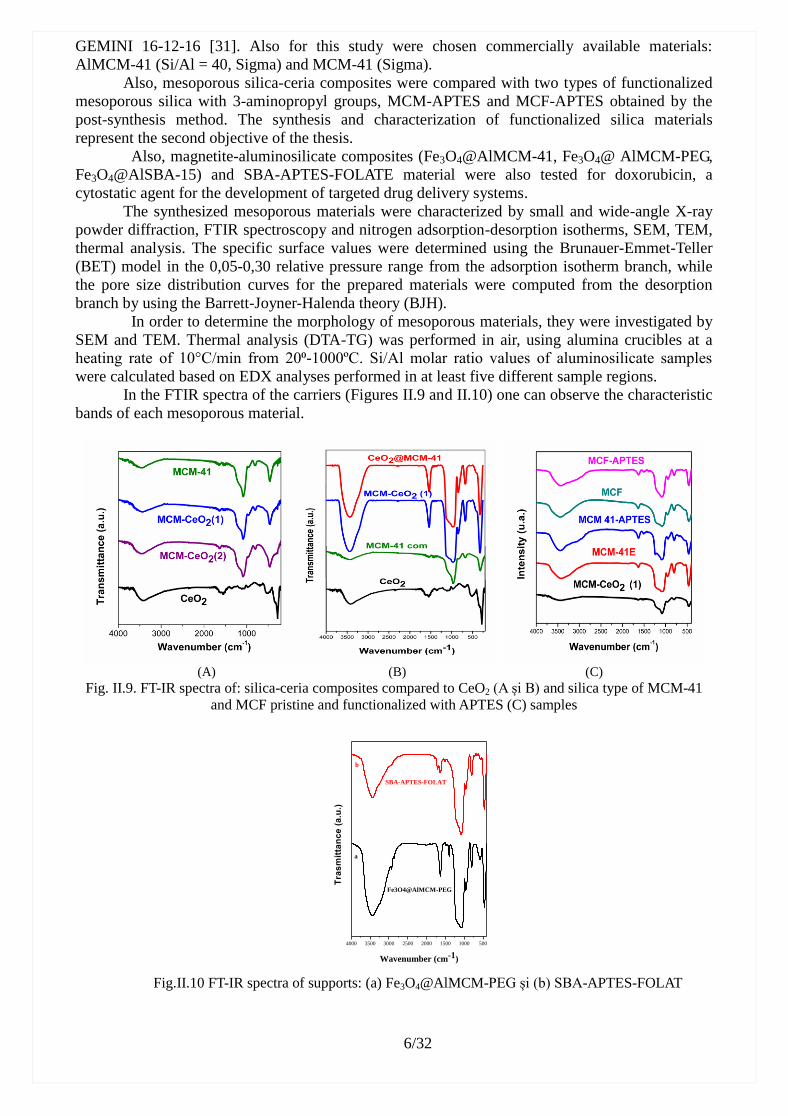
In the FTIR spectra of the carriers (Figures II.9 and II.10) one can observe the characteristic
bands of each mesoporous material.
(A) (B) (C)
Fig. II.9. FT-IR spectra of: silica-ceria composites compared to CeO2 (A și B) and silica type of MCM-41
and MCF pristine and functionalized with APTES (C) samples
4000 3500 3000 2500 2000 1500 1000 500
Tra
sm
itta
nc
e (
a.u
.)
Wavenumber (cm-1)
SBA-APTES-FOLAT
Fe3O4@AlMCM-PEG
a
b
Fig.II.10 FT-IR spectra of supports: (a) Fe3O4@AlMCM-PEG și (b) SBA-APTES-FOLAT

7/32
The formation of crystalline cubic fluorite structure for CeO2 (Fig.II.11.A și Fig.II.12.A)
phase in all silica-ceria composite materials was evidenced by wide-angle XRD. An amorphous
silica phase is also presented in the core-shell silica-ceria composite material. Furthermore, the
intensity of the CeO2 diffraction peaks is reduced for the CeO2@MCM-41, MCM-CeO2 materials
with respect to CeO2 nanoparticles, as expected because the weight fraction decrease of the ceria
cubic phase in these composite materials.
The small-angle XRD (Fig.II.11.B și Fig. II.12.B) demonstrate that both core-shell and
ceria-silica composite materials present ordered pore arrays, characteristic for MCM-41 silica.
2 3 4 5 6
Inte
nsi
ty (
a.u
.)
(b) MCM-CeO2(1)
(c) MCM-41
(100)
(110) (200)
2
(a) CeO2@MCM-41
(A) (B)
Fig. II.11. Wide-angle XRD (A) patterns of CeO2-based carriers and the small-angle XRD (B) patterns of
CeO2-based carriers in comparision with MCM-41
10 20 30 40 50 60 70
M CM -CeO2(2)
2(°)
In
ten
sit
ate
(u
.a.)
M CM -CeO2(1)
(311)(220)(200)
(100)
2 3 4 5 6
2(°)
(200)(110)
Inte
nsi
tate
(u.a
.)
(b) MCM-CeO2(2)
(a) MCM-CeO2(1)
(c) MCM-41
(100)
B
(A) (B)
Fig. II.12. XRD patterns of silica-ceria composites calcined at 550°C/5h: wide angle (A) and small angle
in comparison with MCM-41 (B)
The small-angle XRD for the mesoporous silica and aluminosilicate samples showed the
formation of mesostructured materials with a hexagonal pore framework belonging to the p6m
space group symmetry. All samples have at least three Bragg reflections characteristic for
MCM-41-type materials.
The wide-angle XRD for composite materials containing magnetite nanoparticles (Fig.
II.13.a) demonstrated the preservation of diffraction peaks of magnetite phase, as well as the
formation of the wide peak characteristic for amorphous silica.

8/32
10 20 30 40 50 60 70
Fe3O
4
Fe3O
4@AlMCM-PEG
Fe3O
4@AlMCM-41
2
Fe3O
4@AlSBA-15
(422)(400)
(220) (511)
(311)
Inte
nsi
ty (
a.u
.)
(440)
Fig.II.13. Wide-angle XRD patterns (a) for the materials containing magnetite nanoparticles
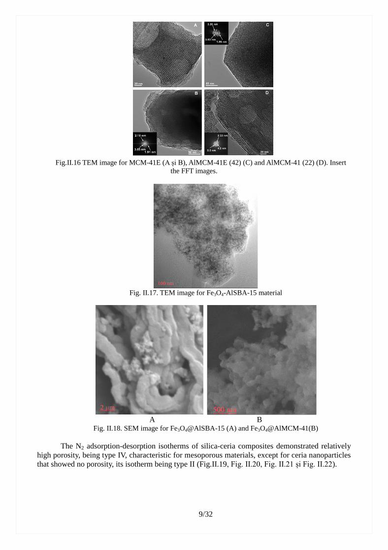
The morphology of mesoporous carriers was investigated by SEM (Fig. II.14, Fig. II.15.B, C,
D și Fig. II.18) and TEM investigation (Fig. II.15.A, Fig.II.16 și Fig. II.17).
Fig.II.14. SEM micrographs of CeO2 NP (A), CeO2@MCM-41(B) and MCM-CeO2 (C)
Fig. II.15. TEM image for MCM-CeO 2 (1)(A) and SEM micrographs for MCM-CeO2 (2) (B), MCM-41
sample, calcined at 550°C (C) and MCM-41E samples(D)
9/32
Fig.II.16 TEM image for MCM-41E (A și B), AlMCM-41E (42) (C) and AlMCM-41 (22) (D). Insert
the FFT images.
Fig. II.17. TEM image for Fe3O4-AlSBA-15 material
A B Fig. II.18. SEM image for Fe3O4@AlSBA-15 (A) and Fe3O4@AlMCM-41(B)
The N2 adsorption-desorption isotherms of silica-ceria composites demonstrated relatively
high porosity, being type IV, characteristic for mesoporous materials, except for ceria nanoparticles
that showed no porosity, its isotherm being type II (Fig.II.19, Fig. II.20, Fig. II.21 și Fig. II.22).

10/32
0,0 0,2 0,4 0,6 0,8 1,00
200
400
600
800
1000
2 4 6 8 10
D
C
B
B
C
A
Vo
lum
of
ad
sorb
ed
ga
s (c
m3g
-1)
p/p0
A) CeO2@MCM-41
B) MCM-CeO2(1)
C) MCM-CeO2(2)
D) NP-CeO2
A
Distributie pori (nm)
dV
/lo
gd
(a.u
.)
Fig. II.19. N2 adsorption-desorption isotherms of: CeO2@MCM-41 (A), MCM-CeO2 (1) (B)
and CeO2 NP (C)
2 4 6 8 10 12
Pore size (nm)
dV
logd (
a.u
.) CeO2@MCM-41
MCM-CeO2 (B)
Fig. II.20. Pore size distribution curves for MCM-CeO2 (1) (red curve) and CeO2@MCM-41 (1)
0,0 0,2 0,4 0,6 0,8 1,0
200
400
600
P /P0
Vo
lum
of
ad
sorb
ed g
as
(cm
3g
-1)
MCM-CeO2(1)
MCM-CeO2(2)
MCM-41(E)
MCM-41
A
2 4 6 8 10 12
(nm)
MCM-CeO2(1)
MCM-CeO2(2)
MCM-41E
d
V /lo
gd (
cm
3g
-1n
m-1
)
MCM-41
B
Fig. II.21. N2 adsorption-desorption isotherm of: MCM-CeO2 (1) , MCM-CeO2 (2), MCM -41 and
MCM-41E (A) and pore size distribution (B)
The textural parameters (specific surface area, SBET, total pore volume, Vp, and average pore
diameter, dBJH) of the carriers, determined from N2 adsorption-desorption isotherms are presented
in the Tables II.3- II.5.
Table II.3. Textural parameters of silica-ceria composites in comparison with those of MCM-41-type silica
and ceria samples
Sample dBJH (nm) SBET (m2g
-1) Vpore (cm
3g
-1)
MCM-41 (Sigma) 3,0 959 0,91
CeO2 NP - 88 0,10
CeO2@MCM-41 3,30 717 1,40
MCM-CeO2 (1) 2,66 683 0,67
MCM-CeO2 (2) 2,67 512 1,14
MCM-41E 2,82 818 0,78

11/32
MCM-41 (550°C) 2,82 1039 1,06
Table. II.4. Synthesis conditions, structural and the textural properties for mesoporous silica and
aluminosilicate supports Support
(Si/Al molar
ratio)
Synthesis conditions d100
(nm)
a0
(nm)
wt
(nm)
SBET
(m2/g)
Vpore
(cm3/g)
dBJH
(nm)
MCM-41E C16TAB 4,060 4,688 1,87 819 0,73 2,82
MCM-41 Comercial 4,105 4,740 1,98 1010 0,99 2,76
AlMCM-41E(51) C14TAB, Al(Osec
Bu), 600 ºC
3,620 4,180 1,61 836 0,87 2,57
AlMCM-41 (40) Comercial 3,914 4,520 1,64 824 0,81 2,88
MCM-48 C16TAB, 600 ºC - 8,93 0,69 1292 1,44 2,76
MCM-48G Gemini, 600 ºC - 8,04 0,83 854 0,97 2,26
MCM-APTES - - - 585 0,51 2,39
MCF - - - 876 2,49 10,40
MCF APTES - - - 518 1,98 9,02
Table II.5. The textural parameters of magnetite-aluminosilica composites
Magnetic mesopous
matrices
SBET (m2/g) Vpori (cm3/g) dBJH des
(nm)
dBJH ads
(nm)
Fe3O4@AlMCM-41 609 0,57 2,66 2,66
Fe3O4@AlSBA-15 829 1,12
0.88 (dpor< 10 nm)
6,81 8,99
-10000 -5000 0 5000 10000
-6
-4
-2
0
2
4
6
-6
-4
-2
0
2
4
6
Ma
gn
etiza
tio
n (
em
u/g
)
Magnetic Field (Gauss)
Fe3O
4@AlMCM-41
Ms= 6.42 [emu/g]
/k = 1.11 [K/Oe]
-10000 -5000 0 5000 10000
-4
-2
0
2
4
-4
-2
0
2
4
Mag
neti
zati
on
(em
u/g
)
Magnetic Field (Gauss)
Fe3O
4@AlSBA-15
Ms= 4.21 [emu/g]
/k = 0.95 [K/Oe]
-10000 -5000 0 5000 10000-8
-6
-4
-2
0
2
4
6
-10000 -5000 0 5000 10000
-8
-6
-4
-2
0
2
4
6
Mag
neti
zati
on
(em
u/g
)
Magnetic field (Gauss)
Fe3O
4@AlMCM-PEG
MS = 5.86 (emu/g)
/k=1.29 (K/Oe)
rex = 3.15 *10-3
C
Fig. II.23. Magnetisation curve function of applied magnetic field for Fe3O4@AlMCM-41 (A),
Fe3O4@AlSBA-15 (B) and Fe3O4@AlMCM-PEG (C) composites
The synthesized magnetite-aluminosilicate composites exhibit superparamagnetic behavior
(Figure II.23) and high porosity, characteristics that recommend them to be tested as vehicles in
targeted drug delivery systems.
Composites based on tetracyclines
The third objective of this thesis was the development of antibiotic delivery systems based
on inorganic carriers such as: MCM-41 and MCM-48 silica, silica-ceria composites and
aluminosilicates. Doxycycline and oxytetracycline were used as antibiotic molecules. The in vitro
release studies and determination of antimicrobial activity of drug-loaded materials are presented in
Chapters II.3 and II.4.
The antibiotics were adsorbed into the mesopores of silica-type carriers by incipient
wetness impregnation method [32, 33], using silica-ceria composites and MCM-41 silica in the
case of doxycycline and MCM-41, MCM-48, MCM-41-type silica-ceria composites with 10% (mol)
and 20% (mol) ceria and aluminosilicates for oxytetracycline. The content of doxycycline
drug-loaded silica-ceria composites was 25% (wt), as well as in Doxy1/MCM-41 material, while in
the case of Doxy2/MCM-41 sample the content of drug was 40% (wt).

12/32
The antibiotic-loaded silica samples were investigated by small- and wide-angle XRD,
FTIR spectroscopy and N2 adsorption-desorption isotherms. The presence of antibiotics in all
samples was evidenced by FT-IR spectroscopy (Fig.II.24. și Fig. II.29).
4000 3500 3000 2500 2000 1500 1000 500
(d) Doxy/MCM-CeO2
(e) Doxy/CeO2@MCM-41
(c) CeO2@MCM-41
(b) MCM-CeO2
Tra
nsm
itta
nce
(a
.u.)
Wavenumber (cm-1)
(a) Doxycycline
Fig. II.24. FT-IR spectra for: doxycycline, carriers (MCM-CeO2, CeO2@MCM-41) and
doxycycline-loaded silica samples
Fig.II.29. FTIR spectra of oxytetracycline-loaded silica-type supports and oxytetracycline
The vibrations of doxycycline can be noticed in the range of 1700-1300 cm-1
in the spectra
of doxycline-loaded samples, besides the characteristic vibrations of carrier: Si-O-Si (1225, 1090,
465 cm-1
) and Si-OH (966 cm-1
) (Fig. II.24).
In the FTIR spectra of drug-loaded samples it can be observed the vibration bands that
belong to the support, besides the ones of the drug located in the range of: 2850-2950 cm-1
attributed to ϑas,s(CH)), 1590-1650 cm-1
ascribed to the deformation of amide moieties and
1310-1410 cm-1
assigned to the phenol groups (Fig. II.29).
Wide-angle XRD patterns of antibiotic-loaded materials (Figures II.25 and II.30) indicate
that the drug molecules are adsorbed amorphous phase into the carrier mesopores due to the
absence of antibiotic-specific Bragg reflections. The ordered pore array is preserved after the drug
loading procedure, as the small-angle XRD patterns of the doxycycline-loaded silica-ceria
composites showed (Fig. II.25-inset). In the wide-angle XRD patterns of the
oxytetracycline-loaded samples (Fig. II.30), only the Bragg reflections of fluorite phase can be
noticed, demonstrating that the antibiotic molecules were adsorbed into the carrier mesopores in
amorphous state. The lack of oxytetracyclene diffraction peaks suggested that no crystalline drug
was present on the surface of the drug-loaded samples, as it was expected.

13/32
20 40
2 3 4 5 6
In
ten
sita
te (
u.a
.)
2
(a) Doxy/CeO2@MCM-41
(b) Doxy/MCM-CeO2
Inte
nsi
tate
(a
.u.)
2
(a)Doxy/CeO2@MCM-41
(b) Doxy/MCM-CeO2
(10)
(11) (20)
Fig. II.25. Wide-angle XRD patterns of doxycycline-loaded ceria-silica composites. Inset shows the
small-angle XRD data for the same samples
Fig.II.30 Wide-angle XRD patterns of representative oxytetracycline-loaded supports and oxytetracycline
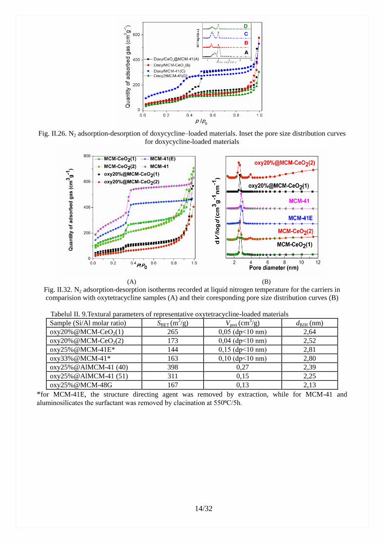
All antibiotics-loaded materials exhibit type IV N2 adsorption-desorption isotherm. (Fig.
II.26 and II.32). The loading of therapeutic agent into the mesochannels is supported by the
decrease of the pore volume and specific surface area values for the antibiotic-loaded composites (Tables II.6 and II.9). The adsorption of either doxycycline or oxytetracycline did not cause
significant changes in the average pore size (Fig. II.26-inset and II.33), indicating poor interactions
between the mesoporous carrier and the therapeutic agent were established. Oxitetracycline-loaded
MCM-48 or MCM-48G supports showed that the adsorption of antibiotic molecules led to less
ordered systems as small-angle XRD data demonstrated.
Table II.6. Textural parameters of doxycycline–loaded materials
Sample Doxycycline (%) dBJH (nm) SBET
(m2g
-1)
Vmezopore
(cm3g
-1)
Doxy/CeO2@MCM-41 25 3.20 266 0.61
Doxy/MCM-CeO2 25 2.70 269 0.19
Doxy/MCM-41 25 2.80 553 0.54
Doxy2/MCM-41 40 2.80 236 0.26
14/32
Fig. II.26. N2 adsorption-desorption of doxycycline–loaded materials. Inset the pore size distribution curves
for doxycycline-loaded materials
(A) (B)
Fig. II.32. N2 adsorption-desorption isotherms recorded at liquid nitrogen temperature for the carriers in
comparision with oxytetracycline samples (A) and their coresponding pore size distribution curves (B)
Tabelul II. 9.Textural parameters of representative oxytetracycline-loaded materials
Sample (Si/Al molar ratio) SBET (m2/g) Vpori (cm
3/g) dBJH (nm)
oxy20%@MCM-CeO2(1) 265 0,05 (dp˂10 nm) 2,64
oxy20%@MCM-CeO2(2) 173 0,04 (dp˂10 nm) 2,52
oxy25%@MCM-41E* 144 0,15 (dp˂10 nm) 2,81
oxy33%@MCM-41* 163 0,10 (dp˂10 nm) 2,80
oxy25%@AlMCM-41 (40) 398 0,27 2,39
oxy25%@AlMCM-41 (51) 311 0,15 2,25
oxy25%@MCM-48G 167 0,13 2,13
*for MCM-41E, the structure directing agent was removed by extraction, while for MCM-41 and
aluminosilicates the surfactant was removed by clacination at 550⁰C/5h.
15/32
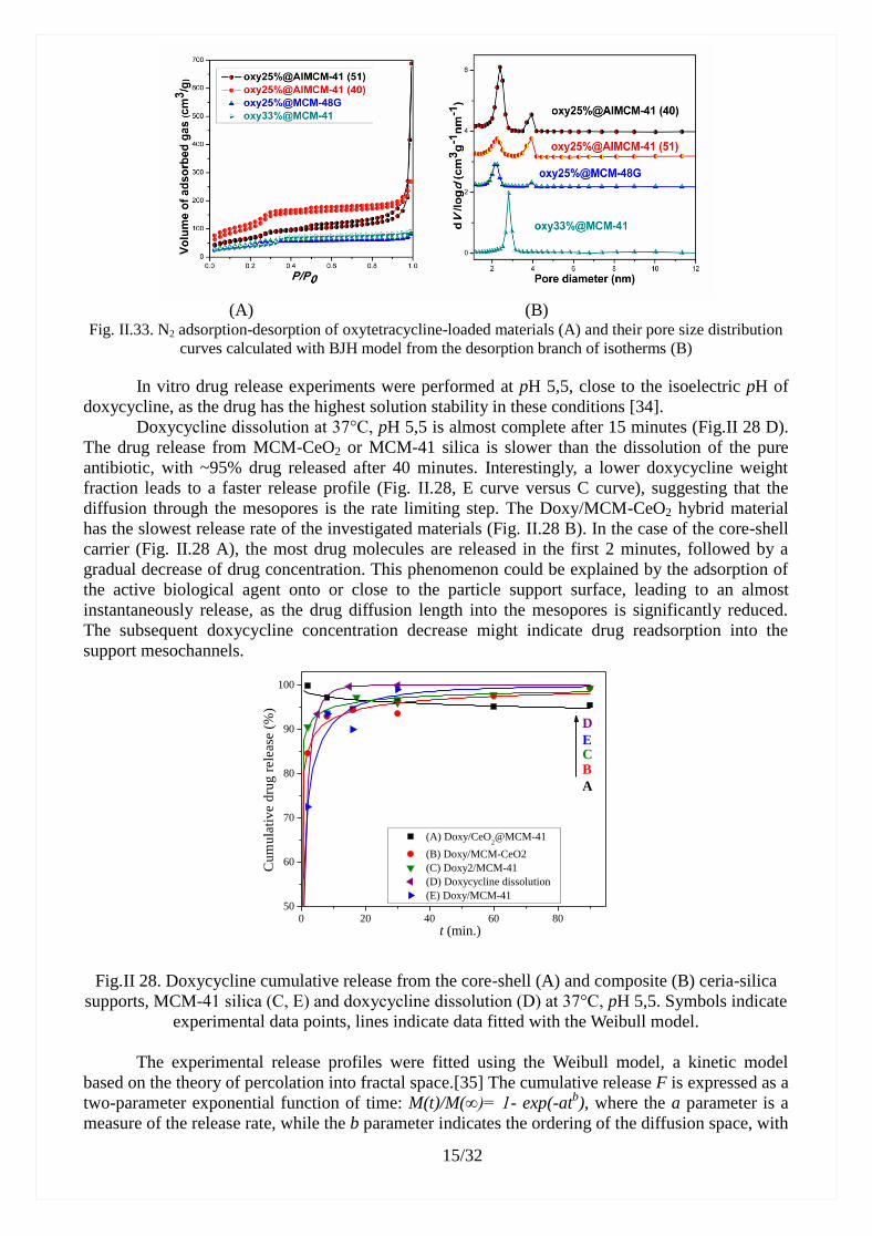
(A) (B)
Fig. II.33. N2 adsorption-desorption of oxytetracycline-loaded materials (A) and their pore size distribution
curves calculated with BJH model from the desorption branch of isotherms (B)
In vitro drug release experiments were performed at pH 5,5, close to the isoelectric pH of
doxycycline, as the drug has the highest solution stability in these conditions [34].
Doxycycline dissolution at 37°C, pH 5,5 is almost complete after 15 minutes (Fig.II 28 D).
The drug release from MCM-CeO2 or MCM-41 silica is slower than the dissolution of the pure
antibiotic, with ~95% drug released after 40 minutes. Interestingly, a lower doxycycline weight
fraction leads to a faster release profile (Fig. II.28, E curve versus C curve), suggesting that the
diffusion through the mesopores is the rate limiting step. The Doxy/MCM-CeO2 hybrid material
has the slowest release rate of the investigated materials (Fig. II.28 B). In the case of the core-shell
carrier (Fig. II.28 A), the most drug molecules are released in the first 2 minutes, followed by a
gradual decrease of drug concentration. This phenomenon could be explained by the adsorption of
the active biological agent onto or close to the particle support surface, leading to an almost
instantaneously release, as the drug diffusion length into the mesopores is significantly reduced.
The subsequent doxycycline concentration decrease might indicate drug readsorption into the
support mesochannels.
0 20 40 60 8050
60
70
80
90
100
E
D
CB
(A) Doxy/CeO2@MCM-41
(B) Doxy/MCM-CeO2
(C) Doxy2/MCM-41
(D) Doxycycline dissolution
(E) Doxy/MCM-41
Cu
mu
lati
ve d
rug
rele
ase
(%
)
t (min.)
A
Fig.II 28. Doxycycline cumulative release from the core-shell (A) and composite (B) ceria-silica
supports, MCM-41 silica (C, E) and doxycycline dissolution (D) at 37°C, pH 5,5. Symbols indicate
experimental data points, lines indicate data fitted with the Weibull model.
The experimental release profiles were fitted using the Weibull model, a kinetic model
based on the theory of percolation into fractal space.[35] The cumulative release F is expressed as a
two-parameter exponential function of time: M(t)/M(∞)= 1- exp(-atb), where the a parameter is a
measure of the release rate, while the b parameter indicates the ordering of the diffusion space, with

16/32
values greater than ~0.69 indicating diffusion in Euclidian space and lower values signifying
diffusion in fractal space [36]. The Weibull model parameters (Table II.8) show that the release
mechanism of the antibiotic molecules from silica and ceria-silica composites corresponds to
diffusion in fractal space, as opposed to doxycycline dissolution (Euclidian space diffusion). Unlike
in the case of SBA-15 mesoporous silica with larger pores [37], the increase in drug weight fraction
leads to modifications of the diffusion mechanism, which can be explained by a decrease in drug
diffusion and solvent counter diffusion rates as the free mesopore volume decreases. Regarding the
ceria- silica mesoporous support MCM-CeO2, it can be concluded that the doxycycline diffusion
process is similar with the pristine MCM-41 silica carrier.
Table II.8. Weibull function parameters describing the experimental doxycycline release data
Parameter Doxy
dissolution Doxy/CeO2@MCM-41
Doxy)/
MCM-CeO2
Doxy/
MCM-41
Doxy2/
MCM-41
a 0,914 4,357 1,657 1,006 2,105
b 0,679 -0,086 0,194 0,389 0,153
R2 1 0,9988 0,9986 0,9922 0,9994
In vitro oxytetracycline release experiments were performed in saline phosphate buffer
solution at pH 5,7 as simulated body fluid [26]. Oxytetracycline, very soluble in water, has low
photochemical and chemical stability, especially in basic solutions. From all studied carriers, the
antibiotic molecules were completely delivered within a period between 60 to 150 min. (Fig.II.34
and II.35), all release profiles exhibiting a pronounced burst effect, required in the treatment with
antibiotics.
Fig.II.34. Oxytetracycline release profiles from silica-ceria composites compared with MCM-41 silica
The drug delivery from silica-ceria composites was slightly slower than from MCM-41. The
higher content of silanol groups in MCM-41E slows down the antibiotic delivery kinetics in
comparison with the corresponding calcined MCM-41 carrier. Recently, we reported a similar
behavior for doxycycline that had a slower release kinetics from MCM-CeO2 (1) than MCM-41
silica matrix.
All oxytetracycline-loaded materials containing MCM-41-type silica or aluminosilicate carrier
exhibited a delivery profile with a pronounced burst effect, about 60% (wt) of the antibiotic amount
being delivered in the first 10 min of the experiment, followed by a gradually release up to 3 h (Fig.
II.35). A higher drug amount into the carriers led to slower kinetics (Fig. II.35 B) and a higher
aluminum content into the MCM-41-type silica matrix resulted in a lower drug delivery rate (Fig.
II.35 A). The cubic pore geometry characteristic for MCM-48 silica didn’t favor a gradually drug
release, faster kinetics being noticed than for oxytetracycline loaded on MCM-41-type supports
(Fig. II.35 B) [41].

17/32
Fig. II.35. The oxytetracycline release profil from aluminosilicate carriers (A) and silica supports (B)
Antimicrobial activity of drug-loaded mesoporous silica-type supports
The antimicrobial activity of doxycycline-loaded MCM-41 or MCM-CeO2 (1) supports with
an antibiotic content of 20% (Table II.7) compared to that of doxycycline cyclate solid powder was
evaluated against Klebsiella ATCC 10031 strain in triplicate.
Tabel II.7. Antimicrobial activity of samples containing doxycycline against Klebsiella ATCC 10031
strain
Sample Klebsiella pneumoniae
ATCC 10031
Growth total inhibition
zone diameter
(mm)
Growth partial inhibition
zone diameter
Doxy ++++ 35 39
Doxy/MCM-41 ++++ 35 39
Doxy/MCM-CeO2(1) ++++ 33 38
The bactericidal activity of representative oxytetracycline-loaded materials containing
mesoporous silica or aluminosilicate vehicle against five clinical isolates of Staphylococcus aureus
(SA1-SA5) and the reference ATCC 43300 strains were tested. The oxytetracycline-loaded samples
containing MCM-41-type carrier exhibited a significant sensitivity against S. aureus ATCC 43300,
similar with oxytetracycline alone, while a lower antimicrobial activity for oxytetracycline-loaded
MCM-48 sample was found (Table II.11).
Tabel II.11. The bactericidal activity of oxitetracycline-loaded materials
Samples
Microbial strains
Staphylococcus aureus
SA1 SA2 SA3 SA4 SA5 ATCC
43300
oxy@AlMCM-41 (51) +++ +++ +++ ++ ++ ++++
oxy@AlMCM-41 (40) +++ ++ +++ ++ ++ ++++
oxy@MCM-41E +++ +++ +++ +++ +++ ++++
oxy@MCM-48 ++ ++ ++ ++ ++ ++
oxytetracycline +++ ++ +++ +++ ++ ++++
where: + low sensitivity (the measured diameter of the clear area < 10 mm); ++ sensitive (the measured diameter of the
clear area between 10–15 mm); +++ distinct sensitivity (the measured diameter of the clear area between 15–25 mm);
++++ significant sensitivity (the measured diameter of the clear area ≥ 30 mm)
The antimicrobial assays demonstrated that oxytetracycline-loaded MCM-41-type carriers
inhibited the clinical isolates of S. aureus development, the effect being similar or better compared
with the antibiotic alone. The most intense inhibitory effect against S. aureus 43300 was noticed for
18/32
the oxy@MCM-41E material (D=39 mm), while for the drug-loaded on commercial AlMCM-41
(40) the average measured inhibition diameter (D=31 mm) had the lowest value.
II.5. Composite based on ketoprofen
The influence of mesoporous silica support properties (pore size and geometry, surface
functionalization, the drug content) on the ketoprofen delivery kinetics was studied. The
ketoprofen-loaded silica samples were then encapsulated into alginate beads and the drug release
kinetics from these complex composite carriers into simulated intestinal fluid (pH 7.4) was
determined.
For obtaining drug-loaded silica materials, the therapeutic agent was adsorbed into the
mesopores of silica-type carriers by incipient wetness impregnation method. In order to study the
influence of pore array geometry, two different pristine silica carriers were used, MCM-41 with an
ordered 1D hexagonal pore array, presenting a pore diameter of 2.8 nm and a total pore volume of 1
cm3/g, and MCF exhibiting a higher porosity with a total pore volume up to 3.25 cm
3/g [27,.28] as
a result of a continuous three-dimensional disordered pore framework consisting of large spherical
pores interconnected by smaller windows [45]. MCF-type silica has the capacity to accommodate a
larger amount of guest molecules into its mesopores than MCM-41 or SBA-15. Nevertheless, there
are only few studies on MCF silica used as carrier in drug delivery systems, comparing to MCM-41
and SBA-15, especially for poor-soluble therapeutic agents [28, 29]. For the enhancement of
acid–base interactions between ketoprofen molecules and silica support, two aminopropyl
functionalized silica materials, MCM-APTES and MCF-APTES, with similar content of organic
moieties, were also employed. The aminopropyl groups content, determined from
thermogravimetric analysis of functionalized silica samples, was 12%(wt) and 12.4%(wt) for
MCM-APTES and MCF-APTES, respectively.
The supports and ketoprofen-silica samples were investigated by small- and wide-angle
XRD, FTIR spectroscopy and N2 adsorption-desorption isotherms. The successful functionalization
of silica carrier with aminopropyl groups was proved by FTIR spectroscopy, which evidenced the
characteristic vibrations of aminopropyl groups.
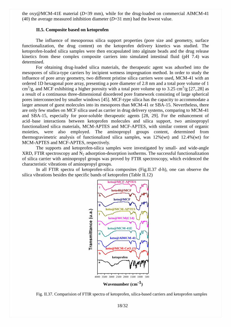
In all FTIR spectra of ketoprofen-silica composites (Fig.II.37 d-h), one can observe the
silica vibrations besides the specific bands of ketoprofen (Table II.12)
4000 3500 3000 2500 2000 1500 1000 500
keto@MCF
keto@MCM-APTES
keto@MCM(C14)
keto@MCM-41E
keto@AlMCM-41
keto@MCM-CeO2(1)
b
Tra
ns
mit
tan
ce
(u
.a.)
Wavenumber (cm-1)
a
c
d
e
f
g
h
i
ketoprofen
keto40@MCF
keto@MCF-APTES
Fig. II.37. Comparision of FTIR spectra of ketoprofen, silica-based carriers and ketoprofen samples
19/32
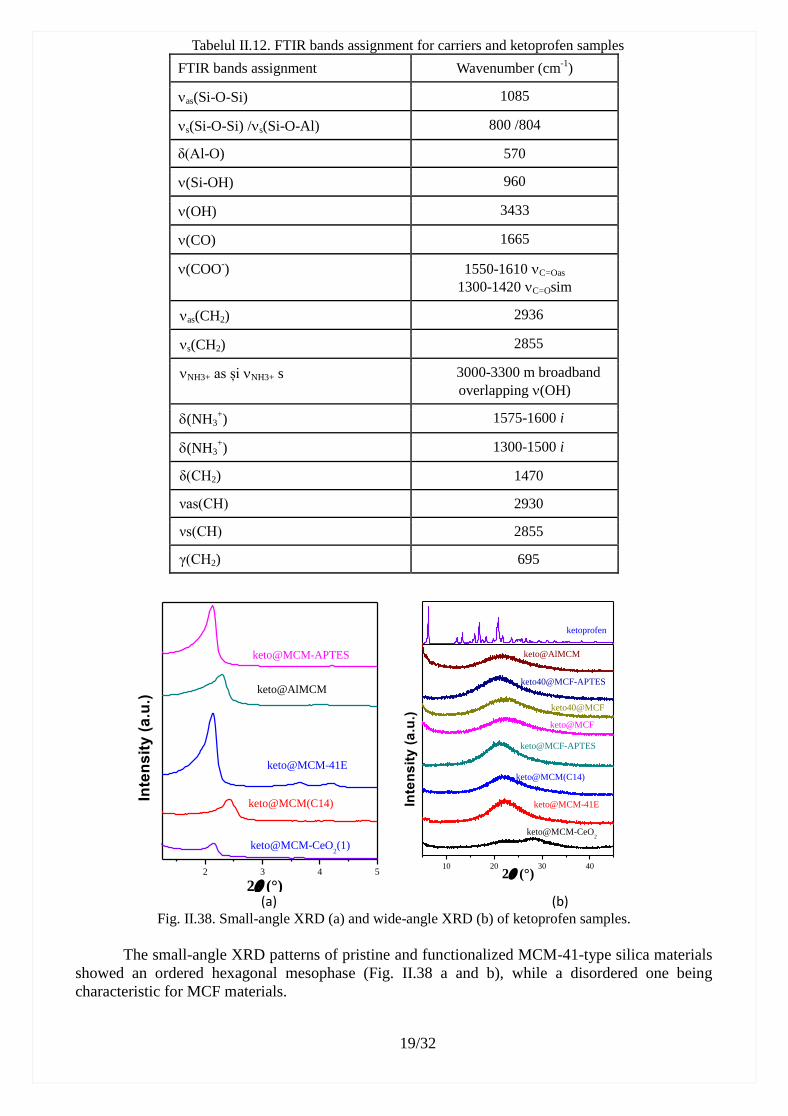
Tabelul II.12. FTIR bands assignment for carriers and ketoprofen samples
FTIR bands assignment Wavenumber (cm-1
)
as(Si-O-Si) 1085
s(Si-O-Si) /s(Si-O-Al) 800 /804
δ(Al-O) 570
(Si-OH) 960
(OH) 3433
(CO) 1665
(COO-) 1550-1610 C=Oas
1300-1420 C=Osim
as(CH2) 2936
s(CH2) 2855
NH3+ as și NH3+ s 3000-3300 m broadband
overlapping (OH)
(NH3+) 1575-1600 i
(NH3+) 1300-1500 i
δ(CH2) 1470
νas(CH) 2930
νs(CH) 2855
γ(CH2) 695
2 3 4 5
keto@MCM-CeO2(1)
keto@MCM(C14)
keto@MCM-41E
keto@AlMCM
Inte
ns
ity
(a
.u.)
2 (°)
keto@MCM-APTES
10 20 30 40
keto@MCM-CeO2
keto@MCF-APTES
keto@MCF
keto40@MCF
Inte
ns
ity
(a
.u.)
2 (°)
keto@AlMCM
keto40@MCF-APTES
keto@MCM(C14)
keto@MCM-41E
ketoprofen
(a) (b)
Fig. II.38. Small-angle XRD (a) and wide-angle XRD (b) of ketoprofen samples.
The small-angle XRD patterns of pristine and functionalized MCM-41-type silica materials
showed an ordered hexagonal mesophase (Fig. II.38 a and b), while a disordered one being
characteristic for MCF materials.

20/32
The small-angle XRD of ketoprofen-loaded MCM-41 samples exhibit the intense (100) and
less intense (110) and (200) Bragg reflections proving the mesostructure preservation of MCM-41
supports (Fig.II.38 a), after the drug encapsulation. In the wide-angle XRD patterns of the
drug-loaded silica samples (Fig. II.38 b), the diffraction peaks of crystalline ketoprofen are not
present, indicating that the drug molecules were absorbed into the silica mesopores in amorphous
state and no crystalline ketoprofen was form on the silica surface.
The mesoporous silica supports and ketoprofen-loaded silica materials exhibit type IV N2
adsorption-desorption isotherm (Fig. II.39, II.40 și II.41), characteristic for mesoporus materials,
completely reversible for MCM-41-type samples and with a large H1 hysteresis loop for
MCF-based materials because of sharp capillary condensation in the 0,65-0,9 relative pressure
range. The adsorption-desorption isotherms of ketoprofen-loaded silica materials proved the
adsorption of drug molecules into the support mesopores, their total porosity being lower than that
of the corresponding carrier (Fig.II.39 A and II.40 A). The textural parameters (specific surface area,
SBET, total pore volume, Vpore, and average pore diameter, d) determined from N2
adsorption-desorption isotherms for the ketoprofen-loaded silica samples compared with silica
carriers are shown in Tabel II.13.
Tabelul II.13. The textural parameters for carriers and ketoprofen-loaded silica samples
Carrier SBET
(m2/g)
Vp
(cm3/g)
dBJH
(nm) Composite
SBET
(m2/g)
Vp
(cm3/g)
dBJH
(nm)
MCM-CeO2 (1) 683 0.67 2.66 keto@MCM-CeO2(1) 235 0.08 2.00
MCM-41E 818 0.78 2.82 keto@MCM-41E 536 0.51 2.25
MCM(C14) 654 0.80 2.13 keto@MCM(C14) 161 0.09 1.76
MCM-APTES 585 0.51 2.39 keto@MCM-APTES 279 0.19 1.76
MCF 876 2.29 16.68 keto@MCF 377 1.49 16.09
keto40@MCF 183 0.87 13.94
MCF-APTES 518 1.98 9.02 keto@MCF-APTES 377 1.75 9.03
AlMCM-41 729 0.66 2.38 keto@AlMCM-41 580 0.34 1.99
0,0 0,2 0,4 0,6 0,8 1,00
200
400
600
keto@MCM-APTES
MCM-APTES
keto@MCM-41
MCM-41
Vo
lum
e o
f ad
sorb
ed
gas
(cm
3g
-1)
P/P0
A
2 4 6 8 10 12 14
keto@MCM-41
keto@MCM-APTES
MCM-APTES
dV
(lo
gd
) (c
m3g
-1n
m)
Pore size (nm)
MCM-41
B
Fig.II.39. N2 adsorption-desorption isotherms of MCM-41-type supports and ketoprofen-loaded samples
containing MCM-41-type carriers (A) and their pore size distribution curves (B)
0,0 0,2 0,4 0,6 0,8 1,00
1000
2000
3000
4000e
d
c
b
keto@MCF-APTES
keto40@MCF
keto@MCF
MCF-APTES
Volu
mof
ad
sorb
ed
ga
s(c
m3/g
)
P/P0
MCFa
A
10 20 30 40 50
d
c
b
dV
(lo
gd
)
Pore size(nm)
keto40@MCF
keto@MCF
MCF-APTES
MCF a
B
Fig. II.40. N2 adsorption-desorption isotherms of MCF-type supports and ketoprofen-loaded samples
containing MCM-41-type carriers (A) and their pore size distribution curves computed with DFT model (B)

21/32
0,0 0,2 0,4 0,6 0,8 1,0
0
100
200
300
400
500
600
700
Vo
lum
of
ad
sorb
ed
ga
s (c
m3
g-1
)
P/P0
keto@AlMCM
MCM-CeO2(1)
keto@MCM-CeO2(1)
Fig. II.41 N2 adsorption-desorption isotherms for ketoprofen-loaded samples
Both 3-aminopropyl functionalized silica supports present lower specific surface area and
average pore diameter than the pristine silica materials from which they were obtained meaning
that the organic moieties are linked on the internal silica pore walls surface (Table II.13). Although
the functionalization with aminopropyl groups of silica materials caused a slight decrease of
textural parameter values, all supports exhibited large porosity required for their application as
vehicles for biologically active molecules. The samples containing ketoprofen presented some
porosity, which diminished with the increase of therapeutic agent content. The pore size
distribution curves and average pore diameter were calculated using Barrett-Joyner-Halenda (BJH)
method for MCM-41-type samples from the desorption branch of isotherms (Fig. II.39B) and DFT
model for MCF-type materials (Fig. 40B). One can observe a decrease of the average pore diameter
of drug-loaded materials compared with that of the corresponding carrier (Fig.II.40 and II.41)
because of either the ketoprofen adsorption into carrier mesopores or the interactions between drug
molecules and silica-type support (Table II.13).
The determination of ketoprofen in vitro release profile from mesoporous silica carriers (Fig.
II. 42.A) was performed in simulated intestinal fluid, saline PBS, pH 7.4, at 37°C, under constant
magnetic stirring, using a dialysis membrane. The ketoprofen dissolution test was carried out in the
same conditions using also a dialysis bag. The drug delivery profiles are plotted in figure II.42, as
drug cumulative release with respect to the time.
The therapeutic agent exhibited fast release kinetics from pristine silica carriers in the first
hour of experiment, even faster than its dissolution, in the case of keto@MCF, followed by a
sustained release, more pronounced in the case of aminopropyl-functionalized silica supports due
to acid-base interactions (Fig.II.45). The fast first stage of ketoprofen release from silica supports,
especially for pristine ones, could be explained by the diffusion of drug molecules, which are more
soluble in basic medium in amorphous state than as crystals. The second stage of ketoprofen
release is slower when drug molecules, which are stronger linked on silica surface through
acid-base interactions, are gradually delivery in simulated intestinal fluid. One can observe a lower
drug release rate at high amount of therapeutic agent adsorbed into the mesopores of pristine MCF
silica.
The ketoprofen experimental release data were fitted with good correlation coefficients,
R2
w>0.95, using the Weibull model, a two parameters exponential function based on the theory of
percolation in both Euclidian and fractal space (Fig.II.42A), which is highly applied for the drug
dissolution from different matrices. Table II.14 lists the obtained a and b parameters, as well as the
correlation coefficients, R2
W, for the Weibull model. One can notice that the b parameter values
correspond to a drug Fick’s diffusion in fractal space, being lower than 0,69 [31].

22/32
0 50 100 150 200 2500
20
40
60
80
100
Cum
ula
tive k
eto
pro
fen r
ele
ase (
%)
Timp (min)
2 3 4 5 6 7 80
10
20
30
40
50
60
70
80
ketoprofen
keto@AlMCM-41
keto@MCM-CeO2
keto@MCM-41E
keto@MCM(C14)
keto@MCM-APTES
keto@MCF
keto40@MCF
keto@MCF-APTES C
um
ula
tiv
e k
eto
pro
fen
re
lea
se
(%
)
t 0.5
B
Fig. II.42. Ketoprofen release profiles from pristine and functionalized silica-type carriers fitted with
Weibull function (A) and Higuchi model (B)
To evaluate the ketoprofen release rate in the burst stage, the drug delivery experimental
data from the first hour of the experiments were fitted with the Higuchi`s model: m(t)/m(∞)=kH*t1/2
,
were kH is the rate constant (Fig.II.42B) [36]. The kH values and the correlation coefficients, R2
H,
obtained for all ketoprofen-loaded silica materials are listed in Table II.14. The slowest ketoprofen
delivery kinetics for the burst stage of drug release was obtained in the case of MCF-APTES carrier,
followed by MCM-APTES support due to the acid-base interactions between amino moieties
linked on carrier mesopores surface and ketoprofen carboxyl groups. In the case of pristine silica
support, lower delivery rate of ketoprofen was noticed for MCM-41E than for MCF, which could
be explained by either a smaller average pore diameter and an ordered pore array or a higher
content of silanol groups as the support was not calcined, unlike MCF carrier.
Table II.14 WEIBULL and HIGUCHI parameters obtained by fitting the experimental data of drug release
from silica-type carriers
Samples Weibull model Higuchi’s function
a b R2
W kH R2
H
ketoprofen 0.009 1.111 0.994 8.698 0.975
keto@MCM-41E 0.157 0.351 0.951 6.594 0.979
keto@MCM-APTES 0.076 0.284 0.955 3.498 0.936
keto@MCF 0.1072 0.626 0.983 9.316 0.979
keto40@MCF 0.0543 0.585 0.981 7.621 0.973
keto@MCF-APTES 0.0082 0.473 0.972 2.919 0.956
To prepare drug-silica-alginate beads, keto@MCM-41E and keto@MCF-APTES were
chosen, both samples having 20%(wt) drug content. The chosen drug-loaded silica samples
presented the lowest total cumulative release and the slowest release kinetics among pristine silica
and functionalized silica carriers, respectively. The obtained ketoprofen-silica-alginate, denoted
keto@MCM-alg and keto@MCF-APTES-alg, were investigated by SEM-EDX and thermal
analysis. The content of ketoprofen, which remained in the silica-alginate beads, was determined
by measuring the drug amount delivered in calcium chloride solution during the beads formation
using UV-vis spectroscopy. About a third part of ketoprofen was released from either MCM-41
support or MCF-APTES material in calcium chloride aqueous solution, resulting a content of drug
in beads of 2,2%(wt).
The SEM investigation of ketoprofen-silica-alginate composites revealed the formation of
spherical beads with a diameter in the range of 0.8-1.2 mm (Fig. II.48). On the beads surface one
can observe the formation of sodium chloride crystals, though the beads were intensively washed
with ultrapure water (Fig. II.48). The EDX analysis coupled with SEM investigation showed that
silica particles are inside the alginate beads, calcium ions penetrate the polymer network and on the
alginate surface there are still sodium chloride crystals.

23/32
Fig. II.49. SEM images of: A) keto@MCM-alg (inset its surface), B) keto@MCF-APTES-alg (inset its surface), and
EDX elemental mapping of: C) keto@MCM-alg and D) keto@MCF-APTES-alg beads
Fig. II.53. DTA-TG analysis of ketoprofen-silica-alginate beads
The DTA-TG analysis of ketoprofen-silica-alginate beads showed a thermal decomposition in
several steps (Fig. II.53). In the temperature range of 20⁰-170⁰C, both-types of beads lose water
from alginate hydrogel and silica mesopores. At 170⁰C, alginate begins to decompose, and the
process continues up to 600⁰C [37]. On the DTA curves, two strong exothermic effects can be
noticed in the 400⁰-580⁰C temperature range. The residue mass for keto@MCM-alg and
keto@MCF-APTES-alg was 25.6% and 24.5%, respectively, which consisted of 15.9% (wt) silica
and 9.7% (wt) compounds containing calcium and sodium ions for the first sample and 14% and
10.5% for the second one. Ketoprofen melts at 97⁰C, it is stable up to around 170⁰C and totally
decomposes at 376⁰C [38, 39]. Because in the silica-alginate beads the content of ketoprofen is low,
its thermal decomposition, in the temperature range of 170⁰-376⁰C, is difficult to notice, being
overlapped with that of alginate, which is in a higher amount in the samples.
The experiments of ketoprofen delivery from silica-alginate beads, carried out in the same
conditions as for drug-loaded silica samples, showed slower kinetics in comparison with the

24/32
ketoprofen-loaded silica carriers. The drug release experimental data were also fitted with the
Weibull function (Fig. 9) with the correlation coefficient of 0.977 and 0.954 for keto@MCM-alg
sample and keto@MCF-APTES-alg beads, respectively. As in the case of the ketoprofen delivery
from silica-type carriers, the exponential b parameter of the Weibull function has values of 0,514
and 0,321 for keto@MCM-alg and keto@MCF-APTES-alg, respectively, corresponding to a
Fickian diffusion of the therapeutic agent. For both type of alginate beads, the pre-exponential
parameter a of the Weibull function has the same value of 0.0103, because it depends on the
surface through which ketoprofen diffusion occurs. This can be explained by the high contribution
of alginate which is in large and similar amount in both type of ketoprofen-loaded silica-alginate
beads. One can notice a higher release rate of ketoprofen from MCM-alg beads than from
MCF-APTES-alg, the similar trend being also observed for the corresponding ketoprofen-loaded
silica samples.
Fig. II.54 Ketoprofen release profiles from silica-alginate beads fitted with the Weibull function
II.6. Composites based on doxorubicin
The last objective of this thesis was the encapsulation of doxorubicin, a cytostatic agent
used in the treatment of several forms of cancer, in magnetite-aluminosilicate-type composite
materials, and studies on the drug release profiles in the biological fluid that simulates the tumor
cell environment. For doxorubicin, Fe3O4@AlMCM-41, Fe3O4@AlSBA-15,
Fe3O4@AlMCM-41-PEG and SBA-15 fuctionalized with folate groups as carriers were chosen.
Doxorubicin was adsorbed into carrier mesopores by the same method, incipient wetness
impregnation.
For all doxorubicin-loaded magnetic mesoporous matrix, FTIR spectra exhibit the
vibrations corresponding to the main functional groups of doxorubicin, besides the bands
characteristic for magnetic mesoporous carrier (Fig. II.55).

25/32
4000 3500 3000 2500 2000 1500 1000 500
Tra
ns
mit
tan
ce
(a
.u.)
Wavenumber (cm-1)
a
b
c
d
e
Doxorubicina
Dox@Fe3O4@AlMCM-41
Dox@Fe3O
4@AlMCM-PEG
Dox@Fe3O
4@AlSBA-15
Dox@SBA-APTES-FOLAT
Fig. II.55. FTIR spectra of doxorubicin and doxorubicin-loaded magnetic carrier
Wide-angle XRD (Fig II.55A) patterns for doxorubicin-loaded materials exhibit only the
Bragg reflections characteristic of crystalline magnetite phase along with the large diffraction peak
in the range of 20-40° 2, characteristic for amorphous silica. The DOX@Fe3O4-AlMCM-PEG
material exhibited a hexagonal pore framework with mesochannels preservation, characteristic of
MCM-41 materials as small-angle proved (Figure II.55B).
Fig. II.55. Wide-angle XRD for doxorubicin-loaded samples (A) and small-angle of
DOX@Fe3O4-AlMCM-41-PEG (B).
N2 adsorption-desorption isotherms, recorded at 77K showed that the porosity of
doxorubicin-loaded materials is lower than that of the corresponding carrier (Fig. II.56) due to the
presence of therapeutic agent molecules into the carrier mesopores. Table II.15 lists the textural
parameters of doxorubicin-loaded samples and those of the correspondig vehicles. The presence of
aluminum into the silica matrix, which leads to Lewis acidity and thus enhances the interactions
between the cytostatic agent and carrier, evidenced by the decrease in the average pore size of
doxorubicin-loaded materials compared to coresponding carrier (Table II.15).

26/32
0,0 0,3 0,6 0,9
0
800
1600
2400
Vo
lum
of
ad
so
rbe
d g
as
(c
m3
/g)
P/P0
DOX@Fe3O4@AlMCM
DOX@Fe3O4@AlSBA-15
DOX@Fe3O4@AlMCM-41-PEG
DOX@SBA-APTES-FOLAT
a
b
c
d
10
0
20
40
dV
/lo
gd
(c
m3
g-1
nm
-1)
Pore size (nm)
DOX@Fe3O4@AlMCM
DOX@Fe3O4@AlSBA-15
DOX@Fe3O4@AlMCM-41-PEG
DOX@SBA-APTES-FOLAT
a
b
c
d
f
Fig. II.56. N2 adsorption-desorption isotherms for
doxorubicin composites Fig. II.57. Pore size distribution for doxorubicin
composites Tabelul II.15. The textural parameters of therapeutic agent-loaded materials in comparision with
coresponding magnetic mesoporous matrix
Samples Doxorubicin-loaded materials Carrier
SBET
(m2/g)
Vpori
(cm3/g)
dBJH
(nm)
SBET
(m2/g)
Vpori
(cm3/g)
dBJH
(nm)
Ms
(emu/g)
DOX@ Fe3O4@AlMCM 497 0.46 2.51 609 0.57 2.66 6.42
DOX@ Fe3O4@AlMCM-PEG 680 0.61
d<10 nm
2.97 852 0.90 2.98 ND*
DOX@Fe3O4@AlSBA-15 469 0.70
d<10 nm
6.29 829 0.88
d<10nm
6.81 4.21
DOX@SBA-APTES-FOLAT 447 0.90 6.29 472 0.906 6.28 ND* ND* = indefinite
0 1000 2000 3000 4000 50000
10
20
30
40
50
60
DOX@SBA-APTES-FOLAT
Cu
mu
lati
ve
rele
ase
of
DO
X (
%)
Timp (min)
Fig. II.58. Doxorubicin release profile from
SBA-APTES-FOLATE carrier
Fig. II.59. Doxorubicin release profile from
Fe3O4@AlMCM-41 and Fe3O4@AlMCM-PEG in
comparisin with doxorubicin dissolution
One can observe that doxorubicin release from Fe3O4@AlMCM-41 (Fig. II.59) was slower
than from Fe3O4@MCM-41, which can be explained by acid-base interactions between the acidic
aluminosilicate surface and cytostatic agent, which has base nature. This is beneficial, leading to a
decrease in its toxicity for feathy tissues, improving the treatment efficiency.

27/32
Fig. II.60. Release profile of DOX from Fe3O4@AlSBA-15
Due to a good DOX release kinetics from Fe3O4@AlMCM-41 carrier, a similar material
functionalized with polyethylene glycol (PEG) moieties was synthesized, characterized and applied
as vehicle for doxorubicin. The sample prepared by doxorubicin adsorption into mesopores of this
carrier, in the same amount as in DOX-Fe3O4@AlMCM-41 material, was denoted
DOX-Fe3O4@AlMCM-PEG, and its drug release profile is shown in figure II.59 in comparison
with that of DOX@Fe3O4@AlMCM-41. A slight increase in the release rate of the therapeutic
agent can be observed, probabily because of polymer linked on aluminosilicate surface. The
Fe3O4@AlMCM-41-PEG material can be used as vehicle for targeted tumoral tissue through
passive accumulation due to the presence of PEG moieties. One can notice that for
DOX-Fe3O4@AlMCM-PEG sample, the total cumulative drug release is higher than that for
DOX-Fe3O4@AlMCM-41 material.
Fig. II.62 Toxicity assays of doxorubicin and doxorubicin-loaded magnetite-aluminosilicat carriers
The toxicity of doxorubicin-loaded was assesed on healthy human lymphocytes line by
using the CellTiter 96R AQueous One Solution Cell Proliferation Assay (MTS, Promega).[46] The
cytotoxicity was evaluated using a standardized assay based on colored formazan production in
metabolically active cells. After 24 h and 48 h exposure to 5 µg mL-1
DOX or equivalent cytostatic
agent in drug-loaded samples. The doxorubicin encapsulation into mesopores of
magnetite-aluminosilicate or SBA-APTES-FOLATE carriers determined an increase of cell
viability in comparison with the drug aqueous solution, in the same concentration in cellular
medium.
Conclusions
In this PhD thesis, several inorganic mesostructured materials were synthesized and
characterized: silica-ceria composites, pristine and aminopropyl functionalized silica materials,
28/32
aluminosilicate, magnetite coated with AlMCM-41 and AlSBA -15-type aluminosilicate.
Silica-ceria mesoporous composites were obtained by two methods. The first approach
consists in obtaining ceria nanoparticles by the hydrothermal method in the presence of CTAB as a
stabilizer, foolowed by their coating with MCM-41-type silica. The second method consists in
obtaining the composites in a single step, in basic medium, in the presence of CTAB as a
structuring agent, through sol-gel technique assisted by a hydrothermal treatment that led to the
formation of a mesophase with an ordered pore array, as well as the crystallization of ceria phase
with fluorite structure and cubic symmetry. By the second method, two composites with different
ceria content, MCM-CeO2 (1) with 10% (mol) ceria and MCM-CeO2 (2) with 20% (mol) ceria
were synthesized and characterized. The removal of surfactant molecules for all silica-ceria
composites was performed by calcination at 550 °C/5h. In the case of second method, the TEM
investigation evidenced the formation of the mesostructured silica coated with ceria nanoparticles
with a diameter of about 5 nm.
For the first time, ceria-silica composites with an ordered hexagonal pore framework were
used as carriers in drug delivery systems, their behavior being compared to that of MCM-41 and
MCF silica and AlMCM-41-type aluminosilicates.
Three mesoporous composite materials with superparamagnetic behavior were synthesized
for the development of targeted drug delivery systems. The composite materials were obtained by
coating Fe3O4 nanoparticles priviously obtained through the decomposition of Fe(III)
tris-acetylacetonate, with AlMCM-41 and AlSBA-15-type aluminosilicates. For passive
accumulation in tumor tissue, the magnetite nanoparticles were coated with AlMCM-41
functionalized with polyethylene glycol moieties. The behavior of SBA-APTES-material as carrier
for doxorubicin was also studied.
The carriers and drug–loaded samples were characterized by various techniques: wide- and
small-angles XRD, FTIR spectroscopy, N2 adsorption-desorption isotherms, SEM and TEM etc.
Two types of mesoporous silica were functionalized with aminopropyl groups by
post-synthesis method, which were further employed as carriers for ketoprofen. These
functionalized supports led to a slower ketoprofen delivery kinetics than in the case of pristine
silica due to acid-base interactions between drug molecules and carrier, which are stronger than van
der Waals and hydrogen bonds formed between pristine silica and ketoprofen.
Another objective of the PhD thesis was the development of antibiotic delivery systems
containing MCM-41-type supports: silica, silica-ceria composites and aluminosilicate. Doxycyline
and oxytetracycline molecules from the tetracycline class, were chosen, both being very soluble in
water. Their adsorption into carrier mesopores was carried out by incipient wetness impregnation
method. Wide-angle XRD analysis demonstrated that antibiotic molecules were adsorbed onto
carrier mesopores in amourphous state and N2 adsorption-desorption isotherms of antibiotic-loaded
materials evidenced a decrease in their porosity and average pore diameter values in comparison
with that of the corresponding support. The evaluation of antimicrobial activity demonstrated that
the adsorption of antibiotics into carrier mesopores did not influence their bactericidal activity.
The introduction of aluminum atoms into the silica framework determine an increase of
Lewis acidity and a decrease of antibiotic release rate. A higher amount of antibiotic into MCM-41
carrier mesopores slowed down the drug delivery kinetics. The experimental data of doxycyclin
and oxytetracycline release profiles were fitted with Weibull model, a two parameters exponential
function. For both antibiotics, based on b parameter values of Weibull function, the drug transport
mechanism was a fickian diffusion.
Also, ketoprofen-based composites containing MCM-41 and MCF pristine and
aminopropyl functionalized silica, aluminosilicate and MCM-CeO2 (1) as carriers were synthesized
and characterized. The slowest kinetics of ketoprofen release from mesoporous silica-type carrier
in simulated intestinal fluid, phosphate buffer saline, pH 7.4, was obtained for the
keto@MCF-APTES sample.
The keto@MCM-41 and keto@MCF-APTES samples with 20% drug content were selected
to obtain more complex drug delivery systems by encapsulating the ketoprofen-silica composite in
alginate beads by ionotropic gelation technique. The SEM analysis evidenced the formation of
alginate beads comprising ketoprofen-loaded mesoporous silica particles, as well as the presence of

29/32
calcium ions inside the polymer. Ketoprofen release from silica-alginate beads was slower than
from functionalized silica demonstrating that the use of alginate is beneficial. Alginate is insoluble
in acidic medium, so ketoprofen can reach the intestinal tract where it is released from
silica-alginate beads, therefore the side-effects in stomach are reduced.
The final objective of PhD thesis was the encapsulation of doxorubicin, a cytostatic agent
used in the treatment of many forms of cancer, in inorganic magnetite-aluminosilicate composites.
The doxorubicin release profiles were determined in the biological fluid simulating the tumor cell
environment, the phosphate buffer solution, pH 5.7. A high efficiency of cytostatic agent
encapsulation was observed, its desorption from the magnetic carriers being very slow. The
presence of aluminum in the silica matrix has led to better drug encapsulation due to strong Lewis
acid-base interactions between doxorubicin posively charged and magnetite-aluminosilicate-type
carrier. The slowest drug release kinetics was obtained for DOX-Fe3O4@AlMCM-41 sample.
The entrapment of doxorubicin into magnetite-aluminosilicate-type or SBA-APTES-FOLATE
carriers led to an enhanced cell viability in comparison with that for the cytostatic agent aqueous
solution, in the same concentration in the cellular medium.
Original contribution
The aim of this PhD thesis was the development of drug delivery systems containing
different active pharmaceutical ingredeints and various types of pure and functionalized
mesoporous silica, silica-ceria and magnetite-aluminosilicate composites as carriers. For the first
time, silica-ceria MCM-41-type composites were tested as supports in drug delivery systems by
combining ceria antioxidant properties with the remarkable loading capacity of mesostructured
silica. In this work, doxycycline delivery systems containing silica-ceria mesoporous composite
carriers were obtained and compared with doxyxline-loaded MCM-41 silica. Also, oxytetracycline
delivery systems were prepared and characterized containing MCM-41 silica, silica-ceria
composites with different ceria content (10% and 20% mol), AlMCM-41 and MCM-48 carriers.
The influence of ceria nanoparticles or aluminum atoms in MCM-41 silica network on the in vitro
release profiles was studied. The novelty of this thesis consists also in the development of more
complex systems obtained by encapsulation of ketoprofen-silica materials in alginate beads for
gastric protection because ketoprofen leads to gastric ulceration as side-effect, the developed
systems being able to reach the intestinal tract. Another active pharmaceutical ingredient used for
the design of targeted drug delivery systems was doxorubicin. In this case, the vehicles employed
for the doxorubicin encapsulation were magnetite coated with mesoporous aluminosilicate, the
presence of aluminum in the silica network favoring a more efficient entrapment of the cytostatic
agent due to strong acid-base interactions between the support and drug molecules.
Perspectives
Biocompatibility and toxicity evaluation of mezoporous silica-ceria carriers and their
antioxidant properties.
Development of targeted drug delivery systems based on other cytostatic agents using
functionalised magnetite-aluminosilicate composite as vehicles.
Toxycity assays on doxorubicin-targeted delivery systems on several tumoral cells line.

30/32
Articles published in ISI journals
D. Berger, S. Nastase, R.A. Mitran, M. Petrescu, E. Vasile, C. Matei, T. Negreanu-Pirjol,
Mesostructured silica and aluminosilicate carriers for oxytetracycline delivery systems,
International Journal of Pharmaceutics 510 (2016) 524-531 (FI=3.649)
M. Petrescu, R.A. Mitran, C. Matei, D. Berger, Mesoporous silica-ceria composites as
carriers for drug delivery systems, Revue Roumaine de Chimie, 2016, 61(6-7), 557-563
(FI=0.246)
M. Petrescu, R. A. Mitran, A.M. Luchian, C. Matei, D. Berger, Mesoporous ceria-silica
composites as carriers for doxycycline, UPB Sci Bull., Series B, 77, Iss 3 (2015)13-24
(ISSN: 1454-2331) - without IF
M. Petrescu, R.A. Mitran, C. Matei, M. Radulescu, D. Berger, Silica-Alginate Composites
for Intestinal Ketoprofen Delivery, Revista de Chimie 12 (2018) – in press
Conferences
M. Petrescu, R.A. Mitran, C. Matei, D. Berger, Mesoporous silica-ceria composites as
carriers for drug delivery systems, Zilele Universităţii "Alexandru Ioan Cuza” din Iaşi, Iași,
2015
M. Petrescu, R.A. Mitran, C. Matei, D. Berger, Studies on mesoporous silica as carriers for
ketoprofen, PRIOCHEM XII – 2016, secțiunea a 3-a – Materiale multifuncționale și
nanocompozite
II.8. References
[1] M. Vallet-Regí, F. Balas, and D. Arcos, Mesoporous materials for drug delivery, Angew.
Chemie - Int. Ed., 46(40), 2007, 7548–7558,
[2] R. C. S. Azevedo, R. G. Sousa, W. A. A. Macedo, E. M. B. Sousa, Combining mesoporous
silica–magnetite and thermally-sensitive polymers for applications in hyperthermia, J
Sol-Gel Sci Technol, 72 (2), 2014, 208-218
[3] R.-A. Mitran, D. Berger, L. Băjenaru, S. Năstase, C. Andronescu, C. Matei, Azobenzene
functionalized mesoporous AlMCM-41-type support for drug release applications,
cent.eur.j.chem., 12 (7), 2014, 788-795
[4] R. Liang, M. Wei, D. G. Evans, X. Duan, Inorganic nanomaterials for bioimagin, targeted
drug delivery and therapeutics, Chemical Communications, 50, 2014, 14071-14081
[5] R.-A. Mitran, S. Nastase, C. Matei, D. Berger, Tailoring the dissolution rate enhancement of
aminoglutethimide by functionalization of MCM-41 silica: a hydrogen bonding propensity
approach, RSC Advances, 5 (4), 2015, 2592-2601
[6] D. Schubert, R. Dargusch, J. Raitano, S.W. Chan, Cerium and yttrium oxide nanoparticles
are neuroprotective, Biochem. Biophys. Res. Com, 42, 2006, 86–91,
[7] I. Celardo, J.Z. Pedersen, E. Traversa, L. Ghibelli, Nanoscale, 3, 2011, 1411-1420
[8] L. Kong, X. Cai, X. H. Zhou, L. L. Wong, A. S. Karakoti, S. Seal, J. F. McGinnis,
Nanoceria extend photoreceptor cell lifespan in tubby mice by modulation of
apoptosis/survival signaling pathways, Neurobiol Dis., 42, 2011, 514-523,
[9] J. L. Niu, A. Azfer, L. M. Rogers, X. H. Wang, P. E. Kolattukudy, Cardioprotective effects
of cerium oxide nanoparticles in a transgenic murine model of cardiomyopathy, Cardiovasc.
Res., 73, 2007, 549–559
[10] C. K. Kim, T. Kim, I.-Y. Choi, M. Soh, Dg Kim, Y.-Ju Kim, H. Jang, H-S. Yang, J. Y. Kim,
H.K. Park, S. P. Park, S. Park, T. Yu. B.-W. Yoon, S.-H. Lee, T. wan Hyeon, Ceria
nanoparticles that can protect against ischemic stroke, Angew. Chem. Int. Ed., 51, 2012,
11039-11043,
[11] A. Hayat, D. Andreescu, G. Bulbul, S. Andreescu, Redox reactivity of cerium oxide
nanoparticles against dopamine, J. Coll. Inter. Sci, 418, 2014, 240–245

31/32
[12] H. Iwasaki, H. Inoue, Y. Mitsuk, A. Badran, S. Ikegaya, T. Ueda, Doxycycline induces
apoptosis by way of caspase-3 activation with inhibition of matrix metalloproteinase in
human T-lymphoblastic leukemia CCRF-CEM cells, Journal of Laboratory and Clinical
Medicine, 140 (6), 2002, 382-386
[13] W. C. M. Duivenvoorden, S. V. Popović, Š. Lhoták, E. Seidlitz, H. W. Hirte, R. G. Tozer, G.
Singh, Doxycycline Decreases Tumor Burden in a Bone Metastasis Model of Human Breast
Cancer, Cancer Research, 62 (6), 2002, 1588-1591
[14] N. Holmes, P. Charles, Safety and efficacy review of doxycycline, Clinical Medicine:
Therapeutics, 1, 2009, 471–482
[15] E.C. Perreira-Maia, P.P. Silva, W.B. De Almeida, H.F. Dos Santos, B.L. Marcial, R.
Ruggeiro, W. Guerra, Tetracyclines and glycylclines: An overview, Quimica Nova, 33,
2010, 700-706,
[16] K. Chatterjee, J. Zhang, N.Honbo, J. S. Karliner, Doxorubicin Cardiomyopathy,
Cardiology, 115, 2010, 155-162
[17] F. Maseras, K.Morokuma, IMOMM: A new integrated ab initio + molecular mechanics
geometry optimization scheme of equilibrium structures and transition states ,
J.Comput.Chem, 16 (9), 1995, 1170-1179
[18] J. R. Shoemaker, L. W. Burggraf, M. S. Gordon, An ab initio cluster study of the structure
of the Si(001) surface, J. Chem. Phys. A, 112, 2000, 2994
[19] J.R.Shoemaker, L.W.Burggraf, M.S.Gordon, J. Phys. Chem. A, 103, 1999, 3245-51
[20] C. H. Choi, M. S. Gordon, J. Am. Chem. Soc, 121, 1999, 11311-7
[21] Y. Jung, C. H. Choi, M. S. Gordon, J. Chem. Phys. B, 105(18), 2001, 4039-44
[22] C. H. Choi, D.-J. Liu, J. W. Evans, M. S. Gordon, Passive and Active Oxidation of Si(100)
by Atomic Oxygen: A Theoretical Study of Possible Reaction Mechanisms, J. Am. Chem.
Soc, 124 (29), 2002, 8730-8740
[23] Y. Jung, Y. Akinaga, K. D. Jordan, M. S. Gordon, Theoret. Chem. Acc, 109, 2003, 268-73
[24] C. H. Choi, M. S. Gordon, J. Am. Chem. Soc, 124, 2002, 6162
[25] Jan H. Jensen, Molecular Modeling Basics, CRC Press, Taylor and Francis Group, ISBN
978-1-4200-7527-4, 2010, 25-76
[26] M. R. C. Marques, R. Loebenberg, M. Almukainzi, Simulated Biological Fluids with
Possible Application in Dissolution Testing, Dissolution Technologies, 18(3), 2011, 15-28,
[27] Wan, Y., Zhao, D. Chem. Rev. 107, 2007, 2821
[28] Jia. L. J., Shen. J. Y., Li. Z.Y., Zhang. D.R., Zhang. Q., Lin. G.P., Zheng. D.D., Tian. X.N.,
In vitro and in vivo evaluation of paclitaxel-loaded mesoporous silica nanoparticles with
three pore sizes, Int. J. Pharm, 445, 2013, 12–19
[29] Martínez. L., Villalobos. R., Sánchez. M., Cruz. J., Ganem. A., Melgoza. L.M., Monte
Carlo simulations for the study of drug release from cylindrical matrix systems with an inert
nucleus, Int. J. Pharm. 369, 2009, 38–46.
[30] Nastase. S., Bajenaru. L., Matei. C., Mitran. R.A., Berger. D., Ordered mesoporous silica
and aluminosilicate-type matrix for amikacin delivery systems, Microporous Mesoporous
Mater. 182, 2013, 32–39
[31] D. Berger, L. Băjenaru, S. Năstase, R.A. Mitran, C. Munteanu, C. Matei, Influence of
structural, textural and surface properties of mesostructured silica and aluminosilicate
carriers on aminoglycoside uptake and in vitro delivery, Microporous and Mesoporous
Materials, 206, 2015, 150-160
[32] R. A. Mitran, S. Nastase, C. Stan, A. I. Iorgu, C. Matei, D. Berger, Doxycycline encapsulation
studies into mesoporous SBA-15 silica type carriers and its in vitro release, 14th International
Multidisciplinary Scientific GeoConference on Na Bio and Green: Technologies for
Sustainable Future, 1, 2014, 53-60.
[33] R.A. Mitran, S. Nastase, L. Bajenaru, C. Matei, D. Berger, Mesostructured Aluminosilicates
As Carriers For Doxycycline-Based Drug Delivery Systems, 14th International
Multidisciplinary Scientific GeoConference on Nano, Bio and Green: Technologies for
Sustainable Future, 1, 2014, 113-120
32/32
[34] R. Injac, V. Djordjevic-Milic, B. Srdjenovic, Thermostability Testing and Degradation
Profiles of Doxycycline in Bulk, Tablets, and Capsules by HPLC, Journal of Chromatographic
Science, 45 (9), 2007, 623-628
[35] K. Kosmidis, P. Argyrakis, P. Machera, Fractal kinetics in drug release from finite fractal
matrices, The Journal of Chemical Physics, 119 (12), 2003, 6373-6377
[36] V. Papadopoulou, K. Kosmidis, M. Vlachou, P. Macheras, On the use of the Weibull
function for the discernment of drug release mechanisms, International Journal of
Pharmaceutics, 309 (1–2), 2006, 44-50
[37] R. A. Mitran, S. Nastase, L. Bajenaru, C. Matei, D. Berger, Mesostructured
Aluminosilicates As Carriers For Doxycycline-Based Drug Delivery Systems, 14th
International Multidisciplinary Scientific GeoConference on Nano, Bio and Green:
Technologies for Sustainable Future, 1, 2014, 113-120
[38] M. Petrescu, R. A. Mitran, A.M. Luchian, C. Matei, D. Berger, Mesoporous ceria-silica
composites as carriers for doxycycline, UPB Sci Bull., Series B, 77, Iss 3, 2015, 13-24
[39] Doi, A.M., Stokof, M.K.,The kinetics of oxytetraciclyne degradation in deionized water
under varying temperature pH, light, substrate, and organic matter., J.Aquat. Anim. Health
12, 2001, 246-253
[40] Janssen, A.H., Koster, A.J., de Jong, K.P., On the shape of the mesopores in zeolite Y: a
three-dimensional transmission electron microscopy study combined with texture analysis, J.
Phys. Chem. B 106, 2002, 11905-11909
[41] M. Petrescu, R.A. Mitran, C. Matei, D. Berger, Mesoporous silica-ceria composites as
carriers for drug delivery systems, Revue Roumaine de Chimie, 61(6-7), 2016, 557-563
[42] Martínez, L., Villalobos, R., Sánchez, M., Cruz, J., Ganem, A., Melgoza, L.K., Monte Carlo
simulations for the study of drug release from cylindrical matrix systems with an inert
nucleus, Int. J. Pharm., 369, 2009, 38-46
[43] Papadopoulou, V., Kosmidis, K., Vlachou, M., Macheras, P., On the use of the Weibull
function for the discernment of drug release mechanism,Int. J. Pharm., 309, 2006, 44-50
[44] D. Berger, S. Nastase, R.A. Mitran, M. Petrescu, E. Vasile, C. Matei, T. Negreanu-Pirjol,
Mesostructured silica and aluminosilicate carriers for oxytetracycline delivery systems,
International Journal of Pharmaceutics, 510, 2016, 524-531
[45] R. A. Mitran, D. Berger, C. Munteanu, C. Matei, Evaluation of different Mesoporous Silica
Supports for Energy Storage in Shape-Stabilized Phase Change Materials with Dual Thermal
Responses, J. Phys. Chem. C., 119, 2015, 15177-15184
[46] Popa, A. C., Stan, G. E., Enculescu, M., et al. Superior biofunctionality of dental implant
fixtures uniformly coated with durable bioglass films by magnetron sputtering, Journal of
the Mechanical Behavior of Biomedical Materials, 51, 2015, 313-327
[47] Popa, A. C., Stan, G. E., Besleaga, C., et al., Submicrometer Hollow Bioglass Cones
Deposited by Radio Frequency Magnetron Sputtering: Formation Mechanism, Properties,
and Prospective Biomedical Applications, Acs Applied Materials & Interfaces 8 (7),
2016, 4357-4367