Embed Size (px)
Citation preview

9/2/14
1
Catherine “Casey” S. Jones, PhD, RN, ANP-C, AE-C
Community Acquired Pneumonia
Catherine “Casey” S. Jones, PhD, RN, ANP-C, AE-C
Texas Pulmonary & Critical Care
Consultants, PA &
Adjunct Professor at Texas Woman’s University in Dallas
Disclosures
No financial relationship with any pharmaceutical manufacturer or medical
device company
9/2/14
2
Objectives
� Compare the different types of pneumonia according to the patient’s current location or residence and risk factors.
� Assess the patient’s susceptibility for hospitalization using CURB-65.
� Recommend appropriate therapy for individuals with community acquired pneumonia.
Types of Pneumonia
� Community acquired pneumonia – (CAP) � Hospital acquired (nosocomial) pneumonia
(HAP) – occurs 48 hours or more after admission
� Ventilator acquired (VAP) – more than 48-72 hours after endotracheal intubation
Types of Pneumonia
� Healthcare-associated - (HCAP) - ¡ Nursing homes ¡ Dialysis centers ¡ Outpatient clinics ¡ Within 90 days of discharge from acute or
chronic care facility ¡ Recent IV antibiotic therapy, chemotherapy
or wound care within the past 30 days � Aspiration pneumonitis & pneumonia
9/2/14
3
Prevalence
� 2009 – 1.1 million in U.S. hospitalized with pneumonia
� Average length of stay - 5.2 days � Nursing home – 33,700 residents with
pneumonia or 2.3 % in 2004 � More than 50,000 deaths in 2010
� CDC
How do we defend against pneumonia?
� Nose � Coughing & sneezing reflexes � Mucus Blanket � Cilia (mucociliary escalator) � Macrophages � Leukocytes
Etiology
� Most pneumonias are caused by micro-aspiration or inhalation of bacteria or viruses into the lung.
� Usually the body’s defenses will prevent infection, but at times of low resistance pathogenic organisms may overwhelm the usual protective mechanisms.
� Commonly 10-14 days after an upper respiratory infection (URI).
9/2/14
4
Who is at risk for developing pneumonia?
� Elderly � Dormitory or Barrack
Conditions � Hospitalized � Exposure to Smoke
and Chemicals � Genetics
� Drug & Alcohol Users � Chronic Lung
Conditions � Compromised
Immunity � Asthmatics � Newborns
Risk Factors Continued
� Age � Stroke � Neuromuscular
disease � Sedatives & Alcohol
� Poor Nutrition � Prior Infections � Anatomic Changes � Tumor � Granulocytopenia
Microbial diagnosis made in only 7.6 % of cases in 2009
Bacterial > Viral
Community Acquired Pneumonia
9/2/14
5
CAP: Definition
� CAP occurs outside the hospital or within 24 hours of admission to a hospital or LTC facility.
� By definition, the person must NOT have been in a
LTC facility within 90 days prior to onset of symptoms
Common Bacterium
� Streptococcus pneumoniae (65%) � Mycoplasma pneumoniae – historically
children & adolescents – increasing high rates in adults – especially elderly adults
� Chlamydophilia pneumoniae (previously named Chlamydia) (0-20%)
� Legionella (2-9%) – classically contaminated water sources in hospitals & hotels – resistant to all beta-lactams
Common Bacterium
� Haemophilus influenzae � Neisseria meningitidis � Moraxella catarrhalis � Klebsiella pneumoniae � Staphylococcus aureus - infrequent
pulmonary pathogen – watch for patients with recent influenza – (MRSA – only 2 % of infections were pneumonia)
9/2/14
6
Common Viruses
� Influenza virus � Respiratory syncytial virus (RSV) � Adenovirus � Parainfluenza virus � Human metapneumovirus � Middle East respiratory syndrome
coronavirus – patients from Saudi Arabia or other Middle East countries - 2012
Etiology of Viral Pneumonias
� Most common causative organisms are Respiratory Syncytial Virus (RSV), influenza, parainfluenza, adenoviruses, measles, and chicken pox.
� Symptoms usually milder than bacterial
pneumonia. Initially fever, dry cough, headache, muscle pain and weakness. In 12-36 hours dyspnea occurs, fever increases, and cough produces a scant sputum.
Viral Pneumonia
� An acute infection of the pulmonary parenchyma with viral origin
� Perhaps accounts for half of all pneumonia cases.
� Symptoms subside in 2-5 days.
9/2/14
7
Examples of “Exposure-Specific” Infections
� Chlamydia psittaci (psittacosis) � Coxiella burnetii (Q Fever) � Francisella tularensis (Tularemia) � Endemic Fungi (blastomyces, coccidioides,
histoplasma) � Sin Nombre virus (hantavirus pulmonary syndrome) � Yersinia pestis (pneumonic plague)
Pleural Effusion
� If a pleural effusion is evident on the chest x-ray, the patient should be referred for evaluation promptly
� Failure to recognize an early empyema may mean
therapy involves thoracotomy rather than simpler procedures such as thoracentesis or chest tube placement
Clinical Pearl
� The chest x-ray should normalize in 8 weeks in normals, 12 weeks in those with underlying lung disease (COPD)
� You must show resolution of the pneumonia on chest x-ray in this time frame
� If the pneumonia does not resolve on chest x-ray, refer to specialist
9/2/14
8
Symptoms of Community-Acquired Pneumonia
Fever (80 %) Cough
Mucopurulent – bacterial Scant/watery - atypical
Dyspnea Pleuritic Chest Pain (30 %) Hypoxia Tachypnea (45-70 %) Tachycardia
Chills (40-50 %) Sweats &/or Rigors (15 %) Crackles &/or Rhonchi Hemoptysis Fatigue Myalgias GI symptoms (nausea,
vomiting, diarrhea) Mental status changes
Typical Presentation
� TYPICAL PNEUMONIA: ¡ Sudden onset of fever ¡ Cough productive of purulent sputum ¡ Chest pain ¡ Shaking chills ¡ Headache ¡ Dullness with bronchial signs of lung consolidation
Typical Pneumonia
� Localized X-ray findings � Leukocytosis – 15,000 – 30,000 per mm3 � Bacterial
9/2/14
9
Atypical Pneumonia
� Gradual onset � Dry cough � Headache � Myalgia � Fatigue � Sore throat � Nausea, vomiting
� Diarrhea � Physical findings
minimal � Leukocyte count <15,000 � Examples:
¡ Viral ¡ Mycoplasma pneumoniae ¡ Chlamydophila
pneumoniae
Elderly
Ø Elderly patients may have fewer symptoms than younger patients or no symptoms at all
Ø If an elderly person has a minor cough and weakness for 1 day, they need to be evaluated
Ø Some exhibit only confusion, lethargy, and general disorientation
Elderly Presentation
� Mental status change � Falls � Incontinence � Failure to thrive � Metabolic changes � Fever - frequently absent
9/2/14
10
Subjective Data
� Recent URI � Cough: ranges from hacking, non-productive
(mycoplasma, viral) to productive with rusty or yellow sputum (bacterial)
� Fever, chills � Myalgia, pleuritic pain, dyspnea � Malaise, headache, loss of appetite � Nausea, vomiting � Occasional sore throat
Objective Data
� Physical exam may be normal in early stages � Increased temperature, pulse � Nasal flaring, tachypnea � Lungs: dullness to percussion and auscultation
over site of consolidation, diffuse crackles and wheezes, rhonchi
Physical Examination
� Auscultation ¡ Crackles or rhonchi ¡ Bronchial breath
sounds ¡ Consolidation
� Percussion � Palpation
¡ Feel Tactile Fremitus
� Signs of consolidation: ¡ Bronchophony
÷ Exaggerated vocal resonance over consolidated area
¡ Egophony ÷ (E to A)
¡ Whispered pectoriloquy ÷ Increased resonance
9/2/14
11
Diagnosis & Initial Assessment of CAP
� Chest X-Ray – gold standard – not helpful with identifying pathogen
� Screening pulse oximetry � Routine diagnostic testing is optional ● Initial assessment of severity
Differential Diagnosis
� Chronic pulmonary disease: asthma, COPD, chronic bronchitis
� Atelectasis � Damage from physical
agents: near drowning, smoke inhalation
� CHF � Neoplasms � Lung abscess � Tuberculosis � Pulmonary embolism
Severity of Illness Scoring
� CURB-65 ¡ Confusion of new onset ¡ Urea greater than 7 mmol/l (19 mg/dL)* ¡ Respiratory rate of ≥ 30 breaths/minute ¡ Blood pressure < 90 mmHg systolic or diastolic ≤ 60 mm Hg ¡ 65 or older
* May omit if unavailable in office setting
9/2/14
12
CURB-65
� Scoring ¡ 0 to 1 treat as out-patient ¡ 2 short stay @ hospital ¡ 3 to 5 hospital with probable ICU
admission
Severity of Illness Scoring
� Pneumonia Severity Index (PSI) ¡ Need more laboratory values ¡ More complicated ¡ Calculator online @
÷ http://pda.ahrq.gov/clinic/psi/psicalc.asp Risk classes I - V
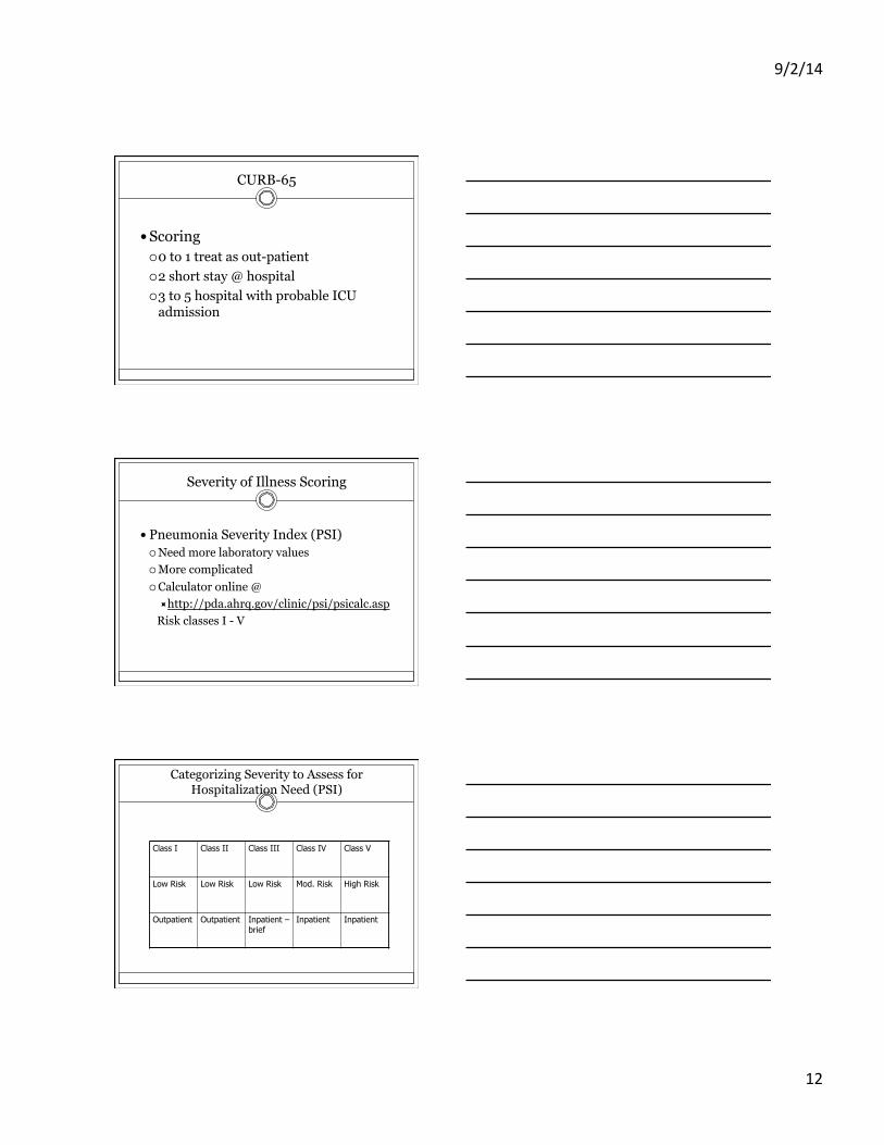
Categorizing Severity to Assess for Hospitalization Need (PSI)
Class I Class II Class III Class IV Class V
Low Risk Low Risk Low Risk Mod. Risk High Risk
Outpatient Outpatient Inpatient – brief
Inpatient Inpatient
9/2/14
13
Outpatient Versus Hospitalization
� Cost of inpatient versus outpatient management is up to 25 times greater!
� Outpatients resume normal activity sooner. � 80 % prefer outpatient therapy. � Hospitalization increases thromboembolic
events & superinfection by more-virulent or resistant hospital bacteria.
Criteria for Hospitalization
� ~ 10 % of hospitalized patients with CAP requires ICU admission
� One of most important determinants for ICU care is presence of chronic comorbid conditions
� 1/3 of patients with severe CAP were previously healthy
Antibiotics of Choice: Outpatient Therapy
� Previously healthy & no risk factors for drug-resistant S. pneumoniae infection: ¡ Macrolide (azithromycin, clarithromycin or erythromycin) ¡ Doxycycline
� Comorbidities or use of antimicrobials within previous 3 months: ¡ Respiratory fluoroquinolone (moxifloxacin, gemifloxacin, or
levofloxacin 750 mg) ¡ Β-lactam PLUS a macrolide (high-dose amoxicillin or
amoxicillin-clavulanate) ¡ Alternatives – ceftriaxone, cefpodoxime & cefuroxime,
doxycycline
9/2/14
14
Antibiotic Choice in the Elderly
� Use macrolide for those 65 and older � Proven to increase survival
Antibiotic Stewardship
� Avoid use of respiratory quinolones if not indicated.
� Save quinolones for patients who really need these medications! No new antibiotics in the near future.
� Limit duration of therapy to recommended time periods.
� Probiotics probably help limit development of C. diff, decreasing use of subsequent antibiotics
Ancillary Therapies
� Increased fluids, good nutrition � Expectorants (marginal utility) � Cough suppressants with care, usually just at
bedtime � Analgesics, acetaminophen for high fever � If likely diagnosis influenza pneumonia,
consider Tamiflu � Tobacco cessation
9/2/14
15
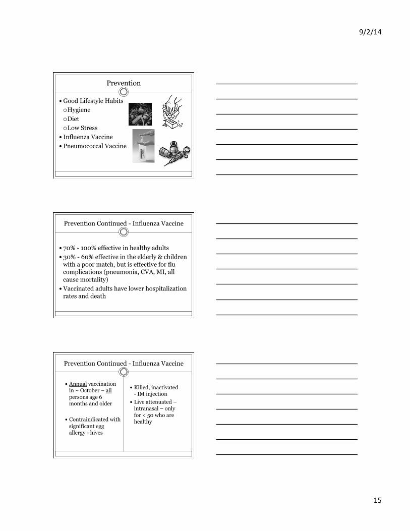
Prevention
� Good Lifestyle Habits ¡ Hygiene ¡ Diet ¡ Low Stress
� Influenza Vaccine � Pneumococcal Vaccine
Prevention Continued - Influenza Vaccine
� 70% - 100% effective in healthy adults � 30% - 60% effective in the elderly & children
with a poor match, but is effective for flu complications (pneumonia, CVA, MI, all cause mortality)
� Vaccinated adults have lower hospitalization rates and death
Prevention Continued - Influenza Vaccine
� Annual vaccination in ~ October – all persons age 6 months and older
� Contraindicated with significant egg allergy - hives
� Killed, inactivated - IM injection
� Live attenuated – intranasal – only for < 50 who are healthy
9/2/14
16
Prevention Continued – Pneumovax Vaccine
� PPSV23: � Those 65 and older � Chronic comorbidities � All cigarette smokers � Asthmatics
� Booster - one after age 65
� PCV13: Immunocompromised or children
� Now approved for adults
Hospitalization
Diagnosis for Hospitalized Patients
� Chest X-Ray – gold standard � WBC (leukocytosis or leukopenia) � Blood Cultures � Sputum Gram stain & Culture � Urine Antigens for Legionella & pneumococcus � CT scan (rarely) � PPD (R/O TB)
9/2/14
17
Diagnosis for Hospitalized Patients
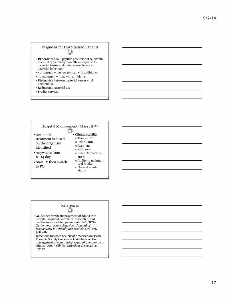
� Procalcitonin – peptide precursor of calcitonin released by parenchymal cells in response to bacterial toxins – elevated serum levels with bacterial infections
� <0.1 mcg/L = too low to treat with antibiotics � >0.25 mcg/L = treat with antibiotics � Distinguish between bacterial versus viral
pneumonia � Reduce antibacterial use � Predict survival
Hospital Management (Class III-V)
� Antibiotic treatment is based on the organism identified
� Anywhere from 10-14 days
� Start IV then switch to PO
� Clinical stability: ¡ Temp <100 ¡ Pulse <100 ¡ Resp <24 ¡ SBP >90 ¡ Pulse Oximeter ≥ 90 % ¡ Ability to maintain
oral intake ¡ Normal mental
status
References
� Guidelines for the management of adults with hospital-acquired, ventilator-associated, and healthcare-associated pneumonia. ATS/IDSA Guidelines. (2005). American Journal of Respiratory & Critical Care Medicine, vol 171, 388-416.
� Infectious Diseases Society of America/American Thoracic Society Consensus Guidelines on the management of community-acquired pneumonia in adults. (2007). Clinical Infectious Diseases. 44, S27-72.

9/2/14
18
References
� Rello, J. & Chastre, J. (2013). Update in pulmonary infections 2012. American Journal of Respiratory & Critical Care Medicine. Vol. 187, 1061-1066.
Thank you!