Embed Size (px)
Citation preview
Phase Segregation in Silicon Carbide-Carbon SolidSolutions from XRD and NMR Studies
Oleksandr O. Mykhaylyk,† Yaroslav Z. Khimyak, and J. Paul Attfield*
Department of Chemistry, University of Cambridge, Lensfield Road,Cambridge CB2 1EW, United Kingdom
Mykola P. Gadzira
Institute for Problems of Materials Science, NAS of Ukraine, 3 Krzhyzhanivskogo str.,Kyiv 03142, Ukraine
Received September 24, 2001. Revised Manuscript Received December 5, 2001
SiC-C solid solution powders have been analyzed by X-ray diffraction (XRD) and magic-angle spinning nuclear magnetic resonance (MAS NMR). The XRD data reveal a previouslyunreported phase separation of the as-synthesized material into two cubic silicon carbidephases characterized by slightly different lattice parameters [4.35287(9) and 4.35841(6) Å].The 29Si MAS NMR spectra of the powders after different heat treatments (annealing in avacuum and high-pressure sintering) show that the small excess of carbon (<1 at %) in theâ-SiC crystal structure has a large influence on the 29Si chemical shift resulting in adisplacement of the 29Si MAS NMR peak from -18.5 ppm to lower field. At least fivenonequivalent silicon sites have been detected in Si1-xC1+x: the solid solution formed byhigh-pressure sintering (4 GPa, 1800 °C) of the as-synthesized SiC-C powder. These sitesare assigned to point (carbon antisite) defects in the cubic silicon carbide structure.
Introduction
Silicon carbide (SiC) is important as an ultrahardceramic material. It is known to exist in variouspolytypic forms, of which the common forms are 3C, 4H,6H, and 15R.1 All forms of silicon carbide are based onthe diamond-like structure, with alternating, tetra-hedrally bonded, silicon and carbon atoms. The cubicpolytype, 3C, has a stacking sequence of diamond-likeplanes of Si and C with only one type of crystallographicsite for Si and C atoms. All the other SiC polytypes areformed by other stacking sequences of Si and C layersand have trigonal, hexagonal, or rhombohedral sym-metries. It is well established that the arrangement ofthe layer units in the silicon carbide polytypes caninclude stacking faults.2,3 Recently, it was shown thatin addition to stacking faults, certain synthesis pro-cesses can produce diamond-like C layers in the cubicsilicon carbide (â-SiC) crystal structure forming aSiC-C solid solution.4 Thus, the superstoichiometriccarbon atoms are located in the silicon carbide crystalstructure as planar defects along {111}. These defects
reduce the lattice parameter and produce strain ani-sotropy in the â-SiC crystal structure.5 It was revealedthat a diffusion of the superstoichiometric carbon atomsduring high-pressure and high-temperature sintering(4 GPa, 1800 °C) breaks down the planar defectsproducing an ideal solid solution of carbon in the siliconsublattice of the â-SiC crystal structure. The latticeparameter of this sintered sample has been used toestimate the composition through Vegard’s law asSi1-xC1+x with x ) 0.007.4 The presence of supersto-ichiometric carbon in the silicon carbide crystal struc-ture changes the local atomic environment and canproduce electronically nonequivalent sites.
During the last two decades, it has been shown thatsolid-state magic-angle spinning nuclear magnetic reso-nance (MAS NMR) spectroscopy is ideally suited todetermine the number of crystallographically non-equivalent sites in SiC polytypes.6-8 The 29Si and 13Cchemical shifts demonstrate high sensitivity to theenvironments of these atoms beyond their nearestneighbors. One can therefore expect MAS NMR spec-troscopy to be sensitive to the number and types of localenvironments in the SiC-C solid solutions and providesupplementary information to elucidate the location ofthe superstoichiometric carbon atoms in the structure.Thus, the application of X-ray diffraction (XRD) mea-surements to study long-range structural order along
* To whom correspondence should be addressed. E-mail: [email protected].
† Permanent address: Institute for Problems of Materials Science,NAS of Ukraine, 3 Krzhyzhanivskogo str., Kyiv 03142, Ukraine.
(1) Verma, A. R.; Krishna, P. Polymorphism and Polytypism inCrystals; Wiley: New York, 1966; Gmelin Handbook of InorganicChemistry, 8th ed.; Springer-Verlag: Berlin/Heidelberg/New York;Suppl. Vol. B2, Si-Silicon, “Properties of Crystalline Silicon Carbide”,1984; Suppl. Vol. B3, Si-Silicon, “System Si-C”, 1986.
(2) Jagodzinski, H. Acta Crystallogr. 1954, 7, 300.(3) Pandey, D.; Lele, S.; Krishna, P. Proc. R. Soc. London 1980,
A369, 435.(4) Mykhaylyk, O. O.; Gadzira, M. P. Acta Crystallogr. 1999, B55,
297.
(5) Mykhaylyk, O. O.; Gadzira, M. P. J. Mater. Chem. 2001, 11,217.
(6) Hartman, J. S.; Richardson, M. F.; Sherriff, B. L.; Winsborrow,B. G. J. Am. Chem. Soc. 1987, 109, 6059.
(7) Guth, J. R.; Petuskey, W. T. J. Phys. Chem. 1987, 91, 5361.(8) Dybowski, C.; Gaffney, E. J.; Sayir, A.; Rabinowitz, M. J.
Colloids Surf. 1996, A118, 171.
1348 Chem. Mater. 2002, 14, 1348-1353
10.1021/cm011246l CCC: $22.00 © 2002 American Chemical SocietyPublished on Web 02/16/2002
with MAS NMR spectroscopy can provide detailedinsights about the structure of SiC-C solid solutions,as described below. An excellent example of this ap-proach is an analysis of the stacking fault distributionin â-SiC.9
Experimental Section
Sample Preparation and Characterization. The SiC-Csolid solution powder was synthesized by self-propagatinghigh-temperature synthesis (the ignition temperature was inthe range of 1200-1300 °C) in an argon atmosphere from amixture of fine elemental silicon powder and thermally ex-foliated graphite (TEG). The particle size of the synthesizedpowder is less than 1 µm. The details of the preparation ofthe powder are described elsewhere.10 The XRD patternshowed the presence of stacking faults in the cubic SiC-Ccrystal structure,4 and together with the main phase, thesynthesized powder included small quantities of Si3N4 andSi2N2O. There were no significant amount of amorphouscomponents identified by the XRD experiment. Subsequently,the as-synthesized SiC-C powder was annealed in a vacuumat different temperatures in the range of 1400-1900 °C andsintered under pressures in the range of 4-8 GPa andtemperatures of 1400 and 1800 °C. The mechanical propertiesof these and other samples are reported elsewhere.5 To carryout the NMR measurements, the sintered samples werecrushed in an agate mortar. These samples were tested byXRD before and after crushing, and no significant change inthe lattice parameter and peak broadening was observed.
Instrumental and Measuring Techniques. Initial pow-der XRD data were collected on a DRON-UM1 powderdiffractometer [graphite (002) monochromator, Cu KR radia-tion] operated in a reflection scan mode. To obtain higherresolution diffraction measurements, a STOE STADI P powderdiffractometer [position sensitive detector, curved germanium(111) monochromator, Cu KR1 radiation, λ ) 1.5405981 Å]operated in transmission scan mode was used. The upper 2θlimit of 130° enabled the diffraction pattern of cubic siliconcarbide to be recorded up to the 224 reflection. Throughoutthe experiments, the ambient temperature was maintained at21 ( 0.5 °C. The systematic errors of the measurements suchas the zero shift of diffractometer (<(0.003°) and displacementof specimens from the goniometer axis (<(0.02 mm) weredetermined at the beginning and at the end of each pattern.The diffraction patterns were fitted by a full profile Rietveldmethod using the Fullprof software package.11 Individualpeaks were also fitted with the CSD software package (version4.11)12 using a pseudo-Voigt function. This package was alsoapplied for least-squares refinement of lattice parameters. Theshifts of diffraction lines caused by the displacement of thespecimens from the goniometer axis were taken into accountin the calculations. The lattice parameter values were notcorrected for refraction.
The alignment of the STOE diffractometer was tested by asilicon standard (SRM640b) and â-SiC (the fine powder wassynthesized13 by the reaction of SiO2 and C and annealed at1800 °C to obtain a stacking-fault-free structure of cubic siliconcarbide). The lattice parameters determined for these materi-als [5.43093(5) and 4.35883(7) Å, respectively] were in a goodagreement with literature data [5.430969(39)14 and 4.3589(1)Å,15 respectively]. The instrumental resolution of the STOE
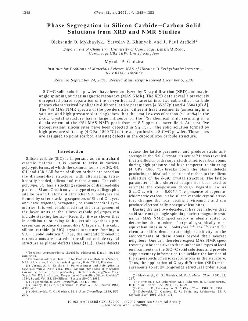
diffractometer estimated by the full widths at half-maximum(FWHM) of the silicon standard and â-SiC peaks was ca. 0.10°at lower 2θ diffraction angles and ca. 0.20° at higher 2θdiffraction angles (Figure 1).
Solid-state 29Si and 13C MAS NMR spectra were recordedat 9.4 T (79.44 MHz for 29Si and 100.56 MHz for 13C) using aChemagnetics CMX-400 spectrometer with a 4 mm diameterzirconia rotor spun in nitrogen at 8.0 kHz. All MAS NMRspectra presented in the paper were obtained by using π/4pulses of 2.0 µs duration with a recycle delay of 600 s. At least68 repeated scans were acquired. Chemical shifts are quotedin ppm from external tetramethylsilane. A deconvolution ofthe NMR spectra was carried out using the Spinsight 4.1software package. Spin-lattice relaxation times (T1) weredetermined using the standard saturation-recovery method.The curve-fitting analysis of variable delay time data was usedto calculate the T1. The above â-SiC powder was used as theâ-SiC standard for the MAS NMR measurements.
Electron spin resonance (ESR) spectra were recorded on aBruker ER 200 D EPR spectrometer with an X-band cavityand a microwave frequency of 8.97 GHz. This instrument issensitive to ∼1 ppm of unpaired spins.
Results and Discussion
XRD Analysis. On the basis of diffraction peakbroadening (Williamson-Hall plots) and an analysis ofthe behavior of the lattice parameter following differenttreatments at high temperatures in a vacuum or underhigh pressures, a structural model of the solid solutionSiC-C was previously suggested.4 In this model, su-perstoichiometric carbon atoms are located in diamond-like layers and form planar defects along {111} in cubicSiC-C. Such defects lead to a decrease of the latticeparameter of silicon carbide, broadening of the diffrac-tion peaks, and strain anisotropy of the crystal struc-ture.4,5 The higher resolution of XRD in the presentinvestigation reveals a previously unobserved splittingof the diffraction peaks for the as-synthesized powder,which is most clearly observed at high 2θ angles (Figure2). The absence of an hkl dependence of the splittingeliminates the possibility of a slight hexagonal or otherlattice distortion. The splittings are consistent with a
(9) Tateyama, H.; Noma, H.; Adachi, Y.; Komatsu, M. Chem. Mater.1997, 9, 766.
(10) Gadzyra, N. F.; Gnesin, G. G.; Andreev, A. V.; Kravets, V. G.;Kasyanenko, A. A. Inorg. Mater. 1996, 32, 721.
(11) Rodriguez-Carvajal, J. Fullprof Program, Version 3.5d. Labo-ratoire Leon Brillouin, France, 1998.
(12) Akselrud, L. G.; Gryn, Yu. N.; Zavalii, P. Y.; Pecharskii, V. K.;Fundamentskii, V. S. Twelfth European Crystallographic Meeting,Moscow, USSR, August 20-29, 1989; Collected Abstracts; Olden-burg: Munchen, 1990; Vol. 3, p 155.
(13) Khaenko, B. V.; Prilutskii, E. V.; Mikhailik, A. A.; Karpets,M. V.; Krainikov, A. V. Inorg. Mater. 1995, 31, 304.
(14) Yoder-Short, D. J. Appl. Crystallogr. 1993, 26, 272.(15) Powder Diffraction File No. 29-1129; a ) 4.3589(1) Å; the
wavelength for Cu KR radiation was taken to be 1.54178 Å.
Figure 1. Instrumental resolution of STOE STADI P diffrac-tometer. The FWHM vs 2θ for diffraction lines of siliconstandard (SRM640b) and â-SiC powder.
Silicon Carbide-Carbon Solid Solutions Chem. Mater., Vol. 14, No. 3, 2002 1349
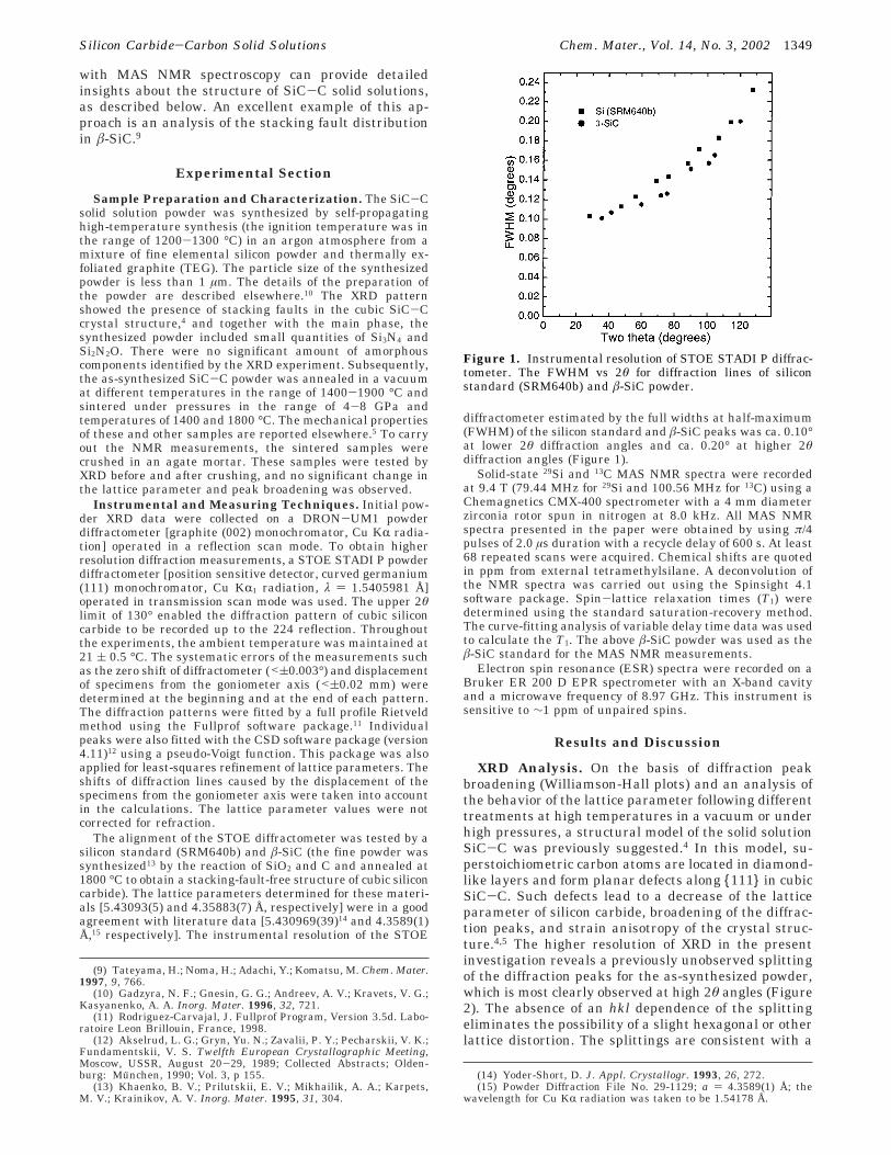
two-phase behavior, showing that the concentration ofthe superstoichiometric carbon layers in the SiC-Cstructure is not uniform, and the previous model for theas-synthesized SiC-C materials4 requires modification.
Rietveld fitting of two cubic silicon carbide phases(space group F4h3m) to the XRD data was unsuccessfulbecause of the similarity in the cell parameters of thetwo phases. However, an individual deconvolution ofeach of the XRD peaks enabled the two contributionsto be distinguished and the estimated peak positionscorrespond to two face-centered cubic crystal structures,SiC-C and â-SiC,with lattice parameters of 4.35287(9)and 4.35841(6) Å, respectively. The latter is slightlylower than the value of 4.3589(1) Å for standard â-SiC.15
The deconvoluted relative intensities of the reflectionsfor the two phases appear to be different from thecalculated intensities for cubic silicon carbide,16 but ahigher resolution diffraction experiment using synchro-tron radiation will be needed to characterize the twostructures more accurately.
The sintering of the as-synthesized powder at a highpressure (4-8 GPa) and temperature (1800 °C) leadsto the disappearance of the splitting in the siliconcarbide diffraction pattern, and sharp peaks from asingle cubic phase with a lattice parameter of 4.35286-(4) Å were observed after sintering at the lowestpressure of 4 GPa (Figure 2). If a mixture of twoindividual phases of silicon carbide (â-SiC and SiC-C)is assumed to occur in the as-synthesized powder, thenthe â-SiC lattice parameter should decrease after suchtreatments. However, no lattice parameter decrease isobserved after heat treatment of stoichiometric â-SiCunder such pressures17 and the model discussed belowshows that a mixture of the two individual phases isnot a good description of the structural transformationon sintering.
A small correction to the previous structural model4
of the SiC-C solid solution is needed to satisfy theresults of the diffraction experiments. In this model,planar diamond-like carbon defect layers exist in siliconcarbide. If these tend to aggregate in some regions ofthe SiC, then domains of â-SiC and SiC-C will exist inthe crystallites, and if they are sufficiently large, asplitting of the diffraction peaks will be observed.Diffusion at high pressures and a temperature of >1800°C breaks down the superstoichiometric carbon layersin the SiC-C crystal structure and establishes a uni-form concentration of the excess of carbon atoms in allparts of the crystallites. Thus, the thermal diffusion at4 GPa and 1800 °C equalizes the composition of theâ-SiC and SiC-C domains and an ideal, homogeneous,single-phase solid solution (Si1-xC1+x) is produced. Thediffusion of superstoichiometric carbon atoms at the (4GPa, 1800 °C) sintering changes the microstructurefrom planar long-range defects in the phase-segregated,as-synthesized SiC-C solid solution to point defects (CSiantisites) in the ideal solid solution, so that the averagelattice parameter is not conserved during the process.Sintering the as-synthesized powder at a lower temper-ature of 1400 °C (when this diffusion process is notactive) does not change the fine structure of the XRDpattern, and only a broadening of the peaks created byan increase of the dislocation density was observed.5
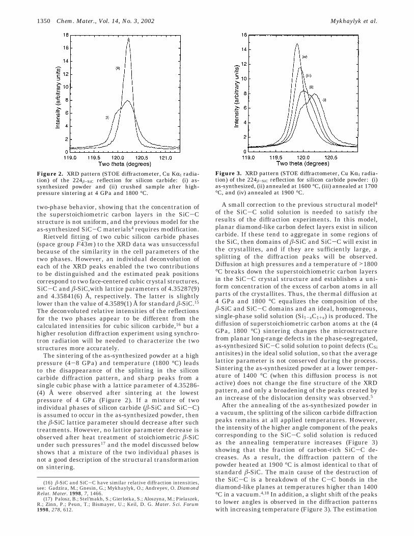
After the annealing of the as-synthesized powder ina vacuum, the splitting of the silicon carbide diffractionpeaks remains at all applied temperatures. However,the intensity of the higher angle component of the peakscorresponding to the SiC-C solid solution is reducedas the annealing temperature increases (Figure 3)showing that the fraction of carbon-rich SiC-C de-creases. As a result, the diffraction pattern of thepowder heated at 1900 °C is almost identical to that ofstandard â-SiC. The main cause of the destruction ofthe SiC-C is a breakdown of the C-C bonds in thediamond-like planes at temperatures higher than 1400°C in a vacuum.4,18 In addition, a slight shift of the peaksto lower angles is observed in the diffraction patternswith increasing temperature (Figure 3). The estimation
(16) â-SiC and SiC-C have similar relative diffraction intensities,see: Gadzira, M.; Gnesin, G.; Mykhaylyk, O.; Andreyev, O. DiamondRelat. Mater. 1998, 7, 1466.
(17) Palosz, B.; Stel’makh, S.; Gierlotka, S.; Aloszyna, M.; Pielaszek,R.; Zinn, P.; Peun, T.; Bismayer, U.; Keil, D. G. Mater. Sci. Forum1998, 278, 612.
Figure 2. XRD pattern (STOE diffractometer, Cu KR1 radia-tion) of the 224â-SiC reflection for silicon carbide: (i) as-synthesized powder and (ii) crushed sample after high-pressure sintering at 4 GPa and 1800 °C.
Figure 3. XRD pattern (STOE diffractometer, Cu KR1 radia-tion) of the 224â-SiC reflection for silicon carbide powder: (i)as-synthesized, (ii) annealed at 1600 °C, (iii) annealed at 1700°C, and (iv) annealed at 1900 °C.
1350 Chem. Mater., Vol. 14, No. 3, 2002 Mykhaylyk et al.
of the lattice parameters based on the deconvolution ofthe most highly resolved, 224, reflection shows that theparameter increases slightly for both the â-SiC andSiC-C regions in the crystallites (Figure 4). Thissuggests that the two different types of silicon carbidein a crystallite have a mutual influence on each other,presumably through epitaxial interfaces, and a highfraction of SiC-C domains leads to a slight decrease ofthe lattice parameter for the rest of the silicon carbide(â-SiC). The growth of the cubic silicon carbide fractionat high annealing temperatures (Figure 3) leads to agradual increase of the â-SiC lattice parameter until thestandard value is reached (Figure 4).
NMR Analysis. The excess carbon atoms in SiC-Ccould change the local bonding of the atoms and createdangling bonds as in amorphous Si1-xCx.19 However, noESR signal was detected for the as-synthesized powdershowing that SiC-C is diamagnetic and has a negligibleconcentration of dangling bonds or incorporated impuri-ties such as nitrogen20 with unpaired electrons. Theabsence of paramagnetic centers enabled MAS NMRspectroscopy to be applied to these silicon carbidesamples. Attempts to record 13C NMR spectra wereunsuccessful, as in other NMR studies6,7 of cubic siliconcarbide. This is probably due to the extremely longspin-lattice relaxation time. Hence, only 29Si MASNMR results were used to analyze the SiC-C materials.
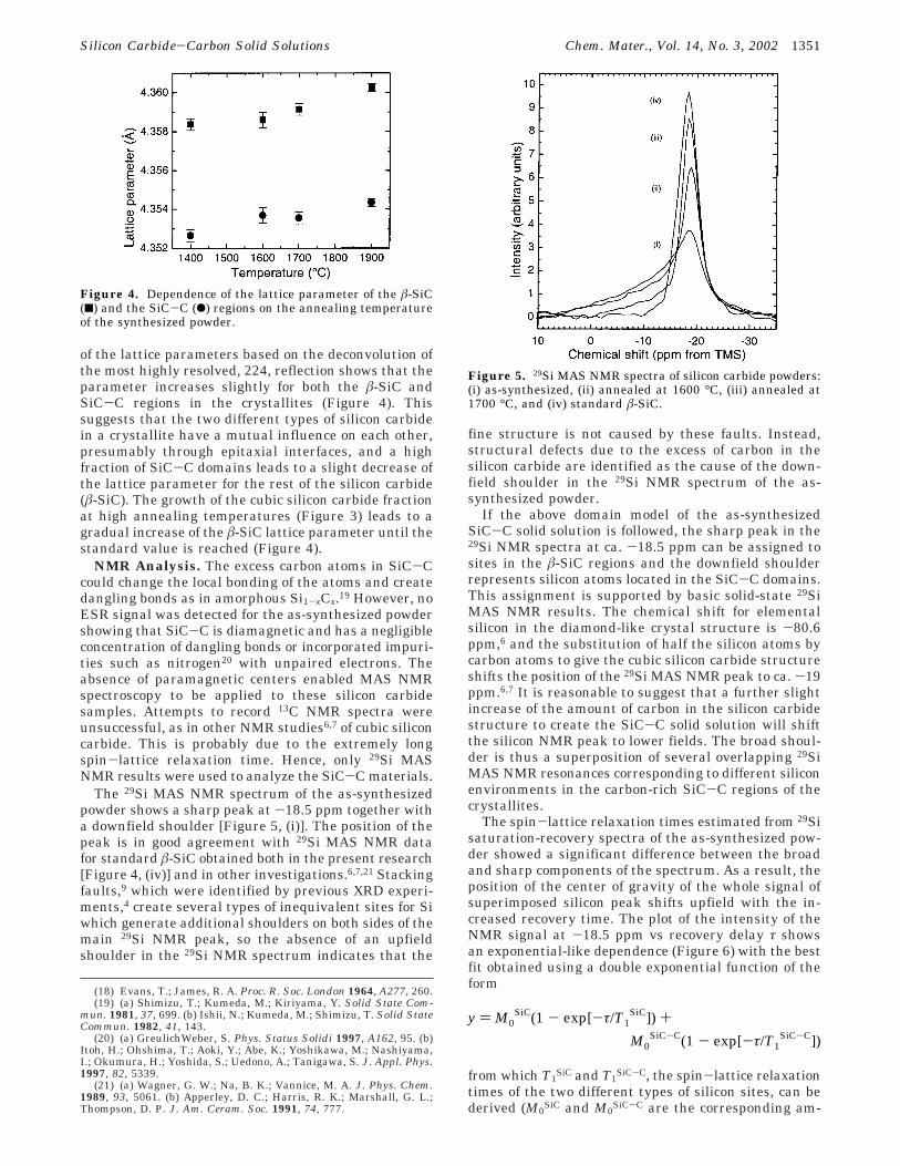
The 29Si MAS NMR spectrum of the as-synthesizedpowder shows a sharp peak at -18.5 ppm together witha downfield shoulder [Figure 5, (i)]. The position of thepeak is in good agreement with 29Si MAS NMR datafor standard â-SiC obtained both in the present research[Figure 4, (iv)] and in other investigations.6,7,21 Stackingfaults,9 which were identified by previous XRD experi-ments,4 create several types of inequivalent sites for Siwhich generate additional shoulders on both sides of themain 29Si NMR peak, so the absence of an upfieldshoulder in the 29Si NMR spectrum indicates that the
fine structure is not caused by these faults. Instead,structural defects due to the excess of carbon in thesilicon carbide are identified as the cause of the down-field shoulder in the 29Si NMR spectrum of the as-synthesized powder.
If the above domain model of the as-synthesizedSiC-C solid solution is followed, the sharp peak in the29Si NMR spectra at ca. -18.5 ppm can be assigned tosites in the â-SiC regions and the downfield shoulderrepresents silicon atoms located in the SiC-C domains.This assignment is supported by basic solid-state 29SiMAS NMR results. The chemical shift for elementalsilicon in the diamond-like crystal structure is -80.6ppm,6 and the substitution of half the silicon atoms bycarbon atoms to give the cubic silicon carbide structureshifts the position of the 29Si MAS NMR peak to ca. -19ppm.6,7 It is reasonable to suggest that a further slightincrease of the amount of carbon in the silicon carbidestructure to create the SiC-C solid solution will shiftthe silicon NMR peak to lower fields. The broad shoul-der is thus a superposition of several overlapping 29SiMAS NMR resonances corresponding to different siliconenvironments in the carbon-rich SiC-C regions of thecrystallites.
The spin-lattice relaxation times estimated from 29Sisaturation-recovery spectra of the as-synthesized pow-der showed a significant difference between the broadand sharp components of the spectrum. As a result, theposition of the center of gravity of the whole signal ofsuperimposed silicon peak shifts upfield with the in-creased recovery time. The plot of the intensity of theNMR signal at -18.5 ppm vs recovery delay τ showsan exponential-like dependence (Figure 6) with the bestfit obtained using a double exponential function of theform
from which T1SiC and T1
SiC-C, the spin-lattice relaxationtimes of the two different types of silicon sites, can bederived (M0
SiC and M0SiC-C are the corresponding am-
(18) Evans, T.; James, R. A. Proc. R. Soc. London 1964, A277, 260.(19) (a) Shimizu, T.; Kumeda, M.; Kiriyama, Y. Solid State Com-
mun. 1981, 37, 699. (b) Ishii, N.; Kumeda, M.; Shimizu, T. Solid StateCommun. 1982, 41, 143.
(20) (a) GreulichWeber, S. Phys. Status Solidi 1997, A162, 95. (b)Itoh, H.; Ohshima, T.; Aoki, Y.; Abe, K.; Yoshikawa, M.; Nashiyama,I.; Okumura, H.; Yoshida, S.; Uedono, A.; Tanigawa, S. J. Appl. Phys.1997, 82, 5339.
(21) (a) Wagner, G. W.; Na, B. K.; Vannice, M. A. J. Phys. Chem.1989, 93, 5061. (b) Apperley, D. C.; Harris, R. K.; Marshall, G. L.;Thompson, D. P. J. Am. Ceram. Soc. 1991, 74, 777.
Figure 4. Dependence of the lattice parameter of the â-SiC(9) and the SiC-C (b) regions on the annealing temperatureof the synthesized powder.
Figure 5. 29Si MAS NMR spectra of silicon carbide powders:(i) as-synthesized, (ii) annealed at 1600 °C, (iii) annealed at1700 °C, and (iv) standard â-SiC.
y ) M0SiC(1 - exp[-τ/T1
SiC]) +
M0SiC-C(1 - exp[-τ/T1
SiC-C])
Silicon Carbide-Carbon Solid Solutions Chem. Mater., Vol. 14, No. 3, 2002 1351
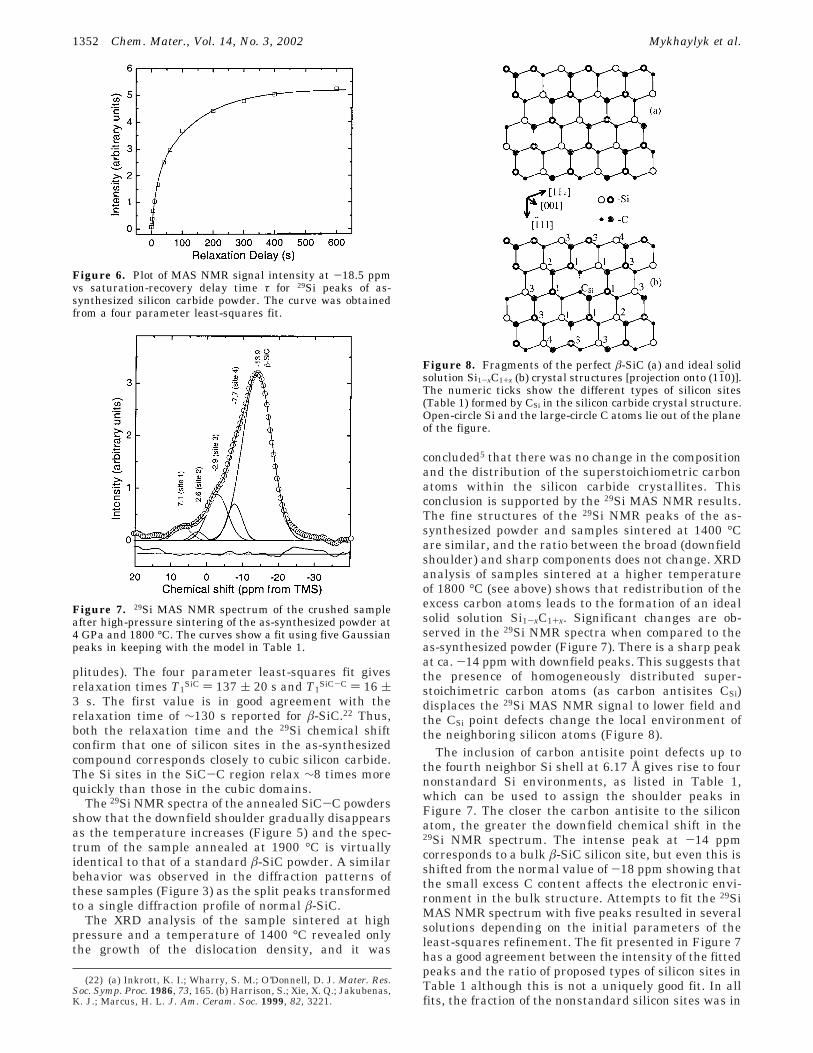
plitudes). The four parameter least-squares fit givesrelaxation times T1
SiC ) 137 ( 20 s and T1SiC-C ) 16 (
3 s. The first value is in good agreement with therelaxation time of ∼130 s reported for â-SiC.22 Thus,both the relaxation time and the 29Si chemical shiftconfirm that one of silicon sites in the as-synthesizedcompound corresponds closely to cubic silicon carbide.The Si sites in the SiC-C region relax ∼8 times morequickly than those in the cubic domains.
The 29Si NMR spectra of the annealed SiC-C powdersshow that the downfield shoulder gradually disappearsas the temperature increases (Figure 5) and the spec-trum of the sample annealed at 1900 °C is virtuallyidentical to that of a standard â-SiC powder. A similarbehavior was observed in the diffraction patterns ofthese samples (Figure 3) as the split peaks transformedto a single diffraction profile of normal â-SiC.
The XRD analysis of the sample sintered at highpressure and a temperature of 1400 °C revealed onlythe growth of the dislocation density, and it was
concluded5 that there was no change in the compositionand the distribution of the superstoichiometric carbonatoms within the silicon carbide crystallites. Thisconclusion is supported by the 29Si MAS NMR results.The fine structures of the 29Si NMR peaks of the as-synthesized powder and samples sintered at 1400 °Care similar, and the ratio between the broad (downfieldshoulder) and sharp components does not change. XRDanalysis of samples sintered at a higher temperatureof 1800 °C (see above) shows that redistribution of theexcess carbon atoms leads to the formation of an idealsolid solution Si1-xC1+x. Significant changes are ob-served in the 29Si NMR spectra when compared to theas-synthesized powder (Figure 7). There is a sharp peakat ca. -14 ppm with downfield peaks. This suggests thatthe presence of homogeneously distributed super-stoichimetric carbon atoms (as carbon antisites CSi)displaces the 29Si MAS NMR signal to lower field andthe CSi point defects change the local environment ofthe neighboring silicon atoms (Figure 8).
The inclusion of carbon antisite point defects up tothe fourth neighbor Si shell at 6.17 Å gives rise to fournonstandard Si environments, as listed in Table 1,which can be used to assign the shoulder peaks inFigure 7. The closer the carbon antisite to the siliconatom, the greater the downfield chemical shift in the29Si NMR spectrum. The intense peak at -14 ppmcorresponds to a bulk â-SiC silicon site, but even this isshifted from the normal value of -18 ppm showing thatthe small excess C content affects the electronic envi-ronment in the bulk structure. Attempts to fit the 29SiMAS NMR spectrum with five peaks resulted in severalsolutions depending on the initial parameters of theleast-squares refinement. The fit presented in Figure 7has a good agreement between the intensity of the fittedpeaks and the ratio of proposed types of silicon sites inTable 1 although this is not a uniquely good fit. In allfits, the fraction of the nonstandard silicon sites was in
(22) (a) Inkrott, K. I.; Wharry, S. M.; O’Donnell, D. J. Mater. Res.Soc. Symp. Proc. 1986, 73, 165. (b) Harrison, S.; Xie, X. Q.; Jakubenas,K. J.; Marcus, H. L. J. Am. Ceram. Soc. 1999, 82, 3221.
Figure 6. Plot of MAS NMR signal intensity at -18.5 ppmvs saturation-recovery delay time τ for 29Si peaks of as-synthesized silicon carbide powder. The curve was obtainedfrom a four parameter least-squares fit.
Figure 7. 29Si MAS NMR spectrum of the crushed sampleafter high-pressure sintering of the as-synthesized powder at4 GPa and 1800 °C. The curves show a fit using five Gaussianpeaks in keeping with the model in Table 1.
Figure 8. Fragments of the perfect â-SiC (a) and ideal solidsolution Si1-xC1+x (b) crystal structures [projection onto (11h0)].The numeric ticks show the different types of silicon sites(Table 1) formed by CSi in the silicon carbide crystal structure.Open-circle Si and the large-circle C atoms lie out of the planeof the figure.
1352 Chem. Mater., Vol. 14, No. 3, 2002 Mykhaylyk et al.
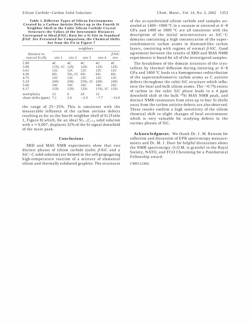
the range of 25-35%. This is consistent with themeasurable influence of the carbon antisite defectsreaching as far as the fourth neighbor shell of Si (Table1, Figure 8) which, for an ideal Si1-xC1+x solid solutionwith x ) 0.007, displaces 32% of the Si signal downfieldof the main peak.
Conclusions
XRD and MAS NMR experiments show that twodistinct phases of silicon carbide (cubic â-SiC and aSiC-C solid solution) are formed in the self-propagatinghigh-temperature reaction of a mixture of elementalsilicon and thermally exfoliated graphite. The structures
of the as-synthesized silicon carbide and samples an-nealed at 1400-1900 °C in a vacuum or sintered at 4-8GPa and 1400 or 1800 °C are all consistent with thedescription of the initial microstructure as SiC-Cdomains containing a high concentration of the super-stoichiometric carbon atoms in diamond-like carbonlayers, coexisting with regions of normal â-SiC. Goodagreement between the results of XRD and MAS NMRexperiments is found for all of the investigated samples.
The breakdown of the domain structure of the crys-tallites by thermal diffusion during sintering at 4-8GPa and 1800 °C leads to a homogeneous redistributionof the superstoichiometric carbon atoms as C antisitedefects throughout the cubic SiC structure which influ-ence the local and bulk silicon atoms. The ∼0.7% excessof carbon in the cubic SiC phase leads to a 4 ppmdownfield shift of the bulk 29Si MAS NMR peak, anddistinct NMR resonances from sites up to four Si shellsaway from the carbon antisite defects are also observed.These results confirm a high sensitivity of the siliconchemical shift to slight changes of local environmentwhich is very valuable for studying defects in thevarious phases of SiC.
Acknowledgment. We thank Dr. J. M. Rawson forcollection and discussion of EPR spectroscopy measure-ments and Dr. M. J. Duer for helpful discussions aboutthe NMR spectroscopy. O.O.M. is grateful to the RoyalSociety, NATO, and FCO Chevening for a PostdoctoralFellowship award.
CM011246L
Table 1. Different Types of Silicon EnvironmentsCreated by a Carbon Antisite Defect up to the Fourth Si
Neighbor Shell in the Cubic Silicon Carbide CrystalStructure; the Values of the Interatomic Distances
Correspond to Ideal â-SiC; Data for a Si Site in Standardâ-SiC Are Presented for Comparison; the Chemical Shifts
Are from the Fit in Figure 7
neighbors
distance tocentral Si (Å) site 1 site 2 site 3 site 4
â-SiCsite
1.89 4C 4C 4C 4C 4C3.08 11Si, 1C 12Si 12Si 12Si 12Si3.61 12C 12C 12C 12C 12C4.36 6Si 5Si, 1C 6Si 6Si 6Si4.75 12C 12C 12C 12C 12C5.34 24Si 24Si 23Si, 1C 24Si 24Si5.66 16C 16C 16C 16C 16C6.17 12Si 12Si 12Si 11Si, 1C 12Si
multiplicity 12 6 24 12chem shifts (ppm) 7.1 2.6 -2.9 -7.7 -13.9
Silicon Carbide-Carbon Solid Solutions Chem. Mater., Vol. 14, No. 3, 2002 1353