Embed Size (px)
Citation preview

Pharmacology in the Newborn with HIE
Jackie B. Martin, DNP, RN, NNP-BC, CCNSAssociate Nurse EducatorMEDNAX, Inc.
Disclosures
Financial No relevant financial relationships to
disclose
FDA Nothing to disclose

Objectives
Discuss the medications involved in the care of an infant with Hypoxic-Ischemic Encephalopathy
Define Hypoxic-Ischemic Encephalopathy or HIE
Describe the mechanisms of the protection of hypothermia
Maternal History Mother is a 28 y.o., gravida 1, African-
American woman
Blood type: A negative
Prenatal Labs: RPR – NR, Rubella-immune, HbsAg – neg, GBS – neg, HIV –neg, Herpes-neg.
Received early and regular prenatal care
Delivery was planned in her home town at a community hospital
Delivery History Mother presented to the community
hospital at 39 weeks gestation in labor
Rapid second stage dilatation
SROM with MSAF
Episodes of fetal bradycardia with recovery
Vaginal delivery assisted by vacuum extraction
Tight nuchal cord x 1, with the cord tearing during the attempt to remove the cord from the neck and deliver the baby

Delivery (continued) Moderate blood loss with the clamping and
cutting of the cord
Infant was rapidly placed in a warmer, apneic, without spontaneous activity, HR < 100 bpm
The nurse suctioned out the mouth and a physician immediately intubated the infant with a 4.0 ETT and suctioned ETT receiving meconium below the cords. A second intubation received no meconium, so the infant was reintubated and the ETT was secured at 9.5 cm at the lip.

Delivery (continued) The infant continued to be apneic and the
HR was now 40 bpm. Positive pressure ventilation was provided and chest compressions were begun with the heart rate rising to about 50 bpm. The infant was given a dose of epinephrine ET while equipment was prepared for umbilical line insertion for future doses if needed. With the epinephrine and continued PPV, the infant’s heart rate increased to 70 bpm and very slowly continued to increase.

Epinephrine Therapeutic Category: Adrenergic Agonist
Agent
Uses: Acute cardiovascular collapse/ cardiac arrest. Short-term use for systemic hypotension. May also be used for treatment of bronchospasm.
Young,T.E., Mangum, B. (2010). Epinephrine. Neofax 2010. (23rd
Ed.). Montvale, NJ:Thomson Reuters, 164.
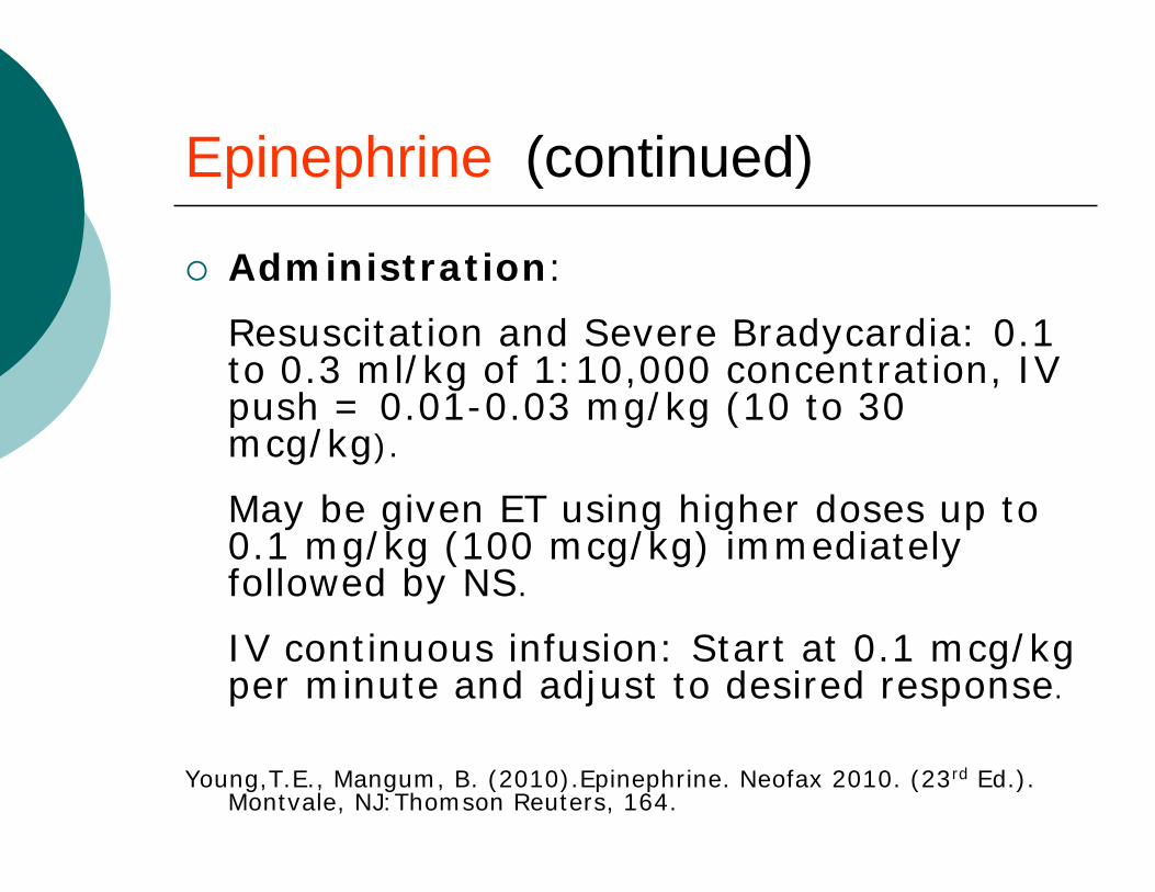
Epinephrine (continued)
Administration:
Resuscitation and Severe Bradycardia: 0.1 to 0.3 ml/kg of 1:10,000 concentration, IV push = 0.01-0.03 mg/kg (10 to 30 mcg/kg).
May be given ET using higher doses up to 0.1 mg/kg (100 mcg/kg) immediately followed by NS.
IV continuous infusion: Start at 0.1 mcg/kg per minute and adjust to desired response.
Young,T.E., Mangum, B. (2010).Epinephrine. Neofax 2010. (23rd Ed.). Montvale, NJ:Thomson Reuters, 164.
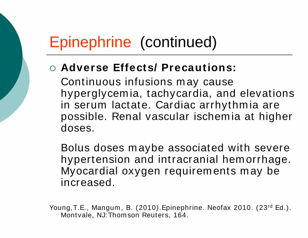
Epinephrine (continued) Adverse Effects/Precautions:
Continuous infusions may cause hyperglycemia, tachycardia, and elevations in serum lactate. Cardiac arrhythmia are possible. Renal vascular ischemia at higher doses.
Bolus doses maybe associated with severe hypertension and intracranial hemorrhage. Myocardial oxygen requirements may be increased.
Young,T.E., Mangum, B. (2010).Epinephrine. Neofax 2010. (23rd Ed.). Montvale, NJ:Thomson Reuters, 164.
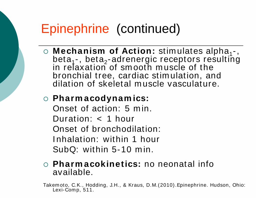
Epinephrine (continued) Mechanism of Action: stimulates alpha1-,
beta1-, beta2-adrenergic receptors resulting in relaxation of smooth muscle of the bronchial tree, cardiac stimulation, and dilation of skeletal muscle vasculature.
Pharmacodynamics: Onset of action: 5 min.Duration: < 1 hourOnset of bronchodilation:Inhalation: within 1 hourSubQ: within 5-10 min.
Pharmacokinetics: no neonatal info available.
Takemoto, C.K., Hodding, J.H., & Kraus, D.M.(2010).Epinephrine. Hudson, Ohio: Lexi-Comp, 511.

Delivery continued Very little respiratory effort was noted, only
an occasional gasp. The infant remained extremely pale, and limp, but was becoming pinker. Apgars were 1 at 1 minute and 3 at five minutes and 5 at ten minutes. The pH on the venous cord gas was 6.95.
The radiant warmer was turned off to provide passive hypothermia for this asphyxiated infant.
The decision was made to give the infant a normal saline bolus due to probable hypovolemia. A low lying UVC was inserted for this.
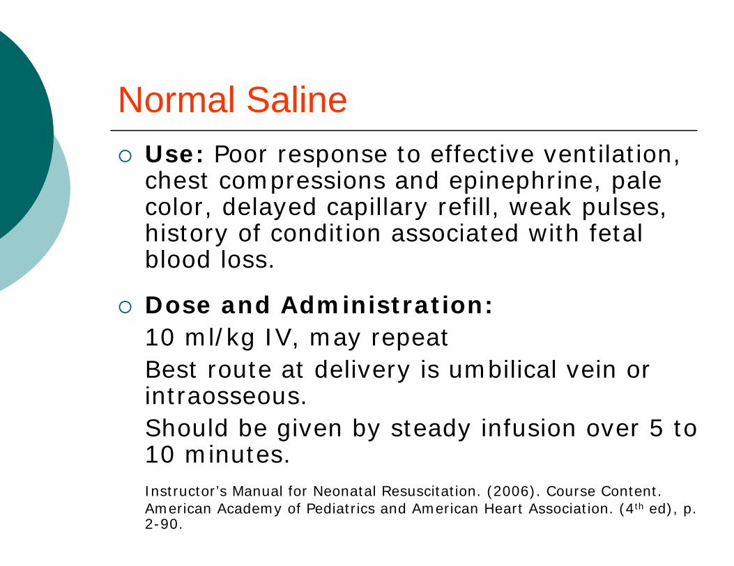
Normal Saline Use: Poor response to effective ventilation,
chest compressions and epinephrine, pale color, delayed capillary refill, weak pulses, history of condition associated with fetal blood loss.
Dose and Administration:10 ml/kg IV, may repeatBest route at delivery is umbilical vein or intraosseous.Should be given by steady infusion over 5 to 10 minutes. Instructor’s Manual for Neonatal Resuscitation. (2006). Course Content. American Academy of Pediatrics and American Heart Association. (4th ed), p. 2-90.

Normal Saline (continued) Dosage and administration continued:
Administer slowly in preterm infants, as they are especially susceptible to intracranial hemorrhage with rapid volume administration
Mechanism of Action: volume expansion for hypovolemia due to causes such as placenta previa and abruption, twin-to-twin transfusion, blood loss from the umbilical cord, or a significant fetal-maternal hemorrhage without obvious blood loss.
Instructor’s Manual for Neonatal Resuscitation. (2006). Course Content. American Academy of Pediatrics and American Heart Association. (4th ed), p. 2-90
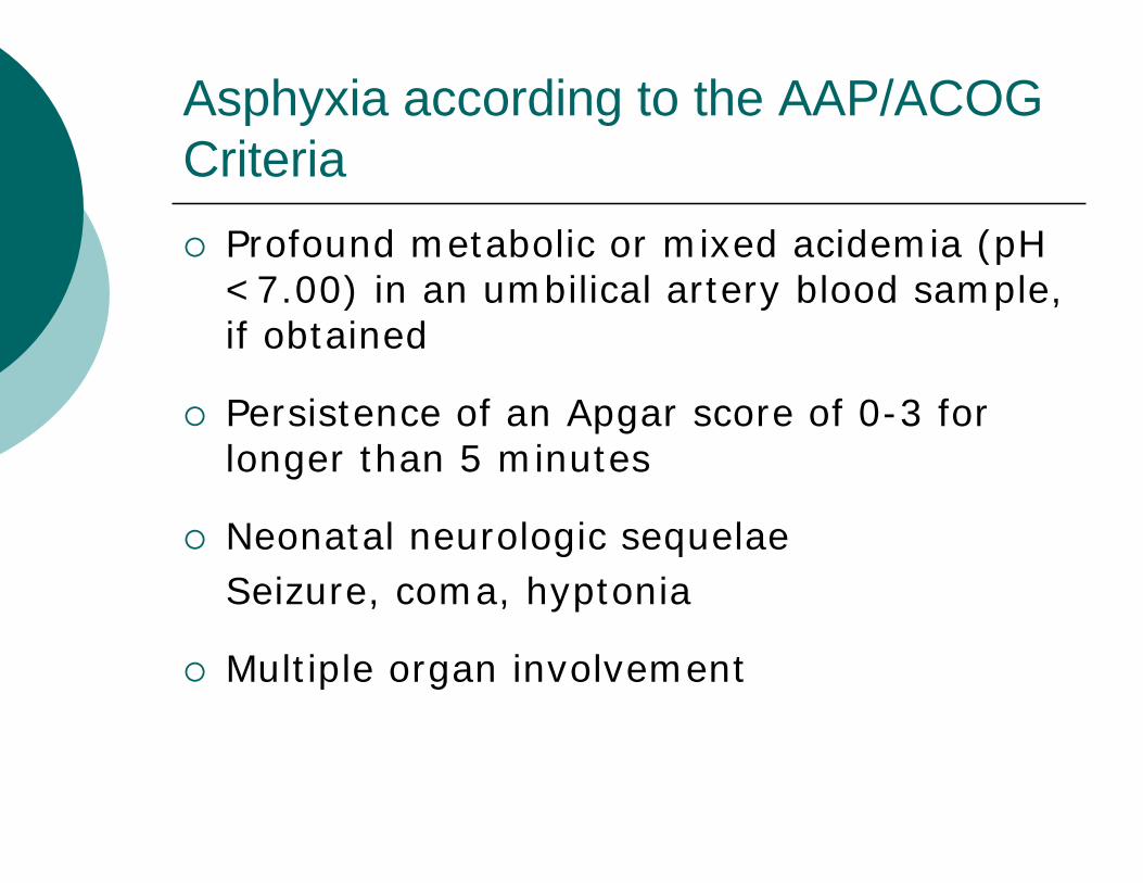
Asphyxia according to the AAP/ACOG Criteria Profound metabolic or mixed acidemia (pH
<7.00) in an umbilical artery blood sample, if obtained
Persistence of an Apgar score of 0-3 for longer than 5 minutes
Neonatal neurologic sequelaeSeizure, coma, hyptonia
Multiple organ involvement
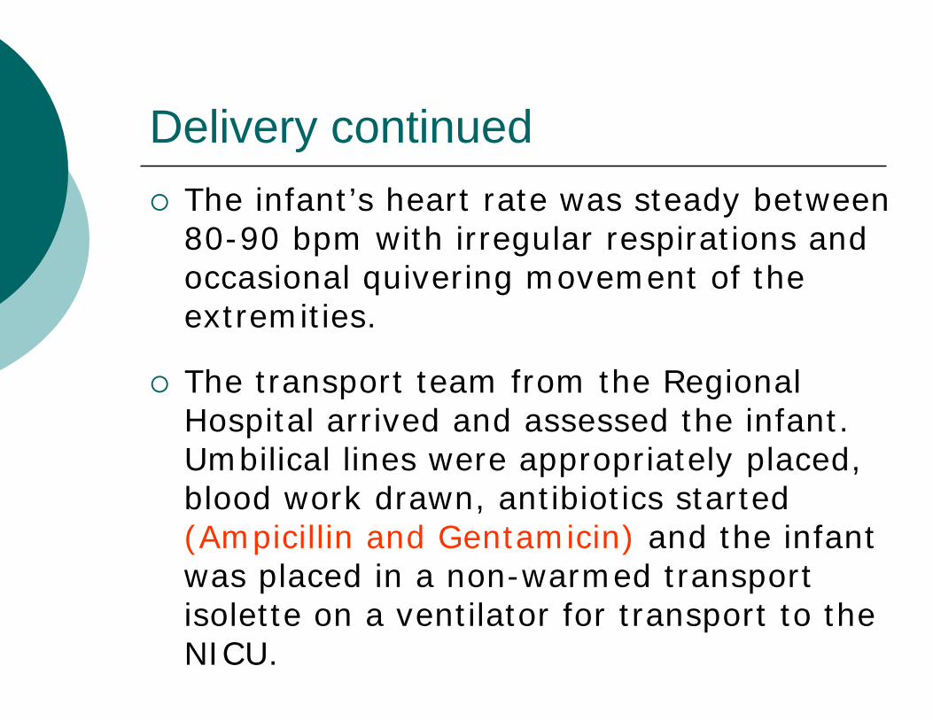
Delivery continued The infant’s heart rate was steady between
80-90 bpm with irregular respirations and occasional quivering movement of the extremities.
The transport team from the Regional Hospital arrived and assessed the infant. Umbilical lines were appropriately placed, blood work drawn, antibiotics started (Ampicillin and Gentamicin) and the infant was placed in a non-warmed transport isolette on a ventilator for transport to the NICU.
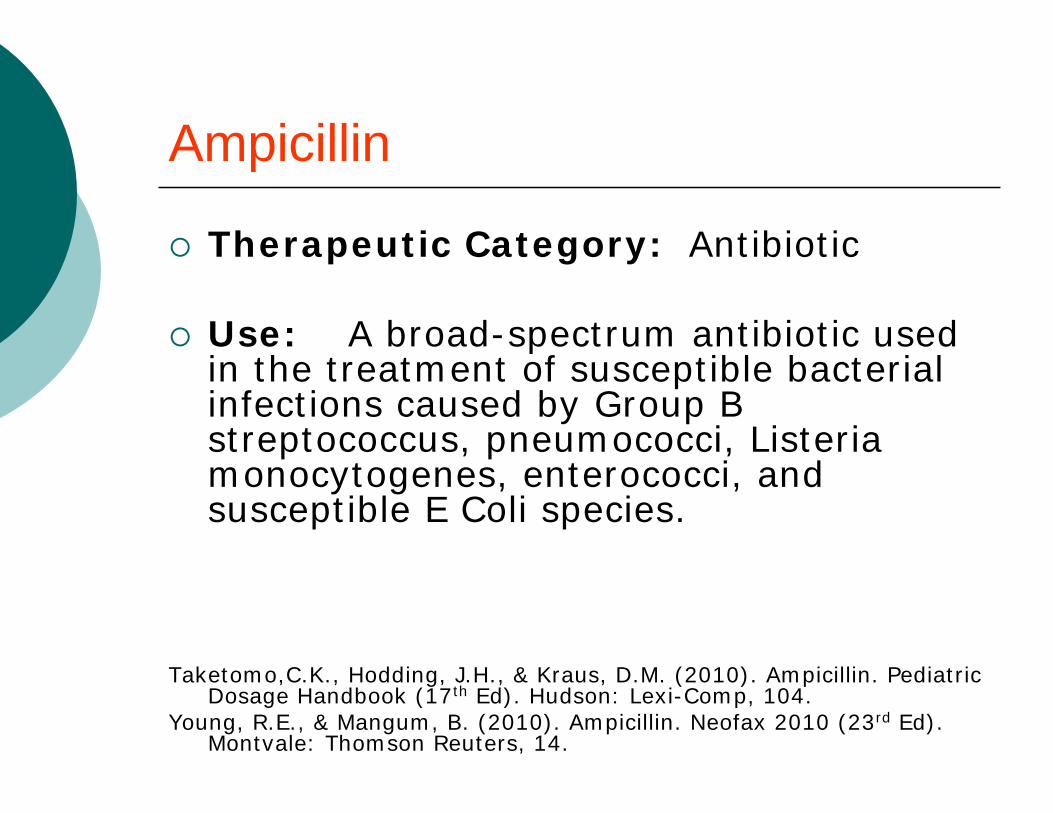
Ampicillin
Therapeutic Category: Antibiotic
Use: A broad-spectrum antibiotic used in the treatment of susceptible bacterial infections caused by Group B streptococcus, pneumococci, Listeria monocytogenes, enterococci, and susceptible E Coli species.
Taketomo,C.K., Hodding, J.H., & Kraus, D.M. (2010). Ampicillin. Pediatric Dosage Handbook (17th Ed). Hudson: Lexi-Comp, 104.
Young, R.E., & Mangum, B. (2010). Ampicillin. Neofax 2010 (23rd Ed). Montvale: Thomson Reuters, 14.
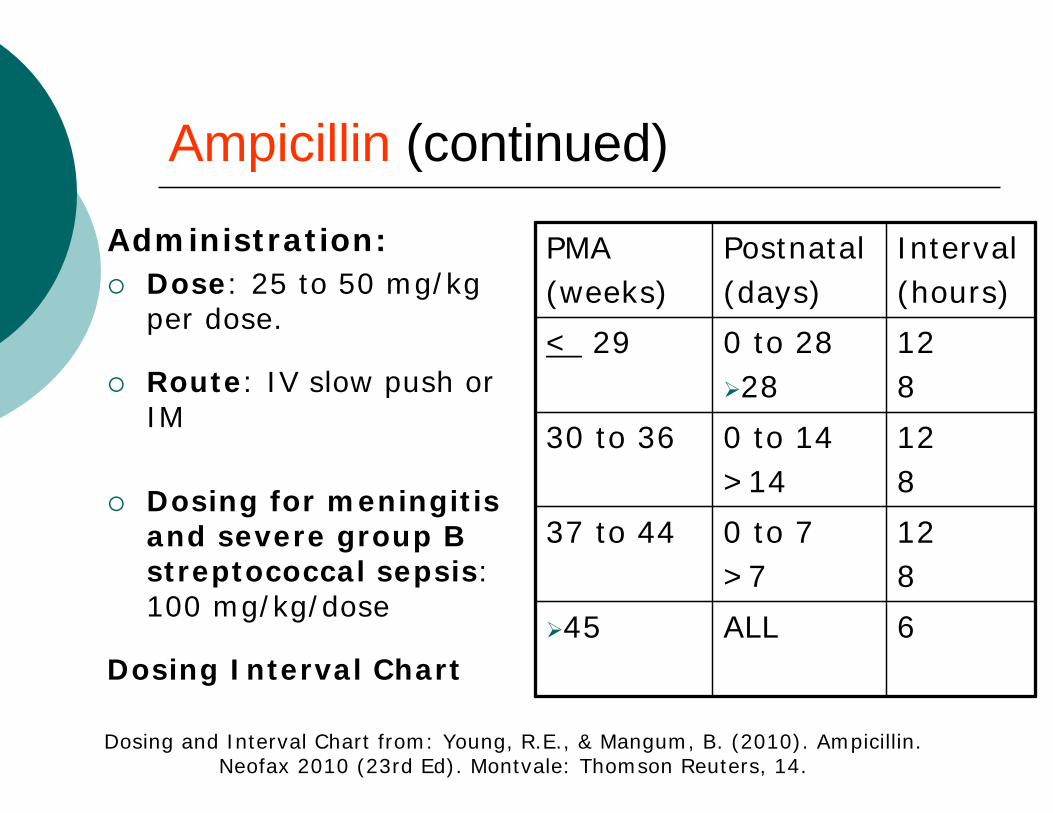
Ampicillin (continued)
Administration: Dose: 25 to 50 mg/kg
per dose.
Route: IV slow push or IM
Dosing for meningitis and severe group B streptococcal sepsis: 100 mg/kg/dose
Dosing Interval Chart6ALL45
128
0 to 7>7
37 to 44
128
0 to 14>14
30 to 36
128
0 to 2828
< 29
Interval(hours)
Postnatal(days)
PMA(weeks)
Dosing and Interval Chart from: Young, R.E., & Mangum, B. (2010). Ampicillin. Neofax 2010 (23rd Ed). Montvale: Thomson Reuters, 14.
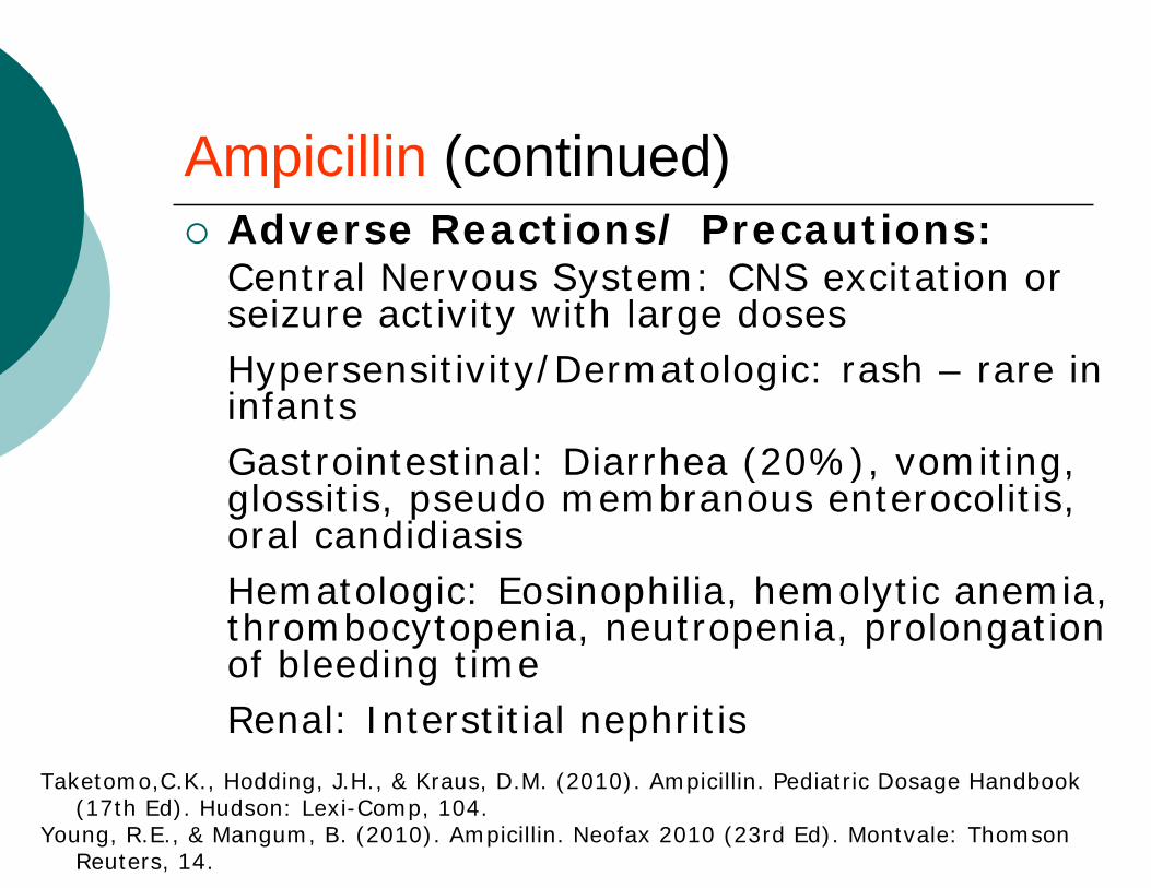
Ampicillin (continued) Adverse Reactions/ Precautions:
Central Nervous System: CNS excitation or seizure activity with large dosesHypersensitivity/Dermatologic: rash – rare in infantsGastrointestinal: Diarrhea (20%), vomiting, glossitis, pseudo membranous enterocolitis, oral candidiasisHematologic: Eosinophilia, hemolytic anemia, thrombocytopenia, neutropenia, prolongation of bleeding timeRenal: Interstitial nephritis
Taketomo,C.K., Hodding, J.H., & Kraus, D.M. (2010). Ampicillin. Pediatric Dosage Handbook (17th Ed). Hudson: Lexi-Comp, 104.
Young, R.E., & Mangum, B. (2010). Ampicillin. Neofax 2010 (23rd Ed). Montvale: Thomson Reuters, 14.

Ampicillin (continued) Mechanism of Action: Ampicillin binds to
one or more penicillin-binding proteins during the active multiplication phase of bacterial cell wall synthesis, interfering with the synthesis and causing cell wall death. 1
Pharmacokinetics:Half life:Neonates < 7 days of age: 4 hoursClearance: Primarily renal and inversely related to postnatal age.2
Taketomo,C.K., Hodding, J.H., & Kraus, D.M. (2010). Ampicillin. Pediatric Dosage Handbook (17th Ed). Hudson: Lexi-Comp, 104.
Young, R.E., & Mangum, B. (2010). Ampicillin. Neofax 2010 (23rd Ed). Montvale: Thomson Reuters, 14.

Gentamicin Therapeutic Category: Antibiotic,
Aminoglycoside
Use: Used in combination with ß-lactam antibiotics as empiric therapy for sepsis in newborns. Treatment of bacterial infections caused by aerobic gram-negative bacilli such as Pseudomonas, E.Coli, Klebsiella, Proteus, Seratia, and gram-positive staphylococcus. Also used for the treatment of bone, CNS, respiratory tract, skin and soft tissue infections.
Young, R.E., & Mangum, B. (2010). Gentamicin. Neofax 2010 (23rd Ed). Montvale: Thomson Reuters, 50.

Gentamicin (continued) Administration:
IV- on syringe pump over 30 minutes.IM- associated with variable absorption
Dosing Chart and Intervals->*or significant asphyxia, PDA, or treatment with indomethacin
244ALL>35
3624
4.54
0 to 7>8
30 to 34
483624
544
0 to 78 to 28> 29
< 29*
Interval(hours)
Dose(mg/kg)
Post-natal
(days)
PMA(weeks)
Dosing Chart from Neofax 2010. Young, R.E., & Mangum, B. (2010). Gentamicin. Neofax 2010 (23rd Ed). Montvale: Thomson Reuters, 50.
Gentamicin (continued)
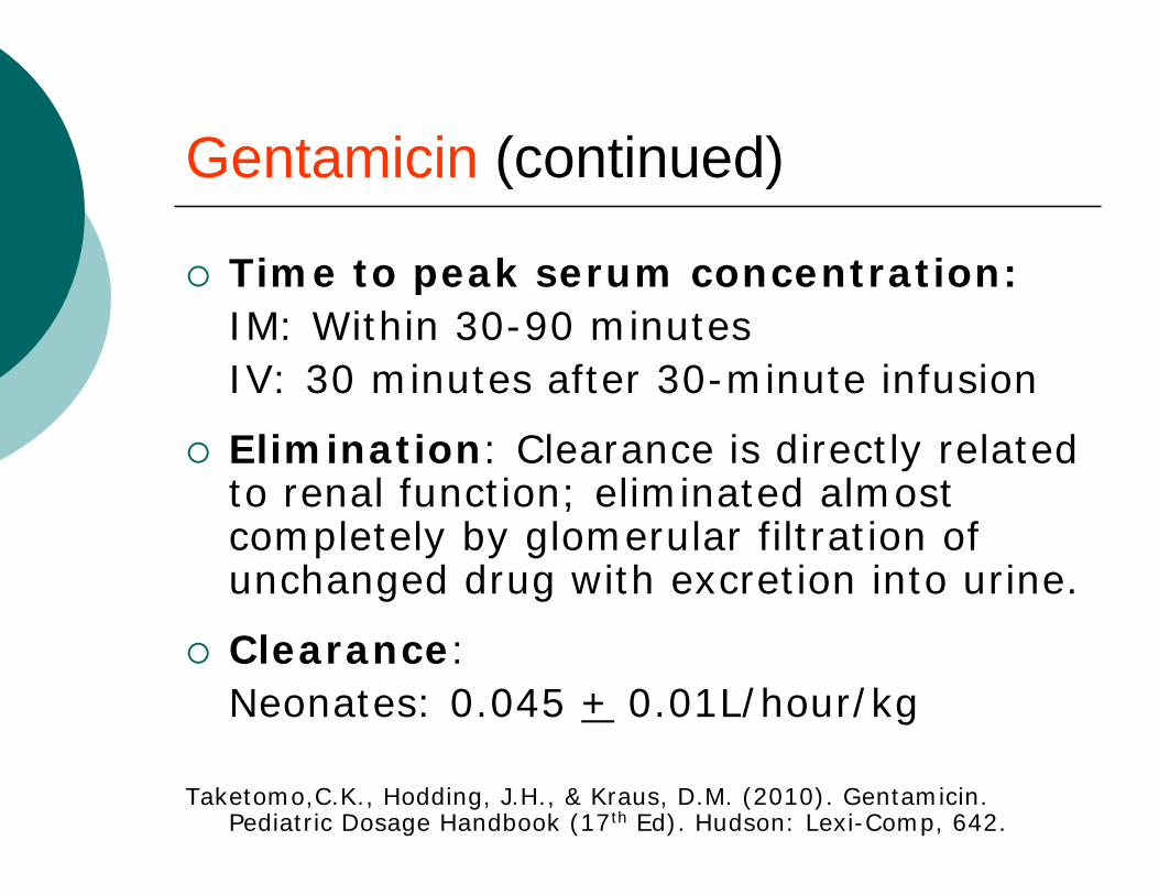
Time to peak serum concentration: IM: Within 30-90 minutesIV: 30 minutes after 30-minute infusion
Elimination: Clearance is directly related to renal function; eliminated almost completely by glomerular filtration of unchanged drug with excretion into urine.
Clearance:Neonates: 0.045 + 0.01L/hour/kg
Taketomo,C.K., Hodding, J.H., & Kraus, D.M. (2010). Gentamicin. Pediatric Dosage Handbook (17th Ed). Hudson: Lexi-Comp, 642.

Gentamicin (continued) If treating for more than 48 hours,
measure serum concentrations.
The peak concentration should be obtained 30 minutes after the end of the infusion.
The trough concentration should be obtained just prior to the next dose.
Therapeutic serum concentrations:Peak: 5 to 12 mcg/mlTrough: 0.5 to 1 mcg/ml
Young, R.E., & Mangum, B. (2010). Gentamicin. Neofax 2010 (23rd Ed). Montvale: Thomson Reuters, 50.

Gentamicin (continued) Mechanism of Action: Gentamicin binds to
30S and 50S ribosomal subunits, which inhibits cellular initiation of bacterial protein synthesis and results in a defective bacterial cell membrane.
Pharmacokinetics:Distribution: increased in neonates with fever, edema, ascites, fluid overload. Decreased in patients with dehydration:Vd Neonates: 0.45 + 0.1 L/kgProtein binding: < 30%Half-life:Neonates: < 1 week: 3-11.5 hours
1 week to 1 month: 3-6 hours
Taketomo,C.K., Hodding, J.H., & Kraus, D.M. (2010). Gentamicin. Pediatric Dosage Handbook (17th Ed). Hudson: Lexi-Comp, 643.
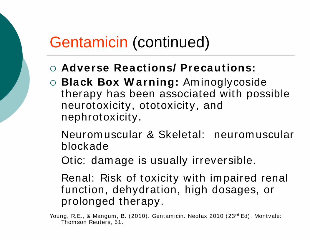
Gentamicin (continued) Adverse Reactions/Precautions: Black Box Warning: Aminoglycoside
therapy has been associated with possible neurotoxicity, ototoxicity, and nephrotoxicity.
Neuromuscular & Skeletal: neuromuscular blockadeOtic: damage is usually irreversible.
Renal: Risk of toxicity with impaired renal function, dehydration, high dosages, or prolonged therapy.
Young, R.E., & Mangum, B. (2010). Gentamicin. Neofax 2010 (23rd Ed). Montvale: Thomson Reuters, 51.
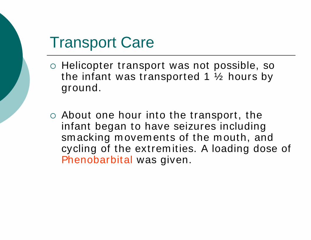
Transport Care Helicopter transport was not possible, so
the infant was transported 1 ½ hours by ground.
About one hour into the transport, the infant began to have seizures including smacking movements of the mouth, and cycling of the extremities. A loading dose of Phenobarbital was given.
Phenobarbital
Therapeutic Category: Anticonvulsant, Barbiturate; Hypnotic; Sedative
Uses: Management of generalized tonic-clonic and partial seizures; neonatal seizures. May improve outcome in severely asphyxiated infants. Neonatal abstinence syndrome in nonopiate- or polydrug-exposed infants. May lower bilirubin in chronic cholestasis.
Taketomo,C.K., Hodding, J.H., & Kraus, D.M. (2010).Phenobarbital. Pediatric Dosage Handbook (17th Ed). Hudson: Lexi-Comp, 1097.
Young, R.E., & Mangum, B. (2010).Phenobarbital. Neofax 2010 (23rd Ed). Montvale: Thomson Reuters, 232.
Phenobarbital (continued) Dose and Administration:
Anticonvulsant: Loading dose of 20 mg/kg given IV over 10 to 15 minutes. For refractory seizures an additional 5 mg/kg may be given up to a total of 40 mg/kg.Maintenance: 3 to 4 mg/kg per day beginning 12 to 24 hours after loading dose.Neonatal Abstinence Syndrome:Load of 16 mg/kg po on day 1.Maintenance of 1-4 mg/kg/dose PO q 12 hours.Based on abstinence scoring.
Young, R.E., & Mangum, B. (2010).Phenobarbital. Neofax 2010 (23rd Ed). Montvale: Thomson Reuters, 232.

Phenobarbital (continued) Adverse Reactions:
Cardiovascular: hypotension, circulatory collapse, bradycardiaCentral Nervous System: sedation at serum concentrations above 40 mcg/ml.Respiratory: depression at concentrations above 60 mcg/mlIrritating to veins.
Mechanism of Action: Limits the spread of seizure activity, possibly by increasing inhibitory neurotransmission. Depresses the CNS activity by binding to the barbiturate site at the GABA-receptor complex. Depresses the reticular activating system.
Taketomo,C.K., Hodding, J.H., & Kraus, D.M. (2010).Phenobarbital. Pediatric Dosage Handbook (17th Ed). Hudson: Lexi-Comp, 1099.
Young, R.E., & Mangum, B. (2010).Phenobarbital. Neofax 2010 (23rd Ed). Montvale: Thomson Reuters, 232.
Phenobarbital (continued) Pharmacodynamics:
Onset of action:Oral: within 20-60 minutesIV: within 5 minutesMaximum Effect: IV within 30 minutesDuration: Oral: 6-10 hoursIV: 4-10 hours
Taketomo,C.K., Hodding, J.H., & Kraus, D.M. (2010).Phenobarbital. Pediatric Dosage Handbook (17th Ed). Hudson: Lexi-Comp, 1099.
Phenobarbital (continued) Pharmacokinetics:
Distribution Vd: 0.8-1 L/kgProtein binding: 35-50%Metabolism: in the liver by hydroxylation and glucuronide conjugationHalf-life: 45-500 hoursTime to peak concentration: oral 1-6 hoursElimination: 20-50% excreted unchanged in urine.
Taketomo,C.K., Hodding, J.H., & Kraus, D.M. (2010).Phenobarbital. Pediatric Dosage Handbook (17th Ed). Hudson: Lexi-Comp, 1099.
NICU Admission Upon admission to the NICU, the
infant was noted to be lethargic, with decreased muscle tone. He had increased tendon reflexes and overactive dolls-eye reflex, a weak suck and incomplete moro. Frequent seizure activity continued so a standard EEG was obtained documenting the seizure activity and a second anticonvulsant, phenytoin was added.

Phenytoin Therapeutic Category: antiarrhythmic,
anticonvulsant Uses: anticonvulsant used to treat seizures
that are refractory to phenobarbital. Dosage and administration:
Loading dose: 15 to 20 mg/kg IV infusion over 30 minutes.Maintenance dose:4 to 8 mg/kg q 24 hours IV slow push or PO.
Young, R.E., & Mangum, B. (2010).Phenytoin. Neofax 2010 (23rd Ed). Montvale: Thomson Reuters, 234.
Phenytoin (continued) Dosage and administration con’t:
(up to 8 mg/kg per dose q 8-12 hours after 1 week of age)Maximum infusion rate 0.5 mg/kg/min.Flush IV with saline before and after.
Warning: Phenytoin is highly unstable in any IV solution. Avoid use in central lines due to precipitation. IM not acceptable as drug crystallizes in the muscle.
Young, R.E., & Mangum, B. (2010).Phenytoin. Neofax 2010 (23rd
Ed). Montvale: Thomson Reuters, 234.

Phenytoin (continued) Adverse Reactions:
Extravasation causes tissue inflammation and necrosis.
High serum concentrations are associated with seizures.
Hypersensitivity has been reported in infants.
Toxicity includes: cardiac arrhythmias, hypotension, gingivitis, nystagmus, rickets, hyperglycemia and hypoinsulinemia.
Young, R.E., & Mangum, B. (2010).Phenytoin. Neofax 2010 (23rd Ed). Montvale: Thomson Reuters, 234

Phenytoin (continued) Mechanism of Action: Stabilizes
neuronal membranes and decreases seizure activity by increasing efflux or decreasing influx of sodium ions across cell membranes in the motor cortex during generation of nerve impulses
Pharmacokinetics:Distribution: Vd
Preterm: 1-1.2 L/kgFull term: 0.8-0.9 L/kg
Taketomo,C.K., Hodding, J.H., & Kraus, D.M. (2010).Phenytoin. Pediatric Dosage Handbook (17th Ed). Hudson: Lexi-Comp, 1106.
Phenytoin (continued)
Pharmacokinetics:Protein binding: Adults 90-95%, decreased protein binding in neonates (up to 20% free)
Metabolism: is dose-dependent, half-life is dependent on serum concentration.
Elimination: < 5% excreted unchanged in urine
Taketomo,C.K., Hodding, J.H., & Kraus, D.M. (2010).Phenytoin. Pediatric Dosage Handbook (17th Ed). Hudson: Lexi-Comp, 1106.
NICU Admission Lorazepam or fosphenytoin would be
other anticonvulsant options. The infant met criteria for Stage 2-Moderate Encephalopathy (Hypoxic Ischemic Encephalopathy) and met the hospital criteria for therapeutic body cooling. The infant was placed on a cooling blanket with the radiant warmer turned off. An esophageal probe was inserted to monitor the infant’s temperature. The infant was on a ventilator, a cardiorespiratory monitor, pulse oximetry, and an aEEG machine.
aEEG During the time the infant is on whole body
cooling, a continuous EEG or aEEG may be in place.
An aEEG or amplitude-integrated EEG, or Cerebral Function Monitor (CFM), is a device that measures background electrocortical activity in the brain.
Uses a single lead, consisting of three wires placed over the biparietal or frontal region of the head
Indicates the generalized level of electrical activity that is occurring across the brain
McNamara, P., Keyzers, M. (2006). A protocol for Cerebral Function Monitoring in the NICU. Hospital for Sick Children, Toronto, Canada, p 1.
aEEG (continued) Research has shown the aEEG to be
a sensitive tool for predicting severity of HIE if applied in the first 6-12 hours following perinatal asphyxia
Also valuable detection tool for neonates experiencing clinical or subclinical seizures
McNamara, P., Keyzers, M. (2006). A protocol for Cerebral Function Monitoring in the NICU. Hospital for Sick Children, Toronto, Canada, p 1.

Indications for an aEEG Hypoxic ischemic encephalopathy Seizures Significant neurological disorders Post cardiac arrest Inborn error of metabolism Neonatal abstinence syndrome
Pitfalls:The aEEG requires lead stabilization to
decrease or eliminate artifact caused by HFOV.
McNamara, P., Keyzers, M. (2006). A protocol for Cerebral Function Monitoring in the NICU. Hospital for Sick Children, Toronto, Canada, p 1.
Lorazepam Therapeutic Category: Antianxiety agent,
anticonvulsant, benzodiazepine.
Uses: Acute management of seizures refractory to conventional therapy.
Dosage and Administration:0.05 – 0.1 mg/kg per dose IV slow push. Repeat dose based on clinical response.
Adverse Effects:Respiratory Depression. Some preterm infants have had rhythmic myoclonic jerking when receiving lorazepam for sedation.
Taketomo,C.K., Hodding, J.H., & Kraus, D.M. (2010).Lorazepam. Pediatric Dosage Handbook (17th Ed). Hudson: Lexi-Comp, 845.
Young, R.E., & Mangum, B. (2010).Lorazepam. Neofax 2010 (23rd Ed). Montvale: Thomson Reuters, 216.

Lorazepam (continued) Mechanism of Action: Depresses all levels
of the CNS including the limbic and reticular formation by binding to the benzodiazepine site on the GABA receptor complex.
Pharmacodynamics:Onset of action:Oral: within 60 minutesIM: 30-60 minutesIV: 15-30 minutesDuration: 8-12 hours
Taketomo,C.K., Hodding, J.H., & Kraus, D.M. (2010).Lorazepam. Pediatric Dosage Handbook (17th Ed). Hudson: Lexi-Comp, 846.
Lorazepam (continued) Pharmacokinetics:
Distribution: Vd = 0.76 L/kgProtein binding: 85%Metabolism: primarily by glucuronide conjugation in the liverHalf-life: Full term neonates: 40.2 hours; range 18-73 hoursElimination: in urine primarily as glucuronide conjugate
Taketomo,C.K., Hodding, J.H., & Kraus, D.M. (2010).Lorazepam. Pediatric Dosage Handbook (17th Ed). Hudson: Lexi-Comp, 846.
Fosphenytoin Therapeutic Category: Anticonvulsant,
Hydantoin
Uses: treatment of seizures refractory to phenobarbital. Can administer with lorazepam for rapid onset seizure control.
Dosage and Administration:Note: Fosphenytoin is expressed as phenytoin equivalents (PE). (Fosphenytoin 1 mg PE = phenytoin 1 mg)
Young, R.E., & Mangum, B. (2010).Fosphenytoin. Neofax 2010 (23rd
Ed). Montvale: Thomson Reuters, 210.
Fosphenytoin (continued)
Dosage and Administration con’t:Loading dose: 15 to 20 mg PE/kg IM or IV infusion over at least 10 minutes.Maintenance dose: 4 to 8 mg PE/kg q 24 hours IM or IV slow push. Begin 24 hours after load.Maximum rate of infusion 1.5 mg PE/kg per minute. Flush with saline before and after administration.
Young, R.E., & Mangum, B. (2010).Fosphenytoin. Neofax 2010 (23rd
Ed). Montvale: Thomson Reuters, 210.
Fosphenytoin (continued) Adverse Reactions: Signs of toxicity
include drowsiness that is dose and rate dependent. Minor irritation at IV site.
Mechanism of Action: Fosphenytoin is water-soluble and is a prodrug of phenytoin. It is rapidly converted by phosphatases in the blood and tissue. Has no known pharmacologic activity before conversion to phenytoin.
Taketomo,C.K., Hodding, J.H., & Kraus, D.M. (2010).Fosphenytoin.Pediatric Dosage Handbook (17th Ed). Hudson: Lexi-Comp, 630.
Young, R.E., & Mangum, B. (2010).Fosphenytoin. Neofax 2010 (23rd
Ed). Montvale: Thomson Reuters, 210.

Fosphenytoin (continued) Mechanism of Action continued:
Phenytoin works by stabilizing neuronal membranes and decreasing seizure activity by increasing efflux or decreasing influx of sodium ions across cell membranes in the motor cortex.
Pharmacokinetics:Pharmacokinetics for fosphenytoin-derived phenytoin are the same as those for phenytoin.
Taketomo,C.K., Hodding, J.H., & Kraus, D.M. (2010).Fosphenytoin.Pediatric Dosage Handbook (17th Ed). Hudson: Lexi-Comp, 631.
Maintenance Care
Pain medication and sedation were provided for this infant during the cooling process. A fentanyl or morphine drip would be appropriate for the treatment of pain during this cooling process and midazolam for sedation.
The infant is also paralyzed with pancuronium in an effort to decrease shivering that might increase the body temperature during the cooling process.
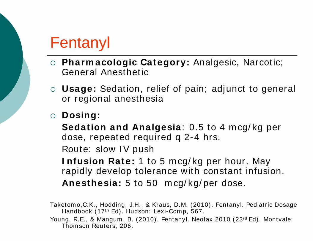
Fentanyl Pharmacologic Category: Analgesic, Narcotic;
General Anesthetic
Usage: Sedation, relief of pain; adjunct to general or regional anesthesia
Dosing:Sedation and Analgesia: 0.5 to 4 mcg/kg per dose, repeated required q 2-4 hrs.Route: slow IV pushInfusion Rate: 1 to 5 mcg/kg per hour. May rapidly develop tolerance with constant infusion.Anesthesia: 5 to 50 mcg/kg/per dose.
Taketomo,C.K., Hodding, J.H., & Kraus, D.M. (2010). Fentanyl. Pediatric Dosage Handbook (17th Ed). Hudson: Lexi-Comp, 567.
Young, R.E., & Mangum, B. (2010). Fentanyl. Neofax 2010 (23rd Ed). Montvale: Thomson Reuters, 206.
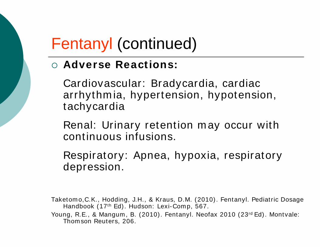
Fentanyl (continued) Adverse Reactions:
Cardiovascular: Bradycardia, cardiac arrhythmia, hypertension, hypotension, tachycardia
Renal: Urinary retention may occur with continuous infusions.
Respiratory: Apnea, hypoxia, respiratory depression.
Taketomo,C.K., Hodding, J.H., & Kraus, D.M. (2010). Fentanyl. Pediatric Dosage Handbook (17th Ed). Hudson: Lexi-Comp, 567.
Young, R.E., & Mangum, B. (2010). Fentanyl. Neofax 2010 (23rd Ed). Montvale: Thomson Reuters, 206.
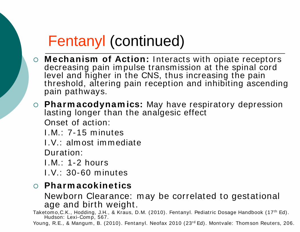
Fentanyl (continued) Mechanism of Action: Interacts with opiate receptors
decreasing pain impulse transmission at the spinal cord level and higher in the CNS, thus increasing the pain threshold, altering pain reception and inhibiting ascending pain pathways.
Pharmacodynamics: May have respiratory depression lasting longer than the analgesic effectOnset of action: I.M.: 7-15 minutesI.V.: almost immediateDuration:I.M.: 1-2 hoursI.V.: 30-60 minutes
PharmacokineticsNewborn Clearance: may be correlated to gestational age and birth weight.
Taketomo,C.K., Hodding, J.H., & Kraus, D.M. (2010). Fentanyl. Pediatric Dosage Handbook (17th Ed). Hudson: Lexi-Comp, 567.
Young, R.E., & Mangum, B. (2010). Fentanyl. Neofax 2010 (23rd Ed). Montvale: Thomson Reuters, 206.
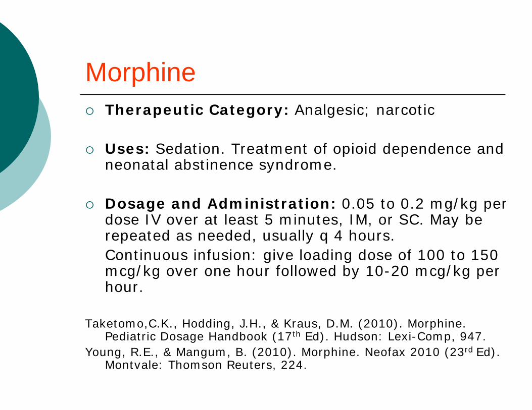
Morphine Therapeutic Category: Analgesic; narcotic
Uses: Sedation. Treatment of opioid dependence and neonatal abstinence syndrome.
Dosage and Administration: 0.05 to 0.2 mg/kg per dose IV over at least 5 minutes, IM, or SC. May be repeated as needed, usually q 4 hours.Continuous infusion: give loading dose of 100 to 150 mcg/kg over one hour followed by 10-20 mcg/kg per hour.
Taketomo,C.K., Hodding, J.H., & Kraus, D.M. (2010). Morphine. Pediatric Dosage Handbook (17th Ed). Hudson: Lexi-Comp, 947.
Young, R.E., & Mangum, B. (2010). Morphine. Neofax 2010 (23rd Ed). Montvale: Thomson Reuters, 224.
Morphine (continued) Dosage and administration con’t:
Treatment of opioid dependence: begin at most recent IV morphine dose equivalent. Taper 10 to 20% per day as tolerated. Oral dose is 3-5 times IV dose.Initial treatment of neonatal abstinence syndrome: 0.03-0.1 mg/kg per dose po q 3-4 hours, wean by 10-20% every 2 to 3 days based on scoring. Use a 0.4 mg/ml dilution.
Adverse Reactions: Marked respiratory depression, hypotension, bradycardia, and transient hypotonia. An ileus and delayed gastric emptying and urine retention may occur. Tolerance may develop after prolonged use, wean slowly.
Young, R.E., & Mangum, B. (2010). Morphine. Neofax 2010 (23rd
Ed). Montvale: Thomson Reuters, 224.

Morphine (continued) Mechanism of action: binds to opiate receptors in
the CNS, causing inhibition of ascending pain pathways, altering patient perception of and response to pain. Also causes generalized CNS depression.
Pharmacodynamics:Oral solution: Peak: 1 hr. Duration: 2-5 hours.IM injection: Peak: 30-60 min. Duration: 3-5 hours.IV injection: Peak: 20 min. Duration: 3-5 hours.
Taketomo,C.K., Hodding, J.H., & Kraus, D.M. (2010). Morphine. Pediatric Dosage Handbook (17th Ed). Hudson: Lexi-Comp, 949.
Morphine (continued) Pharmacokinetics:
Distribution: to skeletal muscle, liver, kidneys, lungs, intestinal tract, spleen, brain, and into breast milk, crosses placenta.Protein binding: premature infants < 20%.
Metabolism: in the liver via glucuronide conjugation Half-life: Premature Infants: 10-20 hoursNeonates: 7.6 hours, range 4.5 -13.3 hrs.Clearance:Premature: 0.5-3 ml/minute/kgNeonates 1-7 days: median 5.5 mL/min/kg, range
3.2-8.4 ml/min/kgNeonates 8-30 days: median 7.4 ml/min/kg, range
3.4-13.8 ml/min/kgTaketomo,C.K., Hodding, J.H., & Kraus, D.M. (2010). Morphine. Pediatric Dosage
Handbook (17th Ed). Hudson: Lexi-Comp, 949.
MidazolamTherapeutic Category: Sedative/Hypnotic, anticonvulsant
Usage: Conscious sedation, treatment of refractory seizures, anxiolysis, and amnesia prior to a procedure or before anesthesia
Administration:Sedation:IV: 0.5 to 0.15 mg/kg over at least 5 minutes. Repeat as
needed q2-4hrs.Continuous IV infusion: 0.01 to 0.06 mg/kg per hour (10 to
60 mcg/kg/hour). May need to be increased with development of tolerance and/or increased clearance.
Intranasal: 0.2 to 0.3 mg/kg per dose using 5-mg/ml injectable form.
Sublingual: 0.2 mg/kg per dose using 5-mg/ml injectable form mixed with a small amount of flavored syrup.
Oral: 0.25 mg/kg per dose using Versed® oral syrup.
Taketomo,C.K., Hodding, J.H., & Kraus, D.M. (2010). Midazolam. Pediatric Dosage Handbook (17th Ed). Hudson: Lexi-Comp, 928.
Young, R.E., & Mangum, B. (2010). Midazolam. Neofax 2010 (23rd Ed). Montvale: Thomson Reuters, 220.

Midazolam (continued) Dosage and Administration Continued:
Anticonvulsant: Loading dose: 0.15 mg/kg (150 mcg/kg) IV over at least 5 minutes, followed byMaintenance Infusion: 0.06 to 0.4 mg/kg per hour (1 to 7 mcg/kg per minute).
Adverse Reactions/ Precautions:Black Box Warning: Midazolam has been associated with respiratory depression and arrest when used for sedation in a non-critical care setting. Rapid administration of midazolam has been associated with severe hypotension and seizures in neonates. Nasal administration may cause a burning sensation.
Young, R.E., & Mangum, B. (2010). Midazolam. Neofax 2010 (23rd Ed). Montvale: Thomson Reuters, 220.
Midazolam (continued) Mechanism of Action: “ Depresses all levels of
CNS, including the limbic and reticular formation, by binding to the benzodiazepine site on the gamma-aminobutyric acid (GABA) receptor complex and modulating GABA, which is a major inhibitory neurotransmitter in the brain.”
Pharmacodynamics: Onset of ActionChildren:I.M.: within 5 minutesI.V.: within 5 minutesIntranasal: within 5 minutesDuration:I.M.: Mean: 2 hours, up to 6 hoursI.V.: 20-30 minutesIntranasal: 30-60 minutesNote: Full recovery may take more than 24 hours
Taketomo,C.K., Hodding, J.H., & Kraus, D.M. (2010). Midazolam. Pediatric Dosage Handbook (17th Ed). Hudson: Lexi-Comp, 928.
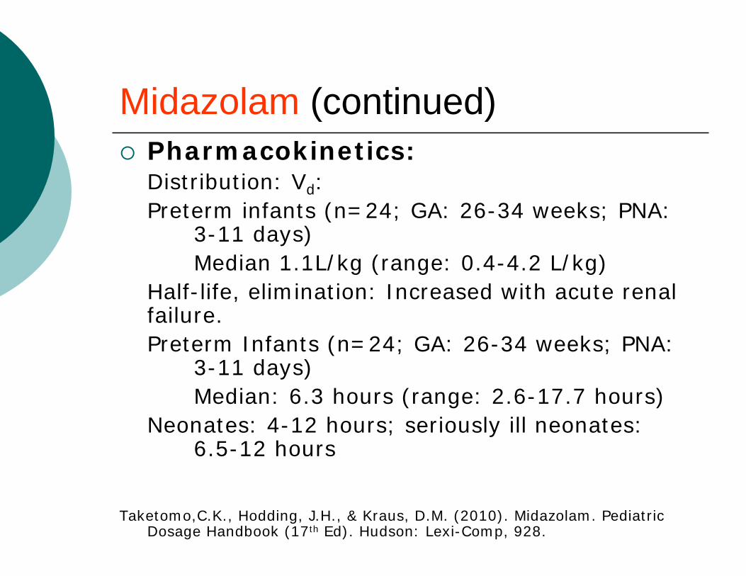
Midazolam (continued) Pharmacokinetics:
Distribution: Vd:Preterm infants (n=24; GA: 26-34 weeks; PNA:
3-11 days) Median 1.1L/kg (range: 0.4-4.2 L/kg)
Half-life, elimination: Increased with acute renal failure.Preterm Infants (n=24; GA: 26-34 weeks; PNA:
3-11 days)Median: 6.3 hours (range: 2.6-17.7 hours)
Neonates: 4-12 hours; seriously ill neonates: 6.5-12 hours
Taketomo,C.K., Hodding, J.H., & Kraus, D.M. (2010). Midazolam. Pediatric Dosage Handbook (17th Ed). Hudson: Lexi-Comp, 928.
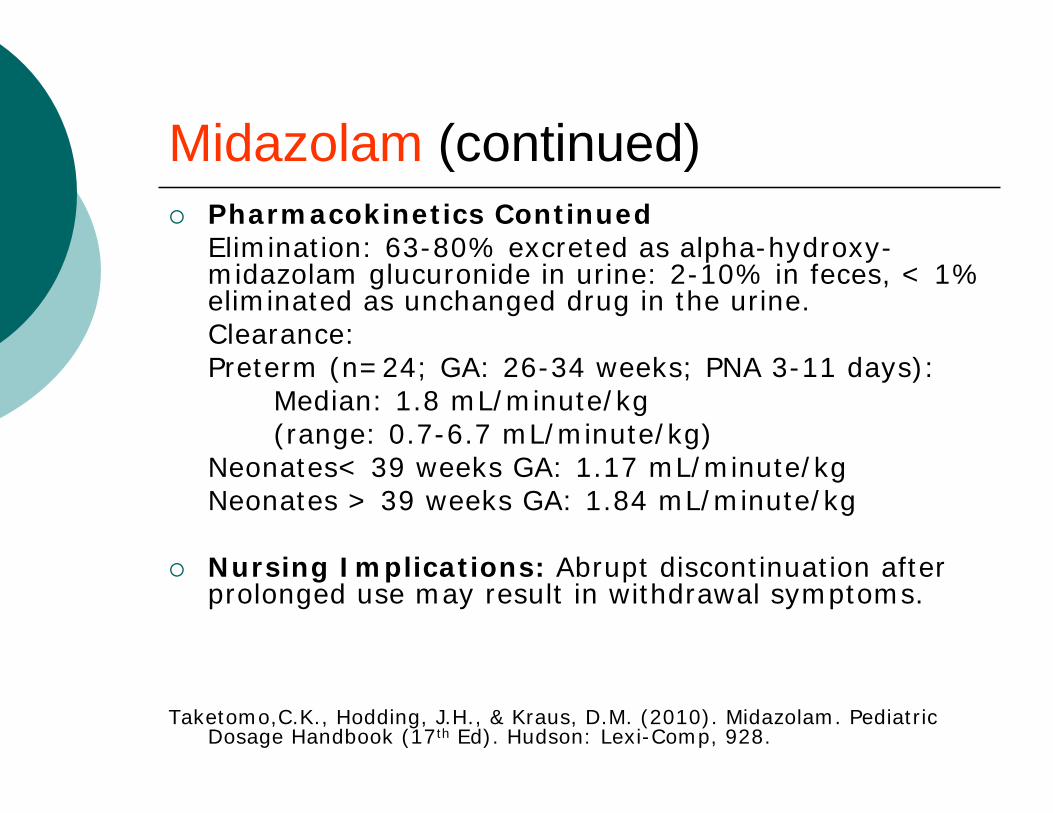
Midazolam (continued) Pharmacokinetics Continued
Elimination: 63-80% excreted as alpha-hydroxy-midazolam glucuronide in urine: 2-10% in feces, < 1% eliminated as unchanged drug in the urine.Clearance:Preterm (n=24; GA: 26-34 weeks; PNA 3-11 days):
Median: 1.8 mL/minute/kg(range: 0.7-6.7 mL/minute/kg)
Neonates< 39 weeks GA: 1.17 mL/minute/kgNeonates > 39 weeks GA: 1.84 mL/minute/kg
Nursing Implications: Abrupt discontinuation after prolonged use may result in withdrawal symptoms.
Taketomo,C.K., Hodding, J.H., & Kraus, D.M. (2010). Midazolam. Pediatric Dosage Handbook (17th Ed). Hudson: Lexi-Comp, 928.
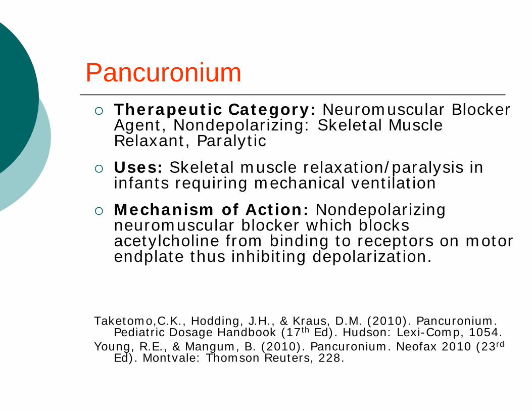
Pancuronium Therapeutic Category: Neuromuscular Blocker
Agent, Nondepolarizing: Skeletal Muscle Relaxant, Paralytic
Uses: Skeletal muscle relaxation/paralysis in infants requiring mechanical ventilation
Mechanism of Action: Nondepolarizing neuromuscular blocker which blocks acetylcholine from binding to receptors on motor endplate thus inhibiting depolarization.
Taketomo,C.K., Hodding, J.H., & Kraus, D.M. (2010). Pancuronium.Pediatric Dosage Handbook (17th Ed). Hudson: Lexi-Comp, 1054.
Young, R.E., & Mangum, B. (2010). Pancuronium. Neofax 2010 (23rd
Ed). Montvale: Thomson Reuters, 228.
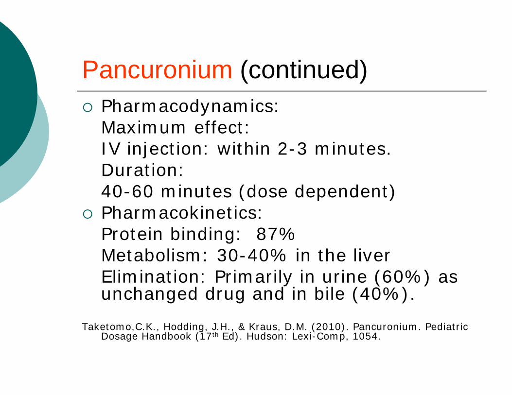
Pancuronium (continued) Pharmacodynamics:
Maximum effect: IV injection: within 2-3 minutes.Duration:40-60 minutes (dose dependent)
Pharmacokinetics: Protein binding: 87%Metabolism: 30-40% in the liverElimination: Primarily in urine (60%) as unchanged drug and in bile (40%).
Taketomo,C.K., Hodding, J.H., & Kraus, D.M. (2010). Pancuronium. Pediatric Dosage Handbook (17th Ed). Hudson: Lexi-Comp, 1054.
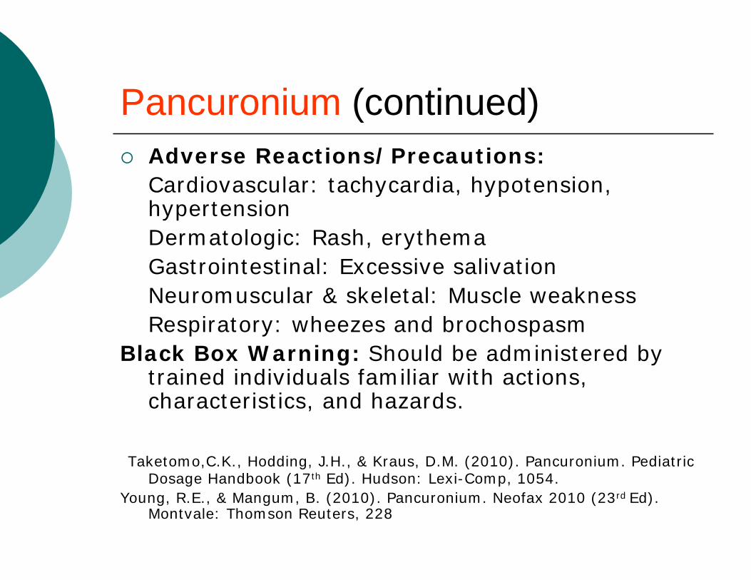
Pancuronium (continued) Adverse Reactions/Precautions:
Cardiovascular: tachycardia, hypotension, hypertensionDermatologic: Rash, erythemaGastrointestinal: Excessive salivationNeuromuscular & skeletal: Muscle weaknessRespiratory: wheezes and brochospasm
Black Box Warning: Should be administered by trained individuals familiar with actions, characteristics, and hazards.
Taketomo,C.K., Hodding, J.H., & Kraus, D.M. (2010). Pancuronium. Pediatric Dosage Handbook (17th Ed). Hudson: Lexi-Comp, 1054.
Young, R.E., & Mangum, B. (2010). Pancuronium. Neofax 2010 (23rd Ed). Montvale: Thomson Reuters, 228

What is HIE?
Hypoxic-Ischemic Encephalopathy Acute brain injury due to prenatal,
intrapartum, or postnatal hypoxic events Hypoxia and ischemia leading to
neurologic dysfunction It is also known as perinatal asphyxia or
neonatal encephalopathy Occurs most often in term or near-term
infants.

Incidence of HIE
1-2/1000 live term births (developed countries)
10-20% mortality (postnatal) 25% childhood disabilities
Cerebral palsyEpilepsyMental retardationlearning disabilities
Zanelli, S.A., Naylor, M., Dobbins, N., Quigg, M., Goodkin, H., Matsumoto, J.A, and Fairchild, K. Implementation of a “Hypothermia for HIE” program: 2-year experience in a single NICU. (2008). Journal of Perinatalogy, 28, 171-175.
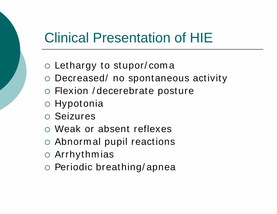
Clinical Presentation of HIE
Lethargy to stupor/coma Decreased/ no spontaneous activity Flexion /decerebrate posture Hypotonia Seizures Weak or absent reflexes Abnormal pupil reactions Arrhythmias Periodic breathing/apnea
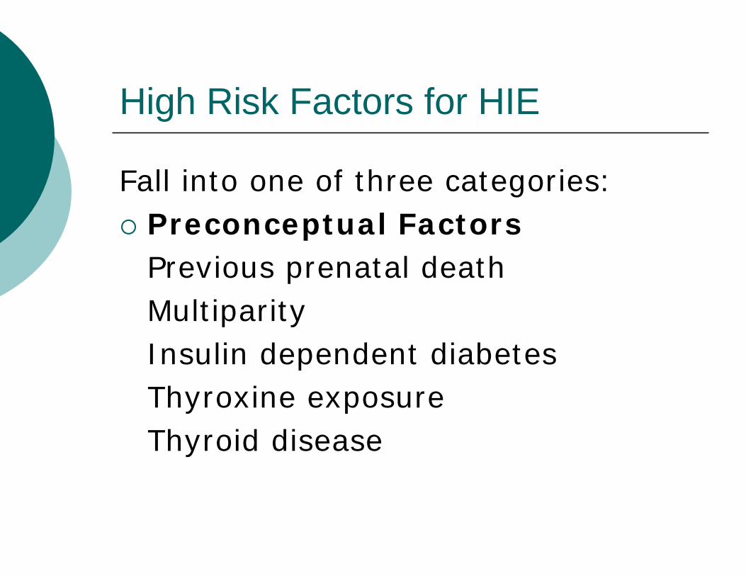
High Risk Factors for HIE
Fall into one of three categories: Preconceptual Factors
Previous prenatal deathMultiparityInsulin dependent diabetesThyroxine exposureThyroid disease
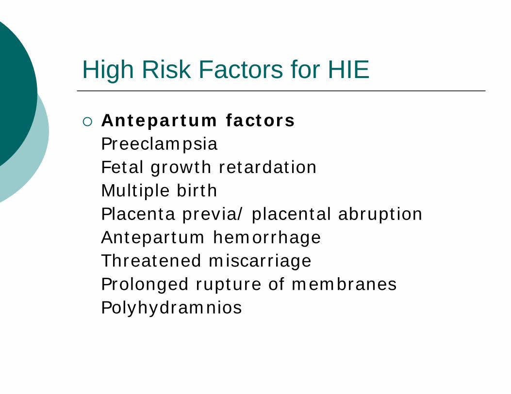
High Risk Factors for HIE
Antepartum factorsPreeclampsiaFetal growth retardationMultiple birthPlacenta previa/ placental abruptionAntepartum hemorrhageThreatened miscarriageProlonged rupture of membranesPolyhydramnios
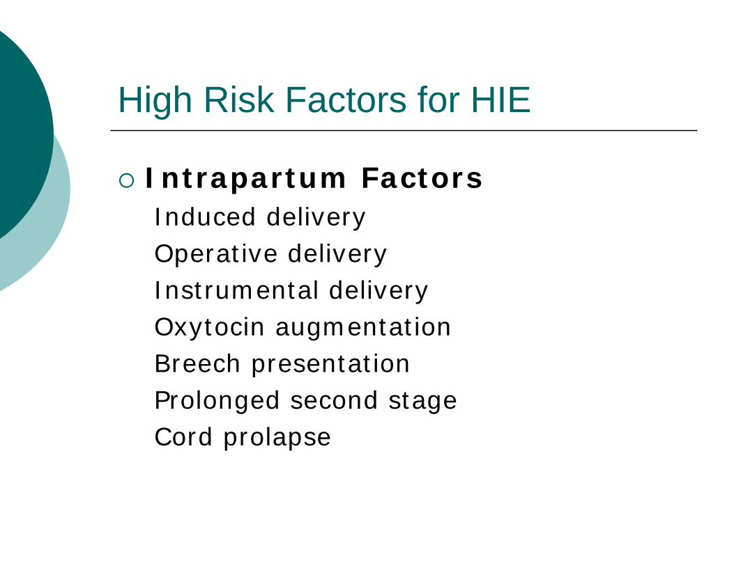
High Risk Factors for HIE
Intrapartum FactorsInduced deliveryOperative deliveryInstrumental deliveryOxytocin augmentationBreech presentationProlonged second stageCord prolapse
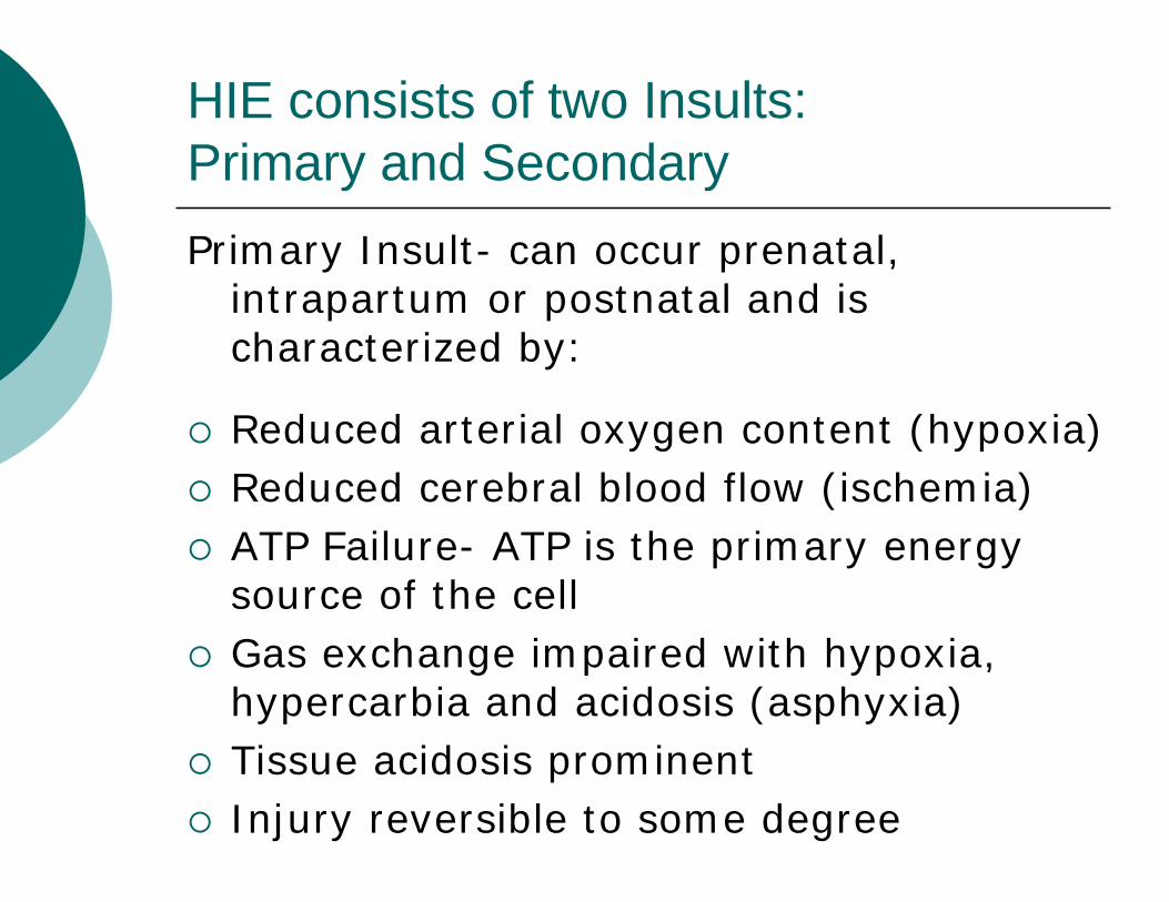
HIE consists of two Insults:Primary and SecondaryPrimary Insult- can occur prenatal,
intrapartum or postnatal and is characterized by:
Reduced arterial oxygen content (hypoxia) Reduced cerebral blood flow (ischemia) ATP Failure- ATP is the primary energy
source of the cell Gas exchange impaired with hypoxia,
hypercarbia and acidosis (asphyxia) Tissue acidosis prominent Injury reversible to some degree

HIE consists of two Insults:Primary and SecondarySecondary Insult
Occurs 6-12 hours after birth Tissue injury occurs when blood flow
returns and molecular oxygen is reintroduced to the tissues
During post-ischemic reperfusion cytotoxic oxygen-derived free radicals are generated
The highest post-asphyxial cerebral blood flow is correlated with the severity of brain damage.

Staging of NeonatalEncephalopathy per Sarnat &Sarnat
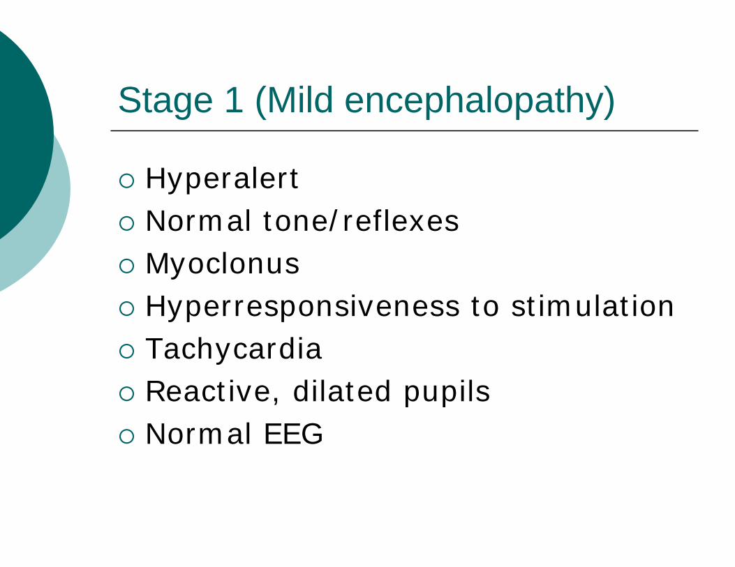
Stage 1 (Mild encephalopathy)
Hyperalert Normal tone/reflexes Myoclonus Hyperresponsiveness to stimulation Tachycardia Reactive, dilated pupils Normal EEG

Stage 2 (Moderate Encephalopathy) Lethargy Hypotonia Increased tendon reflexes Myoclonus Frequent seizure activity Weak suck Incomplete Moro reflex Overactive dolls-eye reflex Pupils constrictive, reactive Periodic respirations may be present
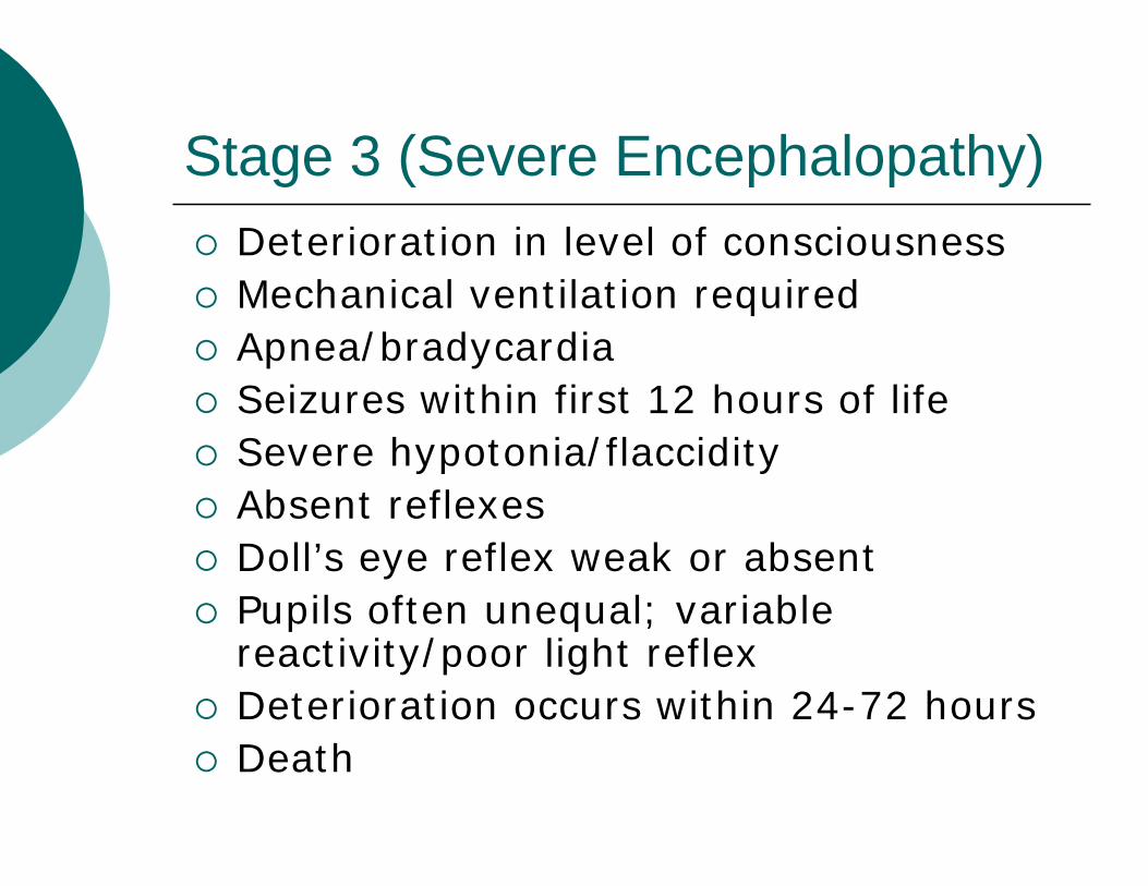
Stage 3 (Severe Encephalopathy) Deterioration in level of consciousness Mechanical ventilation required Apnea/bradycardia Seizures within first 12 hours of life Severe hypotonia/flaccidity Absent reflexes Doll’s eye reflex weak or absent Pupils often unequal; variable
reactivity/poor light reflex Deterioration occurs within 24-72 hours Death

Diagnosis of HIE Look for events which could compromise
blood or oxygen supply to the fetus No clear diagnostic test for
encephalopathy due to hypoxia-ischemia Maternal temperature elevations
increased risk Consider placental pathology Detailed neonatal neurological exam to
determine degree of encephalopathy Most infants with encephalopathy do not
have an obvious cause
Diagnostic Tools
Cranial ultrasound
CT
MRI
EEG
Therapeutic Window for Treatment Interval between the primary and
secondary energy failure Initiation of therapies in animals
during this latent phase were successful in reducing brain damage
Duration of the therapeutic window~ 6 hours in near term fetal sheep treated with brain cooling after brain ischemia.
Management of HIE Prompt resuscitation Maintain physiologic oxygenation & acid-
base balance Homeostasis of fluid and electrolyte
abnormalities Monitor blood volume Maintain perfusion Control seizures Thorough neurological exams Monitor and manage disturbances with
other body systems Consider cerebral hypothermia

Management Specifics
VentilationMaintain the CO2 within normal range (35-45)High CO2 increases cerebral blood flowLow CO2 decreases cerebral blood flow
PerfusionPromptly treat hypotensionAvoid hypertension
Fluid StatusEstablish immediate access (UVC or PIV) and start IVFWatch urine closely for ATN or SIADH in the first 48 hours – dec. total fluids to 60 ml/kg/day if present
More Management Specifics
Blood Glucose-Establish immediate access (UVC or PIV)-Maintain in normal range-Avoid hypoglycemia
Seizure-Treat clinical seizures
Monitor Electrolytes Coagulation (disseminated intravascular
coagulation [DIC] may occur)-Monitor platelets, PT/PTT
Why Hypothermia? Cooling brain temperature 3-5 degrees
reduces brain injury in animals and adults Prevents edema Temperature reduction decreases the
metabolic rate of the brain by 5.3% and reduces the amount of oxygen required by the brain by 6-7%.
May reduce the severity of the reperfusion injury to the brain, protect neurons by lowering cerebral metabolism, and delaying the reperfusion injury allowing time for other interventions.
Inhibits platelet-activating factor, inflammatory cascade.
Reduces the Extent of Brain Injury
Reduction of infarct size Decrease in neuronal cell loss Retention of sensory motor function Preservation of hippocampal
structures Recovery of
electroencephalographic activity
Who Benefits?
Outcomes of infants who were cooled showed decreased rates of cerebral palsy and mental impairments.
Infants with moderate HIE improved the most. The effects of cooling were not improved for infants with either mild or severe HIE.
Exclusion Criteria
Strong Clinical indicators of:
Severe Sepsis Meningitis Pneumonia Active Bleeding

Different Cooling Techniques
Selective head Cooling: thought to decrease the systemic effects of hypothermia but may not be as effective in cooling the deep regions of the brain. Head cooling penetrates 1 cm.
http://medgadget.com/archives/2006

Different Cooling Techniques
Whole Body Cooling: Cooling all areas of the brain could only be achieved by lowering the core temperature of the baby.
While both head and body cooling are associated with reduction in brain injury following HIE, whole body cooling results in more consistency in temperature regulation.
Temperature regulation is beneficial because small fluctuations in temp may result in varied neurological protection.

Different Cooling Techniques
childrenscentralcal.org Whole Body Cooling
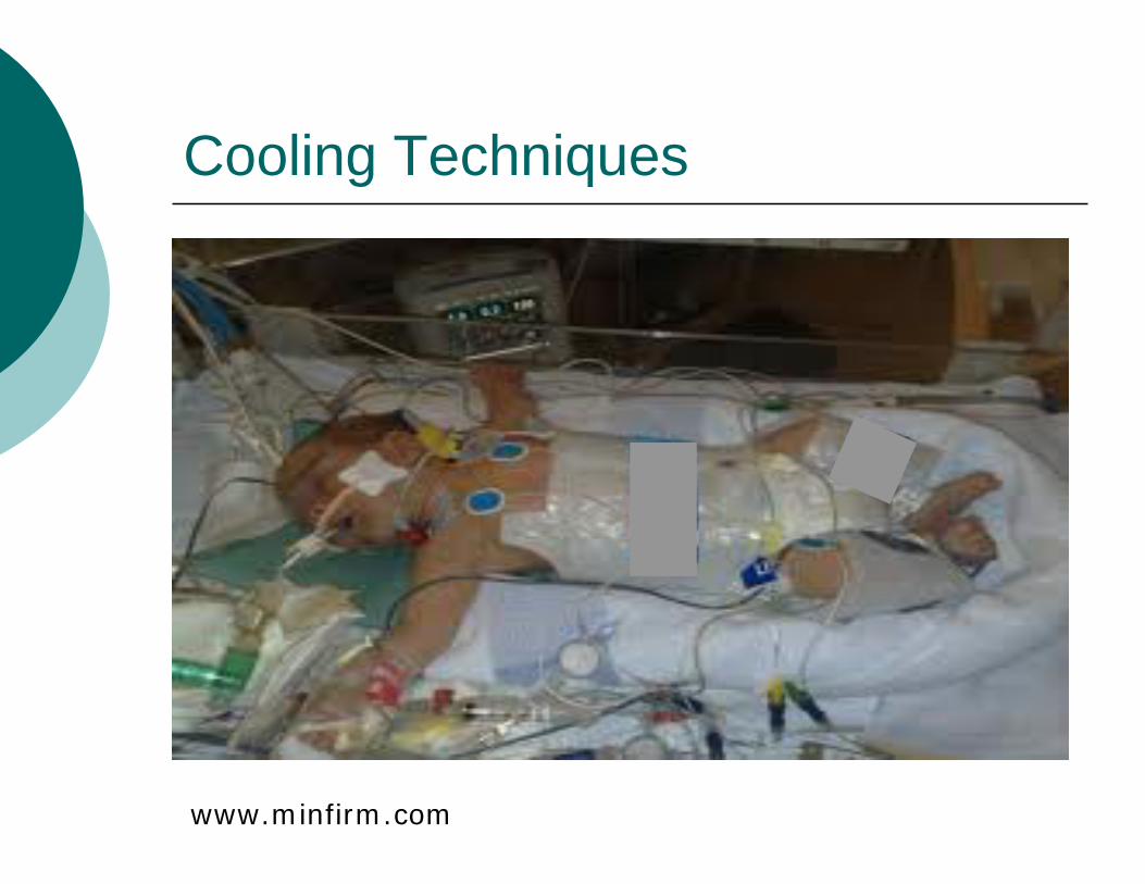
Cooling Techniques
www.minfirm.com
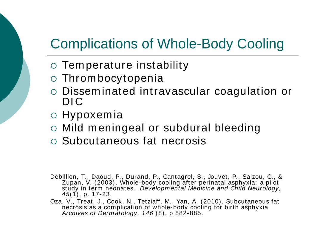
Complications of Whole-Body Cooling Temperature instability Thrombocytopenia Disseminated intravascular coagulation or
DIC Hypoxemia Mild meningeal or subdural bleeding Subcutaneous fat necrosis
Debillion, T., Daoud, P., Durand, P., Cantagrel, S., Jouvet, P., Saizou, C., & Zupan, V. (2003). Whole-body cooling after perinatal asphyxia: a pilot study in term neonates. Developmental Medicine and Child Neurology, 45(1), p. 17-23.
Oza, V., Treat, J., Cook, N., Tetziaff, M., Yan, A. (2010). Subcutaneous fat necrosis as a complication of whole-body cooling for birth asphyxia. Archives of Dermatology, 146 (8), p 882-885.

How Mild to Moderate Hypothermia Affects Drug Metabolism
The medications used in this presentation have been listed with the normal pharmacodynamics and pharmacokinetics up to this point, however, hypothermia may affect the half-life, distribution and elimination. This section of the presentation covers changes that may occur.

How Mild to Moderate Hypothermia Affects Drug Metabolism
Affects both: pharmacokinetics or distribution and
clearance pharmacodynamics or drug effects
Changes are due to altered enzyme kinetics and/or altered blood flow affecting tissue distribution and hepatic and renal clearance
Zanelli, S., & Fairchild, K. (2009). Physiologic and Pharmacologic Effects of Therapeutic Hypothermia for Neonatal Hypoxic Ischemic Encephalopathy. Newborn &Infant Nursing Reviews, 9(1), p.10-16.
Sedation and Pain Control DrugsEffect of Hypothermia on medication
Morphine during hypothermia:- decreases metabolism- increases plasma concentration- Risk of toxic levels at standard doses- This is at a temperature of 33-34° C
Recommendations:- Titrate dose to effect- Consider a lower starting dose
Zanelli, S., & Fairchild, K. (2009). Physiologic and Pharmacologic Effects of Therapeutic Hypothermia for Neonatal Hypoxic Ischemic Encephalopathy. Newborn &Infant Nursing Reviews, 9(1), p.10-16.
Sedation and Pain Control DrugsEffect of Hypothermia on medication
Fentanyl during hypothermia:- decreases metabolism and clearance- increases plasma concentration- decreases volume of distribution- may be sequestered in periphery- this is at a temperature of < 34°C
Recommendations:- titrate dose to effect- consider a lower starting dose
Fentanyl during rewarming:- monitor for overdosing if the fentanyl was started before cooling
Zanelli, S., & Fairchild, K. (2009). Physiologic and Pharmacologic Effects of Therapeutic Hypothermia for Neonatal Hypoxic Ischemic Encephalopathy. Newborn &Infant Nursing Reviews, 9(1), p.10-16.
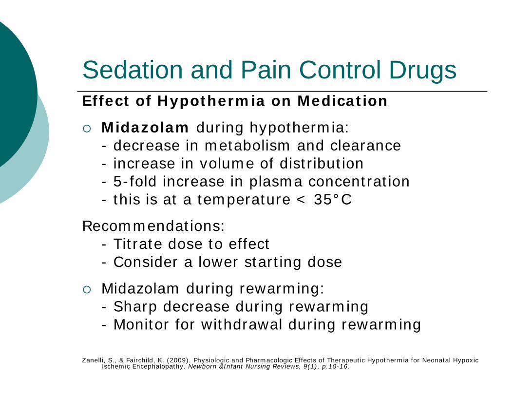
Sedation and Pain Control DrugsEffect of Hypothermia on Medication
Midazolam during hypothermia:- decrease in metabolism and clearance- increase in volume of distribution- 5-fold increase in plasma concentration- this is at a temperature < 35°C
Recommendations:- Titrate dose to effect- Consider a lower starting dose
Midazolam during rewarming:- Sharp decrease during rewarming- Monitor for withdrawal during rewarming
Zanelli, S., & Fairchild, K. (2009). Physiologic and Pharmacologic Effects of Therapeutic Hypothermia for Neonatal Hypoxic Ischemic Encephalopathy. Newborn &Infant Nursing Reviews, 9(1), p.10-16.
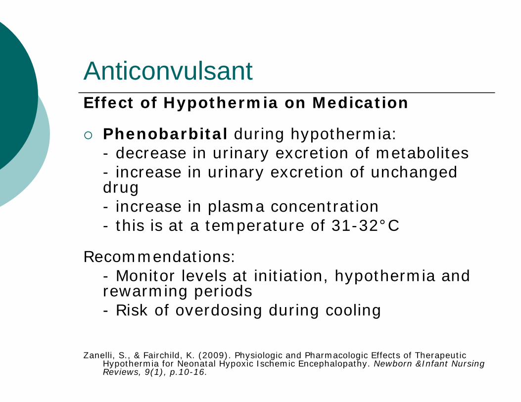
AnticonvulsantEffect of Hypothermia on Medication
Phenobarbital during hypothermia:- decrease in urinary excretion of metabolites- increase in urinary excretion of unchanged drug- increase in plasma concentration- this is at a temperature of 31-32°C
Recommendations:- Monitor levels at initiation, hypothermia and rewarming periods- Risk of overdosing during cooling
Zanelli, S., & Fairchild, K. (2009). Physiologic and Pharmacologic Effects of Therapeutic Hypothermia for Neonatal Hypoxic Ischemic Encephalopathy. Newborn &Infant Nursing Reviews, 9(1), p.10-16.

AnticonvulsantEffect of Hypothermia on Medication
Phenytoin during hypothermia:- decrease in metabolism and clearance- increase in plasma concentration-risk of overdosing during cooling- this is at a temperature of 34°C
Recommendations:- Monitor levels at initiation, during hypothermia and during rewarming periods
Phenytoin during rewarming:- risk of underdosing after rewarming
Zanelli, S., & Fairchild, K. (2009). Physiologic and Pharmacologic Effects of Therapeutic Hypothermia for Neonatal Hypoxic Ischemic Encephalopathy. Newborn &Infant Nursing Reviews, 9(1), p.10-16.

AntibioticEffect of Hypothermia on Medication
Gentamicin during hypothermia:- decreased clearance, volume of distribution- increased half-life- this is at a temperature of 29°C- no change in metabolism at 35°C
Recommendations:- Monitor levels at initiation, and during
hypothermia and rewarming period
Gentamicin during rewarming:- risk of underdosing after rewarming
Zanelli, S., & Fairchild, K. (2009). Physiologic and Pharmacologic Effects of Therapeutic Hypothermia for Neonatal Hypoxic Ischemic Encephalopathy. Newborn &Infant Nursing Reviews, 9(1), p.10-16.

Continued Patient Care and Discharge After 72 hours of cooling the infant is
gradually rewarmed (an increase of 0.2°C every 30 minutes).
The ventilator was gradually weaned and the infant was extubated.
No further seizure activity was noted.
A neurological consult was obtained to determine the need for further Phenobarbital and to determine follow-up.
Enteral feeds progressed slowly.
Feeding volume advanced nicely, but the infant was a poor po feeder.
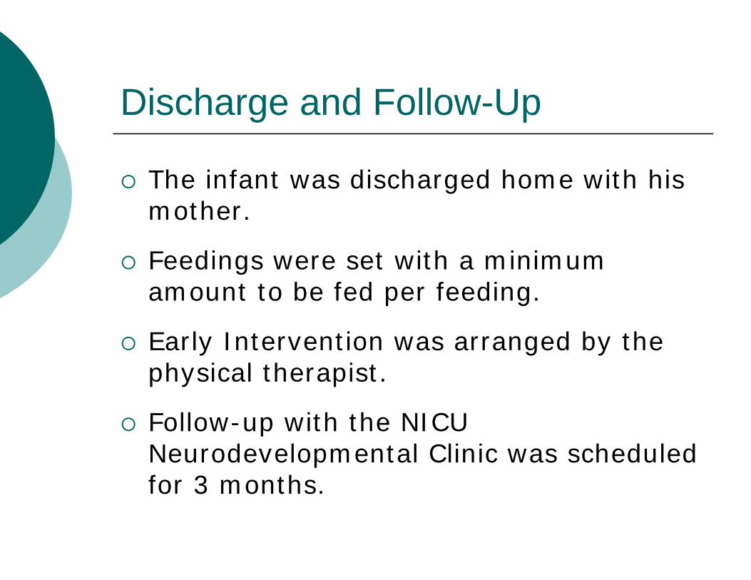
Discharge and Follow-Up
The infant was discharged home with his mother.
Feedings were set with a minimum amount to be fed per feeding.
Early Intervention was arranged by the physical therapist.
Follow-up with the NICU Neurodevelopmental Clinic was scheduled for 3 months.
ReferencesDebillion, T., Daoud, P., Durand, P., Cantagrel, S., Jouvet, P.,
Saizou, C., & Zupan, V. (2003). Whole-body cooling after perinatal asphyxia: a pilot study in term neonates. Developmental Medicine and Child Neurology, 45(1), p. 17-23.
Instructor’s Manual for Neonatal Resuscitation. (2006). Course Content. American Academy of Pediatrics and American Heart Association. (4th ed), p. 2-90
McNamara, P., Keyzers, M. (2006). A protocol for Cerebral Function Monitoring in the NICU. Hospital for Sick Children, Toronto, Canada, p 1.
Oza, V., Treat, J., Cook, N., Tetziaff, M., Yan, A. (2010). Subcutaneous fat necrosis as a complication of whole-body cooling for birth asphyxia. Archives of Dermatology, 146 (8), p 882-885.
Taketomo, C.K., Hodding, J.H., & Kraus, D.M.(2010). Hudson, Ohio: Lexi-Comp.
The Report of ACOG's Task Force on Neonatal Encephalopathy and Cerebral Palsy: Executive Summary. (2011).ACOG Guidelines. Retrieved April 15, 2011 from www.acog.org/from_home/misc/neonatalencephalopathy.cfm.
Young,T.E., Mangum, B. (2010). Neofax 2010. (23rd Ed.). Montvale, NJ:Thomson Reuters.
ReferencesZanelli, S.A., Naylor, M., Dobbins, N., Quigg, M., Goodkin, H.,
Matsumoto, J.A, and Fairchild, K. Implementation of a “Hypothermia for HIE” program: 2-year experience in a single NICU. (2008). Journal of Perinatalogy, 28, 171-175.
Zanelli, S., & Fairchild, K. (2009). Physiologic and Pharmacologic Effects of Therapeutic Hypothermia for Neonatal Hypoxic Ischemic Encephalopathy. Newborn &Infant Nursing Reviews, 9(1), p.10-16.


![HIE-Project-note acceptance test actuator · the HIE-DB and its instruments can be found in Ref. [1]. Figure 1 — Layout of the prototype HIE DB. A first prototype HIE-DB was designed](https://img.pdfslide.us/doc/110x75/5f079f267e708231d41de6c5/hie-project-note-acceptance-test-actuator-the-hie-db-and-its-instruments-can-be.jpg)