Embed Size (px)
Citation preview
LUND UNIVERSITY
PO Box 117221 00 Lund+46 46-222 00 00
Pharmacokinetics and pharmacodynamics of pentoxifylline and metabolites
Magnusson, Marie V
Published: 2009-01-01
Link to publication
Citation for published version (APA):Magnusson, M. V. (2009). Pharmacokinetics and pharmacodynamics of pentoxifylline and metabolites LundUniversity, Faculty of Medicine
General rightsCopyright and moral rights for the publications made accessible in the public portal are retained by the authorsand/or other copyright owners and it is a condition of accessing publications that users recognise and abide by thelegal requirements associated with these rights.
• Users may download and print one copy of any publication from the public portal for the purpose of privatestudy or research. • You may not further distribute the material or use it for any profit-making activity or commercial gain • You may freely distribute the URL identifying the publication in the public portalTake down policyIf you believe that this document breaches copyright please contact us providing details, and we will removeaccess to the work immediately and investigate your claim.
Pharmacokinetics and pharmacodynamics of pentoxifylline
and metabolites in humans
Marie Magnusson

ISSN 1652-8220
ISBN 978-91-86253-16-5
Lund University
Faculty of Medicine
Doctorial Dissertation Series 2009: 29
Printed in Lund, Sweden, by Media-Tryck 2009
© Marie Magnusson
2

To Linn, Erik and Andreas
3

4
Table of Contents
ORIGINAL PAPERS....................................................................................................... 7
ABBREVIATIONS ........................................................................................................... 8
INTRODUCTION ........................................................................................................... 9
1. Pharmacokinetics of pentoxifylline ........................................................................... 9
2. Therapeutics and pharmacodynamics ...................................................................... 12
A) Blood flow ................................................................................................................... 12
B) Fibrosis ........................................................................................................................ 16
AIMS ................................................................................................................................ 19
MATERIAL AND METHODS ..................................................................................... 20
Methods for analysis of pentoxifylline and metabolites .............................................. 20
Paper I .............................................................................................................................. 20
Paper II ............................................................................................................................ 21
Paper III .......................................................................................................................... 23
Paper IV .......................................................................................................................... 23
RESULTS & DISCUSSION .......................................................................................... 26
Papers I and IV ............................................................................................................... 26
Paper I .............................................................................................................................. 31
Paper II ............................................................................................................................ 33
Paper III .......................................................................................................................... 34
Paper IV ........................................................................................................................... 36
CONCLUSIONS ............................................................................................................. 41
SUMMARY .................................................................................................................... 42
SWEDISH SUMMARY ................................................................................................ 44
ACKNOWLEDGEMENTS .......................................................................................... 46
REFERENCES ............................................................................................................... 48
5

6
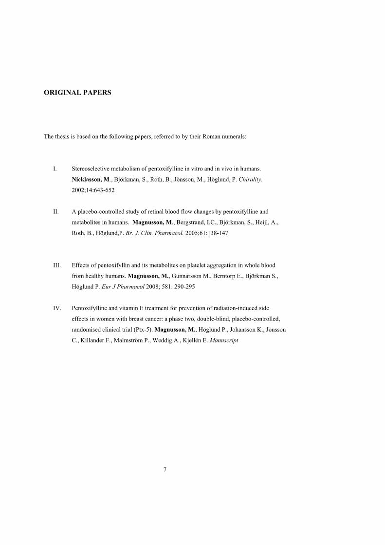
ORIGINAL PAPERS
The thesis is based on the following papers, referred to by their Roman numerals:
I. Stereoselective metabolism of pentoxifylline in vitro and in vivo in humans.
Nicklasson, M., Björkman, S., Roth, B., Jönsson, M., Höglund, P. Chirality.
2002;14:643-652
II. A placebo-controlled study of retinal blood flow changes by pentoxifylline and
metabolites in humans. Magnusson, M., Bergstrand, I.C., Björkman, S., Heijl, A.,
Roth, B., Höglund,P. Br. J. Clin. Pharmacol. 2005;61:138-147
III. Effects of pentoxifyllin and its metabolites on platelet aggregation in whole blood
from healthy humans. Magnusson, M., Gunnarsson M., Berntorp E., Björkman S.,
Höglund P. Eur J Pharmacol 2008; 581: 290-295
IV. Pentoxifylline and vitamin E treatment for prevention of radiation-induced side
effects in women with breast cancer: a phase two, double-blind, placebo-controlled,
randomised clinical trial (Ptx-5). Magnusson, M., Höglund P., Johansson K., Jönsson
C., Killander F., Malmström P., Weddig A., Kjellén E. Manuscript
7
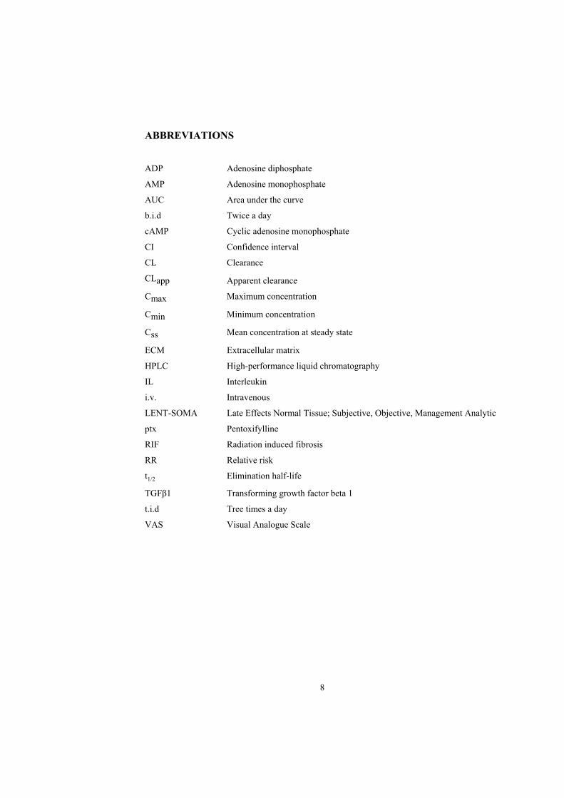
ABBREVIATIONS
ADP Adenosine diphosphate
AMP Adenosine monophosphate
AUC Area under the curve
b.i.d Twice a day
cAMP Cyclic adenosine monophosphate
CI Confidence interval
CL Clearance
CLapp Apparent clearance
Cmax Maximum concentration
Cmin Minimum concentration
Css Mean concentration at steady state
ECM Extracellular matrix
HPLC High-performance liquid chromatography
IL Interleukin
i.v. Intravenous
LENT-SOMA Late Effects Normal Tissue; Subjective, Objective, Management Analytic
ptx Pentoxifylline
RIF Radiation induced fibrosis
RR Relative risk
t1/2 Elimination half-life
TGF 1 Transforming growth factor beta 1
t.i.d Tree times a day
VAS Visual Analogue Scale
8
INTRODUCTION
In this thesis the pharmacokinetics and pharmacodynamics of pentoxifylline have been studied.
Pharmacokinetics describes how a drug is absorbed, distributed, metabolised in and eliminated
from the body. Pharmacodynamics describes the relationship between the drug concentration and
effect. Pentoxifylline is an interesting drug to study since it exhibits complex pharmacokinetics
with both reversible metabolism, and active metabolites. Difficulties in finding consistent clinical
effects of pentoxifylline may be due to the drug acting at least in part through formation of active
metabolites, the rate and extent of which may vary between individuals.
1. Pharmacokinetics of pentoxifylline
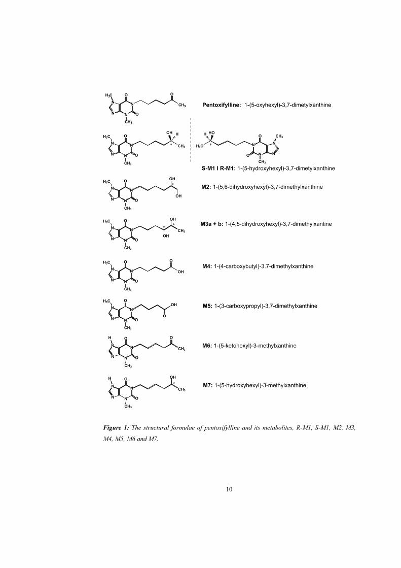
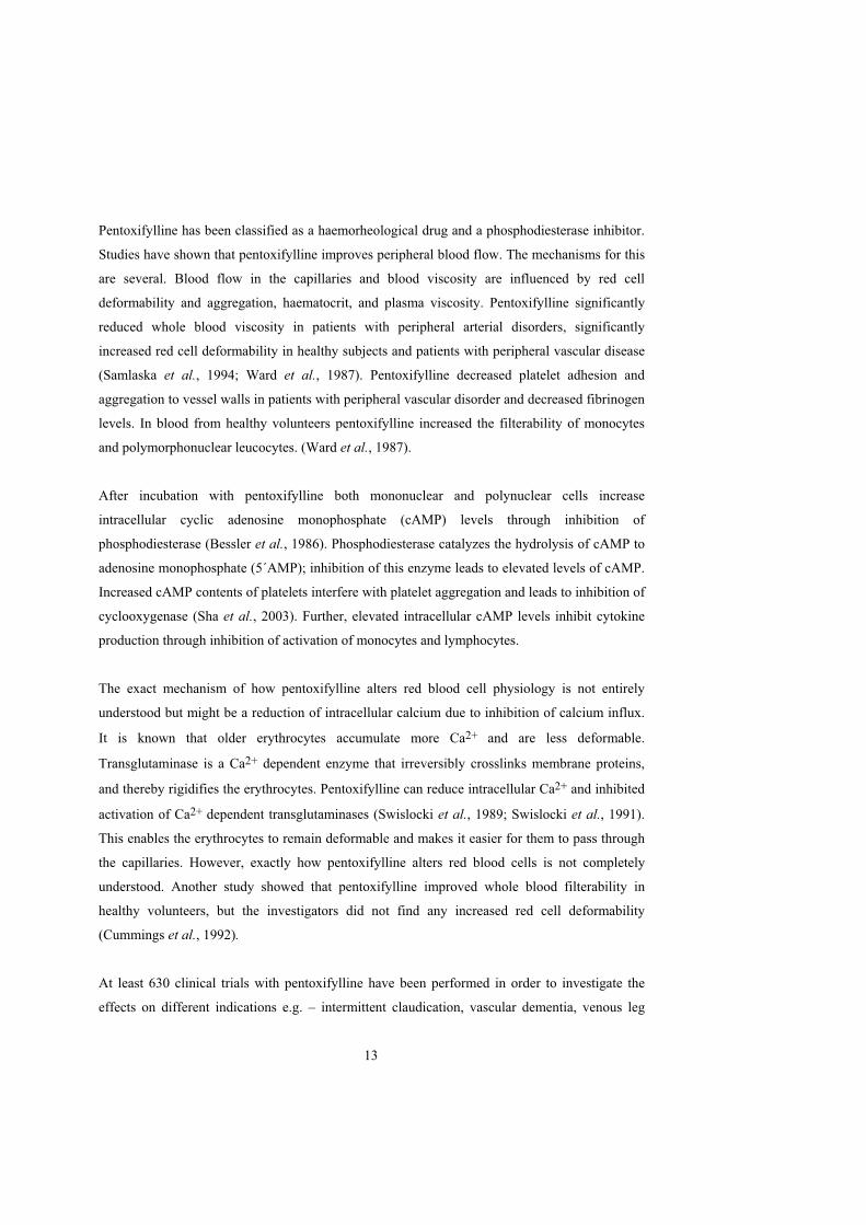
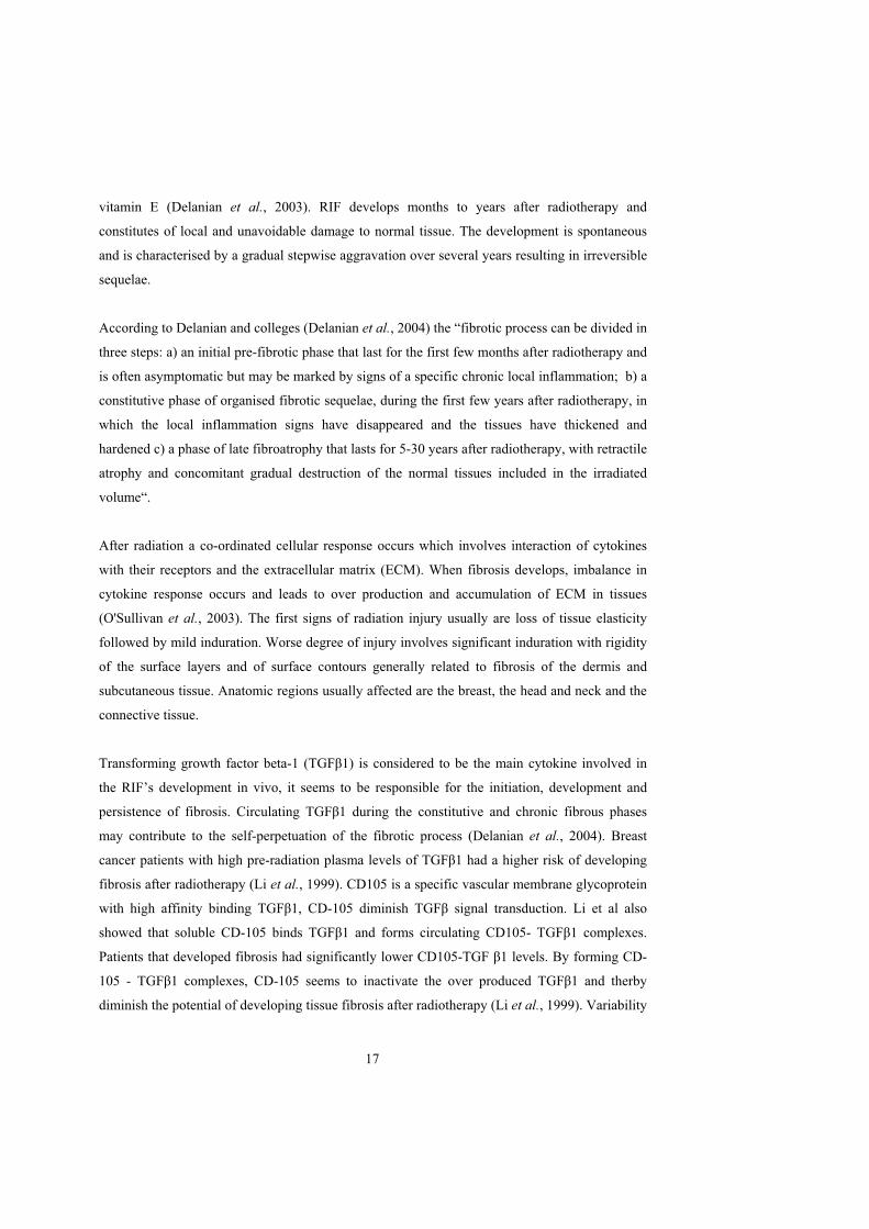
When the biotransformation of pentoxifylline was studied in man, seven phase 1 metabolites
(denoted M1-M7) were identified in human urine (Hinze, 1972b). The structures of the
metabolites were determined: the biotransformation yields three hydroxy metabolites (M1, M2,
M3a, and M3b), two carboxy metabolites (M4, M5) and two demethylated metabolites (M6,
M7), figure 1. The major metabolite excreted in urine is M5 followed by M4. The excretion of
unchanged pentoxifylline and M1 each accounts for less than 1% of the dose (Hinze, 1972a).
When pentoxifylline is administered to healthy humans the areas under the plasma concentration
curves (AUC) for M5 and M1, but not M4 are larger than pentoxifyllines (Beermann et al., 1985;
Bryce et al., 1989; Smith et al., 1986).
9
O
Figure 1: The structural formulae of pentoxifylline and its metabolites, R-M1, S-M1, M2, M3,
M4, M5, M6 and M7.
CH3
N
N
ON
3HC
O
N
C3H
Pentoxifylline: 1-(5-oxyhexyl)-3,7-dimetylxanthine
OH
* * N
N
N
H3C
CH3
N
O
N
O
CH3
H
N
CH3
N
CH3
N
O
O
H3C
H HO
S-M1 I R-M1: 1-(5-hydroxyhexyl)-3,7-dimetylxanthine
OH
N
N
H3C
CH3
N
O
N
O
* M2: 1-(5,6-dihydroxyhexyl)-3,7-dimethylxanthine
OH
OH
OH
N
N
H3C
CH3
N
O
NM3a + b: 1-(4,5-dihydroxyhexyl)-3,7-dimethylxantine
O
CH**
3
N
N
H3C
CH3
N
O
N
O
OH
O
M4: 1-(4-carboxybutyl)-3.7-dimethylxanthine
N
N
H3C
CH3
N
OOH
NM5: 1-(3-carboxypropyl)-3,7-dimethylxanthine
OO
M6: 1-(5-ketohexyl)-3-methylxanthine
CH3
N
N
H
N
O
N
O
CH3
O
N
N
H
CH3
N
O OH
N
O
CH3
* M7: 1-(5-hydroxyhexyl)-3-methylxanthine
10
Studies have shown that clearance (CL) for pentoxifylline was much higher than hepatic blood
flow and higher than, or in the same level as, cardiac output (Ings et al., 1982; Rames et al.,
1990). Since the metabolism of pentoxifylline to M1 is reversible the clearance is further
underestimated, as the AUC is increased due to continuous addition of pentoxifylline formed
from M1 (therefore the term CLapparent is used in this these studies). The high clearance
indicates that pentoxifylline is metabolised also at other sites than the liver, such as blood, since
CL is not limited by hepatic blood flow. Patients with impairment of liver function due to
cirrhosis had significantly increased AUCs for pentoxifylline and M1 compared with healthy
volunteers but the AUC ratio for Pentoxifylline/M1 was the same in the two groups (Rames et
al., 1990). Studies have shown that pentoxifylline is metabolised to M1 when incubated in whole
blood (Bryce et al., 1980; Ings et al., 1982). Taken together this suggests that erythrocytes are
the major site for pentoxifylline - M1 interconversion.
Metabolites M1 and M5 of pentoxifylline have significant haemorheologic effects but M2, M3,
M4, M6 and M7 had little haemorrheologic effect (Ambrus et al., 1995). Consequently, the
present thesis focuses on the active metabolites M1 and M5 and the M4 metabolite due to the
pharmacokinetic data.
The hydroxy metabolite M1 is formed by reduction of pentoxifylline, the reaction is rapid and
reversible (Lee et al., 1997). It takes place both in the liver (Lillibridge et al., 1996) and the
erythrocytes (Bryce et al., 1989; Ings et al., 1982; Poirier et al., 1989). M1 has a carbon atom
(marked *) that carries four different substituents and is therefore a chiral compound. A chiral
molecule has two non-superimposable mirror forms called enantiomers that are as one's left and
right hands to each other: "the same" but opposite. The R (rectus=right) and S (sinister=left)
nomenclature refer to the absolute configuration based on atomic number of the substituents.
Different enantiomers of chiral compounds can have different effects as drugs. The enantiomers
of M1 are shown in figure 1.
The R – enantiomer of M1 was manufactured as a drug in its own right, lisofylline. Lisofylline
has been tested in several clinical trials with varying results. A study in healthy volunteers
showed that lisofylline causes a prolonged and marked decrease in the levels of circulating free
fatty acids but these effects were not seen when lisofylline was administered to patients with
acute lung injury or acute respiratory distress syndrome (ARDS-Network, 2002; Bursten et al.,
1998) Lisofylline 3mg/kg t.i.d. (but not 2mg/kg) reduced the incidence of infections and
11
improved 100-day survival in patients receiving related-donor allogeneic bone marrow
transplantation; but no effects were seen on time to neutrophil recovery and platelet recovery
(List et al., 2000). Co-administration with lisofylline did not decrease the toxicities of high dose
i.v. IL-2 (Margolin et al., 1997). Recently, a new therapeutic use for lisofylline was proposed,
suggesting that lisofylline can prevent autoimmune disorders including type 1 diabetes (Yang et
al., 2005). However, despite many clinical trials lisofylline is still in the experimental and
development stages and has not been approved for sale due to limitations in its therapeutic
efficacy.
It is unclear how much R-M1 is formed after administration of pentoxifylline. Limited in vitro
data indicates that R-M1 is formed to a much lesser extent than S-M1 in humans (Lillibridge et
al., 1996). The stereoselective formation of M1 has only been studied in vitro in human liver
microsomes and in a few cancer patients who received pentoxifylline together with ciprofloxacin
and interleukin-2 (Thompson et al., 1994). There is a pharmacokinetic interaction between
ciprofloxacin and methylxanthines that may influence the formation of M1 (Fuhr et al., 1992). A
recent study showed that the mechanism behind the interaction is inhibition of CYP1A2 and that
it seems likely that CYP1A2 catalyses xanthine 7-demethylation of pentoxifylline to M6 and M1
to M7 (Peterson et al., 2004; Raoul et al., 2007).
Since the pharmacokinetics for the formation of the stereoisomers of M1 has only been partly
investigated this prompted us to perform paper I. The aims in paper I were to study the kinetics
of the reversible transformation of pentoxifylline to R-M1 and S-M1 in erythrocyte suspension
and also to quantify the formation of the enantiomers of M1, as well as M4 and M5 after
intravenous and oral administration to healthy volunteers.
2. Therapeutics and pharmacodynamics
A) Blood flow
Pentoxifylline was developed by Hoechst Aktiengesellschaft in the seventies. Pentoxifylline
obtained marketing authorisation in Germany 1972 and in the USA 1984 for the treatment of
intermittent claudication on the basis of chronic occlusive arterial disease of the limbs.
Pentoxifylline has never been granted authorisation in Sweden but it is possible to prescribe and
obtain permission for its use on a named patient bases from the Medical Product Agency.
12
Pentoxifylline has been classified as a haemorheological drug and a phosphodiesterase inhibitor.
Studies have shown that pentoxifylline improves peripheral blood flow. The mechanisms for this
are several. Blood flow in the capillaries and blood viscosity are influenced by red cell
deformability and aggregation, haematocrit, and plasma viscosity. Pentoxifylline significantly
reduced whole blood viscosity in patients with peripheral arterial disorders, significantly
increased red cell deformability in healthy subjects and patients with peripheral vascular disease
(Samlaska et al., 1994; Ward et al., 1987). Pentoxifylline decreased platelet adhesion and
aggregation to vessel walls in patients with peripheral vascular disorder and decreased fibrinogen
levels. In blood from healthy volunteers pentoxifylline increased the filterability of monocytes
and polymorphonuclear leucocytes. (Ward et al., 1987).
After incubation with pentoxifylline both mononuclear and polynuclear cells increase
intracellular cyclic adenosine monophosphate (cAMP) levels through inhibition of
phosphodiesterase (Bessler et al., 1986). Phosphodiesterase catalyzes the hydrolysis of cAMP to
adenosine monophosphate (5´AMP); inhibition of this enzyme leads to elevated levels of cAMP.
Increased cAMP contents of platelets interfere with platelet aggregation and leads to inhibition of
cyclooxygenase (Sha et al., 2003). Further, elevated intracellular cAMP levels inhibit cytokine
production through inhibition of activation of monocytes and lymphocytes.
The exact mechanism of how pentoxifylline alters red blood cell physiology is not entirely
understood but might be a reduction of intracellular calcium due to inhibition of calcium influx.
It is known that older erythrocytes accumulate more Ca2+ and are less deformable.
Transglutaminase is a Ca2+ dependent enzyme that irreversibly crosslinks membrane proteins,
and thereby rigidifies the erythrocytes. Pentoxifylline can reduce intracellular Ca2+ and inhibited
activation of Ca2+ dependent transglutaminases (Swislocki et al., 1989; Swislocki et al., 1991).
This enables the erythrocytes to remain deformable and makes it easier for them to pass through
the capillaries. However, exactly how pentoxifylline alters red blood cells is not completely
understood. Another study showed that pentoxifylline improved whole blood filterability in
healthy volunteers, but the investigators did not find any increased red cell deformability
(Cummings et al., 1992).
At least 630 clinical trials with pentoxifylline have been performed in order to investigate the
effects on different indications e.g. – intermittent claudication, vascular dementia, venous leg
13
ulcers, bone marrow transplant, cerebrovascular disease, diabetes mellitus, endometriosis, male
infertility and many more (Cochrane, 2009; Martindale, 2009). Most of the studies are small and
the magnitude of effects varies. The only approved indication for pentoxifylline is intermittent
claudication. Below a short summary will follow for the most common therapeutic usages of
pentoxifylline.
Intermittent claudication develops during exercise, such as walking, when the flow to the lower
limb is insufficient to meet the needs of the exercising muscle. The patient experience muscle
pain that disappears when resting. The reduced blood flow is often caused by atherosclerosis.
Intermittent claudication is often associated with coronary heart disease and morbidity and
mortality is raised in patients with intermittent claudication. The risk factors for developing
intermittent claudication are the same as coronary heart disease: hypertension, diabetes, smoking,
and high cholesterol levels. Intermittent claudication reduces the patients’ mobility due to pain
when walking, but at the same time the walking exercise together with smoking cessation are the
primary treatment (LB, 2007-2008; Watson et al., 2008). Pentoxifylline has been used in the
treatment of intermittent claudication for a long time due to its haemorheologic properties (Ernst
et al., 1992; Moher et al., 2000; Porter et al., 1982; Reich et al., 1987).
In a meta-analysis all randomised, placebo-controlled, double blind clinical trials on the
indication intermittent claudication were reviewed: 52 studies met the inclusion criteria of the
meta-analysis and pentoxifylline were studied in 17 of these, including 1041 patients (Moher et
al., 2000). The meta-analysis showed that, after 24 weeks treatment (but not 8 weeks),
pentoxifylline was more effective than placebo and the other drugs for the primary effect
parameter, maximum walking distance. The other primary effect parameter was pain free
walking distance: pentoxifylline seems better than the other medications here as well, but this
was not significant. Mortality was studied in seven of the studies including 479 patients,
conclusions regarding mortality could not be made from this, only one patient in the treatment
group and two in the placebo group died. The authors conclude that pentoxifylline therapy may
be efficacious in improving the walking capacity, although its treatment effect is modest (Moher
et al., 2000).
Patients with vascular dementia have reduced cerebrovascular perfusion. Pentoxifylline has been
tested in clinical trials and used for treatment of vascular dementia (Black et al., 1992; Hartmann,
1985), its mechanism of action in vascular dementia was said to be due to reduced blood
14
viscosity and inhibition of the production of pro-inflammatory cytokines (Sha et al., 2003). A
systematic review was performed in order to evaluate the use of pentoxifylline in patients with
vascular dementia (Sha et al., 2003). Only four studies including 469 patients met their inclusion
criteria i.e. randomised, double blinded, and placebo controlled. None of the four studies showed
a significant improvement in the pentoxifylline group compared with the placebo group, but a
trend towards improvement could be noticed. A subgroup analysis was done in three of the
studies, using a stricter definition for vascular dementia. The subgroup analysis showed
significant improvement or decreased impairment in the pentoxifylline group compared with the
placebo group (Sha et al., 2003). The authors conclude that pentoxifylline has a potential role in
treatment of vascular dementia, but that few studies met the quality criteria for inclusion in the
review and that new larger well contained clinical trials are needed to confirm the results.
When a patient has chronic venous hypertension in the tissues of the lower leg, this leads to a
chronic inflammatory condition and a leukocyte activation and may eventually result in venous
leg ulcer (Pascarella et al., 2005). Pentoxifylline has been tested in clinical trials for treatment of
venous leg ulcer (Barbarino, 1992; Dale et al., 1999; Falanga et al., 1999). A Cochrane review
was performed in order to investigate the effects of pentoxifylline for treating venous leg ulcer:
11 randomised, placebo controlled clinical trials including 841 patients met the inclusion criteria
(Jull et al., 2007). A random effect model was used to combine the results from the studies and it
was found that treatment with pentoxifylline leads to significant improvement or complete
healing more often compared with placebo (RR 1.70, 95% CI 1.30-2.24) (Jull et al., 2007).
Pentoxifylline has been tested and used in the treatment of microalbuminuria (Navarro et al.,
2005; Rodriguez-Moran et al., 2005). A systematic review on all studies with patients with
kidney disease and pentoxifylline treatment where done: 10 randomised controlled clinical trials
were found including 476 patients (McCormick et al., 2008). The effect parameter was change in
proteinurea. Proteinurea increases the risk for cardiovascular mortality and morbidity and end
stage renal disease. By decreasing proteinurea the cardiovascular mortality and morbidity
decreases and the progression of chronic kidney diseases is delayed. The meta-analysis showed
that pentoxifylline significantly decreased proteinurea compared with placebo or standard care
(McCormick et al., 2008). The most likely explanation for the antiproteinuric effect of
pentoxifylline was said to be inhibition of the production of proinflammatory cytokines. The
authors conclude that pentoxifylline seems to decrease proteinuria in patients with chronic
15
kidney diseases, but that the studies were small and few and that larger high quality studies with
mortality and morbidity as endpoint parameters are needed to verify this.
It has been suggested that pentoxifylline may be used in diseases affecting retinal blood flow,
such as diabetic retinopathy (Schmetterer et al., 1996; Sebag et al., 1994; Sonkin et al., 1993a;
Sonkin et al., 1993b) or macular degeneration (Kruger et al., 1998), but a recent Cochrane
review on diabetic retinopathy (Lopes de Jesus et al., 2008) stated that no conclusions could be
drawn due to lack of large randomised controlled clinical trials.
Pentoxifylline was generally well tolerated in all studies with few undesirable effects, nausea
being the most common one. Few patients discontinue treatment with pentoxifylline due to side
effects.
Measurement of blood flow is difficult since the capillaries are “hidden” in the body. Retinal
blood flow is an exception since it can be measured through the eye using a validated method
quantifying all the flows in a selected temporal area of the retina (Michelson et al., 1995;
Michelson et al., 1998). The aim of paper II was to investigate the possible contribution of the
metabolites (R-M1, S-M1, M4, and M5) of pentoxifylline to its effect, assessed as retinal blood
flow, in healthy humans.
Since pentoxifylline inhibits platelet aggregation we used platelet aggregation in whole blood as
effect parameter in paper III (Ambrus et al., 1995; de la Cruz et al., 1993). The aim in paper III
was to investigate the relative potencies of pentoxifylline and metabolite R-M1, S-M1, M4 and
M5 to inhibit platelet aggregation in whole blood, and in particular to clarify contributions of the
two enantiomers of M1, to this effect.
B) Fibrosis
More recent studies have shown that pentoxifylline in combination with vitamin E can reduce
radiation induced fibrosis (RIF) (Chiao et al., 2005; Delanian, 1998; Delanian et al., 1999;
Delanian et al., 2003; Lefaix et al., 1999; Okunieff et al., 2004). Previously, RIF was considered
irreversible. But in a double-blind, placebo-controlled study in women previously treated with
radiotherapy for breast cancer, co-administration of pentoxifylline and vitamin E reduced the
mean area of RIF up to 60% compared with placebo or single treatment with pentoxifylline or
16
vitamin E (Delanian et al., 2003). RIF develops months to years after radiotherapy and
constitutes of local and unavoidable damage to normal tissue. The development is spontaneous
and is characterised by a gradual stepwise aggravation over several years resulting in irreversible
sequelae.
According to Delanian and colleges (Delanian et al., 2004) the “fibrotic process can be divided in
three steps: a) an initial pre-fibrotic phase that last for the first few months after radiotherapy and
is often asymptomatic but may be marked by signs of a specific chronic local inflammation; b) a
constitutive phase of organised fibrotic sequelae, during the first few years after radiotherapy, in
which the local inflammation signs have disappeared and the tissues have thickened and
hardened c) a phase of late fibroatrophy that lasts for 5-30 years after radiotherapy, with retractile
atrophy and concomitant gradual destruction of the normal tissues included in the irradiated
volume“.
After radiation a co-ordinated cellular response occurs which involves interaction of cytokines
with their receptors and the extracellular matrix (ECM). When fibrosis develops, imbalance in
cytokine response occurs and leads to over production and accumulation of ECM in tissues
(O'Sullivan et al., 2003). The first signs of radiation injury usually are loss of tissue elasticity
followed by mild induration. Worse degree of injury involves significant induration with rigidity
of the surface layers and of surface contours generally related to fibrosis of the dermis and
subcutaneous tissue. Anatomic regions usually affected are the breast, the head and neck and the
connective tissue.
Transforming growth factor beta-1 (TGF 1) is considered to be the main cytokine involved in
the RIF’s development in vivo, it seems to be responsible for the initiation, development and
persistence of fibrosis. Circulating TGF 1 during the constitutive and chronic fibrous phases
may contribute to the self-perpetuation of the fibrotic process (Delanian et al., 2004). Breast
cancer patients with high pre-radiation plasma levels of TGF 1 had a higher risk of developing
fibrosis after radiotherapy (Li et al., 1999). CD105 is a specific vascular membrane glycoprotein
with high affinity binding TGF 1, CD-105 diminish TGF signal transduction. Li et al also
showed that soluble CD-105 binds TGF 1 and forms circulating CD105- TGF 1 complexes.
Patients that developed fibrosis had significantly lower CD105-TGF 1 levels. By forming CD-
105 - TGF 1 complexes, CD-105 seems to inactivate the over produced TGF 1 and therby
diminish the potential of developing tissue fibrosis after radiotherapy (Li et al., 1999). Variability
17
in incidence and severity of fibrosis after radiotherapy can partly be explained by TGF 1 gene
polymorphism. Patients with -509 polymorphic allele can partly be predisposed to severe fibrosis
due to over expression of TGF 1. (Giotopoulos et al., 2007; Quarmby et al., 2003).
Pentoxifylline appears to stimulate prostacyclin release from normal endothelial cells to inhibit
some of the cytokine cascade resulting from tissue injury, and it indirectly inhibits the production
of thromboxane, a potent vasoconstrictor and a strong stimulator of platelet aggregation. In vitro
studies have indicated that pentoxifylline inhibits human dermal fibroblast proliferation and
extracellular matrix production and increases collagenase activity (Berman et al., 1989; Berman
et al., 1990).
During inflammatory reactions and RIF development reactive oxygen species, such as singlet
oxygen, superoxide anion hydrogen peroxide, and hydroxyl radicals are generated (Delanian et
al., 2004). If they are not scavenged efficiently, oxidative stress may lead to cell necrosis or
apoptosis. Vitamin E is used due to its antioxidant properties; it protects membrane
phospholipids from oxidative damage. Vitamin E deficiency has been associated with abnormal
connective tissue repair, resulting in the formation of scar like tissue.
The first report that the combination of pentoxifylline and vitamin E can reduce RIF was
published in 1999. It was shown that pentoxifylline/vitamin E treated pigs had a histopatholgic
normalisation of the subcutaneous tissues surrounding a small residual scar, as well as a large
reduction in the immunohistochemical expression for the TGF 1 (Lefaix et al., 1999). Since then
more studies have shown that therapy with pentoxifylline and vitamin E can decrease or even
reverse RIF in humans (Chiao et al., 2005; Delanian et al., 1999; Delanian et al., 2003). Still it
would be even better if the fibrosis could be prevented. We hypothesised that treatment with
pentoxifylline and vitamin E could prevent or reduce the development of fibrotic tissue. To
determine the preventive effects of pentoxifylline and vitamin E against a range of radiation-
induced side effects, a randomised, double-blind, placebo-controlled clinical trial was planned
(paper IV). The primary aim in paper IV was to investigate if pentoxifylline + vitamin E prevent
radiation induced side effects measured as impaired shoulder mobility in women treated for
breast cancer with radiotherapy to the axilla and breast.
18
AIMS
To investigate the reversible transformation of pentoxifylline to R-M1 and S-M1 in
erythrocyte suspension (Paper I)
To investigate the pharmacokinetics of pentoxifylline and some important metabolites
after administration of pentoxifylline to healthy volunteers (Paper I, II) and breast cancer
patients (Paper IV)
To investigate the effects of pentoxifylline and metabolites R-M1, S-M1, M4 and M5 on
retinal blood flow in healthy humans after intravenous infusions of pentoxifylline (Paper
II)
To investigate the relative potencies of pentoxifylline and metabolites R-M1, S-M1, M4
and M5 to inhibit platelet aggregation in whole blood, and in particular to clarify
contributions of the two enantiomers of M1, to this effect (Paper III)
To investigate if the combination of pentoxifylline and vitamin E can prevent radiation
induced side effects in women with breast cancer (Paper IV)
19
MATERIAL AND METHODS
The aim of this section is to highlight some of the most important methods used in this thesis.
Detailed descriptions of the material and methods used are given in the papers.
Methods for analysis of pentoxifylline and metabolites
HPLC method for determination of pentoxifylline, R-M1 and S- M1 (Paper I, II, IV)
The concentrations of pentoxifylline and the enantiomers of M1 were determined by reversed-
phase HPLC after chiral derivatisation of the metabolite.12 Thalidomide was used as internal
standard. The sample was extracted with dichloromethane and diacetyl-L-tartaric acid anhydride
solution was used for derivatisation. A LiChrosorb RP-18, 250 4 mm, 7 m particle size column
was used with 17% acetonitrile in 0.05 M NaH2PO4 buffer, pH 4.0. as mobile phase. The
detection wavelength was 274 nm.
HPLC method for determination of M4 and M5 (Paper I-II)
For the determination of metabolites 4 and 5, enprofylline was used as internal standard. The
sample was extracted with ethyl acetate. A RP-18 column was used with 13% acetonitrile in
0.005 M NaH2PO4 buffer, pH 3.6 as mobile phase and the detection wavelength was 274 nm.
Paper I
Interconversion of pentoxifylline, R-M1 and S-M1 by erythrocytes
The reductive metabolism of pentoxifylline to R-M1 and S-M1 and the oxidative metabolism of
R-M1 and S-M1 to pentoxifylline were investigated in haemolysed erythrocyte suspension from
healthy humans.
For the reductive metabolism of pentoxifylline to R-M1 and S-M1, NADPH and Mg2+ were
added to the aliquots of haemolysed erythrocytes. The incubations were started by the addition of
pentoxifylline in the respective samples. The mixtures were incubated for 20 min at 37 C.
20
For the oxidative metabolism of R-M1 and S-M1 to pentoxifylline, NADP and Mg2+ were added
to the aliquots of haemolysed erythrocytes. The incubations were started by the addition of R-M1
or S-M1 respectively. The samples were incubated for 20 min at 37 C.
NONMEM version V (The NONMEM project group, San Francisco, CA, USA) was used to
determine the Michaelis – Menten parameters, Vmax and Km, of the enzymes involved in the
metabolism of pentoxifylline to R-M1 and S-M1. Two models were used, representing the action
of either one or two enzymes:
][][max
CKCVV
m ][][
][][
)2(
)2max(
)1(
)1max(
CKCV
CKCV
Vmm
In these equations, V is the measured rate of metabolite formation in nmoles/min and [C] is the
concentration of substrate (mM). From the obtained values of Vmax and Km for the various
enzymes, the rates of conversion of pentoxifylline to R-M1 and S-M1 at a therapeutic blood
concentration were calculated and compared.
Pharmacokinetics in humans
The study was performed according to the declaration of Helsinki and approved by the Ethics
Committee of Lund University and by the Swedish Medical Products Agency. After giving
written informed consent six healthy, non-smoking volunteers were included in the study. They
received, in a randomised cross-over design, three doses of pentoxifylline with washout periods
of at least one week in between. The three doses were: 300 mg and 600 mg as intravenous
infusion and a 600 mg controlled release tablet (Trental®). Blood was sampled from an
indwelling venous catheter before start of infusion, during infusion and until 6 h after
termination; for oral administration before and until 25 h after intake of the tablet.
Paper II
Study design
The study was performed according to the declaration of Helsinki and approved by the Ethics
Committee of Lund University and by the Swedish Medical Products Agency. After giving
written informed consent eight healthy, non-smoking volunteers were included in the study. Each
21
subject passed a pre-study ophthalmic examination. The study was randomized, placebo
controlled, observer-blinded, and partly blinded for the subject in a four period cross-over design.
The subjects were given the four treatments in random order. During one session they were given
placebo (0.9% saline solution) as intravenous infusion. During the three other sessions they were
given pentoxifylline by intravenous infusion. In one session the subjects were pre-treated with
ciprofloxacin and in another with rifampicin.
The pre-treatments with ciprofloxacin and rifampicin could not be blinded but the pentoxifylline
and placebo administrations were.
Measurement and analysis of retinal blood flow
Retinal blood flow was assessed using the Scanning Laser Doppler Flowmetry (SLDF)
(Heidelberg Retina Flowmeter, Heidelberg Engineering) (Michelson et al., 1995). The method of
SLDF provides a high definition tomographic image of perfused retinal vessels with
simultaneous evaluation of blood flow using an optical Doppler effect. The measurements were
performed in a selected area (2.7x0.7mm) of the central temporal retina, at baseline, during the
infusion period and up to 5 hours after termination.
Quantification of capillary retinal blood flow was stated in arbitrary units (AUs) describing the
product of mean flow velocity and mean amount of blood cells in a standardised volume. The
mean values from each time point have been used in the calculations.
Data presentation and statistical considerations
Two main statistical models were tested after an initial check for lack of period effects. The first
one comprised only the treatments given as fixed effects, thus disregarding the obtained plasma
concentration data. In the second model treatments were not included; instead the AUCs of
pentoxifylline and its metabolites were used as regressor variables. Such a model can only be
successfully used if the correlations between the regressors are low. All models used subjects as
random effects, and a repeated measures spatial exponential covariance structure design.
22
Paper III
Platelet aggregation
The study was performed according to the declaration of Helsinki and approved by the Ethics
committee of Lund University. The subjects were informed about the study and gave oral
informed consent prior to blood sampling. Measurement of platelet aggregation were done in
whole blood from 8 healthy volunteers by impedance technology (electrical resistance between
two electrodes immersed in whole blood), using a whole blood lumi-aggregometer, (Chrono log
modell 560 Ca, Chrono Log Corp, Havertown, PA, USA) (Podczasy, 1997; Vucenik, 1998).
Blood was sampled at two occasions from each subject by venepuncture. At one occasion
pentoxifylline, rac-M1, M4 and M5 were investigated and on another occasion rac-M1 and R-
M1.
Platelet aggregation was studied in whole blood diluted in a 1:1 ratio with 0.9 % saline, the
samples were incubated in 37ºC prior to testing. The test procedure was started by adding
luciferin luciferase to the sample; the samples were incubated in 37 ºC for 1 minute prior to
addition of the test substances. Pentoxifylline, rac-M1, R-M1, M4, M5 or saline (positive
control) was added to the sample and incubated in 37ºC for 1 minute prior to addition of
Adenosindiphosphate (ADP), aggregation was monitored for 6 minutes. The effects of S-M1
were calculated from a comparison between R-M1 and rac-M1 since we did not have access to
pure S-M1.
Paper IV
Study design
The study was a randomised, placebo controlled for pentoxifylline, double blinded clinical trial
with a parallel study design. Randomisation was stratified for previous use of chemotherapy.
The study was conducted at Lund University Hospital, Department of Oncology in accordance
with the principles of Good Clinical Practice (GCP) and the ethical principles stated in the
current revision of the declaration of Helsinki. The study was approved by the Ethics Committee
of Lund University and by the Swedish Medical Products Agency. The trial is registered on the
ISRCTN.org website, number ISRCTN39143623.
23
Patients
Patients fulfilling the following criteria were included in the study: primary breast cancer,
axillary dissection, mastectomy or segmental resection of the breast, radiotherapy to the
breast/thoracic wall, and axilla and fossa supra/infra calvicularis. The inclusion period was one to
three months after termination of radiotherapy. Exclusion criteria were; known sensitivity to
pentoxifylline or vitamin E, disorders related to muscles or joints, treatment with corticosteroids
during the radiotherapy treatment.
Treatments
After written informed consent and baseline assessments were obtained, patients were randomly
assigned on a 1:1 basis, with stratification for chemotherapy, to pentoxifylline 400 mg
(Pentoxifylline Ratiopharm®, extended-release tablet) 3 times daily or a matching placebo. All
patients were treated with vitamin E 100 mg 3 times daily. The duration of treatment was 12
months.
Safety assessment
Patients were assessed for breast cancer recurrence and survival as a measurement of drug safety.
At each visit patients were asked if they had experienced any change in health status. Adverse
events and serious adverse events were classified and reported according to the ICH GCP
guidelines.
Efficacy assessment
The endpoints consisted of change in the passive abduction of the shoulder, and changes in arm
volume, Late Effects on Normal Tissue; Subjective, Objective, Management and Analytic, breast
score (LENT-SOMA) as assessed by the patients’ physicians (Rubin et al., 1995); and patients’
subjective assessment of somatic sensations and discomfort during the last week as measured by
Visual Analogue Scale (VAS) (Scott et al., 1976). Abduction of the shoulder was measured
using a goniometer. Arm volume was measured by the water displacement method; the volumes
were presented as percentage difference between arms (Bednarczyk et al., 1992; Kettle et al.,
1958). Blood samples were collected by venipuncture into vacuum collection tubes containing
sodium heparin. Plasma concentrations of pentoxifylline, R-M1 and S-M1, were determined by
HPLC.
24

Statistical Methods
Continuous variables are presented as medians and quartiles. Categorical are presented as
frequencies. Abduction, percentage difference in arm volumes, LENT-SOMA score and VAS
observations were regarded as ordinal data. A two-way method developed for repeated measures
of ordinal scale data (Shah et al., 2004) using SAS statistical software version 8.2 (SAS, Cary,
NC, USA) was used to elucidate differences between treatment over time and the time by
treatment interaction in the effect data. Statistical significance was defined as two tail p < 0.05.
25
RESULTS & DISCUSSION
Papers I and IV
Pharmacokinetics in humans
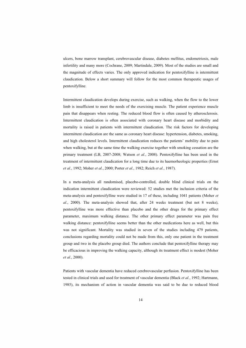
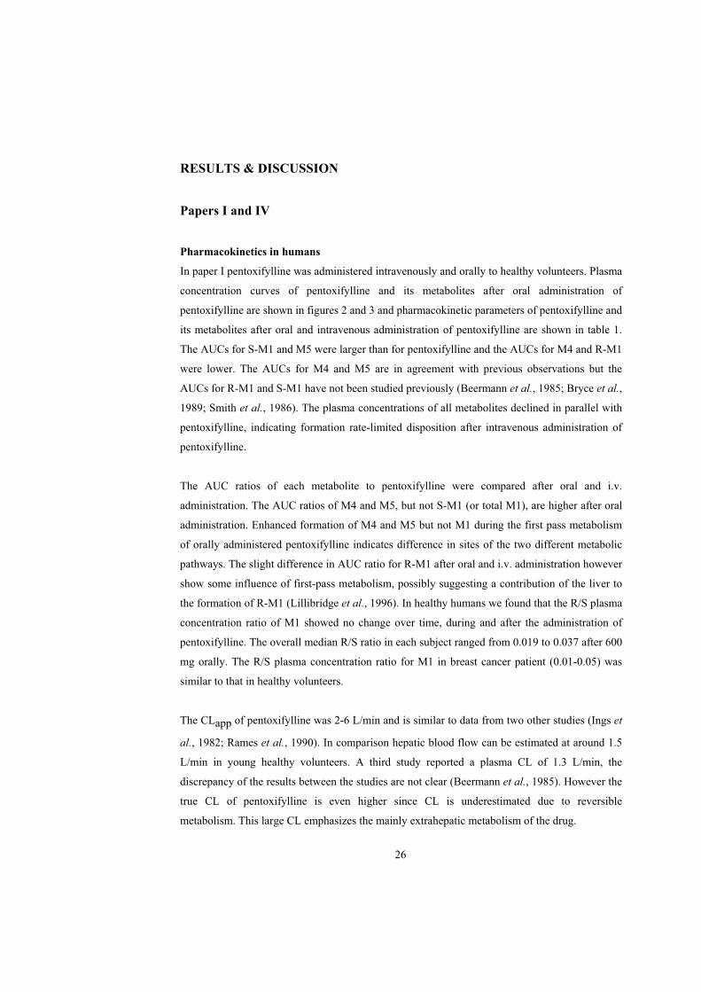
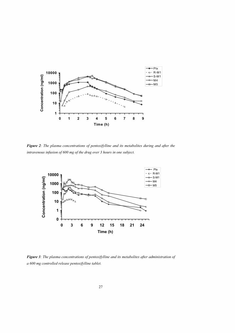
In paper I pentoxifylline was administered intravenously and orally to healthy volunteers. Plasma
concentration curves of pentoxifylline and its metabolites after oral administration of
pentoxifylline are shown in figures 2 and 3 and pharmacokinetic parameters of pentoxifylline and
its metabolites after oral and intravenous administration of pentoxifylline are shown in table 1.
The AUCs for S-M1 and M5 were larger than for pentoxifylline and the AUCs for M4 and R-M1
were lower. The AUCs for M4 and M5 are in agreement with previous observations but the
AUCs for R-M1 and S-M1 have not been studied previously (Beermann et al., 1985; Bryce et al.,
1989; Smith et al., 1986). The plasma concentrations of all metabolites declined in parallel with
pentoxifylline, indicating formation rate-limited disposition after intravenous administration of
pentoxifylline.
The AUC ratios of each metabolite to pentoxifylline were compared after oral and i.v.
administration. The AUC ratios of M4 and M5, but not S-M1 (or total M1), are higher after oral
administration. Enhanced formation of M4 and M5 but not M1 during the first pass metabolism
of orally administered pentoxifylline indicates difference in sites of the two different metabolic
pathways. The slight difference in AUC ratio for R-M1 after oral and i.v. administration however
show some influence of first-pass metabolism, possibly suggesting a contribution of the liver to
the formation of R-M1 (Lillibridge et al., 1996). In healthy humans we found that the R/S plasma
concentration ratio of M1 showed no change over time, during and after the administration of
pentoxifylline. The overall median R/S ratio in each subject ranged from 0.019 to 0.037 after 600
mg orally. The R/S plasma concentration ratio for M1 in breast cancer patient (0.01-0.05) was
similar to that in healthy volunteers.
The CLapp of pentoxifylline was 2-6 L/min and is similar to data from two other studies (Ings et
al., 1982; Rames et al., 1990). In comparison hepatic blood flow can be estimated at around 1.5
L/min in young healthy volunteers. A third study reported a plasma CL of 1.3 L/min, the
discrepancy of the results between the studies are not clear (Beermann et al., 1985). However the
true CL of pentoxifylline is even higher since CL is underestimated due to reversible
metabolism. This large CL emphasizes the mainly extrahepatic metabolism of the drug.
26
1
10
100
1000
10000
0 1 2 3 4 5 6 7 8 9
Time (h)
Co
nce
ntr
atio
n (
ng
/ml)
PtxR-M1S-M1M4M5
Figure 2: The plasma concentrations of pentoxifylline and its metabolites during and after the
intravenous infusion of 600 mg of the drug over 3 hours in one subject.
0
1
10
100
1000
10000
0 3 6 9 12 15 18 21 24
Time (h)
Co
nce
ntr
atio
n (
ng
/ml)
Ptx
R-M1
S-M1
M4
M5
Figure 3: The plasma concentrations of pentoxifylline and its metabolites after administration of
a 600 mg controlled release pentoxifylline tablet.
27
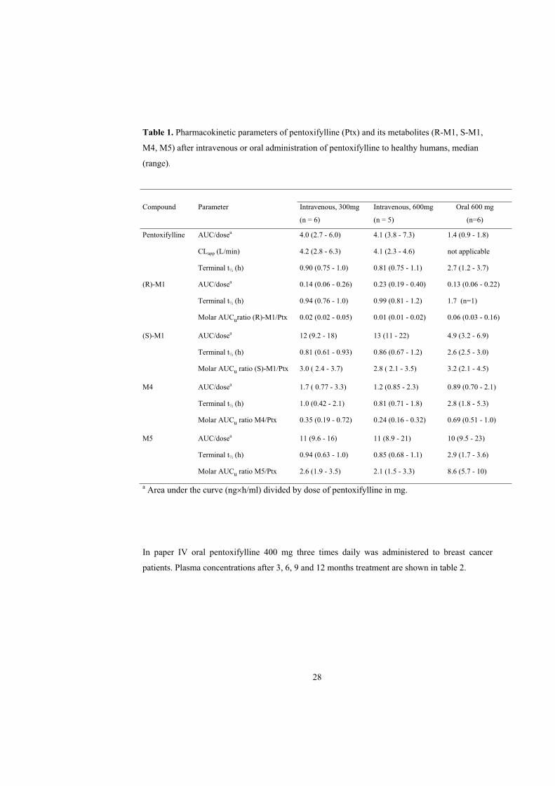
Table 1. Pharmacokinetic parameters of pentoxifylline (Ptx) and its metabolites (R-M1, S-M1,
M4, M5) after intravenous or oral administration of pentoxifylline to healthy humans, median
(range).
Compound Parameter Intravenous, 300mg
(n = 6)
Intravenous, 600mg
(n = 5)
Oral 600 mg
(n=6)
Pentoxifylline AUC/dosea 4.0 (2.7 - 6.0) 4.1 (3.8 - 7.3) 1.4 (0.9 - 1.8)
CLapp (L/min) 4.2 (2.8 - 6.3) 4.1 (2.3 - 4.6) not applicable
Terminal t½ (h) 0.90 (0.75 - 1.0) 0.81 (0.75 - 1.1) 2.7 (1.2 - 3.7)
(R)-M1 AUC/dosea 0.14 (0.06 - 0.26) 0.23 (0.19 - 0.40) 0.13 (0.06 - 0.22)
Terminal t½ (h) 0.94 (0.76 - 1.0) 0.99 (0.81 - 1.2) 1.7 (n=1)
Molar AUCuratio (R)-M1/Ptx 0.02 (0.02 - 0.05) 0.01 (0.01 - 0.02) 0.06 (0.03 - 0.16)
(S)-M1 AUC/dosea 12 (9.2 - 18) 13 (11 - 22) 4.9 (3.2 - 6.9)
Terminal t½ (h) 0.81 (0.61 - 0.93) 0.86 (0.67 - 1.2) 2.6 (2.5 - 3.0)
Molar AUCu ratio (S)-M1/Ptx 3.0 ( 2.4 - 3.7) 2.8 ( 2.1 - 3.5) 3.2 (2.1 - 4.5)
M4 AUC/dosea 1.7 ( 0.77 - 3.3) 1.2 (0.85 - 2.3) 0.89 (0.70 - 2.1)
Terminal t½ (h) 1.0 (0.42 - 2.1) 0.81 (0.71 - 1.8) 2.8 (1.8 - 5.3)
Molar AUCu ratio M4/Ptx 0.35 (0.19 - 0.72) 0.24 (0.16 - 0.32) 0.69 (0.51 - 1.0)
M5 AUC/dosea 11 (9.6 - 16) 11 (8.9 - 21) 10 (9.5 - 23)
Terminal t½ (h) 0.94 (0.63 - 1.0) 0.85 (0.68 - 1.1) 2.9 (1.7 - 3.6)
Molar AUCu ratio M5/Ptx 2.6 (1.9 - 3.5) 2.1 (1.5 - 3.3) 8.6 (5.7 - 10)
a Area under the curve (ng h/ml) divided by dose of pentoxifylline in mg.
In paper IV oral pentoxifylline 400 mg three times daily was administered to breast cancer
patients. Plasma concentrations after 3, 6, 9 and 12 months treatment are shown in table 2.
28
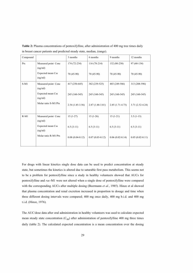
Table 2: Plasma concentrations of pentoxifylline, after administration of 400 mg tree times daily
in breast cancer patients and predicted steady state, median, (range).
Compound 3 months 6 months 9 months 12 months
Ptx Measured point Conc
(ng/ml)
Expected mean Css
(ng/ml)
174 (72-234)
70 (45-90)
116 (78-214)
70 (45-90)
152 (80-230)
70 (45-90)
97 (60-134)
70 (45-90)
S-M1 Measured point Conc
(ng/ml)
Expected mean Css
(ng/ml)
Molar ratio S-M1/Ptx
417 (250-645)
245 (160-345)
2.54 (1.85-3.56)
382 (239-525)
245 (160-345)
2.87 (1.88-3.81)
403 (249-586)
245 (160-345)
2.85 (1.71-4.73)
313 (208-596)
245 (160-345)
3.71 (2.52-4.24)
R-M1 Measured point Conc
(ng/ml)
Expected mean Css
(ng/ml)
Molar ratio R-M1/Ptx
15 (1-27)
6.5 (3-11)
0.08 (0.04-0.12)
15 (1-26)
6.5 (3-11)
0.07 (0.03-0.12)
15 (1-21)
6.5 (3-11)
0.06 (0.02-0.14)
3.5 (1-13)
6.5 (3-11)
0.05 (0.02-0.11)
For drugs with linear kinetics single dose data can be used to predict concentration at steady
state, but sometimes the kinetics is altered due to saturable first pass metabolism. This seems not
to be a problem for pentoxifylline since a study in healthy volunteers showed that AUCs for
pentoxifylline and rac-M1 were not altered when a single dose of pentoxifylline were compared
with the corresponding AUCs after multiple dosing (Beermann et al., 1985). Hinze et al showed
that plasma concentration and renal excretion increased in proportion to dosage and time when
three different dosing intervals were compared; 400 mg once daily, 400 mg b.i.d. and 400 mg
t.i.d. (Hinze, 1976).
The AUC/dose data after oral administration in healthy volunteers was used to calculate expected
mean steady state concentration (Css) after administration of pentoxifylline 400 mg three times
daily (table 2). The calculated expected concentration is a mean concentration over the dosing
29
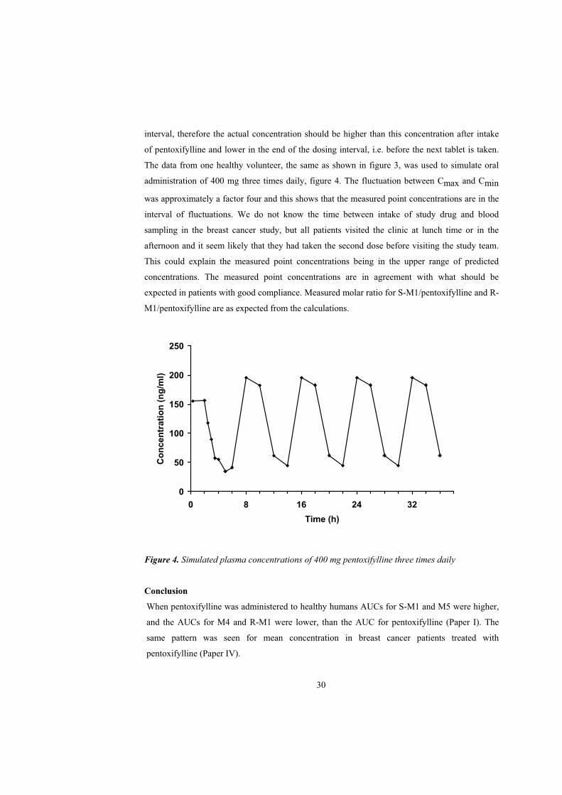
interval, therefore the actual concentration should be higher than this concentration after intake
of pentoxifylline and lower in the end of the dosing interval, i.e. before the next tablet is taken.
The data from one healthy volunteer, the same as shown in figure 3, was used to simulate oral
administration of 400 mg three times daily, figure 4. The fluctuation between Cmax and Cmin
was approximately a factor four and this shows that the measured point concentrations are in the
interval of fluctuations. We do not know the time between intake of study drug and blood
sampling in the breast cancer study, but all patients visited the clinic at lunch time or in the
afternoon and it seem likely that they had taken the second dose before visiting the study team.
This could explain the measured point concentrations being in the upper range of predicted
concentrations. The measured point concentrations are in agreement with what should be
expected in patients with good compliance. Measured molar ratio for S-M1/pentoxifylline and R-
M1/pentoxifylline are as expected from the calculations.
0
50
100
150
200
250
0 8 16 24 32
Time (h)
Co
nce
ntr
atio
n (
ng
/ml)
Figure 4. Simulated plasma concentrations of 400 mg pentoxifylline three times daily
Conclusion
When pentoxifylline was administered to healthy humans AUCs for S-M1 and M5 were higher,
and the AUCs for M4 and R-M1 were lower, than the AUC for pentoxifylline (Paper I). The
same pattern was seen for mean concentration in breast cancer patients treated with
pentoxifylline (Paper IV).
30
Paper I
Interconversion of pentoxifylline and M1 by erythrocytes
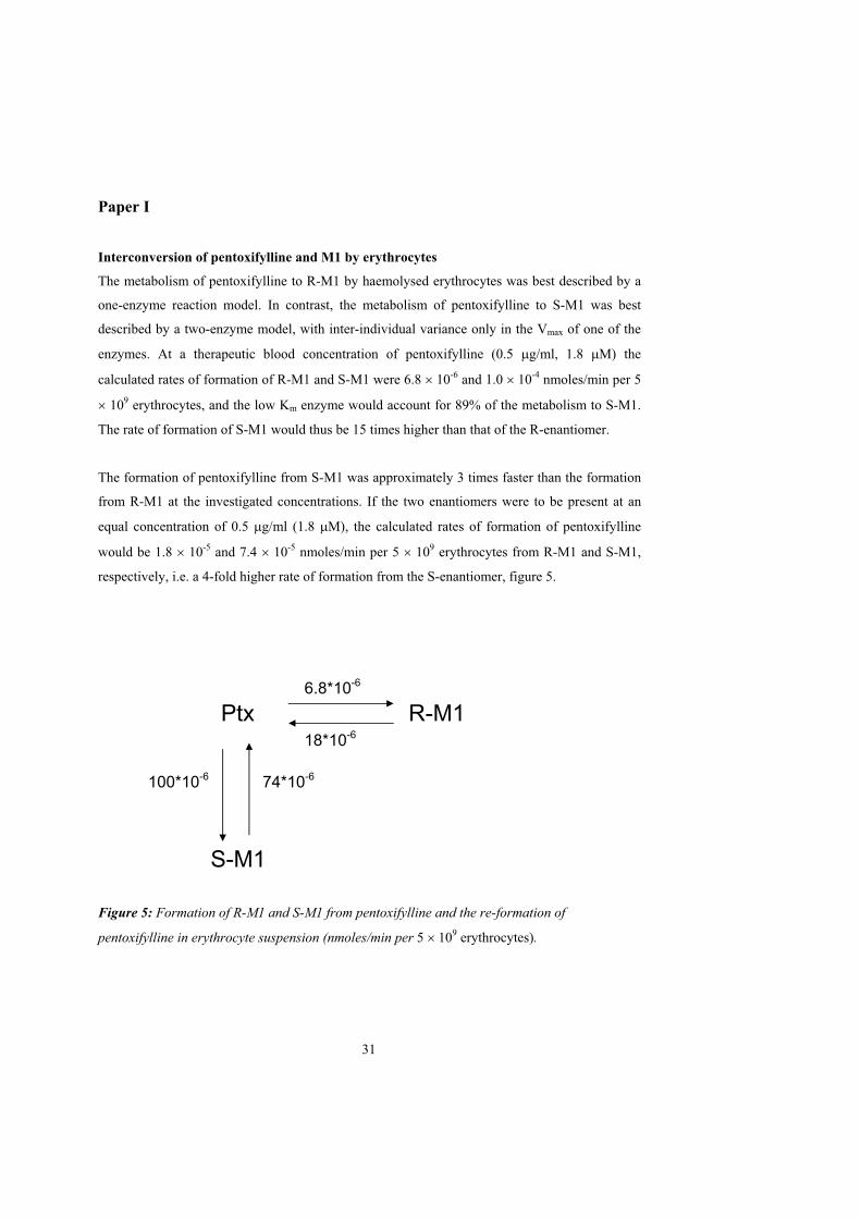
The metabolism of pentoxifylline to R-M1 by haemolysed erythrocytes was best described by a
one-enzyme reaction model. In contrast, the metabolism of pentoxifylline to S-M1 was best
described by a two-enzyme model, with inter-individual variance only in the Vmax of one of the
enzymes. At a therapeutic blood concentration of pentoxifylline (0.5 g/ml, 1.8 M) the
calculated rates of formation of R-M1 and S-M1 were 6.8 10-6 and 1.0 10-4 nmoles/min per 5
109 erythrocytes, and the low Km enzyme would account for 89% of the metabolism to S-M1.
The rate of formation of S-M1 would thus be 15 times higher than that of the R-enantiomer.
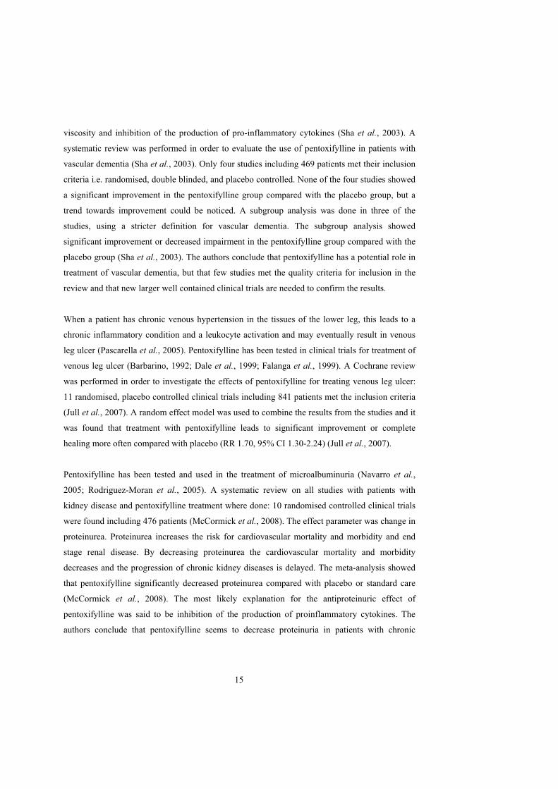
The formation of pentoxifylline from S-M1 was approximately 3 times faster than the formation
from R-M1 at the investigated concentrations. If the two enantiomers were to be present at an
equal concentration of 0.5 g/ml (1.8 M), the calculated rates of formation of pentoxifylline
would be 1.8 10-5 and 7.4 10-5 nmoles/min per 5 109 erythrocytes from R-M1 and S-M1,
respectively, i.e. a 4-fold higher rate of formation from the S-enantiomer, figure 5.
Figure 5: Formation of R-M1 and S-M1 from pentoxifylline and the re-formation of
pentoxifylline in erythrocyte suspension (nmoles/min per 5 109 erythrocytes).
Ptx6.8*10-6
R-M118*10-6
100*10-6 74*10-6
S-M1
31
Conclusion
In erythrocyte suspension the transformation of pentoxifylline to M1 was highly stereospecific
in favor of the S-enantiomer and was reversible from both enantiomers. A 15 fold faster
formation of S-M1 than of R-M1 was estimated in erythrocytes. This seems to be the main
reason for the marked difference in plasma concentration and AUC of the two enantiomers.
Paper II
This study was planned in order to investigate the possible contributions of the metabolites of
pentoxifylline to the haemorheologic effect in humans. Measurement of retinal blood flow was
chosen as a convenient experimental model. By pre-treatment of the subjects with ciprofloxacin a
known inhibitor of CYP1A2 (Fuhr et al., 1992; Peterson et al., 2004) and rifampicin, an inducer
of several of the cytochrome P450 enzymes (Niemi et al., 2003) we succeeded to diminish the
usual high correlation between the concentrations of pentoxifylline and metabolites. Thereby
enabling us to investigate whether pentoxifylline´s metabolites have any effect on retinal blood
flow. We found that AUCs for pentoxifylline and S-M1 were significantly higher after pre-
treatment with ciprofloxacin compared with pentoxifylline administration alone. The effect was
seen on R-M1 as well but was not statistically significant. However the R/S plasma concentration
ratio remained unchanged. The AUC for M5 was significantly lower after pre-treatment with
rifampicin compared with after pentoxifylline alone. The estimated intercept and coefficients
from the mixed model applying a simple linear AUC-effect model are shown in table 3. We
found that pentoxifylline; R-M1 and M5 had coefficients with positive signs indicating that they
all enhanced retinal blood flow. In contrast M4 and S-M1 had negative sign and therefore
appeared to either decrease retinal blood flow or to counteract the pentoxifylline, R-M1 and M5.
R-M1 has a high potency, this is reflected in the values of the coefficients (Table 3). It is
particularly noticeable that R-M1 exerts a significant positive effect in spite of being present in
concentrations that are approximately two orders of magnitude lower than those of pentoxifylline
and M5.
32

The expected mean flow should be calculated as:
245 + 17.1*(AUCptx ) + 303*(AUCR-M1 ) - 9.58* (AUCS-M1) - 54.5*(AUCM4) + 6.66* (AUCM5)
Table 3. Estimates (95% CI) of the intercept and gradients from the regression analysis of the
influence from pentoxifylline (Ptx) and its metbolites on retinal blood flow measured as mean
flow.
Mean flow
Intercept AU† 245 (186 - 304)AUC Ptx AU†/(μg*h/ml)
17.1* (4.31 - 29.9)
AUC R-M1 AU†/(μg*h/ml)
303* (147 - 459)
AUC S-M1 AU†/(μg*h/ml)
-9.58* (-13.6 - -5.52)
AUC M4 AU†/(μg*h/ml)
-54.5* (-93.4 - -15.5)
AUC M5 AU†/(μg*h/ml)
6.66* (0.675 - 12.6)
* P< 0.05
Conclusion
The R-M1 and M5 metabolites of pentoxifylline contribute significantly to pentoxifyllines
enhancement on retinal blood flow in humans.
Paper III
The aim of this study was to investigate the relative potencies of pentoxifylline and metabolite R-
M1, S-M1, M4 and M5 to inhibit platelet aggregation in whole blood, and in particular to clarify
contributions of the two enantiomers of M1, that are formed to very different extents in vivo, to
this effect.
33
0 1 2 3 4
01
020
304
0
A
Concentration (mM)
Imp
edan
ce (
Ohm
)
_
_
__
__
_
_
_
_
__
__
_
_
_
_
_ _ __
__
__
_ _ __
_ _ _ _
0 1 2 3 4
01
020
304
0
B
Concentration (mM)
Imp
edan
ce (
Ohm
)
_
_
_
_
_
_
_
_
_
_
__
_
_
_
_
_
_
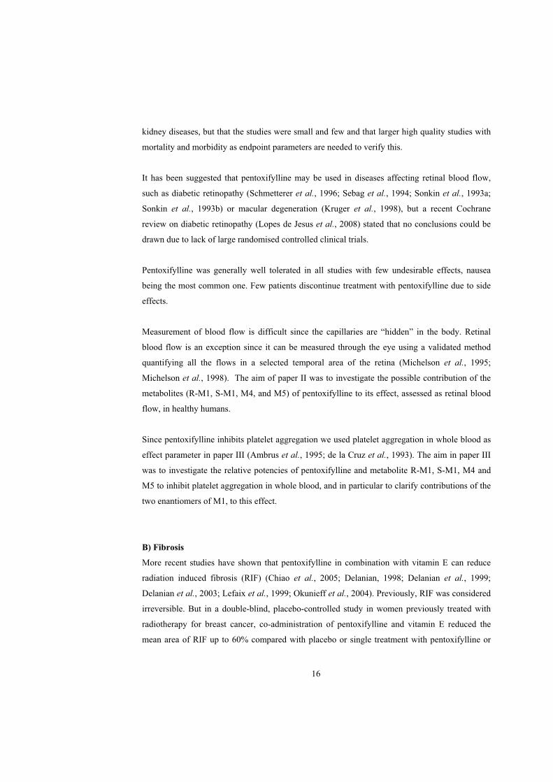
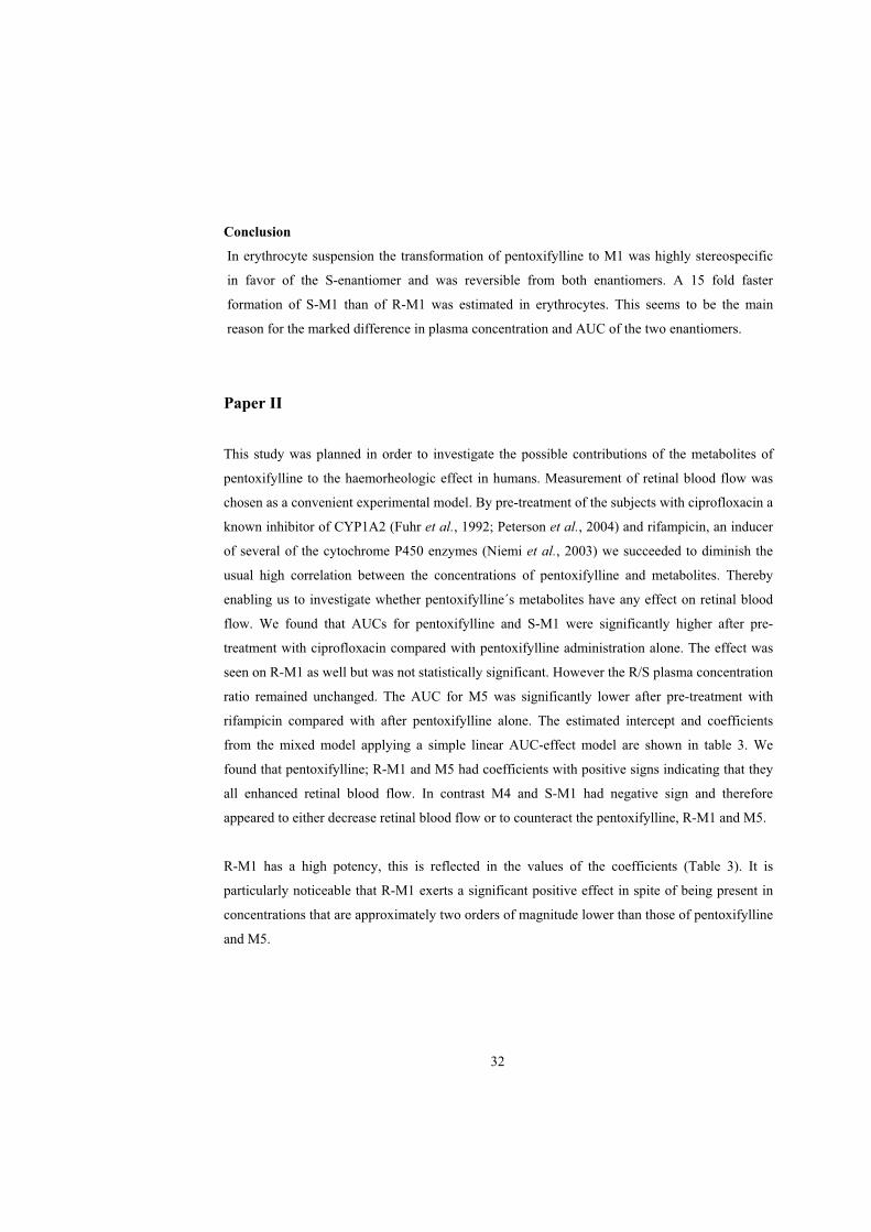
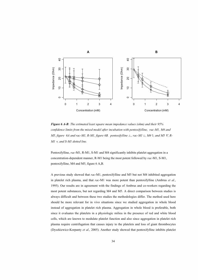
Figure 6 A-B: The estimated least square mean impedance values (ohm) and their 95%
confidence limits from the mixed model after incubation with pentoxifylline, rac-M1, M4 and
M5, figure 6A and rac-M1, R-M1, figure 6B. pentoxifylline , rac-M1 , M4 , and M5 , R-
M1 , and S-M1 dotted line.
Pentoxifylline, rac-M1, R-M1, S-M1 and M4 significantly inhibits platelet aggregation in a
concentration-dependent manner, R-M1 being the most potent followed by rac-M1, S-M1,
pentoxifylline, M4 and M5, figure 6 A,B.
A previous study showed that rac-M1, pentoxifylline and M5 but not M4 inhibited aggregation
in platelet rich plasma, and that rac-M1 was more potent than pentoxifylline (Ambrus et al.,
1995). Our results are in agreement with the findings of Ambrus and co-workers regarding the
most potent substances, but not regarding M4 and M5. A direct comparison between studies is
always difficult and between these two studies the methodologies differ. The method used here
should be more relevant for in vivo situations since we studied aggregation in whole blood
instead of aggregation in platelet rich plasma. Aggregation in whole blood is preferable, both
since it evaluates the platelets in a physiologic milieu in the presence of red and white blood
cells, which are known to modulate platelet function and also since aggregation in platelet rich
plasma require centrifugation that causes injury to the platelets and loss of giant thrombocytes
(Dyszkiewicz-Korpanty et al., 2005). Another study showed that pentoxifylline inhibits platelet
34
aggregation in whole blood more than in platelet rich plasma (de la Cruz et al., 1993). In
addition, Ambrus et al. could not distinguish between the enantiomers of M1, which, as shown
here, differ significantly in their potencies.
In vivo there will always be a mixture of the parent compound and its metabolites after
administration of pentoxifylline, which allows pharmacological interactions between the
different species (an aspect that has not been investigated in vitro, where only one specimen is
added at the time, with the exception of the racemate). In addition, the relative plasma
concentrations of pentoxifylline and the metabolites will be very different from those in the in
vitro experiments, so that activities are compared for different parts of the underlying
concentration – effect curves.
We therefore wanted to estimate the relative contribution of each substance to the total effect of
pentoxifylline on platelet aggregation in vivo. The potencies found in this study was used
together with the concentration data of pentoxifylline and its metabolites in humans after
administration of pentoxifylline (paper I). When combing these results we conclude that the main
effect on platelet aggregation in vivo should actually be brought about by S-M1 and
pentoxifylline, and that the remaining metabolites would contribute by less than 10 % each.
Thus, even if R-M1 is twice as potent as S-M1 in vitro the low concentration achieved after
administration of pentoxifylline results in only a small contribution to the total effect on platelet
aggregation in vivo. However, further studies are needed in order to confirm this.
In this thesis the effects of the R and S enantiomers of M1 have been described for the first time.
Both enantiomers were active in inhibiting platelet aggregation in whole blood whereas only R-
M1 was effective in increasing retinal blood flow. The activity of M4 and M5 also differs
between the two effect models, M5 increases retinal blood flow but does not inhibit platelet
aggregation in whole blood, whereas M4 inhibits platelet aggregation but does not increase
retinal blood flow. The effects of both S-M1, M4 and M5 are limited whereas R-M1 was more
effective than pentoxifylline in both effect models. On retinal blood flow the R-enantiomer was
up to 17 times as effective as pentoxifylline but on aggregation it was only approximately 1.25 as
effective. The differences in activities for the metabolites may be explained by different
mechanism of action in the two effect models.
35
Conclusion
In the following potency order R-M1, rac-M1, pentoxifylline, S-M1 and M4 all significantly
inhibit platelet aggregation in whole blood in vitro.
Paper IV
Between May 2004 and May 2007, a total of 83 patients were included in the study, 42 patients
were randomised to pentoxifylline treatment and 41 to the placebo group. Most patients (67)
were treated with previous chemotherapy, 33 in the pentoxifylline group and 34 in the placebo
group.
Both treatments were generally well tolerated. Only four patients discontinued due to adverse
events: two due to nausea and one due to bruising in the pentoxifylline group and one due to
neuropathic pain in the placebo group. A safety analysis was done in September 2008 when all
patients had been included in the study for a median period of 31 months (range 16-52 months).
Although the study was not dimensioned for safety, we observed no significant differences
between the study groups in terms of safety, including disease recurrence, death, and adverse
events. This shows that pentoxifylline in combination with vitamin E is safe to use
prophylactically.
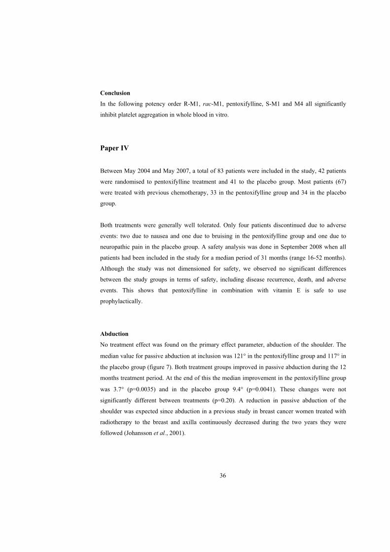
Abduction
No treatment effect was found on the primary effect parameter, abduction of the shoulder. The
median value for passive abduction at inclusion was 121 in the pentoxifylline group and 117 in
the placebo group (figure 7). Both treatment groups improved in passive abduction during the 12
months treatment period. At the end of this the median improvement in the pentoxifylline group
was 3.7 (p=0.0035) and in the placebo group 9.4 (p=0.0041). These changes were not
significantly different between treatments (p=0.20). A reduction in passive abduction of the
shoulder was expected since abduction in a previous study in breast cancer women treated with
radiotherapy to the breast and axilla continuously decreased during the two years they were
followed (Johansson et al., 2001).
36
Instead, the fortunate result was found that shoulder abduction improved in both treatment
groups. Changes that can explain this has been made in both in the physiotherapy and the
radiation technique in order to diminish radiation-induced side effects. The changes in
physiotherapy involve changes in the exercise program that patients receives after radiotherapy,
before the exercise program only included shoulder motion. Now, all patients receive a training
program mostly focused on stretching the shoulder and breast area. Additionally, the radiation
technique has been refined to diminish radiation-induced side effects. The radiation dose to the
muscle tissue around the caput humerus and tissue around the axilla has been diminished. The
discrepancies between the studies could be explained by one or a combination of both factors. A
third but less likely explanation could be that vitamin E causes the improvement since all patients
received vitamin E.
0 3 6 9 12
608
01
0012
014
01
6018
0
Time (months)
Abd
uct
ion
(deg
ree
s)
0 3 6 9 12
608
01
0012
014
01
6018
0
Figure 7: Box-plot of passive abduction of the shoulder by treatment group (blue placebo, red
pentoxifylline) and visit.
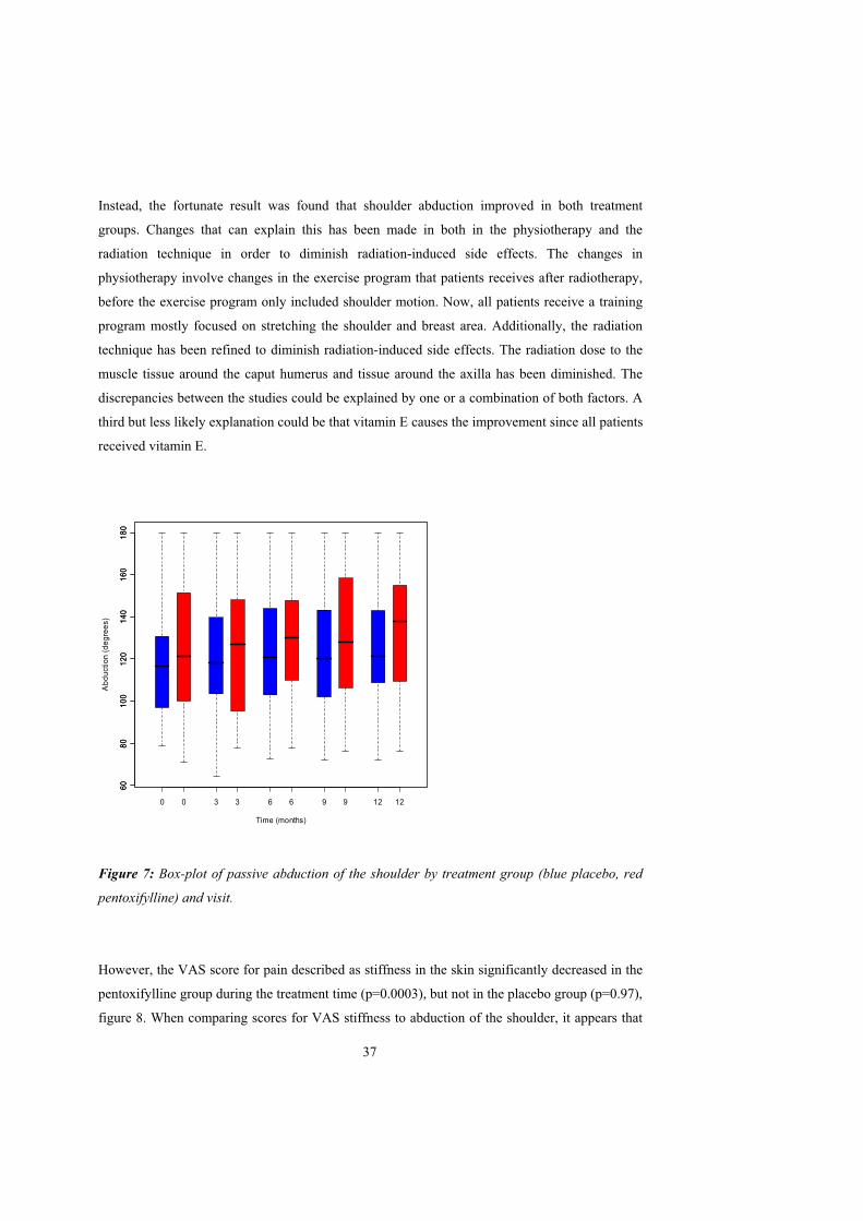
However, the VAS score for pain described as stiffness in the skin significantly decreased in the
pentoxifylline group during the treatment time (p=0.0003), but not in the placebo group (p=0.97),
figure 8. When comparing scores for VAS stiffness to abduction of the shoulder, it appears that
37
high VAS stiffness score is connected to lower degrees of abduction, i.e. less ability to move the
arm. The sense of stiffness may be a harbinger of reduced abduction of the shoulder. Radiation
induced side effect develops over time and all patients will therefore be followed for 5 years.
0 3 6 9 12
02
04
06
0
Time (months)
VA
S (
mm
)
0 3 6 9 12
02
04
06
0
Figure 8: Box-plot of VAS for stiffness of the skin by treatment group (blue placebo, red
pentoxifylline) and visit in the 38 patients reporting this phenomenon at least once during the
study.
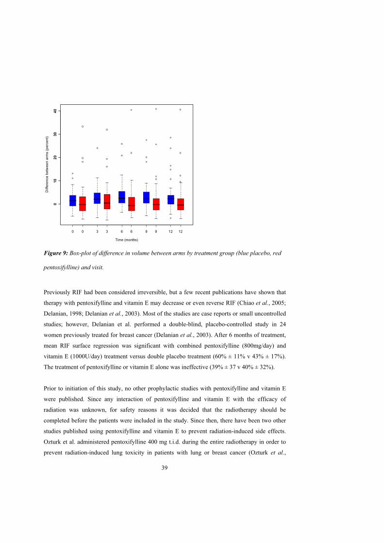
Volume
At study start there was no significant median difference in arm volume between the patient’s
affected and unaffected arms (figure 9). Arm volume increased over time in the placebo group
but not in the pentoxifylline group. At the end of the treatment period the median increase was
1.04% in the placebo group and 0.50% in the pentoxifylline group, and was significant between
groups (p=0.0172).
38
0 3 6 9 12
01
02
03
04
0
0 3 6 9 12
01
02
03
04
0
Time (months)
Diff
ere
nce
bet
we
en
arm
s (p
erc
en
t)
Figure 9: Box-plot of difference in volume between arms by treatment group (blue placebo, red
pentoxifylline) and visit.
Previously RIF had been considered irreversible, but a few recent publications have shown that
therapy with pentoxifylline and vitamin E may decrease or even reverse RIF (Chiao et al., 2005;
Delanian, 1998; Delanian et al., 2003). Most of the studies are case reports or small uncontrolled
studies; however, Delanian et al. performed a double-blind, placebo-controlled study in 24
women previously treated for breast cancer (Delanian et al., 2003). After 6 months of treatment,
mean RIF surface regression was significant with combined pentoxifylline (800mg/day) and
vitamin E (1000U/day) treatment versus double placebo treatment (60% ± 11% v 43% ± 17%).
The treatment of pentoxifylline or vitamin E alone was ineffective (39% ± 37 v 40% ± 32%).
Prior to initiation of this study, no other prophylactic studies with pentoxifylline and vitamin E
were published. Since any interaction of pentoxifylline and vitamin E with the efficacy of
radiation was unknown, for safety reasons it was decided that the radiotherapy should be
completed before the patients were included in the study. Since then, there have been two other
studies published using pentoxifylline and vitamin E to prevent radiation-induced side effects.
Ozturk et al. administered pentoxifylline 400 mg t.i.d. during the entire radiotherapy in order to
prevent radiation-induced lung toxicity in patients with lung or breast cancer (Ozturk et al.,
39
2004). A significant protective effect of pentoxifylline compared with placebo for both early and
late lung radiotoxicity was reported. In another study, pentoxifylline 400 mg t.i.d. was given
during radiotherapy to patients with squamous cell carcinoma of the head and neck (Aygenc et
al., 2004). Late skin changes, fibrosis, and soft tissue necrosis were more severe in the control
group than in the pentoxifylline group. These studies indicate that it is safe to give pentoxifylline
and vitamin E during the radiotherapy. When planning further studies, we intend to administer
these treatments concomitant with the radiotherapy; earlier treatment may lead to greater
protective effects.
Conclusion
The combination of pentoxifylline and vitamin E is safe and can be used to prevent some
radiation-induced side effects. It was found that pentoxifylline in combination with vitamin E
diminished the development of increased arm volume. No significant effects on abduction of the
shoulder were seen but VAS for stiffness in the skin was significantly decreased in the
pentoxifylline group.
40
CONCLUSIONS
In erythrocyte suspension the transformation of pentoxifylline to M1 was highly
stereospecific in favor of the S-enantiomer and was reversible from both enantiomers
(Paper I).
When pentoxifylline was administered to healthy humans AUCs for S-M1 and M5 were
higher, and the AUCs for M4 and R-M1 were lower, than the AUC for pentoxifylline
(Paper I, II). The same pattern was seen for mean concentration in breast cancer patients
treated with pentoxifylline (Paper IV).
Pentoxifylline, R-M1 and M5 all significantly increased retinal blood flow. In contrast S-
M1 and M4 appeared to decrease the blood flow after intravenous infusions of
pentoxifylline to healthy humans (Paper II).
In the following potency order R-M1, rac-M1, pentoxifylline, S-M1 and M4 all
significantly inhibit platelet aggregation in whole blood in vitro (Paper III).
Pentoxifylline in combination with vitamin E is safe and can be used to prevent some
radiation induced side effects such as increased arm volume in women with breast cancer
(Paper IV).
41
SUMMARY
Pentoxifylline is a haemorheologic drug that has been used for a long time for the treatment of
intermittent claudication and other diseases with impaired microcirculation. Pentoxifylline is an
interesting drug to study since it exhibit complex pharmacokinetics with both reversible
metabolism, and active metabolites. Difficulties in finding consistent clinical effects of
pentoxifylline may be due to the drug acting at least in part through formation of active
metabolites, the rate and extent of which may vary between individuals. In humans,
pentoxifylline is metabolised into at least seven phase 1 metabolites (M1-M7). The reversible
metabolism of pentoxifylline to the enantiomers of M1 has only been partly studied.
This thesis investigates the pharmacokinetics of pentoxifylline and metabolites and their
contributions to the haemorheological effects.
When pentoxifylline is administered either orally or intravenously to healthy humans the plasma
concentrations of M5 and S-M1 are higher than the pentoxifylline concentrations, whereas the
ones for M4 are lower and R-M1 much lower. In-vitro studies showed that this can be mainly
explained by a 15 times faster formation of S-M1 than R-M1 from pentoxifylline. Had the
enantiomers been present at equal concentrations the reversible metabolism would have been 4
times faster from S-M1 than from R-M1.
Pentoxifylline was administered to healthy volunteers and retinal blood flow was measured. By
pre-treatment of the subjects with ciprofloxacin and rifampicin, the usual high correlation
between the concentrations of pentoxifylline and the metabolites was diminished and the effects
of pentoxifylline and its metabolites could be studied. Pentoxifylline, R-M1 and M5 seem to
increase retinal blood. When present at equal concentration R-M1 is approximately 15 times as
effective as pentoxifylline. M4 and S-M1 appears to either decrease retinal blood flow or to
counteract the pentoxifylline, R-M1 and M5.
Further, the effects of pentoxifylline and its metabolites on platelet aggregation in whole blood
was investigated. The study showed that pentoxifylline, rac-M1, R-M1, S-M1 and M4
significantly inhibits platelet aggregation in a concentration-dependent manner. R-M1 being the
most potent followed by rac-M1, S-M1, pentoxifylline, M4 and M5.
42
More recent studies have shown that pentoxifylline in combination with vitamin E can reduce
radiation induced fibrosis (RIF). A study was planned to investigate whether the same drug can
prevent radiation induced fibrosis in women with breast cancer, treated with radiotherpay to the
breast and axilla. The clinical trial was randomised, double-blind, placebo-controlled, and the 83
patients included in the study were treated with pentoxifylline or placebo in combination with
vitamin E for 12 months. Abduction of the shoulder was the primary effect parameter and arm
volume the secondary. The study showed that pentoxifylline in combination with vitamin E
diminishes the development of increased arm volume. Significant effects on abduction of the
shoulder were not seen, but the VAS for stiffness in the skin decreased: this may be a harbinger
of reduced abduction of the shoulder. Radiation induced side effect develops over time and all
patients will be followed for 5 years. The combination of pentoxifylline and vitamin E appeared
safe and may be used for prevention of some radiation-induced side effects.
43
POPULÄRVETENSKAPLIG SAMMANFATTNING – Swedish summary
Farmakokinetik beskriver hur ett läkemedel omsätts i kroppen och farmakodynamik beskriver
sambandet mellan koncentration och effekt. Pentoxifyllin är ett läkemedel med en komplicerad
farmakokinetik med aktiva metaboliter och reversibel metabolism. Pentoxifyllin har aldrig
registrerats i Sverige, till stor del beroende på bristande och/eller föråldrad dokumentation;
emellertid förekommer en omfattande förskrivning av läkemedlet på licens. Studier med
pentoxifyllin har gjorts på många olika indikationer men läkemedlet används framför allt vid
claudicatio intermittens, fönstertittarsjukan. Fönstertittarsjukan innebär att blodflödet till framför
allt benen är otillräckligt pga. ateroskleros, detta medför nedsatt rörlighet och smärta för
patienten. Pentoxifyllin förbättrar blodets reologi, d.v.s. hur ”bra” blodet flyter i blodkärlen.
Huvudmålet med denna avhandling var att titta noggrannare på pentoxifyllins farmakokinetik
och farmakodynamik, samt att objektivt försöka mäta effekten av pentoxifyllin och dess
metaboliter. Detta för att se om någon eller några av metaboliterna bidrar till pentoxifyllins
effekter och därmed förklarar skillnaderna i effekt.
När pentoxifyllin bryts ner i kroppen bildas ett flertal metaboliter, metabolit 1, 4 och 5 (M1, M4
och M5) bildas i sådan utsträckning att de kan vara kliniskt betydelsefulla. M1 har dessutom ett
kiralt centrum och metaboliten förekommer således som två stereoisomerer, benämnda R-M1
och S-M1. R-M1 och S-M1 är varandras spegelbilder och förhåller sig till varandra som en
högerhand till en vänsterhand. Dessa stereoisomerer kan ha olika farmakologiska effekter. Det är
inte tidigare klarlagt i vilken omfattning R och S-M1 bildas efter administration av pentoxifyllin.
Detta undersöktes i det första arbetet, en klinisk prövning på friska frivilliga. Försökspersonerna
fick pentoxifyllin som intravenös injektion eller som en tablett. Studien visade att vid dosering av
pentoxifyllin så bildas framför allt S-M1, kvoten av plasmakoncentrationerna för R/S M1 var
0,01-0,04. Vidare undersöktes varför framför allt S-M1 bildas, detta gjordes i erytrocyter
eftersom en betydande del av metabolismen sker där. Studien visar att både R-M1 och S-M1
tillbakabildas till pentoxifyllin och att hastigheten för bildandet av S-M1 var 15 gånger högre än
R-M1. Detta är huvudförklaringen till S-M1:s höga och R-M1:s låga plasmakoncentrationer.
I de två följande delarbeterna studeras effekterna av pentoxifyllin och det undersöks om dess
metaboliter bidrar till dess effekter vilket skulle kunna förklara variabiliteten i effekt hos
patienterna.
44
En klinisk prövning på friska frivilliga genomfördes för att undersöka pentoxifyllin och dess
metaboliters effekt på ögonblodflöde i näthinnan mätt med objektiv metodik. Pentoxifyllin
visade sig öka retinalt ögonblodflöde jämfört med placebo. När area under kurva (AUC) av
plasmakoncentrationerna användes som förklaringsvariabler till retinalt ögonblodflöde fann vi att
R-M1 och M5 ökar retinalt blodflöde medan S-M1 och M4 minskar retinalt blodflöde eller
minskar effekten av pentoxifyllin och R-M1.
I delarbete III undersöktes om pentoxifyllin och dess metaboliter hämmar trombocyt aggregation
i helblod. Det visade sig att pentoxifyllin racemiskt M1 (50% R-M1, 50% S-M1), R-M1, S-M1
och M4 hämmar trombocyt aggregation i helblod på ett koncentrationsberoende sätt. Vi fann att
R-M1 var den potentaste substansen följt av rac-M1, S-M1, pentoxifyllin och M4.
På senare tid har studier visat att pentoxifyllin kan minska eller till och med tillbakabilda
fibrosområden i huden vilka uppkommit efter strålbehandling. Dessa fibrosytor utvecklas ofta
några år efter avslutad strålning och medför besvär med nedsatt rörlighet, smärta och
ödemutveckling. I delarbete IV undersöks om pentoxifyllin kan förhindra dessa biverkningar
efter strålbehandling.
En randomiserad, placebokontrollerad, dubbelblind klinisk prövning genomfördes på 83 kvinnor
som behandlats för bröstcancer med kirurgi följt av strålning. Målet med studien var att förhindra
biverkningar från strålbehandlingen såsom nedsatt axelrörlighet och armlymfödem som är
vanligt förekommande. Patienterna behandlades under ett år med pentoxifyllin eller placebo i
kombination med E-vitamin. En första analys av pentoxifyllins effekter gjordes efter
behandlingstidens slut. Pentoxifyllin visade sig minska armödemutveckling och känslan av
stramhet i armen, däremot sågs inte någon signifikant effekt på patientens axelrörlighet.
Biverkningar efter radioterapi utvecklas under flera år efter avslutad strålning, det är möjligt att
större/mer effekter av pentoxifyllin ses om något år. Studien pågår fortfarande och patienterna
kommer att följas minst 5 år efter avslutad strålning.
45

ACKNOWLEDGEMENTS
I wish to thank:
First and foremost my supervisor Peter Höglund for guiding me through my Ph.D years, for sharing your great scientific knowledge with me, and for always taking time to answer my questions.
My second supervisor Sven Björkman, for all your skilfull work and supportespecially during my first years as a Ph. D student.
Pål Stenberg, former head of the Hospital Pharmacy in Malmö, for employing me and making it possible to have a “normal job” and perform/finish a Ph. D at the same time.
The Department of Clinical Chemistry and Pharmacology for giving me the opportunity to be a Ph. D student and defend my thesis at the department.
Bodil Roth, co author, for all your invaluable help in the lab, technical assistance and support throughout the years.
Elisabeth Kjellén, co author and principal investigator, for all your positive energy, support, encouragement and all your work in the Ptx-5 study.
Anna Weddig, co author, for your support and encouragement and for taking such good care of the patients in the Ptx-5 study.
Margoth Gunnarsson, co author, for all your help in the lab with the whole blood aggregation.
All other co-authors that have worked with me and helped me during my Ph. D years.
All present and former colleagues at the Hospital Pharmacy for creating an enjoyable and inspiring athmosphere, in particular; Lisbet, Maria, Emil and Camilla for working with me in the Clinical trial team and for making the work with clinical trials so fun. To Anna and Sofia for your encouragement and support and for keeping my spirits up and making me laugh even during the ”bad days”.
To Kajsa, for sharing your office at BMC with me while I write this thesis, and for being such a great friend.
46
Jag vill också tacka min familj och mina vänner utanför jobbet, ni förgyller mitt liv, särskilt tack till:
”Lundaligan”, Anna M, Anna W, Annika, Cilla, Kajsa, Ingalill och Ulrika för er ovärderliga vänskap, stöd, och uppmuntran, för alla fikakvällar, fester och annat roligt med er och era familjer.
Linda och Maria för fantastisk vänskap under alla år, för alla roliga upplevelser tillsammans med er och era familjer.
”Semestergänget” Pernilla, Frédéric, Henrik, Jenny, Olof, Gunilla, Tomas, och Linnea för alla härliga semestrar och annat skoj. Extra tack till Pernilla och Frédéric för att jag får vara gudmor åt Amélie.
Mina underbara föräldrar, Margareta och Allan, för att ni finns där för mig och alltid ställer upp för mig och mina barn. Min syster Lena för att du är den bästa syster man kan tänka sig. Till Alfons, Didrik och Daniel för att ni sprider glädje i mitt liv.
Nisse och Aina för att ni är bästa tänkbara farmor och farfar till Linn och Erik.
Andreas, för all kärlek, för att du finns vid min sida, stöttar mig och gör mig glad och lycklig.
Linn och Erik, mina älskade barn, för all glädje och kärlek, ni är solstrålarna i mitt liv. Att komma hem efter en lång dag och mötas av två glada barn, Linn som säger – Mamma jag har saknat dig, och Erik som kommer lite efter men kämpar tappert för att få kramas först, det är lycka!
Finally, financial support is gratefully acknowledged from the Swedish CancerSociety, Carl Gustav and Lilly Lennhoffs foundation at the Swedish Academy of Pharmaceutical Sciences, and Cancer Research Foundation in Northern Sweden.
47
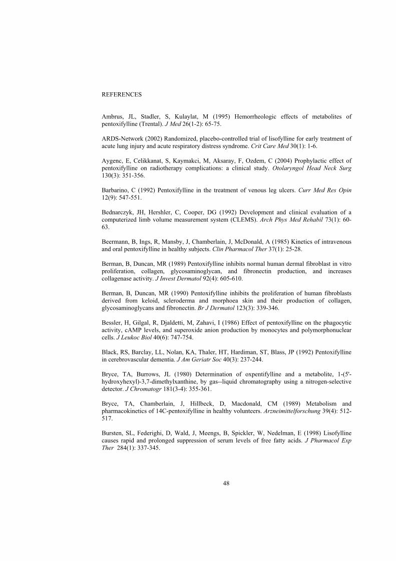
REFERENCES
Ambrus, JL, Stadler, S, Kulaylat, M (1995) Hemorrheologic effects of metabolites of pentoxifylline (Trental). J Med 26(1-2): 65-75.
ARDS-Network (2002) Randomized, placebo-controlled trial of lisofylline for early treatment of acute lung injury and acute respiratory distress syndrome. Crit Care Med 30(1): 1-6.
Aygenc, E, Celikkanat, S, Kaymakci, M, Aksaray, F, Ozdem, C (2004) Prophylactic effect of pentoxifylline on radiotherapy complications: a clinical study. Otolaryngol Head Neck Surg130(3): 351-356.
Barbarino, C (1992) Pentoxifylline in the treatment of venous leg ulcers. Curr Med Res Opin12(9): 547-551.
Bednarczyk, JH, Hershler, C, Cooper, DG (1992) Development and clinical evaluation of a computerized limb volume measurement system (CLEMS). Arch Phys Med Rehabil 73(1): 60-63.
Beermann, B, Ings, R, Mansby, J, Chamberlain, J, McDonald, A (1985) Kinetics of intravenous and oral pentoxifylline in healthy subjects. Clin Pharmacol Ther 37(1): 25-28.
Berman, B, Duncan, MR (1989) Pentoxifylline inhibits normal human dermal fibroblast in vitro proliferation, collagen, glycosaminoglycan, and fibronectin production, and increasescollagenase activity. J Invest Dermatol 92(4): 605-610.
Berman, B, Duncan, MR (1990) Pentoxifylline inhibits the proliferation of human fibroblasts derived from keloid, scleroderma and morphoea skin and their production of collagen, glycosaminoglycans and fibronectin. Br J Dermatol 123(3): 339-346.
Bessler, H, Gilgal, R, Djaldetti, M, Zahavi, I (1986) Effect of pentoxifylline on the phagocytic activity, cAMP levels, and superoxide anion production by monocytes and polymorphonuclearcells. J Leukoc Biol 40(6): 747-754.
Black, RS, Barclay, LL, Nolan, KA, Thaler, HT, Hardiman, ST, Blass, JP (1992) Pentoxifylline in cerebrovascular dementia. J Am Geriatr Soc 40(3): 237-244.
Bryce, TA, Burrows, JL (1980) Determination of oxpentifylline and a metabolite, 1-(5'-hydroxyhexyl)-3,7-dimethylxanthine, by gas--liquid chromatography using a nitrogen-selectivedetector. J Chromatogr 181(3-4): 355-361.
Bryce, TA, Chamberlain, J, Hillbeck, D, Macdonald, CM (1989) Metabolism and pharmacokinetics of 14C-pentoxifylline in healthy volunteers. Arzneimittelforschung 39(4): 512-517.
Bursten, SL, Federighi, D, Wald, J, Meengs, B, Spickler, W, Nedelman, E (1998) Lisofyllinecauses rapid and prolonged suppression of serum levels of free fatty acids. J Pharmacol Exp Ther 284(1): 337-345.
48
Chiao, TB, Lee, AJ (2005) Role of pentoxifylline and vitamin E in attenuation of radiation-induced fibrosis. Ann Pharmacother 39(3): 516-522.
Cochrane (2009) Pentoxifylline. The Cochrane Library.
Cummings, DM, Ballas, SK, Ellison, MJ (1992) Lack of effect of pentoxifylline on red bloodcell deformability. J Clin Pharmacol 32(11): 1050-1053.
Dale, JJ, Ruckley, CV, Harper, DR, Gibson, B, Nelson, EA, Prescott, RJ (1999) Randomised,double blind placebo controlled trial of pentoxifylline in the treatment of venous leg ulcers. BMJ319(7214): 875-878.
de la Cruz, JP, Romero, MM, Sanchez, P, Sanchez de la Cuesta, F (1993) Antiplatelet effect ofpentoxifylline in human whole blood. Gen Pharmacol 24(3): 605-609.
Delanian, S (1998) Striking regression of radiation-induced fibrosis by a combination of pentoxifylline and tocopherol. Br J Radiol 71(848): 892-894.
Delanian, S, Balla-Mekias, S, Lefaix, JL (1999) Striking regression of chronic radiotherapy damage in a clinical trial of combined pentoxifylline and tocopherol. J Clin Oncol 17(10): 3283-3290.
Delanian, S, Lefaix, JL (2004) The radiation-induced fibroatrophic process: therapeutic perspective via the antioxidant pathway. Radiother Oncol 73(2): 119-131.
Delanian, S, Porcher, R, Balla-Mekias, S, Lefaix, JL (2003) Randomized, placebo-controlledtrial of combined pentoxifylline and tocopherol for regression of superficial radiation-inducedfibrosis. J Clin Oncol 21(13): 2545-2550.
Dyszkiewicz-Korpanty, AM, Frenkel, EP, Sarode, R (2005) Approach to the assessment of platelet function: comparison between optical-based platelet-rich plasma and impedance-basedwhole blood platelet aggregation methods. Clin Appl Thromb Hemost 11(1): 25-35.
Ernst, E, Kollar, L, Resch, KL (1992) Does pentoxifylline prolong the walking distance in exercised claudicants? A placebo-controlled double-blind trial. Angiology 43(2): 121-125.
Falanga, V, Fujitani, RM, Diaz, C, Hunter, G, Jorizzo, J, Lawrence, PF, Lee, BY, Menzoian, JO,Tretbar, LL, Holloway, GA, Hoballah, J, Seabrook, GR, McMillan, DE, Wolf, W (1999) Systemic treatment of venous leg ulcers with high doses of pentoxifylline: efficacy in a randomized, placebo-controlled trial. Wound Repair Regen 7(4): 208-213.
Fuhr, U, Anders, EM, Mahr, G, Sorgel, F, Staib, AH (1992) Inhibitory potency of quinolone antibacterial agents against cytochrome P450IA2 activity in vivo and in vitro. Antimicrob AgentsChemother 36(5): 942-948.
Giotopoulos, G, Symonds, RP, Foweraker, K, Griffin, M, Peat, I, Osman, A, Plumb, M (2007)The late radiotherapy normal tissue injury phenotypes of telangiectasia, fibrosis and atrophy in breast cancer patients have distinct genotype-dependent causes. Br J Cancer 96(6): 1001-1007.
49
Hartmann, A (1985) Comparative randomized study of cerebral blood flow after long-termadministration of pentoxifylline and co-dergocrine mesylate in patients with chroniccerebrovascular disease. Curr Med Res Opin 9(7): 475-479.
Hinze, HJ (1976) Bioavailability and pharmacokinetics of pentoxiyflline from `Trental 400´ in man. Pharmatherapeutica 1(3): 160-171.
Hinze, HJ (1972a) [Pharmacokinetics of 3,7-dimethyl-1-(5-oxo-hexyl)-xanthine (BL 191) in man]. Arzneimittelforschung 22(9): 1492-1495.
Hinze, HJ (1972b) Struktur der Ausscheidungsprodukte des 3,7-Dimethyl-1-(5-oxo-hexyl)-xanthins (BL 191) beim Menschen. Arzneimittelforschung 22(7): 1144-1151.
Ings, RM, Nudemberg, F, Burrows, JL, Bryce, TA (1982) The pharmacokinetics of oxpentifylline in man when administered by constant intravenous infusion. Eur J Clin Pharmacol 23(6): 539-543.
Johansson K, Ingvar C, Albertsson M, Ekdahl C (2001) Arm lymphoedema, shoulder mobilityand muscle strength after a breast cancer treatment – a prospective 2- year study. Adv Phsiother 3:55-66.
Jull, A, Arroll, B, Parag, V, Waters, J (2007) Pentoxifylline for treating venous leg ulcers. Cochrane Database Syst Rev(3): CD001733.
Kettle, JH, Rundle, FF, Oddie, TH (1958) Measurement of upper limb volumes: a clinical method. Aust N Z J Surg 27(4): 263-270.
Kruger, A, Matulla, B, Wolzt, M, Pieh, S, Strenn, K, Findl, O, Eichler, HG, Schmetterer, L (1998) Short-term oral pentoxifylline use increases choroidal blood flow in patients with age-related macular degeneration. Arch Ophthalmol 116(1): 27-30.
LB (2007-2008) Läkemedelsboken.
Lee, SH, Slattery, JT (1997) Cytochrome P450 isozymes involved in lisofylline metabolism topentoxifylline in human liver microsomes. Drug Metab Dispos 25(12): 1354-1358.
Lefaix, JL, Delanian, S, Vozenin, MC, Leplat, JJ, Tricaud, Y, Martin, M (1999) Striking regression of subcutaneous fibrosis induced by high doses of gamma rays using a combination of pentoxifylline and alpha-tocopherol: an experimental study. Int J Radiat Oncol Biol Phys 43(4): 839-847.
Li, C, Wilson, PB, Levine, E, Barber, J, Stewart, AL, Kumar, S (1999) TGF-beta1 levels in pre-treatment plasma identify breast cancer patients at risk of developing post-radiotherapy fibrosis. Int J Cancer 84(2): 155-159.
Lillibridge, JA, Kalhorn, TF, Slattery, JT (1996) Metabolism of lisofylline and pentoxifylline inhuman liver microsomes and cytosol. Drug Metab Dispos 24(11): 1174-1179.
List, AF, Maziarz, R, Stiff, P, Jansen, J, Liesveld, J, Andrews, F, Schuster, M, Wolff, S, Litzow, M, Karanes, C, Dahlberg, S, Kirkhart, B, Bianco, JA, Singer, JW (2000) A randomized placebo-controlled trial of lisofylline in HLA-identical, sibling-donor, allogeneic bone marrow transplant
50
recipients. The Lisofylline Marrow Transplant Study Group. Bone Marrow Transplant 25(3):283-291.
Lopes de Jesus, CC, Atallah, AN, Valente, O, Moca Trevisani, VF (2008) Pentoxifylline for diabetic retinopathy. Cochrane Database Syst Rev(2): CD006693.
Margolin, K, Atkins, M, Sparano, J, Sosman, J, Weiss, G, Lotze, M, Doroshow, J, Mier, J, O'Boyle, K, Fisher, R, Campbell, E, Rubin, J, Federighi, D, Bursten, S (1997) Prospective randomized trial of lisofylline for the prevention of toxicities of high-dose interleukin 2 therapyin advanced renal cancer and malignant melanoma. Clin Cancer Res 3(4): 565-572.
Martindale (2009) DRUGDEX® Evaluations. Martindale.
McCormick, BB, Sydor, A, Akbari, A, Fergusson, D, Doucette, S, Knoll, G (2008) The effect of pentoxifylline on proteinuria in diabetic kidney disease: a meta-analysis. Am J Kidney Dis 52(3): 454-463.
Michelson, G, Schmauss, B (1995) Two dimensional mapping of the perfusion of the retina and optic nerve head. Br J Ophthalmol 79(12): 1126-1132.
Michelson, G, Welzenbach, J, Pal, I, Harazny, J (1998) Automatic full field analysis of perfusion images gained by scanning laser Doppler flowmetry. Br J Ophthalmol 82(11): 1294-1300.
Moher, D, Pham, B, Ausejo, M, Saenz, A, Hood, S, Barber, GG (2000) Pharmacologicalmanagement of intermittent claudication: a meta-analysis of randomised trials. Drugs 59(5): 1057-1070.
Navarro, JF, Mora, C, Muros, M, Garcia, J (2005) Additive antiproteinuric effect of pentoxifylline in patients with type 2 diabetes under angiotensin II receptor blockade: a short-term, randomized, controlled trial. J Am Soc Nephrol 16(7): 2119-2126.
Niemi, M, Backman, JT, Fromm, MF, Neuvonen, PJ, Kivisto, KT (2003) Pharmacokineticinteractions with rifampicin : clinical relevance. Clin Pharmacokinet 42(9): 819-850.
O'Sullivan, B, Levin, W (2003) Late radiation-related fibrosis: pathogenesis, manifestations, andcurrent management. Semin Radiat Oncol 13(3): 274-289.
Okunieff, P, Augustine, E, Hicks, JE, Cornelison, TL, Altemus, RM, Naydich, BG, Ding, I, Huser, AK, Abraham, EH, Smith, JJ, Coleman, N, Gerber, LH (2004) Pentoxifylline in thetreatment of radiation-induced fibrosis. J Clin Oncol 22(11): 2207-2213.
Ozturk, B, Egehan, I, Atavci, S, Kitapci, M (2004) Pentoxifylline in prevention of radiation-induced lung toxicity in patients with breast and lung cancer: a double-blind randomized trial. IntJ Radiat Oncol Biol Phys 58(1): 213-219.
Pascarella, L, Schonbein, GW, Bergan, JJ (2005) Microcirculation and venous ulcers: a review. Ann Vasc Surg 19(6): 921-927.
Peterson, TC, Peterson, MR, Wornell, PA, Blanchard, MG, Gonzalez, FJ (2004) Role of CYP1A2 and CYP2E1 in the pentoxifylline ciprofloxacin drug interaction. Biochem Pharmacol68(2): 395-402.
51
Podczasy, JJ (1997) Evaluation of whole-blood lumiaggregation. Clin Appl Thromb Hemost 3(3): 190-195.
Poirier, JM, Lebot, M, Cheymol, G (1989) [In vitro demonstration of hydroxylation of pentoxifylline]. Therapie 44(1): 69-70.
Porter, JM, Cutler, BS, Lee, BY, Reich, T, Reichle, FA, Scogin, JT, Strandness, DE (1982) Pentoxifylline efficacy in the treatment of intermittent claudication: multicenter controlleddouble-blind trial with objective assessment of chronic occlusive arterial disease patients. AmHeart J 104(1): 66-72.
Quarmby, S, Fakhoury, H, Levine, E, Barber, J, Wylie, J, Hajeer, AH, West, C, Stewart, A, Magee, B, Kumar, S (2003) Association of transforming growth factor beta-1 single nucleotide polymorphisms with radiation-induced damage to normal tissues in breast cancer patients. Int JRadiat Biol 79(2): 137-143.
Rames, A, Poirier, JM, LeCoz, F, Midavaine, M, Lecocq, B, Grange, JD, Poupon, R, Cheymol,G, Jaillon, P (1990) Pharmacokinetics of intravenous and oral pentoxifylline in healthy volunteers and in cirrhotic patients. Clin Pharmacol Ther 47(3): 354-359.
Raoul, JM, Peterson, MR, Peterson, TC (2007) A novel drug interaction between the quinolone antibiotic ciprofloxacin and a chiral metabolite of pentoxifylline. Biochem Pharmacol 74(4): 639-646.
Reich, T, Gillings, D (1987) Effects of pentoxifylline on severe intermittent claudication.Angiology 38(9): 651-656.
Rodriguez-Moran, M, Guerrero-Romero, F (2005) Pentoxifylline is as effective as captopril inthe reduction of microalbuminuria in non-hypertensive type 2 diabetic patients--a randomized,equivalent trial. Clin Nephrol 64(2): 91-97.
Rubin, P, Constine, LS, Fajardo, LF, Phillips, TL, Wasserman, TH (1995) RTOG Late Effects Working Group. Overview. Late Effects of Normal Tissues (LENT) scoring system. Int J Radiat Oncol Biol Phys 31(5): 1041-1042.
Samlaska, CP, Winfield, EA (1994) Pentoxifylline. J Am Acad Dermatol 30(4): 603-621.
Schmetterer, L, Kemmler, D, Breiteneder, H, Alschinger, C, Koppensteiner, R, Lexer, F, Fercher, AF, Eichler, HG, Wolzt, M (1996) A randomized, placebo-controlled, double-blindcrossover study of the effect of pentoxifylline on ocular fundus pulsations. Am J Ophthalmol121(2): 169-176.
Scott, J, Huskisson, EC (1976) Graphic representation of pain. Pain 2(2): 175-184.
Sebag, J, Tang, M, Brown, S, Sadun, AA, Charles, MA (1994) Effects of pentoxifylline onchoroidal blood flow in nonproliferative diabetic retinopathy. Angiology 45(6): 429-433.
Sha, MC, Callahan, CM (2003) The efficacy of pentoxifylline in the treatment of vascular dementia: a systematic review. Alzheimer Dis Assoc Disord 17(1): 46-54.
52
Shah, DA, Madden, LV (2004) Nonparametric analysis of ordinal data in designed factorial experiments. Phytopathology 94(1): 33-43.
Smith, RV, Waller, ES, Doluisio, JT, Bauza, MT, Puri, SK, Ho, I, Lassman, HB (1986) Pharmacokinetics of orally administered pentoxifylline in humans. J Pharm Sci 75(1): 47-52.
Sonkin, PL, Kelly, LW, Sinclair, SH, Hatchell, DL (1993a) Pentoxifylline increases retinalcapillary blood flow velocity in patients with diabetes. Arch Ophthalmol 111(12): 1647-1652.
Sonkin, PL, Sinclair, SH, Hatchell, DL (1993b) The effect of pentoxifylline on retinal capillary blood flow velocity and whole blood viscosity. Am J Ophthalmol 115(6): 775-780.
Swislocki, NI, Tierney, JM (1989) Effects of pentoxifylline on Ca2+-dependent transglutaminasein rat erythrocytes. J Clin Pharmacol 29(9): 775-780.
Swislocki, NI, Tierney, JM (1991) Prevention by pentoxifylline of transient Ca2+ accumulationand transglutaminase activation in rat erythrocytes. J Clin Pharmacol 31(6): 531-536.
Thompson, JA, Bianco, JA, Benyunes, MC, Neubauer, MA, Slattery, JT, Fefer, A (1994) Phase Ib trial of pentoxifylline and ciprofloxacin in patients treated with interleukin-2 and lymphokine-activated killer cell therapy for metastatic renal cell carcinoma. Cancer Res 54(13): 3436-3441.
Ward, A, Clissold, SP (1987) Pentoxifylline. A review of its pharmacodynamic and pharmacokinetic properties, and its therapeutic efficacy. Drugs 34(1): 50-97.
Watson, L, Ellis, B, Leng, GC (2008) Exercise for intermittent claudication. Cochrane Database Syst Rev(4): CD000990.
Vucenik, I (1998) Whole blood lumiaggregation: Evaluation of reagents. Clin Appl Thromb Hemost 4(4): 253-256.
Yang, Z, Chen, M, Nadler, JL (2005) Lisofylline: a potential lead for the treatment of diabetes. Biochem Pharmacol 69(1): 1-5.
53

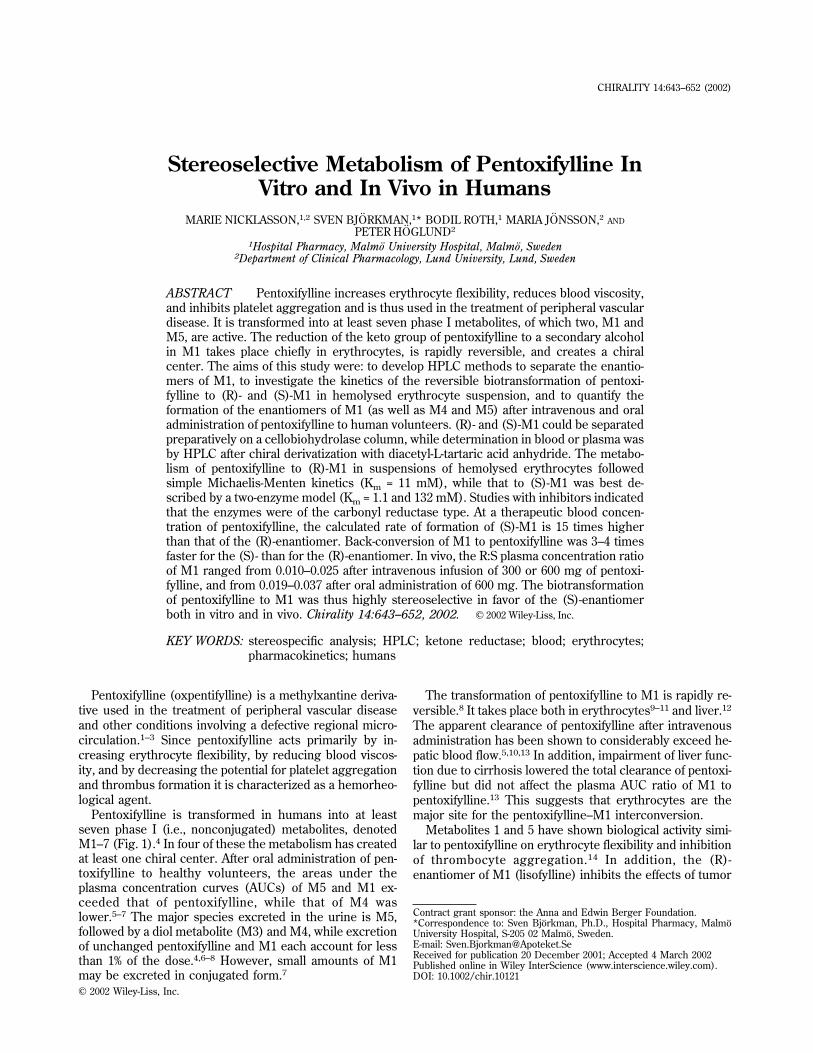
Stereoselective Metabolism of Pentoxifylline InVitro and In Vivo in Humans
MARIE NICKLASSON,1,2 SVEN BJORKMAN,1* BODIL ROTH,1 MARIA JONSSON,2 ANDPETER HOGLUND2
1Hospital Pharmacy, Malmö University Hospital, Malmo, Sweden2Department of Clinical Pharmacology, Lund University, Lund, Sweden
ABSTRACT Pentoxifylline increases erythrocyte flexibility, reduces blood viscosity,and inhibits platelet aggregation and is thus used in the treatment of peripheral vasculardisease. It is transformed into at least seven phase I metabolites, of which two, M1 andM5, are active. The reduction of the keto group of pentoxifylline to a secondary alcoholin M1 takes place chiefly in erythrocytes, is rapidly reversible, and creates a chiralcenter. The aims of this study were: to develop HPLC methods to separate the enantio-mers of M1, to investigate the kinetics of the reversible biotransformation of pentoxi-fylline to (R)- and (S)-M1 in hemolysed erythrocyte suspension, and to quantify theformation of the enantiomers of M1 (as well as M4 and M5) after intravenous and oraladministration of pentoxifylline to human volunteers. (R)- and (S)-M1 could be separatedpreparatively on a cellobiohydrolase column, while determination in blood or plasma wasby HPLC after chiral derivatization with diacetyl-L-tartaric acid anhydride. The metabo-lism of pentoxifylline to (R)-M1 in suspensions of hemolysed erythrocytes followedsimple Michaelis-Menten kinetics (Km = 11 mM), while that to (S)-M1 was best de-scribed by a two-enzyme model (Km = 1.1 and 132 mM). Studies with inhibitors indicatedthat the enzymes were of the carbonyl reductase type. At a therapeutic blood concen-tration of pentoxifylline, the calculated rate of formation of (S)-M1 is 15 times higherthan that of the (R)-enantiomer. Back-conversion of M1 to pentoxifylline was 3–4 timesfaster for the (S)- than for the (R)-enantiomer. In vivo, the R:S plasma concentration ratioof M1 ranged from 0.010–0.025 after intravenous infusion of 300 or 600 mg of pentoxi-fylline, and from 0.019–0.037 after oral administration of 600 mg. The biotransformationof pentoxifylline to M1 was thus highly stereoselective in favor of the (S)-enantiomerboth in vitro and in vivo. Chirality 14:643–652, 2002. © 2002 Wiley-Liss, Inc.
KEY WORDS: stereospecific analysis; HPLC; ketone reductase; blood; erythrocytes;pharmacokinetics; humans
Pentoxifylline (oxpentifylline) is a methylxantine deriva-tive used in the treatment of peripheral vascular diseaseand other conditions involving a defective regional micro-circulation.1–3 Since pentoxifylline acts primarily by in-creasing erythrocyte flexibility, by reducing blood viscos-ity, and by decreasing the potential for platelet aggregationand thrombus formation it is characterized as a hemorheo-logical agent.
Pentoxifylline is transformed in humans into at leastseven phase I (i.e., nonconjugated) metabolites, denotedM1–7 (Fig. 1).4 In four of these the metabolism has createdat least one chiral center. After oral administration of pen-toxifylline to healthy volunteers, the areas under theplasma concentration curves (AUCs) of M5 and M1 ex-ceeded that of pentoxifylline, while that of M4 waslower.5–7 The major species excreted in the urine is M5,followed by a diol metabolite (M3) and M4, while excretionof unchanged pentoxifylline and M1 each account for lessthan 1% of the dose.4,6–8 However, small amounts of M1may be excreted in conjugated form.7
The transformation of pentoxifylline to M1 is rapidly re-versible.8 It takes place both in erythrocytes9–11 and liver.12
The apparent clearance of pentoxifylline after intravenousadministration has been shown to considerably exceed he-patic blood flow.5,10,13 In addition, impairment of liver func-tion due to cirrhosis lowered the total clearance of pentoxi-fylline but did not affect the plasma AUC ratio of M1 topentoxifylline.13 This suggests that erythrocytes are themajor site for the pentoxifylline–M1 interconversion.
Metabolites 1 and 5 have shown biological activity simi-lar to pentoxifylline on erythrocyte flexibility and inhibitionof thrombocyte aggregation.14 In addition, the (R)-enantiomer of M1 (lisofylline) inhibits the effects of tumor
Contract grant sponsor: the Anna and Edwin Berger Foundation.*Correspondence to: Sven Bjorkman, Ph.D., Hospital Pharmacy, MalmoUniversity Hospital, S-205 02 Malmo, Sweden.E-mail: [email protected] for publication 20 December 2001; Accepted 4 March 2002Published online in Wiley InterScience (www.interscience.wiley.com).DOI: 10.1002/chir.10121
CHIRALITY 14:643–652 (2002)
© 2002 Wiley-Liss, Inc.
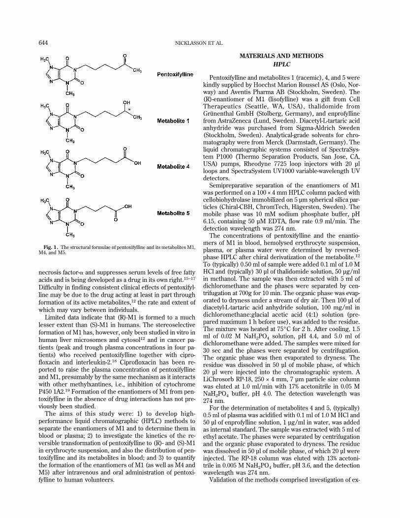
necrosis factor-� and suppresses serum levels of free fattyacids and is being developed as a drug in its own right.15–17
Difficulty in finding consistent clinical effects of pentoxifyl-line may be due to the drug acting at least in part throughformation of its active metabolites,12 the rate and extent ofwhich may vary between individuals.
Limited data indicate that (R)-M1 is formed to a muchlesser extent than (S)-M1 in humans. The stereoselectiveformation of M1 has, however, only been studied in vitro inhuman liver microsomes and cytosol12 and in cancer pa-tients (peak and trough plasma concentrations in four pa-tients) who received pentoxifylline together with cipro-floxacin and interleukin-2.18 Ciprofloxacin has been re-ported to raise the plasma concentration of pentoxifyllineand M1, presumably by the same mechanism as it interactswith other methylxantines, i.e., inhibition of cytochromeP450 1A2.19 Formation of the enantiomers of M1 from pen-toxifylline in the absence of drug interactions has not pre-viously been studied.
The aims of this study were: 1) to develop high-performance liquid chromatographic (HPLC) methods toseparate the enantiomers of M1 and to determine them inblood or plasma; 2) to investigate the kinetics of the re-versible transformation of pentoxifylline to (R)- and (S)-M1in erythrocyte suspension, and also the distribution of pen-toxifylline and its metabolites in blood; and 3) to quantifythe formation of the enantiomers of M1 (as well as M4 andM5) after intravenous and oral administration of pentoxi-fylline to human volunteers.
MATERIALS AND METHODSHPLC
Pentoxifylline and metabolites 1 (racemic), 4, and 5 werekindly supplied by Hoechst Marion Roussel AS (Oslo, Nor-way) and Aventis Pharma AB (Stockholm, Sweden). The(R)-enantiomer of M1 (lisofylline) was a gift from CellTherapeutics (Seattle, WA, USA), thalidomide fromGrunenthal GmbH (Stolberg, Germany), and enprofyllinefrom AstraZeneca (Lund, Sweden). Diacetyl-L-tartaric acidanhydride was purchased from Sigma-Aldrich Sweden(Stockholm, Sweden). Analytical-grade solvents for chro-matography were from Merck (Darmstadt, Germany). Theliquid chromatographic systems consisted of SpectraSys-tem P1000 (Thermo Separation Products, San Jose, CA,USA) pumps, Rheodyne 7725 loop injectors with 20 µlloops and SpectraSystem UV1000 variable-wavelength UVdetectors.
Semipreparative separation of the enantiomers of M1was performed on a 100 × 4 mm HPLC column packed withcellobiohydrolase immobilized on 5 µm spherical silica par-ticles (Chiral-CBH, ChromTech, Hagersten, Sweden). Themobile phase was 10 mM sodium phosphate buffer, pH6.15, containing 50 µM EDTA, flow rate 0.9 ml/min. Thedetection wavelength was 274 nm.
The concentrations of pentoxifylline and the enantio-mers of M1 in blood, hemolysed erythrocyte suspension,plasma, or plasma water were determined by reversed-phase HPLC after chiral derivatization of the metabolite.12
To (typically) 0.50 ml of sample were added 0.1 ml of 1.0 MHCl and (typically) 30 µl of thalidomide solution, 50 µg/mlin methanol. The sample was then extracted with 5 ml ofdichloromethane and the phases were separated by cen-trifugation at 700g for 10 min. The organic phase was evap-orated to dryness under a stream of dry air. Then 100 µl ofdiacetyl-L-tartaric acid anhydride solution, 100 mg/ml indichloromethane:glacial acetic acid (4:1) solution (pre-pared maximum 1 h before use), was added to the residue.The mixture was heated at 75°C for 2 h. After cooling, 1.5ml of 0.02 M NaH2PO4 solution, pH 4.4, and 5.0 ml ofdichloromethane were added. The samples were mixed for30 sec and the phases were separated by centrifugation.The organic phase was then evaporated to dryness. Theresidue was dissolved in 50 µl of mobile phase, of which20 µl were injected into the chromatographic system. ALiChrosorb RP-18, 250 × 4 mm, 7 µm particle size columnwas eluted at 1.0 ml/min with 17% acetonitrile in 0.05 MNaH2PO4 buffer, pH 4.0. The detection wavelength was274 nm.
For the determination of metabolites 4 and 5, (typically)0.5 ml of plasma was acidified with 0.1 ml of 1.0 M HCl and50 µl of enprofylline solution, 1 µg/ml in water, was addedas internal standard. The sample was extracted with 5 ml ofethyl acetate. The phases were separated by centrifugationand the organic phase evaporated to dryness. The residuewas dissolved in 50 µl of mobile phase, of which 20 µl wereinjected. The RP-18 column was eluted with 13% acetoni-trile in 0.005 M NaH2PO4 buffer, pH 3.6, and the detectionwavelength was 274 nm.
Validation of the methods comprised investigation of ex-
Fig. 1. The structural formulae of pentoxifylline and its metabolites M1,M4, and M5.
644 NICKLASSON ET AL.
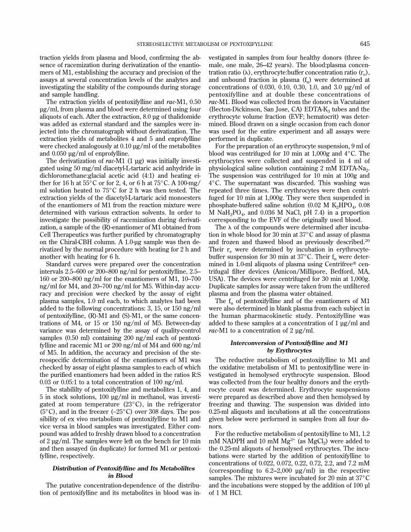
traction yields from plasma and blood, confirming the ab-sence of racemization during derivatization of the enantio-mers of M1, establishing the accuracy and precision of theassays at several concentration levels of the analytes andinvestigating the stability of the compounds during storageand sample handling.
The extraction yields of pentoxifylline and rac-M1, 0.50µg/ml, from plasma and blood were determined using fouraliquots of each. After the extraction, 8.0 µg of thalidomidewas added as external standard and the samples were in-jected into the chromatograph without derivatization. Theextraction yields of metabolites 4 and 5 and enprofyllinewere checked analogously at 0.10 µg/ml of the metabolitesand 0.050 µg/ml of enprofylline.
The derivatization of rac-M1 (1 µg) was initially investi-gated using 50 mg/ml diacetyl-L-tartaric acid anhydride indichloromethane:glacial acetic acid (4:1) and heating ei-ther for 16 h at 55°C or for 2, 4, or 6 h at 75°C. A 100-mg/ml solution heated to 75°C for 2 h was then tested. Theextraction yields of the diacetyl-L-tartaric acid monoestersof the enantiomers of M1 from the reaction mixture weredetermined with various extraction solvents. In order toinvestigate the possibility of racemization during derivati-zation, a sample of the (R)-enantiomer of M1 obtained fromCell Therapeutics was further purified by chromatographyon the Chiral-CBH column. A 1.0-µg sample was then de-rivatized by the normal procedure with heating for 2 h andanother with heating for 6 h.
Standard curves were prepared over the concentrationintervals 2.5–600 or 200–800 ng/ml for pentoxifylline, 2.5–160 or 200–800 ng/ml for the enantiomers of M1, 10–700ng/ml for M4, and 20–700 ng/ml for M5. Within-day accu-racy and precision were checked by the assay of eightplasma samples, 1.0 ml each, to which analytes had beenadded to the following concentrations: 3, 15, or 150 ng/mlof pentoxifylline, (R)-M1 and (S)-M1, or the same concen-trations of M4, or 15 or 150 ng/ml of M5. Between-dayvariance was determined by the assay of quality-controlsamples (0.50 ml) containing 200 ng/ml each of pentoxi-fylline and racemic M1 or 200 ng/ml of M4 and 600 ng/mlof M5. In addition, the accuracy and precision of the ste-reospecific determination of the enantiomers of M1 waschecked by assay of eight plasma samples to each of whichthe purified enantiomers had been added in the ratios R:S0.03 or 0.05:1 to a total concentration of 100 ng/ml.
The stabililty of pentoxifylline and metabolites 1, 4, and5 in stock solutions, 100 µg/ml in methanol, was investi-gated at room temperature (23°C), in the refrigerator(5°C), and in the freezer (−25°C) over 308 days. The pos-sibility of ex vivo metabolism of pentoxifylline to M1 andvice versa in blood samples was investigated. Either com-pound was added to freshly drawn blood to a concentrationof 2 µg/ml. The samples were left on the bench for 10 minand then assayed (in duplicate) for formed M1 or pentoxi-fylline, respectively.
Distribution of Pentoxifylline and Its Metaboliltesin Blood
The putative concentration-dependence of the distribu-tion of pentoxifylline and its metabolites in blood was in-
vestigated in samples from four healthy donors (three fe-male, one male, 26–42 years). The blood:plasma concen-tration ratio (�), erythrocyte:buffer concentration ratio (re),and unbound fraction in plasma (fu) were determined atconcentrations of 0.030, 0.10, 0.30, 1.0, and 3.0 µg/ml ofpentoxifylline and at double these concentrations ofrac-M1. Blood was collected from the donors in Vacutainer(Becton-Dickinson, San Jose, CA) EDTA-K3 tubes and theerythrocyte volume fraction (EVF; hematocrit) was deter-mined. Blood drawn on a single occasion from each donorwas used for the entire experiment and all assays wereperformed in duplicate.
For the preparation of an erythrocyte suspension, 9 ml ofblood was centrifuged for 10 min at 1,000g and 4°C. Theerythrocytes were collected and suspended in 4 ml ofphysiological saline solution containing 2 mM EDTA-Na2.The suspension was centrifuged for 10 min at 100g and4°C. The supernatant was discarded. This washing wasrepeated three times. The erythrocytes were then centri-fuged for 10 min at 1,000g. They were then suspended inphosphate-buffered saline solution (0.02 M K2HPO4, 0.08M NaH2PO4, and 0.036 M NaCl, pH 7.4) in a proportioncorresponding to the EVF of the originally used blood.
The � of the compounds were determined after incuba-tion in whole blood for 30 min at 37°C and assay of plasmaand frozen and thawed blood as previously described.20
Their re were determined by incubation in erythrocyte-buffer suspension for 30 min at 37°C. Their fu were deter-mined in 1.0-ml aliquots of plasma using Centrifree� cen-trifugal filter devices (Amicon/Millipore, Bedford, MA,USA). The devices were centrifuged for 30 min at 1,000g.Duplicate samples for assay were taken from the unfilteredplasma and from the plasma water obtained.
The fu of pentoxifylline and of the enantiomers of M1were also determined in blank plasma from each subject inthe human pharmacokinetic study. Pentoxifylline wasadded to these samples at a concentration of 1 µg/ml andrac-M1 to a concentration of 2 µg/ml.
Interconversion of Pentoxifylline and M1by Erythrocytes
The reductive metabolism of pentoxifylline to M1 andthe oxidative metabolism of M1 to pentoxifylline were in-vestigated in hemolysed erythrocyte suspension. Bloodwas collected from the four healthy donors and the eryth-rocyte count was determined. Erythrocyte suspensionswere prepared as described above and then hemolysed byfreezing and thawing. The suspension was divided into0.25-ml aliquots and incubations at all the concentrationsgiven below were performed in samples from all four do-nors.
For the reductive metabolism of pentoxifylline to M1, 1.2mM NADPH and 10 mM Mg2+ (as MgCl2) were added tothe 0.25-ml aliquots of hemolysed erythrocytes. The incu-bations were started by the addition of pentoxifylline toconcentrations of 0.022, 0.072, 0.22, 0.72, 2.2, and 7.2 mM(corresponding to 6.2–2,000 µg/ml) in the respectivesamples. The mixtures were incubated for 20 min at 37°Cand the incubations were stopped by the addition of 100 µlof 1 M HCl.
STEREOSELECTIVE METABOLISM OF PENTOXIFYLLINE 645
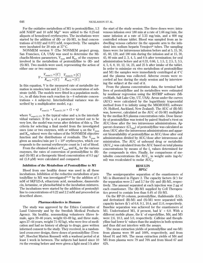
For the oxidative metabolism of M1 to pentoxifylline, 1.3mM NADP and 10 mM Mg2+ were added to the 0.25-mlaliquots of hemolysed erythrocytes. The incubations werestarted by the addition of (R)- or (S)-M1, to final concen-trations of 0.022 and 0.072 mM, respectively. The sampleswere incubated for 20 min at 37°C.
NONMEM version V (The NONMEM project group,San Francisco, CA, USA) was used to determine the Mi-chaelis-Menten parameters, Vmax and Km, of the enzymesinvolved in the metabolism of pentoxifylline to (R)- and(S)-M1. Two models were used, representing the action ofeither one or two enzymes:
V =Vmax�1� × �C �
Km�1� + �C �+
Vmax(2) × �C �
Km�2� + �C �
In this equation, V is the measured rate of metabolite for-mation in nmoles/min and [C] is the concentration of sub-strate (mM). The models were fitted in a population mode,i.e., to all data from each reaction (n = 6 substrate concen-trations × 4 subjects). Interindividual variance was de-scribed by a multiplicative model, e.g.:
Vmax(1) = Vmax(1),TV × (1 + �)
where Vmax(1),TV is the typical value and � is the interindi-vidual variance. If the � of a parameter turned out to bevery low, the model was rerun after deletion of this �. Thecriteria for selecting a final model from the investigatedones (one or two enzymes, with or without � on the Vmaxand Km values) were the values of the NONMEM objectivefunction and the distributions of residuals. The Vmax isgiven as nmoles/min per 5 × 10⁹ erythrocytes, which cor-responds to the normal erythrocyte count in 1 ml of blood.
From the obtained values of Vmax and Km for the variousenzymes, the rates of conversion of pentoxifylline to (R)-and (S)-M1 at a therapeutic blood concentration of 0.5 µg/ml (1.8 µM) were calculated and compared.
Inhibition of the Metabolism of Pentoxifylline to M1
Blood from one healthy donor was used in all theseincubations. Inhibition of the reductive metabolism of pen-toxifylline to M1 was investigated21,22 by the addition of 5mM of SKF525-A, ethacrynic acid, menadione, daunorubi-cin, ketamine, or phenobarbital to the incubation mixtures.The incubations were started by the addition of pentoxifyl-line to concentrations of 0.22 and 2.2 mM and proceeded asdescribed above.
Pharmacokinetics in Humans
The study was approved by the Ethics Committee ofLund University and by the Swedish Medical ProductsAgency. Six healthy, nonsmoking volunteers (three fe-male, ages 39–46 years, weight 63–69 kg, and three male,ages 27–44 years, weight 71–92 kg), who were free of medi-cation and had no history of allergy to drugs, gave writteninformed consent to the study. They received, in a random-ized cross-over design, three doses of pentoxifylline (Tren-tal�, Hoechst Marion Roussel) with a washout period of atleast 1 week in between. The subjects had fasted since 10PM the evening before and were given a light meal 5 h after
the start of the study session. The three doses were: intra-venous infusion over 180 min at a rate of 1.66 mg/min, thesame infusion at a rate of 3.33 mg/min, and a 600 mgcontrolled release tablet. Blood was sampled from an in-dwelling venous catheter (in the opposite arm to the infu-sion) into sodium heparin Venoject� tubes. The samplingtimes were: for intravenous infusion before and at 5, 15, 30,45, 60, 120, and 180 min during the infusion and at 15, 30,45, 60 min and 2, 3, 4, 5, and 6 h after termination; for oraladministration before and at 0.33, 0.66, 1, 1.5, 2, 2.5, 3, 3.5,4, 5, 6, 8, 10, 12, 14, 23, and 25 h after intake of the tablet.In order to minimize ex vivo metabolism of pentoxifyllineand M1 the samples were immediately centrifuged at 4°Cand the plasma was collected. Adverse events were re-corded ad hoc during the study session and by interview-ing the subject at the end of it.
From the plasma concentration data, the terminal half-lives of pentoxifylline and its metabolites were estimatedby nonlinear regression using the RSTRIP software (Mi-croMath, Salt Lake City, UT, USA). Areas under the curves(AUC) were calculated by the logarithmic trapezoidalmethod from 0 to infinity using the MKMODEL software(N. Holford, Auckland, New Zealand). The AUC of (R)-M1was, however, calculated as the AUC of (S)-M1 multipliedby the median R:S plasma concentration ratio. Dose linear-ity of pentoxifylline was tested by paired Student’s t-test onAUC/dose after the two intravenous administrations. Ap-parent clearance (CLapp) of pentoxifylline was calculated asdose/AUC after the intravenous administrations and appar-ent bioavailability of pentoxifylline as AUC/dose after oraladministration divided by AUC/dose after intravenous ad-ministration. The AUC of unbound drug or metabolite(AUCu) was calculated from the AUC based on total plasmaconcentrations by means of the fu values determined forthe compounds in vitro. Finally, for comparisons of me-tabolite concentrations the AUCu in weight units (ng×h/ml) was recalculated to molar AUCu.
RESULTSHPLC
The semipreparative separation of the enantiomers ofM1 is illustrated in Figure 2. The capacity factors (k�) forthe separation were 2.7 and 3.7 for (S)- and (R)-M1, respec-tively. The amount separated at each injection was 2 µg ofeach enantiomer. The (R)-M1 supplied by Cell Therapeu-tics proved to contain less than 0.4% of (S)-M1.
On the RP-18 column, pentoxifylline, thalidomide (I.S.),and derivatized (R)-M1 and (S)-M1 were separated withcapacity factors (k�) of 6.8, 9.1, 10.4, and 11.0, respectively.Baseline separation was achieved for the enantiomers ofM1. Underivatized M1, if present, had k� = 6.0. With adifferent mobile phase, the k� of enprofylline, M4, and M5were 2.6, 10.3, and 5.0, respectively. Caffeine and theoph-ylline had lower k� values than the analytes in both systemsand thus did not interfere with the assays.
The mean extraction yields of pentoxifylline and rac-M1from plasma were 99 and 100%, respectively, and fromblood 91 and 94%. The mean extraction yields of M4 andM5 from plasma were 79 and 76% and from blood 67 and62%.
646 NICKLASSON ET AL.
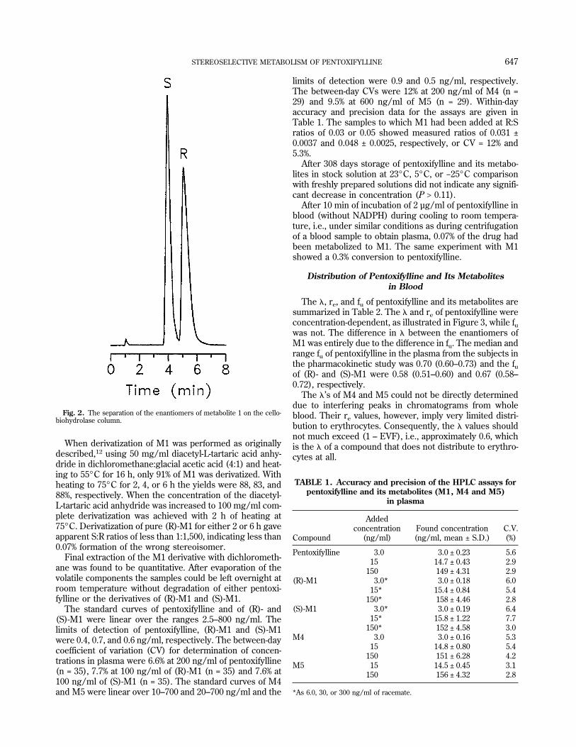
When derivatization of M1 was performed as originallydescribed,12 using 50 mg/ml diacetyl-L-tartaric acid anhy-dride in dichloromethane:glacial acetic acid (4:1) and heat-ing to 55°C for 16 h, only 91% of M1 was derivatized. Withheating to 75°C for 2, 4, or 6 h the yields were 88, 83, and88%, respectively. When the concentration of the diacetyl-L-tartaric acid anhydride was increased to 100 mg/ml com-plete derivatization was achieved with 2 h of heating at75°C. Derivatization of pure (R)-M1 for either 2 or 6 h gaveapparent S:R ratios of less than 1:1,500, indicating less than0.07% formation of the wrong stereoisomer.
Final extraction of the M1 derivative with dichlorometh-ane was found to be quantitative. After evaporation of thevolatile components the samples could be left overnight atroom temperature without degradation of either pentoxi-fylline or the derivatives of (R)-M1 and (S)-M1.
The standard curves of pentoxifylline and of (R)- and(S)-M1 were linear over the ranges 2.5–800 ng/ml. Thelimits of detection of pentoxifylline, (R)-M1 and (S)-M1were 0.4, 0.7, and 0.6 ng/ml, respectively. The between-daycoefficient of variation (CV) for determination of concen-trations in plasma were 6.6% at 200 ng/ml of pentoxifylline(n = 35), 7.7% at 100 ng/ml of (R)-M1 (n = 35) and 7.6% at100 ng/ml of (S)-M1 (n = 35). The standard curves of M4and M5 were linear over 10–700 and 20–700 ng/ml and the
limits of detection were 0.9 and 0.5 ng/ml, respectively.The between-day CVs were 12% at 200 ng/ml of M4 (n =29) and 9.5% at 600 ng/ml of M5 (n = 29). Within-dayaccuracy and precision data for the assays are given inTable 1. The samples to which M1 had been added at R:Sratios of 0.03 or 0.05 showed measured ratios of 0.031 ±0.0037 and 0.048 ± 0.0025, respectively, or CV = 12% and5.3%.
After 308 days storage of pentoxifylline and its metabo-lites in stock solution at 23°C, 5°C, or −25°C comparisonwith freshly prepared solutions did not indicate any signifi-cant decrease in concentration (P > 0.11).
After 10 min of incubation of 2 µg/ml of pentoxifylline inblood (without NADPH) during cooling to room tempera-ture, i.e., under similar conditions as during centrifugationof a blood sample to obtain plasma, 0.07% of the drug hadbeen metabolized to M1. The same experiment with M1showed a 0.3% conversion to pentoxifylline.
Distribution of Pentoxifylline and Its Metabolitesin Blood
The �, re, and fu of pentoxifylline and its metabolites aresummarized in Table 2. The � and re of pentoxifylline wereconcentration-dependent, as illustrated in Figure 3, while fuwas not. The difference in � between the enantiomers ofM1 was entirely due to the difference in fu. The median andrange fu of pentoxifylline in the plasma from the subjects inthe pharmacokinetic study was 0.70 (0.60–0.73) and the fuof (R)- and (S)-M1 were 0.58 (0.51–0.60) and 0.67 (0.58–0.72), respectively.
The �’s of M4 and M5 could not be directly determineddue to interfering peaks in chromatograms from wholeblood. Their re values, however, imply very limited distri-bution to erythrocytes. Consequently, the � values shouldnot much exceed (1 – EVF), i.e., approximately 0.6, whichis the � of a compound that does not distribute to erythro-cytes at all.
TABLE 1. Accuracy and precision of the HPLC assays forpentoxifylline and its metabolites (M1, M4 and M5)
in plasma
Compound
Addedconcentration
(ng/ml)Found concentration(ng/ml, mean ± S.D.)
C.V.(%)
Pentoxifylline 3.0 3.0 ± 0.23 5.615 14.7 ± 0.43 2.9
150 149 ± 4.31 2.9(R)-M1 3.0* 3.0 ± 0.18 6.0
15* 15.4 ± 0.84 5.4150* 158 ± 4.46 2.8
(S)-M1 3.0* 3.0 ± 0.19 6.415* 15.8 ± 1.22 7.7
150* 152 ± 4.58 3.0M4 3.0 3.0 ± 0.16 5.3
15 14.8 ± 0.80 5.4150 151 ± 6.28 4.2
M5 15 14.5 ± 0.45 3.1150 156 ± 4.32 2.8
*As 6.0, 30, or 300 ng/ml of racemate.
Fig. 2. The separation of the enantiomers of metabolite 1 on the cello-biohydrolase column.
STEREOSELECTIVE METABOLISM OF PENTOXIFYLLINE 647
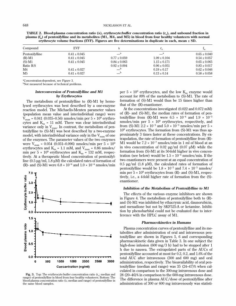
Interconversion of Pentoxifylline and M1by Erythrocytes
The metabolism of pentoxifylline to (R)-M1 by hemo-lysed erythrocytes was best described by a one-enzymereaction model. The Michaelis-Menten parameter values(population mean value and interindividual range) wereVmax = 0.041 (0.035–0.56) nmoles/min per 5 × 10⁹ erythro-cytes and Km = 11 mM. There was clear interindividualvariance only in Vmax. In contrast, the metabolism of pen-toxifylline to (S)-M1 was best described by a two-enzymemodel, with interindividual variance only in the Vmax of oneof the enzymes. The parameter values of the two enzymeswere Vmax = 0.054 (0.031–0.096) nmoles/min per 5 × 10⁹
erythrocytes and Km = 1.1 mM, and Vmax = 0.86 nmoles/min per 5 × 10⁹ erythrocytes and Km = 132 mM, respec-tively. At a therapeutic blood concentration of pentoxifyl-line (0.5 µg/ml, 1.8 µM) the calculated rates of formation of(R)- and (S)-M1 were 6.8 × 10⁻⁶ and 1.0 × 10⁻⁴ nmoles/min
per 5 × 10⁹ erythrocytes, and the low Km enzyme wouldaccount for 89% of the metabolism to (S)-M1. The rate offormation of (S)-M1 would thus be 15 times higher thanthat of the (R)-enantiomer.
At the concentrations investigated (0.022 and 0.072 mM)of (R)- and (S)-M1, the median rates of formation of pen-toxifylline from (R)-M1 were 6.5 × 10⁻⁴ and 1.9 × 10⁻³
nmoles/min per 5 × 10⁹ erythrocytes, respectively, andfrom (S)-M1 2.2 × 10⁻³ and 5.0 × 10⁻³ nmoles/min per 5 ×10⁹ erythrocytes. The formation from (S)-M1 was thus ap-proximately 3 times faster at these concentrations. By ex-trapolation, the rate of formation of pentoxifylline from (R)-M1 would be 7.2 × 10⁻⁷ nmoles/min in 1 ml of blood at anin vivo concentration of 0.02 µg/ml (0.07 µM) while theformation from (S)-M1 at its 50-fold higher in vivo concen-tration (see below) would be 1.5 × 10⁻⁴ nmoles/min. If thetwo enantiomers were present at an equal concentration of0.5 µg/ml (1.8 µM), the calculated rates of formation ofpentoxifylline would be 1.8 × 10⁻⁵ and 7.4 × 10⁻⁵ nmoles/min per 5 × 10⁹ erythrocytes from (R)- and (S)-M1, respec-tively, i.e., a 4-fold higher rate of formation from the (S)-enantiomer.
Inhibition of the Metabolism of Pentoxifylline to M1
The effects of the various enzyme inhibitors are shownin Figure 4. The metabolism of pentoxifylline both to (R)-and (S)-M1 was inhibited by ethacrynic acid, daunorubicin,and menadione but not by SKF525-A or ketamine. Inhibi-tion by phenobarbital could not be evaluated due to inter-ference with the HPLC assay of M1.
Pharmacokinetics in Humans
Plasma concentration curves of pentoxifylline and its me-tabolites after administration of oral and intravenous pen-toxifylline are shown in Figures 5, 6 and correspondingpharmacokinetic data given in Table 3. In one subject thehigh-dose infusion (600 mg/3 h) had to be stopped after 1h due to nausea. The extrapolated parts of the AUCs ofpentoxifylline accounted at most for 0.2, 0.2, and 1.6% of thetotal AUC after intravenous (300 and 600 mg) and oraladministration, respectively. The bioavailability of oral pen-toxifylline (median and range) was 35 (24–47)% when cal-culated in comparison to the 300-mg intravenous dose and28 (20–46)% in comparison to the 600-mg intravenous dose.The difference in plasma AUC/dose of pentoxifylline afteradministration of 300 or 600 mg intravenously was statisti-
TABLE 2. Blood:plasma concentration ratio (�), erythrocyte:buffer concentration ratio (re), and unbound fraction inplasma (fu) of pentoxifylline and its metabolites (M1, M4, and M5) in blood from four healthy volunteers with normal
erythrocyte volume fractions (EVF). Figures are five determinations in duplicate in each, mean ± SD.
Compound EVF � re fu
Pentoxifylline 0.41 ± 0.045 —a —a 0.65 ± 0.040(R)-M1 0.41 ± 0.045 0.77 ± 0.059 1.09 ± 0.104 0.54 ± 0.057(S)-M1 0.41 ± 0.045 0.84 ± 0.063 1.15 ± 0.171 0.65 ± 0.065Ratio R:S 0.92 ± 0.004 0.96 ± 0.051 0.83 ± 0.017M4 0.41 ± 0.027 —b 0.19 ± 0.17 0.62 ± 0.046M5 0.41 ± 0.027 —b 0.13 ± 0.14 0.58 ± 0.058
aConcentration-dependent, see Figure 3.bNot measured because of technical problems.
Fig. 3. Top: The erythrocyte:buffer concentration ratio (re; median andrange) of pentoxifylline in blood from four healthy volunteers. Bottom: Theblood:plasma concentration ratio (�; median and range) of pentoxifylline inthe same blood samples.
648 NICKLASSON ET AL.
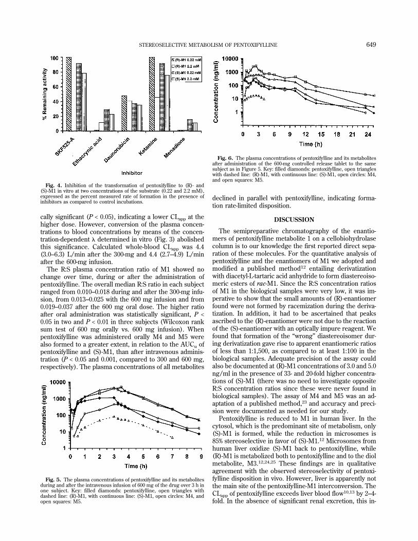
cally significant (P < 0.05), indicating a lower CLapp at thehigher dose. However, conversion of the plasma concen-trations to blood concentrations by means of the concen-tration-dependent � determined in vitro (Fig. 3) abolishedthis significance. Calculated whole-blood CLapp was 4.4(3.0–6.3) L/min after the 300-mg and 4.4 (2.7–4.9) L/minafter the 600-mg infusion.
The R:S plasma concentration ratio of M1 showed nochange over time, during or after the administration ofpentoxifylline. The overall median R:S ratio in each subjectranged from 0.010–0.018 during and after the 300-mg infu-sion, from 0.013–0.025 with the 600 mg infusion and from0.019–0.037 after the 600 mg oral dose. The higher ratioafter oral administration was statistically significant, P <0.05 in two and P < 0.01 in three subjects (Wilcoxon ranksum test of 600 mg orally vs. 600 mg infusion). Whenpentoxifylline was administered orally M4 and M5 werealso formed to a greater extent, in relation to the AUCu ofpentoxifylline and (S)-M1, than after intravenous adminis-tration (P < 0.05 and 0.001, compared to 300 and 600 mg,respectively). The plasma concentrations of all metabolites
declined in parallel with pentoxifylline, indicating forma-tion rate-limited disposition.
DISCUSSION
The semipreparative chromatography of the enantio-mers of pentoxifylline metabolite 1 on a cellobiohydrolasecolumn is to our knowledge the first reported direct sepa-ration of these molecules. For the quantitative analysis ofpentoxifylline and the enantiomers of M1 we adopted andmodified a published method12 entailing derivatizationwith diacetyl-L-tartaric acid anhydride to form diastereoiso-meric esters of rac-M1. Since the R:S concentration ratiosof M1 in the biological samples were very low, it was im-perative to show that the small amounts of (R)-enantiomerfound were not formed by racemization during the deriva-tization. In addition, it had to be ascertained that peaksascribed to the (R)-enantiomer were not due to the reactionof the (S)-enantiomer with an optically impure reagent. Wefound that formation of the “wrong” diastereoisomer dur-ing derivatization gave rise to apparent enantiomeric ratiosof less than 1:1,500, as compared to at least 1:100 in thebiological samples. Adequate precision of the assay couldalso be documented at (R)-M1 concentrations of 3.0 and 5.0ng/ml in the presence of 33- and 20-fold higher concentra-tions of (S)-M1 (there was no need to investigate oppositeR:S concentration ratios since these were never found inbiological samples). The assay of M4 and M5 was an ad-aptation of a published method,23 and accuracy and preci-sion were documented as needed for our study.
Pentoxifylline is reduced to M1 in human liver. In thecytosol, which is the predominant site of metabolism, only(S)-M1 is formed, while the reduction in microsomes is85% stereoselective in favor of (S)-M1.12 Microsomes fromhuman liver oxidize (S)-M1 back to pentoxifylline, while(R)-M1 is metabolized both to pentoxifylline and to the diolmetabolite, M3.12,24,25 These findings are in qualitativeagreement with the observed stereoselectivity of pentoxi-fylline disposition in vivo. However, liver is apparently notthe main site of the pentoxifylline-M1 interconversion. TheCLapp of pentoxifylline exceeds liver blood flow10,13 by 2–4-fold. In the absence of significant renal excretion, this in-
Fig. 4. Inhibition of the transformation of pentoxifylline to (R)- and(S)-M1 in vitro at two concentrations of the substrate (0.22 and 2.2 mM),expressed as the percent measured rate of formation in the presence ofinhibitors as compared to control incubations.
Fig. 5. The plasma concentrations of pentoxifylline and its metabolitesduring and after the intravenous infusion of 600 mg of the drug over 3 h inone subject. Key: filled diamonds: pentoxifylline, open triangles withdashed line: (R)-M1, with continuous line: (S)-M1, open circles: M4, andopen squares: M5.
Fig. 6. The plasma concentrations of pentoxifylline and its metabolitesafter administration of the 600-mg controlled release tablet to the samesubject as in Figure 5. Key: filled diamonds: pentoxifylline, open triangleswith dashed line: (R)-M1, with continuous line: (S)-M1, open circles: M4,and open squares: M5.
STEREOSELECTIVE METABOLISM OF PENTOXIFYLLINE 649
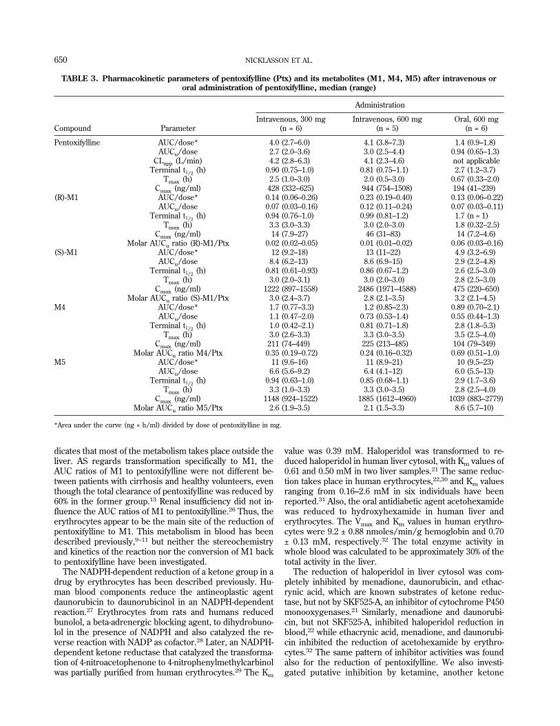
dicates that most of the metabolism takes place outside theliver. AS regards transformation specifically to M1, theAUC ratios of M1 to pentoxifylline were not different be-tween patients with cirrhosis and healthy volunteers, eventhough the total clearance of pentoxifylline was reduced by60% in the former group.13 Renal insufficiency did not in-fluence the AUC ratios of M1 to pentoxifylline.26 Thus, theerythrocytes appear to be the main site of the reduction ofpentoxifylline to M1. This metabolism in blood has beendescribed previously,9–11 but neither the stereochemistryand kinetics of the reaction nor the conversion of M1 backto pentoxifylline have been investigated.
The NADPH-dependent reduction of a ketone group in adrug by erythrocytes has been described previously. Hu-man blood components reduce the antineoplastic agentdaunorubicin to daunorubicinol in an NADPH-dependentreaction.27 Erythrocytes from rats and humans reducedbunolol, a beta-adrenergic blocking agent, to dihydrobuno-lol in the presence of NADPH and also catalyzed the re-verse reaction with NADP as cofactor.28 Later, an NADPH-dependent ketone reductase that catalyzed the transforma-tion of 4-nitroacetophenone to 4-nitrophenylmethylcarbinolwas partially purified from human erythrocytes.29 The Km
value was 0.39 mM. Haloperidol was transformed to re-duced haloperidol in human liver cytosol, with Km values of0.61 and 0.50 mM in two liver samples.21 The same reduc-tion takes place in human erythrocytes,22,30 and Km valuesranging from 0.16–2.6 mM in six individuals have beenreported.31 Also, the oral antidiabetic agent acetohexamidewas reduced to hydroxyhexamide in human liver anderythrocytes. The Vmax and Km values in human erythro-cytes were 9.2 ± 0.88 nmoles/min/g hemoglobin and 0.70± 0.13 mM, respectively.32 The total enzyme activity inwhole blood was calculated to be approximately 30% of thetotal activity in the liver.
The reduction of haloperidol in liver cytosol was com-pletely inhibited by menadione, daunorubicin, and ethac-rynic acid, which are known substrates of ketone reduc-tase, but not by SKF525-A, an inhibitor of cytochrome P450monooxygenases.21 Similarly, menadione and daunorubi-cin, but not SKF525-A, inhibited haloperidol reduction inblood,22 while ethacrynic acid, menadione, and daunorubi-cin inhibited the reduction of acetohexamide by erythro-cytes.32 The same pattern of inhibitor activities was foundalso for the reduction of pentoxifylline. We also investi-gated putative inhibition by ketamine, another ketone
TABLE 3. Pharmacokinetic parameters of pentoxifylline (Ptx) and its metabolites (M1, M4, M5) after intravenous ororal administration of pentoxifylline, median (range)
Compound Parameter
Administration
Intravenous, 300 mg(n = 6)
Intravenous, 600 mg(n = 5)
Oral, 600 mg(n = 6)
Pentoxifylline AUC/dose* 4.0 (2.7–6.0) 4.1 (3.8–7.3) 1.4 (0.9–1.8)AUCu/dose 2.7 (2.0–3.6) 3.0 (2.5–4.4) 0.94 (0.65–1.3)
CLapp (L/min) 4.2 (2.8–6.3) 4.1 (2.3–4.6) not applicableTerminal t1/2 (h) 0.90 (0.75–1.0) 0.81 (0.75–1.1) 2.7 (1.2–3.7)
Tmax (h) 2.5 (1.0–3.0) 2.0 (0.5–3.0) 0.67 (0.33–2.0)Cmax (ng/ml) 428 (332–625) 944 (754–1508) 194 (41–239)
(R)-M1 AUC/dose* 0.14 (0.06–0.26) 0.23 (0.19–0.40) 0.13 (0.06–0.22)AUCu/dose 0.07 (0.03–0.16) 0.12 (0.11–0.24) 0.07 (0.03–0.11)
Terminal t1/2 (h) 0.94 (0.76–1.0) 0.99 (0.81–1.2) 1.7 (n = 1)Tmax (h) 3.3 (3.0–3.3) 3.0 (2.0–3.0) 1.8 (0.32–2.5)
Cmax (ng/ml) 14 (7.9–27) 46 (31–83) 14 (7.2–4.6)Molar AUCu ratio (R)-M1/Ptx 0.02 (0.02–0.05) 0.01 (0.01–0.02) 0.06 (0.03–0.16)
(S)-M1 AUC/dose* 12 (9.2–18) 13 (11–22) 4.9 (3.2–6.9)AUCu/dose 8.4 (6.2–13) 8.6 (6.9–15) 2.9 (2.2–4.8)
Terminal t1/2 (h) 0.81 (0.61–0.93) 0.86 (0.67–1.2) 2.6 (2.5–3.0)Tmax (h) 3.0 (2.0–3.1) 3.0 (2.0–3.0) 2.8 (2.5–3.0)
Cmax (ng/ml) 1222 (897–1558) 2486 (1971–4588) 475 (220–650)Molar AUCu ratio (S)-M1/Ptx 3.0 (2.4–3.7) 2.8 (2.1–3.5) 3.2 (2.1–4.5)
M4 AUC/dose* 1.7 (0.77–3.3) 1.2 (0.85–2.3) 0.89 (0.70–2.1)AUCu/dose 1.1 (0.47–2.0) 0.73 (0.53–1.4) 0.55 (0.44–1.3)
Terminal t1/2 (h) 1.0 (0.42–2.1) 0.81 (0.71–1.8) 2.8 (1.8–5.3)Tmax (h) 3.0 (2.6–3.3) 3.3 (3.0–3.5) 3.5 (2.5–4.0)
Cmax (ng/ml) 211 (74–449) 225 (213–485) 104 (79–349)Molar AUCu ratio M4/Ptx 0.35 (0.19–0.72) 0.24 (0.16–0.32) 0.69 (0.51–1.0)
M5 AUC/dose* 11 (9.6–16) 11 (8.9–21) 10 (9.5–23)AUCu/dose 6.6 (5.6–9.2) 6.4 (4.1–12) 6.0 (5.5–13)
Terminal t1/2 (h) 0.94 (0.63–1.0) 0.85 (0.68–1.1) 2.9 (1.7–3.6)Tmax (h) 3.3 (1.0–3.3) 3.3 (3.0–3.5) 2.8 (2.5–4.0)
Cmax (ng/ml) 1148 (924–1522) 1885 (1612–4960) 1039 (883–2779)Molar AUCu ratio M5/Ptx 2.6 (1.9–3.5) 2.1 (1.5–3.3) 8.6 (5.7–10)
*Area under the curve (ng × h/ml) divided by dose of pentoxifylline in mg.
650 NICKLASSON ET AL.
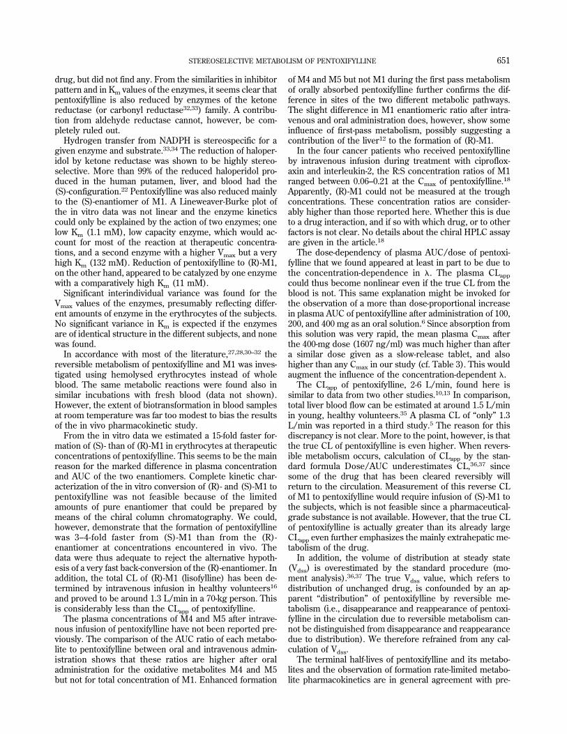
drug, but did not find any. From the similarities in inhibitorpattern and in Km values of the enzymes, it seems clear thatpentoxifylline is also reduced by enzymes of the ketonereductase (or carbonyl reductase32,33) family. A contribu-tion from aldehyde reductase cannot, however, be com-pletely ruled out.
Hydrogen transfer from NADPH is stereospecific for agiven enzyme and substrate.33,34 The reduction of haloper-idol by ketone reductase was shown to be highly stereo-selective. More than 99% of the reduced haloperidol pro-duced in the human putamen, liver, and blood had the(S)-configuration.22 Pentoxifylline was also reduced mainlyto the (S)-enantiomer of M1. A Lineweaver-Burke plot ofthe in vitro data was not linear and the enzyme kineticscould only be explained by the action of two enzymes; onelow Km (1.1 mM), low capacity enzyme, which would ac-count for most of the reaction at therapeutic concentra-tions, and a second enzyme with a higher Vmax but a veryhigh Km (132 mM). Reduction of pentoxifylline to (R)-M1,on the other hand, appeared to be catalyzed by one enzymewith a comparatively high Km (11 mM).
Significant interindividual variance was found for theVmax values of the enzymes, presumably reflecting differ-ent amounts of enzyme in the erythrocytes of the subjects.No significant variance in Km is expected if the enzymesare of identical structure in the different subjects, and nonewas found.
In accordance with most of the literature,27,28,30–32 thereversible metabolism of pentoxifylline and M1 was inves-tigated using hemolysed erythrocytes instead of wholeblood. The same metabolic reactions were found also insimilar incubations with fresh blood (data not shown).However, the extent of biotransformation in blood samplesat room temperature was far too modest to bias the resultsof the in vivo pharmacokinetic study.
From the in vitro data we estimated a 15-fold faster for-mation of (S)- than of (R)-M1 in erythrocytes at therapeuticconcentrations of pentoxifylline. This seems to be the mainreason for the marked difference in plasma concentrationand AUC of the two enantiomers. Complete kinetic char-acterization of the in vitro conversion of (R)- and (S)-M1 topentoxifylline was not feasible because of the limitedamounts of pure enantiomer that could be prepared bymeans of the chiral column chromatography. We could,however, demonstrate that the formation of pentoxifyllinewas 3–4-fold faster from (S)-M1 than from the (R)-enantiomer at concentrations encountered in vivo. Thedata were thus adequate to reject the alternative hypoth-esis of a very fast back-conversion of the (R)-enantiomer. Inaddition, the total CL of (R)-M1 (lisofylline) has been de-termined by intravenous infusion in healthy volunteers16
and proved to be around 1.3 L/min in a 70-kg person. Thisis considerably less than the CLapp of pentoxifylline.
The plasma concentrations of M4 and M5 after intrave-nous infusion of pentoxifylline have not been reported pre-viously. The comparison of the AUC ratio of each metabo-lite to pentoxifylline between oral and intravenous admin-istration shows that these ratios are higher after oraladministration for the oxidative metabolites M4 and M5but not for total concentration of M1. Enhanced formation
of M4 and M5 but not M1 during the first pass metabolismof orally absorbed pentoxifylline further confirms the dif-ference in sites of the two different metabolic pathways.The slight difference in M1 enantiomeric ratio after intra-venous and oral administration does, however, show someinfluence of first-pass metabolism, possibly suggesting acontribution of the liver12 to the formation of (R)-M1.
In the four cancer patients who received pentoxifyllineby intravenous infusion during treatment with ciproflox-axin and interleukin-2, the R:S concentration ratios of M1ranged between 0.06–0.21 at the Cmax of pentoxifylline.18
Apparently, (R)-M1 could not be measured at the troughconcentrations. These concentration ratios are consider-ably higher than those reported here. Whether this is dueto a drug interaction, and if so with which drug, or to otherfactors is not clear. No details about the chiral HPLC assayare given in the article.18
The dose-dependency of plasma AUC/dose of pentoxi-fylline that we found appeared at least in part to be due tothe concentration-dependence in �. The plasma CLappcould thus become nonlinear even if the true CL from theblood is not. This same explanation might be invoked forthe observation of a more than dose-proportional increasein plasma AUC of pentoxifylline after administration of 100,200, and 400 mg as an oral solution.6 Since absorption fromthis solution was very rapid, the mean plasma Cmax afterthe 400-mg dose (1607 ng/ml) was much higher than aftera similar dose given as a slow-release tablet, and alsohigher than any Cmax in our study (cf. Table 3). This wouldaugment the influence of the concentration-dependent �.
The CLapp of pentoxifylline, 2-6 L/min, found here issimilar to data from two other studies.10,13 In comparison,total liver blood flow can be estimated at around 1.5 L/minin young, healthy volunteers.35 A plasma CL of “only” 1.3L/min was reported in a third study.5 The reason for thisdiscrepancy is not clear. More to the point, however, is thatthe true CL of pentoxifylline is even higher. When revers-ible metabolism occurs, calculation of CLapp by the stan-dard formula Dose/AUC underestimates CL,36,37 sincesome of the drug that has been cleared reversibly willreturn to the circulation. Measurement of this reverse CLof M1 to pentoxifylline would require infusion of (S)-M1 tothe subjects, which is not feasible since a pharmaceutical-grade substance is not available. However, that the true CLof pentoxifylline is actually greater than its already largeCLapp even further emphasizes the mainly extrahepatic me-tabolism of the drug.
In addition, the volume of distribution at steady state(Vdss) is overestimated by the standard procedure (mo-ment analysis).36,37 The true Vdss value, which refers todistribution of unchanged drug, is confounded by an ap-parent “distribution” of pentoxifylline by reversible me-tabolism (i.e., disappearance and reappearance of pentoxi-fylline in the circulation due to reversible metabolism can-not be distinguished from disappearance and reappearancedue to distribution). We therefore refrained from any cal-culation of Vdss.
The terminal half-lives of pentoxifylline and its metabo-lites and the observation of formation rate-limited metabo-lite pharmacokinetics are in general agreement with pre-
STEREOSELECTIVE METABOLISM OF PENTOXIFYLLINE 651
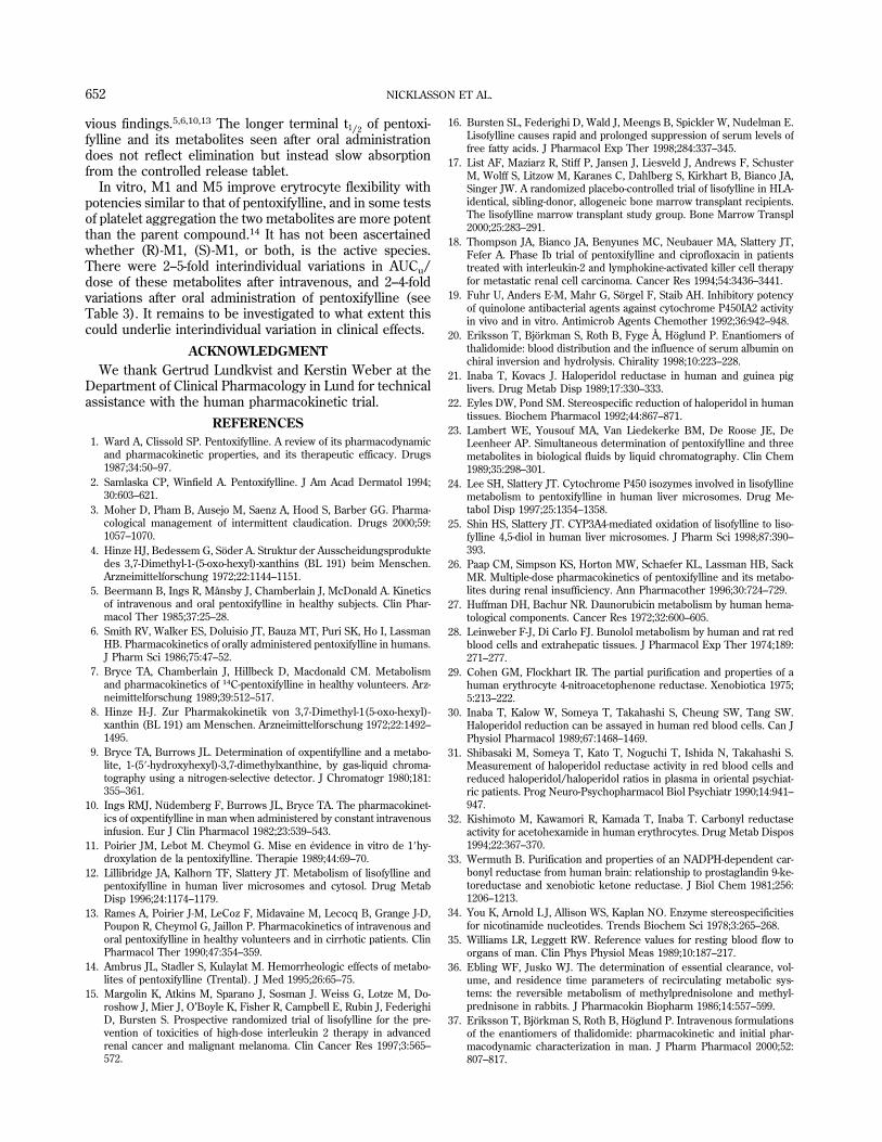
vious findings.5,6,10,13 The longer terminal t1/2 of pentoxi-fylline and its metabolites seen after oral administrationdoes not reflect elimination but instead slow absorptionfrom the controlled release tablet.
In vitro, M1 and M5 improve erytrocyte flexibility withpotencies similar to that of pentoxifylline, and in some testsof platelet aggregation the two metabolites are more potentthan the parent compound.14 It has not been ascertainedwhether (R)-M1, (S)-M1, or both, is the active species.There were 2–5-fold interindividual variations in AUCu/dose of these metabolites after intravenous, and 2–4-foldvariations after oral administration of pentoxifylline (seeTable 3). It remains to be investigated to what extent thiscould underlie interindividual variation in clinical effects.
ACKNOWLEDGMENTWe thank Gertrud Lundkvist and Kerstin Weber at the
Department of Clinical Pharmacology in Lund for technicalassistance with the human pharmacokinetic trial.
REFERENCES1. Ward A, Clissold SP. Pentoxifylline. A review of its pharmacodynamic
and pharmacokinetic properties, and its therapeutic efficacy. Drugs1987;34:50–97.
2. Samlaska CP, Winfield A. Pentoxifylline. J Am Acad Dermatol 1994;30:603–621.
3. Moher D, Pham B, Ausejo M, Saenz A, Hood S, Barber GG. Pharma-cological management of intermittent claudication. Drugs 2000;59:1057–1070.
4. Hinze HJ, Bedessem G, Soder A. Struktur der Ausscheidungsproduktedes 3,7-Dimethyl-1-(5-oxo-hexyl)-xanthins (BL 191) beim Menschen.Arzneimittelforschung 1972;22:1144–1151.
5. Beermann B, Ings R, Månsby J, Chamberlain J, McDonald A. Kineticsof intravenous and oral pentoxifylline in healthy subjects. Clin Phar-macol Ther 1985;37:25–28.
6. Smith RV, Walker ES, Doluisio JT, Bauza MT, Puri SK, Ho I, LassmanHB. Pharmacokinetics of orally administered pentoxifylline in humans.J Pharm Sci 1986;75:47–52.
7. Bryce TA, Chamberlain J, Hillbeck D, Macdonald CM. Metabolismand pharmacokinetics of 14C-pentoxifylline in healthy volunteers. Arz-neimittelforschung 1989;39:512–517.
8. Hinze H-J. Zur Pharmakokinetik von 3,7-Dimethyl-1(5-oxo-hexyl)-xanthin (BL 191) am Menschen. Arzneimittelforschung 1972;22:1492–1495.
9. Bryce TA, Burrows JL. Determination of oxpentifylline and a metabo-lite, 1-(5�-hydroxyhexyl)-3,7-dimethylxanthine, by gas-liquid chroma-tography using a nitrogen-selective detector. J Chromatogr 1980;181:355–361.
10. Ings RMJ, Nudemberg F, Burrows JL, Bryce TA. The pharmacokinet-ics of oxpentifylline in man when administered by constant intravenousinfusion. Eur J Clin Pharmacol 1982;23:539–543.
11. Poirier JM, Lebot M. Cheymol G. Mise en evidence in vitro de 1�hy-droxylation de la pentoxifylline. Therapie 1989;44:69–70.
12. Lillibridge JA, Kalhorn TF, Slattery JT. Metabolism of lisofylline andpentoxifylline in human liver microsomes and cytosol. Drug MetabDisp 1996;24:1174–1179.
13. Rames A, Poirier J-M, LeCoz F, Midavaine M, Lecocq B, Grange J-D,Poupon R, Cheymol G, Jaillon P. Pharmacokinetics of intravenous andoral pentoxifylline in healthy volunteers and in cirrhotic patients. ClinPharmacol Ther 1990;47:354–359.
14. Ambrus JL, Stadler S, Kulaylat M. Hemorrheologic effects of metabo-lites of pentoxifylline (Trental). J Med 1995;26:65–75.
15. Margolin K, Atkins M, Sparano J, Sosman J. Weiss G, Lotze M, Do-roshow J, Mier J, O’Boyle K, Fisher R, Campbell E, Rubin J, FederighiD, Bursten S. Prospective randomized trial of lisofylline for the pre-vention of toxicities of high-dose interleukin 2 therapy in advancedrenal cancer and malignant melanoma. Clin Cancer Res 1997;3:565–572.
16. Bursten SL, Federighi D, Wald J, Meengs B, Spickler W, Nudelman E.Lisofylline causes rapid and prolonged suppression of serum levels offree fatty acids. J Pharmacol Exp Ther 1998;284:337–345.
17. List AF, Maziarz R, Stiff P, Jansen J, Liesveld J, Andrews F, SchusterM, Wolff S, Litzow M, Karanes C, Dahlberg S, Kirkhart B, Bianco JA,Singer JW. A randomized placebo-controlled trial of lisofylline in HLA-identical, sibling-donor, allogeneic bone marrow transplant recipients.The lisofylline marrow transplant study group. Bone Marrow Transpl2000;25:283–291.
18. Thompson JA, Bianco JA, Benyunes MC, Neubauer MA, Slattery JT,Fefer A. Phase Ib trial of pentoxifylline and ciprofloxacin in patientstreated with interleukin-2 and lymphokine-activated killer cell therapyfor metastatic renal cell carcinoma. Cancer Res 1994;54:3436–3441.
19. Fuhr U, Anders E-M, Mahr G, Sorgel F, Staib AH. Inhibitory potencyof quinolone antibacterial agents against cytochrome P450IA2 activityin vivo and in vitro. Antimicrob Agents Chemother 1992;36:942–948.
20. Eriksson T, Bjorkman S, Roth B, Fyge Å, Hoglund P. Enantiomers ofthalidomide: blood distribution and the influence of serum albumin onchiral inversion and hydrolysis. Chirality 1998;10:223–228.
21. Inaba T, Kovacs J. Haloperidol reductase in human and guinea piglivers. Drug Metab Disp 1989;17:330–333.
22. Eyles DW, Pond SM. Stereospecific reduction of haloperidol in humantissues. Biochem Pharmacol 1992;44:867–871.
23. Lambert WE, Yousouf MA, Van Liedekerke BM, De Roose JE, DeLeenheer AP. Simultaneous determination of pentoxifylline and threemetabolites in biological fluids by liquid chromatography. Clin Chem1989;35:298–301.
24. Lee SH, Slattery JT. Cytochrome P450 isozymes involved in lisofyllinemetabolism to pentoxifylline in human liver microsomes. Drug Me-tabol Disp 1997;25:1354–1358.
25. Shin HS, Slattery JT. CYP3A4-mediated oxidation of lisofylline to liso-fylline 4,5-diol in human liver microsomes. J Pharm Sci 1998;87:390–393.
26. Paap CM, Simpson KS, Horton MW, Schaefer KL, Lassman HB, SackMR. Multiple-dose pharmacokinetics of pentoxifylline and its metabo-lites during renal insufficiency. Ann Pharmacother 1996;30:724–729.
27. Huffman DH, Bachur NR. Daunorubicin metabolism by human hema-tological components. Cancer Res 1972;32:600–605.
28. Leinweber F-J, Di Carlo FJ. Bunolol metabolism by human and rat redblood cells and extrahepatic tissues. J Pharmacol Exp Ther 1974;189:271–277.
29. Cohen GM, Flockhart IR. The partial purification and properties of ahuman erythrocyte 4-nitroacetophenone reductase. Xenobiotica 1975;5:213–222.
30. Inaba T, Kalow W, Someya T, Takahashi S, Cheung SW, Tang SW.Haloperidol reduction can be assayed in human red blood cells. Can JPhysiol Pharmacol 1989;67:1468–1469.
31. Shibasaki M, Someya T, Kato T, Noguchi T, Ishida N, Takahashi S.Measurement of haloperidol reductase activity in red blood cells andreduced haloperidol/haloperidol ratios in plasma in oriental psychiat-ric patients. Prog Neuro-Psychopharmacol Biol Psychiatr 1990;14:941–947.
32. Kishimoto M, Kawamori R, Kamada T, Inaba T. Carbonyl reductaseactivity for acetohexamide in human erythrocytes. Drug Metab Dispos1994;22:367–370.
33. Wermuth B. Purification and properties of an NADPH-dependent car-bonyl reductase from human brain: relationship to prostaglandin 9-ke-toreductase and xenobiotic ketone reductase. J Biol Chem 1981;256:1206–1213.
34. You K, Arnold L J, Allison WS, Kaplan NO. Enzyme stereospecificitiesfor nicotinamide nucleotides. Trends Biochem Sci 1978;3:265–268.
35. Williams LR, Leggett RW. Reference values for resting blood flow toorgans of man. Clin Phys Physiol Meas 1989;10:187–217.
36. Ebling WF, Jusko WJ. The determination of essential clearance, vol-ume, and residence time parameters of recirculating metabolic sys-tems: the reversible metabolism of methylprednisolone and methyl-prednisone in rabbits. J Pharmacokin Biopharm 1986;14:557–599.
37. Eriksson T, Bjorkman S, Roth B, Hoglund P. Intravenous formulationsof the enantiomers of thalidomide: pharmacokinetic and initial phar-macodynamic characterization in man. J Pharm Pharmacol 2000;52:807–817.
652 NICKLASSON ET AL.
British Journal of Clinical Pharmacology
DOI:10.1111/j.1365-2125.2005.02549.x
Br J Clin Pharmacol
61
:2 138–147 138 © 2005 Blackwell Publishing Ltd
Correspondence
Peter Höglund,
Department of Clinical Pharmacology, Lund University Hospital, SE-221 85 Lund, Sweden.
Tel
: + 46 4617 7979
Fax:
+ 46 4617 6085
E-mail:
Keywords
ciprofloxacin, metabolite effects, pentoxifylline, pharmacokinetics, retinal blood flow, rifampicin
Received
6 April 2005
Accepted
21 June 2005
Published
OnlineEarly
20 December 2005
A placebo-controlled study of retinal blood flow changes
by pentoxifylline and metabolites in humans
Marie Magnusson,
1,2
Ingar C. Bergstrand,
3
Sven Björkman,
2
Anders Heijl,
3
Bodil Roth
2
& Peter Höglund
1
1
Department of Clinical Pharmacology, Lund University Hospital, Lund,
2
Hospital Pharmacy and
3
Department of Ophthalmology, Malmö University Hospital, Malmö, Sweden
Aim
To investigate the possible effects of pentoxifylline metabolites on retinal blood flowin humans.
Methods
A randomized, placebo-controlled, four-period cross-over study that was observerblinded and partly blinded for the eight participants. On one occasion a placebo wasgiven as an intravenous (i.v.) infusion over 100 min. On the other three occasionspentoxifylline was administered as i.v. infusions over 100 min at a rate of 3 mg min
−
1
.Before two of the pentoxifylline infusions the subjects were pretreated with eitherciprofloxacin or rifampicin. Retinal blood flow was measured by scanning laser dop-pler flowmetry (SLDF) in a selected area of the central temporal retina before, duringand until 5 h after the end of infusion. Blood samples for concentration analyses ofpentoxifyllin, R-M1, S-M1, M4 and M5 were taken serially and areas under thecurves (AUCs) were calculated. Linear mixed models were used for the statisticalanalyses.
Results
Mean AUCs (ng h ml
−
1
) were significantly increased for pentoxifylline (1964
vs
. 1453)and S-M1 (5804
vs
. 4227), but not R-M1 when pentoxifylline was co-administeredwith ciprofloxacin. The mean AUC for M5 was significantly reduced when subjectswere pretreated with rifampicin (2041
vs
. 3080). Pentoxifylline with and withoutpretreatment with rifampicin significantly increased retinal blood flow assessed asmean flow, pulsation (i.e. 1-systole/diastole), and diastolic flow (but not duringsystole), compared with placebo. The increases over placebo were more pronouncedon diastolic flow, 9.7% (95% confidence interval 4.2, 15.5) than on mean flow, 4.6%(1.1, 8.3) after pentoxifylline administration. With pentoxifylline after rifampicin pre-treatment the corresponding differences were 11.7% (5.8, 17.9) and 5.1% (1.4, 7.8)over placebo, respectively. After co-administration of pentoxifylline and ciprofloxacinwe saw only a nonsignificant trend towards increased flow during diastole, but asignificant decrease in pulsation. When AUCs for pentoxifylline and its metaboliteswere used as regressor variables to retinal mean flow we found that pentoxifylline,R-M1 and M5 had coefficients with a positive sign indicating that they enhanced theretinal blood flow. In contrast, S-M1 and M4 had coefficients with negative sign andthus appeared to decrease the blood flow in subjects treated with pentoxifylline.
Conclusion
The R-M1 and M5 metabolites of pentoxifylline contributed significantly to the effectsof pentoxifylline on retinal blood flow.
Retinal blood flow changes by pentoxifylline in humans
Br J Clin Pharmacol
61
:2 139
Introduction
Pentoxifylline, 3,7-dimethyl-1(5
′
-oxo-hexyl)xanthine,is a haemorheological drug widely used for the treat-ment of intermittent claudication [1–3]. However, theclinical efficacy of the drug for this indication is stillcontroversial. It has also been suggested that pentoxifyl-line may be used in diseases affecting retinal blood flow,such as diabetic retinopathy [4–7] or macular degener-ation [8]. The therapeutic effect of pentoxifylline inthese conditions would be to increase capillary bloodflow by increasing deformability of both erythrocytesand leucocytes as well as by a possible direct vasodila-tory effect.
Pentoxifylline is metabolized in humans into at leastseven metabolites, denoted M1–M7 [9–12]. Reductionof the 5
′
-oxo group gives the hydroxy metabolite M1,3,7-dimetyl-1(5
′
hydroxyhexyl)xanthine. This creates achiral centre in the molecule, yielding S-M1 as a majorand R-M1 as minor metabolite [13, 14]. This metabo-lism takes place both in the liver [13] and in the eryth-rocytes [14, 15] and is reversible [10, 13, 14]. Thus,during treatment with pentoxifylline the plasma concen-trations of the parent drug, R-M1 and S-M1 rapidlyattain equilibrium. Hepatic metabolism produces thecarboxylic acid metabolites M4 and M5; M5 is themajor excreted form [9–12]. Both M1 (as the racemicform) and M5 have been shown to influence erythrocytedeformability and platelet aggregation
in vitro
and maythus contribute to the
in vivo
haemorheological effectsof pentoxifylline [16].
The aim of this study was to investigate the pos-sible contributions of the metabolites (M1, M4 andM5) of pentoxifylline to its haemorheological effect,assessed as retinal blood flow, in humans. Scanninglaser doppler flowmetry (SLDF) [17] in a selectedarea of the central temporal retina was used to investi-gate drug effects on blood flow in small capillaries.When pentoxifylline is administered there is consider-able co-variation between the concentrations of pen-toxifylline and its metabolites [14, unpublished]. Thismakes it very difficult to separate any effects of themetabolites from that of the parent compound. In thisstudy we therefore co-administered pentoxifylline andan inhibitor of CYP1A2 (ciprofloxacin) on one occa-sion [18, 19], and gave pentoxifylline after pretreat-ment with an inducer of several enzymes of thecytochrome P450 system (rifampicin) on anotheroccasion [20], in order to create different blood con-centration ratios between the metabolites and pentoxi-fylline in the same subject. The study was placebocontrolled with a cross-over design using four treat-ment arms.
Methods
Study design
The study was approved by the Ethics Committee ofLund University and by the Swedish Medical ProductsAgency. After giving written informed consent, eighthealthy, nonsmoking volunteers (six males, 24–42 years, 73–100 kg, and two females, 21–24 years, 65–66 kg), who were free of medication and had no historyof allergy to drugs, were included in the study. Eachsubject passed a prestudy ophthalmic examination,where inclusion criteria were normal ocular findings, nopreviously known eye pathology, full visual acuity andametropia less than 3 diopters. The study was random-ized, placebo controlled, observer blinded, and partlyblinded for the subject in a four-period cross-overdesign.
All subjects were given the four treatments in randomorder. During one session they were given placebo(0.9% saline solution) by intravenous (i.v.) infusion over100 min. During the three other sessions they weregiven pentoxifylline (Trental, Hoechst Marion Roussel)by i.v. infusion over 100 min at a rate of 3 mg min
−
1
.The volumes infused and the concentrations of pentox-ifylline in the solution were measured. In one sessionthe subjects were pretreated with a 750-mg ciprofloxa-cin tablet (Ciproxin, Bayer) 1 h before start of the infu-sion. Before another they took one rifampicin 600-mgtablet (Rimactan, Swedish Orphan) daily for 7 days, thelast dose being taken approximately 24 h before the startof the pentoxifylline infusion.
The pretreatments with ciprofloxacin and rifampicincould not be blinded but the pentoxifylline and placeboadministrations were.
The subjects had fasted since 22.00 h the eveningbefore the study day and were not allowed to take anyother medication except for occasional paracetamol 48 hpreceding a study day and during the pretreatment withrifampicin. They were given a light meal 4 h after thestart of the infusion. The study periods were separatedby at least 1 week and after the pretreatment withrifampicin by 2 weeks. Adverse events were assessedbefore and during the study days by open-endedquestions.
Analysis of pentoxifylline and its metabolites
Blood was sampled from an indwelling venous cathe-ter (in the opposite arm from the infusion) intosodium heparin Venoject
®
tubes. The sampling timeswere: before and at 20, 45 and 85 min during theinfusion period and at 0.5, 1, 2, 3, 4 and 5 h after itstermination (in the first four subjects also at 6 h). Inorder to minimize
ex vivo
interconversion of pentoxi-
M. Magnusson et al.
140
61
:2
Br J Clin Pharmacol
fylline and M1 the samples were immediately centri-fuged at 4
o
C and the plasma was collected and frozen[14].
Concentrations of pentoxifylline and metabolites R-M1, S-M1, M4 and M5 in plasma were determined byhigh-performance liquid chromatography (HPLC) aspreviously described [14]. From the plasma concentra-tion data, the terminal half-lives of pentoxifylline andits metabolites were estimated using the RSTRIP soft-ware (MicroMath, Salt Lake City, UT, USA) and usedfor extrapolation of the areas under the curves (AUCs).The AUCs were calculated by the logarithmic trapezoi-dal method from 0 to infinity using the MKMODELsoftware (Biosoft, Cambridge, UK).
Measurement and analysis of retinal blood flow
Retinal blood flow was assessed using the SLDF at670 nm (Heidelberg Retina Flowmeter, HeidelbergEngineering) [17, 21]. The method of SLDF provides ahigh-definition tomographic image of perfused retinalvessels with simultaneous evaluation of blood flowusing an optical Doppler effect. The measurements wereperformed in a selected area of the central temporalretina, and one picture included an area of 2.7
×
0.7 mm,at baseline, at 45 and 85 min during the infusion periodand at 0.5, 1, 2, 3, 4 and 5 h after termination (in thefirst four subjects also at 6 h). At all the time points formeasurements four pictures of the same area were takenand the three technically best ones were assessed.
Quantification of capillary retinal blood flow wasstated in arbitrary units (AUs) describing the product ofmean flow velocity and mean quantity of blood cells ina standardized volume. The mean values from each timepoint were used in the calculations. The readings wereanalysed using the AFFPIA software (automatic fullfield perfusion image analyser program) [21]. The vari-ables used were mean blood flow, blood flow duringsystole, blood flow during diastole, and pulsation (i.e.1-diastole/systole).
Data presentation and statistical considerations
The number of subjects recruited was based on the fol-lowing assumptions. In an earlier study on 10 healthysubjects [7] an i.v. infusion over 90 min of 200 mg ofpentoxifylline gave an increase by 17
±
9% (mean
±
SDbetween subjects) and the 400-mg dose gave an increaseby 27
±
12% in ocular fundus pulsation. We anticipatedthat a 300-mg dose over 100 min would give a 23
±
10%increase in ocular fundus pulsation between subjects,compared with placebo. Since, in the current study, wecompare the effects within the same subject, we made a
series of power calculations assuming different within-subject correlations, number of subjects, and detectabledifferences, e.g. using eight subjects we would be ableto detect a difference of 8% with a power of at least80%, at a two-tailed
P
-value
<
0.05, if the intrasubjectcorrelation was 0.75. This was calculated for a singlemeasurement of maximum effect. When calculationswere based on integrated effects (i.e. several measure-ments over time) the power to detect differences wereanticipated to be even better.
The volumes infused and the concentrations of pen-toxifylline in the solution were used for calculation ofthe actual doses given. The resulting concentrationswere re-calculated to a nominal dose of 300 mg. In thepharmacokinetic analyses we similarly used AUCsrecalculated to a nominal dose of 300 mg. However,AUCs calculated from the observed plasma concentra-tions were used as input in the concentration–effectanalyses described below. For statistical analysis theMIXED procedure in SAS (version 8.2; SAS Institute,Cary, NC, USA) was used. In the analysis of the AUCswe used the treatments given as fixed effects and sub-jects were entered as random effects.
We found that there were no significant changes overtime within each study day. Thus, we used all observa-tions after dose in a repeated measures design. Further,the observed effects were rather low, limiting the possi-bility for detection of deviations from linear relationsbetween effect and regressor variables. Two main statis-tical models were tested after an initial check for lackof period effects. The first one comprised only the treat-ments given as fixed effects, thus disregarding theobtained plasma concentration data. In the secondmodel treatments were not included; instead, the AUCsof pentoxifylline and its metabolites were used asregressor variables. Such a model can be successfullyused only if the correlations between the regressors arelow. Both models used subjects as random effects, anda repeated measures spatial exponential covariancestructure design.
Since one subject dropped out, the study was notcompletely balanced and for both the kinetic and thedynamic analyses least-square means were obtainedfrom the mixed model, thus providing the marginalmeans that would be expected had the study been com-pletely balanced with no missing data. Arithmetic least-square means and 95% confidence limits are given,except when relative changes in the effect model com-prising treatments as fixed effects are presented, inwhich case geometric least-square means and 95% con-fidence limits are given. Statistical significance wasaccepted at
P
<
0.05 (two-tailed).
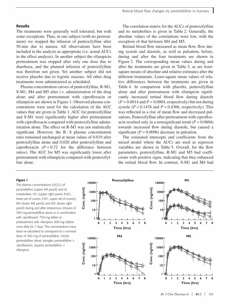
Retinal blood flow changes by pentoxifylline in humans
Br J Clin Pharmacol
61
:2 141
Results
The treatments were generally well tolerated, but withsome exceptions. Thus, in one subject (with no pretreat-ment) we stopped the infusion of pentoxifylline after50 min due to nausea. All observations have beenincluded in the analysis as appropriate (i.e. actual AUCsin the effect analysis). In another subject the rifampicinpretreatment was stopped after only one dose due todiarrhoea, and the planned infusion of pentoxifyllinewas therefore not given. Yet another subject did notreceive placebo due to logistic reasons. All other drugtreatments were administered as scheduled.
Plasma concentration curves of pentoxifylline, R-M1,S-M1, M4 and M5 after i.v. administration of the drugalone and after pretreatment with ciprofloxacin orrifampicin are shown in Figure 1. Observed plasma con-centrations were used for the calculation of the AUCvalues that are given in Table 1. AUC for pentoxifyllineand S-M1 were significantly higher after pretreatmentwith ciprofloxacin compared with pentoxifylline admin-istration alone. The effect on R-M1 was not statisticallysignificant. However, the R : S plasma concentrationratio remained unchanged at mean values of 0.019 afterpentoxifylline alone and 0.020 after pentoxifylline andciprofloxacin (
P
=
0.72 for the difference betweenratios). The AUC for M5 was significantly lower afterpretreatment with rifampicin compared with pentoxifyl-line alone.
The correlation matrix for the AUCs of pentoxifyllineand its metabolites is given in Table 2. Generally, theabsolute values of the correlations were low, with theexception of that between M4 and M5.
Retinal blood flow measured as mean flow, flow dur-ing systole and diastole, as well as pulsation, before,during and after the four treatments are shown inFigure 2. The corresponding mean values during andafter the treatments are given in Table 3, as are least-square means of absolute and relative estimates after thedifferent treatments. Least-square mean values of rela-tive differences between the treatments are given inTable 4. In comparison with placebo, pentoxifyllinealone and after pretreatment with rifampicin signifi-cantly increased retinal blood flow during diastole(
P
=
0.0014 and
P
=
0.0004, respectively) but not duringsystole (
P
=
0.1476 and
P
=
0.4366, respectively). Thiswas reflected in a rise of mean flow and decreased pul-sations. Pentoxifylline after pretreatment with ciproflox-acin resulted only in a nonsignificant trend (
P
=
0.0604)towards increased flow during diastole, but caused asignificant (
P
=
0.0096) decrease in pulsation.The estimated intercepts and coefficients from the
mixed model where the AUCs are used as regressorvariables are shown in Table 5. Overall, for the flowparameters, pentoxifylline, R-M1 and M5 had coeffi-cients with positive signs, indicating that they enhancedthe retinal blood flow. In contrast, S-M1 and M4 had
Figure 1
The plasma concentrations (
±
S.D.) of
pentoxifylline (upper left panel) and its
metabolites: M1 (upper right panel: R-M1,
lower set of curves; S-M1, upper set of curves),
M4 (lower left panel) and M5 (lower right
panel) during and after intravenous infusion of
300 mg pentoxifylline alone or in combination
with ciprofloxacin 750-mg tablet or
pretreatment with rifampicin 600-mg tablets
once daily for 7 days. The concentrations have
been re-calculated to correspond to a nominal
dose of 300 mg of pentoxifylline. Circles
pentoxifylline alone, triangles pentoxifylline +
ciprofloxacin, squares pentoxifylline +
rifampicin
0 1 2 3 4 5 6 7 8
Co
nc. (
ng/m
l)
Time (hrs)
Pentoxifylline
0 1 2 3 4 5 6 7 8
Co
nc. (
ng/m
l)
Time (hrs)
M4
0 1 2 3 4 5 6 7 8
Co
nc. (
ng/m
l)
Time (hrs)
M1
0 1 2 3 4 5 6 7 8
Co
nc. (
ng/m
l)
Time (hrs)
M5
10000
1000
100
10
1
10000
1000
100
10
1
10000
1000
100
10
1
10000
1000
100
10
1
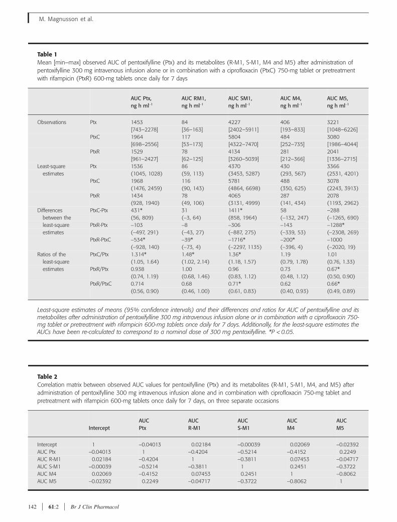
M. Magnusson et al.
142
61
:2
Br J Clin Pharmacol
Table 1
Mean [min–max] observed AUC of pentoxifylline (Ptx) and its metabolites (R-M1, S-M1, M4 and M5) after administration of pentoxifylline 300 mg intravenous infusion alone or in combination with a ciprofloxacin (PtxC) 750-mg tablet or pretreatment with rifampicin (PtxR) 600-mg tablets once daily for 7 days
AUC Ptx,ng h ml
−
1
AUC RM1,ng h ml
−
1
AUC SM1,ng h ml
−
1
AUC M4,ng h ml
−
1
AUC M5, ng h ml
−
1
Observations Ptx 1453 84 4227 406 3221[743–2278] [36–163] [2402–5911] [193–833] [1048–6226]
PtxC 1964 117 5804 484 3080[698–2556] [53–173] [4322–7470] [252–735] [1986–4044]
PtxR 1529 78 4134 281 2041[961–2427] [62–125] [3260–5039] [212–366] [1336–2715]
Least-square Ptx 1536 86 4370 430 3366estimates (1045, 1028) (59, 113) (3453, 5287) (293, 567) (2531, 4201)
PtxC 1968 116 5781 488 3078(1476, 2459) (90, 143) (4864, 6698) (350, 625) (2243, 3913)
PtxR 1434 78 4065 287 2078(928, 1940) (49, 106) (3131, 4999) (141, 434) (1193, 2962)
Differences PtxC-Ptx 431* 31 1411* 58 –288between the (56, 809) (
−
3, 64) (858, 1964) (
−
132, 247) (
−
1265, 690)least-square PtxR-Ptx
−
103
−
8
−
306
−
143
−
1288*estimates (
−
497, 291) (
−
43, 27) (
−
887, 275) (
−
339, 53) (
−
2308, 269)PtxR-PtxC
−
534*
−
39*
−
1716*
−
200*
−
1000(
−
928, 140) (
−
73, 4) (
−
2297, 1135) (
−
396, 4) (
−
2020, 19)Ratios of the PtxC/Ptx 1.314* 1.48* 1.36* 1.19 1.01
least-square (1.05, 1.64) (1.02, 2.14) (1.18, 1.57) (0.79, 1.78) (0.76, 1.33)estimates PtxR/Ptx 0.938 1.00 0.96 0.73 0.67*
(0.74, 1.19) (0.68, 1.46) (0.83, 1.12) (0.48, 1.12) (0.50, 0.90)PtxR/PtxC 0.714 0.68 0.71* 0.62 0.66*
(0.56, 0.90) (0.46, 1.00) (0.61, 0.83) (0.40, 0.93) (0.49, 0.89)
Least-square estimates of means (95% confidence intervals) and their differences and ratios for AUC of pentoxifylline and itsmetabolites after administration of pentoxifylline 300 mg intravenous infusion alone or in combination with a ciprofloxacin 750-mg tablet or pretreatment with rifampicin 600-mg tablets once daily for 7 days. Additionally, for the least-square estimates theAUCs have been re-calculated to correspond to a nominal dose of 300 mg pentoxifylline. *P
<
0.05.
Table 2
Correlation matrix between observed AUC values for pentoxifylline (Ptx) and its metabolites (R-M1, S-M1, M4, and M5) after administration of pentoxifylline 300 mg intravenous infusion alone and in combination with ciprofloxacin 750-mg tablet and pretreatment with rifampicin 600-mg tablets once daily for 7 days, on three separate occasions
InterceptAUCPtx
AUCR-M1
AUCS-M1
AUCM4
AUCM5
Intercept 1
−
0.04013 0.02184
−
0.00039 0.02069
−
0.02392AUC Ptx
−
0.04013 1
−
0.4204
−
0.5214
−
0.4152 0.2249AUC R-M1 0.02184
−
0.4204 1
−
0.3811 0.07453
−
0.04717AUC S-M1
−
0.00039
−
0.5214
−
0.3811 1 0.2451
−
0.3722AUC M4 0.02069
−
0.4152 0.07453 0.2451 1
−
0.8062AUC M5
−
0.02392 0.2249
−
0.04717
−
0.3722
−
0.8062 1
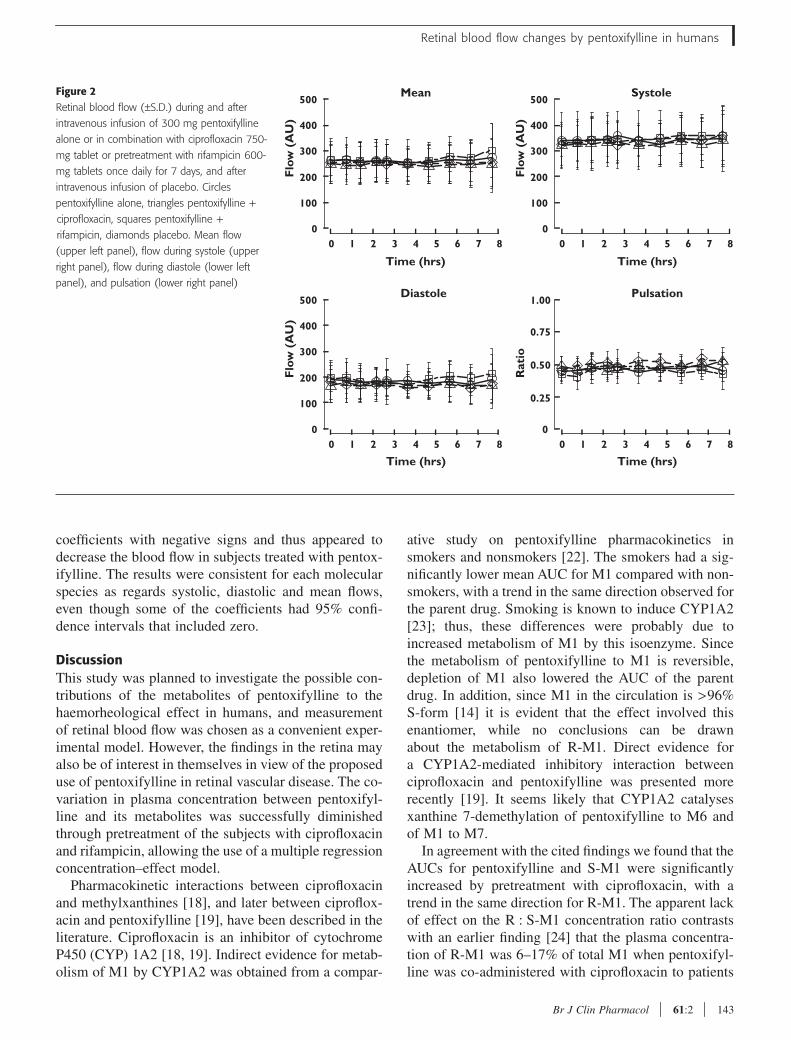
Retinal blood flow changes by pentoxifylline in humans
Br J Clin Pharmacol
61
:2 143
coefficients with negative signs and thus appeared todecrease the blood flow in subjects treated with pentox-ifylline. The results were consistent for each molecularspecies as regards systolic, diastolic and mean flows,even though some of the coefficients had 95% confi-dence intervals that included zero.
Discussion
This study was planned to investigate the possible con-tributions of the metabolites of pentoxifylline to thehaemorheological effect in humans, and measurementof retinal blood flow was chosen as a convenient exper-imental model. However, the findings in the retina mayalso be of interest in themselves in view of the proposeduse of pentoxifylline in retinal vascular disease. The co-variation in plasma concentration between pentoxifyl-line and its metabolites was successfully diminishedthrough pretreatment of the subjects with ciprofloxacinand rifampicin, allowing the use of a multiple regressionconcentration–effect model.
Pharmacokinetic interactions between ciprofloxacinand methylxanthines [18], and later between ciproflox-acin and pentoxifylline [19], have been described in theliterature. Ciprofloxacin is an inhibitor of cytochromeP450 (CYP) 1A2 [18, 19]. Indirect evidence for metab-olism of M1 by CYP1A2 was obtained from a compar-
ative study on pentoxifylline pharmacokinetics insmokers and nonsmokers [22]. The smokers had a sig-nificantly lower mean AUC for M1 compared with non-smokers, with a trend in the same direction observed forthe parent drug. Smoking is known to induce CYP1A2[23]; thus, these differences were probably due toincreased metabolism of M1 by this isoenzyme. Sincethe metabolism of pentoxifylline to M1 is reversible,depletion of M1 also lowered the AUC of the parentdrug. In addition, since M1 in the circulation is
>
96%S-form [14] it is evident that the effect involved thisenantiomer, while no conclusions can be drawnabout the metabolism of R-M1. Direct evidence fora CYP1A2-mediated inhibitory interaction betweenciprofloxacin and pentoxifylline was presented morerecently [19]. It seems likely that CYP1A2 catalysesxanthine 7-demethylation of pentoxifylline to M6 andof M1 to M7.
In agreement with the cited findings we found that theAUCs for pentoxifylline and S-M1 were significantlyincreased by pretreatment with ciprofloxacin, with atrend in the same direction for R-M1. The apparent lackof effect on the R : S-M1 concentration ratio contrastswith an earlier finding [24] that the plasma concentra-tion of R-M1 was 6–17% of total M1 when pentoxifyl-line was co-administered with ciprofloxacin to patients
Figure 2Retinal blood flow (±S.D.) during and after
intravenous infusion of 300 mg pentoxifylline
alone or in combination with ciprofloxacin 750-
mg tablet or pretreatment with rifampicin 600-
mg tablets once daily for 7 days, and after
intravenous infusion of placebo. Circles
pentoxifylline alone, triangles pentoxifylline +
ciprofloxacin, squares pentoxifylline +
rifampicin, diamonds placebo. Mean flow
(upper left panel), flow during systole (upper
right panel), flow during diastole (lower left
panel), and pulsation (lower right panel)
Mean
0 1 2 3 4 5 6 7 8
100
0
200
300
400
500
Time (hrs)
Flo
w (
AU
)
Systole
0 1 2 3 4 5 6 7 8
100
0
200
300
400
500
Time (hrs)
Flo
w (
AU
)
Pulsation
0 1 2 3 4 5 6 7 80
0.25
0.50
0.75
1.00
Time (hrs)
Rat
io
Diastole
0 1 2 3 4 5 6 7 8
100
0
200
300
400
500
Time (hrs)
Flo
w (
AU
)
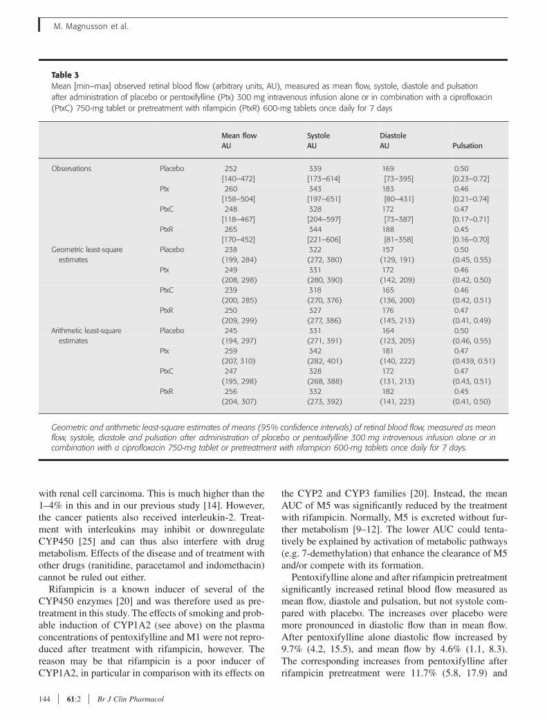
M. Magnusson et al.
144 61:2 Br J Clin Pharmacol
with renal cell carcinoma. This is much higher than the1–4% in this and in our previous study [14]. However,the cancer patients also received interleukin-2. Treat-ment with interleukins may inhibit or downregulateCYP450 [25] and can thus also interfere with drugmetabolism. Effects of the disease and of treatment withother drugs (ranitidine, paracetamol and indomethacin)cannot be ruled out either.
Rifampicin is a known inducer of several of theCYP450 enzymes [20] and was therefore used as pre-treatment in this study. The effects of smoking and prob-able induction of CYP1A2 (see above) on the plasmaconcentrations of pentoxifylline and M1 were not repro-duced after treatment with rifampicin, however. Thereason may be that rifampicin is a poor inducer ofCYP1A2, in particular in comparison with its effects on
the CYP2 and CYP3 families [20]. Instead, the meanAUC of M5 was significantly reduced by the treatmentwith rifampicin. Normally, M5 is excreted without fur-ther metabolism [9–12]. The lower AUC could tenta-tively be explained by activation of metabolic pathways(e.g. 7-demethylation) that enhance the clearance of M5and/or compete with its formation.
Pentoxifylline alone and after rifampicin pretreatmentsignificantly increased retinal blood flow measured asmean flow, diastole and pulsation, but not systole com-pared with placebo. The increases over placebo weremore pronounced in diastolic flow than in mean flow.After pentoxifylline alone diastolic flow increased by9.7% (4.2, 15.5), and mean flow by 4.6% (1.1, 8.3).The corresponding increases from pentoxifylline afterrifampicin pretreatment were 11.7% (5.8, 17.9) and
Table 3Mean [min–max] observed retinal blood flow (arbitrary units, AU), measured as mean flow, systole, diastole and pulsation after administration of placebo or pentoxifylline (Ptx) 300 mg intravenous infusion alone or in combination with a ciprofloxacin (PtxC) 750-mg tablet or pretreatment with rifampicin (PtxR) 600-mg tablets once daily for 7 days
Mean flowAU
SystoleAU
DiastoleAU Pulsation
Observations Placebo 252 339 169 0.50[140–472] [173–614] [73–395] [0.23–0.72]
Ptx 260 343 183 0.46[158–504] [197–651] [80–431] [0.21–0.74]
PtxC 248 328 172 0.47[118–467] [204–597] [73–387] [0.17–0.71]
PtxR 265 344 188 0.45[170–452] [221–606] [81–358] [0.16–0.70]
Geometric least-square Placebo 238 322 157 0.50estimates (199, 284) (272, 380) (129, 191) (0.45, 0.55)
Ptx 249 331 172 0.46(208, 298) (280, 390) (142, 209) (0.42, 0.50)
PtxC 239 318 165 0.46(200, 285) (270, 376) (136, 200) (0.42, 0.51)
PtxR 250 327 176 0.47(209, 299) (277, 386) (145, 213) (0.41, 0.49)
Arithmetic least-square Placebo 245 331 164 0.50estimates (194, 297) (271, 391) (123, 205) (0.46, 0.55)
Ptx 259 342 181 0.47(207, 310) (282, 401) (140, 222) (0.439, 0.51)
PtxC 247 328 172 0.47(195, 298) (268, 388) (131, 213) (0.43, 0.51)
PtxR 256 332 182 0.45(204, 307) (273, 392) (141, 223) (0.41, 0.50)
Geometric and arithmetic least-square estimates of means (95% confidence intervals) of retinal blood flow, measured as meanflow, systole, diastole and pulsation after administration of placebo or pentoxifylline 300 mg intravenous infusion alone or incombination with a ciprofloxacin 750-mg tablet or pretreatment with rifampicin 600-mg tablets once daily for 7 days.
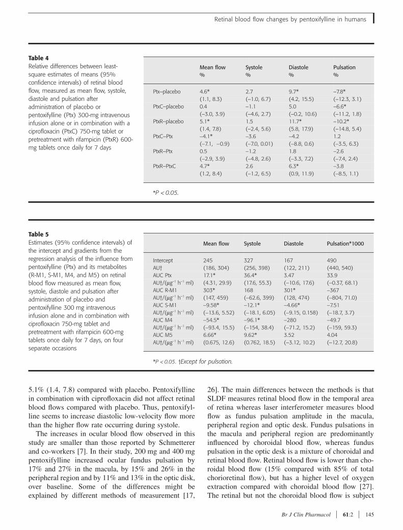
Retinal blood flow changes by pentoxifylline in humans
Br J Clin Pharmacol 61:2 145
5.1% (1.4, 7.8) compared with placebo. Pentoxifyllinein combination with ciprofloxacin did not affect retinalblood flows compared with placebo. Thus, pentoxifyl-line seems to increase diastolic low-velocity flow morethan the higher flow rate occurring during systole.
The increases in ocular blood flow observed in thisstudy are smaller than those reported by Schmettererand co-workers [7]. In their study, 200 mg and 400 mgpentoxifylline increased ocular fundus pulsation by17% and 27% in the macula, by 15% and 26% in theperipheral region and by 11% and 13% in the optic disk,over baseline. Some of the differences might beexplained by different methods of measurement [17,
26]. The main differences between the methods is thatSLDF measures retinal blood flow in the temporal areaof retina whereas laser interferometer measures bloodflow as fundus pulsation amplitude in the macula,peripheral region and optic desk. Fundus pulsations inthe macula and peripheral region are predominantlyinfluenced by choroidal blood flow, whereas funduspulsation in the optic desk is a mixture of choroidal andretinal blood flow. Retinal blood flow is lower than cho-roidal blood flow (15% compared with 85% of totalchorioretinal flow), but has a higher level of oxygenextraction compared with choroidal blood flow [27].The retinal but not the choroidal blood flow is subject
Mean flow%
Systole%
Diastole%
Pulsation%
Ptx–placebo 4.6* 2.7 9.7* −7.8*(1.1, 8.3) (−1.0, 6.7) (4.2, 15.5) (−12.3, 3.1)
PtxC–placebo 0.4 −1.1 5.0 −6.6*(−3.0, 3.9) (−4.6, 2.7) (−0.2, 10.6) (−11.2, 1.8)
PtxR–placebo 5.1* 1.5 11.7* −10.2*(1.4, 7.8) (−2.4, 5.6) (5.8, 17.9) (−14.8, 5.4)
PtxC–Ptx −4.1* −3.6 −4.2 1.2(−7.1, −0.9) (−7.0, 0.01) (−8.8, 0.6) (−3.5, 6.3)
PtxR–Ptx 0.5 −1.2 1.8 −2.6(−2.9, 3.9) (−4.8, 2.6) (−3.3, 7.2) (−7.4, 2.4)
PtxR–PtxC 4.7* 2.6 6.3* −3.8(1.2, 8.4) (−1.2, 6.5) (0.9, 11.9) (−8.5, 1.1)
*P < 0.05.
Table 4Relative differences between least-square estimates of means (95% confidence intervals) of retinal blood flow, measured as mean flow, systole, diastole and pulsation after administration of placebo or pentoxifylline (Ptx) 300-mg intravenous infusion alone or in combination with a ciprofloxacin (PtxC) 750-mg tablet or pretreatment with rifampicin (PtxR) 600-mg tablets once daily for 7 days
Mean flow Systole Diastole Pulsation*1000
Intercept 245 327 167 490AU† (186, 304) (256, 398) (122, 211) (440, 540)AUC Ptx 17.1* 36.4* 3.47 33.9AU†/(µg−1 h−1 ml) (4.31, 29.9) (17.6, 55.3) (−10.6, 17.6) (−0.37, 68.1)AUC R-M1 303* 168 301* −367AU†/(µg−1 h−1 ml) (147, 459) (−62.6, 399) (128, 474) (−804, 71.0)AUC S-M1 −9.58* −12.1* −4.66* −7.51AU†/(µg−1 h−1 ml) (−13.6, 5.52) (−18.1, 6.05) (−9.15, 0.158) (−18.7, 3.7)AUC M4 −54.5* −96.1* −280 −49.7AU†/(µg−1 h−1 ml) (−93.4, 15.5) (−154, 38.4) (−71.2, 15.2) (−159, 59.3)AUC M5 6.66* 9.62* 3.52 4.04AU†/(µg−1 h−1 ml) (0.675, 12.6) (0.762, 18.5) (−3.12, 10.2) (−12.7, 20.8)
*P < 0.05. †Except for pulsation.
Table 5Estimates (95% confidence intervals) of the intercept and gradients from the regression analysis of the influence from pentoxifylline (Ptx) and its metabolites (R-M1, S-M1, M4, and M5) on retinal blood flow measured as mean flow, systole, diastole and pulsation after administration of placebo and pentoxifylline 300 mg intravenous infusion alone and in combination with ciprofloxacin 750-mg tablet and pretreatment with rifampicin 600-mg tablets once daily for 7 days, on four separate occasions
M. Magnusson et al.
146 61:2 Br J Clin Pharmacol
to autoregulation [27], therefore making the retinalblood flow less affected by systemic factors than thechoroidal blood flow. Another advantage with the SLDFis that it scans an area of 2.7 × 0.7 mm and measures allflow in this area. Thus, more capillaries are found andthe influence of erroneous values is smaller. With SLDFit is also easier to find the same area for all the measure-ments compared with laser interferometer that measuresin one point. Pentoxifylline increases both choroidaland retinal blood flow but the increase is more pro-nounced in choroidal blood flow, possibly because reti-nal blood flow is autoregulated.
This study was performed in healthy volunteers partlybecause pentoxifylline is not approved for general usein Sweden and partly because we needed to co-admin-ister pentoxifylline with ciprofloxacin and rifampicin inorder to create different concentration ratios betweenpentoxifylline and its metabolites. It is difficult to makeassumptions about the magnitude of the effect in apatient group with reduced retinal flow after administra-tion to healthy volunteers, but we have shown that anincrease in retinal blood flow is obtainable. This has notbeen shown previously either in patients or in healthyvolunteers. In a study by Kruger et al., ocular funduspulsation and retinal blood flow were measured by laserinterferometry and SLDF in patients with age-relatedmacular degeneration after administration of pentoxifyl-line or placebo, 400 mg three times daily for 3 months[8]. They found that pentoxifylline increased ocular fun-dus pulsation amplitude up to 28% after 3 months’ treat-ment but could not find any effects on retinal blood flow.In the present study we investigated the effects of pen-toxifylline and metabolites on healthy eyes. Thus, ourfindings are not dependent on pathophysiologicalchanges of the retinal vascular bed. In addition, SLDFcombined with the AFFPIA software analysis used inthe present study resulted in better reproducibility andless bias compared with the technique in Krueger’sstudy. Our results are in agreement with previous stud-ies, using the subjective blue field entoptic phenomenoncomputer simulation technique, that have shown anincrease in capillary blood flow velocity in healthy vol-unteers and diabetes patients [4, 5].
A previous study has shown a clear time dependencyof the effects on blood flow after a single dose of pen-toxifylline [7]. We found no such time dependency(Figure 2). We cannot explain this, but we measuredretinal blood flow, whereas other investigators havemeasured a total of retinal and the more abundant cho-roidal blood flow. In order to study the contributionsfrom pentoxifylline and its metabolites we needed todiminish the naturally occurring high correlations
between these compounds. The correlation matrix(Table 2) shows that we were successful, possibly withthe exception of a remaining rather high correlationbetween M4 and M5. Applying a simple linear AUC–effect model, we found that pentoxifylline, R-M1 andM5 had coefficients with positive signs indicating thatthey all contribute to the effects. It is particularlynoticeable that R-M1 exerts a significant effect in spiteof being present in concentrations that are two ordersof magnitude lower than those of pentoxifylline andM5. The high potency of this compound is alsoreflected in the values of the coefficients (Table 5),which are accordingly one to two orders of magnitudegreater than those of the other compounds. Two of themetabolites, S-M1 and M4, showed negative coeffi-cients. This should not necessarily be interpreted asthese substances, per se, having a negative effect onblood flow, only that they, in the mix of substancesobtained after pentoxifylline administration, tend tomodify the effects in a direction opposite that ofpentoxifylline.
Our results showing biological activity of R-M1 andM5 are to some extent in agreement with those of pre-vious studies. The haemorheological effects of pentox-ifylline and its metabolites have been investigated invitro [16]. As regards enhancement of erythrocyte filter-ability, there was little difference between pentoxifyl-line, racemic M1 and M5. In a test for adenosinediphosphate-induced platelet aggregation M5 was notquite as effective as the parent drug; however, racemicM1 was approximately 10 times more potent than pen-toxifylline. R-M1 has been investigated as an immuno-modulatory drug, under the generic name of lisofylline.In this context R-M1 was shown to be 800 times morepotent than pentoxifylline for inhibiting release ofinflammatory mediators from monocytic leukaemiacells [28]. All these results, in conjunction with our own,point to R-M1 as a biologically very active molecule.
In conclusion, using a linear multiple-regressionmodel we were able to demonstrate that R-M1 and M5metabolites of pentoxifylline contribute significantlyto the haemorheological effects of pentoxifylline inhumans.
Johnny Ring, Department of Ophthalmology, MalmöUniversity Hospital, obtained the SLDF registrationsand Louise Goldberg, Department of Ophthalmology,Malmö University Hospital analysed the SLDF registra-tions. The project has received funds from the Anna andEdwin Berger Foundation and the Faculty of Medicine,Lund University.
Retinal blood flow changes by pentoxifylline in humans
Br J Clin Pharmacol 61:2 147
References1 Ward A, Clissold SP. Pentoxifylline. A review of its
pharmacodynamic and pharmacokinetic properties, and its therapeutic efficacy. Drugs 1987; 34: 50–97.
2 Samlaska CP, Winfield A. Pentoxifylline. J Am Acad Dermatol 1994; 30: 603–21.
3 Moher D, Pham B, Ausejo M, Saenz A, Hood S, Barber GG. Pharmacological management of intermittent claudication. Drugs 2000; 59: 1057–70.
4 Sonkin PL, Sinclair SH, Hatchell DL. The effect of pentoxifylline on retinal capillary blood flow velocity and whole blood viscosity. Am J Ophthalmol 1993; 115: 775–80.
5 Sonkin PL, Kelly LW, Sinclair SH, Hatchell DL. Pentoxifylline increases retinal capillary blood flow velocity in patients with diabetes. Arch Ophthalmol 1993; 111: 1647–52.
6 Sebag J, Tang M, Brown S, Sadun AA, Charles MA. Effects of pentoxifylline on choriodal blood flow in nonproliferative diabetic retinopathy. Angiology 1994; 45: 429–33.
7 Schmetterer L, Kemmler D, Breiteneder RN, Alschinger C, Koppensteiner R, Lexer F, Fercher AF, Eichler H-G, Wolzt M. A randomized, placebo-controlled, double-blind crossover study of the effects of pentoxifylline on ocular fundus pulsation. Am J Ophthamol 1996; 121: 169–76.
8 Kruger A, Matulla B, Woltz M, Pieh S, Strenn K, Findl O, Eichler H-G, Schmetterer L. Short-term oral pentoxifylline use increases choriodal blood flow in patients with age related macular degeneration. Arch Ophthalmol 1998; 116: 27–30.
9 Hinze HJ, Bedessem G, Söder A. Struktur der Ausscheidungsprodukte des 3,7-Dimetyl-1-(5-oxo-hexyl)-xantins (BL 191) beim Menschen. Arzneimittelforschung 1972; 22: 1144–51.
10 Hinze HJ. Zur Pharmakokinetik von 3,7-Dimetyl-1-(5-oxo-hexyl)-xantin (BL 191) am Menchen. Arzneimittelforschung 1972; 22: 1492–5.
11 Smith RV, Walker ES, Doluisio JT, Bauza MT, Puri SK, Ho I, Lassman HB. Pharmacokinetics of orally administered pentoxifylline in humans. J Pharm Sci 1986; 75: 47–52.
12 Bryce TA, Hillbeck CD, Maceonald CM. Metabolism and pharmacokinetics of 14C-pentoxifylline in healthy volunteers. Arzneim-Forsch/Drug Res 1989; 3: 512–7.
13 Lillibridge JA, Kalhorn TF, Slattery JT. Metabolism of lisofylline and pentoxifylline in human liver and cytosol. Drug Metab Disp 1996; 24: 1174–9.
14 Nicklasson M, Björkman S, Roth B, Jönsson M, Höglund P.
Stereoselective metabolism of pentoxifylline in vitro and in vivo in humans. Chirality 2002; 14: 643–52.
15 Ings RMJ, Nüdemberg F, Burrows JL, Bryce TA. The pharmacokinetics of oxpentifylline in man when administered by constant intravenous infusion. Eur J Clin Pharmacol 1982; 23: 539–43.
16 Ambrus JL, Stadler S, Kulaylat M. Hemorrheologic effects of metabolites of pentoxifylline (Trental). J Med 1995; 26: 65–75.
17 Michelson G, Schmasuss B. Two dimensional mapping of the perfusion of the retina and optic nerve head. Br J Ophthamol 1995; 79: 1126–32.
18 Fuhr U, Anders EM, Mahr G, Sörgel F, Staib AH. Inhibitory potency of quinolone antibacterial agents against cytochrome P4501A2 activity in vivo and in vitro. Antimicrob Agents Chemother 1992; 36: 942–8.
19 Peterson TC, Peterson MR, Wornell PA, Blanchard MG, Gonzalez FJ. Role of CYP 1A2 and CYP 2E1 in the pentoxifylline ciprofloxacin drug interaction. Biochem Pharmacol 2004; 68: 395–402.
20 Niemi M, Backman JT, Fromm MF, Neuvonen PJ, Kivistö KT. Pharmacokinetic interactions with rifampicin. Clin Pharmacokinet 2003; 42: 819–50.
21 Michelson G, Welzenbach J, Pal I, Harzny J. Automatic full field analysis of perfusion images gained by scanning laser Doppler flowmetry. Br J Ophthamol 1998; 82: 1294–300.
22 Mauro VF, Mauro LS, Hageman JH. Comparison of pentoxifylline pharmacokinetics between smokers and nonsmokers. J Clin Pharmacol 1992; 32: 1054–8.
23 Zevin S, Benowitz NL. Drug interaction with tobacco smoking. An update. Clin Pharmacokinet 1999; 36: 425–38.
24 Thompson JA, Bianco JA, Benyunes MC, Neubauer MA, Slattery JT, Fefer A. Phase 1b trial of pentoxifylline and ciprofloxacin in patients treated with interleukin-2 and lymphokine-activated killer cell therapy for metastatic renal cell carcinoma. Cancer Res 1994; 54: 3436–41.
25 Piscitelli SC, Reiss WG, Figg WD, Petros WP. Pharmacokinetic studies with recombinant cytokines. Scientific issues and practical considerations. Clin Pharmacokinet 1997; 32: 368.
26 Schmetterer LF, Lexer F, Unfried CJ, Sattmann H, Fercher AF. Topical measurment of fundus pulsations. Opt Engineering 1995; 34: 711–6.
27 Delaey C, Voorde JVD. Regulatory mechanisms in the retinal and choroidal circulation. Ophthalmic Res 2000; 32: 249–56.
28 Rice GC, Brown PA, Nelson RJ, Bianco JA, Singer JW, Bursten S. Protection from endotoxic shock in mice by pharmacologic inhibition of phosphatidic acid. Proc Natl Acad Sci USA 1994; 91: 3857–61.
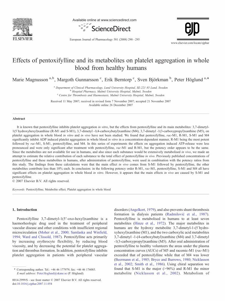
Effects of pentoxifylline and its metabolites on platelet aggregation in wholeblood from healthy humans
Marie Magnusson a,b, Margoth Gunnarsson c, Erik Berntorp c, Sven Björkman b, Peter Höglund a,⁎
a Department of Clinical Pharmacology, Lund University Hospital, SE-221 85 Lund, Swedenb Hospital Pharmacy, Malmö University Hospital, Malmö, Sweden
c Centre for Thrombosis and Haemostasis, Malmö University Hospital, Malmö, Sweden
Received 11 May 2007; received in revised form 7 November 2007; accepted 21 November 2007Available online 26 December 2007
Abstract
It is known that pentoxifylline inhibits platelet aggregation in vitro, but the effects from pentoxifylline and its main metabolites: 3,7-dimetyl-1(5´hydroxyhexyl)xanthine (R-M1 and S-M1), 3,7-dimetyl -1(4-carboxybutyl)xanthine (M4), 3,7-dimetyl -1(3-carboxypropyl)xanthine (M5), onplatelet aggregation in whole blood in vitro and in vivo have not been studied. We found that pentoxifylline, rac-M1, R-M1, S-M1 and M4significantly inhibit ADP induced platelet aggregation in whole blood in vitro in a concentration-dependent manner, R-M1 being the most potentfollowed by rac-M1, S-M1, pentoxifylline, and M4. In this series of experiments the effects on aggregation induced ATP-release were lesspronounced and were only significant after treatment with pentoxifylline, rac-M1 and R-M1, but the potency order appears to be the same.Since the metabolites are not available for use in humans, and also since each substance would be extensively metabolised in vivo, we made anattempt to estimate the relative contribution of each substance to the total effect of pentoxifylline in vivo. Previously published concentrations ofpentoxifylline and these metabolites in humans, after administration of pentoxifylline, were used in combination with the potency ratios fromthis study. The findings from these calculations were that the main effect in vivo comes from S-M1 followed by pentoxifylline, the othermetabolites contribute less than 10% each. In conclusion: in the following potency order R-M1, rac-M1, pentoxifylline, S-M1 and M4 all havesignificant effects on platelet aggregation in whole blood in vitro. However, it appears that the main effects in vivo are caused by S-M1 andpentoxifylline.© 2007 Elsevier B.V. All rights reserved.
Keywords: Pentoxifylline; Metabolite effect; Platelet aggregation in whole blood
1. Introduction
Pentoxifylline 3,7-dimetyl-1(5′-oxo-hexyl)xanthine is ahaemorheologic drug used in the treatment of peripheralvascular disease and other conditions with insufficient regionalmicrocirculation (Moher et al., 2000; Samlaska and Winfield,1994; Ward and Clissold, 1987). Pentoxifylline acts primarilyby increasing erythrocyte flexibility, by reducing bloodviscosity, and by decreasing the potential for platelet aggrega-tion and thrombus formation. In addition, pentoxifylline inhibitsplatelet aggregation in patients with peripheral vascular
disorders (Angelkort, 1979), and also prevents shunt thrombosisformation in dialysis patients (Radmilović et al., 1987).Pentoxifylline is metabolised in humans to at least sevenmetabolites (Hinze et al., 1972). The major metabolites inhumans are the hydroxy metabolite 3,7-dimetyl-1-(5´hydro-xyhexyl)xanthine (M1), and the two carboxylic acid metabolites3,7-dimetyl -1-(4-carboxybutyl)xanthine (M4) and 3,7-dimetyl-1(3-carboxypropyl)xanthine (M5). After oral administration ofpentoxifylline to healthy volunteers the areas under the plasmaconcentration curves (AUCs) of M5 and racemic-M1 (rac-M1)exceeded that of pentoxifylline while that of M4 was lower(Beermann et al., 1985; Bryce and Burrows, 1980; Nicklassonet al., 2002; Smith et al., 1986). Using chiral separation wefound that S-M1 is the major (N96%) and R-M1 the minormetabolite (Nicklasson et al., 2002). Metabolism of
Available online at www.sciencedirect.com
European Journal of Pharmacology 581 (2008) 290–295www.elsevier.com/locate/ejphar
⁎ Corresponding author. Tel.: +46 46 177979; fax: +46 46 176085.E-mail address: [email protected] (P. Höglund).
0014-2999/$ - see front matter © 2007 Elsevier B.V. All rights reserved.doi:10.1016/j.ejphar.2007.11.054
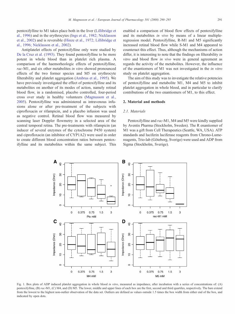
pentoxifylline to M1 takes place both in the liver (Lillibridge etal., 1996) and in the erythrocytes (Ings et al., 1982; Nicklassonet al., 2002) and is reversible (Hinze et al., 1972; Lillibridge etal., 1996; Nicklasson et al., 2002).
Antiplatelet effects of pentoxifylline only were studied byDe la Cruz et al. (1993). They found pentoxifylline to be morepotent in whole blood than in platelet rich plasma. Acomparison of the haemorheologic effects of pentoxifylline,rac-M1, and six other metabolites in vitro showed pronouncedeffects of the two former species and M5 on erythrocytefilterability and platelet aggregation (Ambrus et al., 1995). Wehave previously investigated the effect of pentoxifylline and itsmetabolites on another of its modes of action, namely retinalblood flow, in a randomised, placebo controlled, four-periodcross over study in healthy volunteers (Magnusson et al.,2005). Pentoxifylline was administered as intravenous infu-sions alone or after pre-treatment of the subjects withciprofloxacin or rifampicin, and a placebo infusion was usedas negative control. Retinal blood flow was measured byscanning laser Doppler flowmetry in a selected area of thecentral temporal retina. The pre-treatments with rifampicin (aninducer of several enzymes of the cytochrome P450 system)and ciprofloxacin (an inhibitor of CYP1A2) were used in orderto create different blood concentration ratios between pentox-ifylline and its metabolites within the same subject. This
enabled a comparison of blood flow effects of pentoxifyllineand its metabolites in vivo by means of a linear multiple-regression model. Pentoxifylline, R-M1 and M5 significantlyincreased retinal blood flow while S-M1 and M4 appeared tocounteract this effect. Thus, although the mechanisms of actiondiffer, it is interesting to note that the findings on filterability invitro and blood flow in vivo were in general agreement asregards the activity of the metabolites. However, the influenceof the enantiomers of M1 was not investigated in the in vitrostudy on platelet aggregation.
The aim of this study was to investigate the relative potenciesof pentoxifylline and metabolite M1, M4 and M5 to inhibitplatelet aggregation in whole blood, and in particular to clarifycontributions of the two enantiomers of M1, to this effect.
2. Material and methods
2.1. Materials
Pentoxifylline and rac-M1, M4 andM5 were kindly suppliedby Aventis Pharma (Stockholm, Sweden). The R enantiomer ofM1 was a gift from Cell Therapeutics (Seattle, WA, USA). ATPstandards and luciferin luciferase reagents from Chrono-Lume-reagents, Trio-lab (Göteborg, Sverige) were used and ADP fromSigma (Stockholm, Sverige).
Fig. 1. Box plots of ADP induced platelet aggregation in whole blood in vitro, measured as impedance, after incubation with a series of concentrations of: (A)pentoxifylline, (B) rac-M1, (C) M4, and (D) M5. The lower, middle and upper lines of each box are the first, second and third quartiles, respectively. The bars extendfrom the lowest to the highest non-outlier observation of the data set. Outliers are defined as values outside 1.5 times the box width from either end of the box, andindicated by open dots.
291M. Magnusson et al. / European Journal of Pharmacology 581 (2008) 290–295
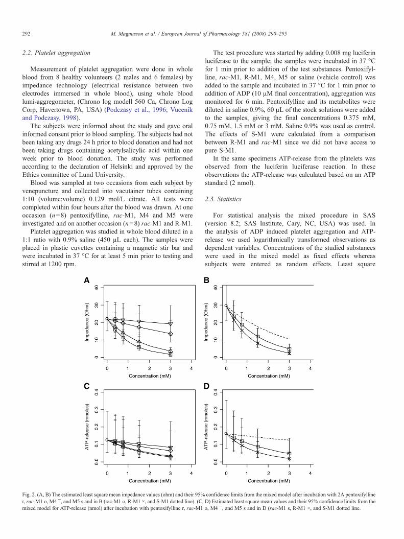
2.2. Platelet aggregation
Measurement of platelet aggregation were done in wholeblood from 8 healthy volunteers (2 males and 6 females) byimpedance technology (electrical resistance between twoelectrodes immersed in whole blood), using whole bloodlumi-aggregometer, (Chrono log modell 560 Ca, Chrono LogCorp, Havertown, PA, USA) (Podczasy et al., 1996; Vucenikand Podczasy, 1998).
The subjects were informed about the study and gave oralinformed consent prior to blood sampling. The subjects had notbeen taking any drugs 24 h prior to blood donation and had notbeen taking drugs containing acetylsalicylic acid within oneweek prior to blood donation. The study was performedaccording to the declaration of Helsinki and approved by theEthics committee of Lund University.
Blood was sampled at two occasions from each subject byvenepuncture and collected into vacutainer tubes containing1:10 (volume:volume) 0.129 mol/L citrate. All tests werecompleted within four hours after the blood was drawn. At oneoccasion (n=8) pentoxifylline, rac-M1, M4 and M5 wereinvestigated and on another occasion (n=8) rac-M1 and R-M1.
Platelet aggregation was studied in whole blood diluted in a1:1 ratio with 0.9% saline (450 µL each). The samples wereplaced in plastic cuvettes containing a magnetic stir bar andwere incubated in 37 °C for at least 5 min prior to testing andstirred at 1200 rpm.
The test procedure was started by adding 0.008 mg luciferinluciferase to the sample; the samples were incubated in 37 °Cfor 1 min prior to addition of the test substances. Pentoxifyl-line, rac-M1, R-M1, M4, M5 or saline (vehicle control) wasadded to the sample and incubated in 37 °C for 1 min prior toaddition of ADP (10 µM final concentration), aggregation wasmonitored for 6 min. Pentoxifylline and its metabolites werediluted in saline 0.9%, 60 µL of the stock solutions were addedto the samples, giving the final concentrations 0.375 mM,0.75 mM, 1.5 mM or 3 mM. Saline 0.9% was used as control.The effects of S-M1 were calculated from a comparisonbetween R-M1 and rac-M1 since we did not have access topure S-M1.
In the same specimens ATP-release from the platelets wasobserved from the luciferin luciferase reaction. In theseobservations the ATP-release was calculated based on an ATPstandard (2 nmol).
2.3. Statistics
For statistical analysis the mixed procedure in SAS(version 8.2; SAS Institute, Cary, NC, USA) was used. Inthe analysis of ADP induced platelet aggregation and ATP-release we used logarithmically transformed observations asdependent variables. Concentrations of the studied substanceswere used in the mixed model as fixed effects whereassubjects were entered as random effects. Least square
Fig. 2. (A, B) The estimated least square mean impedance values (ohm) and their 95% confidence limits from the mixed model after incubation with 2A pentoxifylliner, rac-M1 o, M4 ¯, and M5 s and in B (rac-M1 o, R-M1 ×, and S-M1 dotted line). (C, D) Estimated least square mean values and their 95% confidence limits from themixed model for ATP-release (nmol) after incubation with pentoxifylline r, rac-M1 o, M4 ¯, and M5 s and in D (rac-M1 s, R-M1 ×, and S-M1 dotted line.
292 M. Magnusson et al. / European Journal of Pharmacology 581 (2008) 290–295
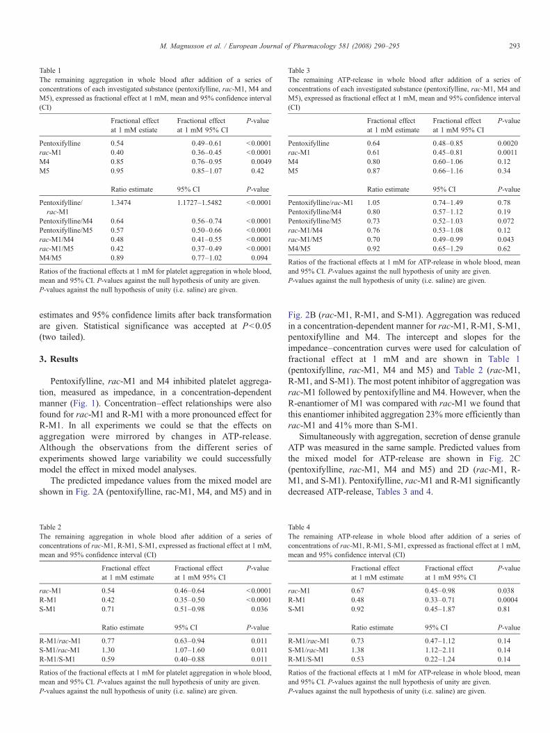
estimates and 95% confidence limits after back transformationare given. Statistical significance was accepted at Pb0.05(two tailed).
3. Results
Pentoxifylline, rac-M1 and M4 inhibited platelet aggrega-tion, measured as impedance, in a concentration-dependentmanner (Fig. 1). Concentration–effect relationships were alsofound for rac-M1 and R-M1 with a more pronounced effect forR-M1. In all experiments we could se that the effects onaggregation were mirrored by changes in ATP-release.Although the observations from the different series ofexperiments showed large variability we could successfullymodel the effect in mixed model analyses.
The predicted impedance values from the mixed model areshown in Fig. 2A (pentoxifylline, rac-M1, M4, and M5) and in
Fig. 2B (rac-M1, R-M1, and S-M1). Aggregation was reducedin a concentration-dependent manner for rac-M1, R-M1, S-M1,pentoxifylline and M4. The intercept and slopes for theimpedance–concentration curves were used for calculation offractional effect at 1 mM and are shown in Table 1(pentoxifylline, rac-M1, M4 and M5) and Table 2 (rac-M1,R-M1, and S-M1). The most potent inhibitor of aggregation wasrac-M1 followed by pentoxifylline and M4. However, when theR-enantiomer of M1 was compared with rac-M1 we found thatthis enantiomer inhibited aggregation 23% more efficiently thanrac-M1 and 41% more than S-M1.
Simultaneously with aggregation, secretion of dense granuleATP was measured in the same sample. Predicted values fromthe mixed model for ATP-release are shown in Fig. 2C(pentoxifylline, rac-M1, M4 and M5) and 2D (rac-M1, R-M1, and S-M1). Pentoxifylline, rac-M1 and R-M1 significantlydecreased ATP-release, Tables 3 and 4.
Table 1The remaining aggregation in whole blood after addition of a series ofconcentrations of each investigated substance (pentoxifylline, rac-M1, M4 andM5), expressed as fractional effect at 1 mM, mean and 95% confidence interval(CI)
Fractional effectat 1 mM estiate
Fractional effectat 1 mM 95% CI
P-value
Pentoxifylline 0.54 0.49–0.61 b0.0001rac-M1 0.40 0.36–0.45 b0.0001M4 0.85 0.76–0.95 0.0049M5 0.95 0.85–1.07 0.42
Ratio estimate 95% CI P-value
Pentoxifylline/rac-M1
1.3474 1.1727–1.5482 b0.0001
Pentoxifylline/M4 0.64 0.56–0.74 b0.0001Pentoxifylline/M5 0.57 0.50–0.66 b0.0001rac-M1/M4 0.48 0.41–0.55 b0.0001rac-M1/M5 0.42 0.37–0.49 b0.0001M4/M5 0.89 0.77–1.02 0.094
Ratios of the fractional effects at 1 mM for platelet aggregation in whole blood,mean and 95% CI. P-values against the null hypothesis of unity are given.P-values against the null hypothesis of unity (i.e. saline) are given.
Table 2The remaining aggregation in whole blood after addition of a series ofconcentrations of rac-M1, R-M1, S-M1, expressed as fractional effect at 1 mM,mean and 95% confidence interval (CI)
Fractional effectat 1 mM estimate
Fractional effectat 1 mM 95% CI
P-value
rac-M1 0.54 0.46–0.64 b0.0001R-M1 0.42 0.35–0.50 b0.0001S-M1 0.71 0.51–0.98 0.036
Ratio estimate 95% CI P-value
R-M1/rac-M1 0.77 0.63–0.94 0.011S-M1/rac-M1 1.30 1.07–1.60 0.011R-M1/S-M1 0.59 0.40–0.88 0.011
Ratios of the fractional effects at 1 mM for platelet aggregation in whole blood,mean and 95% CI. P-values against the null hypothesis of unity are given.P-values against the null hypothesis of unity (i.e. saline) are given.
Table 3The remaining ATP-release in whole blood after addition of a series ofconcentrations of each investigated substance (pentoxifylline, rac-M1, M4 andM5), expressed as fractional effect at 1 mM, mean and 95% confidence interval(CI)
Fractional effectat 1 mM estimate
Fractional effectat 1 mM 95% CI
P-value
Pentoxifylline 0.64 0.48–0.85 0.0020rac-M1 0.61 0.45–0.81 0.0011M4 0.80 0.60–1.06 0.12M5 0.87 0.66–1.16 0.34
Ratio estimate 95% CI P-value
Pentoxifylline/rac-M1 1.05 0.74–1.49 0.78Pentoxifylline/M4 0.80 0.57–1.12 0.19Pentoxifylline/M5 0.73 0.52–1.03 0.072rac-M1/M4 0.76 0.53–1.08 0.12rac-M1/M5 0.70 0.49–0.99 0.043M4/M5 0.92 0.65–1.29 0.62
Ratios of the fractional effects at 1 mM for ATP-release in whole blood, meanand 95% CI. P-values against the null hypothesis of unity are given.P-values against the null hypothesis of unity (i.e. saline) are given.
Table 4The remaining ATP-release in whole blood after addition of a series ofconcentrations of rac-M1, R-M1, S-M1, expressed as fractional effect at 1 mM,mean and 95% confidence interval (CI)
Fractional effectat 1 mM estimate
Fractional effectat 1 mM 95% CI
P-value
rac-M1 0.67 0.45–0.98 0.038R-M1 0.48 0.33–0.71 0.0004S-M1 0.92 0.45–1.87 0.81
Ratio estimate 95% CI P-value
R-M1/rac-M1 0.73 0.47–1.12 0.14S-M1/rac-M1 1.38 1.12–2.11 0.14R-M1/S-M1 0.53 0.22–1.24 0.14
Ratios of the fractional effects at 1 mM for ATP-release in whole blood, meanand 95% CI. P-values against the null hypothesis of unity are given.P-values against the null hypothesis of unity (i.e. saline) are given.
293M. Magnusson et al. / European Journal of Pharmacology 581 (2008) 290–295
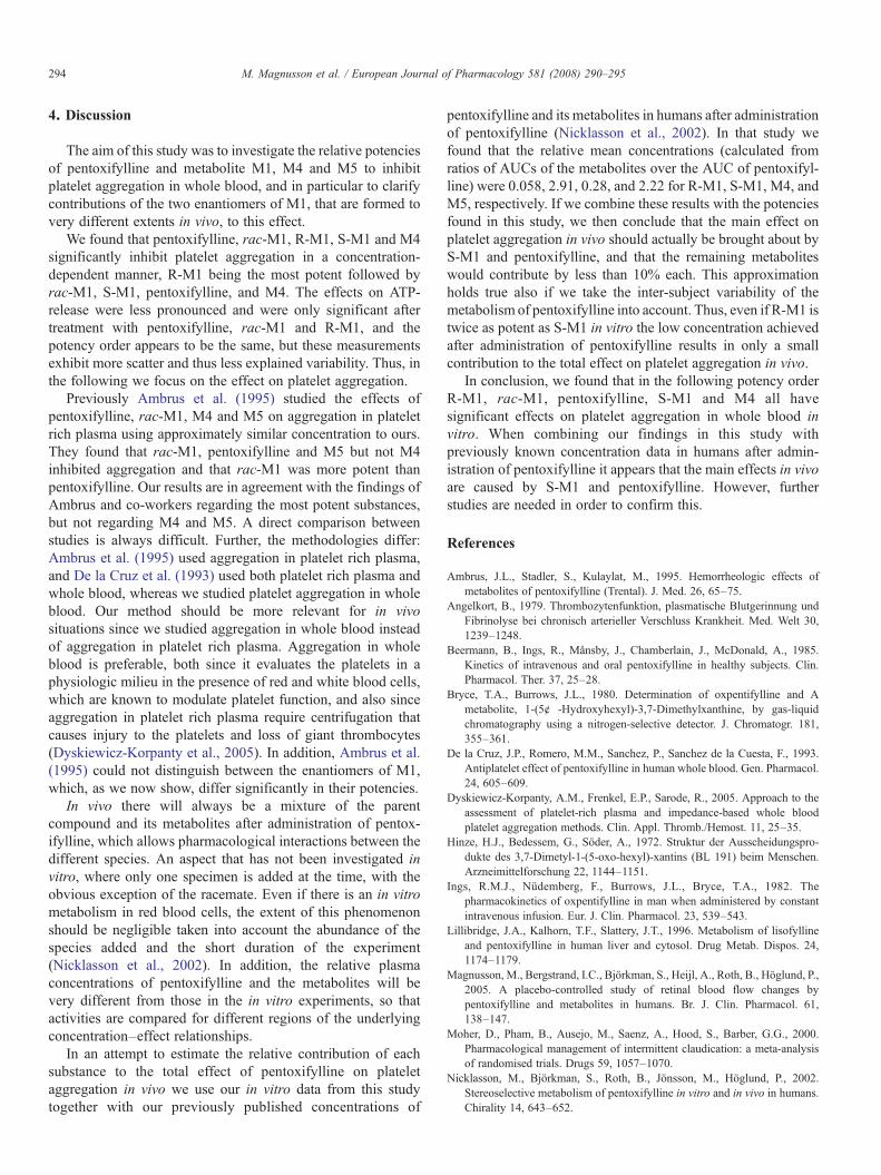
4. Discussion
The aim of this study was to investigate the relative potenciesof pentoxifylline and metabolite M1, M4 and M5 to inhibitplatelet aggregation in whole blood, and in particular to clarifycontributions of the two enantiomers of M1, that are formed tovery different extents in vivo, to this effect.
We found that pentoxifylline, rac-M1, R-M1, S-M1 and M4significantly inhibit platelet aggregation in a concentration-dependent manner, R-M1 being the most potent followed byrac-M1, S-M1, pentoxifylline, and M4. The effects on ATP-release were less pronounced and were only significant aftertreatment with pentoxifylline, rac-M1 and R-M1, and thepotency order appears to be the same, but these measurementsexhibit more scatter and thus less explained variability. Thus, inthe following we focus on the effect on platelet aggregation.
Previously Ambrus et al. (1995) studied the effects ofpentoxifylline, rac-M1, M4 and M5 on aggregation in plateletrich plasma using approximately similar concentration to ours.They found that rac-M1, pentoxifylline and M5 but not M4inhibited aggregation and that rac-M1 was more potent thanpentoxifylline. Our results are in agreement with the findings ofAmbrus and co-workers regarding the most potent substances,but not regarding M4 and M5. A direct comparison betweenstudies is always difficult. Further, the methodologies differ:Ambrus et al. (1995) used aggregation in platelet rich plasma,and De la Cruz et al. (1993) used both platelet rich plasma andwhole blood, whereas we studied platelet aggregation in wholeblood. Our method should be more relevant for in vivosituations since we studied aggregation in whole blood insteadof aggregation in platelet rich plasma. Aggregation in wholeblood is preferable, both since it evaluates the platelets in aphysiologic milieu in the presence of red and white blood cells,which are known to modulate platelet function, and also sinceaggregation in platelet rich plasma require centrifugation thatcauses injury to the platelets and loss of giant thrombocytes(Dyskiewicz-Korpanty et al., 2005). In addition, Ambrus et al.(1995) could not distinguish between the enantiomers of M1,which, as we now show, differ significantly in their potencies.
In vivo there will always be a mixture of the parentcompound and its metabolites after administration of pentox-ifylline, which allows pharmacological interactions between thedifferent species. An aspect that has not been investigated invitro, where only one specimen is added at the time, with theobvious exception of the racemate. Even if there is an in vitrometabolism in red blood cells, the extent of this phenomenonshould be negligible taken into account the abundance of thespecies added and the short duration of the experiment(Nicklasson et al., 2002). In addition, the relative plasmaconcentrations of pentoxifylline and the metabolites will bevery different from those in the in vitro experiments, so thatactivities are compared for different regions of the underlyingconcentration–effect relationships.
In an attempt to estimate the relative contribution of eachsubstance to the total effect of pentoxifylline on plateletaggregation in vivo we use our in vitro data from this studytogether with our previously published concentrations of
pentoxifylline and its metabolites in humans after administrationof pentoxifylline (Nicklasson et al., 2002). In that study wefound that the relative mean concentrations (calculated fromratios of AUCs of the metabolites over the AUC of pentoxifyl-line) were 0.058, 2.91, 0.28, and 2.22 for R-M1, S-M1, M4, andM5, respectively. If we combine these results with the potenciesfound in this study, we then conclude that the main effect onplatelet aggregation in vivo should actually be brought about byS-M1 and pentoxifylline, and that the remaining metaboliteswould contribute by less than 10% each. This approximationholds true also if we take the inter-subject variability of themetabolism of pentoxifylline into account. Thus, even if R-M1 istwice as potent as S-M1 in vitro the low concentration achievedafter administration of pentoxifylline results in only a smallcontribution to the total effect on platelet aggregation in vivo.
In conclusion, we found that in the following potency orderR-M1, rac-M1, pentoxifylline, S-M1 and M4 all havesignificant effects on platelet aggregation in whole blood invitro. When combining our findings in this study withpreviously known concentration data in humans after admin-istration of pentoxifylline it appears that the main effects in vivoare caused by S-M1 and pentoxifylline. However, furtherstudies are needed in order to confirm this.
References
Ambrus, J.L., Stadler, S., Kulaylat, M., 1995. Hemorrheologic effects ofmetabolites of pentoxifylline (Trental). J. Med. 26, 65–75.
Angelkort, B., 1979. Thrombozytenfunktion, plasmatische Blutgerinnung undFibrinolyse bei chronisch arterieller Verschluss Krankheit. Med. Welt 30,1239–1248.
Beermann, B., Ings, R., Månsby, J., Chamberlain, J., McDonald, A., 1985.Kinetics of intravenous and oral pentoxifylline in healthy subjects. Clin.Pharmacol. Ther. 37, 25–28.
Bryce, T.A., Burrows, J.L., 1980. Determination of oxpentifylline and Ametabolite, 1-(5¢ -Hydroxyhexyl)-3,7-Dimethylxanthine, by gas-liquidchromatography using a nitrogen-selective detector. J. Chromatogr. 181,355–361.
De la Cruz, J.P., Romero, M.M., Sanchez, P., Sanchez de la Cuesta, F., 1993.Antiplatelet effect of pentoxifylline in human whole blood. Gen. Pharmacol.24, 605–609.
Dyskiewicz-Korpanty, A.M., Frenkel, E.P., Sarode, R., 2005. Approach to theassessment of platelet-rich plasma and impedance-based whole bloodplatelet aggregation methods. Clin. Appl. Thromb./Hemost. 11, 25–35.
Hinze, H.J., Bedessem, G., Söder, A., 1972. Struktur der Ausscheidungspro-dukte des 3,7-Dimetyl-1-(5-oxo-hexyl)-xantins (BL 191) beim Menschen.Arzneimittelforschung 22, 1144–1151.
Ings, R.M.J., Nüdemberg, F., Burrows, J.L., Bryce, T.A., 1982. Thepharmacokinetics of oxpentifylline in man when administered by constantintravenous infusion. Eur. J. Clin. Pharmacol. 23, 539–543.
Lillibridge, J.A., Kalhorn, T.F., Slattery, J.T., 1996. Metabolism of lisofyllineand pentoxifylline in human liver and cytosol. Drug Metab. Dispos. 24,1174–1179.
Magnusson, M., Bergstrand, I.C., Björkman, S., Heijl, A., Roth, B., Höglund, P.,2005. A placebo-controlled study of retinal blood flow changes bypentoxifylline and metabolites in humans. Br. J. Clin. Pharmacol. 61,138–147.
Moher, D., Pham, B., Ausejo, M., Saenz, A., Hood, S., Barber, G.G., 2000.Pharmacological management of intermittent claudication: a meta-analysisof randomised trials. Drugs 59, 1057–1070.
Nicklasson, M., Björkman, S., Roth, B., Jönsson, M., Höglund, P., 2002.Stereoselective metabolism of pentoxifylline in vitro and in vivo in humans.Chirality 14, 643–652.
294 M. Magnusson et al. / European Journal of Pharmacology 581 (2008) 290–295

Podczasy, J.J., Lee, J., Vucenik, I., 1996. Evaluation of whole-bloodlumiaggregation. Clin. Appl. Thromb./Hemost. 3, 190–195.
Radmilović, A., Borić, Z., Naumović, T., Stemenković, M., Mušikivić, P.,1987. Shunt thrombosis prevention in hemodialysis patients — a double-blind, randomized study: pentoxifylline vs placebo. Angiology 38,499–505.
Samlaska, C.P., Winfield, A., 1994. Pentoxifylline. J. Am. Acad. Dermatol. 30,603–621.
Smith, R.V., Waller, E.S., Doluisio, J.T., Bauza, M.T., Puri, S.K., Ho, I.,Lassman, H.B., 1986. Pharmacokinetics of orally administered pentoxifyl-line in humans. J. Pharm. Sci. 75, 47–52.
Vucenik, I., Podczasy, J.J., 1998. Whole blood lumiaggregation: evaluation ofreagents. Clin. Appl. Thromb./Hemost. 4, 253–256.
Ward, A., Clissold, S.P., 1987. Pentoxifylline. A review of its pharmacodynamicand pharmacokinetic properties, and its therapeutic efficacy. Drugs 34,50–97.
295M. Magnusson et al. / European Journal of Pharmacology 581 (2008) 290–295

![Clinical Study Role of Pentoxifylline and Sparfloxacin in ...downloads.hindawi.com/archive/2014/595213.pdfClinical Study Role of Pentoxifylline and Sparfloxacin in ... ... Spar]. ]](https://img.pdfslide.us/doc/110x75/5ac369327f8b9a5c558bb6e8/clinical-study-role-of-pentoxifylline-and-sparfloxacin-in-study-role-of-pentoxifylline.jpg)