Embed Size (px)
Citation preview

Available online at www.scholarsresearchlibrary.com
Scholars Research Library
Der Pharmacia Lettre, 2013, 5 (1):121-135
(http://scholarsresearchlibrary.com/archive.html)
ISSN 0975-5071 USA CODEN: DPLEB4
121
Scholar Research Library
Pharmacognostical evaluation of roots of Cassia fistula Linn.
Supriya Deshpande, Shailesh Kewatkar and Vivek Paithankar
Department of Pharmaceutical Sciences, Bhagwant University, Ajmer, Rajasthan _____________________________________________________________________________________________ ABSTRACT Since the advent of modern drug treatments, traditional medicine has greatly receded in occidental societies. Moreover, only a limited number of medicinal plants have received detailed scientific scrutiny thereby prompting the World Health Organization to recommend that this area be comprehensively investigated. Cassia fistula Linn. belonging to the family Casesalpinaceae is commonly called as Amaltas an Indian Labernum and is native to India, the Amazon, Sri Lanka and is extensively diffused in various countries. Cassia fistula Linn. is used extensively in various parts of the world against a wide range of ailments, the synergistic action of its metabolite production being most probably responsible for the plant’s beneficial effects. Cassia fistula Linn. plant organs are known to be an important source of secondary metabolites, notably phenolic compounds. The study provides taxonomical, Pharmacognostical, physicochemical details helpful in laying down standardization and pharmacopoeial parameters. The innumerable medicinal properties and therapeutic uses of Cassia Fistula Linn. as well as its phytochemical investigations prove its importance as a valuable medicinal plant. Keywords: Cassia fistula Linn., Casesalpinaceae, Microscopy, Pharmacognosy _____________________________________________________________________________________________
INTRODUCTION
Native to India, the Amazon and Sri Lanka, Cassia fistula Linn., a semi-wild Indian Labernum also known as the Golden Shower, has become extensively diffused in various countries including Mauritius, India, South Africa, Mexico, China, West Indies, East Africa and Brazil as an `ornamental tree for its beautiful bunches of yellow flowers. Recognized by the British Pharmacopoeia [1], C. fistula, a member of the Casesalpinaceae family, is widely used for its medicinal properties, its main property being that of a mild laxative suitable for children and pregnant women. It is also a purgative due to the wax aloin and a tonic [2] and has been reported to treat many other intestinal disorders like healing ulcers [3][4]. The plant has a high therapeutic value and it exerts an antipyretic and analgesic effect [5]. Besides, it has been found to exhibit antinflammatory and hypoglycaemic activity [6]. In the Indian literature, this plant has been described to be useful against skin diseases, liver troubles, tuberculous glands and its use in the treatment of haematemesis, pruritus, leucoderm and diabetes has been suggested. However, there are no reports on the pharmacognostical studies of the plant. Hence, the present work is an attempt in this direction and includes morphological and physical evaluation, determination of physico-chemical constants and preliminary phytochemical screening of different extracts of Cassia fistula Linn.

Supriya Deshpande et al Der Pharmacia Lettre, 2013, 5 (1):121-135 _____________________________________________________________________________
122
Scholar Research Library
MATERIALS AND METHODS
Plant material: The roots of the plant Cassia fistula Linn. was collected from the interiors of Bhopal, Madhya Pradesh respectively. The plant has been authenticated by Safia College of Science, Peer Gate, Bhopal, (Madhya Pradesh), and were given the voucher specimen number 160/Bot/Safia/2010. The authenticated roots were dried under shade and then coarsely powdered with the help of mechanical grinder. The powdered was passed through sieve no. 40 and stored in an airtight container for extraction. Chemicals and instruments: All the chemicals used were of laboratory grade. Compound microscope, watch glass, glass slides, cover slips, and other common glasswares were used in this experiment. Photographs were taken with using Nikon Labphot 2 Microscopic Unit, and trinocular microscope. Various solvents used mainly petroleum ether, ethanol (95%),and reagents used for staining different sections like phluoroglucinol, iodine solution, safranin, and acetic acid were procured from CDH, Mumbai, India. Macroscopic and microscopic analysis: The Macroscopic and microscopic of the plant was studied according to the methods of Evans, the cross sections were prepared and stained with phluoroglucinol, iodine solution, safranin, and acetic acid. The microscopic analysis of powder was studied [7] [8] [9]. Physico- chemical analysis: Air dried plant material was used for the quantitative determination of ash and extractive values according to standard procedure of Indian Pharmacopoeia, 1996 [10] [11] and WHO/QCMMPM, 1992 [12] [13]. Preliminary phytochemical screening: Preliminary phytochemical evaluation was carried out by using standard procedure [14] [15].
RESULTS AND DISCUSSION
Morphological characters:
Table 1: Morphological characters of the roots of Cassia fistula L.
Sr. No. Particulars Observations 1 Colour Brownish yellow 2 Odour Odourless 3 Taste Characteristics 4 Length 05 – 10 cm 5 Surface Rough 6 Shape Irregular
Microscopic characters of roots The paraffin embedded specimens were sectioned with the help of Rotary Microtone. The thickness of the sections was 10 – 12 µm. Dewaxing of the sections was by customary procedure [16].The section were stained with Toluidine blue as per the method published by O’Brien et al., 1964 [17] [18]. Structure of the root Root measuring 2.2 mm thick was studied. It is circular in outline with more or less smooth outline (Fig 2.1). The root consists of periderm, cortex, secondary phloem and secondary xylem cylinder (Fig 2.2). Periderm It is continuous all around the root and more or less uniform in thickness. It is 300µm thick in radial plane. The periderm includes several layers of dark brown narrowly tabular radial files of phellem cells and 2 layers of wide, squarish phelloderm cells.
Supriya Deshpande et al Der Pharmacia Lettre, 2013, 5 (1):121-135 _____________________________________________________________________________
123
Scholar Research Library
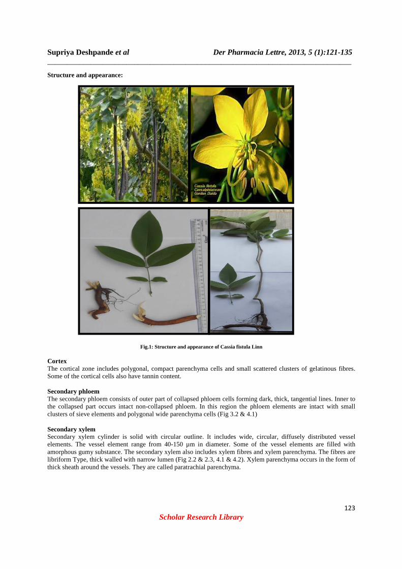
Structure and appearance:
Fig.1: Structure and appearance of Cassia fistula Linn
Cortex The cortical zone includes polygonal, compact parenchyma cells and small scattered clusters of gelatinous fibres. Some of the cortical cells also have tannin content. Secondary phloem The secondary phloem consists of outer part of collapsed phloem cells forming dark, thick, tangential lines. Inner to the collapsed part occurs intact non-collapsed phloem. In this region the phloem elements are intact with small clusters of sieve elements and polygonal wide parenchyma cells (Fig 3.2 & 4.1) Secondary xylem Secondary xylem cylinder is solid with circular outline. It includes wide, circular, diffusely distributed vessel elements. The vessel element range from 40-150 µm in diameter. Some of the vessel elements are filled with amorphous gumy substance. The secondary xylem also includes xylem fibres and xylem parenchyma. The fibres are libriform Type, thick walled with narrow lumen (Fig 2.2 & 2.3, 4.1 & 4.2). Xylem parenchyma occurs in the form of thick sheath around the vessels. They are called paratrachial parenchyma.

Supriya Deshpande et al Der Pharmacia Lettre, 2013, 5 (1):121-135 _____________________________________________________________________________
124
Scholar Research Library
Fig 2.1 T.S. of root – Entire view
Fig 2.2 T.S. of root – Outer sector – Enlarged

Supriya Deshpande et al Der Pharmacia Lettre, 2013, 5 (1):121-135 _____________________________________________________________________________
125
Scholar Research Library
Fig 2.3 T.S. of root – Central Sector – Enlarged
Where, Co- Cortex, Pd- Phelloderm, Pe- Periderm, Pm- Phellem, Sph- Secondary Phloem, Sx- Secondary xylem, Ve- Vessel, G- Gum
Fig 3.1 T.S. of root showing phellem and phelloderm of periderm

Supriya Deshpande et al Der Pharmacia Lettre, 2013, 5 (1):121-135 _____________________________________________________________________________
126
Scholar Research Library
Fig 3.2 Secondary phloem
Where, Co- Cortex, Cph- Collapsed phloem, GF- Gilatinous Fibres , NCph- Non collapsed phloem, Pd- Phelloderm, Pm- Phellem, Ta- Tannin

Supriya Deshpande et al Der Pharmacia Lettre, 2013, 5 (1):121-135 _____________________________________________________________________________
127
Scholar Research Library
Fig 4.1: Secondary phloem elements- Enlarged
Crystals Calcium oxalate crystals are abundant in the middle cortical zone. The crystals are exclusively prismatic type. The crystals also occur in the xylem parenchyma which encloses the vessel (Fig 4.3 & 5.1 & 5.2).

Supriya Deshpande et al Der Pharmacia Lettre, 2013, 5 (1):121-135 _____________________________________________________________________________
128
Scholar Research Library
Fig 4.2: Secondary xylem showing gum inclusions in the vessels

Supriya Deshpande et al Der Pharmacia Lettre, 2013, 5 (1):121-135 _____________________________________________________________________________
129
Scholar Research Library
Fig 4.3: Crystal distribution in the bark
Where, Co- Cortex, Cph- Collapsed phloem, Cr- Crystal, GF- Gilatinous Fibres, NCph- Non collapsed phloem, Pp- Paratrachial Parenchyma, Sph- Secondary Phloem, Sx- Secondary xylem, Ve- Vessel, XFi- Xylem Fibre, G- Gum
Fig 5.1: Crystal in the cortical tissues
Supriya Deshpande et al Der Pharmacia Lettre, 2013, 5 (1):121-135 _____________________________________________________________________________
130
Scholar Research Library
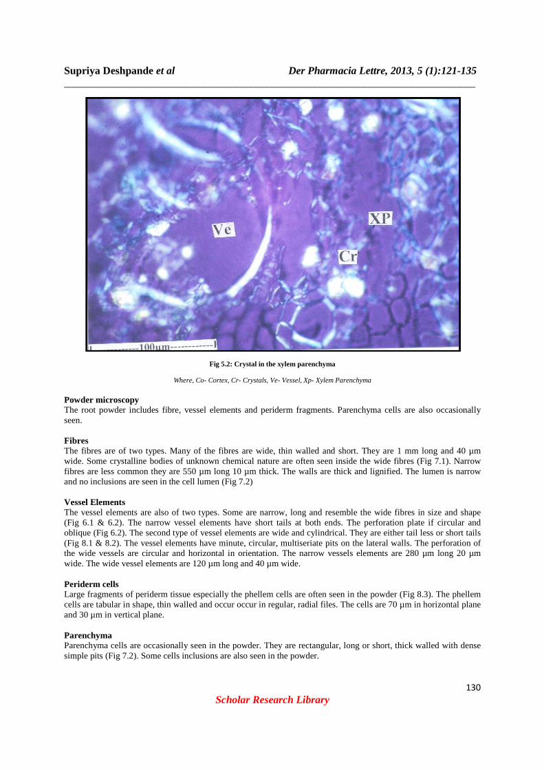
Fig 5.2: Crystal in the xylem parenchyma
Where, Co- Cortex, Cr- Crystals, Ve- Vessel, Xp- Xylem Parenchyma Powder microscopy The root powder includes fibre, vessel elements and periderm fragments. Parenchyma cells are also occasionally seen. Fibres The fibres are of two types. Many of the fibres are wide, thin walled and short. They are 1 mm long and 40 µm wide. Some crystalline bodies of unknown chemical nature are often seen inside the wide fibres (Fig 7.1). Narrow fibres are less common they are 550 µm long 10 µm thick. The walls are thick and lignified. The lumen is narrow and no inclusions are seen in the cell lumen (Fig 7.2) Vessel Elements The vessel elements are also of two types. Some are narrow, long and resemble the wide fibres in size and shape (Fig 6.1 & 6.2). The narrow vessel elements have short tails at both ends. The perforation plate if circular and oblique (Fig 6.2). The second type of vessel elements are wide and cylindrical. They are either tail less or short tails (Fig 8.1 & 8.2). The vessel elements have minute, circular, multiseriate pits on the lateral walls. The perforation of the wide vessels are circular and horizontal in orientation. The narrow vessels elements are 280 µm long 20 µm wide. The wide vessel elements are 120 µm long and 40 µm wide. Periderm cells Large fragments of periderm tissue especially the phellem cells are often seen in the powder (Fig 8.3). The phellem cells are tabular in shape, thin walled and occur occur in regular, radial files. The cells are 70 µm in horizontal plane and 30 µm in vertical plane. Parenchyma Parenchyma cells are occasionally seen in the powder. They are rectangular, long or short, thick walled with dense simple pits (Fig 7.2). Some cells inclusions are also seen in the powder.

Supriya Deshpande et al Der Pharmacia Lettre, 2013, 5 (1):121-135 _____________________________________________________________________________
131
Scholar Research Library
Fig 6.1: Wide fibre and narrow vessel element
Fig 6.2: Narrow vessel element and wide fibres- Enlarged
Where, CI- Cell Inclusion, Pa- Parenchyma, Pe- Perforation, Pi- Pits, VE- Vessel Elements, WF- Wide Fibre

Supriya Deshpande et al Der Pharmacia Lettre, 2013, 5 (1):121-135 _____________________________________________________________________________
132
Scholar Research Library
Fig 7.1: One wide fibre- Enlarged showing cell inclusions
Fig 7.2: Narrow Fibre and Parenchyma cells- Enlarged
Where, CI- Cell Inclusion, NF- Narrow Fibre, Pa- Parenchyma

Supriya Deshpande et al Der Pharmacia Lettre, 2013, 5 (1):121-135 _____________________________________________________________________________
133
Scholar Research Library
Fig 8.1: Narrow and wide vessel element
Fig 8.2: One tailed wide vessel element – Enlarged
Supriya Deshpande et al Der Pharmacia Lettre, 2013, 5 (1):121-135 _____________________________________________________________________________
134
Scholar Research Library
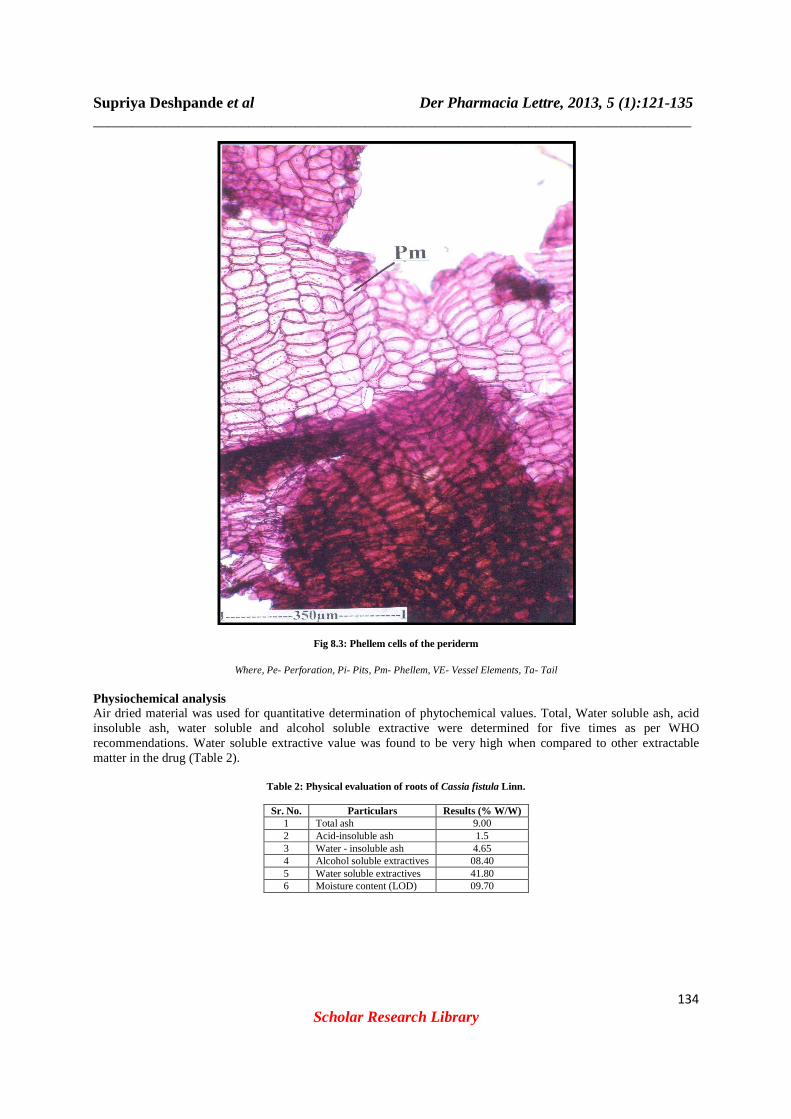
Fig 8.3: Phellem cells of the periderm
Where, Pe- Perforation, Pi- Pits, Pm- Phellem, VE- Vessel Elements, Ta- Tail Physiochemical analysis Air dried material was used for quantitative determination of phytochemical values. Total, Water soluble ash, acid insoluble ash, water soluble and alcohol soluble extractive were determined for five times as per WHO recommendations. Water soluble extractive value was found to be very high when compared to other extractable matter in the drug (Table 2).
Table 2: Physical evaluation of roots of Cassia fistula Linn.
Sr. No. Particulars Results (% W/W) 1 Total ash 9.00 2 Acid-insoluble ash 1.5 3 Water - insoluble ash 4.65 4 Alcohol soluble extractives 08.40 5 Water soluble extractives 41.80 6 Moisture content (LOD) 09.70

Supriya Deshpande et al Der Pharmacia Lettre, 2013, 5 (1):121-135 _____________________________________________________________________________
135
Scholar Research Library
Table 3: Preliminary phytochemical screening of two extracts of the roots of Cassia fistula Linn. (+ positive test, -- negative test).
Sr. No. Test Pet. Ether Ethanol
1 Alkaloids a. Hager’s Reagent -- +
b. Mayer’s Reagent -- +
c. Wagner’s Reagent -- +
d. Dragandorffs Reagent -- +
2 Glycosides a Liebermann-Burchard Reagent + +
b Legals Reagent + +
c Borntragers Reagent + +
3 Saponins a Foam Test + +
b Haemolysis Test + +
4 Flavonoids a Shinoda Test -- +
CONCLUSION
The study of Pharmacognostical features of roots of Cassia fistula Linn. had shown the standards which will be useful the detection of its identity and authenticity. The other study viz. physical evaluation, preliminary phytochemical test add to its quality control and quality assurance for proper identification. Acknowledgement Author is thankful to Safia College of Science, Bhopal, Mr. Manvendra Singh Karchuli and Prof. P. Jayaraman for their guidance and support to conclude this type of research work successfully.
REFERENCES
[1] M Mukhopadhyay; A Saha; A Dutta; B De; A Mukherjee. Food Chem. Toxicol, 1998, 36, 937–940. [2] GV Satyavati; M Sharma. Medicinal plant in India, ICMR, New Delhi, 1989, 55. [3] K Biswas; AB Ghosh. Bharatia Banawasadhi, Calcutta University, Advancement of learning, Calcutta, 1973, 2, 336. [4] KR Kirtikar; BD Basu. Indian Medicinal Plants, International book seller and publisher, India, 2006, pp 865. [5] D Patel; D Karbhari; D Gulati; D Gokhale. Pharm. Biol, 1965, 157, 22–27. [6] A Dutta; B De. Indian J. Pharmacol Sci, 1998, 60, 388-390. [7] WC Evans; Trease. Textbook of pharmacognosy, Saunders, London, 2009, pp 562. [8] M Raja; R Venkataraman. Der Pharmacia Sinica, 2011, 2, 136. [9] S Kumar; VK Garg; N Kumar; PK Sharma; S Chaudhary; A Upadhyay. European Journal of Experimental Biology, 2011, 1, 77. [10] Indian Pharmacopoeia, Government of Indian, Ministry of health and human welfare, New Delhi, India,Controller of publications, 1996, 2, A53. [11] SK Panda; D Das; BN Tripathy; L Nayak. Asian Journal of Plant Science and Research, 2012, 2 151. [12] Nagamani; J Suresh; J Ahuja; V Reddy; D Rajan. Asian Journal of Plant Science and Research, 2012, 2, 25. [13] WHO/ Pharm/ 92.559/ rev.1., Quality control methods for medicinal plant materials, Organization Mondiale De La Sante, Geneva, 1992, 9, 22. [14] CK Kokate. Practical Pharmacognosy, Vallabh Prakashan, New Delhi, India, 1994, pp 54. [15] JB Harbone. Methods of extraction and isolation, in: phytochemical methods, Chapman and Hall, London, 1998, pp 60. [16] DA Johanson. Plant Microtechnique, Mc Graw Hill Book Co, New York, 1940, pp 523. [17] TP O’brien; N Fedar. Mc Cull ME, Protoplasma, 1964, 59, pp 364. [18] V Sodde; N Dashora; K Prabhu; B Jaykumar; R Lobo. Der Pharmacia Sinica, 2011, 2, pp 217.