Embed Size (px)
Citation preview

Limitation of Individual Folding Resources in the ER Leads to Outcomes Distinct from
the Unfolded Protein Response.
Davide Eletto1, Avinash Maganty1,*, Daniela Eletto1,2, Devin Dersh1,3, Catherine Makarewich1,¶,
Chhanda Biswas4,#, James C. Paton5, Adrienne W. Paton5, Shirin Doroudgar6, Christopher C.
Glembotski6 and Yair Argon1,‡
1Dept. of Pathology and Laboratory Medicine, Children’s Hospital of Philadelphia, PA, 2Dept. of
Pharmaceutical Sciences, University of Salerno, Fisciano (SA), Italy, 3Biochemistry and Molecular
Biophysics Graduate Group, Perelman School of Medicine at the University of Pennsylvania, 4Dept. of Surgery The University of Pennsylvania, Philadelphia, PA 19104, 5Research Centre for
Infectious Diseases, School of Molecular and Biomedical Science, University of Adelaide,
Australia, 6San Diego State University Heart Institute and the Department of Biology, San Diego
State University, San Diego CA, 92182.
‡To whom correspondence should be addressed at:
816B ARC, 3615 Civic Center Blvd, Philadelphia, PA 19104.
Tel: 267-426-5131
Fax: 267-426-5165
Email: [email protected]
* Present address: Weill Cornell Medical College, 1300 York Ave., New York, NY 10065 ¶ Present address: Program on Cellular and Molecular Physiology, Temple University School of
Medicine, Philadelphia, PA # Present address: Dept. of Pediatrics, Children’s Hospital of Philadelphia, PA
Running Head: Distinct responses to chaperone ablation
© 2012. Published by The Company of Biologists Ltd.Jo
urna
l of C
ell S
cien
ceA
ccep
ted
man
uscr
ipt
JCS online publication date 1 August 2012

2
Abbreviations: 17AAG, 17-N-Allylamino-17-demethoxygeldanamycin; BiP, binding
immunoglobulin protein; DHEA, dehydroepiandrosterone; DTT, dithiothreitol; GRP94, glucose
regulated protein 94 kDa; HSP, heat-shock protein; KD, knock-down; KO, knock-out; PDIA6,
protein disulfide isomerase A6; RNAi, RNA interference; shRNA, short hairpin RNA; TG,
thapsigargin; Tun, tunicamycin.
Jour
nal o
f Cel
l Sci
ence
Acc
epte
d m
anus
crip
t
3
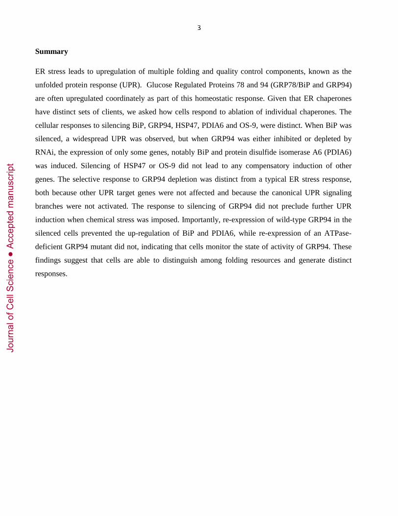
Summary
ER stress leads to upregulation of multiple folding and quality control components, known as the
unfolded protein response (UPR). Glucose Regulated Proteins 78 and 94 (GRP78/BiP and GRP94)
are often upregulated coordinately as part of this homeostatic response. Given that ER chaperones
have distinct sets of clients, we asked how cells respond to ablation of individual chaperones. The
cellular responses to silencing BiP, GRP94, HSP47, PDIA6 and OS-9, were distinct. When BiP was
silenced, a widespread UPR was observed, but when GRP94 was either inhibited or depleted by
RNAi, the expression of only some genes, notably BiP and protein disulfide isomerase A6 (PDIA6)
was induced. Silencing of HSP47 or OS-9 did not lead to any compensatory induction of other
genes. The selective response to GRP94 depletion was distinct from a typical ER stress response,
both because other UPR target genes were not affected and because the canonical UPR signaling
branches were not activated. The response to silencing of GRP94 did not preclude further UPR
induction when chemical stress was imposed. Importantly, re-expression of wild-type GRP94 in the
silenced cells prevented the up-regulation of BiP and PDIA6, while re-expression of an ATPase-
deficient GRP94 mutant did not, indicating that cells monitor the state of activity of GRP94. These
findings suggest that cells are able to distinguish among folding resources and generate distinct
responses.
Jour
nal o
f Cel
l Sci
ence
Acc
epte
d m
anus
crip
t

4
Introduction
Folding of secreted and membrane proteins, their post-translational modifications and their quality
control are performed by endoplasmic reticulum (ER) resident chaperones, enzymes and co-factors.
When these processes are compromised by accumulation of misfolded substrates, a signaling
mechanism initiates the stress response known as the unfolded protein response (UPR), which aims
to restore ER homeostasis (Ron and Walter, 2007; Walter and Ron, 2011). The UPR is initiated not
only by pathological circumstances, but also in physiological situations like differentiation of
secretory cells, in preparation for an increased demand on the ER folding capacity (van Anken et al.,
2003).
In metazoa, the UPR comprises three signaling branches emanating from the transmembrane
transducers inositol-requiring enzyme 1 (IRE1), activated transcription factor 6 (ATF6) and protein
kinase RNA-activated ER kinase (PERK) (Ron and Walter, 2007). The mode of function of these
pathways has been elucidated mostly by using chemically-induced ER stress, such as with
tunicamycin, thapsigargin or DTT (Ron and Walter, 2007; Walter and Ron, 2011). Other
mechanistic insights have come through the expression in the ER of misfolded proteins as models
for various protein conformation diseases (Ron, 2002). These substrates are ‘proteotoxic’ because
they are thought to occupy folding resources that in turn leads to the UPR (Balch et al., 2008). We
sought to explore a complementary approach - limiting individual folding components of the ER by
RNAi in order to assess the consequences to the cell.
In canonical UPR, hundreds of ER genes are co-induced, including many components of the protein
folding machinery (Kamauchi et al., 2005; Murray et al., 2004; Travers et al., 2000). Nonetheless,
because the ER fulfils multiple additional functions, such as calcium homeostasis and lipid
synthesis, different physiological conditions may require distinct outcomes, characterized by the up-
regulation of selective subsets of ER genes. Indeed, recent work in yeast shows that UPR signaling
can cause differential target gene expression depending on the nature of the stress (Thibault et al.,
2011).
Two of the most inducible ER proteins are glucose-regulated protein 94, GRP94 (gp96 or HSP90B1)
and BiP (immunoglobulin binding protein or GRP78), which are hallmarks of both pathological and
physiological UPR (Chang et al., 1989; Shiu et al., 1977; Wiest et al., 1990). BiP functions as the
“first encounter” chaperone of the secretory pathway and interacts with many newly synthesized
secretory proteins (Ma and Hendershot, 2004). BiP is also a negative regulator of the UPR, through
Jour
nal o
f Cel
l Sci
ence
Acc
epte
d m
anus
crip
t

5
its association with IRE1, ATF6 and PERK (Ron and Walter, 2007): its depletion induces ER stress
signaling through all three UPR transducers (Paton et al., 2006). In contrast, less is known about the
identities of GRP94’s clients and interacting proteins, although for the few known clients GRP94 is
essential (Yang et al., 2007). At least in some folding pathways, GRP94 acts later than BiP (Melnick
et al., 1992; Melnick et al., 1994; Muresan and Arvan, 1997). Also in contrast to BiP, GRP94 has
not been found to bind directly to the ER stress transducers.
Even though the two chaperones display no obvious genetic redundancy with each other, Link et al.
described compensatory regulation in C. elegans: loss of either GRP94 or BiP up-regulated
expression of the other and activated IRE1 (Kapulkin et al., 2005). GRP94-deficient murine cells
also expressed more BiP and other ER proteins, but unlike C. elegans, they were less responsive to
ER stress because the level of spliced X-box binding protein 1 (XBP1) was substantially reduced
(Mao et al., 2010). These reports suggest a compensatory network among at least some of the ER
chaperones, but the underlying molecular mechanisms remain unclear.
The present study was designed to ask how the ER responds to limitation of individual folding
resources such as BiP or GRP94. We identified a novel transcriptional network that appears distinct
from the canonical UPR, by which the ER monitors the activity state of GRP94 and responds to the
perturbation of this chaperone’s function. This response does not preclude responses to other stress
conditions, suggesting that the ER is able to distinguish among various metabolic stresses and
respond to each adaptively.
Jour
nal o
f Cel
l Sci
ence
Acc
epte
d m
anus
crip
t

6
Results
Silencing GRP94 produces a response distinct from the UPR.
To determine the consequences of depleting individual ER chaperones we used RNAi knockdown (KD)
approach and monitored the expression of individual proteins via Western blot analysis. An example of
the primary data is shown in Fig. 1A and the data are summarized in the subsequent panels. The effects
of RNAi KDs were compared to the effects of tunicamycin (Tun) or thapsigargin (TG) treatments as
reference points, since it is well known that when cells are treated with these agents they respond by up-
regulating a large battery of ER chaperones, enzymes and components of the quality control system. We
measured the changes in protein levels of eight UPR target proteins, representing a few different classes
of luminal ER stress response genes. In response to Tun, seven of the eight are upregulated (Fig. 1B)
and in response to TG, six of eight were up regulated (Suppl. Fig. 1), as expected from coordinate UPR
regulation.
BiP could generally be depleted only up to 60%, but even this partial depletion was sufficient to up-
regulate a similar pattern of target proteins as that caused by the chemical stress inducers (Fig. 1A-C).
Thus, partial silencing of BiP is sufficient to cause UPR, as also noted by others (Ye et al., 2010). A
more complete ablation of BiP is provided by using subtilase AB (subAB), a bacterial endopeptidase
which cleaves BiP specifically at a di-leucine motif (L416-L417), rendering it nonfunctional (Paton et
al., 2006). SubAB treatment depleted BiP acutely and completely, causing a wide spread ER stress
response (Fig. 4E and (Paton et al., 2006)).
In contrast to the broad consequences of BiP silencing, when other chaperones are depleted by RNAi the
cellular response is much more selective. For example, silencing GRP94 expression by lentiviral
infection of shRNAs triggered over-expression of BiP and of another KDEL-containing protein with an
apparent molecular mass of 50 kDa (Fig. 1D), which we identified as PDIA6 (Supp. Fig. 2). In contrast,
calreticulin, HSP47, OS-9 and GRP170, other ER proteins that have been reported to be physically or
functionally related to GRP94 (Christianson et al., 2008; Meunier et al., 2002), are marginally or not at
all affected by silencing of GRP94 (Fig. 1D). These genes are not responsive to GRP94 depletion even
though they are up regulated by chemical stimulation of ER stress (Fig. 1B) or in some cases by
complete BiP ablation by subAB (data not shown). Particularly informative is the lack of response by
GRP170 to GRP94 depletion, since GRP170 is highly induced by chemical ER stress (Fig. 1A). This
underscores the selective nature of the response and argues against the possibility that all highly
Jour
nal o
f Cel
l Sci
ence
Acc
epte
d m
anus
crip
t

7
responsive genes are induced by GRP94 depletion. Therefore, the response to the silencing of GRP94 is
different from the response to chemically-induced ER stress. It is termed ‘the GRP94-specific response’
throughout this work.
The GRP94-specific response is unique also because it is not the common response to the depletion of
any resident ER protein. Silencing of HSP47, a collagen chaperone in fibroblasts, or of OS-9, a
component of ER-associated degradation, leads to little if any up-regulation of the proteins in our test set
(Fig. 1E and data not shown), and silencing of PDIA6 induces GRP94 and BiP only marginally (Suppl.
Figs. 1B, 2D). Thus, the consequences of depleting individual ER chaperones are distinct rather than
common, from a broad UPR through selective up-regulation to very little response. On the basis of these
data, we conclude that cells are able to distinguish between the ablation of particular chaperones in the
ER, generating distinct outcomes.
The GRP94-specific response is also obtained by means other than RNAi.
The up-regulation of BiP and PDIA6 is not a peculiarity of the method of lentiviral delivery and is not
due to the shRNA method of silencing, since: 1. Infection with a lentivirus expressing a non-targeting
sequence (shRNA-Ctrl) or green fluorescence protein (GFP) does not induce the two genes (Fig. 2A); 2.
BiP and PDIA6 are up-regulated also when the effective shRNAs are introduced by transfection (data
not shown). 3. The GRP94-specific response is observed in multiple cell lines from different species and
tissue types (two shown in Fig. 2A). 4. The extent of BiP induction is inversely proportional to the level
of GRP94 expression: we used five shRNA sequences, with different knock-down efficiencies and
observed that the lesser the expression of GRP94, the higher the induction of BiP (Fig. 2B).
Silencing of GRP94 expression in fibroblastic cell lines is long-lived; it persists for months, and when it
subsides, BiP expression returns to the basal level of expression (data not shown). This is in contrast to
the tolerance of lymphoid or myogenic cells, where silencing of GRP94 expression is shorter-lived
(Kropp et al., 2010; Ostrovsky et al., 2010)
To ask whether the induction is responsive to levels of GRP94 transcripts, protein or activity, we took
advantage of 17-Allylamino-17-demethoxygeldanamycin (17AAG), which inhibits the ATPase activity
of GRP94 (and HSP90). The treatment with 17AAG induces a response comparable to the RNAi, in
dose-dependent fashion: BiP and PDIA6 were induced but GRP170 expression increased only slightly
and OS-9 expression did not change (Fig. 2C-D). Interestingly, the level of expression of GRP94 itself
Jour
nal o
f Cel
l Sci
ence
Acc
epte
d m
anus
crip
t

8
increases, suggesting that cells respond to limit of the activity, rather than of the amount of GRP94
mRNA or protein.
Although the GRP94-specific response was observed consistently in a variety of cell lines, we asked if it
was also evident in vivo. To test this, we examined a conditional GRP94 knockout mouse, where GRP94
is deleted postnatally in skeletal muscle, due to Cre recombinase driven by the skeletal muscle-specific
promoter of muscle creatine kinase (Barton et al., manuscript submitted). As shown in Fig. 2E, GRP94-
deleted muscle displays similar induction of BiP and PDIA6 in murine skeletal muscle. In contrast,
calreticulin expression remains steady. Importantly, distinct regulation of GRP94 and BiP is also
observed in normal physiology: during aging, GRP94 expression, but not BiP expression in skeletal
muscles declines (Fig. 2F). The selective reduction in GRP94 during aging is correlated with the
reduced expression of insulin-like growth factors in aging muscle (Musaro et al., 2001), and are thought
to be contributing factors to aging-related sarcopenia (Bartke, 2009). We showed that the activity of
GRP94 is essential for production of IGF ((Barton et al., 2012; Ostrovsky et al., 2010; Wanderling et al.,
2007).
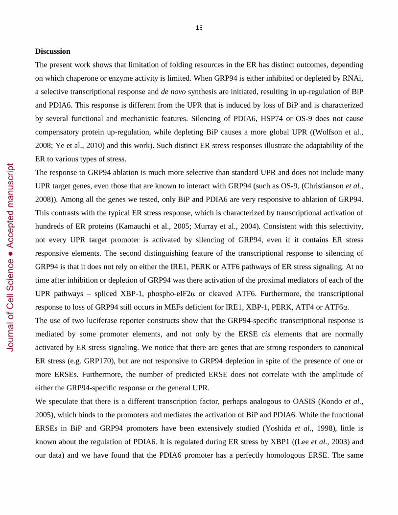
Another example of divergent chaperone regulation is provided by the androgen-responsive prostate
carcinoma cell line LnCap (Supp. Fig. 3). When treated with the androgen analogue 5α-
dehydroepiandrosterone (DHEA), there is a selective increase in GRP94 expression, without a
concomitant increase in BiP in these cells. This increase appears to be cell-type specific, since PC-3,
another prostate cancer cell line that is androgen-insensitive, as well as the embryonic kidney fibroblast
293T, do not respond in this fashion. These data indicate that GRP94 expression can be increased
considerably even without a general UPR.
The GRP94-specific response is transcriptional.
Besides an increase in steady state levels of BiP protein in GRP94-depleted cells, BiP mRNA levels are
also elevated. (Fig. 3A). Moreover, the induction of BiP is abolished when cells are pre-treated with
actinomycin D, to arrest transcription (Fig. 3B). In this experiment, we used inhibition of activity rather
than RNAi, because under this condition the up-regulation is fast and the time course of the experiment
is compatible with cell viability. Yet a third line of evidence that the GRP94 specific response is
transcriptional is provided by a luciferase reporter analysis. Using a construct where luciferase
expression is driven by regulatory regions of the BiP gene, compared to controls, there is approximately
3.5-fold induction of luciferase in 293T cells in which GRP94 was knocked down (Fig. 3C). The three
Jour
nal o
f Cel
l Sci
ence
Acc
epte
d m
anus
crip
t

9
ER stress elements (ERSEs, (Yoshida et al., 1998)) of the BiP promoter are essential for the response to
GRP94 silencing, since luciferase activity is abolished when all three ERSEs are mutated (Fig. 3C).
Thus, ablation of GRP94 initiates a transcription response at the BiP promoter. While we did not check
the transcription of PDIA6 explicitly, its promoter has the same organization of three ERSE elements as
the BiP promoter and ERSE1 shows excellent correspondence to the canonical sequence (Fig. 3D and
Suppl. Fig. 6). Not all ER stress-responsive promoters are induced by silencing of GRP94. One example
is the promoter of PDIA1, which has only one ERSE and its sequence diverges from the canonical
sequence (Suppl. Fig. 6). Furthermore, we tested another reporter, where luciferase is driven by the
promoter of the Herpes Simplex Virus ICP0 gene (Burnett et al.). This reporter is responsive to
chemically induced ER stress (Burnett et al.) and is also responsive to silencing of BiP (Fig. 3C).
However, the ICP0-luciferase reporter is not induced by silencing of GRP94, supporting the conclusion
that depletion of the two major ER chaperones is not equivalent and the GRP94-specific transcriptional
response is mechanistically distinct from UPR.
The response to GRP94 ablation is not mediated by UPR signaling.
Transcriptional responses of ER quality control genes are usually initiated by the UPR signaling
pathways, through activation of IRE1, PERK or ATF6. Therefore, we investigated whether depletion of
GRP94 triggers these three transducers, using two approaches: 1) assaying the response to GRP94
deletion in cells where the signal pathways are genetically ablated (loss-of-function approach) and 2)
measuring the activity of each transducer in GRP94-ablated cells.
The GRP94-specific response does not require IRE1, because it occurs in IRE1-deficient mouse
embryonic fibroblasts (MEF) just as it does in WT MEF (Fig. 4A). Similarly, the GRP94-specific
response is normal in PERK-deficient and in ATF6α-deficient MEF (Fig. 4B,C). Therefore, none of the
three transducers is needed individually for the response.
To test the possibility that in the absence of one transducer, signaling could occur via another UPR
branch, we tested whether the main substrates mediating the signaling are activated. No splicing of
XBP1 was observed at one, two or three days after infection with shRNA-GRP94-encoding virus, during
the time frame needed to silence GRP94, and also not after 27 days, long after silencing was established
(Fig. 4D). In contrast, tunicamycin and DTT treatments induce XBP1 splicing within a few hours, as
expected, serving as positive controls for the assay. Since RNAi silencing requires days, we asked if
there was an earlier XBP1 splicing event when GRP94 was inhibited by 17AAG, under conditions that
Jour
nal o
f Cel
l Sci
ence
Acc
epte
d m
anus
crip
t

10
induce BiP upregulation (Fig. 3). As shown in Fig. 4E, there was no detectable XBP1 splicing within 4
hours of 17AAG treatment. In contrast, efficient XBP1 splicing was observed already at the earliest time
point of BiP ablation by subAB (Fig. 4E), which is known to induce UPR. The inability to detect XBP1
splicing was not because of low sensitivity of detection; as shown in Fig. 4F, splicing is easily detectable
in our hands even at a dose of 100 ng/ml tunicamycin, 10-20X lower than the dose commonly used to
induce UPR. Finally, the GRP94-specific response was intact in XBP1-deficient MEF (Suppl. Fig. 4A),
showing that it is independent of either IRE1 or XBP1.
A second well-studied branch of the UPR machinery, ATF6, is activated by relocation to the Golgi
complex, where proteolytic cleavage frees the N-terminal domain to become a transcription factor (Haze
et al., 1999). We tested the activation of ATF6 by using 293T cells stably expressing an HA-tagged
version of the protein (Wang et al., 2000). As shown in Fig. 4G, in GRP94-silenced cells, the basal level
of the p50 active fragment of ATF6 is equal to that seen in control cells and there is no detectable
accumulation of active ATF6. Note that the p50 fragment is unstable and is best detected when
proteasome degradation is inhibited by MG-132 (Fig. 4G). In contrast, both tunicamycin treatment and
BiP depletion are associated with accumulation of p50-ATF6.
The main substrate of the third UPR sensor, PERK, is the translation initiation factor 2 alpha (eIF2α)
(Harding et al., 2000). Once phosphorylated, eIF2α activates the transcription factor ATF4 that in turn
mediates the expression of ER proteins. Therefore, we assessed the phosphorylation of eIF2α in GRP94-
silenced cells (Fig. 4H). Phospho-eIF2α levels do not appear significantly different in GRP94-depleted
cells; in contrast, phosphorylation of eIF2α is induced by depletion of BiP (Fig. 4H and see also
(Wolfson et al., 2008)). Further evidence against the involvement of PERK is that the GRP94-specific
response occurs in ATF4-deficient MEF (Suppl. Fig. 4B).
On the basis of these combined data, we conclude that the GRP94-specific response does not cause
activation of either IRE1, PERK or ATF6, while depletion of BiP by the same methods does, supporting
that the GRP94-specific response is not a canonical UPR, but rather is mediated by a different
mechanism.
The responsiveness to ER stress is not diminished by ablation of GRP94.
Jour
nal o
f Cel
l Sci
ence
Acc
epte
d m
anus
crip
t

11
If the GRP94-specific response involves up-regulation of two proteins but not the activation of UPR
sensors, is the UPR still functional under these conditions? To address this question we compared the
responses to tunicamycin or thapsigargin of cells that are either GRP94-deficient or -sufficient.
In terms of viability, GRP94-deficient cells survived in increasing concentrations of Tun or TG less well
than GRP94-sufficient cells, but the differences were not dramatic (Fig. 5A-B) when compared to the
effect of BiP-deficient cells (Fig. 5 in (Morinaga et al., 2008)) or to the GRP94-null ES cells (Fig. 6 in
(Biswas et al., 2007)). These observations indicate that despite the ubiquitous up-regulation of GRP94
under ER stress conditions, this chaperone contributes to, but is not essential for coping with ER stress.
A different conclusion was reached in (Biswas et al., 2007; Ostrovsky et al., 2009), showing that GRP94
KO ES cells are hypersensitive to stress, suggesting a pro-survival role for GRP94. The difference is
likely due to the up-regulation of BiP and PDIA6 seen in the KD cells but not in grp94-/- ES cells,
arguing that the up-regulation of the two compensates for some function of GRP94. Long-term loss of
GRP94 function leads to adaptation at a cost of increased sensitivity to ER stress.
As a second readout for UPR responsiveness we measured the extent of BiP induction by Tun or TG
stress. As shown in Fig. 5C, GRP94-silenced cells can over-express BiP when challenged with Tun or
TG beyond the level of the GRP94-specific response, demonstrating that the stable induction of BiP
when GRP94 is silenced is not maximal, and further increase of BiP is possible. GRP94-silenced cells
responded by increasing the level of BiP in a dose-dependent fashion, similarly to the control cells, but
BiP induction appeared saturated at lower concentration of Tun in comparison to the control cells (Fig.
5D). Interestingly, GRP94-silenced cells responded with similar amplitude to the control cells at the
lowest concentration of Tun, indicating that the trigger is not compromised by the lack of GRP94. A
third readout for UPR responsiveness was the XBP1-Venus reporter, where YFP fluorescence is induced
by splicing of the upstream XBP1 sequence (Iwawaki and Akai, 2006). While GRP94-silenced cells
displayed only background reporter levels, when treated with Tun, the reporter in these cells was
activated (Eletto and Argon, data not shown). Together, these readouts show that the GRP94-specific
response does not obviate the ability of cells to mount the UPR transcriptional response.
If depletion of GRP94 does not impair the ER stress response and since GRP94 is normally induced
under such stress conditions, we also tested the effect of over-expressing GRP94 on the ER stress
response. Taking advantage of doxycycline-inducible CHO cells, we expressed a controlled amount of
Flag-tagged GRP94 in cells that were simultaneously treated with subAB (the time course of BiP
ablation by this method is compatible with the time course of doxycycline induction). Depleting cells of
Jour
nal o
f Cel
l Sci
ence
Acc
epte
d m
anus
crip
t

12
BiP resulted in 3-4-fold induction of the endogenous GRP94, even in cells that already over-expressed
Flag-tagged GRP94 (Fig. 5E). This indicates that the ER has a selective mechanism of sensing levels of
BiP that fails to monitor the abundance of GRP94. Surprisingly, the over-expression of GRP94 is not
counter-balanced by a reduction in the level of BiP, as mirrored by the loss-of-function experiment.
Altogether, these results suggest that: a) the relative abundance of GRP94 is monitored only below a
minimal threshold; b) cells employ a distinct mechanism to sense the relative abundance of GRP94; c)
Although GPR94 is one of the major UPR targets, it is not required for mounting the response to
chemical stresses.
Cells monitor the activity, not the protein level of GRP94 in the ER.
Since RNAi was not the only treatment that induced BiP and PDIA6 and chemical inhibition led to the
same response, we asked if cells sense the expression of GRP94 or its activity. To this end, we
determined whether WT GRP94 or an ATPase-deficient (E82A) mutant can substitute for the
endogenous protein when the latter is silenced in doxycycline-inducible CHO cells. The exogenous
GRP94 genes were constructed to be shRNA-resistant (Suppl. Fig. 5). When an active version of GRP94
(Flag-tagged WT GRP94), replaced the silenced endogenous GRP94, the induction of BiP and PDIA6
was abolished. On the other hand, expression of E82A GRP94 failed to prevent the induction of the two
genes (Fig. 6). Together with the effect of geldanamycin, these results strongly suggest that the
mechanism that monitors the amount of GRP94 in the ER is dependent on the activity of the chaperone.
Since GRP94 activity is much more client-selective than BiP activity, loss of this chaperone may
necessitate a more restricted and mechanistically distinct response than UPR.
Jour
nal o
f Cel
l Sci
ence
Acc
epte
d m
anus
crip
t
13
Discussion
The present work shows that limitation of folding resources in the ER has distinct outcomes, depending
on which chaperone or enzyme activity is limited. When GRP94 is either inhibited or depleted by RNAi,
a selective transcriptional response and de novo synthesis are initiated, resulting in up-regulation of BiP
and PDIA6. This response is different from the UPR that is induced by loss of BiP and is characterized
by several functional and mechanistic features. Silencing of PDIA6, HSP74 or OS-9 does not cause
compensatory protein up-regulation, while depleting BiP causes a more global UPR ((Wolfson et al.,
2008; Ye et al., 2010) and this work). Such distinct ER stress responses illustrate the adaptability of the
ER to various types of stress.
The response to GRP94 ablation is much more selective than standard UPR and does not include many
UPR target genes, even those that are known to interact with GRP94 (such as OS-9, (Christianson et al.,
2008)). Among all the genes we tested, only BiP and PDIA6 are very responsive to ablation of GRP94.
This contrasts with the typical ER stress response, which is characterized by transcriptional activation of
hundreds of ER proteins (Kamauchi et al., 2005; Murray et al., 2004). Consistent with this selectivity,
not every UPR target promoter is activated by silencing of GRP94, even if it contains ER stress
responsive elements. The second distinguishing feature of the transcriptional response to silencing of
GRP94 is that it does not rely on either the IRE1, PERK or ATF6 pathways of ER stress signaling. At no
time after inhibition or depletion of GRP94 was there activation of the proximal mediators of each of the
UPR pathways – spliced XBP-1, phospho-eIF2α or cleaved ATF6. Furthermore, the transcriptional
response to loss of GRP94 still occurs in MEFs deficient for IRE1, XBP-1, PERK, ATF4 or ATF6α.
The use of two luciferase reporter constructs show that the GRP94-specific transcriptional response is
mediated by some promoter elements, and not only by the ERSE cis elements that are normally
activated by ER stress signaling. We notice that there are genes that are strong responders to canonical
ER stress (e.g. GRP170), but are not responsive to GRP94 depletion in spite of the presence of one or
more ERSEs. Furthermore, the number of predicted ERSE does not correlate with the amplitude of
either the GRP94-specific response or the general UPR.
We speculate that there is a different transcription factor, perhaps analogous to OASIS (Kondo et al.,
2005), which binds to the promoters and mediates the activation of BiP and PDIA6. While the functional
ERSEs in BiP and GRP94 promoters have been extensively studied (Yoshida et al., 1998), little is
known about the regulation of PDIA6. It is regulated during ER stress by XBP1 ((Lee et al., 2003) and
our data) and we have found that the PDIA6 promoter has a perfectly homologous ERSE. The same
Jour
nal o
f Cel
l Sci
ence
Acc
epte
d m
anus
crip
t

14
motif is either poorly conserved or not present at all in other PDIAs, explaining why they are not
induced and PDIA6 is highly responsive to ER stress.
Despite GRP94 being one of the main hallmarks of the ER stress response, its loss or inhibition do not
impair the cells’ ability to mount a stress response and cope with ER stress. GRP94-depleted cells,
which have increased levels of BiP, can still further up-regulate BiP when ‘insulted’ with chemical
stress inducers. In contrast, a GRP94-null clone that does not show BiP induction, is dramatically more
susceptible to ER stress (Biswas et al., 2007). We propose that ER stress responsiveness is not impaired
without GRP94 as long as there is sufficient activity of BiP and PDIA6. This explanation fits with the
cytoprotective effect of BiP over-expression (Dorner et al., 1992). Also consistent with this explanation
is the inability of cells to survive simultaneous ablation of GRP94 and BiP (Eletto, unpublished data).
Furthermore, KD of GRP94 in myeloma lines, which are specialized secretory cells, is barely achievable
and only lasts for few days (Kropp et al., 2010). In contrast, GRP94 gene remained silenced for
indefinite period of time in non-professionally secretory cells, as reported here. Thus, GRP94 may have
a central role in general UPR only when there is high demand on folding resources, when even mild
accumulation of folding intermediates may be harmful.
It is unclear why BiP and PDIA6 in particular are over-expressed when GRP94 activity is lost. There is
no obvious functional reason why up-regulation of either one would compensate for the loss of another
component. BiP and GRP94 do work in concert on the folding of some client proteins, such as
immunoglobulins or thyroglobulin, but they are not redundant and work sequentially, apparently
recognizing distinct folding intermediates (Melnick et al., 1994). We hypothesize that when GRP94 is
ablated, BiP, which recognizes the less advanced folding intermediates, must be induced to deal with
accumulation of GRP94 clients. PDIA6 is a BiP partner that binds to BiP clients (Jessop et al., 2009),
and its level is co-regulated with BiP under chemically induced ER stress and in response to the
accumulation of misfolded proteins (this work and (Hartley et al., 2010)). GRP94 and BiP may also be
affected by each other due to other shared common functions. Both are calcium-binding proteins,
(Biswas et al., 2007; Lievremont et al., 1997) and both are involved in the degradation of soluble
substrates (Christianson et al., 2008; Kabani et al., 2002). In conclusion, the expression network formed
by BiP, GRP94 and PDIA6 can be rationalized by several known functional parameters that can be
tested in the future.
Cells must have developed mechanisms to monitor the abundance of chaperones in order to keep their
levels within a certain range that is compatible with their cooperative mode of actions. For example,
Jour
nal o
f Cel
l Sci
ence
Acc
epte
d m
anus
crip
t

15
when cytosolic HSP90 is inhibited, HSP70 is up-regulated, due to the cooperation of these two
chaperones in the folding pathways of many kinases and transcription factors (Bagatell et al., 2000; Zou
et al., 1998). Such compensatory mechanisms are likely based on sensing the loss of chaperone activity.
We provide evidence that the presence of GRP94 is also detected based on its activity and not simply the
abundance of the protein. First, the transcriptional response of BiP and PDIA6 is activated not only
when GRP94 is depleted, but also when it is inhibited pharmacologically. Second, the up-regulation of
BiP and PDIA6 occurs even if an inactive GRP94 mutant is over-expressed, but is prevented when
active GRP94 is supplied. Interestingly, the detection mechanism works only when GRP94 activity is
reduced; over-expression of GRP94 does not perturb the normal functioning of the ER stress response
machinery.
These results suggest the following model: when GRP94 activity is insufficient, either because of a high
demand of obligate clients that accumulate in the ER or because of another need, a dedicated sensor is
activated, which reports on the limiting GRP94 through a signaling pathway, culminating in activation
of transcription of BiP, PDIA6, and perhaps other genes.
Our results show that ER proteins are not necessarily upregulated as a cohort. Rather, multiple signal
transduction pathways may be evocated in response to distinct stimuli or stresses. The plasticity of ER
dynamics may be due to its ability to initiate such distinct stress responses, leading to distinct outcomes
and different kinetics.
Jour
nal o
f Cel
l Sci
ence
Acc
epte
d m
anus
crip
t

16
Materials and methods
Ethics Statement
All experiments were approved by the CHOP and the University of Pennsylvania animal care
committees.
Chemicals and Plasmids
Actinomycin D, doxycycline, tunicamycin and thapsigargin were purchased from Sigma Chemicals
(St. Louis, MO). MG-132 was from Calbiochem (San Diego, CA). Lipofectamine 2000 transfection
reagent was from Invitrogen (Carlsbad, CA). 17-Allylamino-17-demethoxygeldanamycin (17AAG),
puromycin and G418 were from InvivoGen (San Diego, CA); the XTT cell proliferation kit from
Biotium, Inc (Hayward, CA). DMEM was from Mediatech, Inc. (Manassess, VA), fetal
bovine serum was from Gemini (West Sacramento, CA). Glutamine, penicillin/streptomycin
supplement was from Gibco-Invitrogen (Grand Island, NY). Subtilase AB toxin was a kind gift of
Dr J.C. Paton (Univ. of Adelaide, Australia). A Flag-tagged GRP94 expressed into the pTRE Vector
(Clontech, Mountain View, CA) was subjected to site-directed mutagenesis using the QuickChange
Kit (Agilent, Santa Clara, CA) to generate the E82A GRP94 ATPase deficient mutant.
Cell culture and GRP94 conditional KO mice
C2C12, 10T1/2, NIH-3T3, 293T, and HeLa cells were from the ATCC. CHO-K1 Tet-On were from
Clontech. 293T cells stably expressing a HA-tagged ATF6 were a kind gift of Drs H. Steiner and
Haass (Ludwig Maximilians University, Germany). These cell lines were grown in DMEM in the
presence of 10% FBS and Gln/Pen/Strept, and, when needed, the proper eukaryotic selection agent
(puromycin or G418).
IRE1 and XBP1 KO, PERK and ATF4 KO MEFs were generous gifts from Dr D Ron (Univ. of
Cambridge, UK). ATF6alpha KO MEFs from Dr K. Mori (Univ. of Kyoto, Japan), Mice containing
a floxed allele of GRP94 (grp94flox) (Yang et al., 2007) were crossed with muscle creatine kinase
(MCK)-Cre transgenic mice (on a C57Bl/6 background; Jackson Labs, stock 006475). Double
heterozygous progeny were then bred to grp94flox/flox mice, in order to deplete GRP94 within skeletal
and cardiac muscle. Tissue samples were collected and processed for immunoblotting as reported in
(Barton et al., 2010).
Immunoblotting
Jour
nal o
f Cel
l Sci
ence
Acc
epte
d m
anus
crip
t

17
Cells were lysed with a 0.5% NP-40/Igepal detergent solution as described in (Ostrovsky et al.,
2009). Images were recorded using an Alpha Innotech (Santa Clara, CA) or Odyssey (Li-Cor,
Lincoln, NE) imagers. Band intensities were normalized for total protein loads using house-keeping
proteins (α−tubulin or 14-3-3). Fold changes were calculated relative to internal references (wt or
control samples), as indicated in the figure legends.
Antibodies: rabbit anti-14-3-3 (C16) and mAb anti-myogenin (F5D) were purchased from Santa
Cruz, Biotechnology, Santa Cruz, CA. MAb anti-desmin(D33) was from Imgenex (San Diego CA);
mAb anti-KDEL was from StressGen (Vancouver, BC); anti-HSP90 was from BD Transduction
Laboratories (San Jose, CA) and anti-caspase 3 and anti-cleaved caspase 3(Asp175) were from Cell
Signaling; the anti-GRP94 monoclonal antibody 9G10 (SPA-850) from Stressgen Biotechnologies
(Victoria, BC). The monoclonal anti-HA antibody HA.11 (clone 16B12) was obtained from Covance
(Princeton, NJ). Secondary antibodies conjugated to HRP were from Jackson ImmunoResearch
Laboratories (West Grove, PA), secondary antibodies conjugated to near-infrared fluorophores were
from Li-Cor.
RNAi silencing
GRP94, BiP, PDIA6, HSP47 or OS-9 were knocked-down, using the following shRNAs from Sigma
Life Science (Saint Louis, MO): SHCLNG-NM_011631 (a set of 5 vectors referred to in this paper
as shRNA23 to 27; Fig. 2B), TRCN0000008455, SHCLNG-NM_027959 (a set of 5 vectors, Suppl.
Fig. 2D), TRCN0000008534, TRCN0000175937, respectively. Briefly, cells were transduced with
lentiviral particles encoding the shRNA sequences, as in (Ostrovsky et al., 2010). The efficiency of
knockdown was consistently >90% for GRP94, PDIA6, HSP47 and OS-9. BiP expression typically
could only be reduced to 40% of control level.
Analysis of XBP1 mRNA splicing and ATF6 endoproteolysis
XBP1 and β-actin were PCR amplified from total RNA as in (Calfon et al., 2002).
293T stably expressing HA-tagged ATF6 were analyzed by immunoblotting with anti HA-antibody
HA.11 after the cells were stressed with Tun and treated with MG132, to block degradation of the
ATF6 fragment. The levels of the ATF6 fragment nuclear fractions were enriched as described in (Li
et al., 2000).
Jour
nal o
f Cel
l Sci
ence
Acc
epte
d m
anus
crip
t

18
Luciferase reporter assay
Constructs composed of nucleotides −284 to +221 of the human GRP78 promoter driving luciferase
were described in (Doroudgar et al., 2009). The construct composed of 1024 nucleotides upstream of
the coding sequence of the herpes simplex virus ICP0 gene fused to luciferase was from Dr. Liu
(Burnett et al., 2012). 293T stably expressing shRNA targeting either GRP94, BiP or a non-relevant
sequence were transfected and analyzed for luciferase activity as in (Doroudgar et al., 2009).
Statistical Analysis
Statistical analysis was performed using a one-way analysis of variance followed by Student's
Newman-Keul's post hoc analysis of variance (*, p < 0.05, unless otherwise stated in the figure
legends).
Jour
nal o
f Cel
l Sci
ence
Acc
epte
d m
anus
crip
t

19
Acknowledgments
We are grateful to Drs. K. Mori (University of Kyoto, Japan), D. Ron (Cambridge University, UK),
J.C. Paton (University of Adelaide), H. Steiner and C. Haass (Ludwig-Maximilians-University,
Germany), R. Prywes (Columbia University) and R. Lu (University of Guelph) for generous gifts of
cell lines, plasmids and other reagents. We also thank Dr. Yina Dong and Erikka Carr for technical
support and Drs. T. Gidalevitz, M. Marzec, M. Chou and A. Gentilella for their comments and
suggestions. This work was funded by NIH grants GM077480 and AG18001 (to Y.A.), and
HL085577, HL75573, HL104535 and EB011698 (to C.G.G.). D.D. was supported by training grant
GM008275. S.D. was supported by the Rees-Stealy Research Foundation, the San Diego Chapter of
the ARCS Foundation, an American Heart Association Predoctoral Fellowship and an Inamori
Foundation Fellowship. The funders had no role in study design, data collection and analysis,
decision to publish, or preparation of the manuscript.
Jour
nal o
f Cel
l Sci
ence
Acc
epte
d m
anus
crip
t

20
References
Bagatell, R., Paine-Murrieta, G. D., Taylor, C. W., Pulcini, E. J., Akinaga, S., Benjamin, I. J. and Whitesell, L. (2000). Induction of a heat shock factor 1-dependent stress response alters the cytotoxic activity of hsp90-binding agents. Clin Cancer Res 6, 3312-8. Balch, W. E., Morimoto, R. I., Dillin, A. and Kelly, J. W. (2008). Adapting proteostasis for disease intervention. Science 319, 916-9. Bartke, A. (2009). The somatotropic axis and aging: Mechanisms and persistent questions about practical implications. Exp Gerontol 44, 372-374. Barton, E., Park, S., James, J. K., Makarewich, C. A., Eletto, D., Philippou, A., Lei, H., Brisson, B., Ostrovsky, O., Li, Z. et al. (2012). Muscle-specific deletion of GRP94 leads to small muscles and impaired organismal growth due to inhibition of muscle-derived IGF production Submitted. Barton, E. R., DeMeo, J. and Lei, H. (2010). The insulin-like growth factor (IGF)-I E-peptides are required for isoform-specific gene expression and muscle hypertrophy after local IGF-I production. J Appl Physiol 108, 1069-76. Biswas, C., Ostrovsky, O., Makarewich, C. A., Wanderling, S., Gidalevitz, T. and Argon, Y. (2007). The peptide binding activity of GRP94 is regulated by Calcium. Biochem. J. 405, 233-241. Burnett, H. F., Audas, T. E., Liang, G. and Lu, R. R. (2012). Herpes simplex virus-1 disarms the unfolded protein response in the early stages of infection. Cell Stress Chaperones. Calfon, M., Zeng, H., Urano, F., Till, J. H., Hubbard, S. R., Harding, H. P., Clark, S. G. and Ron, D. (2002). IRE1 couples endoplasmic reticulum load to secretory capacity by processing the XBP-1 mRNA. Nature 415, 92-6. Chang, S. C., Erwin, A. E. and Lee, A. S. (1989). Glucose-regulated protein (GRP94 and GRP78) genes share common regulatory domains and are coordinately regulated by common trans- acting factors. Mol. Cell. Biol. 9, 2153-62. Christianson, J. C., Shaler, T. A., Tyler, R. E. and Kopito, R. R. (2008). OS-9 and GRP94 deliver mutant alpha1-antitrypsin to the Hrd1-SEL1L ubiquitin ligase complex for ERAD. Nat Cell Biol 10, 272-82. Dorner, A. J., Wasley, L. C. and Kaufman, R. J. (1992). Overexpression of GRP78 mitigates stress induction of glucose regulated proteins and blocks secretion of selective proteins in Chinese hamster ovary cells. EMBO J. 11, 1563-1571. Doroudgar, S., Thuerauf, D. J., Marcinko, M. C., Belmont, P. J. and Glembotski, C. C. (2009). Ischemia activates the ATF6 branch of the endoplasmic reticulum stress response. J Biol Chem 284, 29735-45. Harding, H. P., Zhang, Y., Bertolotti, A., Zeng, H. and Ron, D. (2000). Perk is essential for translational regulation and cell survival during the unfolded protein response. Mol Cell 5, 897-904. Hartley, T., Siva, M., Lai, E., Teodoro, T., Zhang, L. and Volchuk, A. (2010). Endoplasmic reticulum stress response in an INS-1 pancreatic beta-cell line with inducible expression of a folding-deficient proinsulin. BMC Cell Biol 11, 59. Haze, K., Yoshida, H., Yanagi, H., Yura, T. and Mori, K. (1999). Mammalian transcription factor ATF6 is synthesized as a transmembrane protein and activated by proteolysis in response to endoplasmic reticulum stress. Mol Biol Cell 10, 3787-99. Iwawaki, T. and Akai, R. (2006). Analysis of the XBP1 splicing mechanism using endoplasmic reticulum stress-indicators. Biochem Biophys Res Commun 350, 709-15.
Jour
nal o
f Cel
l Sci
ence
Acc
epte
d m
anus
crip
t

21
Jessop, C. E., Watkins, R. H., Simmons, J. J., Tasab, M. and Bulleid, N. J. (2009). Protein disulphide isomerase family members show distinct substrate specificity: P5 is targeted to BiP client proteins. J Cell Sci 122, 4287-95. Kabani, M., Beckerich, J. M. and Brodsky, J. L. (2002). Nucleotide exchange factor for the yeast Hsp70 molecular chaperone Ssa1p. Mol Cell Biol 22, 4677-89. Kamauchi, S., Nakatani, H., Nakano, C. and Urade, R. (2005). Gene expression in response to endoplasmic reticulum stress in Arabidopsis thaliana. FEBS J 272, 3461-76. Kapulkin, V., Hiester, B. G. and Link, C. D. (2005). Compensatory regulation among ER chaperones in C. elegans. FEBS Lett 579, 3063-8. Kondo, S., Murakami, T., Tatsumi, K., Ogata, M., Kanemoto, S., Otori, K., Iseki, K., Wanaka, A. and Imaizumi, K. (2005). OASIS, a CREB/ATF-family member, modulates UPR signalling in astrocytes. Nat Cell Biol 7, 186-94. Kropp, L. E., Garg, M. and Binder, R. J. (2010). Ovalbumin-derived precursor peptides are transferred sequentially from gp96 and calreticulin to MHC class I in the endoplasmic reticulum. J Immunol 184, 5619-27. Lee, A. H., Iwakoshi, N. N. and Glimcher, L. H. (2003). XBP-1 regulates a subset of endoplasmic reticulum resident chaperone genes in the unfolded protein response. Mol Cell Biol 23, 7448-59. Li, M., Baumeister, P., Roy, B., Phan, T., Foti, D., Luo, S. and Lee, A. S. (2000). ATF6 as a transcription activator of the endoplasmic reticulum stress element: thapsigargin stress-induced changes and synergistic interactions with NF-Y and YY1. Mol Cell Biol 20, 5096-106. Lievremont, J. P., Rizzuto, R., Hendershot, L. and Meldolesi, J. (1997). BiP, a major chaperone protein of the endoplasmic reticulum lumen, plays a direct and important role in the storage of the rapidly exchanging pool of Ca2+. J. Biol. Chem. 272, 30873-30879. Ma, Y. and Hendershot, L. M. (2004). ER chaperone functions during normal and stress conditions. J Chem Neuroanat 28, 51-65. Mao, C., Wang, M., Luo, B., Wey, S., Dong, D., Wesselschmidt, R., Rawlings, S. and Lee, A. S. (2010). Targeted mutation of the mouse Grp94 gene disrupts development and perturbs endoplasmic reticulum stress signaling. PLoS One 5, e10852. Melnick, J., Aviel, S. and Argon, Y. (1992). The endoplasmic reticulum stress protein GRP94, in addition to BiP, associates with unassembled immunoglobulin chains. J. Biol. Chem. 267, 21303-6. Melnick, J., Dul, J. L. and Argon, Y. (1994). Sequential interaction of the chaperones BiP and GRP94 with immunoglobulin chains in the endoplasmic reticulum. Nature 370, 373-375. Meunier, L., Usherwood, Y. K., Chung, K. T. and Hendershot, L. M. (2002). A subset of chaperones and folding enzymes form multiprotein complexes in endoplasmic reticulum to bind nascent proteins. Mol Biol Cell 13, 4456-69. Morinaga, N., Yahiro, K., Matsuura, G., Moss, J. and Noda, M. (2008). Subtilase cytotoxin, produced by Shiga-toxigenic Escherichia coli, transiently inhibits protein synthesis of Vero cells via degradation of BiP and induces cell cycle arrest at G1 by downregulation of cyclin D1. Cell Microbiol 10, 921-9. Muresan, Z. and Arvan, P. (1997). Thyroglobulin transport along the secretory pathway. Investigation of the role of molecular chaperone, GRP94, in protein export from the
Jour
nal o
f Cel
l Sci
ence
Acc
epte
d m
anus
crip
t

22
endoplasmic reticulum [published erratum appears in J Biol Chem 1997 Nov 28;272(48):30590]. J. Biol. Chem. 272, 26095-102. Murray, J. I., Whitfield, M. L., Trinklein, N. D., Myers, R. M., Brown, P. O. and Botstein, D. (2004). Diverse and specific gene expression responses to stresses in cultured human cells. Mol Biol Cell 15, 2361-74. Musaro, A., McCullagh, K., Paul, A., Houghton, L., Dobrowolny, G., Molinaro, M., Barton, E. R., Sweeney, H. L. and Rosenthal, N. (2001). Localized Igf-1 transgene expression sustains hypertrophy and regeneration in senescent skeletal muscle. Nat Genet 27, 195-200. Ostrovsky, O., Ahmed, N. T. and Argon, Y. (2009). The chaperone activity of GRP94 toward insulin-like growth factor II is necessary for the stress response to serum deprivation. Mol Biol Cell 20, 1855-64. Ostrovsky, O., Eletto, D., Makarewich, C., Barton, E. R. and Argon, Y. (2010). Glucose Regulated Protein 94 is required for muscle differentiation through its control of the autocrine production of insulin-like growth factors. . Biochim Biophys Acta 1803, 333-341. Paton, A. W., Beddoe, T., Thorpe, C. M., Whisstock, J. C., Wilce, M. C., Rossjohn, J., Talbot, U. M. and Paton, J. C. (2006). AB5 subtilase cytotoxin inactivates the endoplasmic reticulum chaperone BiP. Nature 443, 548-52. Ron, D. (2002). Proteotoxicity in the endoplasmic reticulum: lessons from the Akita diabetic mouse. J Clin Invest 109, 443-445. Ron, D. and Walter, P. (2007). Signal integration in the endoplasmic reticulum unfolded protein response. Nat Rev Mol Cell Biol 8, 519-29. Shiu, R. P., Pouyssegur, J. and Pastan, I. (1977). Glucose depletion accounts for the induction of two transformation-sensitive membrane proteins in Rous sarcoma virus-transformed chick embryo fibroblasts. Proc. Natl. Acad. Sci. USA 74, 3840-3844. Thibault, G., Ismail, N. and Ng, D. T. (2011). The unfolded protein response supports cellular robustness as a broad-spectrum compensatory pathway. Proc Natl Acad Sci U S A 108, 20597-602. Travers, K. J., Patil, C. K., Wodicka, L., Lockhart, D. J., Weissman, J. S. and Walter, P. (2000). Functional and genomic analyses reveal an essential coordination between the unfolded protein response and ER-associated degradation. Cell 101, 249-58. van Anken, E., Romijn, E. P., Maggioni, C., Mezghrani, A., Sitia, R., Braakman, I. and Heck, A. J. (2003). Sequential waves of functionally related proteins are expressed when B cells prepare for antibody secretion. Immunity 18, 243-53. Walter, P. and Ron, D. (2011). The unfolded protein response: from stress pathway to homeostatic regulation. Science 334, 1081-6. Wanderling, S., Simen, B. B., Ostrovsky, O., Ahmed, N. T., Vogen, S., Gidalevitz, T. and Argon, Y. (2007). GRP94 is essential for mesoderm induction and muscle development because it regulates IGF secretion. Mol Biol Cell 18, 3764-3775. Wang, Y., Shen, J., Arenzana, N., Tirasophon, W., Kaufman, R. J. and Prywes, R. (2000). Activation of ATF6 and an ATF6 DNA binding site by the endoplasmic reticulum stress response. J Biol Chem 275, 27013-20. Wiest, D. L., Burkhardt, J. K., Hester, S., Hortsch, M., Meyer, D. I. and Argon, Y. (1990). Membrane biogenesis during B cell differentiation: most endoplasmic reticulum proteins are expressed coordinately. J. Cell Biol. 110, 1501-11.
Jour
nal o
f Cel
l Sci
ence
Acc
epte
d m
anus
crip
t

23
Wolfson, J. J., May, K. L., Thorpe, C. M., Jandhyala, D. M., Paton, J. C. and Paton, A. W. (2008). Subtilase cytotoxin activates PERK, IRE1 and ATF6 endoplasmic reticulum stress-signalling pathways. Cell Microbiol 10, 1775-86. Yang, Y., Liu, B., Dai, J., Srivastava, P. K., Zammit, D. J., Lefrancois, L. and Li, Z. (2007). Heat shock protein gp96 is a master chaperone for toll-like receptors and is important in the innate function of macrophages. Immunity 26, 215-226. Ye, R., Jung, D. Y., Jun, J. Y., Li, J., Luo, S., Ko, H. J., Kim, J. K. and Lee, A. S. (2010). Grp78 Heterozygosity Promotes Adaptive Unfolded Protein Response and Attenuates Diet-Induced Obesity and Insulin Resistance. Diabetes 59, 6-16. Yoshida, H., Haze, K., Yanagi, H., Yura, T. and Mori, K. (1998). Identification of the cis-Acting Endoplasmic Reticulum Stress Response Element Responsible for Transcriptional Induction of Mammalian Glucose- regulated Proteins. Involvement of basic leucine zipper transcription factors. J. Biol. Chem. 273, 33741-33749. Zou, J., Guo, Y., Guettouche, T., Smith, D. F. and Voellmy, R. (1998). Repression of heat shock transcription factor HSF1 activation by HSP90 (HSP90 complex) that forms a stress-sensitive complex with HSF1. Cell 94, 471-80.
Jour
nal o
f Cel
l Sci
ence
Acc
epte
d m
anus
crip
t

24
Figure Legends
Figure 1: Silencing GRP94 produces a response distinct from a general UPR.
A. shRNA-mediated silencing of either BiP or GRP94 leads to upregulation of other ER
components. In this representative experiment, murine 10T1/2 cells stably expressing shRNA to
GRP94, to BiP or an irrelevant shRNA (Ctrl) were lysed and analyzed by immunoblotting with
antibodies to the proteins indicated on the right. Signals were quantified using a LiCor imager,
because of its superior dynamic range. Levels of expression of the indicated proteins were
determined by quantitation of exposures at the linear range and normalized for protein load by
comparison to the levels of tubulin and 14-3-3 in the same sample. The density of each band in the
shCtrl lane is defined as 1.0. The relative fold-increase is indicated below each band. Similar results
were obtained in various other cells, of either human of mouse origin, where the pattern was always
consistent even if the amplitude of the effect differed.
B. Quantitation of the response of the indicated 8 ER proteins to tunicamycin treatment (2μg/mL for
24 hours), as a typical chemical inducer of ER stress. Shown are means ± SD of 4 independent
experiments, performed and quantified as in A.
C. The protein levels of the same eight ER components as in B were determined by immunoblotting
of lysates of cells stably expressing lentivirus shRNA against BiP. Shown are means ± SD of 4
independent experiments.
D. The level of expression of the same protein set in cells silenced by shRNA against GRP94.
Shown are means ± SD of >4 independent experiments.
E. The level of expression of the same protein set in cells silenced by shRNA targeting HSP47. ND,
not determined. Shown are means ± SD of 3 independent experiments.
Figure 2: The GRP94-specific response occurs when GRP94 is ablated by several means in vitro
and in vivo.
Jour
nal o
f Cel
l Sci
ence
Acc
epte
d m
anus
crip
t

25
A. The GRP94-specific RNAi response is observed in several cell lines. Murine myoblasts (C2C12)
and embryonic fibroblasts (10T1/2) were infected with a lentivirus encoding shRNA targeting
GRP94 or with a control, non-targeting, shRNA (Ctrl), or a GFP-expressing lentivirus (GFP).
Lysates were analyzed by immunoblot with anti-KDEL antibody to detect GRP94, BiP and PDIA6
simultaneously. α-tubulin served as loading control.
B. BiP induction is proportional to the degree of silencing of GRP94. GRP94 expression in C2C12
was knocked-down to different extents with the indicated shRNAs. Levels of expression of GRP94
(white) and BiP (dark grey bars) were determined by densitometry of immunoblots. The means ± SD
of 3 independent experiments are shown. The numbers indicate the relative fold change of BiP or
GRP94 with each of the indicated shRNAs.
C. Pharmacological inhibition of GRP94 also induces BiP and PDIA6 expression. 10T1/2 cells were
treated with 17AAG at the indicated concentrations for 24 hours. Lysates were analyzed by
immunoblotting. (D, 0.1%v/v DMSO). Data are from a representative experiment out of three.
D. Pharmacological inhibition of GRP94 does not induce upregulation of calreticulin (CRT) or either
OS-9 isoform, and only marginally induces GRP170 expression (in contrast to tunicamycin
treatment, see Fig. 1B). 10T1/2 cells were treated with 17AAG at the indicated concentrations for 24
hours. Lysates were analyzed as in C. Note that unlike the shRNA treatment, 17AAG causes
destabilization of AKT, indicating that the compound affects cytosolic HSP90 as well as GRP94.
The numbers below the bands are the relative protein levels, determined by densitometry and
normalized to the 14-3-3 loading control.
E. BiP and PDIA6, but not calreticulin (CRT) are induced in GRP94 knocked-out muscle.
Gastrocnemius muscles were obtained from transgenic floxed-GRP94 mice expressing MCK-Cre
recombinase (Mut) or from littermate controls (WT). 40μg of muscle protein extracts were analyzed
by immunoblotting as in A. 14-3-3 served as loading control. Note that the comparisons are
normalized within each pair with the value of each protein in the WT mouse defined as 1.0.
F. Selective decline in GRP94 expression occurs during aging. WT (C57Bl/6) mice were sacrificed
and dissected at the indicated ages. Protein extracts from dissected tibialis anterior muscles were
subjected to immunoblotting with anti-KDEL to determine simultaneously the levels of GRP94
(black circles) and BiP (white squares). Means of relative expression levels ± SD of 2-4 animals are
Jour
nal o
f Cel
l Sci
ence
Acc
epte
d m
anus
crip
t

26
shown, except the 25 months time point, which is based on only one mouse. *, time points when
GRP94 expression values are significantly different from those of BiP in the same samples; p<0.002;
two–tailed t test.
Figure 3: The GRP94-specific response is transcriptional.
A. Total RNA derived from 293T cells stably expressing shRNAs to GRP94, BiP or an irrelevant
shRNA (Ctrl) was subjected to RT-PCR. Amplicons of BiP and β-actin were then resolved on
agarose gels. The gel shown is representative of two independent experiments.
B. 10T1/2 were treated with either 5 μM 17AAG, 0.5 μg/mL actinomycin D (ActD) or both. After
24 hours, cells were lysed and the indicated proteins resolved and quantified by immunoblotting.
C. 293T cells in which an irrelevant gene (white bars, shCtrl), GRP94 (black bars), or BiP (grey
bars) were silenced, were transiently transfected with reporter plasmids carrying luciferase driven
by: the minimal promoter of BiP; a mutant version of the BiP promoter where the ERSEs were
disrupted; or the minimal promoter of HSV ICP0, a UPR responsive viral gene. All cells were also
co-transfected with a plasmid carrying renilla luciferase. Luciferase activity was assayed 24 hours
post-transfection and relative luciferase activity was calculated after normalization against renilla
values (% mean ± SEM). n=6 transfections for the wild type and mutant BiP promoters and 2 for the
ICP0 promoter. Left, schematic representation of intact or mutated promoter-luciferase constructs.
Numbers indicate the nucleotide position from the transcription start site.
D. Schematic representation of the promoters of select murine UPR targets, as well as HSV ICP0
genes. The ERSEs in each promoter, defined as in (Yoshida et al., 1998), are indicated by boxes and
are shaded. The solid grey boxes are identical in sequence, as shown in Suppl. Fig. 6. The numbers
below the ERSEs define the bp position relative to the transcription start site (TSS).
Figure 4: The response to GRP94 ablation is not mediated by UPR signaling.
A-C. The GRP94-specific response is observed in MEF deficient for each of the three UPR
transducers. A. GRP94 and BiP were silenced individually, using lentiviral infections, in IRE1-
Jour
nal o
f Cel
l Sci
ence
Acc
epte
d m
anus
crip
t

27
deficient MEFs. Cell lysates were analyzed by western blotting. Relative band intensities,
normalized to the loading controls (14-3-3) are indicated. B. Response of PERK-deficient MEFs to
lentiviral-mediated silencing of GRP94. C. Response of ATF6α-deficient MEFs to lentiviral-
mediated silencing of GRP94.
D. XBP1 splicing is not triggered in GRP94 depleted cells. Total RNA was extracted from 293T
cells that were either untransduced (U), transduced with a lentivirus with shRNA to GRP94 (GRP94)
or with an irrelevant shRNA (Ctrl). Cells were harvested at the indicated times after viral
transduction and XBP1 was amplified by RT-PCR. The unspliced (u, 473 bp) and spliced (s, 447 bp)
forms differ by 26 nucleotides. Only the unspliced form is seen in all samples, whereas the positive
controls for cells mounting active UPR show either efficient splicing (DTT treatment: 1 μM for 6
hrs) or partial splicing (Tun: 10 μg/mL for 8hrs).
E. Inhibition of GRP94 does not trigger XBP1 splicing, while ablation of BiP does. XBP1 splicing
in 10T1/2 cells that were treated with 100 ng/mL subAB, a toxin that cleaves BiP selectively and
induces global UPR, or exposed to 10 μM 17AAG overnight. Cells were harvested at the indicated
time points. RNA extracts were assayed as in panel D, with β-actin amplification serving as a
loading control.
F. Sensitivity of detection of XBP1 splicing. 293T cells were subjected to the indicated doses of
Tunicamycin overnight and splicing was detected as shown in Fig. 4D-E. % Splicing is the ratio of
the spliced band over the total amplicon (s / (u + s)) in each gel lane. Even though there is a higher
level of basal activity in our 3T3 cells compared to 293T cells or MEFs, splicing in response to as
little as 0.1 μg/ml Tunicamcyin was already detectable. Doses up to 2.0 μg/ml were used in the
other figures.
G. ATF6 endo-proteolysis is induced in BiP-, but not in GRP94-depleted cells. Nuclear extracts of
293T cells stably co-expressing HA-tagged ATF6 with shRNA to BiP, GRP94 or Ctrl genes were
analyzed by immunoblotting. The nuclear ATF6 fragment was detected with anti-HA antibody.
Consistent with previous results (Haze et al., 1999), the nuclear ATF6 fragment was detected when
cells were treated with tunicamycin (Tun; 2 μg/ml for 3h) to induce ER stress. The same fragment
appears induced in BiP-depleted cells, while the pattern of GRP94-depleted is similar to the control
Jour
nal o
f Cel
l Sci
ence
Acc
epte
d m
anus
crip
t

28
cells. Enhanced levels of the ATF6 fragment were detected when cells were treated with MG132 (10
μM for 3h) to prevent degradation. A longer exposure is presented as Suppl. Fig. 4C.
H. Depletion of BiP, but not of GRP94, triggers PERK activity. BiP, GRP94 or an irrelevant gene
(Ctrl) were silenced with the appropriate shRNA encoding lentivirus and then assayed by Western
blots with the indicated antibodies. The anti-phospho-eIF2a measures the phosphorylation site most
indicative of PERK activation. *, a non-specific band.
The data shown in all panels of this figure are representative of at least three independent
experiments.
Figure 5: The responsiveness to ER stress is not diminished by ablation of GRP94.
A-B. HeLa cells stably expressing shRNA against GRP94 (shGRP94, black squares) or shRNA-
CTRL (white squares) were exposed to various doses of thapsigargin (TG, panel A) or tunicamycin
(Tun, B). After 48 hours, cell viability was assayed by XTT and plotted as percentage relative to
DMSO control. Results are the average ± SD of three (TG) and two (Tun) independent experiments
performed in quadruplicate.
C. 293T cells stably expressing shRNA-CTRL or against GRP94 were treated with 10µM
Thapsigargin (Tg), 10µg/mL Tun (Tun) or vehicle control (DMSO) for 18 hours. The protein levels
were quantified by densitometry of immunoblots probed with KDEL antibody to detect BiP and
GRP94. The levels of expression of GRP94 (white bars) and BiP (black) were normalized against
the DMSO levels and plotted in the bar graph. Values are means + SD of three experiments.
D. BiP response to titration of Tun doses. GRP94-silenced or control 293T cells were treated with
Tun at the indicated concentrations for 18hrs. Cell lysates were analyzed as in C. The response
values were renormalized to the response seen in each of cells without Tun, to separate the response
to Tun from the inherent up-regulation of BiP by GRP94 KD. Note that BiP induction at the lower
drug concentration is similar between the two cell lines, whereas they differ at higher concentrations
of Tun. White bars, fold changes of GRP94 expression vs. no Tun. Black bars, fold changes of BiP
expression vs. no Tun. Values are means + SD of three experiments.
Jour
nal o
f Cel
l Sci
ence
Acc
epte
d m
anus
crip
t

29
E. CHO-tet cells were induced with 50 ng/mL doxycycline to express a Flag-tagged WT GRP94
cDNA. After two days, doxycycline or untreated cells were exposed to 10 ng/mL subAB to trigger
ER stress. At the indicated time points after subAB addition, protein extracts were collected and
analyzed as in A. White, level of GPR94; black, BiP; grey bars, FLAG-GRP94. Dox, doxycycline.
The means ± SD of 4 independent experiments are shown.
Figure 6: Cells monitor the level of active GRP94 in the ER.
CHO-tet cells were induced with 50 ng/mL doxycycline to express Flag-tagged WT (panels A-B) or
E82A (C-D) GRP94 cDNA (resistant to shRNA). Cells were then infected with shRNA against
GRP94 or –CTRL lentivirus and selected in presence of 2 mg/mL puromycin, to silence the
endogenous GRP94. After four days, cells were harvested and subjected to immunoblotting to
determine the level of expression of endogenous GRP94, Flag-GRP94, BiP and PDIA6. A and C,
the level of expression of endogenous GRP94 (endo. 94, white bars) and of exogenous, Flag-GRP94
(exo. 94, grey shaded bars) with and without doxycyclin (DOX). B and D, the level of BiP (striped
bars) and of PDIA6 (spotted bars) before and after replacement of GRP94 expression. The means ±
SD of 4 independent experiments are shown. *, p ≤ 0.05; **, p ≤ 0.01.
Jour
nal o
f Cel
l Sci
ence
Acc
epte
d m
anus
crip
t

30
Supplementary figures legends:
Suppl. Fig. 1: Additional ER protein responses.
A. Induction of protein expression levels in response to 24h treatment with 0.5μM thapsigargin.
The experimental details were as in Figure 1.
B. Induction of protein expression levels in response to silencing of PDIA6.
Suppl. Fig. 2: Identification of PDIA6.
A. p50 is a KDEL-containing protein, detectable in murine (MEF) but not human (293T) samples.
Lysates of MEFs and 293T were analyzed by immunoblotting with anti-KDEL antibody. Star
indicates an unidentified band, which is not derived from either GRP94 or BiP.
B. Sequence comparison of the carboxy-terminal six residues of human and mouse PDIA6 and the
epitope recognized by the anti-KDEL antibody. The amino acid in position 2 (highlighted) is critical
for the immuno-reactivity of the monoclonal antibody.
C. PDIA6 co-migrates with the 50K protein. The same blot as in panel A, re-probed with a
polyclonal antibody specific to PDIA6.
D. PDIA6-specific shRNAs silence the expression of the 50K protein. MEFs were transduced with
lentiviruses encoding shRNAs targeting PDIA6, as indicated. PDIA6, GRP94 and BiP were detected
by immunoblotting with anti-KDEL antibody. Numbers below the bands are the relative expression
levels, determined by densitometry. Note that silencing of PDIA6 does not lead to significant
induction of GRP94 or BiP.
Protein Disulfide Isomerase family A, member 6 (PDIA6) is also known as ERp5, NM_027959 in
M. musculus, NM_005742, in H. sapiens (Hayano T, Kikuchi M., Gene 1995),
Suppl. Fig. 3: Up-regulation of GRP94 without change in BiP expression upon treatment with a
steroid analog.
Jour
nal o
f Cel
l Sci
ence
Acc
epte
d m
anus
crip
t

31
A. LnCap and PC-3 prostate carcinoma lines or 293T cells were treated with 20 nM DHEA for 24
hours. Lysates were subjected to immunoblotting with anti-KDEL and the levels of expression of
GRP94 and BiP was determined by densitometry. Actin was used as a loading control.
B. Quantitation of the normalized anti-KDEL signal for LnCap cells. Light, dark gray bars: GRP94
and BIP level of expression, respectively. The means ± SD of 3 independent experiments are shown.
Statistical significance of the difference was determined by Student t test.
C. Quantitation of the normalized anti-KDEL signal for PC-3 cells.C. Quantitation of the normalized
anti-KDEL signal for PC-3 cells.
D. Quantitation of the normalized anti-KDEL signal for 293T cells.
Suppl. Fig. 4: Need for UPR components during the GRP94-specific response.
A-B. GRP94 and BiP in XBP1-deficient (A) or ATF4--deficient (B) MEFs were individually
knocked down as in Fig. 4. Cell lysates were analyzed as in Fig. 4.
C. A longer exposure of the gel shown in Fig. 4D, to highlight the nuclear fragment of ATF6.
Suppl. Fig. 5: Characterization of shRNA-resistant GRP94 cDNA.
293T cells expressing either WT or shRNA-resistant (shR) GRP94 cDNA were transduced with
shRNA-CTRL or -25. After four days, cells were harvested, lysed and protein extracts analyzed by
immunoblotting with anti-FLAG, anti-KDEL and anti14-3-3.
Suppl. Fig. 6: Sequence comparison of the ERSEs of the ER chaperone genes studied in this paper.
Consensus nucleotides are shaded grey. Note that PDIA6, but not PDIA1 ((Yoshida et al., 1998) and
this work), contains a canonical ERSE (ERSE-1). Not all the ERSEs have been tested
experimentally.
Jour
nal o
f Cel
l Sci
ence
Acc
epte
d m
anus
crip
t

0
1
2
3
4
5 shBiP
GRP94 BiP PDIA6 PDI CRT OS9 HSP47 GRP170
Fo
ldC
han
ge
6C.
Fo
ldC
han
ge
0
1
2
3
4
5
6
7
GRP94 BiP PDIA6 PDI CRT OS9 HSP47 GRP170
Tunicamycin
shGRP94
GRP94 BiP PDIA6 PDI CRT OS9 HSP47 GRP170
Fo
ldC
han
ge
0
1
2
3
4
5
6
shHSP47
NDND ND ND
GRP94 BiP PDIA6 PDI CRT OS9 HSP47 GRP170
Fo
ldC
han
ge
0
1
2
3
4
5
6
D.
E.
B.
Tubulin
1.1 1.2 1.0 1.0PDI
1.5 0.2 5.6
1.3 11.1 <0.1 1.0
1.0BiP
GRP94
14-3-3
shRNA: Ctrl
0.7 1.8 0.9 1.0HSP47
1.0 2.2 2.1 1.0Calreticulin
GRP94BiP
A.
Eletto Fig. 1Jo
urna
l of C
ell S
cien
ceA
ccep
ted
man
uscr
ipt

F.
D 0.5 1 2.5 5 10 2517AAG
( mm M):
GRP94
BiP
PDIA6
Tubulin
0.10.01
C.
A.
GRP94
BiP
C2C12 10T1/2Cells:
shRNA: GFPCtrlCtrl GFP
GRP94
GRP94
PDIA6
Tubulin
B.
Rel
ativ
eB
iPE
xpre
ssio
n
2.13
2.6
3.3
0
1
2
3
NT shCtrl sh23 sh26 sh24 sh27 sh25
1.0 0.9 1.01.0
0.7
1.4
0.5
1.6
0.3
2.1
0.2
2.6
0.1
3.3
Eletto Fig. 2.
E.
D.
BiP
GRP94
Rel
ativ
eE
xpre
ssio
n
*
*
1.0
0.5
1.5
20105 15 25Age (month)
BiP
PDIA6
CRT
14-3-3
8 wks4 wksWT WTMut Mut
1 0.2 1 0.3
1 2.2 1 2.6
1 2.9 1 2.1
1 1.3 1 0.9
GRP94
BiP
14-3-3
1 5 25 M
1 1.3 2.5 6.4
OS9.1
OS9.21 1.2 1.1 1.4
CRT1 1.2 1.1 1.3
1GRP170
1.2 1.2 1.9
AKT1 0.5 0.1 <0.1
17AAG:
Jour
nal o
f Cel
l Sci
ence
Acc
epte
d m
anus
crip
t

A.
3.21.0 1.1 0.9
+ +++
BiP
14-3-3
17AAG:
ActD:
Eletto Fig. 3
C.
B.
DNAladder Ctrl GRP94 BiP shRNA
BiP
bb -actin
D.
TSS 3'5'GRP94:
TSS 3'5'PDIA1:
TSS 3'5'GRP170:
TSS 3'
ERSE-3
-262-280
ERSE-2
-230-248
ERSE-1
-197-215
5'BiP:
ERSE
-283-301
TSS 3'
ERSE-3
-742-761
ERSE-2
-632-650
ERSE-1
-86-104
5'PDIA6:
ERSE
-561-579
TSS 3'
ERSE-1
-113-131
5'CRT:ERSE-2
-144-162
ERSE-1
-1-19
ERSE-2
-443-461
TSS 3'
ERSE-3
-595-613
ERSE-2
-141-159
ERSE-1
-29-47
5'OS9:
TSS 3'
ERSE
-864-882
5'ICP0:
Jour
nal o
f Cel
l Sci
ence
Acc
epte
d m
anus
crip
t

D.
DTTTun
shRNA
1 2 3
-RT
-Taq
-cDNA
neg. controls
U
XBP1su
GRP94
CtrlCtrlGRP94
Ctrl GRP94U
shRNA
27 days post-transduction
-RT
-cDNA
su
GRP94
Ctrl
0.5Kb
0.4Kb
-Taq
0.5Kb
0.4Kb
Days aftertransduction
MG-132:Full length
nuclear fragment
+ + + +
TunCtrlshRNA: BiP BiP
GRP94 Ctrl
GRP94
100kD
50kD
G .C.
GRP940.8 <0.1 1.16.3 2.4 0.11.0 1.0
BiP1.1 2.6 0.90.2 0.5 2.41.0 1.0
14-3-3
ire1 a a -/-ire1 a a +/+
CtrlGRP94shRNA: GRP94
CtrlBiPBiP
B.
A.
14-3-3
atf6 aa +/+ atf6 aa -/-
GRP941.0 1.0<0.1 <0.11.2 0.9
BiP1.0 1.02.3 2.21.0 0.8
CtrlGRP94shRNA: GRP94
Ctrl
14-3-3
perk -/- perk +/+
BiP1.0 1.01.0 2.2 0.8 2.3
GRP941.0 1.01.0 0.9 <0.1<0.1
CtrlGRP94shRNA: CtrlGRP94
F .
Eletto Fig. 4
Tunicamycin ( mm g/ml)2.50.1 0.50
10
30
50
70
%S
plic
ing
1.0 1.0 1.0
1.0<0.15.7*
1.03.00.3
shRNA:
1.02.2 0.8
eIF2 aa
Tubulin
BiP
GRP94
p-eIF2 aa(Ser52)
CtrlGRP94BiP
H .
E.
bb -actin1.65Kb
2.0Kb
XBP1
1 12 24 4hrs:
SubAB 17AAG
su0.5Kb
0.4Kb
neg. controls
Jour
nal o
f Cel
l Sci
ence
Acc
epte
d m
anus
crip
t

TG ( m m M)
A.
shGRP94
shCtrl
Per
cen
tS
urv
ival
C.
0
1
2
3
4
5
6
7
DMSO Tg Tun DMSO Tun
shCtrl shGRP94
Tg
Rel
ativ
eE
xpre
ssio
n
GRP94
BiP
E.
0
1
2
3
4
6
5
Rel
ativ
eE
xpre
ssio
n
0 24h 48h
-Dox
0 24h 48h
+Dox
GRP94
FLAG-GRP94
BiP
Eletto Fig. 5
D.
0
1
2
3
4
5
6
0.5 1 5 0.5 1 5
Fo
ldC
han
ge
Vs.
DM
SO
mm g/ml TunshCtrl shGRP94
GRP94
BiP
B.
Tun ( m m g/ml)
shGRP94
shCtrl
Per
cen
tS
urv
ival
Jour
nal o
f Cel
l Sci
ence
Acc
epte
d m
anus
crip
t

0
0.5
1
1.5
2
2.5
3
3.5
shCTRL shC.+Dox shRNA-GRP94 sh94+Dox.shCTRL shCTRL+DOX
Rel
ativ
eE
xpre
ssio
n
D
+DOX
shRNAGRP94
shRNAGRP94
PD
IA6
PD
IA6
PD
IA6
PD
IA6
BiP
BiP
BiP
BiP
0
0.5
1
1.5
2
2.5
3
shCTRL shC.+Dox shRNA-GRP94 sh94+Dox.shCTRL shCTRL+DOX
Rel
ativ
eE
xpre
ssio
n
C Express:E82A GRP94
+DOX
shRNAGRP94
shRNAGRP94
En
do
.94
Exo
.94
En
do
.94
Exo
.94
Exo
.94
Exo
.94
En
do
.94
0
0.5
1
1.5
2
2.5
3
3.5
4
shCTRL shC.+Dox shRNA-GRP94 sh94+Dox.0
0.5
1
1.5
2
2.5
3
shCTRL shC.+Dox shRNA-GRP94 sh94+Dox.
Express:WT GRP94
shCTRL shRNAGRP94
shRNAGRP94
shCTRL+DOX
+DOX
shCTRL shCTRL+DOX
+DOX
Rel
ativ
eE
xpre
ssio
n
Rel
ativ
eE
xpre
ssio
n
A B
***
shRNAGRP94
shRNAGRP94
En
do
.94
En
do
.94
Exo
.94 E
xo.9
4
Exo
.94 Exo
.94
En
do
.94
En
do
.94E
xo.9
4
BiP
PD
IA6
PD
IA6 P
DIA
6
PD
IA6
BiP
BiP
BiP
BiP
Eletto Fig. 6
Express:WT GRP94
Express:E82A GRP94
Jour
nal o
f Cel
l Sci
ence
Acc
epte
d m
anus
crip
t

0
1
2
3
4
5
6
GRP94 BiP PDIA6 PDI CRT OS9 HSP47 GRP170
Fo
ldC
han
ge
shRNA-PDIA6
B.
ND ND NDND
0
1
2
3
4
5
6
7
GRP94 BiP PDIA6 PDI CRT OS9 HSP47 GRP170
Fo
ldC
han
ge
Thapsigargin
A.
Eletto Suppl. Fig. 1
Jour
nal o
f Cel
l Sci
ence
Acc
epte
d m
anus
crip
t

human PDIA6 (residues 416-421): LGGKDEL
mouse PDIA6 (residues 416-421): LEEKDEL
anti-KDEL (clone 10C3) epitope: SEEKDEL
B.
D.
GRP9410.9 0.9 1.4 0.8 1.2 1.1
14-3-3
11.0 <0.1 0.1 0.2 <0.1 <0.1PDIA6( aa KDEL)
BiP10.8 1.3 1.2 1.0 1.5 1.3
70 71 72 73 74 CtrlshRNA:
aa PDIA6
293TMEFs
50kD
C.
GRP94
BiP*
p50
293TMEFs
100kD
75kD
50kD
aa KDEL
A.
Eletto Suppl. Fig. 2
Jour
nal o
f Cel
l Sci
ence
Acc
epte
d m
anus
crip
t
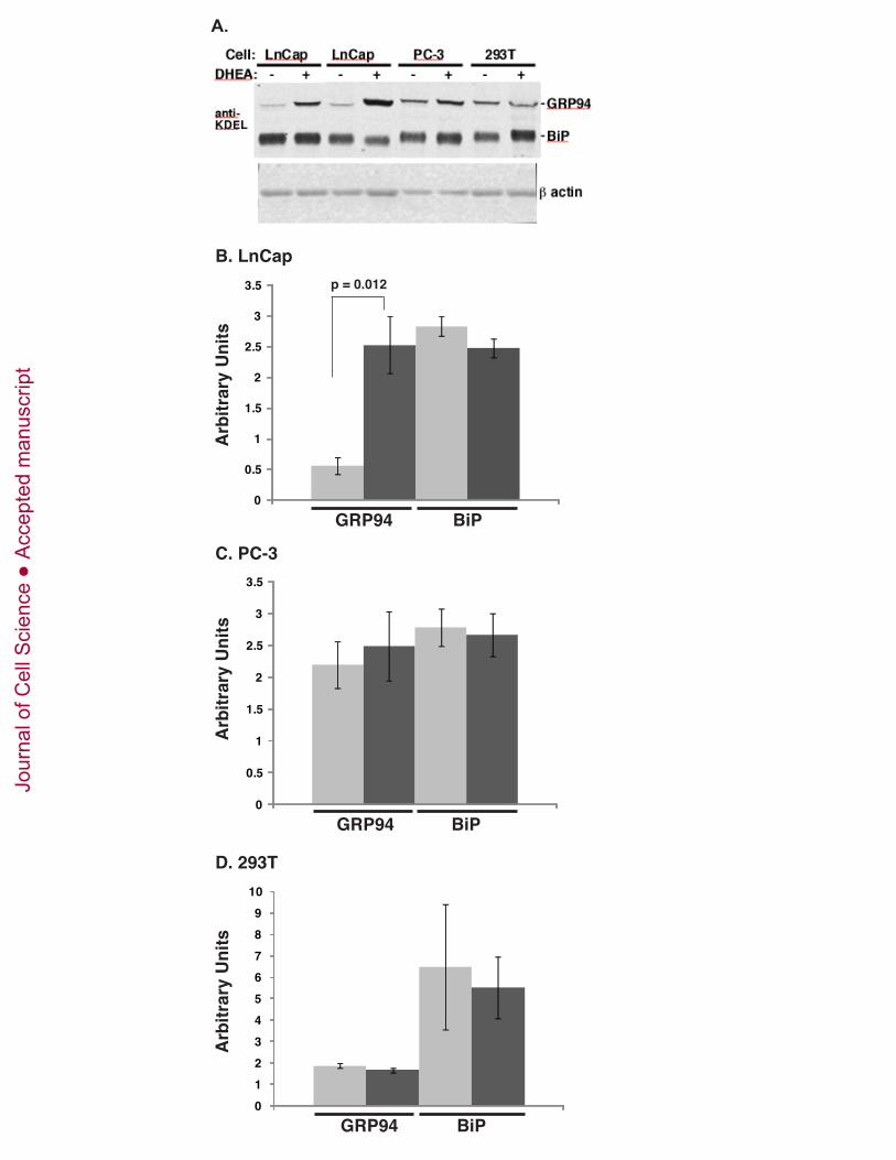
0
0.5
1
1.5
2
2.5
3
3.5
GRP94 BiP
Arb
itrar
y U
nits
0123456789
10
Arb
itrar
y U
nits
GRP94 BiP
B. LnCap
0
0.5
1
1.5
2
2.5
3
3.5
GRP94 BiP
Arb
itrar
y U
nits
p = 0.012
C. PC-3
D. 293T
A. Jo
urna
l of C
ell S
cien
ceA
ccep
ted
man
uscr
ipt

B.
14-3-3
BiP0.9 2.2 0.90.4 0.4 2.31.0 1.0
GRP941.1 <0.1 1.23.7 3.4 <0.11.0 1.0
xbp1-/-xbp1 +/+
shRNA: BiP CtrlGRP94BiP CtrlGRP94
A.
CtrlGRP94
1.0 1.0 <0.10.1 1.00.9
shRNA:
atf4 +/+atf4 -/-
14-3-3
BiP
GRP94
1.1 1.0 2.32.5 1.00.8
CtrlGRP94
Eletto Suppl. Fig. 4
C.
shRNA:
MG-132: ++TUN
Full length
nuclear fragment
++++ ++
BiP Ctrl
GRP94 CtrlGRP94BiPJo
urna
l of C
ell S
cien
ceA
ccep
ted
man
uscr
ipt
14-3-3
FLAG-GRP94
GRP94
1.00.20.70.8
WT shRWTshR
shRNA-CTRL shRNA-25
cDNA:
Eletto Suppl. Fig. 5
Jour
nal o
f Cel
l Sci
ence
Acc
epte
d m
anus
crip
t

ICP0 ccaatcggcggccgccagc
GRP94 ccaatcgcaaggagccacg
PDIA6 ccaatgaaaggcggccacg (ERSE-1)
PDIA1 ccaatcggagggcgcggcg
BiP ccaatcggaggcctccacg (ERSE-1)
CRT ccaatcggaggcctccacg (ERSE-2)
GRP170 ccaatccgtcacaaacacg (ERSE-2)
(ERSE-1)ccaatgaaagcttggggcgOS9
Eletto Suppl. Fig. 6
Jour
nal o
f Cel
l Sci
ence
Acc
epte
d m
anus
crip
t

![Roberta Citro arXiv:2006.03026v1 [cond-mat.supr-con] 4 Jun ... · Via Giovanni Paolo II, 132, I-84084 Fisciano (SA), Italy, CNR-SPIN, Via Giovanni Paolo II, 132, I-84084 Fisciano](https://img.pdfslide.us/doc/110x75/5f582312cb738b5e5629704f/roberta-citro-arxiv200603026v1-cond-matsupr-con-4-jun-via-giovanni-paolo.jpg)