Embed Size (px)
Citation preview
HAL Id: pasteur-00686327https://hal-riip.archives-ouvertes.fr/pasteur-00686327
Submitted on 27 Apr 2013
HAL is a multi-disciplinary open accessarchive for the deposit and dissemination of sci-entific research documents, whether they are pub-lished or not. The documents may come fromteaching and research institutions in France orabroad, or from public or private research centers.
L’archive ouverte pluridisciplinaire HAL, estdestinée au dépôt et à la diffusion de documentsscientifiques de niveau recherche, publiés ou non,émanant des établissements d’enseignement et derecherche français ou étrangers, des laboratoirespublics ou privés.
Phagocyte-specific S100 proteins in the local response tothe Echinococcus granulosus larva.
Tatiana Basika, Natalia Muñoz, Cecilia Casaravilla, Florencia Irigoín, CarlosBatthyány, Mariana Bonilla, Gustavo Salinas, José Pedro Pacheco, Johaness
Roth, Rosario Durán, et al.
To cite this version:Tatiana Basika, Natalia Muñoz, Cecilia Casaravilla, Florencia Irigoín, Carlos Batthyány, et al..Phagocyte-specific S100 proteins in the local response to the Echinococcus granulosus larva.. Para-sitology, Cambridge University Press (CUP), 2012, 139 (2), pp.271-83. �10.1017/S003118201100179X�.�pasteur-00686327�

Phagocyte-specific S100 proteins in the local response to theEchinococcus granulosus larva
TATIANA BASIKA1†, NATALIA MUÑOZ1‡, CECILIA CASARAVILLA1,FLORENCIA IRIGOÍN1§, CARLOS BATTHYÁNY2, MARIANA BONILLA1,GUSTAVO SALINAS1, JOSÉ PEDRO PACHECO3, JOHANESS ROTH4,ROSARIO DURÁN2 and ALVARO DÍAZ1*1Cátedra de Inmunología, Departamento de Biociencias (Facultad de Química) e Instituto de Química Biológica(Facultad de Ciencias), Universidad de la República, Montevideo, Uruguay2Unidad de Bioquímica y Proteómica Analíticas, Instituto Pasteur de Montevideo, Instituto de Investigaciones BiológicasClemente Estable, Montevideo, Uruguay3Departamento de Patología, Facultad de Veterinaria, Universidad de la República, Montevideo, Uruguay4Department of Dermatology, University of Münster, Münster, Germany
(Received 8 April 2011; revised 16 August 2011; accepted 6 September 2011; first published online 5 January 2012)
SUMMARY
Infection by larval Echinococcus granulosus is usually characterized by tight inflammatory control. However, various degreesof chronic granulomatous inflammation are also observed, reaching a high point in infection of cattle by the most prevalentparasite strain worldwide, which is not well adapted to this host species. In this context, epithelioid andmultinucleated giantmacrophages surround the parasite, and the secreted products of these cells often associate with the larval wall. Thephagocyte-specific S100 proteins, S100A8, S100A9 and S100A12, are important non-conventionally secreted amplifiers ofinflammatory responses. We have analysed by proteomics and immunohistochemistry the presence of these proteins at theE. granulosus larva-host interface. We found that, in the context of inflammatory control as observed in human infections,the S100 proteins are not abundant, but S100A9 and S100A8 can be expressed by eosinophils distal to the parasite. In thegranulomatous inflammation context as observed in cattle infections, we found that S100A12 is one of the most abundanthost-derived, parasite-associated proteins, while S100A9 and S100A8 are not present at similarly high levels. As expected,S100A12 derives mostly from the epithelioid and multinucleated giant cells. S100A12, as well as cathepsin K and matrixmetalloproteinase-9, also expressed by E. granulosus-elicited epithelioid cells, are connected to the Th17 arm of immunity,which may therefore be involved in this granulomatous response.
Key words: Echinococcus granulosus, S100 proteins, granuloma, cathepsin K, metalloproteinase-9.
INTRODUCTION
The larval stage (metacestode) of the taeniid cestodeEchinococcus granulosus causes cystic echinococcosis,also called hydatid disease, in a variety of livestockspecies as well as in humans (reviewed by Thompson(1995)). The E. granulosus metacestode is a bladder-like structure (hydatid) that dwells in the paren-chymas of internal organs, most commonly liver andlungs, and can reach up to tens of cm in diameter.
The hydatid is defined by the hydatid wall (HW),a structure comprising a thin inner layer of cells(germinal layer, GL) and themassive outer laminatedlayer (LL). The LL (reviewed by Díaz et al.(2011a,b)) is a peculiar extracellular structure formedby a meshwork of mucins bearing galactose-richglycans. Additionally, in E. granulosus but not otherspecies of the genus, it contains nano-deposits ofcalcium inositol hexakisphosphate (InsP6) (Irigoínet al. 2004). Being up to 3mm thick, and permeableto macromolecules, the LL represents a very largearea for the adsorption of diffusible proteins (Coltortiand Varela-Díaz, 1974; Casaravilla et al. 2006).Whereas establishing larval E. granulosus elicits
local inflammatory responses, these responsesnormally resolve upon parasite deployment of theLL. Thus established hydatids normally growsurrounded by a non-infiltrated or minimallyinfiltrated host-derived collagen capsule (reviewedby Díaz et al. 2011a). This is readily seen in bothsheep and human infections (Yamashita et al. 1961;Mufarrij et al. 1990). Although certainly a key aspectof this parasite’s survival strategy, inflammatory
* Corresponding author: Cátedra de Inmunología,Instituto de Higiene, Avenida Alfredo Navarro 3051,Montevideo, CP 11600, Uruguay. Tel/Fax:+59824874320. E-mail: [email protected].† Current address: Unidad de Biología Parasitaria,Instituto de Higiene y Facultad de Ciencias, Universidadde la República, Montevideo, Uruguay.‡ Current address: Departamento de Produccion yDesarrollo Biotecnológico, Instituto de Higiene, Facultadde Medicina, Universidad de la República, Montevideo,Uruguay.§ Current address: Departamento de Histología yEmbriología, Facultad de Medicina, Universidad de laRepública, Montevideo, Uruguay.
271
Parasitology (2012), 139, 271–283. © Cambridge University Press 2012doi:10.1017/S003118201100179X
resolution does not always take place. Chronicinflammation can often be seen, even in host species(sheep, humans) considered suitable for the G1 strainof the parasite, the most prevalent worldwide(Jenkins et al. 2005). The prototypical case of lackof inflammatory control is infection of cattle by thisparasite strain. As a result of an undetermined host-parasite mismatch, chronic local inflammation is therule, and parasite vitality is accordingly compromised(Rao and Mohiyuddin, 1974; Bortoletti and Ferretti,1978; Sakamoto and Cabrera, 2003; Díaz et al.2011a). Intermediate and variable degrees of inflam-mation are observed in pig infections (Slais andVanek, 1980).
When present, the chronic inflammatory responseto E. granulosus is typically granulomatous. A layerof palisading epithelioid and multinucleated giantmacrophages is directly apposed to the parasite’s LL(Nieberle and Cohrs, 1967; Slais and Vanek, 1980).Behind this first layer is a mononuclear cell infiltrate,featuring lymphocytes, plasmocytes, conventionalmacrophages and some eosinophils. More externallyand/or intermixed with the mononuclear cell infil-trate is a layer of fibroblasts and collagen. A similargranulomatous response is (invariably) elicited by thehighly invasive larval stage of Echinococcus multi-locularis. In this context it has been demonstratedthat the granuloma is T-cell dependent, and that is itdamaging to the parasite (Gottstein and Hemphill,1997; Dai et al. 2004).
The large capacity of the parasite’s LL to adsorbproteins implies that the secreted products of thelocal host reaction tend to accumulate in the HW.Additional host proteins in HW extracts can derivefrom remnants of epithelioid cells that adhere tightlyto the LL. By far the major parasite-derivedmacromolecules inHWextracts are the LL structuralmucins (since the LL is quantitatively very dominantover the GL). As these mucins are highly insoluble,conventionally prepared HW extracts are dominatedby host proteins (Casaravilla and Díaz, 2010; Díazet al. 2011a). WhenHWderive from a non-resolutive(granulomatous) context, these proteins includeprominently the products of the epithelioid andmultinucleated giant cells (MGC) (Díaz et al.2000a,b; Marco et al. 2006). This has led to obser-vations of general interest for granuloma biologybeing made initially in the E. granulosus sytem (Díazet al. 2000b).
In this study, we investigated the association ofphagocyte-specific S100 proteins with the granulo-matous response to the E. granulosus metacestode.S100 is a large family of cytosolic calcium-bindingproteins thought to regulate cytoskeletal functionand other calcium-dependent cellular responses.Three S100 proteins, namely S100A8, S100A9 andS100A12 are expressed prominently bymyeloid cells,and therefore referred to as the phagocyte-specificS100 protein subfamily (reviewed by Ehrchen et al.
2009 and Pietzsch andHoppmann, 2009). They formCa2+ and Zn2+-dependent dimers and higher oligo-mers; while S100A9 and S100A8 most commonlyheterodimerize (oligomerize), S100A12 associatesonly with itself. These proteins are actively secretedby a non-conventional mechanism and fulfil extra-cellular functions. The clearest of these is asamplifiers of the inflammatory response, i.e. asendogenous danger-associated signals (DAMPs).S100A8 and also the S100A9/S100A8 heterodimerare agonists of TLR4, and this interaction has astrong impact in inflammatory disorders (Vogl et al.2007; Loser et al. 2010). S100A12 is thought to be anagonist of the receptor for advanced glycation end-products, RAGE (Hofmann et al. 1999). Functionsare not necessarily well conserved across mammalianspecies: S100A12 is absent in rodents, and rodentS100A8 has been proposed to be the functionalhomologue of human S100A12 (Pietzsch andHoppmann, 2009). We reasoned that the analysis ofthe phagocyte-specific S100 proteins across theinflammation-resolution spectrum in hydatid diseasemay contribute a valuable element towards under-standing the regulation of local inflammation in thisinfection.
MATERIALS AND METHODS
Parasite materials
Hydatids from mouse experimental infections wereretrieved 8–12 months after intraperitoneal in-oculation of protoscoleces obtained from naturalbovine infections. Human hydatid surgical samples(fresh and/or paraffin-embedded) were obtainedfrom the Clínica de Cirugía Pediátrica, HospitalPereira-Rossel (Dr G. Giannini), and the Laboratoriode Anatomía Patológica, Hospital Maciel (DrM. Roldán), both in Montevideo, Uruguay. Bovine,sheep and pig hydatid material was from naturallivestock infections in Uruguay. For the bovine host,a panel of paired fresh (for protein extracts) andparaffin-embedded (for inflammatory scoring andimmunohistochemistry) samples was set up.
Hydatid wall protein extracts
Hydatid walls (HW; comprising LL and GL) wereretrieved from fresh samples as described by (Irigoínet al. 2002). HW were washed with PBS containing0·5 mM CaCl2 to remove loosely bound proteins.They were then extracted using PBS containing thecalcium chelators EGTA or EDTA (in excess ofthe molar amount needed to solubilize the calciumInsP6 deposits (Díaz et al. 2011a), with 2 M NaCl,or sequentially with both agents, as indicated. Theprotease inhibitors PMSF (2mM), iodoacetamide(2 mM), pepstatin A (2 μg/ml), and E-64 (100 μM)were added in each step. Extracts were concentrated
272Tatiana Basika and others

prior to analysis by precipitation with 10% (w/v)trichloroacetic acid.
Antibodies
Rabbit polyclonal antibodies against human andmouse S100A9 and S100A8, and human S100A12were raised as previously described (Zwadlo et al.1988; Roth et al. 1993; Vogl et al. 1999a). Amonoclonal antibody against human S100A9 (cloneS36·48) was purchased in biotinylated form fromBMA Biomedicals AG (Switzerland); this antibodyworks in paraffin-embedded sections but does notreact with the denatured protein in Western blotting.A rabbit polyclonal antibody was raised againstbovine S100A12 recombinantly expressed in E. coli.Total RNA was obtained from bovine peripheralblood leukocytes (buffy coat), reverse-transcribedusing an oligodT primer, and the coding sequencefor S100A12 amplified by nested PCR usingthe primers: tctcctgaaggtgaacgtagt (outer forward),cggatccatgactaagctggaagat (inner forward), cgtcga-caagcttctactctttgtggatatct (inner reverse) and cgtcga-ccgggtaaggcagcctcaggg (outer reverse). The ampliconwas ligated into pGEM-T Easy (Promega), and theinsert then excised using BamHI and SalI andintroduced into the pET28a vector (Novagen) forexpression in E. coli. The fusion protein was purifiedby nickel affinity chromatography from the solublefraction of bacterial lysates. The recombinant proteinwas also bound to CNBr-activated Sepharose(Sigma) for affinity purification of the rabbit anti-bodies obtained.
SDS-PAGE and Western blotting
HW extracts were run under reducing conditions,either in conventional SDS-PAGE (10% (w/v)acrylamide) or in the Tris-tricine system (16·5%(w/v) acrylamide), and Coomassie blue- or silver-stained as indicated. Alternatively, proteins weretransferred to nitrocellulose membranes for Westernblotting. Membranes were probed with the specificrabbit antisera, followed by alkaline phosphatase goatanti-rabbit IgG (Calbiochem), and developed withnitro-blue tetrazolium/ 5-bromo 4-chloro 3-indolylphosphate (NBT/BCIP; Sigma) substrate. Controlmembranes in which normal rabbit serum was usedin the first probing step gave no staining.
One dimensional SDS-PAGE andMALDI-TOF-based proteomics
Selected Coomassie blue-stained protein bands weredigestedwith sequencing-grade trypsin, and peptidesanalysed by MS and MS/MS in the AppliedBiosystems 4800 Analyzer. Proteins were thenidentified by searching the NCBI nr database (2010)
using theMASCOTprogram in the ‘sequence query’mode. The following search parameters were em-ployed: monoisotopic mass tolerance 0·08–0·10Da;fragment mass tolerance 0·2–0·6 Da; methionineoxidation, and in cases also propionamide additionto cysteine residues and/or acetylated proteinN-terminus, as variable modifications; 1 missedtryptic cleavage allowed. Significant peptide and/orprotein scores (P<0·05) were used as criteria forpositive protein identification. In initial experimentsusing equipment without MS/MS capabilities(Voyager DE-Pro also from Applied Biosystems), atryptic peptide fingerprinting approach was taken,leading to presumptive identifications that were laterconfirmed by MS/MS as above and/or by Westernblotting using specific antibodies.
Nano-LC-MS-based proteomics
HW 2M NaCl extracts were dialysed using 3500 kDacut-off membranes against 10mM ammonium bi-carbonate, 0·5 mM EDTA and protein content inthem measured by absorbance at 280 nm. Proteins inthe extracts were reduced, carbamidomethylated, anddigested with sequencing-grade trypsin (1:10 enzymeto total protein ratio, 24 h at 37 °C) in the presenceof 1 M guanidine hydrochloride. Samples were theninjected into a nano-HPLC system (Proxeon easy-nLC, Thermo Scientific) fitted with a reverse-phasecolumn (easy C18 column, 3 μm; 75 μm ID×10 cm;Proxeon, Thermo Scientific) and separated using a0·1% (v/v) formic acid in water – 0·1% (v/v) formicacid in acetonitrile gradient (0–60% acetonitrile in60min; flow 400 nl/min). Online MS detection/analysis was carried out in the LTQ Velos nano-ESI-linear ion trap instrument (Thermo Scientific)in the data-dependent triple play MS2 mode(full scan followed by zoom scan and MS/MS ofthe top 5 peaks in each segment). Proteins wereidentified by searching the IPI database (bovine,2010) using the following parameters in theMascot software in the MS/MS ion search mode:peptide tolerance 400 ppm, MS/MS tolerance0·8 Da, and cysteine carbamidomethylation, proteinN-terminus acetylation, methionine oxidation andasparagine/glutamine deamidation as these allowedvariable modifications. The significance limit forprotein identification was set at P<0·05.
Immunohistochemistry
Microtome sections (0·5 mm thick) were dewaxed,treated with proteinase K as an antigen retrievalprocedure and with 1% H2O2 in methanol to inhibitendogenous peroxidase activity, and blocked using10% (v/v) goat serum. Bovine S100A12 was detectedusing affinity-purified rabbit antibodies; the purifi-cation flow-through was used as negative control, and
273S100 proteins in the host response to larval E. granulosus

gave no staining. Human S100A9 was detected withthe biotinylated S36·48 monoclonal antibody fol-lowed by streptavidin-peroxidase (Sigma). HumanS100A8 was detected with the rabbit polyclonalantiserum; normal rabbit serum used at the sameconcentration as control gave no staining. Sectionsprobed with the rabbit primary antibodies were thenincubated with peroxidase-conjugated goat IgGagainst rabbit IgG (Calbiochem). All sections werefinally developed using diaminobenzidine substrateand counterstained with Mayer’s haematoxylin. Inparallel, sections from each sample were stained onlywith haematoxylin-eosin and scored for inflam-matory status on an arbitrary scale from ‘− ’ (noinflammation, only collagen present) to ‘+++++ ’
(intense granulomatous inflammation featuring a fullrim of epithelioid cells surrounding the parasite).
RESULTS
Phagocyte-specific S100 proteins are associated withthe E. granulosus hydatid in different host species
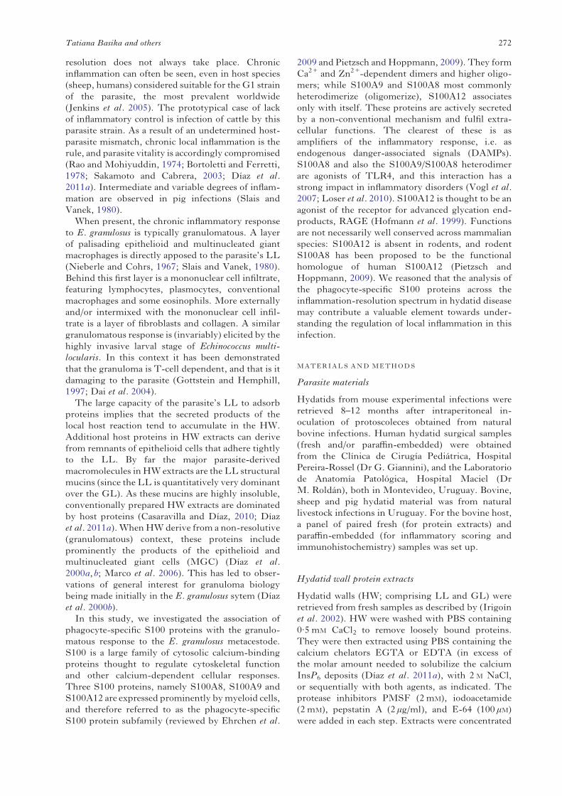
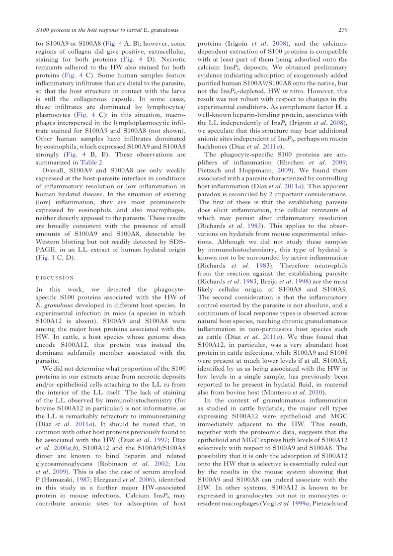
Treating E. granulosus HW with calcium chelatorsdissolves the calcium InsP6 deposits (Irigoín et al.2002, 2004). Being initially interested in proteinsassociated with these deposits, we prepared extractsusing EGTA- or EDTA-containing buffer fromintact hydatids obtained by experimental infectionof mice. The extracts featured 3 major protein bands(Fig. 1 A). For the bands with apparent molecularmasses 14 and 8 KDa, tryptic peptide fingerprintingsuggested that they corresponded to host-derivedS100A9 and S100A8. This was confirmed byMS/MS (Table S1 online version only) and byWestern blotting (Fig. 1 B). Immunoblotting alsoshowed that the association of mouse S100A8 withthe HW was strictly Ca2+-dependent, while that ofS100A9 was less strictly so. Both proteins could beextracted even in the presence of Ca2+, by high ionicstrength.
Hydatids, developing after intraperitoneal infec-tion ofmice, grow loose in the peritoneal cavity, whilethose arising from natural infections are embeddedwithin organ parenchymas. Also, the spectrum ofinflammatory conditions observed in hydatid diseaseis not reproduced in thismodel, in which resolution isalways observed (Richards et al. 1983; Breijo et al.1998). In addition, as mentioned, rodents do notencode S100A12 in their genomes. We thereforeanalysed the presence of host S100 proteins inparasite samples from natural infections in cattle,pig, sheep, and humans. A limited proteomic screen-ing was carried out, using one-dimensional gelsand focusing on the apparent molecular mass ofthe monomeric S100 proteins (6–14 kDa) and ofthe S100A9/S100A8 dimer (24 kDa), which forunknown reasons can run as such even afterdenaturation and reduction (see for example Fig. 1
D, left-hand panel). This analysis showed thatprominent 8 kDa bands in the extracts studied fromcattle, pig and sheep origins corresponded toS100A12 (Fig. 1 C). Part of the S100A12 moleculesfrom all 3 species appeared to be N-terminallyacetylated (Tables S1 and S2, online version only),a modification previously observed for humanS100A9 but not reported for S100A12 proteins(Ilg et al. 1996; Vogl et al. 1999b; McMorran et al.2007). S100A12 was undetectable both by proteomicmethods and by more sensitive Western blotting inthe human sample analysed which, however, didcontain immunochemically detectable S100A9 andS100A8 (Fig. 1 D). An immunochemical assessmentof the presence of S100A9 and S100A8 in the non-human samples was precluded by the lack of suitableantibodies. In sum, the initial analysis suggested thatassociation of host phagocyte-specific S100 proteinswith the HW may be a general phenomenon butdifferences probably exist in terms of individualS100 proteins across different host species and/orinflammatory status of individual hydatids.
Host proteins unrelated to S100 that wereidentified in the limited proteomic studies describedabove included the pentraxin-family acute-phaseproteins serum amyloid P (in mouse-derivedsamples; Fig. 1A; GeneBank Accession no. P12246)and C-reactive protein (in sheep-derived samples;Fig. 1C; GeneBank Accession no. EE780797,identified by TBLASTN search of mammalianESTs). Non-S100 host proteins identified in thebovine sample will be discussed in the context ofsimilar findings by nano-LC-MS-based proteomicsdescribed in the next section.
S100A12 is consistently associated with theE. granulosus larva in the bovine host
The cross-host species studies described in theprevious section were hampered by the limitednumber of samples available and the lack of ahistological assessment of the local inflammatorystatus for each sample.We therefore chose to focus on(readily available to us) bovine-derived samples,scoring individual samples for local inflammatorystatus. Although complete inflammatory resolutionwas never found, cattle hydatid samples displayed awide range of intensities in inflammation, thusallowing the assessment of S100 proteins acrossdifferent biological conditions.
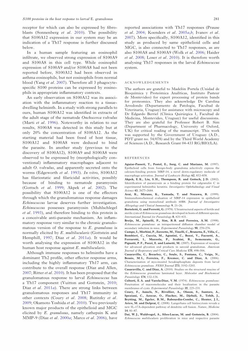
In an analysis based on 1-D SDS-PAGE, aprominent 8 kDa band was identified proteomicallyas S100A12 in each of 9 independent samples studied(Fig. 2 and Table 2). The presence of S100A12 acrossall bovine-derived hydatid samples was confirmedby Western blotting (Fig. 2 and data not shown).S100A12 appeared to be more abundant in extractsfrom samples with middle to high inflammation
274Tatiana Basika and others
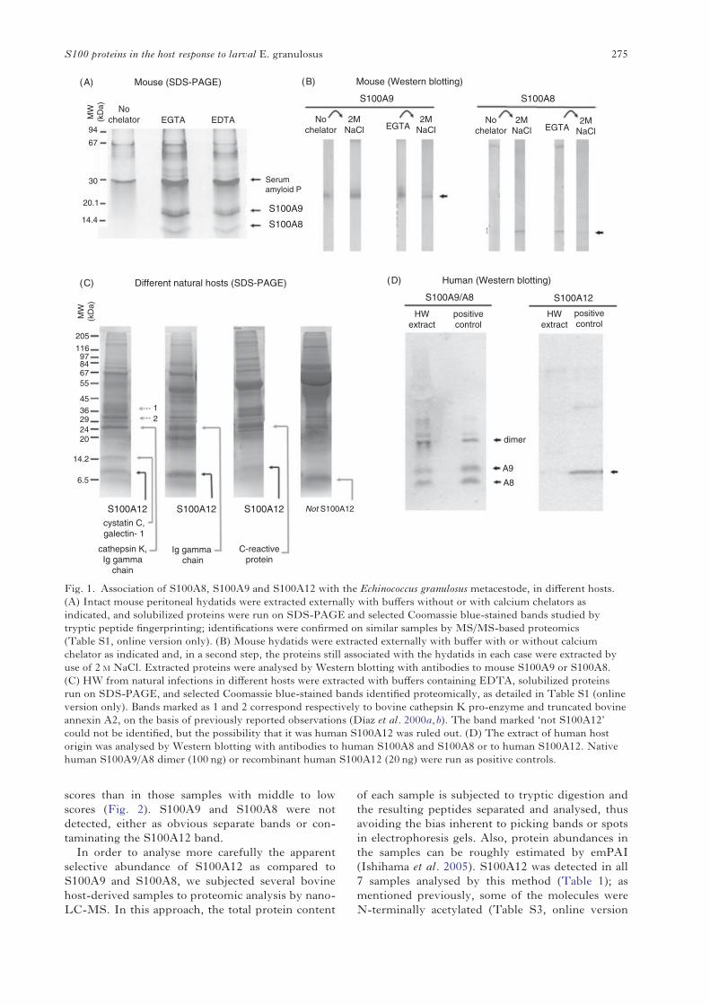
scores than in those samples with middle to lowscores (Fig. 2). S100A9 and S100A8 were notdetected, either as obvious separate bands or con-taminating the S100A12 band.In order to analyse more carefully the apparent
selective abundance of S100A12 as compared toS100A9 and S100A8, we subjected several bovinehost-derived samples to proteomic analysis by nano-LC-MS. In this approach, the total protein content
of each sample is subjected to tryptic digestion andthe resulting peptides separated and analysed, thusavoiding the bias inherent to picking bands or spotsin electrophoresis gels. Also, protein abundances inthe samples can be roughly estimated by emPAI(Ishihama et al. 2005). S100A12 was detected in all7 samples analysed by this method (Table 1); asmentioned previously, some of the molecules wereN-terminally acetylated (Table S3, online version
MW
(kD
a)
MW
(kD
a)
205
11697846755
4536 1
2292420
14.2
6.5
94
67
30
20.1
14.4
Serum amyloid P
S100A9
S100A8
S100A9 S100A8
S100A9/A8 S100A12
Nochelator
Nochelator 2M
NaClNo
chelator2M
NaCl2M
NaClEGTAEGTA EDTA
S100A12 S100A12
Ig gamma chain
S100A12cystatin C,galectin- 1
cathepsin K,Ig gamma
chain
C-reactiveprotein
Not S100A12
HWextract
HWextract
positivecontrol
positivecontrol
dimer
A9
A8
2MNaClEGTA
(A) Mouse (SDS-PAGE) (B) Mouse (Western blotting)
(C) Different natural hosts (SDS-PAGE) (D) Human (Western blotting)
Fig. 1. Association of S100A8, S100A9 and S100A12 with the Echinococcus granulosus metacestode, in different hosts.(A) Intact mouse peritoneal hydatids were extracted externally with buffers without or with calcium chelators asindicated, and solubilized proteins were run on SDS-PAGE and selected Coomassie blue-stained bands studied bytryptic peptide fingerprinting; identifications were confirmed on similar samples by MS/MS-based proteomics(Table S1, online version only). (B) Mouse hydatids were extracted externally with buffer with or without calciumchelator as indicated and, in a second step, the proteins still associated with the hydatids in each case were extracted byuse of 2 M NaCl. Extracted proteins were analysed by Western blotting with antibodies to mouse S100A9 or S100A8.(C) HW from natural infections in different hosts were extracted with buffers containing EDTA, solubilized proteinsrun on SDS-PAGE, and selected Coomassie blue-stained bands identified proteomically, as detailed in Table S1 (onlineversion only). Bands marked as 1 and 2 correspond respectively to bovine cathepsin K pro-enzyme and truncated bovineannexin A2, on the basis of previously reported observations (Díaz et al. 2000a,b). The band marked ‘not S100A12’could not be identified, but the possibility that it was human S100A12 was ruled out. (D) The extract of human hostorigin was analysed by Western blotting with antibodies to human S100A8 and S100A8 or to human S100A12. Nativehuman S100A9/A8 dimer (100 ng) or recombinant human S100A12 (20 ng) were run as positive controls.
275S100 proteins in the host response to larval E. granulosus
only). The estimated abundance of S100A12 rangedbetween 4 and 27% of total host protein, and againsamples with lower inflammatory scores tended todisplay lower relative abundances of S100A12 thanthose with higher scores. In relation to total tissue drymass (of which most corresponds to the LL mucins(Díaz et al. 2011b)), the emPAI data and total proteincontents of extracts allowed us to estimate thatS100A12 is present in the range of 0·1–1mg per gof tissue dry mass. S100A9 was not detected in any ofthe samples, while S100A8 was detected in a singlesample, with an emPAI that was 18-fold lower thanthat of S100A12 (Table 1). The emPAI method isbased on the ratio between experimentally observedsignals and theoretically observable peptides for eachprotein (Ishihama et al. 2005). The Mascot softwareestimates this second figure on the basis of proteinmolecular mass. As the molecular masses of bovine
S100A8 and S100A9 are similar to and larger thanthose of S10012 respectively, the number of ob-servable peptides for S100A8 and S1009 is at least ashigh as that of S100A12. Using the emPAI formulaand the fact that a single peptide can allow significantprotein identification, we estimate that the abun-dances of S100A9 and S100A8 in the bovine-originsamples must be at least an order of magnitude lowerthan that of S100A12. In sum, in the host species inwhich local inflammation against the parasite isstrongest and most maintained, S100A12 is a majorproduct at the host-parasite interface, while S100A8and S100A9 are not present in similarly highamounts.
Non-S100 host proteins detected in theLC-MS-based proteomics search across over halfthe individual samples analysed (and in cases also inthe SDS-PAGE-based experiments shown in Fig. 1)are listed in Table S4, online version only.These include 2 proteins previously known to beabundant in this system (Díaz et al. 2000a,b), namelythe cysteine protease cathepsin K and the cortical(non-conventionally secreted) protein annexin A2.Abundant host proteins newly identified includedcystatin C, an inhibitor of papain-family cysteineproteinases including cathepsin K (reviewed by Turket al. 2008), and regakine-1, a CC chemokine presentat high concentrations in bovine plasma (Struyfet al. 2001). They also included galectin-1, a non-conventionally secreted anti-inflammatory mediator(reviewed by Rabinovich and Ilarregui, 2009) pre-viously observed in hydatid fluid of the same hostorigin (Monteiro et al. 2010). Other proteins foundworth mentioning were the calcification inhibitorα-2-HS-glycoprotein (reviewed by Lee et al. 2009)and the cytotoxic T cell andNK cell cytolytic proteingranulysin (reviewed by Krensky and Clayberger,2009). The proteins mentioned, including S100A12,can be considered representative of the subset ofabundant (bovine) host-derived LL-associated pro-teins that can be solubilized by high ionic strengthand/or EDTA. Other abundant host proteins exist inthis system that require more drastic treatments forextraction, namely immunoglobulins and terminalcomplement components (our unpublished results).
Epithelioid cells and MGC from the host granulomaadjacent to the parasite are the main source ofS100A12 in the bovine host
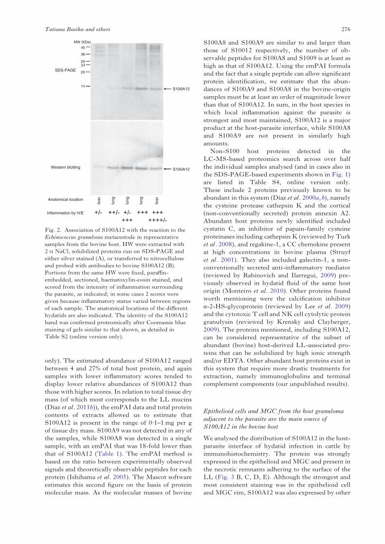
We analysed the distribution of S100A12 in the host-parasite interface of hydatid infection in cattle byimmunohistochemistry. The protein was stronglyexpressed in the epithelioid andMGC and present inthe necrotic remnants adhering to the surface of theLL (Fig. 3 B, C, D, E). Although the strongest andmost consistent staining was in the epithelioid celland MGC rim, S100A12 was also expressed by other
Inflammation by H/E
Anatomical location
Western blotting
lung
lung
lung
liver
liver
45
2924
20
14
36
M
S100A12
S100A12
SDS-PAGE
W (KDa)
Fig. 2. Association of S100A12 with the reaction to theEchinococcus granulosus metacestode in representativesamples from the bovine host. HW were extracted with2 M NaCl, solubilized proteins run on SDS-PAGE andeither silver stained (A), or transferred to nitrocelluloseand probed with antibodies to bovine S100A12 (B).Portions from the same HW were fixed, paraffin-embedded, sectioned, haematoxylin-eosin stained, andscored from the intensity of inflammation surroundingthe parasite, as indicated; in some cases 2 scores weregiven because inflammatory status varied between regionsof each sample. The anatomical locations of the differenthydatids are also indicated. The identity of the S100A12band was confirmed proteomically after Coomassie bluestaining of gels similar to that shown, as detailed inTable S2 (online version only).
276Tatiana Basika and others

cells of the host reaction, namely morphologicallyconventional macrophages present in the mono-nuclear cell infiltrate (Fig. 3 A, C, F), and somefibroblasts (Fig. 3 A, C, G). S100A12 was alsoobserved to be expressed by alveolar macrophages,but this appeared to be independent of the presenceof the parasite as it was also observed in samples takenfrom tens of cm from the infection site (not shown).As expected, when the local inflammatory reactionwas weak, S100A12 staining was also weak, restrictedto some macrophages infiltrating the collagenouscapsule (Fig. 3 A). As for the parasite structures, wedid not find clear-cut evidence of the presence ofS100A12 within the LL itself, but since it is generallyvery difficult to stain proteins within the LLin immunohistochemistry (Stadelmann et al. 2010;Díaz et al. 2011a), we do not take this as evidenceagainst the protein being also present in the interiorof the LL. In contrast, the S100A12 antibodies didstain the GL (not shown). The GL is known to take
up host proteins, as observed by immunohistochem-istry and by proteomics (Díaz et al. 2000a; Monteiroet al. 2010). The immunohistochemical findings onS100A12 are summarized in Table 2. The strongexpression of S100A12 by the epithelioid cells andMGC directly apposed to the HW explains theconsistent presence of this protein in the extractsfrom bovine-origin HW samples (Fig. 2 and Table 1).
Eosinophils distal to the parasite can express S100A9and S100A8 in the human host
Human hydatid samples are, in most cases, charac-terized by the lack of inflammation, in particular inthe tissue directly apposed to the parasite. Thus, thistype of sample could complement the bovine hydatidsamples, which are characterized by continuinginflammation. The non-infiltrated collagenous cap-sule of human hydatid samples generally did not stain
Table 1. Semi-quantitation of S100 proteins in a panel of bovine host samples by LC-MS and emPAI
(The emPAI of S100 proteins detected in each sample was normalized by the added emPAI of all host proteins identified(at the P<0·05 significance level) in the sample, thus giving an indication of relative abundance with respect to total hostprotein. S100A9 was not detected in any of the samples tested. ND stands for not determined.)
Inflammatoryscore of sample
Anatomicalorigin of sample
S100A12 (% oftotal emPAI)
S100A8 (% oftotal emPAI)
+/− Liver 4·0 Not detectedND Lung 6·5 0·35ND Lung 6·8 Not detected++/− Lung 6·8 Not detectedND Lung 11·9 Not detected+/− ; +++ Lung 22·1 Not detected+++ ; ++++/− Liver 26·8 Not detected
Table 2. Summary of immunohistochemical findings
(H, L and N stand for high, low and no immunoreactivity, respectively.)
Bovine Structure/cell type S100A12
HW (GL) LHW (LL) NEpithelioid cells HNecrotic area HFibroblasts H/NMacrophages (in macrophage-dominated infiltrate) HMacrophages (in predominantly lymphoplasmocytic infiltrate) HLymphocytes and plasmocytes N
Human Structure/cell type S100A8 S100A9
HW (GL) H HHW (LL) N NNecrotic area H HFibroblasts L LMacrophages (in predominantly lymphoplasmocytic infiltrate) H HLymphocytes and plasmocytes N NEosinophils H H
277S100 proteins in the host response to larval E. granulosus
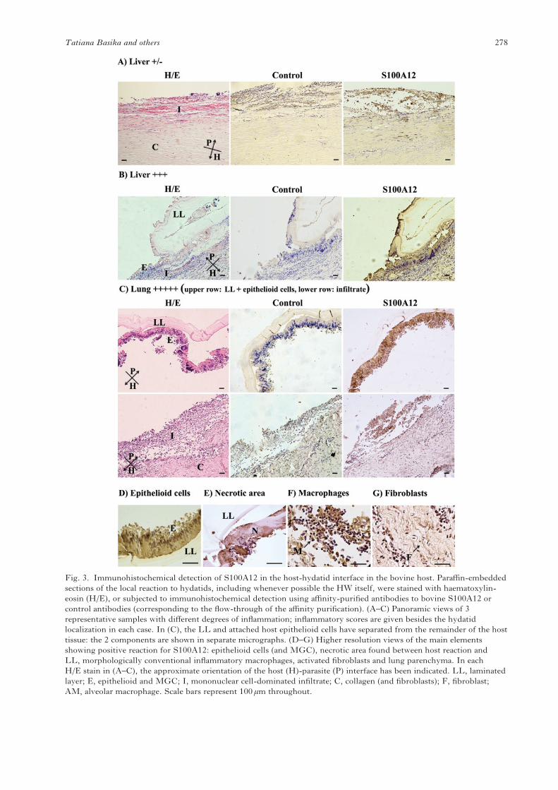
Fig. 3. Immunohistochemical detection of S100A12 in the host-hydatid interface in the bovine host. Paraffin-embeddedsections of the local reaction to hydatids, including whenever possible the HW itself, were stained with haematoxylin-eosin (H/E), or subjected to immunohistochemical detection using affinity-purified antibodies to bovine S100A12 orcontrol antibodies (corresponding to the flow-through of the affinity purification). (A–C) Panoramic views of 3representative samples with different degrees of inflammation; inflammatory scores are given besides the hydatidlocalization in each case. In (C), the LL and attached host epithelioid cells have separated from the remainder of the hosttissue: the 2 components are shown in separate micrographs. (D–G) Higher resolution views of the main elementsshowing positive reaction for S100A12: epithelioid cells (and MGC), necrotic area found between host reaction andLL, morphologically conventional inflammatory macrophages, activated fibroblasts and lung parenchyma. In eachH/E stain in (A–C), the approximate orientation of the host (H)-parasite (P) interface has been indicated. LL, laminatedlayer; E, epithelioid and MGC; I, mononuclear cell-dominated infiltrate; C, collagen (and fibroblasts); F, fibroblast;AM, alveolar macrophage. Scale bars represent 100 μm throughout.
278Tatiana Basika and others
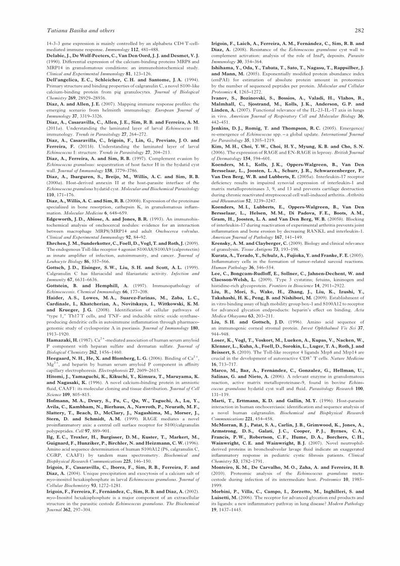
for S100A9 or S100A8 (Fig. 4 A, B); however, someregions of collagen did give positive, extracellular,staining for both proteins (Fig. 4 D). Necroticremnants adhered to the HW also stained for bothproteins (Fig. 4 C). Some human samples featureinflammatory infiltrates that are distal to the parasite,so that the host structure in contact with the larvais still the collagenous capsule. In some cases,these infiltrates are dominated by lymphocytes/plasmocytes (Fig. 4 C); in this situation, macro-phages interspersed in the lymphoplasmocytic infil-trate stained for S100A9 and S100A8 (not shown).Other human samples have infiltrates dominatedby eosinophils, which expressed S100A9 and S100A8strongly (Fig. 4 B, E). These observations aresummarized in Table 2.Overall, S100A9 and S100A8 are only weakly
expressed at the host-parasite interface in conditionsof inflammatory resolution or low inflammation inhuman hydatid disease. In the situation of existing(low) inflammation, they are most prominentlyexpressed by eosinophils, and also macrophages,neither directly apposed to the parasite. These resultsare broadly consistent with the presence of smallamounts of S100A9 and S100A8, detectable byWestern blotting but not readily detected by SDS-PAGE, in an LL extract of human hydatid origin(Fig. 1 C, D).
DISCUSSION
In this work, we detected the phagocyte-specific S100 proteins associated with the HW ofE. granulosus developed in different host species. Inexperimental infection in mice (a species in whichS100A12 is absent), S100A9 and S100A8 wereamong the major host proteins associated with theHW. In cattle, a host species whose genome doesencode S100A12, this protein was instead thedominant subfamily member associated with theparasite.We did not determine what proportion of the S100
proteins in our extracts arose from necrotic depositsand/or epithelioid cells attaching to the LL vs fromthe interior of the LL itself. The lack of stainingof the LL observed by immunohistochemistry (forbovine S100A12 in particular) is not informative, asthe LL is remarkably refractory to immunostaining(Díaz et al. 2011a). It should be noted that, incommonwith other host proteins previously found tobe associated with the HW (Díaz et al. 1997; Díazet al. 2000a,b), S100A12 and the S100A9/S100A8dimer are known to bind heparin and relatedglycosaminoglycans (Robinson et al. 2002; Liuet al. 2009). This is also the case of serum amyloidP (Hamazaki, 1987; Heegaard et al. 2006), identifiedin this study as a further major HW-associatedprotein in mouse infections. Calcium InsP6 maycontribute anionic sites for adsorption of host
proteins (Irigoín et al. 2008), and the calcium-dependent extraction of S100 proteins is compatiblewith at least part of them being adsorbed onto thecalcium InsP6 deposits. We obtained preliminaryevidence indicating adsorption of exogenously addedpurified human S100A9/S100A8 onto the native, butnot the InsP6-depleted, HW in vitro. However, thisresult was not robust with respect to changes in theexperimental conditions. As complement factor H, awell-known heparin-binding protein, associates withthe LL independently of InsP6 (Irigoín et al. 2008),we speculate that this structure may bear additionalanionic sites independent of InsP6, perhaps onmucinbackbones (Díaz et al. 2011a).The phagocyte-specific S100 proteins are am-
plifiers of inflammation (Ehrchen et al. 2009;Pietzsch and Hoppmann, 2009). We found themassociatedwith a parasite characterized by controllinghost inflammation (Díaz et al. 2011a). This apparentparadox is reconciled by 2 important considerations.The first of these is that the establishing parasitedoes elicit inflammation, the cellular remnants ofwhich may persist after inflammatory resolution(Richards et al. 1983). This applies to the obser-vations on hydatids from mouse experimental infec-tions. Although we did not study these samplesby immunohistochemistry, this type of hydatid isknown not to be surrounded by active inflammation(Richards et al. 1983). Therefore neutrophilsfrom the reaction against the establishing parasite(Richards et al. 1983; Breijo et al. 1998) are the mostlikely cellular origin of S100A8 and S100A9.The second consideration is that the inflammatorycontrol exerted by the parasite is not absolute, and acontinuum of local response types is observed acrossnatural host species, reaching chronic granulomatousinflammation in non-permissive host species suchas cattle (Díaz et al. 2011a). We thus found thatS100A12, in particular, was a very abundant hostprotein in cattle infections, while S100A9 and S1008were present at much lower levels if at all. S100A8,identified by us as being associated with the HW inlow levels in a single sample, has previously beenreported to be present in hydatid fluid, in materialalso from bovine host (Monteiro et al. 2010).In the context of granulomatous inflammation
as studied in cattle hydatids, the major cell typesexpressing S100A12 were epithelioid and MGCimmediately adjacent to the HW. This result,together with the proteomic data, suggests that theepithelioid andMGC express high levels of S100A12selectively with respect to S100A9 and S100A8. Thepossibility that it is only the adsorption of S100A12onto the HW that is selective is essentially ruled outby the results in the mouse system showing thatS100A9 and S100A8 can indeed associate with theHW. In other systems, S100A12 is known to beexpressed in granulocytes but not in monocytes orresidentmacrophages (Vogl et al. 1999a; Pietzsch and
279S100 proteins in the host response to larval E. granulosus

Hoppmann, 2009). S100A12 expression in granulo-ma macrophages, including epithelioid cells andMGC specifically, has been reported, in humansystems, in the last few years (Kim et al. 2006;Morbini et al. 2006; Campo et al. 2007); theexpression of S100A9 and S100A8 was not analysedin these works. Other works have reported widelydifferent observations on the expression of S100A9and S100A8 in these cell types; these observationsinclude co-expression of both, expression of onlyS100A9, and lack of expression of either (Zwadloet al. 1988; Delabie et al. 1990; Aguiar-Passeti et al.1997; Arai et al. 1999; Sunderkotter et al. 2004;Kurata et al. 2005; Terasaki et al. 2007). Also, in ahelminth-induced granuloma model in mice (patent
S. mansoni infection), S100A9 and S100A8 werefound to be expressed by macrophages at the edgeof the granuloma but not by the epithelioid andMGCs at the centre of the reaction (Yang et al. 1997).Our results suggest that comparative analysis of all3 phagocyte-specific S100 proteins in epithelioid andMGCs might show that, at least in some stronglyinflammatory contexts, these cells express high levelsof S100A12 selectively.
We also found S100A12 to be expressed byfibroblasts, although only in the samples displayingthemost intense inflammatory reactions. S100A9 andS100A8 are now known to be expressed in certainnon-haematopoetic cells (keratinocytes) in responseto the Th17 cytokine IL-22 (Wolk et al. 2006), the
Fig. 4. Immunohistochemical detection of S100A9 and S100A8 in the host-hydatid interface in the human host.Paraffin-embedded sections of the local reaction to Echinococcus granulosus metacestodes were stained withhaematoxylin-eosin (H/E), or subjected to immunohistochemical detection using antibodies to human S100A9or S100A8, or control antibodies. (A–C) Panoramic views of 3 representative samples with different degrees ofinflammation; inflammatory scores are given besides the hydatid localization in each case. The control stainsshown correspond to normal rabbit serum. (D and E) Higher resolution views of the main elements showingpositive reaction for the proteins under study, namely the collagenous capsule and an eosinophil infiltraterespectively. In (E) an H/E stain has been included in an inset to show that the infiltrating cells are eosinophils. In eachH/E stain in (A–C), the approximate orientation of the host (H)-parasite (P) interface has been indicated.I, mononuclear cell-dominated infiltrate; C, collagen (and fibroblasts); HW, hydatid wall. Scale bars represent 100 μmthroughout.
280Tatiana Basika and others
receptor for which can also be expressed by fibro-blasts (Sonnenberg et al. 2010). The possibilitythat S100A12 expression in our system may be anindication of a Th17 response is further discussedbelow.In a human sample featuring an eosinophil
infiltrate, we observed strong expression of S100A9and S100A8 in this cell type. While eosinophilexpression of S100A9 and/or S100A8 had not beenreported before, S100A12 had been observed inasthma eosinophils, but not eosinophils from normalblood (Yang et al. 2007). Therefore all 3 phagocyte-specific S100 proteins can be expressed by eosino-phils in appropriate inflammatory contexts.An early observation on S100A12 was its associ-
ation with the inflammatory reaction to a tissue-dwelling helminth. In a study with strong parallels toours, human S100A12 was identified in extracts ofthe adult stage of the nematode Onchocerca volvulus(Marti et al. 1996). Noteworthy in relation to ourresults, S100A8 was detected in this study but atonly 20% the concentration of S100A12. As thestarting material had been freed of host tissue,S100A12 and S100A8 were deduced to bindthe parasite. In another study (previous to thediscovery of S100A12), S100A9 and S100A8 wereobserved to be expressed by (morphologically con-ventional) inflammatory macrophages adjacent toadult O. volvulus, and apparently secreted onto theworms (Edgeworth et al. 1993). In vitro, S100A12has filariostatic and filaricidal activities, possiblybecause of binding to nematode paramyosin(Gottsch et al. 1999; Akpek et al. 2002). Thepossibility that S100A12 is one of the effectorsthrough which the granulomatous response damagesEchinococcus larvae deserves further investigation.Echinococcus possesses paramyosin (Muhlschlegelet al. 1993), and therefore binding to this protein isa conceivable anti-parasite mechanism. An inflam-matory response very similar to the chronic granulo-matous version of the response to E. granulosus isnormally elicited by E. multilocularis (Gottstein andHemphill, 1997; Díaz et al. 2011a). It would beworth analysing the expression of S100A12 in thehuman host response against E. multilocularis.Although immune responses to helminths have a
dominant Th2 profile, other effector response arms,including the highly inflammatory Th17 arm, cancontribute to the overall response (Díaz and Allen,2007; Ritter et al. 2010). It has been proposed that thegranulomatous response to larval Echinococcus hasa Th17 component (Vuitton and Gottstein, 2010;Díaz et al. 2011a). There are strong links betweengranulomatous responses and Th17 immunity inother contexts (Coury et al. 2008; Rutitzky et al.2009; Okamoto Yoshida et al. 2010). Two previouslyknown major products of the epithelioid and MGCelicited by E. granulosus, namely cathepsin K andMMP-9 (Díaz et al. 2000a; Marco et al. 2006), have
reported associations with Th17 responses (Prauseet al. 2004; Koenders et al. 2005a,b; Ivanov et al.2007). More specifically, S100A12, identified in thisstudy as produced by same epithelioid cells andMGC, is also connected to Th17 responses, as arealso S100A8 and S100A9 (Wolk et al. 2006; Haideret al. 2008; Loser et al. 2010). It is therefore worthanalysing Th17 responses in the larval Echinococcussystem.
ACKNOWLEDGEMENTS
The authors are grateful to Madelón Portela (Unidad deBioquímica y Proteómica Analíticas, Instituto Pasteurde Montevideo) for expert processing of protein bandsfor proteomics. They also acknowledge Dr CarolinaArredondo (Departamento de Patología, Facultad deVeterinaria, Uruguay) for assistance with microscopy andDr Edgardo Berriel (Clínica Quirúrgica 1, Facultad deMedicina, Montevideo, Uruguay) for useful discussions.They are also grateful for Professor Robert B. Sim(Department of Pharmacology, University of Oxford,UK) for critical reading of the manuscript. This workwas supported by the Government of Uruguay (A.D.,PDT grant no. 54/078) and by the Third World Academyof Sciences (A.D., Research Grant 04-433 RG/BIO/LA).
REFERENCES
Aguiar-Passeti, T., Postol, E., Sorg, C. and Mariano, M. (1997).Epithelioid cells from foreign-body granuloma selectively express thecalcium-binding protein MRP-14, a novel down-regulatory molecule ofmacrophage activation. Journal of Leukocyte Biology 62, 852–858.Akpek, E. K., Liu, S. H., Thompson, R. and Gottsch, J. D. (2002).Identification of paramyosin as a binding protein for calgranulin C inexperimental helminthic keratitis. Investigative Ophthalmology and VisualScience 43, 2677–2684.Arai, K., Mizuno, K., Yamada, T. and Nozawa, R. (1999).Immunohistochemical evaluation of MRP-14 expression in epithelioidgranuloma using monoclonal antibody 60B8. Journal of InvestigativeAllergology and Clinical Immunology 9, 21–26.Bortoletti, G. and Ferretti, G. (1978). Ultrastructural aspects of fertile andsterile cysts ofEchinococcus granulosus developed in hosts of different species.International Journal for Parasitology 8, 421–431.Breijo, M., Spinelli, P., Sim, R. B. and Ferreira, A.M. (1998).Echinococcus granulosus: an intraperitoneal diffusion chamber model ofsecondary infection in mice. Experimental Parasitology 90, 270–276.Campo, I.,Morbini, P., Zorzetto,M., Tinelli, C., Brunetta, E., Villa, C.,Bombieri, C., Cuccia, M., Agostini, C., Bozzi, V., Facoetti, A.,Ferrarotti, I., Mazzola, P., Scabini, R., Semenzato, G.,Pignatti, P. F., Pozzi, E. and Luisetti, M. (2007). Expression of receptorfor advanced glycation end products in sarcoid granulomas. AmericanJournal of Respiratory and Critical Care Medicine 175, 498–506.Casaravilla, C., Brearley, C., Soule, S., Fontana, C., Veiga, N.,Bessio, M. I., Ferreira, F., Kremer, C. and Diaz, A. (2006).Characterization of myo-inositol hexakisphosphate deposits from larvalEchinococcus granulosus. FEBS Journal 273, 3192–3203.Casaravilla, C. and Díaz, A. (2010). Studies on the structural mucins ofthe Echinococcus granulosus laminated layer. Molecular and BiochemicalParasitology 174, 132–136.Coltorti, E. A. and Varela-Díaz, V.M. (1974). Echinococcus granulosus:Penetration of macromolecules and their localization in the parasitemembranes of cysts. Experimental Parasitology 35, 225–231.Coury, F., Annels, N., Rivollier, A., Olsson, S., Santoro, A.,Speziani, C., Azocar, O., Flacher, M., Djebali, S., Tebib, J.,Brytting, M., Egeler, R.M., Rabourdin-Combe, C., Henter, J. I.,Arico, M. and Delprat, C. (2008). Langerhans cell histiocytosis reveals anew IL-17A-dependent pathway of dendritic cell fusion. Nature, Medicine14, 81–87.Dai, W. J., Waldvogel, A., Siles-Lucas, M. and Gottstein, B. (2004).Echinococcus multilocularis proliferation in mice and respective parasite
281S100 proteins in the host response to larval E. granulosus
14-3-3 gene expression is mainly controlled by an alphabeta CD4T-cell-mediated immune response. Immunology 112, 481–488.Delabie, J., DeWolf-Peeters, C., Van Den Oord, J. J. and Desmet, V. J.(1990). Differential expression of the calcium-binding proteins MRP8 andMRP14 in granulomatous conditions: an immunohistochemical study.Clinical and Experimental Immunology 81, 123–126.Dell’angelica, E. C., Schleicher, C. H. and Santome, J. A. (1994).Primary structure and binding properties of calgranulin C, a novel S100-likecalcium-binding protein from pig granulocytes. Journal of BiologicalChemistry 269, 28929–28936.Díaz, A. and Allen, J. E. (2007). Mapping immune response profiles: theemerging scenario from helminth immunology. European Journal ofImmunology 37, 3319–3326.Díaz, A., Casaravilla, C., Allen, J. E., Sim, R. B. and Ferreira, A.M.(2011a). Understanding the laminated layer of larval Echinococcus II:immunology. Trends in Parasitology 27, 264–272.Díaz, A., Casaravilla, C., Irigoín, F., Lin, G., Previato, J. O. andFerreira, F. (2011b). Understanding the laminated layer of larvalEchinococcus I: structure. Trends in Parasitology 27, 204–213.Díaz, A., Ferreira, A. and Sim, R. B. (1997). Complement evasion byEchinococcus granulosus: sequestration of host factor H in the hydatid cystwall. Journal of Immunology 158, 3779–3786.Díaz, A., Ibarguren, S., Breijo, M., Willis, A. C. and Sim, R. B.(2000a). Host-derived annexin II at the host-parasite interface of theEchinococcus granulosus hydatid cyst.Molecular and Biochemical Parasitology110, 171–176.Díaz, A.,Willis, A. C. and Sim,R. B. (2000b). Expression of the proteinasespecialized in bone resorption, cathepsin K, in granulomatous inflam-mation. Molecular Medicine 6, 648–659.Edgeworth, J. D., Abiose, A. and Jones, B. R. (1993). An immunohis-tochemical analysis of onchocercal nodules: evidence for an interactionbetween macrophage MRP8/MRP14 and adult Onchocerca volvulus.Clinical and Experimental Immunology 92, 84–92.Ehrchen, J.M., Sunderkotter, C., Foell, D., Vogl, T. andRoth, J. (2009).The endogenous Toll-like receptor 4 agonist S100A8/S100A9 (calprotectin)as innate amplifier of infection, autoimmunity, and cancer. Journal ofLeukocyte Biology 86, 557–566.Gottsch, J. D., Eisinger, S.W., Liu, S. H. and Scott, A. L. (1999).Calgranulin C has filariacidal and filariastatic activity. Infection andImmunity 67, 6631–6636.Gottstein, B. and Hemphill, A. (1997). Immunopathology ofEchinococcosis. Chemical Immunology 66, 177–208.Haider, A. S., Lowes, M. A., Suarez-Farinas, M., Zaba, L. C.,Cardinale, I., Khatcherian, A., Novitskaya, I., Wittkowski, K.M.and Krueger, J. G. (2008). Identification of cellular pathways of“type 1,” Th17 T cells, and TNF- and inducible nitric oxide synthase-producing dendritic cells in autoimmune inflammation through pharmaco-genomic study of cyclosporine A in psoriasis. Journal of Immunology 180,1913–1920.Hamazaki, H. (1987). Ca2+-mediated association of human serum amyloidP component with heparan sulfate and dermatan sulfate. Journal ofBiological Chemistry 262, 1456–1460.Heegaard, N.H., He, X. and Blomberg, L. G. (2006). Binding of Ca2+,Mg2+, and heparin by human serum amyloid P component in affinitycapillary electrophoresis. Electrophoresis 27, 2609–2615.Hitomi, J., Yamaguchi, K., Kikuchi, Y., Kimura, T., Maruyama, K.and Nagasaki, K. (1996). A novel calcium-binding protein in amnioticfluid, CAAF1: its molecular cloning and tissue distribution. Journal of CellScience 109, 805–815.Hofmann, M. A., Drury, S., Fu, C., Qu, W., Taguchi, A., Lu, Y.,Avila, C., Kambham, N., Bierhaus, A., Nawroth, P., Neurath, M. F.,Slattery, T., Beach, D., McClary, J., Nagashima, M., Morser, J.,Stern, D. and Schmidt, A.M. (1999). RAGE mediates a novelproinflammatory axis: a central cell surface receptor for S100/calgranulinpolypeptides. Cell 97, 889–901.Ilg, E. C., Troxler, H., Burgisser, D.M., Kuster, T., Markert, M.,Guignard, F., Hunziker, P., Birchler, N. andHeizmann, C.W. (1996).Amino acid sequence determination of human S100A12 (P6, calgranulin C,CGRP, CAAF1) by tandem mass spectrometry. Biochemical andBiophysical Research Communications 225, 146–150.Irigoín, F., Casaravilla, C., Iborra, F., Sim, R. B., Ferreira, F. andDíaz, A. (2004). Unique precipitation and exocytosis of a calcium salt ofmyo-inositol hexakisphosphate in larval Echinococcus granulosus. Journal ofCellular Biochemistry 93, 1272–1281.Irigoín, F., Ferreira, F., Fernández, C., Sim, R. B. and Díaz, A. (2002).myo-Inositol hexakisphosphate is a major component of an extracellularstructure in the parasitic cestode Echinococcus granulosus. The BiochemicalJournal 362, 297–304.
Irigoín, F., Laich, A., Ferreira, A.M., Fernández, C., Sim, R. B. andDíaz, A. (2008). Resistance of the Echinococcus granulosus cyst wall tocomplement activation: analysis of the role of InsP6 deposits. ParasiteImmunology 30, 354–364.Ishihama, Y., Oda, Y., Tabata, T., Sato, T., Nagasu, T., Rappsilber, J.and Mann, M. (2005). Exponentially modified protein abundance index(emPAI) for estimation of absolute protein amount in proteomicsby the number of sequenced peptides per protein. Molecular and CellularProteomics 4, 1265–1272.Ivanov, S., Bozinovski, S., Bossios, A., Valadi, H., Vlahos, R.,Malmhall, C., Sjostrand, M., Kolls, J. K., Anderson, G. P. andLinden, A. (2007). Functional relevance of the IL-23-IL-17 axis in lungsin vivo. American Journal of Respiratory Cell and Molecular Biology 36,442–451.Jenkins, D. J., Romig, T. and Thompson, R. C. (2005). Emergence/re-emergence of Echinococcus spp. – a global update. International Journalfor Parasitology 35, 1205–1219.Kim, M.H., Choi, Y.W., Choi, H. Y., Myung, K. B. and Cho, S. N.(2006). The expression of RAGE and EN-RAGE in leprosy.British Journalof Dermatology 154, 594–601.Koenders, M. I., Kolls, J. K., Oppers-Walgreen, B., Van DenBersselaar, L., Joosten, L. A., Schurr, J. R., Schwarzenberger, P.,Van Den Berg, W. B. and Lubberts, E. (2005a). Interleukin-17 receptordeficiency results in impaired synovial expression of interleukin-1 andmatrix metalloproteinases 3, 9, and 13 and prevents cartilage destructionduring chronic reactivated streptococcal cell wall-induced arthritis.Arthritisand Rheumatism 52, 3239–3247.Koenders, M. I., Lubberts, E., Oppers-Walgreen, B., Van DenBersselaar, L., Helsen, M.M., Di Padova, F. E., Boots, A.M.,Gram, H., Joosten, L. A. and Van Den Berg, W. B. (2005b). Blockingof interleukin-17 during reactivation of experimental arthritis prevents jointinflammation and bone erosion by decreasing RANKL and interleukin-1.American Journal of Pathology 167, 141–149.Krensky, A.M. and Clayberger, C. (2009). Biology and clinical relevanceof granulysin. Tissue Antigens 73, 193–198.Kurata, A., Terado, Y., Schulz, A., Fujioka, Y. and Franke, F. E. (2005).Inflammatory cells in the formation of tumor-related sarcoid reactions.Human Pathology 36, 546–554.Lee, C., Bongcam-Rudloff, E., Sollner, C., Jahnen-Dechent, W. andClaesson-Welsh, L. (2009). Type 3 cystatins; fetuins, kininogen andhistidine-rich glycoprotein. Frontiers in Bioscience 14, 2911–2922.Liu, R., Mori, S., Wake, H., Zhang, J., Liu, K., Izushi, Y.,Takahashi, H. K., Peng, B. and Nishibori, M. (2009). Establishment ofin vitro binding assay of highmobility group box-1 and S100A12 to receptorfor advanced glycation endproducts: heparin’s effect on binding. ActaMedica Okayama 63, 203–211.Liu, S. H. and Gottsch, J. D. (1996). Amino acid sequence ofan immunogenic corneal stromal protein. Invest Ophthalmol Vis Sci 37,944–948.Loser, K., Vogl, T., Voskort, M., Lueken, A., Kupas, V., Nacken, W.,Klenner, L., Kuhn, A., Foell, D., Sorokin, L., Luger, T. A., Roth, J. andBeissert, S. (2010). The Toll-like receptor 4 ligands Mrp8 and Mrp14 arecrucial in the development of autoreactive CD8+ T cells. Nature Medicine16, 713–717.Marco, M., Baz, A., Fernández, C., Gonzalez, G., Hellman, U.,Salinas, G. and Nieto, A. (2006). A relevant enzyme in granulomatousreaction, active matrix metalloproteinase-9, found in bovine Echino-coccus granulosus hydatid cyst wall and fluid. Parasitology Research 100,131–139.Marti, T., Erttmann, K. D. and Gallin, M. Y. (1996). Host-parasiteinteraction in human onchocerciasis: identification and sequence analysis ofa novel human calgranulin. Biochemical and Biophysical ResearchCommunications 221, 454–458.McMorran, B. J., Patat, S. A., Carlin, J. B., Grimwood, K., Jones, A.,Armstrong, D. S., Galati, J. C., Cooper, P. J., Byrnes, C. A.,Francis, P.W., Robertson, C. F., Hume, D. A., Borchers, C. H.,Wainwright, C. E. and Wainwright, B. J. (2007). Novel neutrophil-derived proteins in bronchoalveolar lavage fluid indicate an exaggeratedinflammatory response in pediatric cystic fibrosis patients. ClinicalChemistry 53, 1782–1791.Monteiro, K.M., De Carvalho, M. O., Zaha, A. and Ferreira, H. B.(2010). Proteomic analysis of the Echinococcus granulosus meta-cestode during infection of its intermediate host. Proteomics 10, 1985–1999.Morbini, P., Villa, C., Campo, I., Zorzetto, M., Inghilleri, S. andLuisetti, M. (2006). The receptor for advanced glycation end products andits ligands: a new inflammatory pathway in lung disease? Modern Pathology19, 1437–1445.
282Tatiana Basika and others

Mufarrij, A. A., Arnaut, A., Meshefedjian, G. and Matossian, R.M.(1990). Comparative histopathological study in the hepatic and pulmonaryhuman hydatidosis. Helminthologia 27, 279–290.Muhlschlegel, F., Sygulla, L., Frosch, P., Massetti, P. andFrosch,M. (1993). Paramyosin of Echinococcus granulosus: cDNA sequenceand characterization of a tegumental antigen. Parasitol Research 79,660–666.Nieberle, K. and Cohrs, P. (1967). Textbook of the Special PathologicalAnatomy of Domestic Animals. Pergamon Press, Oxford, UK.Okamoto Yoshida, Y., Umemura, M., Yahagi, A., O’brien, R. L.,Ikuta, K., Kishihara, K., Hara, H., Nakae, S., Iwakura, Y. andMatsuzaki, G. (2010). Essential role of IL-17A in the formation of amycobacterial infection-induced granuloma in the lung. Journal ofImmunology 184, 4414–4422.Pietzsch, J. and Hoppmann, S. (2009). Human S100A12: a novel keyplayer in inflammation? Amino Acids 36, 381–389.Prause, O., Bozinovski, S., Anderson, G. P. and Linden, A. (2004).Increased matrix metalloproteinase-9 concentration and activity afterstimulation with interleukin-17 in mouse airways. Thorax 59, 313–317.Rabinovich, G. A. and Ilarregui, J. M. (2009). Conveying glycaninformation into T-cell homeostatic programs: a challenging role forgalectin-1 in inflammatory and tumor microenvironments. ImmunologicalReviews 230, 144–159.Rao, D. G. and Mohiyuddin, S. (1974). Incidence of hydatid cysts inbovines and histopathological changes of pulmonary tissue in hydatidosis.Indian Journal of Animal Science 44, 437–440.Richards, K. S., Arme, C. and Bridges, J. F. (1983). Echinococcusgranulosus equinus: an ultrastructural study of murine tissue response tohydatid cysts. Parasitology 86, 407–417.Ritter, M., Gross, O., Kays, S., Ruland, J., Nimmerjahn, F., Saijo, S.,Tschopp, J., Layland, L. E. and Prazeres Da Costa, C. (2010).Schistosoma mansoni triggers Dectin-2, which activates the Nlrp3inflammasome and alters adaptive immune responses. Proceedings of theNational Academy of Sciences, USA 107, 20459–20464.Robinson, M. J., Tessier, P., Poulsom, R. and Hogg, N. (2002). TheS100 family heterodimer, MRP-8/14, binds with high affinity to heparinand heparan sulfate glycosaminoglycans on endothelial cells. Journal ofBiological Chemistry 277, 3658–3665.Roth, J., Burwinkel, F., Van Den Bos, C., Goebeler, M., Vollmer, E.and Sorg, C. (1993). MRP8 and MRP14, S-100-like proteins associatedwith myeloid differentiation, are translocated to plasma membraneand intermediate filaments in a calcium-dependent manner. Blood 82,1875–1883.Rutitzky, L. I., Smith, P.M. and Stadecker, M. J. (2009). T-bet protectsagainst exacerbation of schistosome egg-induced immunopathology byregulating Th17-mediated inflammation. European Journal of Immunology39, 2470–2481.Sakamoto, T. and Cabrera, P. A. (2003). Immunohistochemical obser-vations on cellular response in unilocular hydatid lesions and lymph nodes ofcattle. Acta Tropica 85, 271–279.Slais, J. and Vanek, M. (1980). Tissue reaction to spherical and lobularhydatid cysts of Echinococcus granulosus (Batsch, 1786). Folia Parasitologica(Praha) 27, 135–143.Sonnenberg, G. F., Fouser, L. A. and Artis, D. (2010). Functionalbiology of the IL-22-IL-22R pathway in regulating immunity andinflammation at barrier surfaces. Advances in Immunology 107, 1–29.Stadelmann, B., Spiliotis, M., Muller, J., Scholl, S., Muller, N.,Gottstein, B. and Hemphill, A. (2010). Echinococcus multilocularisphosphoglucose isomerase (EmPGI): A glycolytic enzyme involved inmetacestode growth and parasite-host cell interactions. International Journalfor Parasitology 40, 1563–1574.
Struyf, S., Proost, P., Lenaerts, J. P., Stoops, G., Wuyts, A. and VanDamme, J. (2001). Identification of a blood-derived chemoattractant forneutrophils and lymphocytes as a novel CC chemokine, Regakine-1. Blood97, 2197–2204.Sunderkotter, C. H., Tomimori-Yamashita, J., Nix, V., Maeda, S.M.,Sindrilaru, A., Mariano, M., Sorg, C. and Roth, J. (2004). Highexpression of myeloid-related proteins 8 and 14 characterizes an inflamma-torily active but ineffective response of macrophages during leprosy.Immunology 111, 472–480.Terasaki, F., Fujita, M., Shimomura, H., Tsukada, B., Otsuka, K.,Otsuka, K., Katashima, T., Ikemoto, M. and Kitaura, Y. (2007).Enhanced expression of myeloid-related protein complex (MRP8/14) inmacrophages and multinucleated giant cells in granulomas of patients withactive cardiac sarcoidosis. Circulation Journal 71, 1545–1550.Thompson, R. C. A. (1995). Biology and systematics of Echinococcus. InEchinococcus and Hydatid Disease (ed. Thompson, R. C. A. and Lymbery,A. J.), pp. 1–50. CAB International, Wallingford, UK.Turk, V., Stoka, V. and Turk, D. (2008). Cystatins: biochemicaland structural properties, and medical relevance. Front Biosci 13, 5406–5420.Vogl, T., Propper, C., Hartmann, M., Strey, A., Strupat, K.,Van Den Bos, C., Sorg, C. and Roth, J. (1999a). S100A12 is expressedexclusively by granulocytes and acts independently from MRP8 andMRP14. Journal of Biological Chemistry 274, 25291–25296.Vogl, T., Roth, J., Sorg, C., Hillenkamp, F. and Strupat, K. (1999b).Calcium-induced noncovalently linked tetramers of MRP8 and MRP14detected by ultraviolet matrix-assisted laser desorption/ionization massspectrometry. Journal of the American Society of Mass Spectrometry 10,1124–1130.Vogl, T., Tenbrock, K., Ludwig, S., Leukert, N., Ehrhardt, C.,Van Zoelen, M. A., Nacken, W., Foell, D., Van Der Poll, T., Sorg, C.and Roth, J. (2007). Mrp8 and Mrp14 are endogenous activators ofToll-like receptor 4, promoting lethal, endotoxin-induced shock. NatureMedicine 13, 1042–1049.Vuitton, D. A. and Gottstein, B. (2010). Echinococcus multilocularis and itsintermediate host: a model of parasite-host interplay. Journal of Biomedicineand Biotechnology 2010, 923193.Wolk, K., Witte, E., Wallace, E., Docke, W. D., Kunz, S.,Asadullah, K., Volk, H. D., Sterry, W. and Sabat, R. (2006). IL-22regulates the expression of genes responsible for antimicrobial defense,cellular differentiation, and mobility in keratinocytes: a potential role inpsoriasis. European Journal of Immunology 36, 1309–1323.Yamashita, J., Ohbayashi, M. and Sakamoto, T. (1961). Studies onechinococcosis XII. Ovine experimental cases of unilocular echinococcosis.Japanese Journal of Veterinary Research 9, 23–30.Yang, T.H., Tzeng, S., Cheng, I., Burnett, M. G., Yoshizawa, Y.,Fukuyama, K., Lee, S. C. and Epstein, W. L. (1997). Identification of themouse calcium-binding proteins, MRP 8 and MRP 14, in Schistosomamansoni-induced granulomas: biochemical and functional characterization.Journal of Leukocyte Biology 61, 258–266.Yang, Z., Yan, W.X., Cai, H., Tedla, N., Armishaw, C., DiGirolamo, N., Wang, H.W., Hampartzoumian, T., Simpson, J. L.,Gibson, P. G., Hunt, J., Hart, P., Hughes, J. M., Perry, M. A.,Alewood, P. F. and Geczy, C. L. (2007). S100A12 provokes mast cellactivation: a potential amplification pathway in asthma and innateimmunity. Journal of Allergy and Clinical Immunology 119, 106–114.Zwadlo, G., Bruggen, J., Gerhards, G., Schlegel, R. and Sorg, C.(1988). Two calcium-binding proteins associated with specific stagesof myeloid cell differentiation are expressed by subsets of macro-phages in inflammatory tissues. Clinical and Experimental Immunology 72,510–515.
283S100 proteins in the host response to larval E. granulosus