Embed Size (px)
Citation preview
Phage Wrapping with Cationic Polymers Eliminates NonspecificBinding between M13 Phage and High pI Target Proteins
Jorge A. Lamboy,†,§ Jessica A. Arter,† Kristeene A. Knopp,⊥ Denise Der,†
Cathie M. Overstreet,⊥ Edmund F. Palermo,‡ Hiromitsu Urakami,† Ting-Bin Yu,†
Ozgul Tezgel,| Gregory N. Tew,| Zhibin Guan,† Kenichi Kuroda,‡,# andGregory A. Weiss*,†,⊥
Department of Chemistry and Department of Molecular Biology and Biochemistry, UniVersity ofCalifornia, IrVine, California 92697, Department of Biologic and Materials Sciences, UniVersity
of Michigan School of Dentistry, Ann Arbor, Michigan 48109, and Department of PolymerScience and Engineering, UniVersity of Massachusetts, Amherst, Massachusetts 01003
Received June 21, 2009; E-mail: [email protected]
Abstract: M13 phage have provided scaffolds for nanostructure synthesis based upon self-assembledinorganic and hard materials interacting with phage-displayed peptides. Additionally, phage display hasbeen used to identify binders to plastic, TiO2, and other surfaces. However, synthesis of phage-basedmaterials through the hybridization of soft materials with the phage surface remains unexplored. Here, wepresent an efficient “phage wrapping” strategy for the facile synthesis of phage coated with soluble, cationicpolymers. Polymers bearing high positive charge densities demonstrated the most effective phage wrapping,as shown by assays for blocking nonspecific binding of the anionic phage coat to a high pI target protein.The results establish the functional group requirements for hybridizing phage with soft materials and solvea major problem in phage displaysnonspecific binding by the phage to high pI target proteins.
Introduction
Phage display provides a powerful tool for the dissection ofprotein binding interactions, isolation of ligands, and affinitymaturation.1-5 In addition, the technique has found diverseapplications in materials science; for example, peptides fromphage display libraries capable of recognizing stereoregularityin polymeric materials have been identified.6 In addition,peptide-directed deposition of hard materials on the phage coathas been used to fabricate nanoscale magnetic and semiconduct-ing materials.7-12 However, hybrids of phage and soluble soft
materials remain unexplored. This paper defines the functionalrequirements for forming self-assembled coats of organicpolymers on the phage surface and applies the information tosolve a major problem in phage display-nonspecific bindingto high pI target proteins.
The surface of M13 bacteriophage largely consists of ∼2700copies of the major coat protein, P8, an R-helical protein withan unstructured N-terminus.13 Three negatively charged P8residues (Glu2, Asp4, and Asp5) contribute to the overall largenegative charge of the phage particle. Thus, phage bindessentially irreversibly and nonspecifically to high pI proteins.Such deleterious binding typically derails phage-based assaysand selections, as every member of the library binds with strongaffinity to the high pI target.
A survey of proteins successfully targeted by phage, ribo-some, and mRNA display reveals an abundance of targetproteins with pI values below 9.5, and only one example witha pI above 9.5 (Table S1, Supporting Information). Targetproteins with pIs above 9.5 correspond to an estimated 35% ofthe human proteome14 and could present attractive targets formany applications. However, such high pI proteins are off-limits
† Department of Chemistry, University of California, Irvine.‡ Macromolecular Science and Engineering Center.⊥ Department of Molecular Biology and Biochemistry, University of
California, Irvine.| Department of Polymer Science and Engineering, University of
Massachusetts.§ Present address: Department of Chemistry and Biochemistry, University
of California, San Diego, La Jolla, CA 92093.# Department of Biologic and Materials Sciences, School of Dentistry,
University of Michigan, Ann Arbor, Michigan 48109.(1) Avrantinis, S. K.; Stafford, R. L.; Tian, X.; Weiss, G. A. ChemBioChem
2002, 3, 1229–1234.(2) Murase, K.; Morrison, K. L.; Tam, P. Y.; Stafford, R. L.; Jurnak, F.;
Weiss, G. A. Chem. Biol. 2003, 10, 161–168.(3) Kehoe, J. W.; Kay, B. K. Chem. ReV. 2005, 105, 4056–4072.(4) Pal, G.; Fong, S. Y.; Kossiakoff, A. A.; Sidhu, S. S. Protein Sci. 2005,
14, 2405–2413.(5) Levin, A. M.; Weiss, G. A. Mol. Biosyst. 2006, 2, 49–57.(6) Serizawa, T.; Sawada, T.; Matsuno, H.; Matsubara, T.; Sato, T. J. Am.
Chem. Soc. 2005, 127, 13780–13781.(7) Lee, S. W.; Mao, C.; Flynn, C. E.; Belcher, A. M. Science 2002, 296,
892–895.(8) Mao, C.; Flynn, C. E.; Hayhurst, A.; Sweeney, R.; Qi, J.; Georgiou,
G.; Iverson, B.; Belcher, A. M. Proc. Natl. Acad. Sci. U.S.A. 2003,100, 6946–6951.
(9) Mao, C.; Solis, D. J.; Reiss, B. D.; Kottmann, S. T.; Sweeney, R. Y.;Hayhurst, A.; Georgiou, G.; Iverson, B.; Belcher, A. M. Science 2004,303, 213–217.
(10) Nam, K. T.; Kim, D. W.; Yoo, P. J.; Chiang, C. Y.; Meethong, N.;Hammond, P. T.; Chiang, Y. M.; Belcher, A. M. Science 2006, 312,885–888.
(11) Nam, K. T.; Wartena, R.; Yoo, P. J.; Liau, F. W.; Lee, Y. J.; Chiang,Y. M.; Hammond, P. T.; Belcher, A. M. Proc. Natl. Acad. Sci. U.S.A.2008, 105, 17227–17231.
(12) Lee, S. W.; Belcher, A. M. Nano Lett. 2004, 4, 387–390.(13) Marvin, D. A.; Hale, R. D.; Nave, C.; Helmer-Citterich, M. J. Mol.
Biol. 1994, 235, 260–286.
Published on Web 10/26/2009
10.1021/ja9050873 CCC: $40.75 2009 American Chemical Society16454 9 J. AM. CHEM. SOC. 2009, 131, 16454–16460
to phage display due to the charge complementarity describedabove. Recently, we described a strategy to block nonspecificbinding by the phage coat to high pI target proteins using shortoligolysine peptides. The approach enabled phage-based experi-ments with otherwise inaccessible targets.15
Though effective, oligolysine phage wrappers require mul-tistep syntheses and extensive purification efforts. Thus, wesought a less expensive, synthetic analogue to oligolysinewrappers. Initial efforts with the commercially available, cationicpolymer polyethyleneimine (PEI) failed to block nonspecificinteractions between the phage and high pI targets (data notshown). The unstructured N-terminus of the phage coat proteinP8 and flexible PEI likely present a nonoptimal arrangement ofoppositely charged side chains sweeping through a wide swathof structural space. Improved phage-wrapper binding could resultfrom a polymer having an appropriately rigidified polymericbackbone and optimal charge density. To define such requirements,we collected cationic polymers from three laboratories (Chart 1)and compared the phage wrapping abilities of each polymer throughassay of phage binding to high pI targets.
Results and Discussion
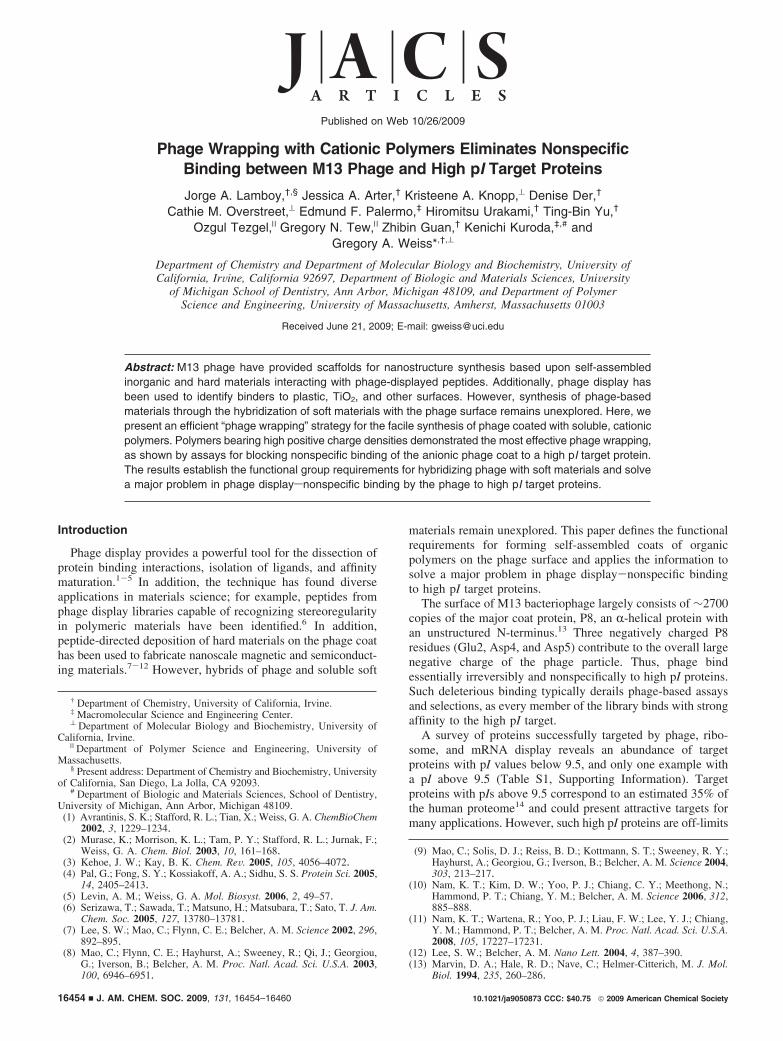
The synthetic, cationic polymer wrappers presented one totwo charged functionalities per 2.5, 5.0, and 32 Å of extendedpolymer backbone. As expected for highly charged molecules,the polymers dissolved readily in aqueous solution (e.g.,phosphate-buffered saline at pH 7.4). In ELISAs targeting thehigh pI colicin E9 DNase (pI 9.5), polymer 5 and thezwitterionic polymers 6-8 failed to wrap phage (Figure S1,Supporting Information). The discussion here, thus, focuses onthe following effective phage wrappers: polymethacrylatederivative 1,16 polyguanidino-oxanorbornene (2),17 poly-diguanidinium (3), and galactaro-oligolysine (4).18
As required for effective phage wrapping, polymers 1-4 donot impinge upon phage or bacterial viability at the concentra-tions required (1-10 µM). However, at higher concentrations(10-100 µM), polymers 2 and 3, but not 1 and 4, eliminatedor reduced phage infectivity without affecting bacterial viability(Tables 1 and S2, Supporting Information). Fixed concentrationsof M13-KO7 phage, which harbors a gene conferring kanamycinresistance, were incubated in the presence of 1, 2, 3, or 4 at theconcentrations indicated in Tables 1 and S2. The polymer-wrapped phage was allowed to infect a tetracycline-resistantEscherichia coli strain; to assess either phage or bacterialviability, the infected bacteria were then spotted on LB platessupplemented with either kanamycin (Tables 1 and S2A-S2C)or tetracycline (Table S2D-F), respectively. Unlike polymers1 and 4, which were tolerated at all concentrations, polymers 2and 3 at concentrations above 10 µM induced formation of whiteinsoluble precipitates of the phage. The precipitated phage haddramatically lower phage infectivity. However, the bacteria wereunaffected at all tested concentrations of polymers (TableS2D-F). Importantly, the optimal polymer concentrations forwrapping the phage (described below) required 10- to 100-foldless material (Figure 1) than the concentrations resulting inphage precipitation.
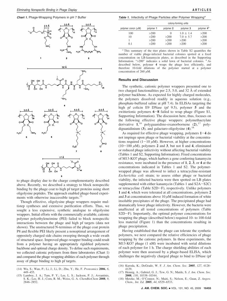
Having established that the phage can tolerate the syntheticpolymers, we next compared the relative efficiencies of phagewrapping by the cationic polymers. In these experiments, theM13-KO7 phage (1 nM) were incubated with serial dilutionsof each polymer for 1 h. The charge shielding abilities of eachpolymer were then assessed by a phage-based ELISA, whichchallenges the negatively charged phage to bind to DNase (pI
(14) Wu, S.; Wan, P.; Li, J.; Li, D.; Zhu, Y.; He, F. Proteomics 2006, 6,449–455.
(15) Lamboy, J. A.; Tam, P. Y.; Lee, L. S.; Jackson, P. J.; Avrantinis,S. K.; Lee, H. J.; Corn, R. M.; Weiss, G. A. ChemBioChem 2008, 9,2846–2852.
(16) Kuroda, K.; DeGrado, W. F. J. Am. Chem. Soc. 2005, 127, 4128–4129.
(17) Hennig, A.; Gabriel, G. J.; Tew, G. N.; Matile, S. J. Am. Chem. Soc.2008, 130, 10338–10344.
(18) Metzke, M.; O’Connor, N.; Maiti, S.; Nelson, E.; Guan, Z. Angew.Chem., Int. Ed. 2005, 44, 6529–6533.
Chart 1. Phage-Wrapping Polymers in pH 7 Buffer Table 1. Infectivity of Phage Particles after Polymer Wrappinga
colony-forming units
polymer concn (µM) polymer 1 polymer 2 polymer 3 polymer 4b
100 >200 0 1.0 ( 1.4 >20010 >200 >200 7.0 ( 5.7 >2001 >200 >200 >200 >2000.1 >200 >200 >200 >200
a This summary of the titer plates shown in Table S2 quantifies thenumber of viable phage-infected bacterial colonies spotted at a fixedconcentration on LB-kanamycin plates, as described in the SupportingInformation. “>200” indicates a solid lawn of bacterial colonies. b Asdescribed below, polymer 4 wraps the phage less efficiently, andtherefore 10-fold dilutions of the polymer started at a polymerconcentration of 260 µM.
J. AM. CHEM. SOC. 9 VOL. 131, NO. 45, 2009 16455
Eliminating Nonspecific Binding in Phage Display A R T I C L E S
9.5) (Figure 1). Polymers 1-4 effectively eliminated theundesirable phage binding to DNase at different concentrations,which reflects their relative effectiveness at wrapping and chargeshielding the phage surface. Polymers 1, 2, and 3 wrapped phagemost effectively when applied in 1000-fold molar excess to thephage (Figure 1A-C, respectively), whereas the less efficientwrapper 4 required a 105-fold molar excess to eliminatenonspecific binding (Figure 1D). Thus, despite the large numberof cationic functionalities in polymer 4 (78 primary amines permolecule), its dispersed charge density delivers less efficientphage wrapping than the other polymers.
The synthetic polymers wrapped the phage as well as or betterthan the previously reported oligolysine wrappers.15 Alsoanalogous to oligolysine wrapping, polymers 1, 2, and 3 causenonspecific binding to many targets, including the blockingagent (bovine serum albumin, BSA), at very high polymerconcentrations (g100 µM). Polymer 4 also demonstrated thiscross-linking effect at a high concentration (∼1 mM); these dataare not shown in Figure 1D, as the resultant high HRP activitysaturated the ELISA absorbance measurement. Excess polymercould cross-link the phage to the blocking agent by saturatingcharged sites on the phage surface and allowing cationicfunctionalities of the polymer to trail off the phage surface. Suchhigh wrapper concentrations far exceed the requirements forblocking nonspecific interactions but demonstrate the importanceof determining optimal wrapper concentrations for new polymersbefore phage-based assays or selections.
The observed cross-linking at higher polymer concentrationsalso correlated with increased polymer length, as demonstratedby comparing two variants of polymer 1. In this experiment,the extended length variant of polymer 1 (1-ext) included 23primary amine functionalities per molecule, and the truncatedlength variant of polymer 1 (1-trunc) included 9 primary aminesper molecule (Figure 2). When used in excess concentrations,the longer, cationic polymer 1-ext results in more deleteriouscross-linking, presumably due to the greater propensity of thelonger polymer to extend off the phage surface. However, thestructure of the polymer and not just the length determines cross-linking ability. For example, the long polymer 4 fails to cross-
link the phage to the target at the concentration (∼100 µM)required for cross-linking by the other polymers (Figure 1); thus,a higher concentration of the less efficient wrapper, polymer 4,is required to saturate charged sites on the phage surface beforecross-linking to the target or blocking agent.
Figure 1. Phage-based ELISAs of M13-KO7 phage wrapped with polymers (A) 1, (B) 2, (C) 3, and (D) 4 binding to either DNase or BSA (negativecontrol). Wrapping the phage (1 nM) with a 1000-fold molar excess (1 µM) of polymer 1, 2, or 3 was sufficient to reduce or eliminate nonspecific bindingto target DNase. Polymer 4, however, required a 105-fold molar excess (∼100 µM) to wrap the phage effectively. Throughout this report, error bars indicatestandard error (n ) 2-3).
Figure 2. Phage-based ELISAs of M13-KO7 phage wrapped by polymers(A) 1-trunc and (B) 1-ext. A fixed concentration of M13-KO7 phage (0.5nM) was incubated with the indicated concentrations of each polymer, andexposed to either DNase or the blocking agent BSA for assay. Polymer1-ext (23 amine functionalities) at high concentrations (∼100 µM) resultedin high levels of cross-linked phage to DNase, but polymer 1-trunc exhibitedonly moderate cross-linking at the same polymer concentrations.
16456 J. AM. CHEM. SOC. 9 VOL. 131, NO. 45, 2009
A R T I C L E S Lamboy et al.
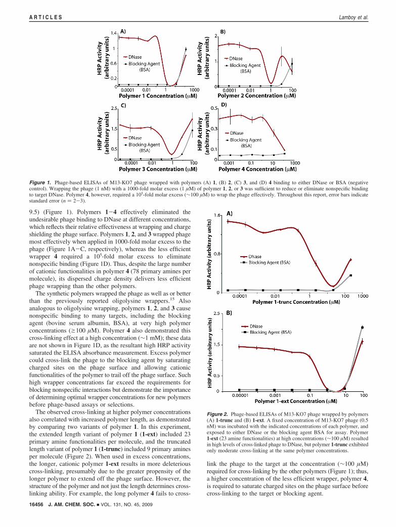
In addition to the potential for novel materials, phagewrapping by cationic polymers could find important applicationsin preventing nonspecific binding between phage and high pItarget proteins. For such applications, the cationic wrapper mustremain attached to the phage during both phage displayselections and screens. To demonstrate the utility of syntheticwrappers in screens, a fixed concentration of wrapper was usedto wrap phage at a range of concentrations. At the optimalpolymer concentration determined by the Figure 1 experiment(1 µM), polymer 1 successfully blocked nonspecific binding ofphage to both DNase and lysozyme (pI 9.5) (Figure 3). Theexperiment demonstrated the absence of spurious binding atphage concentrationse2 nM, while higher phage concentrationselicited nonspecific binding to both targets, presumably due toincomplete wrapping of the phage by an inadequate quantityof wrapping polymer. The experiment also demonstrated thatwrapping the phage with synthetic polymers also blocksnonspecific binding to other high pI targets, such as lysozyme.
To determine the most effective phage wrapper among thepolymers described here and previously,15 an ELISA comparedphage wrapping and cancellation of nonspecific binding bydifferent polymers (Figure 4). In this experiment, 10-fold serialdilutions of 20 nM M13-KO7 phage were incubated with thephage wrappers Lys20, 1-ext, or 2. Polymer 1-ext demonstratedessentially perfect wrapping, as nonspecific binding to the target
DNase was absent at phage concentrations up to the limits ofphage solubility.
Polymer 2 and peptide Lys20 revealed similar levels ofmoderate efficiency for phage wrapping. However, Lys20
exhibited poor efficiency at a high phage concentration (20 nM),where 1-ext demonstrated ∼12.5 times more efficient wrappingthan Lys20. Polymer 1-ext presents a denser collection of cationicamines (one amine side chain per 2.5 Å of polymer backbone)than Lys20 (one amine per 5.0 Å) and polymer 4, which exposestwo amine side chains per galactaro-oligolysine subunit (32 Åbackbone length). As phage wrapping is mediated by an avidityeffect,15 polymers presenting higher positive charge densitiesdemonstrate superior phage wrapping than polymers with alower density of cationic side chains.
In addition to its effectiveness in phage-based screeningassays, the best phage wrapper, 1-ext, works exceptionally wellto block nonspecific interactions during selections with phage-displayed libraries. In the absence of added wrapper, phagedisplay selections targeting high pI proteins generally fail; phagewithout a displayed protein are amplified preferentially, as suchphage place less demand on the bacteria during phage prolifera-tion. During phage display selections and screens, 1-ext wasadded in 1000-fold molar excess, a concentration suggested bythe previously described polymer optimization experiments(Figure 2). The phage-displayed library consisted of 24 differentconfigurations of cystine disulfide-linked peptides displayed asfusions to the N-terminus of P8. With the exception of theinvariant cysteine residues, each peptide included 5-18 residuesencoded by the NNS codon (where N designates any nucleotideand S designates either G or C). Thus, each degenerate positionencodes all 20 naturally occurring amino acids but prevents theoccurrence of the nonsuppressible stop codons TGA and TAA,which could result in nondisplayed peptides. From a combinedpeptide diversity of 2.5 × 10¹⁰ different peptides, two newpeptide ligands to DNase, DNase-1 (amino acid sequence
Figure 3. Phage wrapping ELISAs of M13-KO7 phage mixed with polymer1 (1 µM). (A) Wrapped M13-KO7 phage abrogated nonspecific binding totarget DNase at phage concentrations e2 nM. However, binding betweenphage and DNase was observed in the absence of polymer 1 and at highphage concentrations. (B) Nonspecific binding of M13-KO7 phage tolysozyme was also abolished by wrapping with polymer 1.
Figure 4. Phage-based ELISA comparing the phage wrapping efficienciesof peptide Lys20 with polymers 1-ext and 2. The indicated phage concentra-tions were incubated with 10 µM Lys20 and polymer 1-ext and 3 µMpolymer 2 before testing for binding to DNase. Polymer 1-ext resulted inthe most efficient wrapping, as this wrapper eliminated nonspecific bindingto the target at typically problematic phage concentrations (g5 nM).
J. AM. CHEM. SOC. 9 VOL. 131, NO. 45, 2009 16457
Eliminating Nonspecific Binding in Phage Display A R T I C L E S
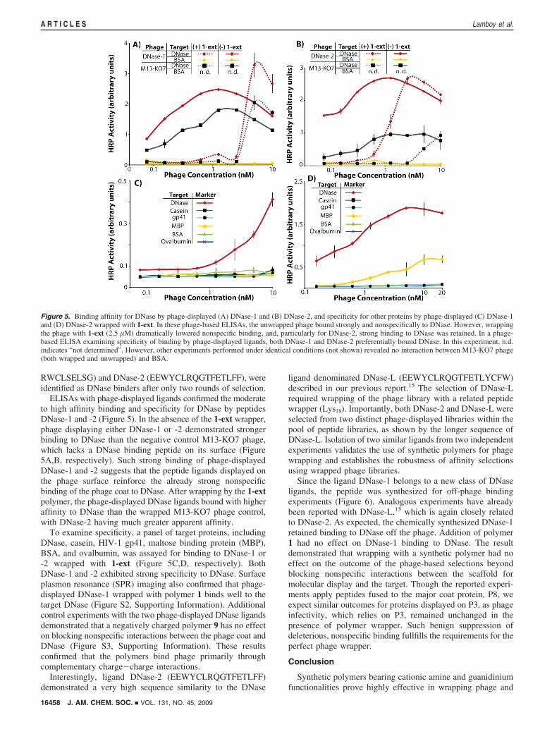
RWCLSELSG) and DNase-2 (EEWYCLRQGTFETLFF), wereidentified as DNase binders after only two rounds of selection.
ELISAs with phage-displayed ligands confirmed the moderateto high affinity binding and specificity for DNase by peptidesDNase-1 and -2 (Figure 5). In the absence of the 1-ext wrapper,phage displaying either DNase-1 or -2 demonstrated strongerbinding to DNase than the negative control M13-KO7 phage,which lacks a DNase binding peptide on its surface (Figure5A,B, respectively). Such strong binding of phage-displayedDNase-1 and -2 suggests that the peptide ligands displayed onthe phage surface reinforce the already strong nonspecificbinding of the phage coat to DNase. After wrapping by the 1-extpolymer, the phage-displayed DNase ligands bound with higheraffinity to DNase than the wrapped M13-KO7 phage control,with DNase-2 having much greater apparent affinity.
To examine specificity, a panel of target proteins, includingDNase, casein, HIV-1 gp41, maltose binding protein (MBP),BSA, and ovalbumin, was assayed for binding to DNase-1 or-2 wrapped with 1-ext (Figure 5C,D, respectively). BothDNase-1 and -2 exhibited strong specificity to DNase. Surfaceplasmon resonance (SPR) imaging also confirmed that phage-displayed DNase-1 wrapped with polymer 1 binds well to thetarget DNase (Figure S2, Supporting Information). Additionalcontrol experiments with the two phage-displayed DNase ligandsdemonstrated that a negatively charged polymer 9 has no effecton blocking nonspecific interactions between the phage coat andDNase (Figure S3, Supporting Information). These resultsconfirmed that the polymers bind phage primarily throughcomplementary charge-charge interactions.
Interestingly, ligand DNase-2 (EEWYCLRQGTFETLFF)demonstrated a very high sequence similarity to the DNase
ligand denominated DNase-L (EEWYCLRQGTFETLYCFW)described in our previous report.15 The selection of DNase-Lrequired wrapping of the phage library with a related peptidewrapper (Lys18). Importantly, both DNase-2 and DNase-L wereselected from two distinct phage-displayed libraries within thepool of peptide libraries, as shown by the longer sequence ofDNase-L. Isolation of two similar ligands from two independentexperiments validates the use of synthetic polymers for phagewrapping and establishes the robustness of affinity selectionsusing wrapped phage libraries.
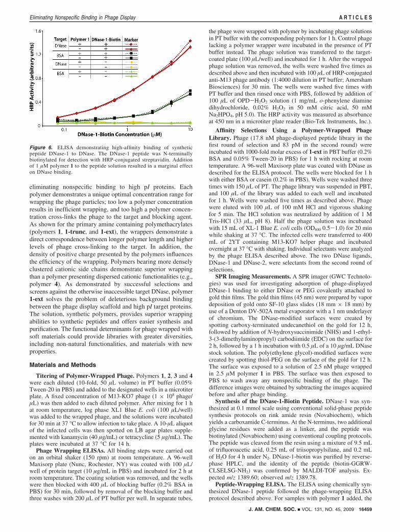
Since the ligand DNase-1 belongs to a new class of DNaseligands, the peptide was synthesized for off-phage bindingexperiments (Figure 6). Analogous experiments have alreadybeen reported with DNase-L,15 which is again closely relatedto DNase-2. As expected, the chemically synthesized DNase-1retained binding to DNase off the phage. Addition of polymer1 had no effect on DNase-1 binding to DNase. The resultdemonstrated that wrapping with a synthetic polymer had noeffect on the outcome of the phage-based selections beyondblocking nonspecific interactions between the scaffold formolecular display and the target. Though the reported experi-ments apply peptides fused to the major coat protein, P8, weexpect similar outcomes for proteins displayed on P3, as phageinfectivity, which relies on P3, remained unchanged in thepresence of polymer wrapper. Such benign suppression ofdeleterious, nonspecific binding fullfills the requirements for theperfect phage wrapper.
Conclusion
Synthetic polymers bearing cationic amine and guanidiniumfunctionalities prove highly effective in wrapping phage and
Figure 5. Binding affinity for DNase by phage-displayed (A) DNase-1 and (B) DNase-2, and specificity for other proteins by phage-displayed (C) DNase-1and (D) DNase-2 wrapped with 1-ext. In these phage-based ELISAs, the unwrapped phage bound strongly and nonspecifically to DNase. However, wrappingthe phage with 1-ext (2.5 µM) dramatically lowered nonspecific binding, and, particularly for DNase-2, strong binding to DNase was retained. In a phage-based ELISA examining specificity of binding by phage-displayed ligands, both DNase-1 and DNase-2 preferentially bound DNase. In this experiment, n.d.indicates “not determined”. However, other experiments performed under identical conditions (not shown) revealed no interaction between M13-KO7 phage(both wrapped and unwrapped) and BSA.
16458 J. AM. CHEM. SOC. 9 VOL. 131, NO. 45, 2009
A R T I C L E S Lamboy et al.
eliminating nonspecific binding to high pI proteins. Eachpolymer demonstrates a unique optimal concentration range forwrapping the phage particles; too low a polymer concentrationresults in inefficient wrapping, and too high a polymer concen-tration cross-links the phage to the target and blocking agent.As shown for the primary amine containing polymethacrylates(polymers 1, 1-trunc, and 1-ext), the wrappers demonstrate adirect correspondence between longer polymer length and higherlevels of phage cross-linking to the target. In addition, thedensity of positive charge presented by the polymers influencesthe efficiency of the wrapping. Polymers bearing more denselyclustered cationic side chains demonstrate superior wrappingthan a polymer presenting dispersed cationic functionalities (e.g.,polymer 4). As demonstrated by successful selections andscreens against the otherwise inaccessible target DNase, polymer1-ext solves the problem of deleterious background bindingbetween the phage display scaffold and high pI target proteins.The solution, synthetic polymers, provides superior wrappingabilities to synthetic peptides and offers easier synthesis andpurification. The functional determinants for phage wrapped withsoft materials could provide libraries with greater diversities,including non-natural functionalities, and materials with newproperties.
Materials and Methods
Titering of Polymer-Wrapped Phage. Polymers 1, 2, 3 and 4were each diluted (10-fold, 50 µL volume) in PT buffer (0.05%Tween-20 in PBS) and added to the designated wells in a microtiterplate. A fixed concentration of M13-KO7 phage (1 × 10⁸ phage/µL) was then added to each diluted polymer. After mixing for 1 hat room temperature, log phase XL1 Blue E. coli (100 µL/well)was added to the wrapped phage, and the solutions were incubatedfor 30 min at 37 °C to allow infection to take place. A 10-µL aliquotof the infected cells was then spotted on LB agar plates supple-mented with kanamycin (40 µg/mL) or tetracycline (5 µg/mL). Theplates were incubated at 37 °C for 14 h.
Phage Wrapping ELISAs. All binding steps were carried outon an orbital shaker (150 rpm) at room temperature. A 96-wellMaxisorp plate (Nunc, Rochester, NY) was coated with 100 µL/well of protein target (10 µg/mL in PBS) and incubated for 2 h atroom temperature. The coating solution was removed, and the wellswere then blocked with 400 µL of blocking buffer (0.2% BSA inPBS) for 30 min, followed by removal of the blocking buffer andthree washes with 200 µL of PT buffer per well. In separate tubes,
the phage were wrapped with polymer by incubating phage solutionsin PT buffer with the corresponding polymers for 1 h. Control phagelacking a polymer wrapper were incubated in the presence of PTbuffer instead. The phage solution was transferred to the target-coated plate (100 µL/well) and incubated for 1 h. After the wrappedphage solution was removed, the wells were washed five times asdescribed above and then incubated with 100 µL of HRP-conjugatedanti-M13 phage antibody (1:4000 dilution in PT buffer; AmershamBiosciences) for 30 min. The wells were washed five times withPT buffer and then rinsed once with PBS, followed by addition of100 µL of OPD-H2O2 solution (1 mg/mL o-phenylene diaminedihydrochloride, 0.02% H2O2 in 50 mM citric acid, 50 mMNa2HPO4, pH 5.0). The HRP activity was measured as absorbanceat 450 nm in a microtiter plate reader (Bio-Tek Instruments, Inc.).
Affinity Selections Using a Polymer-Wrapped PhageLibrary. Phage (17.8 nM phage-displayed peptide library in thefirst round of selection and 83 pM in the second round) wereincubated with 1000-fold molar excess of 1-ext in PBT buffer (0.2%BSA and 0.05% Tween-20 in PBS) for 1 h with rocking at roomtemperature. A 96-well Maxisorp plate was coated with DNase asdescribed for the ELISA protocol. The wells were blocked for 1 hwith either BSA or casein (0.2% in PBS). Wells were washed threetimes with 150 µL of PT. The phage library was suspended in PBT,and 100 µL of the library was added to each well and incubatedfor 1 h. Wells were washed five times as described above. Phagewere eluted with 100 µL of 100 mM HCl and vigorous shakingfor 5 min. The HCl solution was neutralized by addition of 1 MTris-HCl (33 µL, pH 8). Half the phage solution was incubatedwith 15 mL of XL-1 Blue E. coli cells (OD600 0.5-1.0) for 20 minwhile shaking at 37 °C. The infected cells were transferred to 400mL of 2YT containing M13-KO7 helper phage and incubatedovernight at 37 °C with shaking. Individual selectants were analyzedby the phage ELISA described above. The two DNase ligands,DNase-1 and DNase-2, were selectants from the second round ofselections.
SPR Imaging Measurements. A SPR imager (GWC Technolo-gies) was used for investigating adsorption of phage-displayedDNase-1 binding to either DNase or PEG covalently attached togold thin films. The gold thin films (45 nm) were prepared by vapordeposition of gold onto SF-10 glass slides (18 mm × 18 mm) byuse of a Denton DV-502A metal evaporator with a 1 nm underlayerof chromium. The DNase-modified surfaces were created byspotting carboxy-terminated undecanethiol on the gold for 12 h,followed by addition of N-hydroxysuccinimide (NHS) and 1-ethyl-3-(3-dimethylaminopropyl) carbodiimide (EDC) on the surface for2 h, followed by a 1 h incubation with 0.5 µL of a 10 µg/mL DNasestock solution. The poly(ethylene glycol)-modified surfaces werecreated by spotting thiol-PEG on the surface of the gold for 12 h.The surface was exposed to a solution of 2.5 nM phage wrappedin 2.5 µM polymer 1 in PBS. The surface was then exposed toPBS to wash away any nonspecific binding of the phage. Thedifference images were obtained by subtracting the images acquiredbefore and after phage binding.
Synthesis of the DNase-1-Biotin Peptide. DNase-1 was syn-thesized at 0.1 mmol scale using conventional solid-phase peptidesynthesis protocols on rink amide resin (Novabiochem), whichyields a carboxamide C-terminus. At the N-terminus, two additionalglycine residues were added as a linker, and the peptide wasbiotinylated (Novabiochem) using conventional coupling protocols.The peptide was cleaved from the resin using a mixture of 9.5 mLof trifluoroacetic acid, 0.25 mL of triisopropylsilane, and 0.2 mLof H2O for 4 h under N2. DNase-1-biotin was purified by reverse-phase HPLC, and the identity of the peptide (biotin-GGRW-CLSELSG-NH2) was confirmed by MALDI-TOF analysis. Ex-pected m/z 1389.60; observed m/z 1389.78.
Peptide-Wrapping ELISA. The ELISA using chemically syn-thesized DNase-1 peptide followed the phage-wrapping ELISAprotocol described above. For samples with polymer 1 added, the
Figure 6. ELISA demonstrating high-affinity binding of syntheticpeptide DNase-1 to DNase. The DNase-1 peptide was N-terminallybiotinylated for detection with HRP-conjugated streptavidin. Additionof 1 µM polymer 1 to the peptide solution resulted in a marginal effecton DNase binding.
J. AM. CHEM. SOC. 9 VOL. 131, NO. 45, 2009 16459
Eliminating Nonspecific Binding in Phage Display A R T I C L E S
DNase-1 peptide was incubated in the presence of polymer 1 (1µM) in PT buffer for 1 h before being transferred to thecorresponding DNase-coated wells.
Acknowledgment. We gratefully acknowledge support from theNCI, NIGMS and NBIB of the NIH (1R43CA11955-01, 1R01-GM078528-01 to G.A.W.; R01 EB006797 to Z.G.) and a NIHMinority Supplemental Fellowship to J.A.L. We thank Robert Corn,Yulin Chen, and Glenn Eldridge for technical assistance.
Supporting Information Available: Additional experimentsand experimental protocols including stability studies of polymer-wrapped phage, SPR imaging experiments using phage-displayed DNase-1, and wrapping experiments using polymers5-9; updated table of proteins successfully targeted by molec-ular display methods and their estimated pI values. This materialis available free of charge via the Internet at http://pubs.acs.org.
JA9050873
16460 J. AM. CHEM. SOC. 9 VOL. 131, NO. 45, 2009
A R T I C L E S Lamboy et al.