Embed Size (px)
Citation preview
Kaur G et al. Peripheral giant cell granuloma.
58
Case Report
Peripheral Giant Cell Granuloma- A Case Report And Review Of Literature
Geetpriya Kaur, Naveen Puri1, Lalita Yadav
Senior lecturer, 1Professor &Head, Department of Oral Pathology and microbiology, Kalka Dental College Hospital and Research Centre, Meerut.
Corresponding author:
Dr. Geetpiya Kaur
45 Ishwar colony,
New Delhi-110009
E mail: [email protected]
E mail: [email protected]
Received: 12 February 2013 Revised: 10 March 2013 Accepted: 15 March 2013
Abstract: Peripheral giant cell granuloma is the most common oral giant cell lesion. It normally presents as a soft tissue purplish-red nodule consisting of multinucleated giant cells in the background of mononuclear stromal cells and extravasated red blood cells. This article reports a peripheral giant cell granuloma arising at right maxillary alveolus in 56 years old female patient.
Keywords: Giant Cell, Granuloma, monocyte
This article may be cited as: Kaur G, Puri N, Yadav L. Prosthodontic Management Of Patients With Diabetes Mellitus. J Adv Med Dent Scie Res 2013;1(1):58-62.
INTRODUCTION
Peripheral giant cell granuloma(PGCG) is an infrequent reactive, exophytic lesion of the oral cavity, also known as giant-cell epulis, osteoclastoma, giant cell reparative granuloma, or giant cell hyperplasia. It is the most frequent giant cell lesion of the jaws, and originates from the connective tissue of the periosteum or from the periodontal membrane, in response to local irritation or chronic trauma.1
The initiating stimulus has been believed to be due to local irritation or trauma but the cause is not certainly known.2 It has been termed a peripheral giant cell "reparative" granuloma, but whether it is in fact reparative has not been established and their osteoclastic activity nature appears doubtful. Their membrane receptors for calcitonin demonstrated by
immunohistochemistry and their osteoclastic activity when cultured in vitro are evidence that
Kaur G et al. Peripheral giant cell granuloma.
59
they are osteoclasts 3-5,whereas other authors have suggested that the lesion is formed by cells of the mononuclear phagocyte system6.It is not a true neoplasm but rather a benign hyperplastic lesion occurred in response to local irritation such as extraction, poor dental restorations, ill-fitting dentures, plaque, calculus, food impaction and chronic trauma.7 It is more frequent in women than in men, with a slightly higher prevalence in the 30- to 70-year-old-age group, and affects largely the lower jaw (55%) than in the upper jaw.8 Clinically, it manifests as a soft to firm, bright nodule or as a sessile or pediculate mass, which is predominantly bluish red with a smooth shiny or mamillated surface, localized in the gingival tissue or alveolar processes of the incisor and canine region.1 Histologically, PGCG is composed of nodules of multinucleated giant cells in a background of plump ovoid and spindle-shaped mesenchymal cells and extravasated red blood cells.3-6
CASE REPORT
A 56 years old female reported to OPD with a chief complaint of swelling in the upper front tooth region since last 6 months. Patient gives history of extraction 6 months back. The swelling appeared subsequently after 3 days which gradually increased to attain present size.
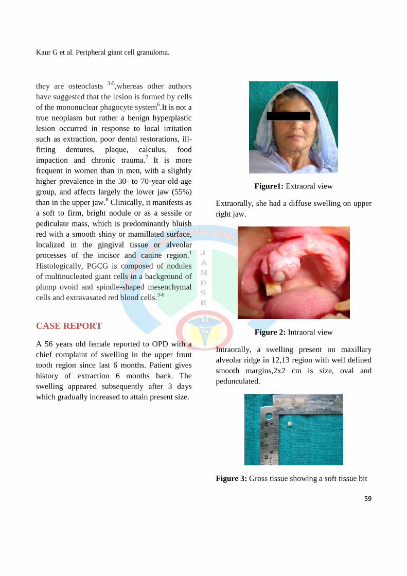
Figure1: Extraoral view
Extraorally, she had a diffuse swelling on upper right jaw.
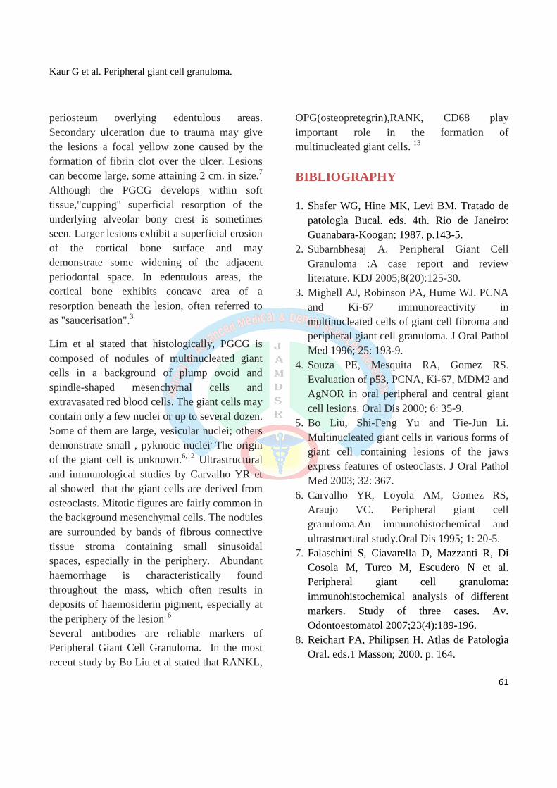
Figure 2: Intraoral view
Intraorally, a swelling present on maxillary alveolar ridge in 12,13 region with well defined smooth margins,2x2 cm is size, oval and pedunculated.
Figure 3: Gross tissue showing a soft tissue bit

Kaur G et al. Peripheral giant cell granuloma.
60
An excisional biopsy was done which revealed a soft tissue bit. The H/E section shows a hyperplastic parakeratinized stratified squamous epithelium with numerous long rete ridges. The epithelium is discontinuous at places with a delicate connective tissue stroma, with a dense chronic inflammatory infiltrate and large proliferating capillaries. Towards the centre of the section, separated by fibrous bonds numerous vesiculated proliferating connective tissue cells intermingled many blood capillaries showing endothelial cell proliferation & plenty of extravasated RBCs are observed. There are many irregularly shaped multinucleated giant cells with uneven distribution and the number of nuclei ranging from 3-10. It is surrounded by a fibrocellular connective tissue stroma with thin interlacing stromds of collagen & plump fibroblasts. Deeper to it, areas of brightly eosinophillic osteoid with plump osteoblastic rimming and large osteocytic lacunae are seen.
Figure 4: Photomicrograph showing a hyperplastic parakeratinized stratified squamous epithelium with numerous long rete ridges. (H& E;X4)
Figure 5: There are many irregularly shaped multinucleated giant cells with uneven distribution and the number of nuclei ranging from 3-10. (H& E;X4)
DISCUSSION
Peripheral Giant Cell Granuloma or so called "Giant cell epulis" is the most common oral giant cell lesion.2 The PGCG occurs throughout life, with peaks in the incidence during the mixed dentitional years and the 30-40-year-old age group. 7 Whereas in our case it occurs at 56 years of age. It is more common among females (60%).9,10 As the term "Giant cell epulis" implies, it occurs on the gingival margins or edentulous alveolar ridge as a focal purplish nodule in either the anterior or posterior regions of the jaws but most frequently between the first permanent molars and the incisors.2 The mandible is affected slightly more often than the maxilla. The lesion can be sessile or pedunculated, spreading through penetration of the periodontal membrane and may or may not be ulcerated.11 Whereas in our case it occurs in maxilla. Occasionally lesions arise from the
Kaur G et al. Peripheral giant cell granuloma.
61
periosteum overlying edentulous areas. Secondary ulceration due to trauma may give the lesions a focal yellow zone caused by the formation of fibrin clot over the ulcer. Lesions can become large, some attaining 2 cm. in size.7
Although the PGCG develops within soft tissue,"cupping" superficial resorption of the underlying alveolar bony crest is sometimes seen. Larger lesions exhibit a superficial erosion of the cortical bone surface and may demonstrate some widening of the adjacent periodontal space. In edentulous areas, the cortical bone exhibits concave area of a resorption beneath the lesion, often referred to as "saucerisation".3
Lim et al stated that histologically, PGCG is composed of nodules of multinucleated giant cells in a background of plump ovoid and spindle-shaped mesenchymal cells and extravasated red blood cells. The giant cells may contain only a few nuclei or up to several dozen. Some of them are large, vesicular nuclei; others demonstrate small , pyknotic nuclei. The origin of the giant cell is unknown.6,12 Ultrastructural and immunological studies by Carvalho YR et al showed that the giant cells are derived from osteoclasts. Mitotic figures are fairly common in the background mesenchymal cells. The nodules are surrounded by bands of fibrous connective tissue stroma containing small sinusoidal spaces, especially in the periphery. Abundant haemorrhage is characteristically found throughout the mass, which often results in deposits of haemosiderin pigment, especially at the periphery of the lesion. 6 Several antibodies are reliable markers of Peripheral Giant Cell Granuloma. In the most recent study by Bo Liu et al stated that RANKL,
OPG(osteopretegrin),RANK, CD68 play important role in the formation of multinucleated giant cells. 13
BIBLIOGRAPHY 1. Shafer WG, Hine MK, Levi BM. Tratado de
patologìa Bucal. eds. 4th. Rio de Janeiro: Guanabara-Koogan; 1987. p.143-5.
2. Subarnbhesaj A. Peripheral Giant Cell Granuloma :A case report and review literature. KDJ 2005;8(20):125-30.
3. Mighell AJ, Robinson PA, Hume WJ. PCNA and Ki-67 immunoreactivity in multinucleated cells of giant cell fibroma and peripheral giant cell granuloma. J Oral Pathol Med 1996; 25: 193-9.
4. Souza PE, Mesquita RA, Gomez RS. Evaluation of p53, PCNA, Ki-67, MDM2 and AgNOR in oral peripheral and central giant cell lesions. Oral Dis 2000; 6: 35-9.
5. Bo Liu, Shi-Feng Yu and Tie-Jun Li. Multinucleated giant cells in various forms of giant cell containing lesions of the jaws express features of osteoclasts. J Oral Pathol Med 2003; 32: 367.
6. Carvalho YR, Loyola AM, Gomez RS, Araujo VC. Peripheral giant cell granuloma.An immunohistochemical and ultrastructural study.Oral Dis 1995; 1: 20-5.
7. Falaschini S, Ciavarella D, Mazzanti R, Di Cosola M, Turco M, Escudero N et al. Peripheral giant cell granuloma: immunohistochemical analysis of different markers. Study of three cases. Av. Odontoestomatol 2007;23(4):189-196.
8. Reichart PA, Philipsen H. Atlas de Patologìa Oral. eds.1 Masson; 2000. p. 164.

Kaur G et al. Peripheral giant cell granuloma.
62
9. Chadwick BL, Crawford PJ, Aldred MJ. Massive giant cell epulis in a child with familial cyclic neutropenia. Brtish Dent J 1989; 167: 279-81.
10. Giansanti JS, Waldron CA. Peripheral giant cell granuloma: review of 720 cases. J Oral Surg 1969; 27: 787-91.
11. Speight PM. Giant cell lesions of the jaws. Oral Diseases I 995;1:6 -7.
12. Lim L, Gibbins JR. Immunohistochemical and structural evidence of a modified microvasculature in the giant cell granuloma of the jaws. Oral Surg Oral Med Oral Pathol 1995;79:190-8.
13. Bo Liu, Shi-Feng Yu and Tie-Jun Li. Multinucleated giant cells in various forms of giant cell containing lesions of the jaws express features of osteoclasts. J Oral Pathol Med 2003; 32:367.
Acknowledgment: The Author would like to
thank the patient for providing consent to use
her photograph in this article.
Source of support: Nil
Conflict of interest: None declared