Embed Size (px)
Citation preview
PFT Interpretation
Presented by:Shazhad Manawar, MDMedical Director Respiratory Care & Pulmonary Rehab McLaren Bay Region
Introduction
History or symptoms suggestive of lung disease.
Risk factors for lung disease are present.
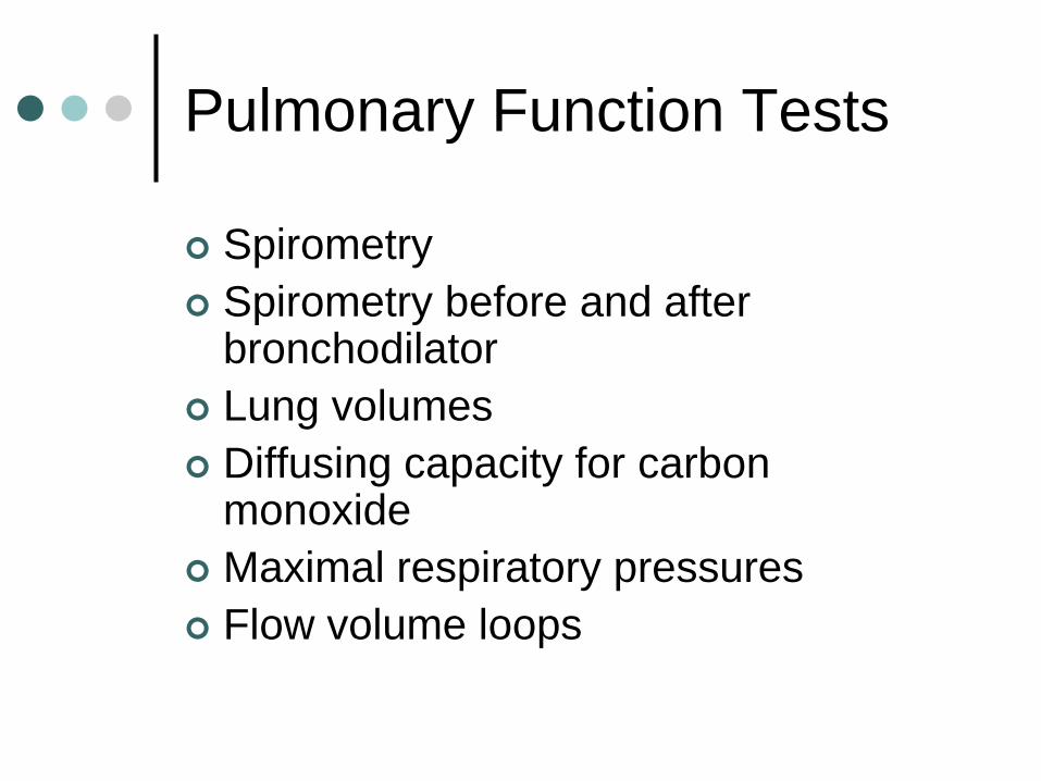
Pulmonary Function Tests
Spirometry Spirometry before and after
bronchodilator Lung volumes Diffusing capacity for carbon
monoxide Maximal respiratory pressures Flow volume loops
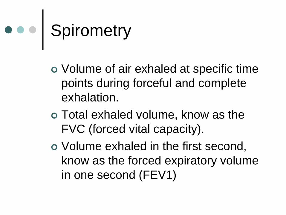
Spirometry
Volume of air exhaled at specific time points during forceful and complete exhalation.
Total exhaled volume, know as the FVC (forced vital capacity).
Volume exhaled in the first second, know as the forced expiratory volume in one second (FEV1)
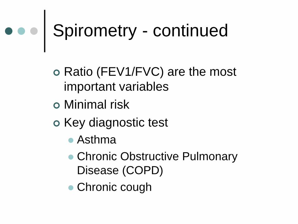
Spirometry - continued
Ratio (FEV1/FVC) are the most important variables
Minimal risk Key diagnostic test Asthma Chronic Obstructive Pulmonary
Disease (COPD) Chronic cough
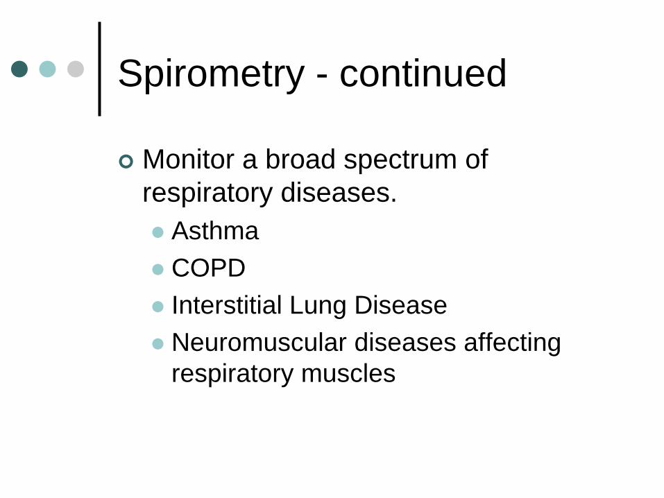
Spirometry - continued
Monitor a broad spectrum of respiratory diseases. Asthma COPD Interstitial Lung Disease Neuromuscular diseases affecting
respiratory muscles
Spirometry - continued
Slow vital capacity (SVC) Useful measurement when FVC is
reduced and airway obstruction is present
Post-bronchodilator
Determine the degree of reversibility Administration of albuterol Technique is important Increase in the FEV1 of more than
12% or greater than 0.2 L suggests acute bronchodilator responsiveness.
Subjective improvements
Post-bronchodilator -continued Thus, the lack of an acute
bronchodilator response on spirometry should not preclude a one to eight week therapeutic trial of bronchodilators and /or inhaled glucocorticoids, with reassessment of clinical status and change in FEV1 at the end of the time.
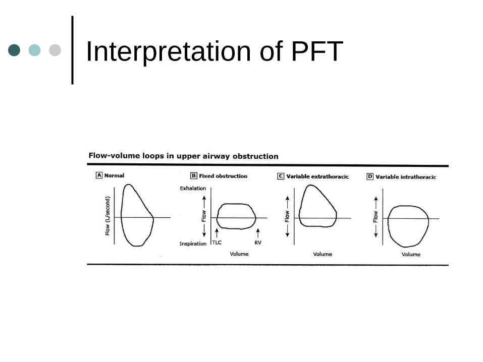
Flow-volume loop
Stridor is heard over the neck Unexplained dyspnea Pharynx, larynx, or trachea Impossible ato detect from standard
FVC Variable extrathoracic Fixed upper airway obstruction (UAO)
Lung volumes
Body plethysmography Helium dilution Nitrogen washout Chest imaging Chest radiograph or high resolution
tomography 15% of those
Lung volumes
Common lung volumes Vital capacity (VC) Functional residual capacity (FRC) Residual volume (RV) Expiratory reserve volume (ERV) Inspiratory capacity (IC) Total lung capacity (TLC)
Interpretation of PFT
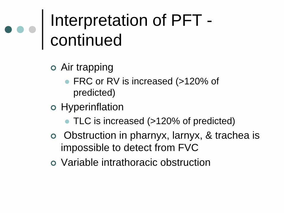
Interpretation of PFT -continued Air trapping
FRC or RV is increased (>120% of predicted)
Hyperinflation TLC is increased (>120% of predicted)
Obstruction in pharnyx, larnyx, & trachea is impossible to detect from FVC
Variable intrathoracic obstruction
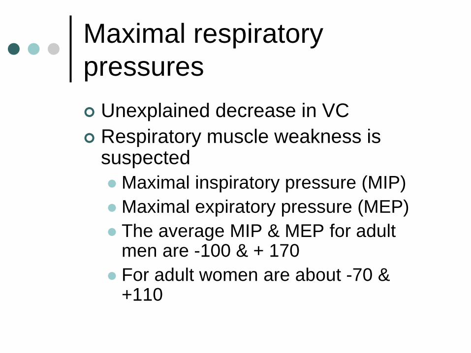
Maximal respiratory pressures Unexplained decrease in VC Respiratory muscle weakness is
suspected Maximal inspiratory pressure (MIP) Maximal expiratory pressure (MEP) The average MIP & MEP for adult
men are -100 & + 170 For adult women are about -70 &
+110
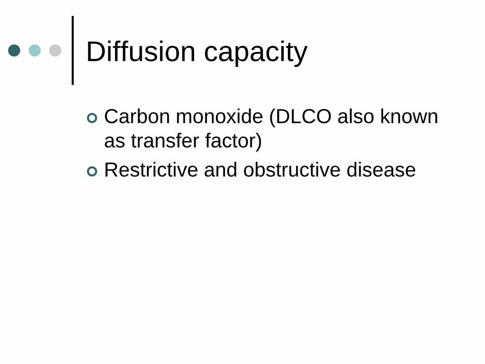
Diffusion capacity
Carbon monoxide (DLCO also known as transfer factor)
Restrictive and obstructive disease
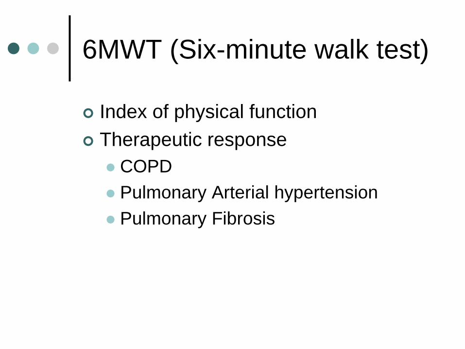
6MWT (Six-minute walk test)
Index of physical function Therapeutic response COPD Pulmonary Arterial hypertension Pulmonary Fibrosis
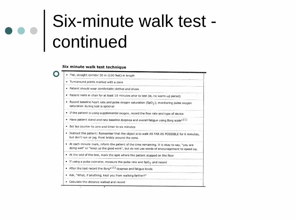
Six-minute walk test -continued
Table 2
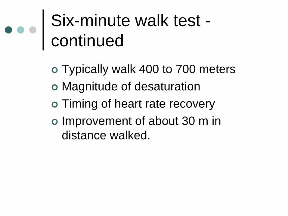
Six-minute walk test -continued Typically walk 400 to 700 meters Magnitude of desaturation Timing of heart rate recovery Improvement of about 30 m in
distance walked.
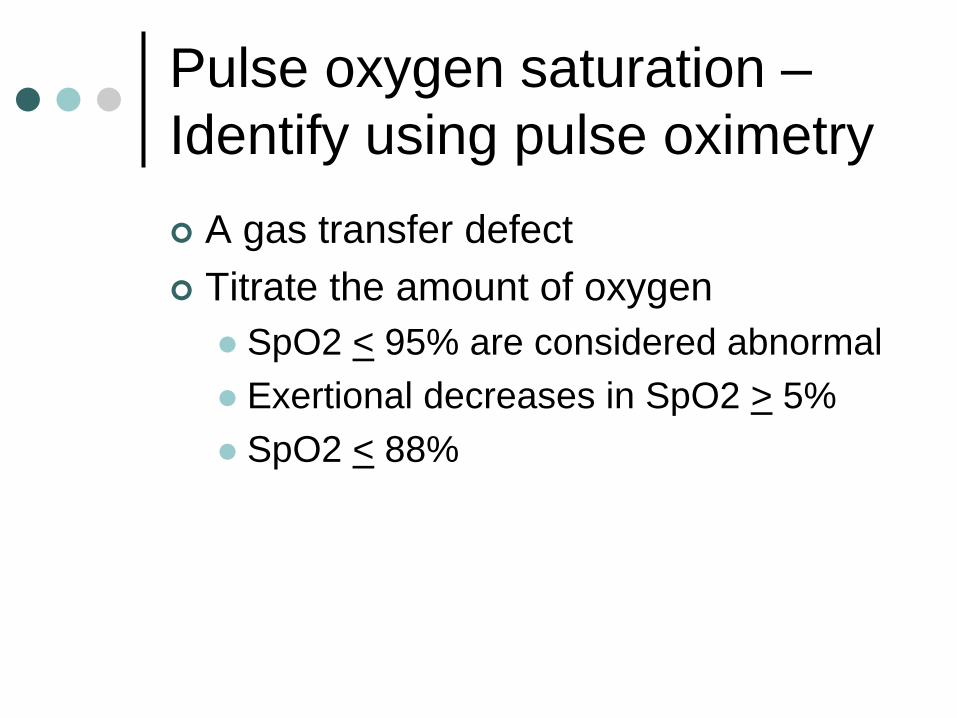
Pulse oxygen saturation –Identify using pulse oximetry A gas transfer defect Titrate the amount of oxygen SpO2 < 95% are considered abnormal Exertional decreases in SpO2 > 5% SpO2 < 88%
Arterial blood gases
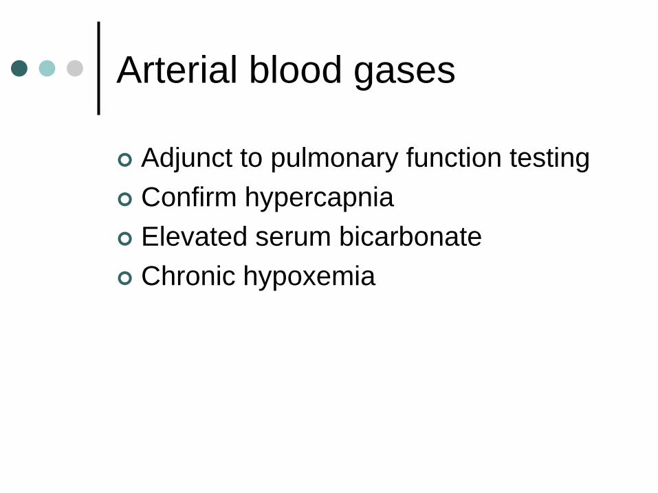
Adjunct to pulmonary function testing Confirm hypercapnia Elevated serum bicarbonate Chronic hypoxemia
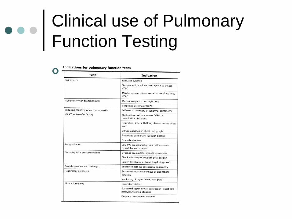
Clinical use of Pulmonary Function Testing #16
Chronic dyspnea
Dyspnea on exertion Spriometry on exertion Spirometry before & after a
bronchodilator
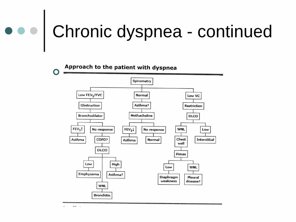
Chronic dyspnea - continued
#18
Asthma
Spirometry before and after a bronchodilator
Follow-up office Bronchial hyperresponsiveness (BHR) Measurement of airway lability
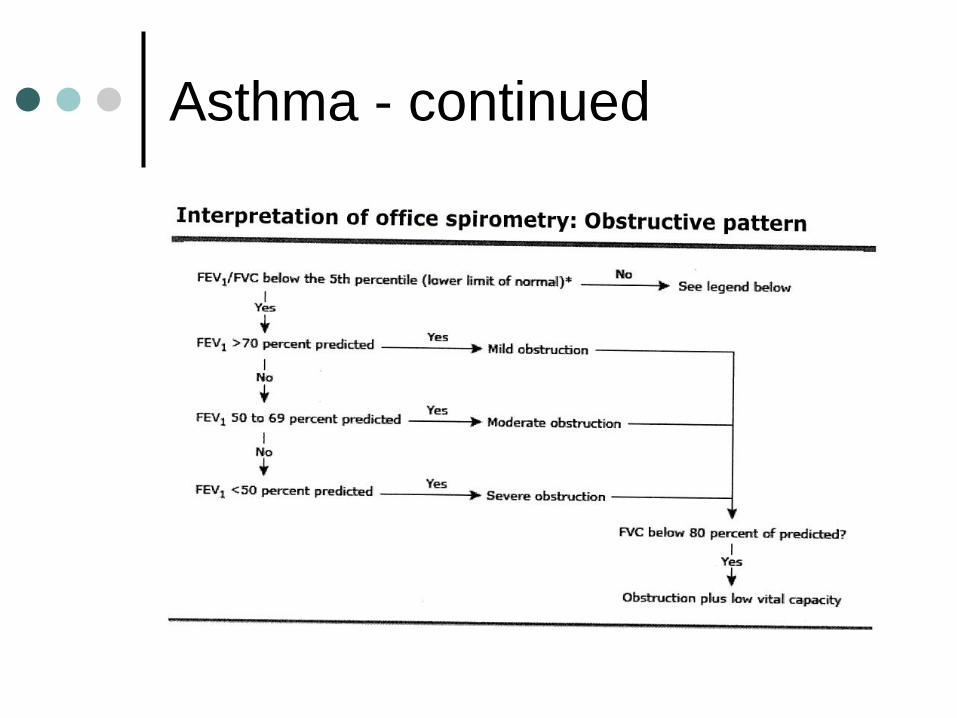
Asthma - continued
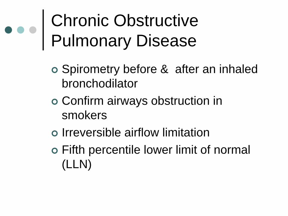
Chronic Obstructive Pulmonary Disease Spirometry before & after an inhaled
bronchodilator Confirm airways obstruction in
smokers Irreversible airflow limitation Fifth percentile lower limit of normal
(LLN)
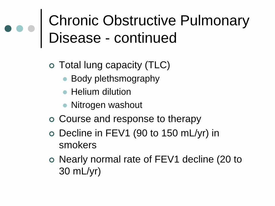
Chronic Obstructive Pulmonary Disease - continued
Total lung capacity (TLC) Body plethsmography Helium dilution Nitrogen washout
Course and response to therapy Decline in FEV1 (90 to 150 mL/yr) in
smokers Nearly normal rate of FEV1 decline (20 to
30 mL/yr)
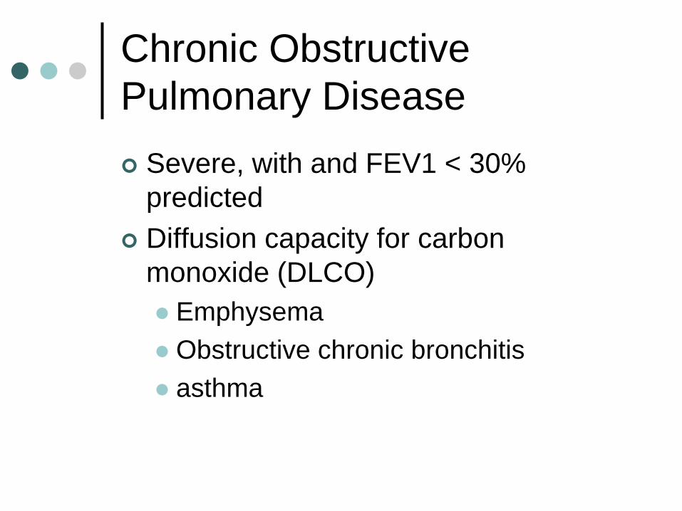
Chronic Obstructive Pulmonary Disease Severe, with and FEV1 < 30%
predicted Diffusion capacity for carbon
monoxide (DLCO) Emphysema Obstructive chronic bronchitis asthma
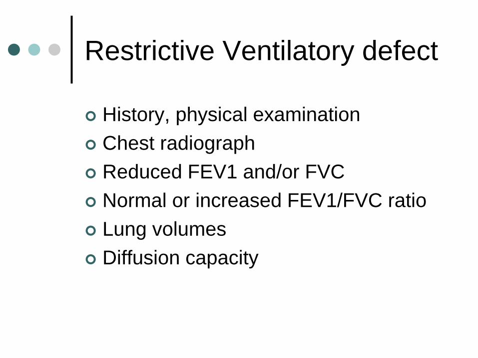
Restrictive Ventilatory defect
History, physical examination Chest radiograph Reduced FEV1 and/or FVC Normal or increased FEV1/FVC ratio Lung volumes Diffusion capacity
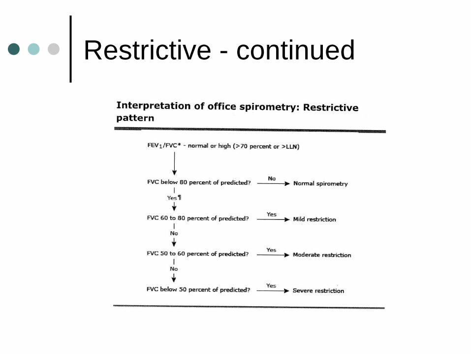
Restrictive - continued
Preoperative testing
COPD or asthma Current smokers Thoracic or upper abdominal surgery Elevated aterial tension of carbon
dioxide (PaCO2) Pneumonia, Prolonged mechanical
ventilation, atelectasis,respiratory failure.
Preoperative testing -continued Surgery can be delayed Should not be used to deny surgery Maximum oxygen uptake
Impairment or disability
Rough indication of an individual’s ability.
Measure maximal oxygen consumption (VO2 max)
Severe impairment Constant severe dyspnea despite
continuous treatment or intermittent extreme dyspnea despite continuous therapy.
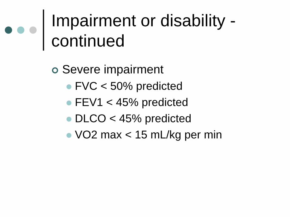
Impairment or disability -continued Severe impairment FVC < 50% predicted FEV1 < 45% predicted DLCO < 45% predicted VO2 max < 15 mL/kg per min