Embed Size (px)
Citation preview
The Christie NHS Foundation Trust
PET-CT in Staging Lung Carcinoma
Dr Rohit Kochhar
Consultant Radiologist
The Christie NHS Foundation Trust
Intended Learning Objectives • Introduction to hybrid imaging
• PET-CT in Lung Cancer
• Assessment-Solitary Pulmonary Nodule
• Revised staging of lung cancer (including 8th Edition of TNM)
• Pitfalls & Limitations of PET-CT Imaging
• Advances and other uses of PET-CT
• Use of IV contrast-one stop shop
• 4D PET-CT
• Prognostication, RT planning and Response Assessment
• Novel Tracers other than FDG
• Take home message-learning points
The Christie NHS Foundation Trust
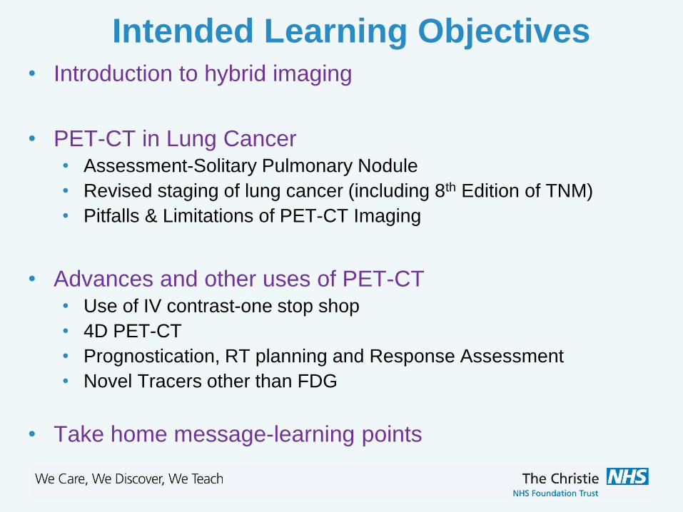
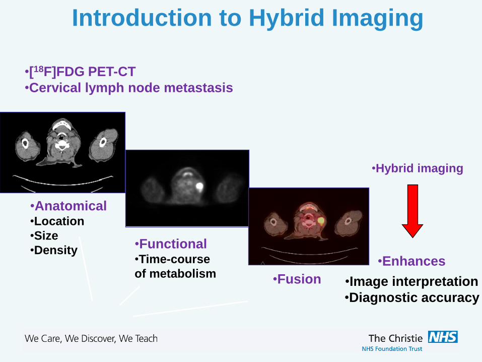
Introduction to Hybrid Imaging
•Anatomical •Location
•Size
•Density •Functional •Time-course
of metabolism •Fusion
•[18F]FDG PET-CT
•Cervical lymph node metastasis
•Hybrid imaging
•Enhances
•Image interpretation
•Diagnostic accuracy
The Christie NHS Foundation Trust
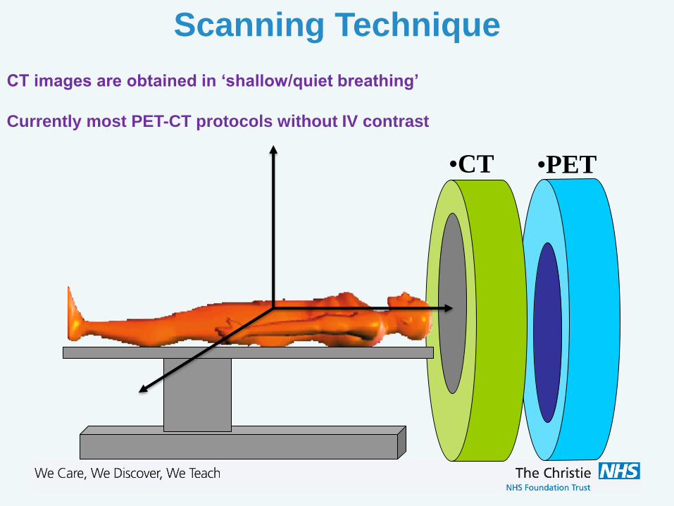
•PET •CT
Scanning Technique
CT images are obtained in ‘shallow/quiet breathing’
Currently most PET-CT protocols without IV contrast
The Christie NHS Foundation Trust
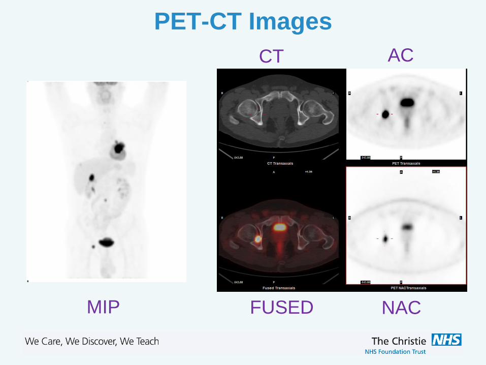
FUSED NAC
CT
MIP
PET-CT Images
AC
The Christie NHS Foundation Trust
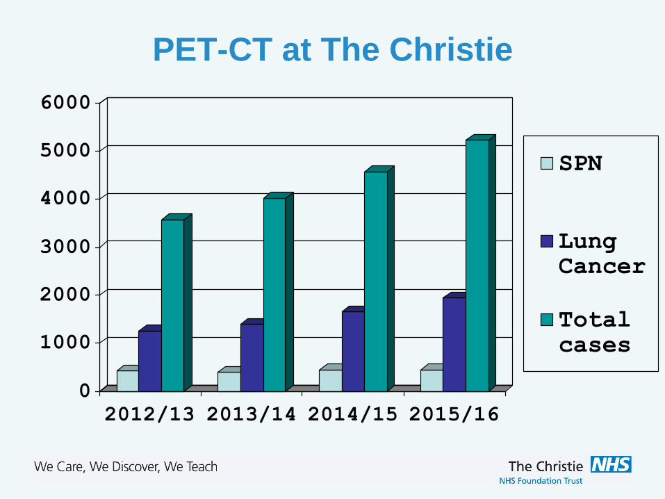
PET-CT at The Christie
0
1000
2000
3000
4000
5000
6000
2012/13 2013/14 2014/15 2015/16
SPN
LungCancer
Totalcases
The Christie NHS Foundation Trust
Solitary Pulmonary Nodule (SPN)
• Definiton
A single spherical lesion of 3 cm or less in diameter completely
surrounded by lung parenchyma without any associated atelectasis or
lymphadenopathy
• The probability of lung cancer increases with tumour size
• The incidence of malignancy in SPN varies widely (5-70%)
• Reliable characterisation frequently not possible on
conventional radiological features – Invasive procedures
AW Tang et al. The solitary pulmonary nodule. Eur J Radiol. 2003;45:69–77
The Christie NHS Foundation Trust
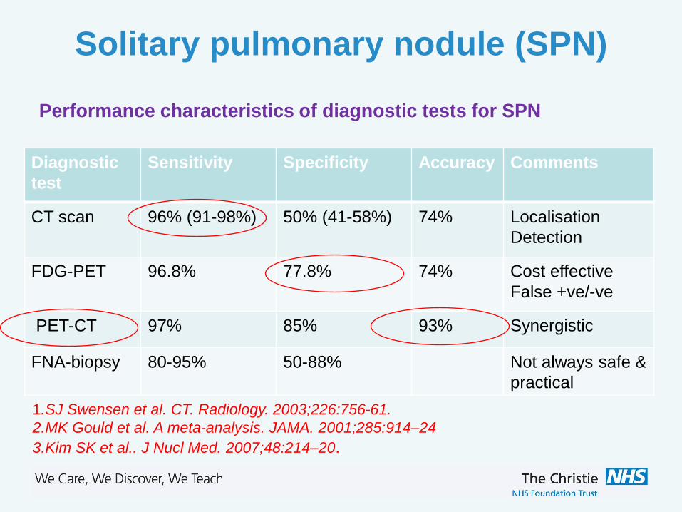
Solitary pulmonary nodule (SPN)
Performance characteristics of diagnostic tests for SPN
Diagnostic
test
Sensitivity Specificity Accuracy Comments
CT scan 96% (91-98%) 50% (41-58%) 74% Localisation
Detection
FDG-PET 96.8% 77.8% 74% Cost effective
False +ve/-ve
PET-CT 97% 85% 93% Synergistic
FNA-biopsy 80-95% 50-88% Not always safe &
practical
1.SJ Swensen et al. CT. Radiology. 2003;226:756-61.
2.MK Gould et al. A meta-analysis. JAMA. 2001;285:914–24
3.Kim SK et al.. J Nucl Med. 2007;48:214–20.
The Christie NHS Foundation Trust
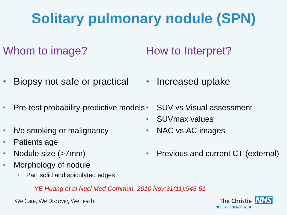
Solitary pulmonary nodule (SPN)
Whom to image?
• Biopsy not safe or practical
• Pre-test probability-predictive models
• h/o smoking or malignancy
• Patients age
• Nodule size (>7mm)
• Morphology of nodule
• Part solid and spiculated edges
How to Interpret?
• Increased uptake
• SUV vs Visual assessment
• SUVmax values
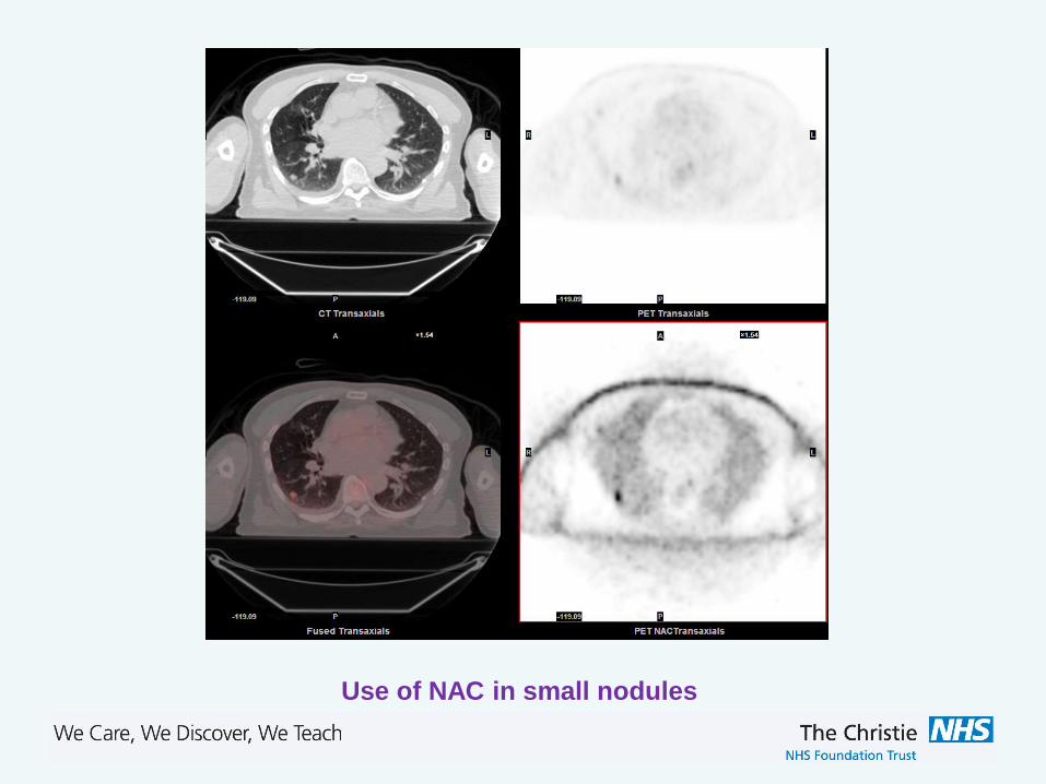
• NAC vs AC images
• Previous and current CT (external)
YE Huang et al Nucl Med Commun. 2010 Nov;31(11):945-51
The Christie NHS Foundation Trust
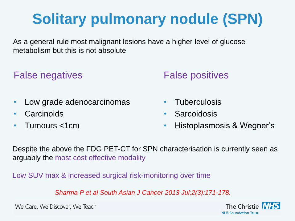
Solitary pulmonary nodule (SPN)
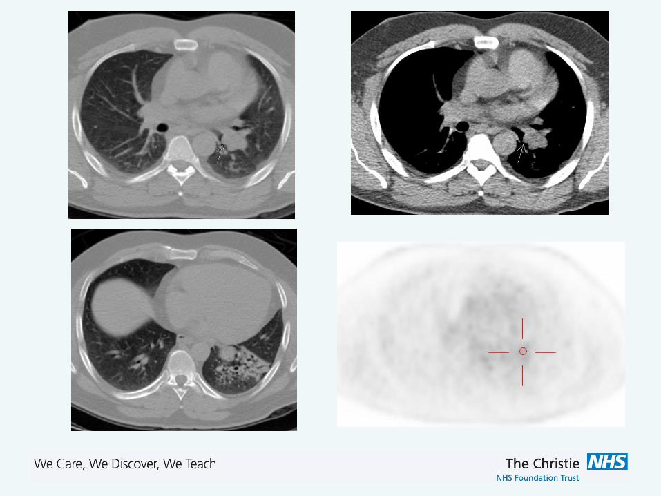
False negatives
• Low grade adenocarcinomas
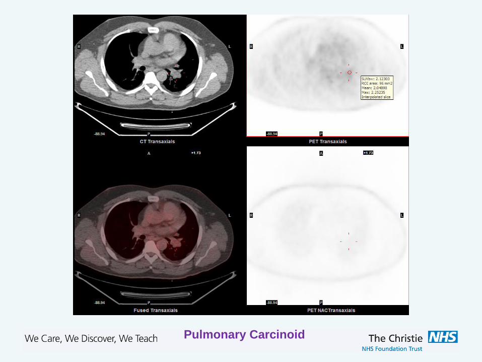
• Carcinoids
• Tumours <1cm
False positives
• Tuberculosis
• Sarcoidosis
• Histoplasmosis & Wegner’s
As a general rule most malignant lesions have a higher level of glucose
metabolism but this is not absolute
Despite the above the FDG PET-CT for SPN characterisation is currently seen as
arguably the most cost effective modality
Low SUV max & increased surgical risk-monitoring over time
Sharma P et al South Asian J Cancer 2013 Jul;2(3):171-178.
The Christie NHS Foundation Trust
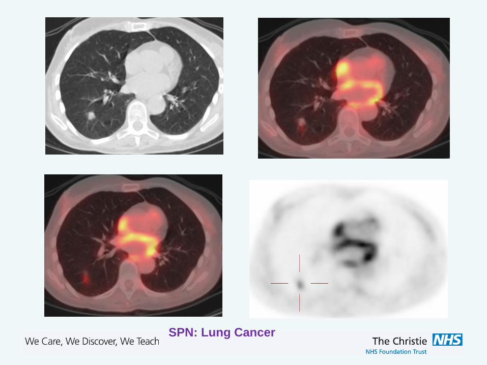
SPN: Lung Cancer
The Christie NHS Foundation Trust
Use of NAC in small nodules
The Christie NHS Foundation Trust
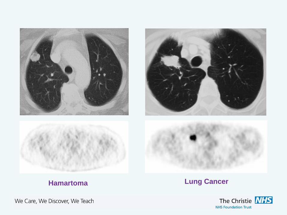
Hamartoma Lung Cancer
The Christie NHS Foundation Trust
The Christie NHS Foundation Trust
Pulmonary Carcinoid
The Christie NHS Foundation Trust
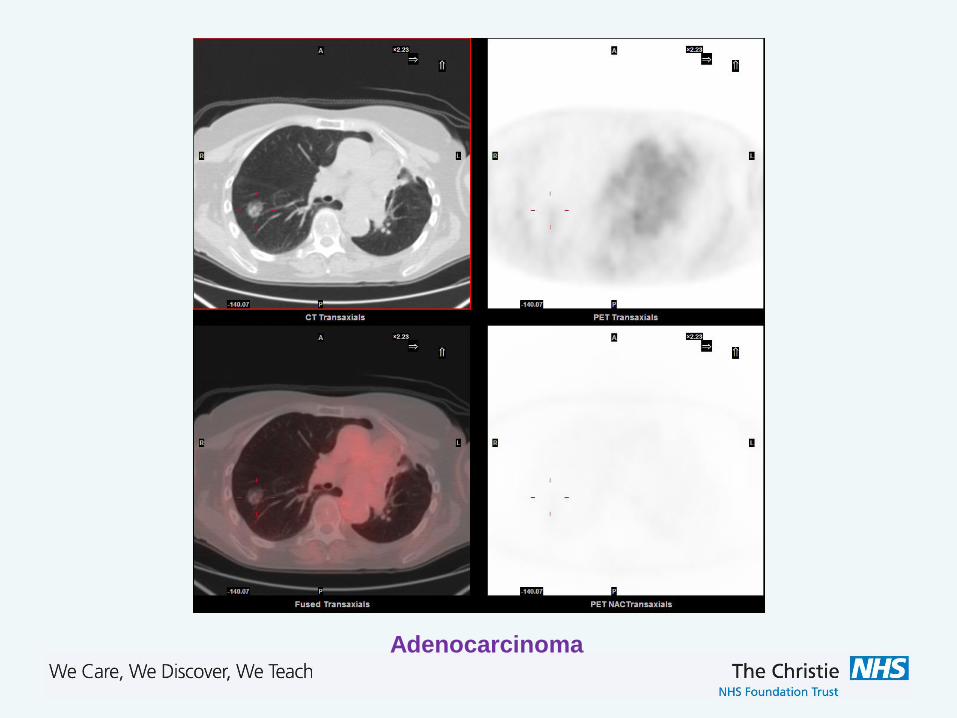
Adenocarcinoma
The Christie NHS Foundation Trust
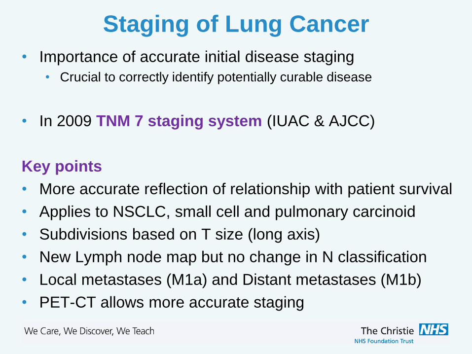
Staging of Lung Cancer
• Importance of accurate initial disease staging
• Crucial to correctly identify potentially curable disease
• In 2009 TNM 7 staging system (IUAC & AJCC)
Key points
• More accurate reflection of relationship with patient survival
• Applies to NSCLC, small cell and pulmonary carcinoid
• Subdivisions based on T size (long axis)
• New Lymph node map but no change in N classification
• Local metastases (M1a) and Distant metastases (M1b)
• PET-CT allows more accurate staging
The Christie NHS Foundation Trust
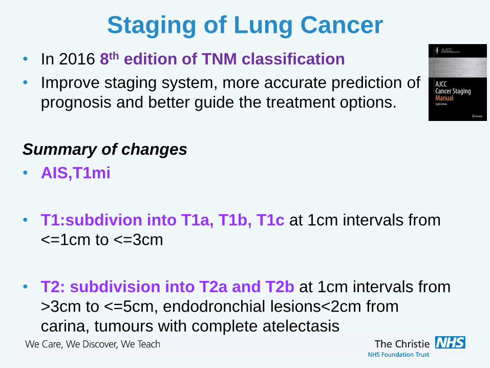
Staging of Lung Cancer
• In 2016 8th edition of TNM classification
• Improve staging system, more accurate prediction of
prognosis and better guide the treatment options.
Summary of changes
• AIS,T1mi
• T1:subdivion into T1a, T1b, T1c at 1cm intervals from
<=1cm to <=3cm
• T2: subdivision into T2a and T2b at 1cm intervals from
>3cm to <=5cm, endodronchial lesions<2cm from
carina, tumours with complete atelectasis
The Christie NHS Foundation Trust
Staging of Lung Cancer
Summary of changes
• T3: >5cm but <=7cm
• T3: Invasion of mediastinal pleura is no longer a predictor
• T4: >7cm, invasion of diaphragm
• M1b: single extrathoracic metastasis in a single organ
• M1c: new category, multiple extrathoracic metastases in one
or multiple organs
The Christie NHS Foundation Trust
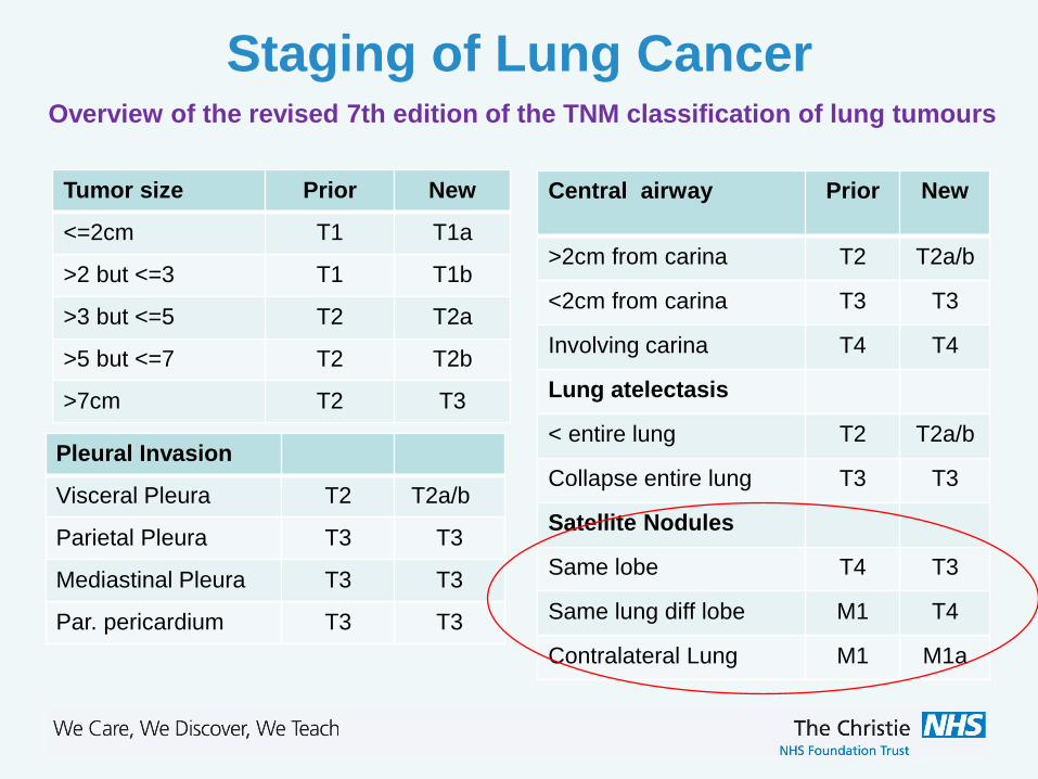
Staging of Lung Cancer
Tumor size Prior New
<=2cm T1 T1a
>2 but <=3 T1 T1b
>3 but <=5 T2 T2a
>5 but <=7 T2 T2b
>7cm T2 T3
Pleural Invasion
Visceral Pleura T2 T2a/b
Parietal Pleura T3 T3
Mediastinal Pleura T3 T3
Par. pericardium T3 T3
Overview of the revised 7th edition of the TNM classification of lung tumours
Central airway Prior New
>2cm from carina T2 T2a/b
<2cm from carina T3 T3
Involving carina T4 T4
Lung atelectasis
< entire lung T2 T2a/b
Collapse entire lung T3 T3
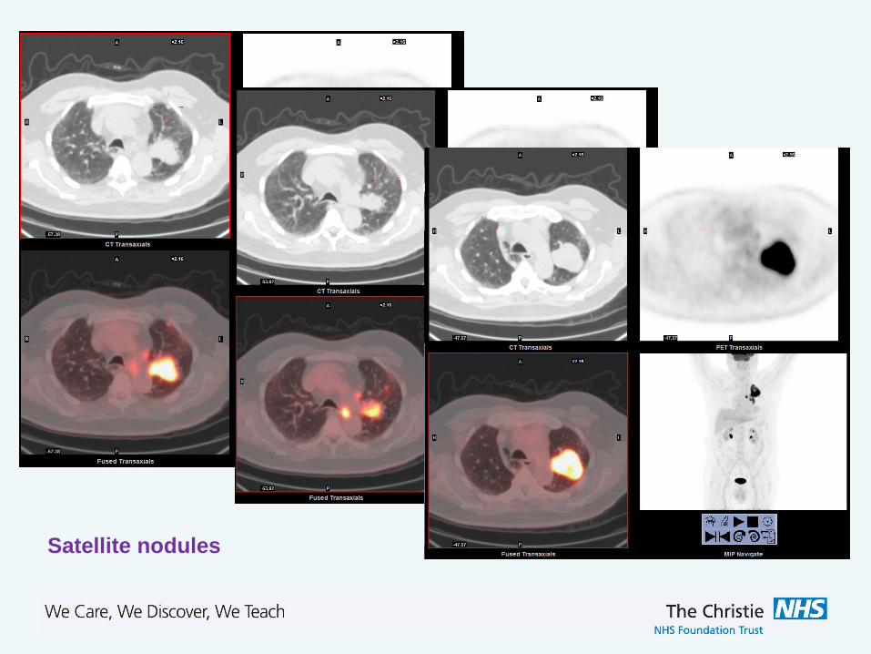
Satellite Nodules
Same lobe T4 T3
Same lung diff lobe M1 T4
Contralateral Lung M1 M1a
The Christie NHS Foundation Trust
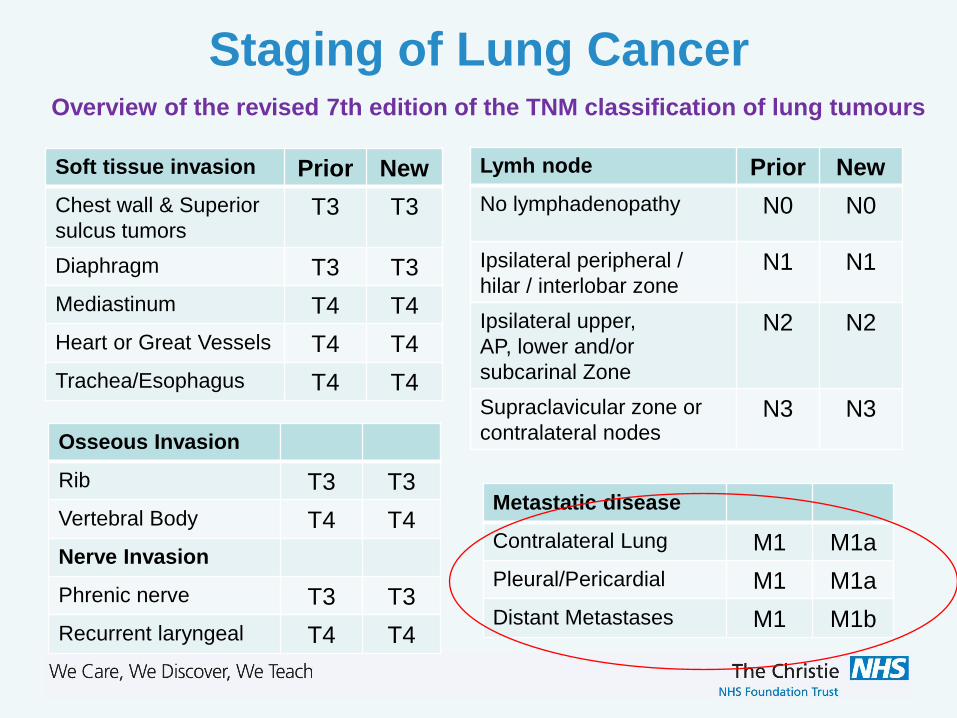
Staging of Lung Cancer
Soft tissue invasion Prior New
Chest wall & Superior
sulcus tumors T3 T3
Diaphragm T3 T3
Mediastinum T4 T4
Heart or Great Vessels T4 T4
Trachea/Esophagus T4 T4
Overview of the revised 7th edition of the TNM classification of lung tumours
Osseous Invasion
Rib T3 T3
Vertebral Body T4 T4
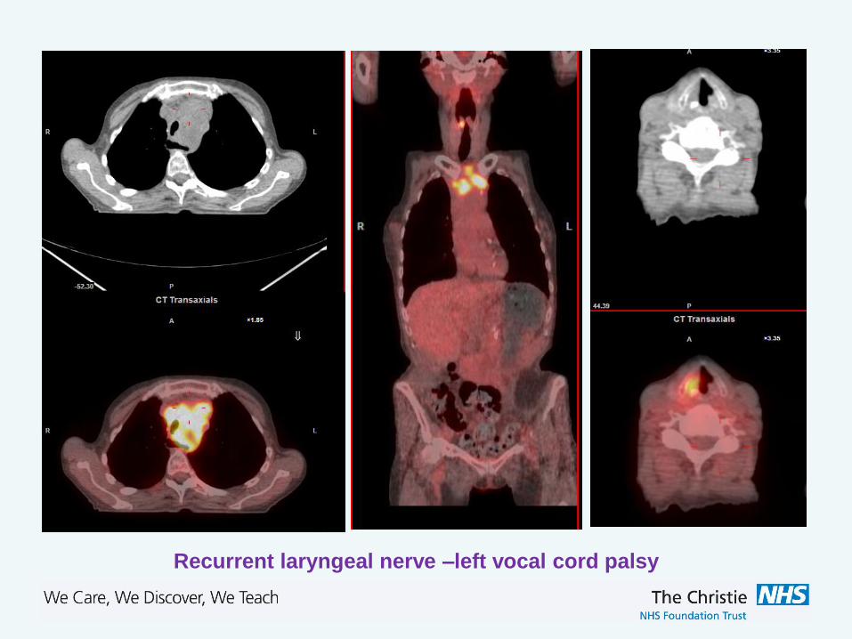
Nerve Invasion
Phrenic nerve T3 T3
Recurrent laryngeal T4 T4
Lymh node Prior New
No lymphadenopathy N0 N0
Ipsilateral peripheral /
hilar / interlobar zone N1 N1
Ipsilateral upper,
AP, lower and/or
subcarinal Zone
N2 N2
Supraclavicular zone or
contralateral nodes N3 N3
Metastatic disease
Contralateral Lung M1 M1a
Pleural/Pericardial M1 M1a
Distant Metastases M1 M1b
The Christie NHS Foundation Trust
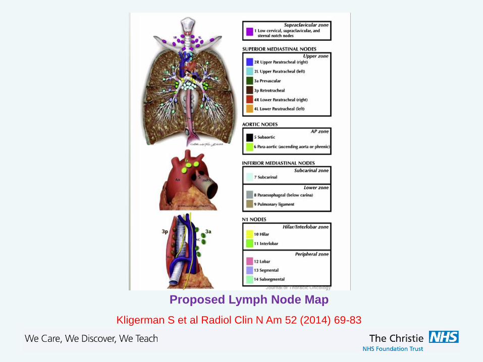
Proposed Lymph Node Map
Kligerman S et al Radiol Clin N Am 52 (2014) 69-83
The Christie NHS Foundation Trust
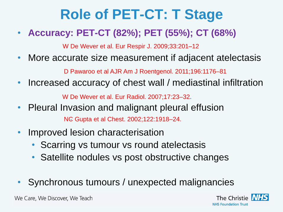
Role of PET-CT: T Stage
• Accuracy: PET-CT (82%); PET (55%); CT (68%)
• More accurate size measurement if adjacent atelectasis
• Increased accuracy of chest wall / mediastinal infiltration
• Pleural Invasion and malignant pleural effusion
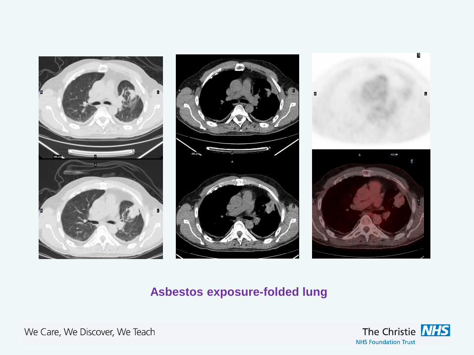
• Improved lesion characterisation
• Scarring vs tumour vs round atelectasis
• Satellite nodules vs post obstructive changes
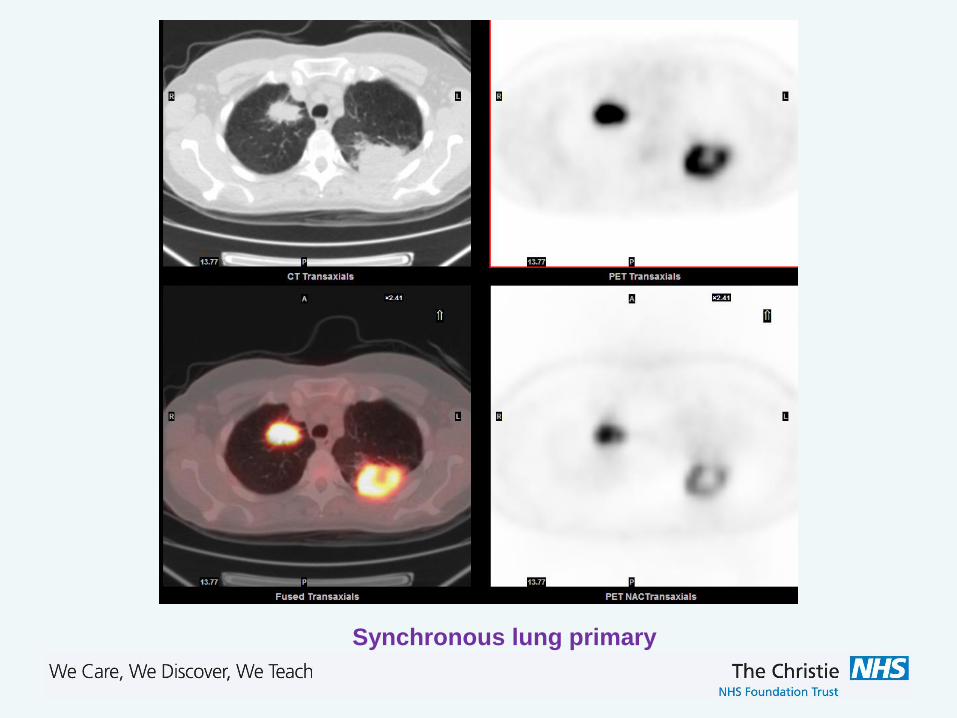
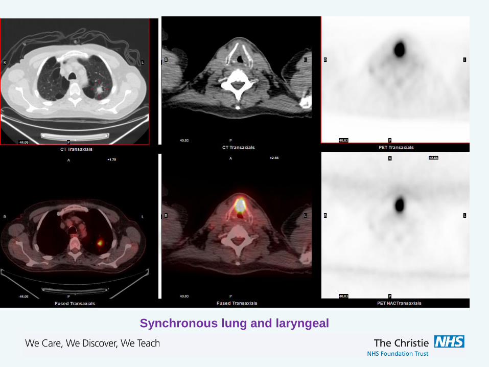
• Synchronous tumours / unexpected malignancies
W De Wever et al. Eur Respir J. 2009;33:201–12
D Pawaroo et al AJR Am J Roentgenol. 2011;196:1176–81
W De Wever et al. Eur Radiol. 2007;17:23–32.
. NC Gupta et al Chest. 2002;122:1918–24.
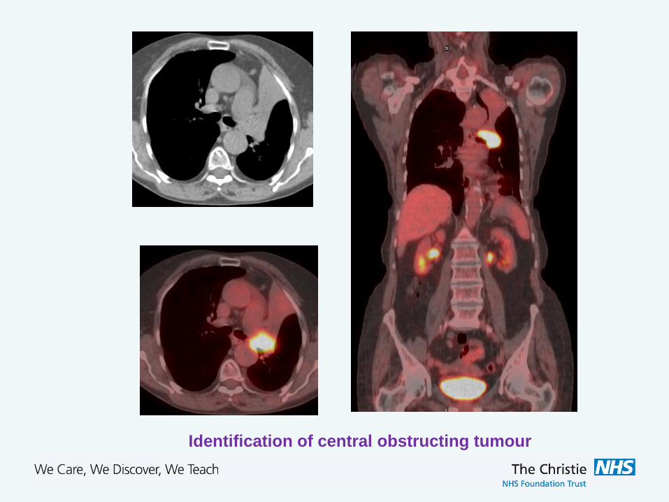
The Christie NHS Foundation Trust
Identification of central obstructing tumour
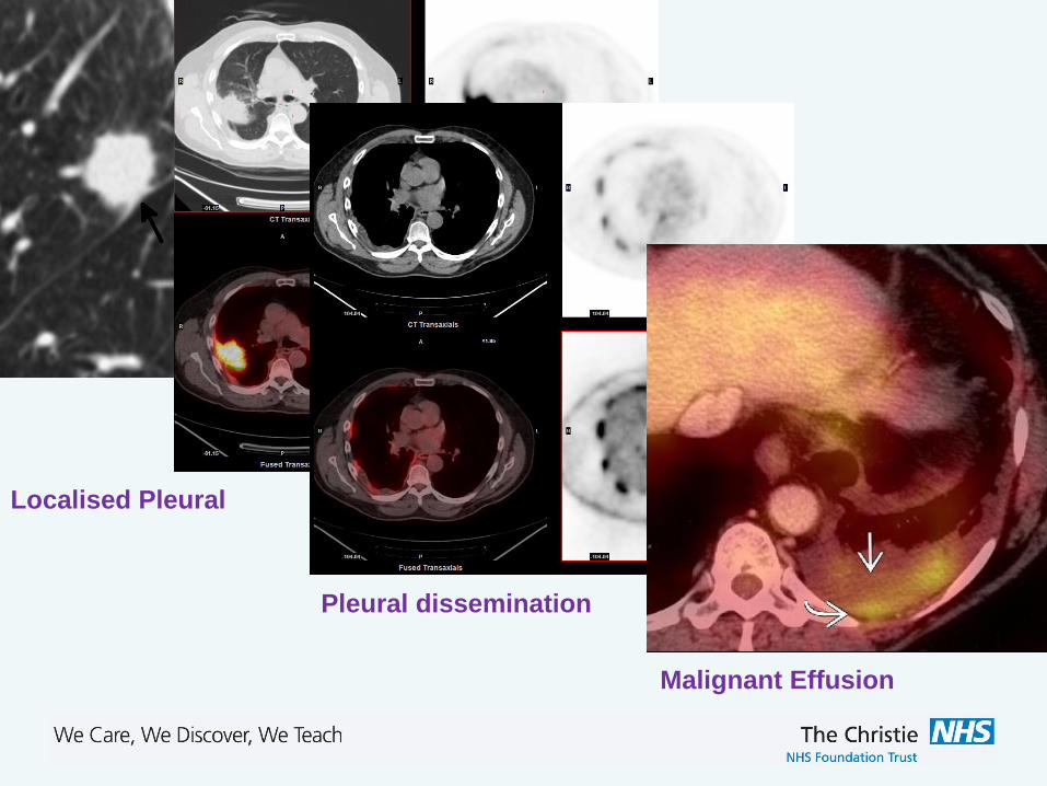
The Christie NHS Foundation Trust
Pleural dissemination
Localised Pleural
Malignant Effusion
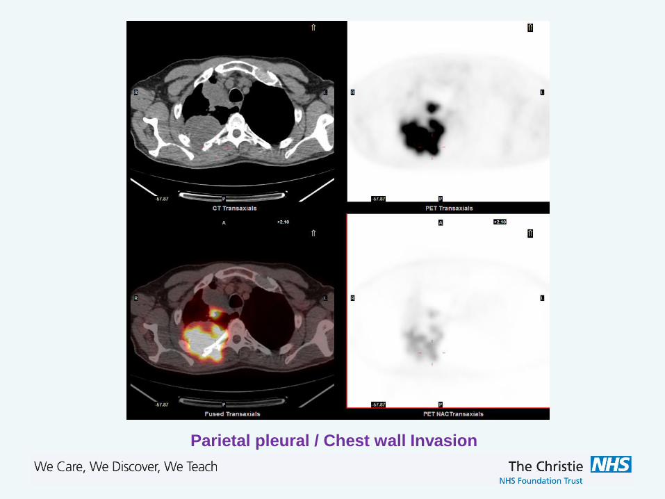
The Christie NHS Foundation Trust
•Examples of T3
Parietal pleural / Chest wall Invasion
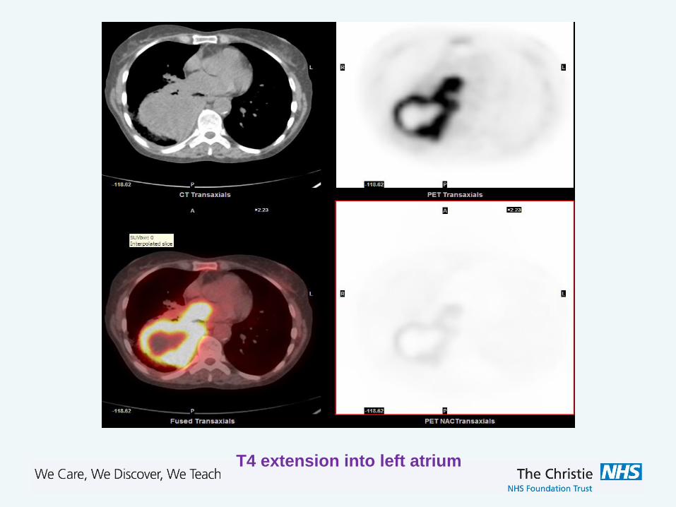
The Christie NHS Foundation Trust
T4 extension into left atrium
The Christie NHS Foundation Trust
Recurrent laryngeal nerve –left vocal cord palsy
The Christie NHS Foundation Trust
•Examples of T3
Satellite nodules
The Christie NHS Foundation Trust
Asbestos exposure-folded lung
The Christie NHS Foundation Trust
Synchronous lung primary
The Christie NHS Foundation Trust
Synchronous lung and laryngeal
The Christie NHS Foundation Trust
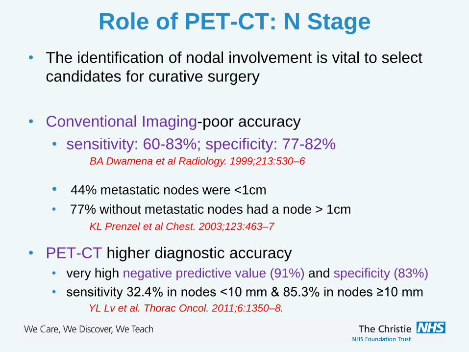
Role of PET-CT: N Stage
• The identification of nodal involvement is vital to select
candidates for curative surgery
• Conventional Imaging-poor accuracy
• sensitivity: 60-83%; specificity: 77-82%
• 44% metastatic nodes were <1cm
• 77% without metastatic nodes had a node > 1cm
• PET-CT higher diagnostic accuracy
• very high negative predictive value (91%) and specificity (83%)
• sensitivity 32.4% in nodes <10 mm & 85.3% in nodes ≥10 mm
BA Dwamena et al Radiology. 1999;213:530–6
KL Prenzel et al Chest. 2003;123:463–7
YL Lv et al. Thorac Oncol. 2011;6:1350–8.
The Christie NHS Foundation Trust
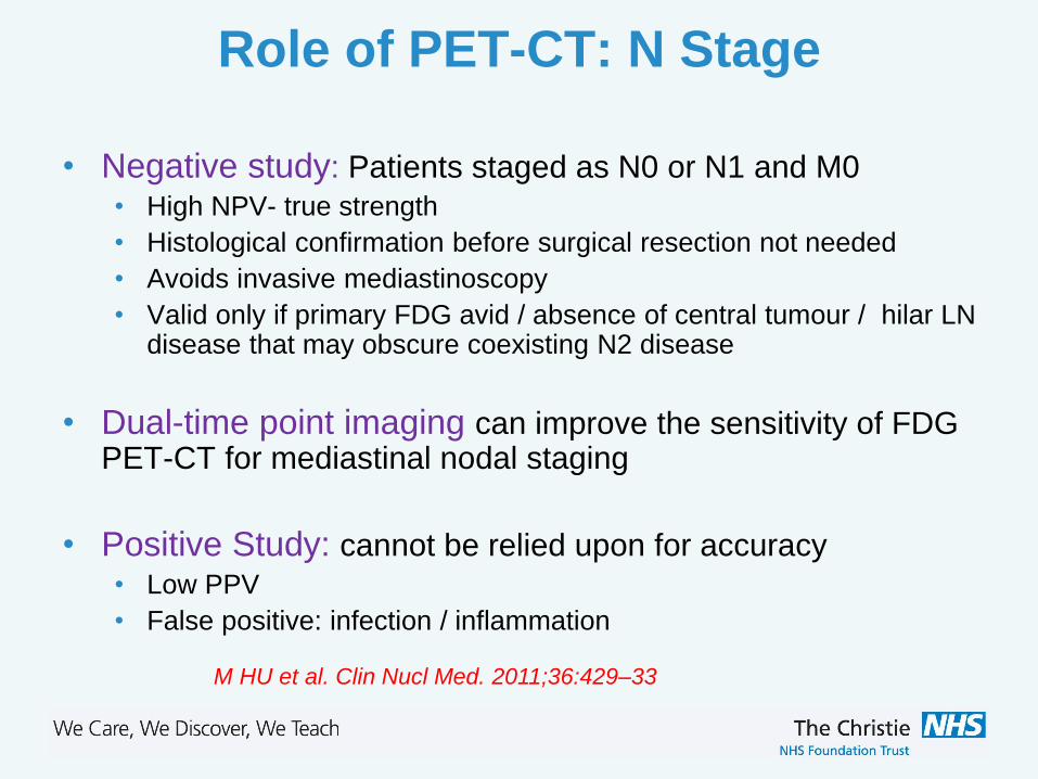
Role of PET-CT: N Stage
• Negative study: Patients staged as N0 or N1 and M0
• High NPV- true strength
• Histological confirmation before surgical resection not needed
• Avoids invasive mediastinoscopy
• Valid only if primary FDG avid / absence of central tumour / hilar LN disease that may obscure coexisting N2 disease
• Dual-time point imaging can improve the sensitivity of FDG PET-CT for mediastinal nodal staging
• Positive Study: cannot be relied upon for accuracy
• Low PPV
• False positive: infection / inflammation
M HU et al. Clin Nucl Med. 2011;36:429–33
The Christie NHS Foundation Trust
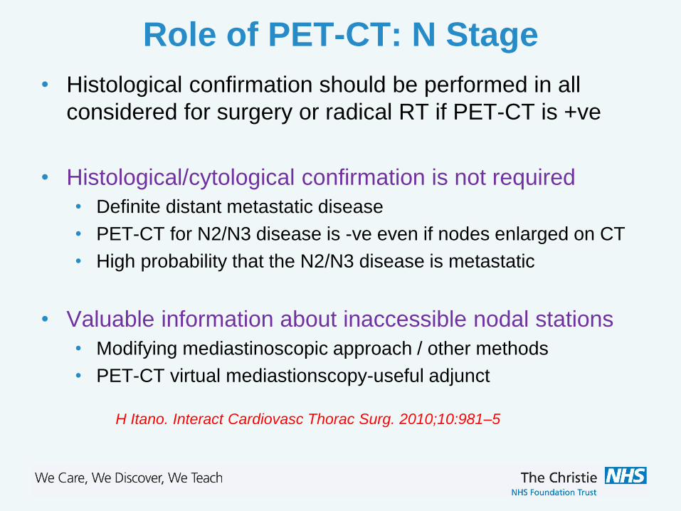
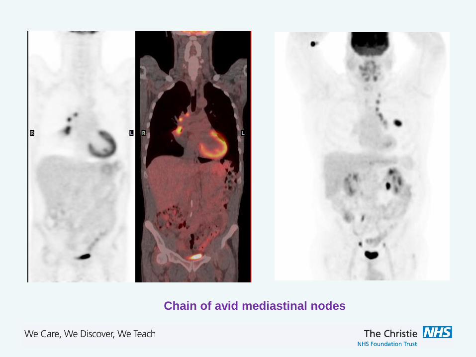
Role of PET-CT: N Stage
• Histological confirmation should be performed in all
considered for surgery or radical RT if PET-CT is +ve
• Histological/cytological confirmation is not required
• Definite distant metastatic disease
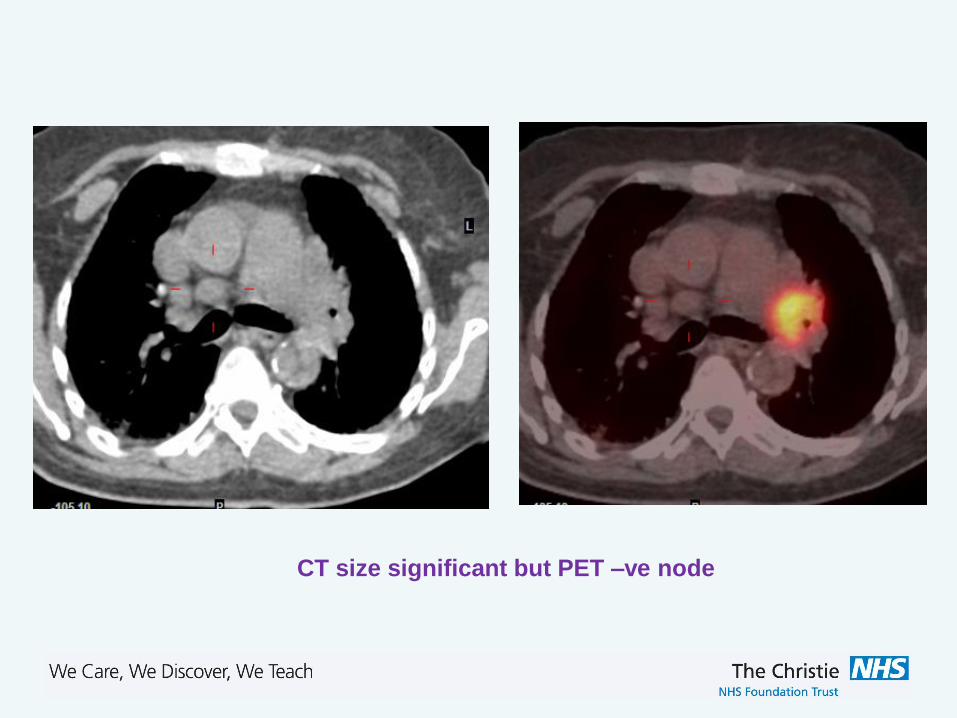
• PET-CT for N2/N3 disease is -ve even if nodes enlarged on CT
• High probability that the N2/N3 disease is metastatic
• Valuable information about inaccessible nodal stations
• Modifying mediastinoscopic approach / other methods
• PET-CT virtual mediastionscopy-useful adjunct
H Itano. Interact Cardiovasc Thorac Surg. 2010;10:981–5
The Christie NHS Foundation Trust
Chain of avid mediastinal nodes
The Christie NHS Foundation Trust
CT size significant but PET –ve node
The Christie NHS Foundation Trust
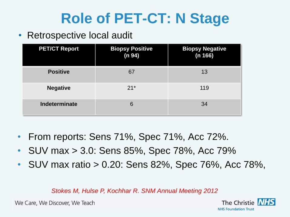
Role of PET-CT: N Stage
Stokes M, Hulse P, Kochhar R. SNM Annual Meeting 2012
PET/CT Report Biopsy Positive
(n 94)
Biopsy Negative
(n 166)
Positive 67 13
Negative 21* 119
Indeterminate 6 34
• From reports: Sens 71%, Spec 71%, Acc 72%.
• SUV max > 3.0: Sens 85%, Spec 78%, Acc 79%
• SUV max ratio > 0.20: Sens 82%, Spec 76%, Acc 78%,
• Retrospective local audit
The Christie NHS Foundation Trust
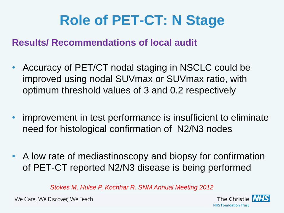
Role of PET-CT: N Stage
Results/ Recommendations of local audit
• Accuracy of PET/CT nodal staging in NSCLC could be
improved using nodal SUVmax or SUVmax ratio, with
optimum threshold values of 3 and 0.2 respectively
• improvement in test performance is insufficient to eliminate
need for histological confirmation of N2/N3 nodes
• A low rate of mediastinoscopy and biopsy for confirmation
of PET-CT reported N2/N3 disease is being performed
Stokes M, Hulse P, Kochhar R. SNM Annual Meeting 2012
The Christie NHS Foundation Trust
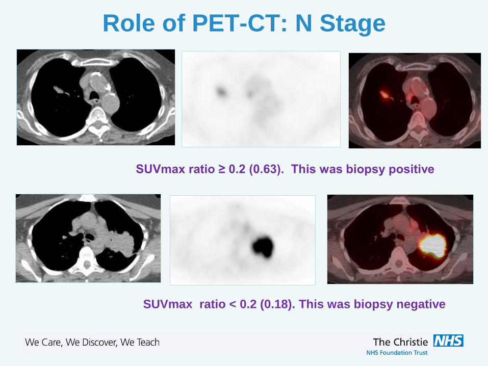
Role of PET-CT: N Stage
SUVmax ratio ≥ 0.2 (0.63). This was biopsy positive
SUVmax ratio < 0.2 (0.18). This was biopsy negative
The Christie NHS Foundation Trust
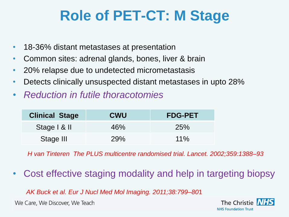
Role of PET-CT: M Stage
• 18-36% distant metastases at presentation
• Common sites: adrenal glands, bones, liver & brain
• 20% relapse due to undetected micrometastasis
• Detects clinically unsuspected distant metastases in upto 28%
• Reduction in futile thoracotomies
• Cost effective staging modality and help in targeting biopsy
Clinical Stage CWU FDG-PET
Stage I & II 46% 25%
Stage III 29% 11%
H van Tinteren The PLUS multicentre randomised trial. Lancet. 2002;359:1388–93
AK Buck et al. Eur J Nucl Med Mol Imaging. 2011;38:799–801
The Christie NHS Foundation Trust
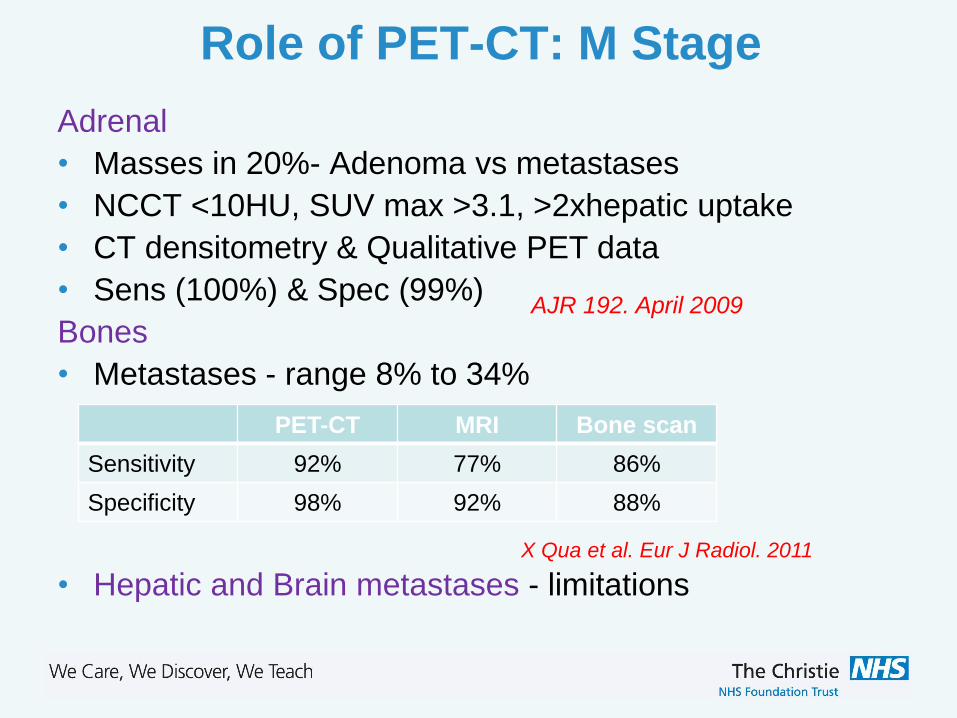
Role of PET-CT: M Stage
Adrenal
• Masses in 20%- Adenoma vs metastases
• NCCT <10HU, SUV max >3.1, >2xhepatic uptake
• CT densitometry & Qualitative PET data
• Sens (100%) & Spec (99%)
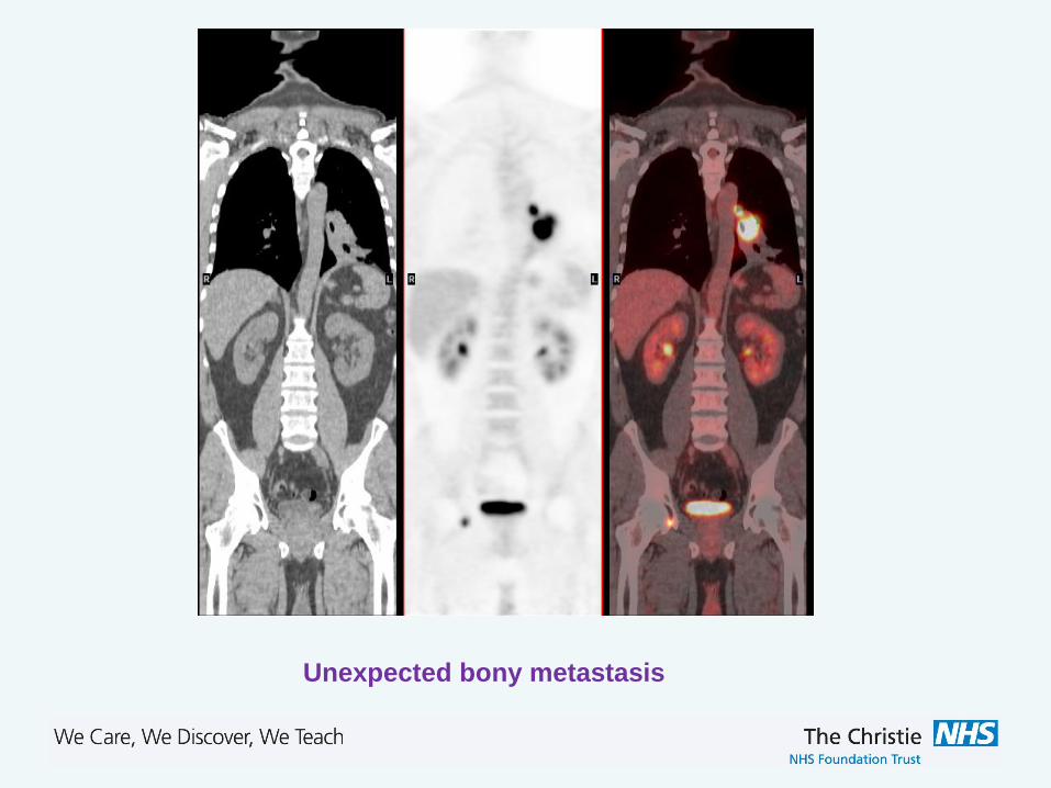
Bones
• Metastases - range 8% to 34%
• Hepatic and Brain metastases - limitations
AJR 192. April 2009
PET-CT MRI Bone scan
Sensitivity 92% 77% 86%
Specificity 98% 92% 88%
X Qua et al. Eur J Radiol. 2011
The Christie NHS Foundation Trust
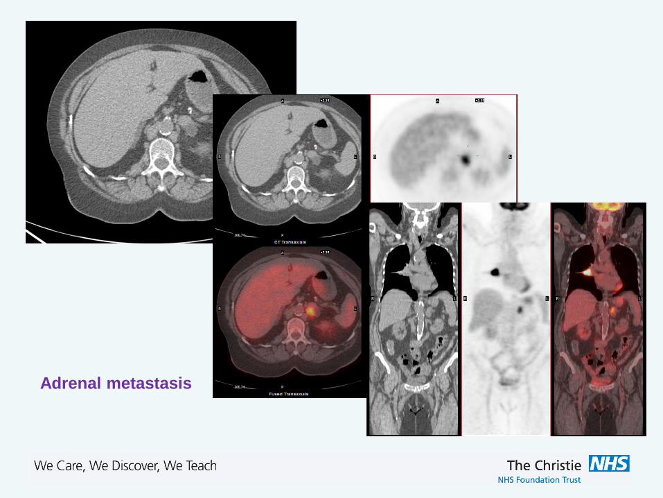
Adrenal metastasis
The Christie NHS Foundation Trust
Unexpected bony metastasis
The Christie NHS Foundation Trust
Role of PET-CT
Small Cell Lung Cancer
• Clinically more aggressive than NSCLC
• 60% to 70% extensive disease at presentation
• Data on SCLC with PET-CT limited
• Modification of stage & management in 10% to 33%
• Staging with PET-CT may separate favourable LS group
A Azad et al. Mol Imaging Biol. 2010;12:443–51
The Christie NHS Foundation Trust
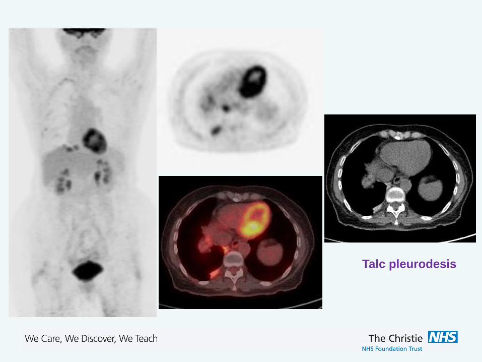
Pitfall and Limitations of PET-CT
False Positives
• Physiological
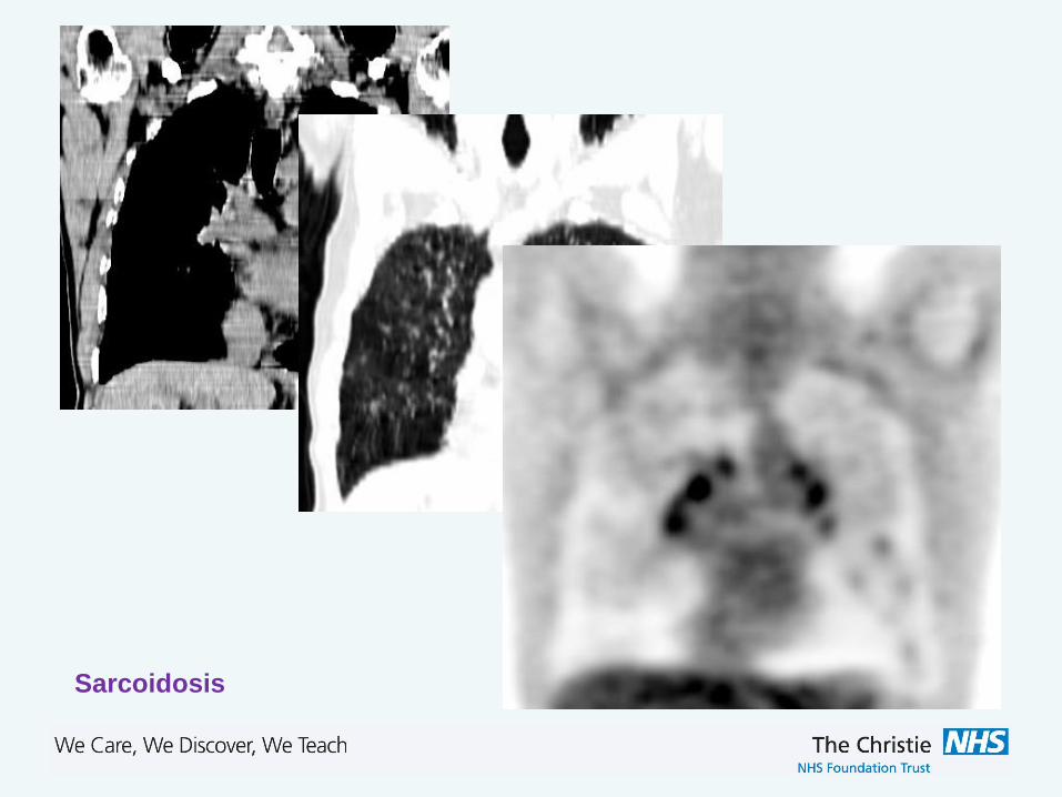
• Inflammation/Infection
• Sarcoidosis, TB, Wegeners
• Infarction
• Embolus
• Iatrogenic
• Pleurodesis
• Post Treatment
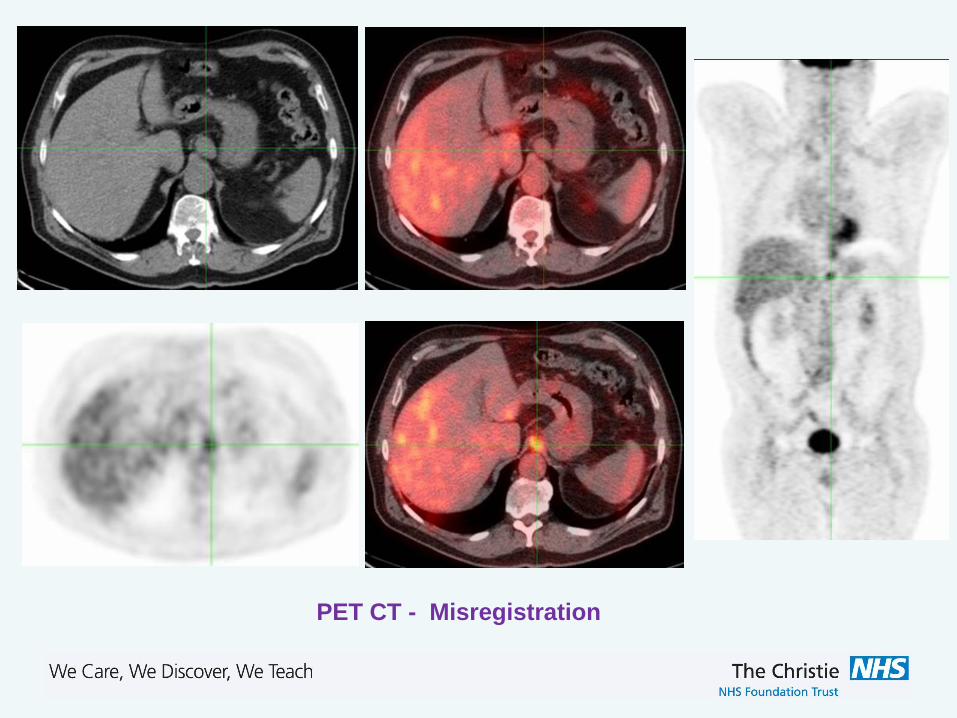
False Negatives
• Small size
• micrometastasis
• Low metabolic tumours
• Carcinoid
• Well diff adenocarcinoma
• Technical factors
• Misregistration
• Glucose serum levels
The Christie NHS Foundation Trust
Sarcoidosis
The Christie NHS Foundation Trust
Talc pleurodesis
The Christie NHS Foundation Trust
PET CT - Misregistration
The Christie NHS Foundation Trust
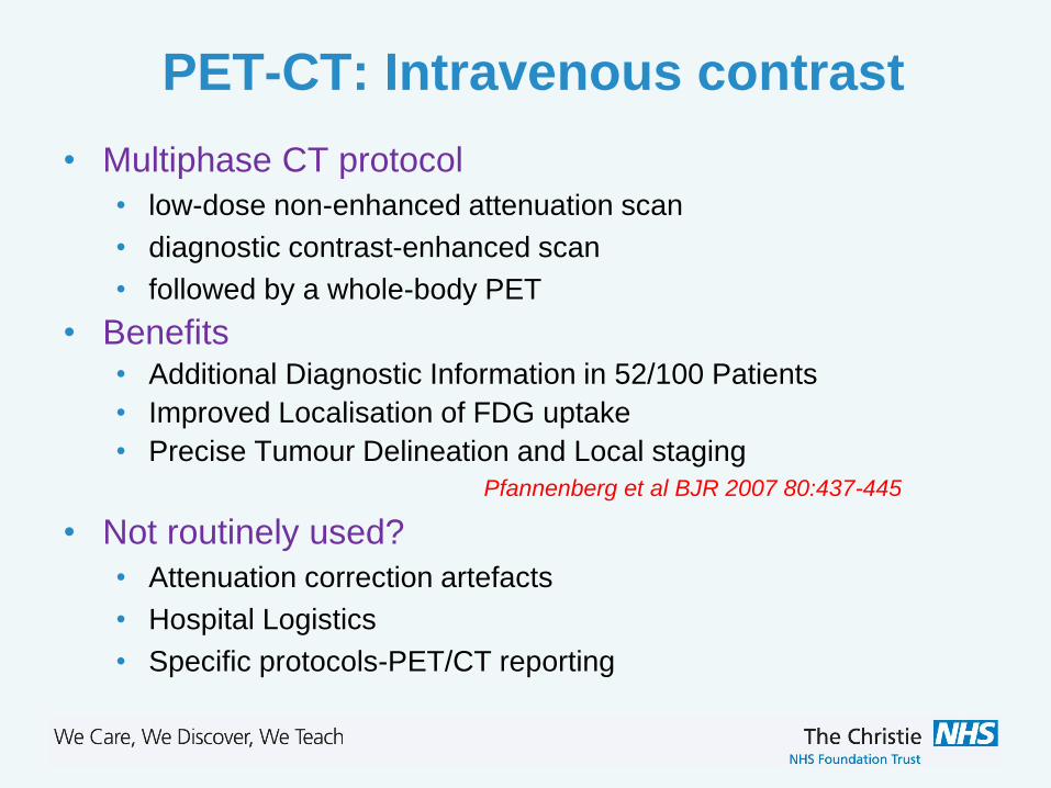
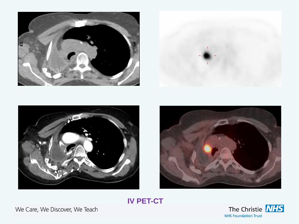
PET-CT: Intravenous contrast
• Multiphase CT protocol
• low-dose non-enhanced attenuation scan
• diagnostic contrast-enhanced scan
• followed by a whole-body PET
• Benefits • Additional Diagnostic Information in 52/100 Patients
• Improved Localisation of FDG uptake
• Precise Tumour Delineation and Local staging
Pfannenberg et al BJR 2007 80:437-445
• Not routinely used?
• Attenuation correction artefacts
• Hospital Logistics
• Specific protocols-PET/CT reporting
The Christie NHS Foundation Trust
IV PET-CT
The Christie NHS Foundation Trust
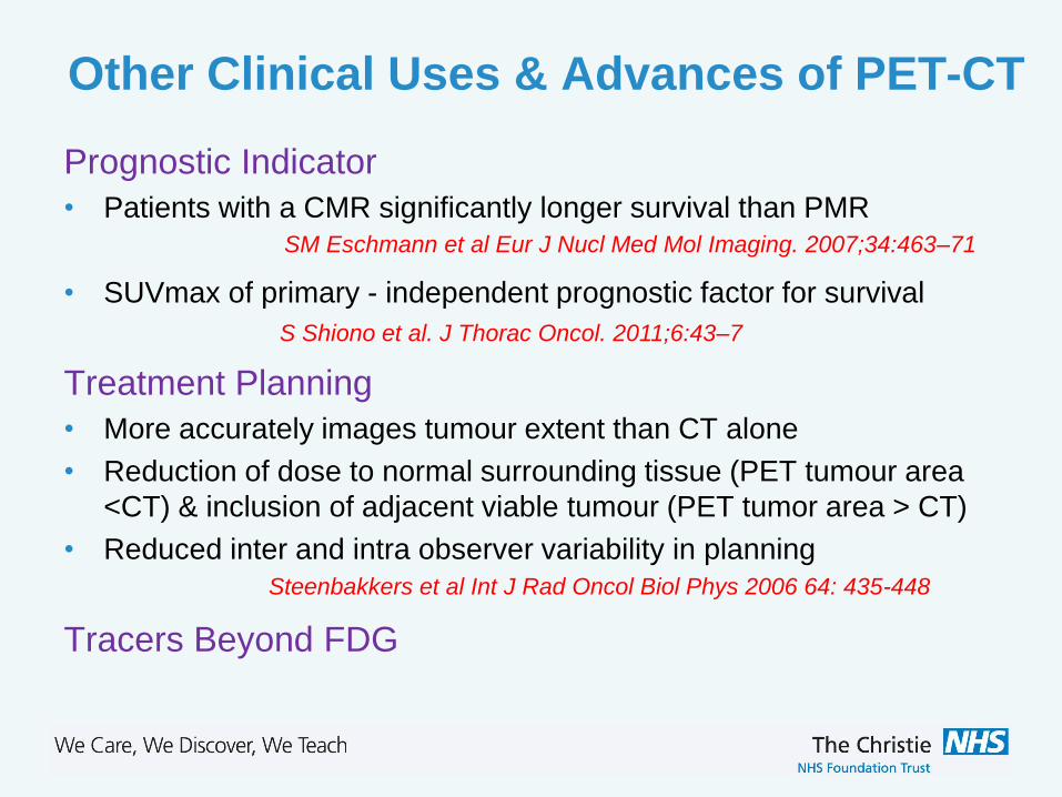
Other Clinical Uses & Advances of PET-CT
Prognostic Indicator
• Patients with a CMR significantly longer survival than PMR
• SUVmax of primary - independent prognostic factor for survival
Treatment Planning
• More accurately images tumour extent than CT alone
• Reduction of dose to normal surrounding tissue (PET tumour area
<CT) & inclusion of adjacent viable tumour (PET tumor area > CT)
• Reduced inter and intra observer variability in planning
Tracers Beyond FDG
SM Eschmann et al Eur J Nucl Med Mol Imaging. 2007;34:463–71
S Shiono et al. J Thorac Oncol. 2011;6:43–7
Steenbakkers et al Int J Rad Oncol Biol Phys 2006 64: 435-448
The Christie NHS Foundation Trust
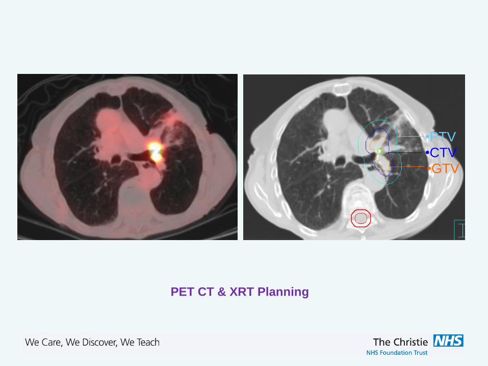
PET CT & XRT Planning
•GTV
•CTV •PTV
The Christie NHS Foundation Trust
Conclusions
• PET-CT has established itself as an important step in the
management of patients with lung cancer
• Useful in characterisation & risk stratification of SPN
• Definite role in staging Lung cancer
• Most accurate and cost effective modality
• Avoid futile thoracotomies and guide biopsies
• Must remember limitations of PET-CT
• PET-CT stage is not the pathological stage
• High negative predictive value-micrometastases
• Histological confirmation of all suspected N2/N3 disease
• Role in RT planning, response monitoring, prognostication

The Christie NHS Foundation Trust
THANK YOU