Embed Size (px)
Citation preview
Remedy Publications LLC., | http://clinicsinsurgery.com/
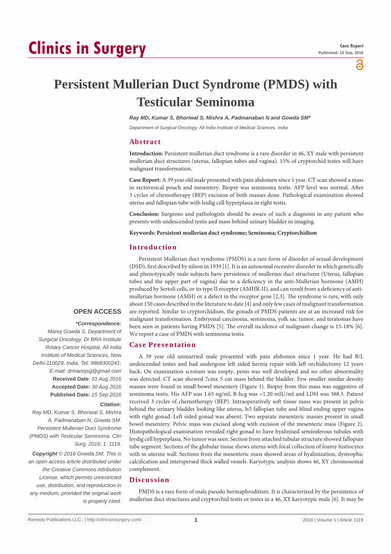
Clinics in Surgery
2016 | Volume 1 | Article 11191
IntroductionPersistent Mullerian duct syndrome (PMDS) is a rare form of disorder of sexual development
(DSD), first described by nilson in 1939 [1]. It is an autosomal recessive disorder in which genetically and phenotypically male subjects have persistence of mullerian duct structures (Uterus, fallopian tubes and the upper part of vagina) due to a deficiency in the anti-Mullerian hormone (AMH) produced by Sertoli cells, or its type II receptor (AMHR-II), and can result from a deficiency of anti-mullerian hormone (AMH) or a defect in the receptor gene [2,3]. The syndrome is rare, with only about 150 cases described in the literature to date [4] and only few cases of malignant transformation are reported. Similar to cryptorchidism, the gonads of PMDS patients are at an increased risk for malignant transformation. Embryonal carcinoma, seminoma, yolk sac tumor, and teratomas have been seen in patients having PMDS [5]. The overall incidence of malignant change is 15-18% [6]. We report a case of PMDS with seminoma testis.
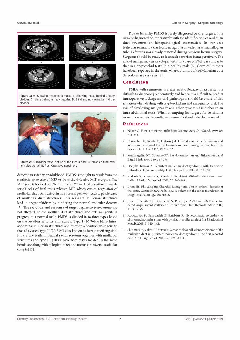
Case PresentationA 39 year old unmarried male presented with pain abdomen since 1 year. He had B/L
undescended testes and had undergone left sided hernia repair with left orchidectomy 12 years back. On examination scrotum was empty, penis was well developed and no other abnormality was detected. CT scan showed 7cmx 5 cm mass behind the bladder. Few smaller similar density masses were found in small bowel mesentery (Figure 1). Biopsy from this mass was suggestive of seminoma testis. His AFP was 1.65 ng/ml, B-hcg was <1.20 mIU/ml and LDH was 388.3. Patient received 3 cycles of chemotherapy (BEP). Intraoperatively soft tissue mass was present in pelvis behind the urinary bladder looking like uterus, b/l fallopian tube and blind ending upper vagina with right gonad. Left sided gonad was absent. Two separate mesenteric masses present in small bowel mesentery. Pelvic mass was excised along with excision of the mesenteric mass (Figure 2). Histopathological examination revealed right gonad to have hyalinised seminiferous tubules with leydig cell hyperplasia. No tumor was seen. Section from attached tubular structure showed fallopian tube segment. Sections of the globular tissue shows uterus with focal collection of foamy histiocytes with in uterine wall. Sections from the mesenteric mass showed areas of hyalinization, dystrophic calcification and interspersed thick walled vessels. Karyotypic analysis shows 46, XY chromosomal complement.
DiscussionPMDS is a rare form of male pseudo hermaphroditism. It is characterized by the persistence of
mullerian duct structures and cryptorchid testis or testes in a 46, XY karyotypic male [6]. It may be
Persistent Mullerian Duct Syndrome (PMDS) with Testicular Seminoma
OPEN ACCESS
*Correspondence:Manoj Gowda S, Department of
Surgical Oncology, Dr BRA Institute Rotary Cancer Hospital, All India
Institute of Medical Sciences, New Delhi-110029, India, Tel: 9968300241;
E-mail: [email protected] Date: 01 Aug 2016Accepted Date: 30 Aug 2016
Published Date: 15 Sep 2016
Citation: Ray MD, Kumar S, Bhoriwal S, Mishra
A, Padmanaban N, Gowda SM. Persistent Mullerian Duct Syndrome
(PMDS) with Testicular Seminoma. Clin Surg. 2016; 1: 1119.
Copyright © 2016 Gowda SM. This is an open access article distributed under
the Creative Commons Attribution License, which permits unrestricted
use, distribution, and reproduction in any medium, provided the original work
is properly cited.
Case ReportPublished: 15 Sep, 2016
AbstractIntroduction: Persistent mullerian duct syndrome is a rare disorder in 46, XY male with persistent mullerian duct structures (uterus, fallopian tubes and vagina). 15% of cryptorchid testes will have malignant transformation.
Case Report: A 39 year old male presented with pain abdomen since 1 year. CT scan showed a mass in rectovesical pouch and mesentery. Biopsy was seminoma testis. AFP level was normal. After 3 cycles of chemotherapy (BEP) excision of both masses done. Pathological examination showed uterus and fallopian tube with leidig cell hyperplasia in right testis.
Conclusion: Surgeons and pathologists should be aware of such a diagnosis in any patient who presents with undescended testis and mass behind urinary bladder in imaging.
Keywords: Persistent mullerian duct syndrome; Seminoma; Cryptorchidism
Ray MD, Kumar S, Bhoriwal S, Mishra A, Padmanaban N and Gowda SM*
Department of Surgical Oncology, All India Institute of Medical Sciences, India
Gowda SM, et al., Clinics in Surgery - Surgical Oncology
Remedy Publications LLC., | http://clinicsinsurgery.com/ 2016 | Volume 1 | Article 11192
detected in infancy or adulthood. PMDS is thought to result from the synthesis or release of MIF or from the defective MIF receptor. The MIF gene is located on Chr 19p. From 7th week of gestation onwards sertoli cells of fetal testis releases MIF which causes regression of mullerian duct. Any defect in this normal pathway leads to persistence of mullerian duct structures. This remnant Mullerian structures lead to cryptorchidism by hindering the normal testicular descent [7]. The secretion and response of target organs to testosterone are not affected, so the wolffian duct structures and external genitalia progress to a normal male. PMDS is divided in to three types based on the location of testes and uterus. Type I (60-70%): Have intra-abdominal mullerian structures and testes in a position analogous to that of ovaries, type II (20-30%) also known as hernia uteri inguinal is have one testis in hernial sac or scrotum together with mullerian structures and type III (10%): have both testes located in the same hernia sac along with fallopian tubes and uterus (transverse testicular ectopia) [2].
Due to its rarity PMDS is rarely diagnosed before surgery. It is usually diagnosed postoperatively with the identification of mullerian duct structures on histopathological examination. In our case testicular seminoma was found in right testis with uterus and fallopian tube. Left testis was already removed during previous hernia surgery. Surgeons should be ready to face such surprises intraoperatively. The risk of malignancy in an ectopic testis in a case of PMDS is similar to that in a cryptorchid testis in a healthy male [8]. Germ cell tumors have been reported in the testis, whereas tumors of the Mullerian duct derivatives are very rare [9].
ConclusionPMDS with seminoma is a rare entity. Because of its rarity it is
difficult to diagnose preoperatively and hence it is difficult to predict intraoperatively. Surgeons and pathologists should be aware of this situation when dealing with cryptorchidism and malignancy in it. The risk of developing malignancy and other symptoms is higher in an intra-abdominal testis. When attempting for surgery for seminoma in such a scenario the mullerian remnants should also be removed.
References1. Nilson O. Hernia uteri inguinalis beim Manne. Acta Chir Scand. 1939; 83:
231-249.
2. Clarnette TD, Sugita Y, Hutson JM. Genital anomalies in human and animal models reveal the mechanisms and hormones governing testicular descent. Br J Urol. 1997; 79: 99-112.
3. MacLaughlin DT, Donahoe PK. Sex determination and differentiation. N Engl J Med. 2004; 350: 367-378.
4. Deepika, Kumar A. Persistent mullerian duct syndrome with transverse testicular ectopia: rare entity. J Clin Diagn Res. 2014; 8: 162-163.
5. Prakash N, Khurana A, Narula B. Persistent Müllerian duct syndrome. Indian J Pathol Microbiol. 2009; 52: 546-548.
6. Levin HS. Philadelphia: Churchill Livingstone. Non neoplastic diseases of the testis. Genitourinary Pathology. A volume in the series foundation in Diagnostic Pathology. 2007; 515.
7. Josso N, Belville C, di Clemente N, Picard JY. AMH and AMH receptor defects in persistent Müllerian duct syndrome. Hum Reprod Update. 2005; 11: 351-356.
8. Aboutorabi R, Feiz zadeh B, Rajabian R. Gynecomastia secondary to choriocarcinoma in a man with persistant mullerian duct. Int J Endocrinol Metab. 2005; 3: 140–142.
9. Shinmura Y, Yokoi T, Tsutsui Y. A case of clear cell adenocarcinoma of the müllerian duct in persistent müllerian duct syndrome: the first reported case. Am J Surg Pathol. 2002; 26: 1231-1234.
A B
C D
Figure 1: A: Showing mesenteric mass. B: Showing mass behind urinary bladder. C: Mass behind urinary bladder. D: Blind ending vagina behind the bladder.
A B
Figure 2: A: Intraoperative picture of the uterus and B/L fallopian tube with right side gonad. B: Post Operative specimen.