Embed Size (px)
Citation preview
Peroxidase Activity of Hemoglobin�Haptoglobin ComplexesCOVALENT AGGREGATION AND OXIDATIVE STRESS IN PLASMA AND MACROPHAGES*□S
Received for publication, July 16, 2009, and in revised form, September 4, 2009 Published, JBC Papers in Press, September 8, 2009, DOI 10.1074/jbc.M109.045567
Alexandr Kapralov‡§, Irina I. Vlasova‡§¶, Weihong Feng‡§, Akihiro Maeda‡§, Karen Walson‡�**, Vladimir A. Tyurin‡§,Zhentai Huang‡§, Rajesh K. Aneja**, Joseph Carcillo**, Hulya Bayır‡§�**, and Valerian E. Kagan‡§1
From the ‡Center for Free Radical and Antioxidant Health, the Departments of §Environmental and Occupational Health and**Critical Care Medicine, and the �Safar Center for Resuscitation Research, University of Pittsburgh, Pittsburgh, Pennsylvania15219 and the ¶Research Institute of Physico-Chemical Medicine, Moscow 119992, Russia
As a hemoprotein, hemoglobin (Hb) can, in the presence ofH2O2, act as a peroxidase. In red blood cells, this activity is reg-ulated by the reducing environment. For stroma-free Hb thisregulation is lost, and the potential for Hb to become a peroxi-dase is high and further increased by inflammatory cells gener-ating superoxide. The latter can be converted into H2O2 andfeedHb peroxidase activity. Haptoglobins (Hp) bind with extra-cellular Hb and reportedly weaken Hb peroxidase activity. Herewe demonstrate that: (i) Hb peroxidase activity is retained uponbinding withHp; (ii) in the presence of H2O2, Hb�Hp peroxidasecomplexes undergo covalent cross-linking; (iii) peroxidaseactivity of Hb�Hp complexes and aggregates consumes reduc-tants such as ascorbate and nitric oxide; (iv) cross-linkedHb�Hpaggregates are takenupbymacrophages at rates exceeding thosefor noncovalently cross-linked Hb�Hp complexes; (v) theengulfed Hb�Hp aggregates activate superoxide production andinduce intracellular oxidative stress (deplete endogenous gluta-thione and stimulate lipid peroxidation); (vi) Hb�Hp aggregatescause cytotoxicity to macrophages; and (vii) Hb�Hp aggregatesare present in septic plasma. Overall, our data suggest thatunder conditions of severe inflammation and oxidative stress,peroxidase activity of Hb�Hp covalent aggregates may causemacrophage dysfunction and microvascular vasoconstriction,which are commonly seen in severe sepsis and hemolyticdiseases.
As a hemoprotein, Hb,2 in the presence of oxidizing equiv-alents such as H2O2, can act as a peroxidase with very highoxidizing potential (1). In red blood cells, this potentiallydangerous activity is strictly regulated by the reducing envi-ronment and the lack of oxidizing equivalents. The inadvert-ently appearing ferric forms of Hb are short-lived, and thehemoprotein is effectively converted into ferro-Hb (deoxy-
Hb) by metHb reductase (2). Normally, less than 2% of totalHb exists in the form ofMetHb because the rate of Hb reduc-tion is far greater than its oxidation (2). For stroma-free Hb,however, this intracellular regulation is lost, and the likeli-hood for Hb to act as a peroxidase is high. This possibility ismarkedly increased in the course of severe inflammation (e.g.in sepsis) by the generation of superoxide radicals byimmune cells. The latter can be spontaneously or catalyti-cally (by extracellular superoxide dismutase) converted intoH2O2, a fuel for Hb peroxidase activity. In line with this,several clinical and experimental investigations have estab-lished that lethality in sepsis is increased in the setting ofhemolysis (3–5).Circulating haptoglobin (Hp) provides an important endog-
enous defense against the toxic effects of Hb (6, 7). The majorbiological function of this abundant plasma protein is bindingand recycling of stroma-free Hb via the macrophage CD163receptor-mediated pathway (8, 9). It has been proposed thatHppossesses antioxidant activity and diminishes oxidative stressinduced by stroma-free Hb (10–12). The antioxidant action ofHp toward Hb has been associated, at least in part, with weak-ening of its peroxidase activity (10) or preventing oxidation andcross-linking of Hb (7).Previous work has demonstrated that peroxidase activity
of different hemoproteins, including Hb, can induce proteinself-oxidation leading to covalent cross-linking and aggrega-tion (13–16). Whether these hetero-oligomeric covalentaggregates retain the peroxidase activity is unknown. If theaggregates retain peroxidase activity, they may continue tobe a source of oxidative stress both in circulation as well as inphagocytizing cells involved in their clearance such asmacrophages.In the current work, we determined the extent to which: (i)
Hb peroxidase activity is decreased by binding with Hp; (ii)peroxidase activity of Hb�Hp complexes initiates cross-link-ing into covalent hetero-oligomers; (iii) peroxidase activityof Hb�Hp complexes and aggregates utilizes nitric oxide(NO�); (iv) Hb�Hp aggregates are taken up by macrophages ascompared with noncovalent Hb�Hp complexes; and (v) theengulfed Hb�Hp aggregates induce oxidative stress and cyto-toxicity. Here, we report that Hb�Hp complexes and aggre-gates are potent peroxidases capable of inducing oxidativestress in both plasma and macrophages. We further demon-strate the presence of Hb�Hp aggregates in septic plasma.
* This work was supported, in whole or in part, by National Institutes of HealthGrants HL70755, HD057587, NS061817, and AHA-0535365N.
□S The on-line version of this article (available at http://www.jbc.org) containssupplemental Figs. S1 and S2.
1 To whom correspondence should be addressed: 100 Technology Dr., Suite350, Pittsburgh, PA 15219-3130. Tel.: 412-624-9479; Fax: 412-624-9361;E-mail: [email protected].
2 The abbreviations used are: Hb, hemoglobin(s); Hp, haptoglobin(s); 4-HNE,4-hydroxynonenal; Hp 1-1, haptoglobin phenotype 1–1; Hp 2-2, haptoglo-bin phenotype 2–2; DTPA, diethylenetriaminepenta-acetic acid; DMPO,5,5-dimethyl-1-pyrroline N-oxide; PMA, phorbol myristate acetate; PBS,phosphate buffered saline; DTT, dithiothreitol; FITC, fluorescein isothio-cyanate; HPLC, high pressure liquid chromatography.
THE JOURNAL OF BIOLOGICAL CHEMISTRY VOL. 284, NO. 44, pp. 30395–30407, October 30, 2009© 2009 by The American Society for Biochemistry and Molecular Biology, Inc. Printed in the U.S.A.
OCTOBER 30, 2009 • VOLUME 284 • NUMBER 44 JOURNAL OF BIOLOGICAL CHEMISTRY 30395
by guest on May 2, 2018
http://ww
w.jbc.org/
Dow
nloaded from
EXPERIMENTAL PROCEDURES
Materials
RPMI 1640 medium, non-heat-inactivated fetal bovineserum were from the American Tissue Culture Collection(Manassas, VA). BioSep-SEC-S4000 column was purchasedfrom Phenomenex, Inc. (Torrance, CA). Human Hb, Hp frompooled human plasma, haptoglobin phenotype 1-1 (Hp 1-1),haptoglobin phenotype 2-2 (Hp 2-2), diethylenetriaminepenta-acetic acid (DTPA), 5,5-dimethyl-1-pyrroline N-oxide(DMPO), phorbol myristate acetate (PMA), dexamethasone,and H2O2 were acquired from Sigma-Aldrich. MultiTox-GloMultiplex cytotoxicity assay kit was from Promega (Madison,WI). Rabbit DMPO nitrone adduct polyclonal antiserum wasfromCayman Chemical Co. (AnnArbor,MI), goat anti-humanHb antibody was from Bethyl Laboratories, Inc. (Montgomery,TX), rabbit anti-human Hp antibody was from Dako Cytoma-tion (Carpinteria, CA), and anti-actin antibody was obtainedfrom Calbiochem (Gibbstown, NJ). Anti-4-HNE antibody andphycoerythrin-conjugated anti-human CD163 antibody werefrom R & D Systems (Minneapolis, MN), goat anti-mousehorseradish peroxidase-conjugated antiserum, goat anti-rabbitalkaline phosphatase-conjugated antiserum, SilverSNAP stainkit, SuperSignal West Pico chemiluminescent substrate, andLumi-Phos WB chemiluminescent substrate were purchasedfrom Fisher.
Culture and Differentiation of THP-1 Cells
THP-1 cells (American Tissue Culture Collection) were cul-tured in the RPMI 1640 medium containing 10% fetal bovineserum, 0.05 mM 2-mercaptoethanol, and 5000 units of penicil-lin/streptomycin and maintained in suspension at 0.5–1.0 �106 cells/ml. The adherent, differentiated macrophages wereobtained by the addition of PMA (20 nM) to culturemedium for4 days. The cells were then treated with fresh culture mediumcontaining dexamethasone (250 nM) for 24 h to trigger CD163expression (17).
Quantification of Cell Surface CD163 Expression
Cell surface CD163 expression was determined by flowcytometry using a phycoerythrin-conjugated anti-humanCD163 antibody. A flow cytometer (Becton-Dickinson, Frank-lin Lakes, NJ) equipped with a 488-nm argon ion laser and sup-plied with the Cell Quest software was used to perform theanalysis. Mean fluorescence intensity from 10,000 cells wasacquired.
Uptake of Fluorescently Labeled Hb�Hp Complexes andAggregates
To quantify Hb or Hb�Hp uptake, the cells were incubatedwith FITC-labeled respective ligands at a concentration of 10�g/ml (as the amount of Hb) in serum-free medium and ana-lyzed using flow cytometry and confocal microscopy.
Flow Cytometry
After 2 h of incubation, the cells were washed with Ca2�-freephosphate-buffered saline (PBS) containing EDTA to dissoci-ate extracellular Hb�Hp from CD163 (18, 19), then collected,
and quantified by mean fluorescence intensity from 10,000acquired cells.
Confocal Microscopy
After 1 h of incubation, the cells were treated with Trypanblue to quench extracellular fluorescence signal followed byfixation with paraformaldehyde. The images were obtainedusing Olympus Fluoview 1000 confocal microscope.
Assessment of Cytotoxicity
THP-1 cells were seeded in 96-well plate and differentiatedwith PMA followed by dexamethasone treatment. The cellswere incubated with either Hb, Hb�Hp complexes, or theiraggregates at a concentration of 0.4 and 0.6 mg/ml (as theamount of Hb) for 1 h in serum-freemedium, after rinsing withCa2�-free PBS. The cells were further incubated for another 6 h(serum was added after 4 h of incubation to minimize the toxiceffect of serumwithdrawal). Cytotoxicity was determined usingPromega MultiTox assay kit according to the manufacturer’smanual by a Fusion � universal microplate analyzer(PerkinElmer Life Sciences). Tomake the data comparable, dig-itonin (30 �g/ml) was used as 100% cytotoxicity standard.
Fluorescence Assay of GSH
Low molecular mass thiols (predominantly GSH) in cellswere determined using ThioGloTM-1, amaleimide reagent thatproduces highly fluorescent adducts with SH groups. Briefly,after differentiated THP-1 cells were incubated with either Hb,Hb-aggregates, Hb�Hp complexes, or Hb�Hp aggregates in thesame condition as in cytotoxicity assay, 0.2 � 105 cells weresuspended in PBS and lysed by freezing and thawing. GSH con-tent was estimated by an immediate fluorescence response toThioGloTM-1. The Fusion�microplate analyzer was employedfor fluorescence assay using excitation at 388 nm and emissionat 500 nm. A standard curve was established by the addition ofGSH (0.04–4.0 �M) to 100 mM phosphate buffer (pH 7.4) con-taining 10 �M ThioGloTM-1. The total amount of protein wasdetermined using the Bradford assay. The GSH level wasexpressed as nmol/mg protein.
Assessment of Superoxide Generation by Flow Cytometry
Oxidation-dependent fluorogenic dye, dihydroethidium(DHE, Molecular Probes) was used to evaluate intracellularproduction of superoxide radicals. Briefly, the cells were incu-bated with 5�MDHE for 10min followed by washing with PBS.The cells were collected and resuspended in PBS after incuba-tionwith eitherHb,Hb aggregates, Hb�Hpcomplexes, or aggre-gates at concentrations of 0.4 mg/ml in cell culture mediumwithout serum for 1 h. The fluorescence of ethidiumwasmeas-ured using a FACscan flow cytometer and Cell Quest software.Mean fluorescence intensity from 10,000 cells was acquiredusing a 585-nm band pass filter (FL-2 channel).
Formation of Hb�Hp Aggregates
Hb aggregates andHb�Hp aggregates were prepared by incu-bation of Hb or Hb�Hp (1:1) in the presence of H2O2 added inequal portions every 15 min (H2O2/Hb ratio 15:1) for 1 h. Thereaction was stopped by the addition of catalase (1400 units/
Hemoglobin�Haptoglobin Aggregation
30396 JOURNAL OF BIOLOGICAL CHEMISTRY VOLUME 284 • NUMBER 44 • OCTOBER 30, 2009
by guest on May 2, 2018
http://ww
w.jbc.org/
Dow
nloaded from
ml). In some cases oleic acid (oleic acid/Hb ratio 100:1) wasadded to increase the amount of Hb�Hp aggregates. To produceFITC-labeled Hb�Hp complexes, the Hb�Hp mixture (1:1) wasincubated in 50 mM phosphate buffer (pH 7.4) for 15 min, andthen FITC was added at a ratio of 150 �g of FITC/1 mg ofprotein and incubated for 1 h.Nonbound FITCwas removed byspin column. To form labeled aggregates, FITC-labeled com-plexes were incubated with H2O2 (H2O2/Hb ratio 15:1) for 1 h.For EPR power saturation measurements, 200 �M ascorbatewas added to the formed aggregates and incubated for 15min toreduce all of the protein immobilized radicals. The residualascorbate was removed by ascorbate oxidase (1 unit/100 �l;5 min).
Peroxidase Activity of Hb, Hb�Hp Complexes, and Aggregates
Formation of Ascorbate Radicals—Formation of ascorbateradicals was monitored by EPR spectroscopy using a JEOL-RE1X EPR spectrometer (Tokyo, Japan) at 25 °C. Ascorbateradical EPR spectra were recorded at the following settings:3350 G, center field; 10 G, sweep width; 10 milliwatt, micro-wave power; 0.5 G, field modulation; 103, receiver gain; 0.03 s,time constant; 4 min, scan time. The time courses of ascorbateradical generation were obtained by repeated scanning of theEPR spectrum (5 G, sweep width; other instrumental condi-tions were the same).UV Absorbance—UV absorbance measurements of ascor-
bate during its oxidation were performed by at 265 nmusing UV160U spectrophotometer (Shimadzu ScientificInstruments).Low Temperature EPR Measurements—To form heme-ni-
trosyl complexes, 10 �M metHb was incubated with a nitroxyldonor, Angeli’s salt (40 �M), for 30 min at room temperature.The reaction was stopped by freezing the samples in liquidnitrogen. The spectra of nitrosylated Hb were recorded underthe following instrumental settings: 3200 G, center field; 500 G,sweep width; 5 G, field modulation; 10 milliwatt, microwavepower; 103, receiver gain; 0.1 s, time constant; 2 min, time scan.EPR spectra of oxoferryl-related tyrosyl radicals weremeasuredat 77 K in 30 s after the addition of H2O2 (50 or 100 �M) to thesamples. EPR settings: center field, 3230G; sweepwidth, 100G;field modulation, 5 G; microwave power, 5 milliwatt; receivergain, 103; time constant, 0.1 s; time scan, 2 min.
Plasma Samples
This studywas approved by the Institutional ReviewBoard ofthe Children’s Hospital of Pittsburgh, and informed consentwas obtained from parents for sample collection. Venous bloodsamples were collected in anticoagulant sodium citrate (3.2%)-containing tubes. The blood samples were centrifuged 10 minat 2400 rpm, and plasma was stored in �80 °C until furtheranalysis.
Electrophoretic Techniques
PAGE—Hb, Hb�Hp complexes, Hb�Hp aggregates, andplasma samples were separated by native, SDS-PAGE, or SDS-DTT-PAGE in Tris-glycine buffer. Because of the high molec-ular mass of aggregates, gels with different porosity (12% acryl-amide, 7.5% acrylamide, 2.5% acrylamide 0.5% agarose) were
used. The gels were stained by SilverSNAP stain kit accordingto the manufacturer’s manual.Western Blotting Analysis—Proteins from SDS-PAGE gels
were electrotransferred to a nitrocellulose membrane. Afterblocking with 5% nonfat milk dissolved in Tris-buffered saline/Tween 20 (0.05%) for 1 h, the membrane was incubated over-night with primary antibodies (at 4 °C). The membranes werewashed several times followed by incubation with the second-ary antibody conjugated with horseradish peroxidase or alka-line phosphatase for 60 min at room temperature.For analysis of intracellular accumulation of Hb�Hp aggre-
gates and 4-hydroxynonenal (4-HNE) protein adducts controland Hb�Hp aggregates treated THP-1 cells were washed threetimes with PBS containing EDTA by centrifugation, suspendedin PBS, lysed bymild sonication, and centrifuged by 500� g for10 min to remove cell debris. The supernatants were separatedon 1% SDS-PAGE, transferred onto a nitrocellulosemembrane,and probed withWest Pico (for peroxidase activity), anti-Hb oranti-Hp antibodies, monoclonal anti-4-HNE antibody, or anti-actin antibody for loading control.The protein bands were visualized by using SuperSignal
West Pico chemiluminescent substrate for the horseradish per-oxidase-conjugated secondary antibody or Lumi-Phos WBchemiluminescent substrate for the alkaline phosphatase-con-jugated antibody. The density of the bands was determinedusing EpiChemi II Darkroom (UVP BioImaging Systems,Upland, CA) or by exposure of Kodak scientific imaging filmX-OMAT LS (Carestream Health Inc., Rochester, NY).
Zymography
The samples were subjected to native or SDS-PAGE in Tris-glycine buffer in 7.5% polyacrylamide gel; peroxidase activitywas determined by the incubation of gels or membranes con-taining electrotransferred proteins with SuperSignalWest PicoChemiluminescent Substrate, which is a typical substrate forperoxidases.
HPLC Chromatography
HPLCchromatography of oxidizedHb�Hpwas performedonBioSep-SEC-S4000 column in 50 mM phosphate buffer (pH7.2). Collected fractions were concentrated by Ultracel YM-30cut -off filters (Millipore, Bedford, MA). Assessment of perox-idase activity of fractions was performed by measuring the flu-orescence of resorufin, an oxidation product of Amplex Red, orby assessment of ascorbate radical EPR signals. The reactionconditions for theAmplexRed assaywere: 50mM sodiumphos-phate buffer (pH 7.4), 100 �M DTPA, 100 �M Amplex Red, 50�MH2O2. The reaction conditions for EPR experiments were: 1�M ascorbate, 500 �M etoposide, 200 �M H2O2.
Statistical Analysis
HPLC ChromatographyData are expressed as means (stand-ard deviation of at least triplicate determinations). The changesin variables were analyzed by a one-way analysis of variance formultiple comparisons. The differences were considered signif-icant at p � 0.05.
Hemoglobin�Haptoglobin Aggregation
OCTOBER 30, 2009 • VOLUME 284 • NUMBER 44 JOURNAL OF BIOLOGICAL CHEMISTRY 30397
by guest on May 2, 2018
http://ww
w.jbc.org/
Dow
nloaded from
RESULTS
Effect of Hp on Peroxidase Activity of Hb
Initially, the involvement of Hb-driven peroxidase activity inits cross-linking with Hp and formation of Hb�Hp aggregateswas explored in model biochemical systems. To assess peroxi-dase activity of Hb, we utilized ascorbate as a well known sub-strate andmonitored its oxidation by EPR spectroscopy. EPR isa specific and reliable method for detection of ascorbate radi-cals formed as one-electron intermediates during oxidation ofascorbate to dehydroascorbate (20). Incubation of Hb withascorbate and H2O2 resulted in the formation of ascorbate rad-icals with a typical doublet signal in the spectrum with hyper-fine splitting constants of 1.7 G (Fig. 1A, inset). Monitoring thetime course of ascorbate radical EPR signals (Fig. 1A) revealedthat Hp increased the lifespan of detectable ascorbate radicals,i.e. delayed its oxidation. This effect of Hp on the lifespan ofascorbate radicals generated by Hb/H2O2 was dependent onHp/Hb ratios and saturated at ratios of 1:1 and higher (Fig. 1B).Without Hb (control), H2O2 did not cause the appearance ofascorbate radical signals (in the presence of the metal chelatorDTPA, 100 �M). Further, the magnitude of Hb-induced signalin the absence ofH2O2was�8-fold lower than that observed inits presence (data not shown). Thus, Hp suppressed oxidationof ascorbate byHb/H2O2, resulting in delayed disappearance ofits radical. In linewith the EPRdata,measurements of ascorbateabsorbance (with a max at 265 nm) showed that Hb/H2O2-induced consumption of ascorbate was progressively inhibitedby increasing concentrations of Hp (Fig. 1C). Similarly to EPRassessments, absorbance measurements demonstrated thatincreasing inhibition of Hb peroxidase activity by Hp reachedsaturation at Hb�Hp ratio of 1:1. This is compatible with theknown very high affinity of Hp for Hb (Kd is on the order ofapproximately �10�14 M) (21).
Peroxidase activity of freeHb andHb�Hp complexes was alsoassessed zymographically after native PAGE. By incubating gelswith a typical chemiluminescent substrate for peroxidases,West Pico, we detected the peroxidase activity in bands, corre-sponding to free Hb as well as to its complexes with either Hp1-1 orHp 2-2. In agreement with the above results on ascorbateoxidation, Hp suppressed peroxidase activity of Hb towardWest Pico (Fig. 1D). Quantification of zymograms revealed thatthe peroxidase activity of Hb�Hp complexes was �30% of thatfor free Hb (Fig. 1E).We showed that at Hp/Hb ratios exceeding 1:1, no free Hb
could be detected on the gels, indicating that the complexeswere the only species with the peroxidase activity. These resultsare consistent with data in the literature that stoichiometry ofHb interactions with Hp is 1:1 (22). Thus, kinetic estimatescould be performed at this ratio of Hb�Hp. The results obtainedwith Amplex Red as a typical peroxidase substrate demon-strated that Hp caused a mixed type of inhibition of Hb perox-idase activity, acting as a competitive inhibitor as well as a non-competitive inhibitor (Fig. 1F). Apparently, Hp itself acted as asubstrate of the peroxidase reaction catalyzed by Hb (seebelow). The noncompetitive mechanism of the inhibitoryaction may derive from covalent modification of Hb�Hp com-plexes leading to the aggregation.
Hb-induced oxidation of several small molecule substrates:ascorbate, West Pico and Amplex Red, was partially andconcentration-dependently inhibited upon binding of Hp, sug-gesting that oxidizing potential of Hb might be competitivelyredirected toward oxidation ofHp. Alternatively, binding ofHpcould block access of small molecules (including H2O2) to theheme-catalytic site of Hb. To experimentally examine this, westudied interactions ofHb and its complexes withHpwithNO�,a small molecule that produces characteristic low temperatureEPR spectra upon binding with the heme moiety in deoxy-Hb(23).When a source of nitroxyl radicals, Angeli’s salt, was addedto free Hb or Hb�Hp complexes, a characteristic EPR spectrumof hexa-coordinate heme-nitrosyl complexes of Hb was readilydetectable at 77 K (supplemental Fig. S1A, panels a and b). Themagnitude of the signals was 15–20% lower in the presence ofHp (Fig. 2A). These results indicate that Hp does not precludeinteractions of Hb with small molecules such as nitroxyl andNO�.
Detection of H2O2-induced Hb Protein-immobilized Radicals;Intermediates of Peroxidase Reaction
In the absence of smallmolecule substrates, peroxidase activ-ity of Hb can cause oxidation of protein itself yielding protein-immobilized radical intermediates, most commonly tyrosylradicals (Tyr�) (13, 24). The production of these radical inter-mediates can be detected by low temperature EPR spectroscopy(25). Hb contains several potentially oxidizable tyrosine resi-dues (Tyr42 and Tyr24 in �-chain). A typical low temperatureEPR spectrum obtained from an incubation system containingeither Hb or Hb�Hp complexes and H2O2 represents a charac-teristic signal of protein-derived (tyrosyl) radicals with a peak-to-trough width of 19 G and a g factor of 2.005 (25). (supple-mental Fig. S1B, panels a and b). The magnitude of Hb�Hp Tyr�radicals was significantly higher than that of Tyr� radicals of Hb(Fig. 2B, bars a and b). The proximity of Tyr� radical interme-diates to the heme moiety of Hb can be probed by power satu-ration experiments. When these measurements were per-formed for Hb/H2O2 and Hb�Hp/H2O2 incubations, nodifferences were found (Fig. 2C). This suggests that Hp did notsignificantly affect the structural organization of Hb as aperoxidase.
H2O2-dependent Aggregation of Hb�Hp
Protein-immobilized (tyrosyl) radicals formed during theperoxidase reaction can realize their oxidizing potential to trig-ger the protein oligomerization process. In this reaction, theradicals recombine, resulting in carbon-carbon cross-linkingthat involves not only the initiating hemoprotein itself but sur-rounding proteins in close proximity yielding hetero-oligomers(13, 14, 16). This type of covalent cross-linking may be partic-ularly effective between proteins in complexes. In the case ofHb�Hp complexes, the formation of covalent hetero-oligomersis indicative of the recombination of radicals formed on bothHb and Hp and can be interpreted in terms of Hp being a sub-strate of theHb-catalyzed peroxidase reaction. BothHb andHpcontain several cysteines that can participate in disulfide bond-ing. Therefore, two different types of aggregates can be formedduring co-oxidation ofHbwithHp: (i) S-S bridges (two coupled
Hemoglobin�Haptoglobin Aggregation
30398 JOURNAL OF BIOLOGICAL CHEMISTRY VOLUME 284 • NUMBER 44 • OCTOBER 30, 2009
by guest on May 2, 2018
http://ww
w.jbc.org/
Dow
nloaded from
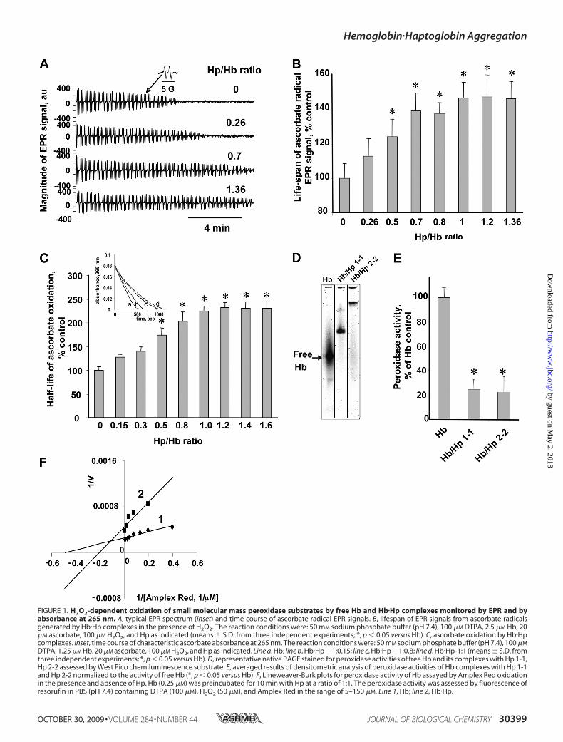
FIGURE 1. H2O2-dependent oxidation of small molecular mass peroxidase substrates by free Hb and Hb�Hp complexes monitored by EPR and byabsorbance at 265 nm. A, typical EPR spectrum (inset) and time course of ascorbate radical EPR signals. B, lifespan of EPR signals from ascorbate radicalsgenerated by Hb�Hp complexes in the presence of H2O2. The reaction conditions were: 50 mM sodium phosphate buffer (pH 7.4), 100 �M DTPA, 2.5 �M Hb, 20�M ascorbate, 100 �M H2O2, and Hp as indicated (means � S.D. from three independent experiments; *, p � 0.05 versus Hb). C, ascorbate oxidation by Hb�Hpcomplexes. Inset, time course of characteristic ascorbate absorbance at 265 nm. The reaction conditions were: 50 mM sodium phosphate buffer (pH 7.4), 100 �M
DTPA, 1.25 �M Hb, 20 �M ascorbate, 100 �M H2O2, and Hp as indicated. Line a, Hb; line b, Hb�Hp �1:0.15; line c, Hb�Hp �1:0.8; line d, Hb�Hp-1:1 (means � S.D. fromthree independent experiments; *, p � 0.05 versus Hb). D, representative native PAGE stained for peroxidase activities of free Hb and its complexes with Hp 1-1,Hp 2-2 assessed by West Pico chemiluminescence substrate. E, averaged results of densitometric analysis of peroxidase activities of Hb complexes with Hp 1-1and Hp 2-2 normalized to the activity of free Hb (*, p � 0.05 versus Hb). F, Lineweaver-Burk plots for peroxidase activity of Hb assayed by Amplex Red oxidationin the presence and absence of Hp. Hb (0.25 �M) was preincubated for 10 min with Hp at a ratio of 1:1. The peroxidase activity was assessed by fluorescence ofresorufin in PBS (pH 7.4) containing DTPA (100 �M), H2O2 (50 �M), and Amplex Red in the range of 5–150 �M. Line 1, Hb; line 2, Hb�Hp.
Hemoglobin�Haptoglobin Aggregation
OCTOBER 30, 2009 • VOLUME 284 • NUMBER 44 JOURNAL OF BIOLOGICAL CHEMISTRY 30399
by guest on May 2, 2018
http://ww
w.jbc.org/
Dow
nloaded from

thiol groups that are dissociable by S-S reducing reagents) and(ii) Tyr-Tyr cross-links (nondissociable by S-S reducingreagents). PAGE analysis of fully denatured proteins in thepresence of SDS and a disulfide reducing agent, DTT, revealedthat incubation of Hbwith Hp in the presence of H2O2 resultedin accumulation of aggregates with different molecular masses.Western blot analysis showed that these aggregates containedboth Hb and Hp as evidenced by their positive staining withanti-Hb and anti-Hp antibodies (Fig. 3). Resistance of Hb�Hpaggregates toDTT suggests that they are covalently linked via nonS-Sbonds.AlthoughbothHp1-1andHp2-2underwentoxidativecross-linking, a greater amountof veryhighmolecularmass aggre-gates was formed after conjugation of Hb with Hp 2-2.
Quenching of Hb�Hp Peroxidase-reactive Intermediates andInhibition of Hb�Hp Hetero-oligomerization by Reductants
The above results indicate that Hp acted as a substrate forHb-driven peroxidase activity. Consequently, small moleculereducing substrates such as NO�, ascorbate, Amplex Red, orDMPO, known to interact with reactive intermediates of Hbperoxidase (15, 26), should prevent cross-linking of Hb withHp. Increasing concentrations of these compounds can out-compete peroxidase activity of Hb toward Hpt and inhibitformation of aggregates. Indeed, we found that increasing con-centrations of Amplex Red or DMPO progressively inhibitedH2O2-dependent aggregation ofHb/Hpt as evidenced by PAGE(Fig. 4A and supplemental Fig. S2). NO� (generated by an NOdonor, PAPANONate) and ascorbate acted in a similar way andinhibited cross-linking of Hb with Hpt (Fig. 4, B andC). Molec-ular proximity of Hp to Hb reactive intermediates, oxoferrylheme and/or protein immobilized (Tyr) radicals, makes Hp apreferred substrate of the peroxidase reaction. Indeed, Tyr-Tyrcross-linking of Hp with Hb was preventable by relatively highconcentration of reducing substrates (for instance, 200-foldhigher concentrations of ascorbate versus those of Hp wererequired to markedly inhibit the formation of aggregates).Recently, a new technique, immuno-spin trapping, has been
developed for specific and sensitive detection of protein-immo-bilized radicals (24, 27, 28). It is based on the use of antibodies toprotein radical-DMPO adducts. When Hb�Hp complexes wereincubated with H2O2 in the presence of DMPO, protein-
FIGURE 2. Detection of heme-nitrosyl complexes and protein-derived(Tyr�) radicals (peroxidase intermediates) by EPR spectroscopy. A, typicallow temperature (77 K) EPR spectra of Hb�Hp complexes and aggregates(insets) and magnitudes of EPR signals of heme-nitrosylated Hb (bar a), Hb�Hpcomplexes (bar b), Hb aggregates (bar c), and Hb�Hp aggregates (bar d).
To form nitrosyl complexes, 40 �M Angeli’s salt was added to the solutioncontaining 10 �M Hb, and incubation was continued for 30 min, after whichthe samples were frozen (means � S.D. from 3–7 independent experiments; *,p � 0.05 versus Hb; **, p � 0.05 versus Hb�Hp complexes). B, H2O2-dependentformation of protein-derived (Tyr�) radicals (peroxidase intermediates) by Hb(bar a), Hb�Hp complexes (bar b), Hb aggregates (bar c), and Hb�Hp aggre-gates (bar d). Shown are typical low temperature (77 K) EPR spectra of Hb�Hpcomplexes and aggregates (insets) (measured at g � 2.005) and magnitudesof the respective protein-derived (Tyr) radicals (means � S.D. from three to sixindependent experiments; *, p � 0.05 versus Hb). C, power saturation curvesfor protein-derived radicals of Hb and Hb�Hp complexes and aggregates. Hbaggregates and Hb�Hp aggregates were obtained by incubating Hb (10 �M)and Hb�Hp in the presence of H2O2 (50 �M added four times with a 15-mininterval). 200 �M ascorbate was added to the formed aggregates and incu-bated for 15 min to reduce all protein immobilized radicals, and the residualascorbate was removed by ascorbate oxidase (1 unit/100 �l; 5 min). 100 �M
H2O2 was added to Hb, Hb aggregates, Hb�Hp complexes, and Hb�Hp aggre-gates, then samples were frozen for 20 s, and EPR spectra of protein-derivedradicals were measured. Catalase (10 �g/ml) was added to stop thereaction.
Hemoglobin�Haptoglobin Aggregation
30400 JOURNAL OF BIOLOGICAL CHEMISTRY VOLUME 284 • NUMBER 44 • OCTOBER 30, 2009
by guest on May 2, 2018
http://ww
w.jbc.org/
Dow
nloaded from
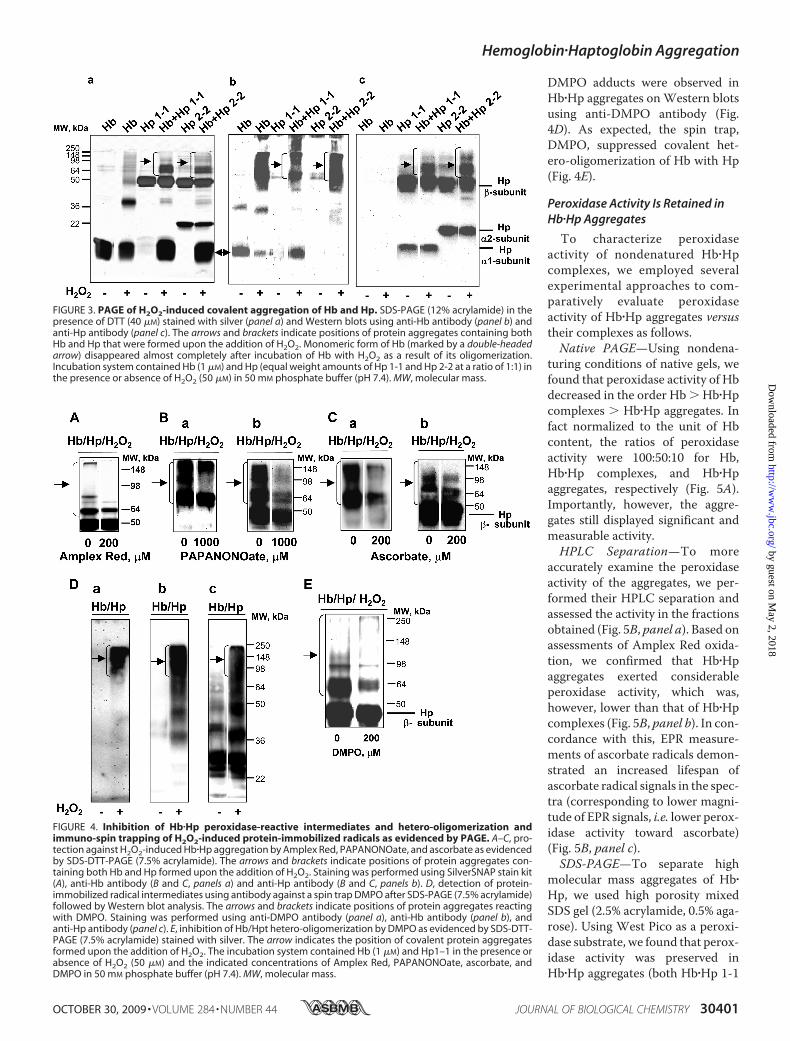
DMPO adducts were observed inHb�Hp aggregates onWestern blotsusing anti-DMPO antibody (Fig.4D). As expected, the spin trap,DMPO, suppressed covalent het-ero-oligomerization of Hb with Hp(Fig. 4E).
Peroxidase Activity Is Retained inHb�Hp Aggregates
To characterize peroxidaseactivity of nondenatured Hb�Hpcomplexes, we employed severalexperimental approaches to com-paratively evaluate peroxidaseactivity of Hb�Hp aggregates versustheir complexes as follows.Native PAGE—Using nondena-
turing conditions of native gels, wefound that peroxidase activity of Hbdecreased in the order Hb � Hb�Hpcomplexes � Hb�Hp aggregates. Infact normalized to the unit of Hbcontent, the ratios of peroxidaseactivity were 100:50:10 for Hb,Hb�Hp complexes, and Hb�Hpaggregates, respectively (Fig. 5A).Importantly, however, the aggre-gates still displayed significant andmeasurable activity.HPLC Separation—To more
accurately examine the peroxidaseactivity of the aggregates, we per-formed their HPLC separation andassessed the activity in the fractionsobtained (Fig. 5B, panel a). Based onassessments of Amplex Red oxida-tion, we confirmed that Hb�Hpaggregates exerted considerableperoxidase activity, which was,however, lower than that of Hb�Hpcomplexes (Fig. 5B, panel b). In con-cordance with this, EPR measure-ments of ascorbate radicals demon-strated an increased lifespan ofascorbate radical signals in the spec-tra (corresponding to lower magni-tude of EPR signals, i.e. lower perox-idase activity toward ascorbate)(Fig. 5B, panel c).SDS-PAGE—To separate high
molecular mass aggregates of Hb�Hp, we used high porosity mixedSDS gel (2.5% acrylamide, 0.5% aga-rose). Using West Pico as a peroxi-dase substrate, we found that perox-idase activity was preserved inHb�Hp aggregates (both Hb�Hp 1-1
FIGURE 3. PAGE of H2O2-induced covalent aggregation of Hb and Hp. SDS-PAGE (12% acrylamide) in thepresence of DTT (40 �M) stained with silver (panel a) and Western blots using anti-Hb antibody (panel b) andanti-Hp antibody (panel c). The arrows and brackets indicate positions of protein aggregates containing bothHb and Hp that were formed upon the addition of H2O2. Monomeric form of Hb (marked by a double-headedarrow) disappeared almost completely after incubation of Hb with H2O2 as a result of its oligomerization.Incubation system contained Hb (1 �M) and Hp (equal weight amounts of Hp 1-1 and Hp 2-2 at a ratio of 1:1) inthe presence or absence of H2O2 (50 �M) in 50 mM phosphate buffer (pH 7.4). MW, molecular mass.
FIGURE 4. Inhibition of Hb�Hp peroxidase-reactive intermediates and hetero-oligomerization andimmuno-spin trapping of H2O2-induced protein-immobilized radicals as evidenced by PAGE. A–C, pro-tection against H2O2-induced Hb�Hp aggregation by Amplex Red, PAPANONOate, and ascorbate as evidencedby SDS-DTT-PAGE (7.5% acrylamide). The arrows and brackets indicate positions of protein aggregates con-taining both Hb and Hp formed upon the addition of H2O2. Staining was performed using SilverSNAP stain kit(A), anti-Hb antibody (B and C, panels a) and anti-Hp antibody (B and C, panels b). D, detection of protein-immobilized radical intermediates using antibody against a spin trap DMPO after SDS-PAGE (7.5% acrylamide)followed by Western blot analysis. The arrows and brackets indicate positions of protein aggregates reactingwith DMPO. Staining was performed using anti-DMPO antibody (panel a), anti-Hb antibody (panel b), andanti-Hp antibody (panel c). E, inhibition of Hb/Hpt hetero-oligomerization by DMPO as evidenced by SDS-DTT-PAGE (7.5% acrylamide) stained with silver. The arrow indicates the position of covalent protein aggregatesformed upon the addition of H2O2. The incubation system contained Hb (1 �M) and Hp1–1 in the presence orabsence of H2O2 (50 �M) and the indicated concentrations of Amplex Red, PAPANONOate, ascorbate, andDMPO in 50 mM phosphate buffer (pH 7.4). MW, molecular mass.
Hemoglobin�Haptoglobin Aggregation
OCTOBER 30, 2009 • VOLUME 284 • NUMBER 44 JOURNAL OF BIOLOGICAL CHEMISTRY 30401
by guest on May 2, 2018
http://ww
w.jbc.org/
Dow
nloaded from
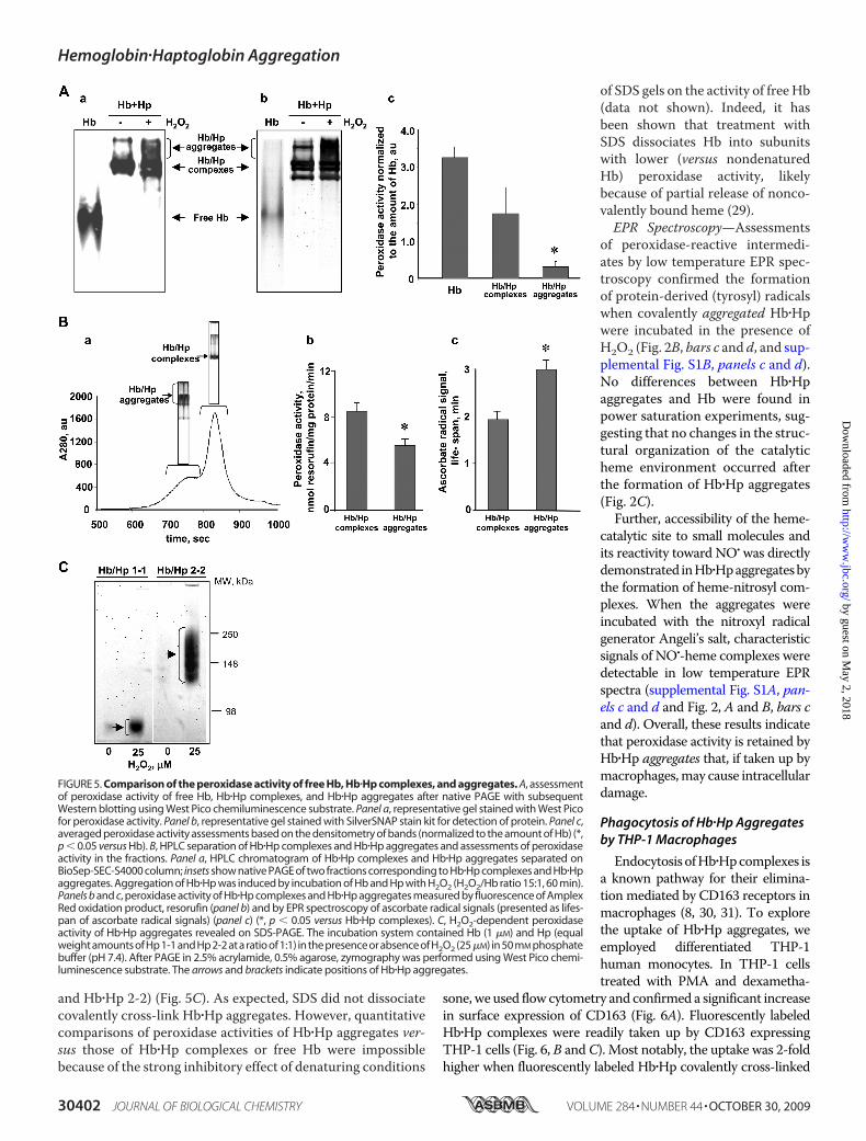
and Hb�Hp 2-2) (Fig. 5C). As expected, SDS did not dissociatecovalently cross-link Hb�Hp aggregates. However, quantitativecomparisons of peroxidase activities of Hb�Hp aggregates ver-sus those of Hb�Hp complexes or free Hb were impossiblebecause of the strong inhibitory effect of denaturing conditions
of SDS gels on the activity of free Hb(data not shown). Indeed, it hasbeen shown that treatment withSDS dissociates Hb into subunitswith lower (versus nondenaturedHb) peroxidase activity, likelybecause of partial release of nonco-valently bound heme (29).EPR Spectroscopy—Assessments
of peroxidase-reactive intermedi-ates by low temperature EPR spec-troscopy confirmed the formationof protein-derived (tyrosyl) radicalswhen covalently aggregated Hb�Hpwere incubated in the presence ofH2O2 (Fig. 2B, bars c and d, and sup-plemental Fig. S1B, panels c and d).No differences between Hb�Hpaggregates and Hb were found inpower saturation experiments, sug-gesting that no changes in the struc-tural organization of the catalyticheme environment occurred afterthe formation of Hb�Hp aggregates(Fig. 2C).Further, accessibility of the heme-
catalytic site to small molecules andits reactivity toward NO� was directlydemonstrated inHb�Hpaggregatesbythe formation of heme-nitrosyl com-plexes. When the aggregates wereincubated with the nitroxyl radicalgenerator Angeli’s salt, characteristicsignals of NO�-heme complexes weredetectable in low temperature EPRspectra (supplemental Fig. S1A, pan-els c and d and Fig. 2, A and B, bars cand d). Overall, these results indicatethat peroxidase activity is retained byHb�Hp aggregates that, if taken up bymacrophages,may cause intracellulardamage.
Phagocytosis of Hb�Hp Aggregatesby THP-1 Macrophages
Endocytosis ofHb�Hpcomplexes isa known pathway for their elimina-tion mediated by CD163 receptors inmacrophages (8, 30, 31). To explorethe uptake of Hb�Hp aggregates, weemployed differentiated THP-1human monocytes. In THP-1 cellstreated with PMA and dexametha-
sone,we used flow cytometry and confirmed a significant increasein surface expression of CD163 (Fig. 6A). Fluorescently labeledHb�Hp complexes were readily taken up by CD163 expressingTHP-1 cells (Fig. 6, B andC). Most notably, the uptake was 2-foldhigher when fluorescently labeled Hb�Hp covalently cross-linked
FIGURE 5. Comparison of the peroxidase activity of free Hb, Hb�Hp complexes, and aggregates. A, assessmentof peroxidase activity of free Hb, Hb�Hp complexes, and Hb�Hp aggregates after native PAGE with subsequentWestern blotting using West Pico chemiluminescence substrate. Panel a, representative gel stained with West Picofor peroxidase activity. Panel b, representative gel stained with SilverSNAP stain kit for detection of protein. Panel c,averaged peroxidase activity assessments based on the densitometry of bands (normalized to the amount of Hb) (*,p � 0.05 versus Hb). B, HPLC separation of Hb�Hp complexes and Hb�Hp aggregates and assessments of peroxidaseactivity in the fractions. Panel a, HPLC chromatogram of Hb�Hp complexes and Hb�Hp aggregates separated onBioSep-SEC-S4000 column; insets show native PAGE of two fractions corresponding to Hb�Hp complexes and Hb�Hpaggregates. Aggregation of Hb�Hp was induced by incubation of Hb and Hp with H2O2 (H2O2/Hb ratio 15:1, 60 min).Panels b and c, peroxidase activity of Hb�Hp complexes and Hb�Hp aggregates measured by fluorescence of AmplexRed oxidation product, resorufin (panel b) and by EPR spectroscopy of ascorbate radical signals (presented as lifes-pan of ascorbate radical signals) (panel c) (*, p � 0.05 versus Hb�Hp complexes). C, H2O2-dependent peroxidaseactivity of Hb�Hp aggregates revealed on SDS-PAGE. The incubation system contained Hb (1 �M) and Hp (equalweight amounts of Hp 1-1 and Hp 2-2 at a ratio of 1:1) in the presence or absence of H2O2 (25�M) in 50 mM phosphatebuffer (pH 7.4). After PAGE in 2.5% acrylamide, 0.5% agarose, zymography was performed using West Pico chemi-luminescence substrate. The arrows and brackets indicate positions of Hb�Hp aggregates.
Hemoglobin�Haptoglobin Aggregation
30402 JOURNAL OF BIOLOGICAL CHEMISTRY VOLUME 284 • NUMBER 44 • OCTOBER 30, 2009
by guest on May 2, 2018
http://ww
w.jbc.org/
Dow
nloaded from
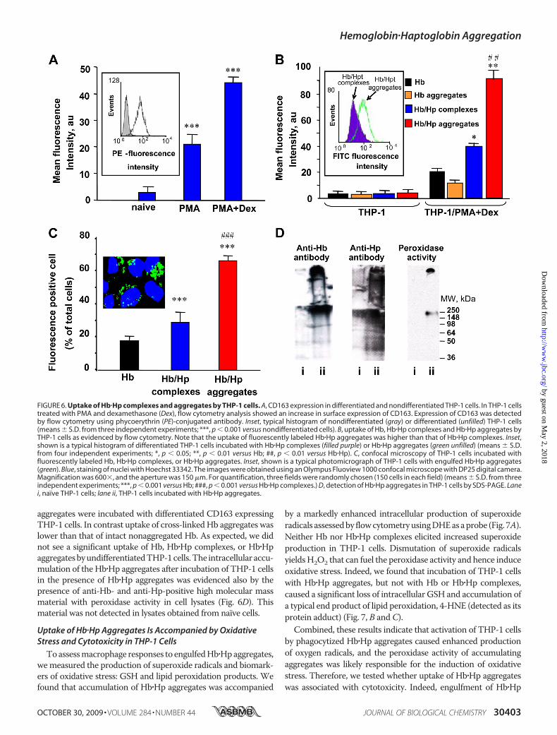
aggregates were incubated with differentiated CD163 expressingTHP-1 cells. In contrast uptake of cross-linkedHb aggregates waslower than that of intact nonaggregated Hb. As expected, we didnot see a significant uptake of Hb, Hb�Hp complexes, or Hb�HpaggregatesbyundifferentiatedTHP-1cells.The intracellularaccu-mulation of the Hb�Hp aggregates after incubation of THP-1 cellsin the presence of Hb�Hp aggregates was evidenced also by thepresence of anti-Hb- and anti-Hp-positive high molecular massmaterial with peroxidase activity in cell lysates (Fig. 6D). Thismaterial was not detected in lysates obtained from naïve cells.
Uptake of Hb�Hp Aggregates Is Accompanied by OxidativeStress and Cytotoxicity in THP-1 Cells
Toassessmacrophage responses to engulfedHb�Hpaggregates,wemeasured the production of superoxide radicals and biomark-ers of oxidative stress: GSH and lipid peroxidation products. Wefound that accumulation of Hb�Hp aggregates was accompanied
by a markedly enhanced intracellular production of superoxideradicals assessedby flowcytometryusingDHEasaprobe (Fig. 7A).Neither Hb nor Hb�Hp complexes elicited increased superoxideproduction in THP-1 cells. Dismutation of superoxide radicalsyieldsH2O2 that can fuel the peroxidase activity and hence induceoxidative stress. Indeed, we found that incubation of THP-1 cellswith Hb�Hp aggregates, but not with Hb or Hb�Hp complexes,caused a significant loss of intracellular GSH and accumulation ofa typical end product of lipid peroxidation, 4-HNE (detected as itsprotein adduct) (Fig. 7, B andC).Combined, these results indicate that activation of THP-1 cells
by phagocytized Hb�Hp aggregates caused enhanced productionof oxygen radicals, and the peroxidase activity of accumulatingaggregates was likely responsible for the induction of oxidativestress. Therefore, we tested whether uptake of Hb�Hp aggregateswas associated with cytotoxicity. Indeed, engulfment of Hb�Hp
FIGURE 6. Uptake of Hb�Hp complexes and aggregates by THP-1 cells. A, CD163 expression in differentiated and nondifferentiated THP-1 cells. In THP-1 cellstreated with PMA and dexamethasone (Dex), flow cytometry analysis showed an increase in surface expression of CD163. Expression of CD163 was detectedby flow cytometry using phycoerythrin (PE)-conjugated antibody. Inset, typical histogram of nondifferentiated (gray) or differentiated (unfilled) THP-1 cells(means � S.D. from three independent experiments; ***, p � 0.001 versus nondifferentiated cells). B, uptake of Hb, Hb�Hp complexes and Hb�Hp aggregates byTHP-1 cells as evidenced by flow cytometry. Note that the uptake of fluorescently labeled Hb�Hp aggregates was higher than that of Hb�Hp complexes. Inset,shown is a typical histogram of differentiated THP-1 cells incubated with Hb�Hp complexes (filled purple) or Hb�Hp aggregates (green unfilled) (means � S.D.from four independent experiments; *, p � 0.05; **, p � 0.01 versus Hb; ##, p � 0.01 versus Hb�Hp). C, confocal microscopy of THP-1 cells incubated withfluorescently labeled Hb, Hb�Hp complexes, or Hb�Hp aggregates. Inset, shown is a typical photomicrograph of THP-1 cells with engulfed Hb�Hp aggregates(green). Blue, staining of nuclei with Hoechst 33342. The images were obtained using an Olympus Fluoview 1000 confocal microscope with DP25 digital camera.Magnification was 600�, and the aperture was 150 �m. For quantification, three fields were randomly chosen (150 cells in each field) (means � S.D. from threeindependent experiments; ***, p � 0.001 versus Hb; ###, p � 0.001 versus Hb�Hp complexes.) D, detection of Hb�Hp aggregates in THP-1 cells by SDS-PAGE. Lanei, naïve THP-1 cells; lane ii, THP-1 cells incubated with Hb�Hp aggregates.
Hemoglobin�Haptoglobin Aggregation
OCTOBER 30, 2009 • VOLUME 284 • NUMBER 44 JOURNAL OF BIOLOGICAL CHEMISTRY 30403
by guest on May 2, 2018
http://ww
w.jbc.org/
Dow
nloaded from
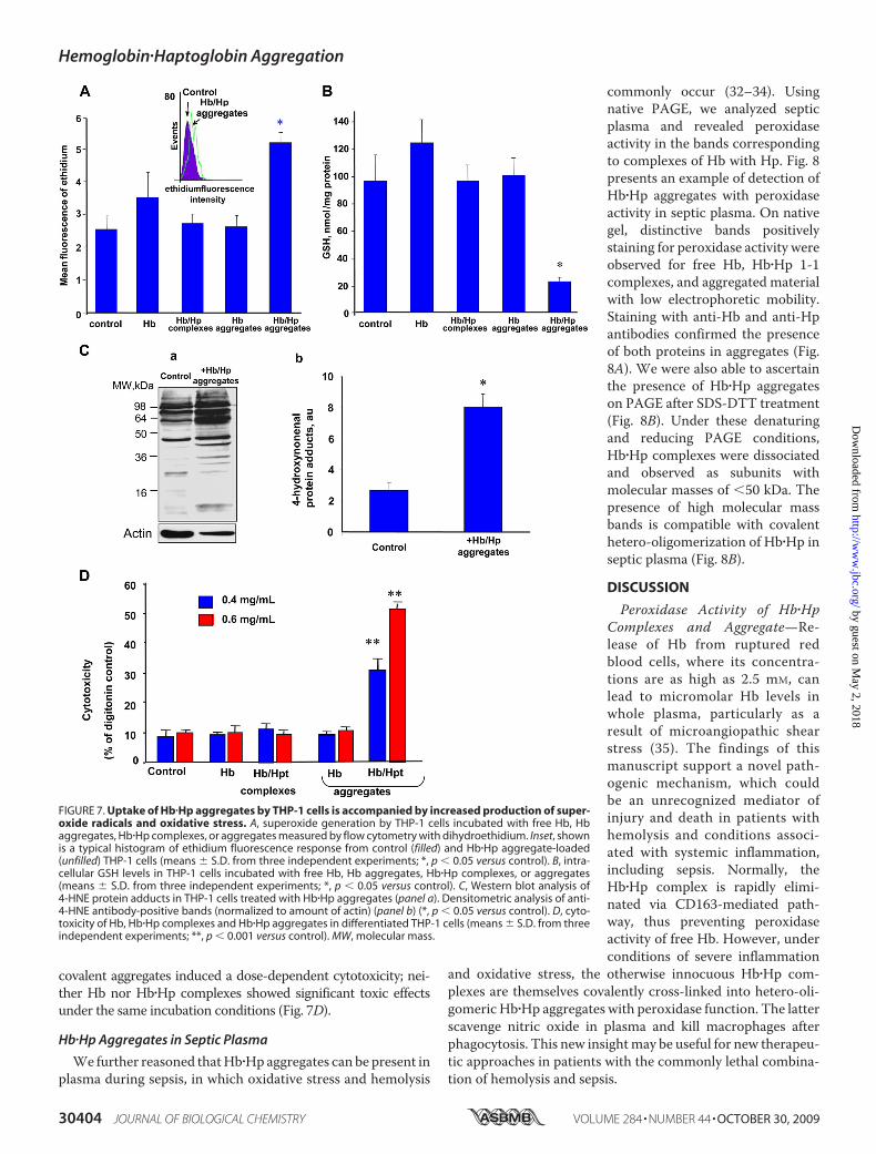
covalent aggregates induced a dose-dependent cytotoxicity; nei-ther Hb nor Hb�Hp complexes showed significant toxic effectsunder the same incubation conditions (Fig. 7D).
Hb�Hp Aggregates in Septic Plasma
We further reasoned thatHb�Hpaggregates can be present inplasma during sepsis, in which oxidative stress and hemolysis
commonly occur (32–34). Usingnative PAGE, we analyzed septicplasma and revealed peroxidaseactivity in the bands correspondingto complexes of Hb with Hp. Fig. 8presents an example of detection ofHb�Hp aggregates with peroxidaseactivity in septic plasma. On nativegel, distinctive bands positivelystaining for peroxidase activity wereobserved for free Hb, Hb�Hp 1-1complexes, and aggregated materialwith low electrophoretic mobility.Staining with anti-Hb and anti-Hpantibodies confirmed the presenceof both proteins in aggregates (Fig.8A). We were also able to ascertainthe presence of Hb�Hp aggregateson PAGE after SDS-DTT treatment(Fig. 8B). Under these denaturingand reducing PAGE conditions,Hb�Hp complexes were dissociatedand observed as subunits withmolecular masses of �50 kDa. Thepresence of high molecular massbands is compatible with covalenthetero-oligomerization of Hb�Hp inseptic plasma (Fig. 8B).
DISCUSSION
Peroxidase Activity of Hb�HpComplexes and Aggregate—Re-lease of Hb from ruptured redblood cells, where its concentra-tions are as high as 2.5 mM, canlead to micromolar Hb levels inwhole plasma, particularly as aresult of microangiopathic shearstress (35). The findings of thismanuscript support a novel path-ogenic mechanism, which couldbe an unrecognized mediator ofinjury and death in patients withhemolysis and conditions associ-ated with systemic inflammation,including sepsis. Normally, theHb�Hp complex is rapidly elimi-nated via CD163-mediated path-way, thus preventing peroxidaseactivity of free Hb. However, underconditions of severe inflammation
and oxidative stress, the otherwise innocuous Hb�Hp com-plexes are themselves covalently cross-linked into hetero-oli-gomeric Hb�Hp aggregates with peroxidase function. The latterscavenge nitric oxide in plasma and kill macrophages afterphagocytosis. This new insightmay be useful for new therapeu-tic approaches in patients with the commonly lethal combina-tion of hemolysis and sepsis.
FIGURE 7. Uptake of Hb�Hp aggregates by THP-1 cells is accompanied by increased production of super-oxide radicals and oxidative stress. A, superoxide generation by THP-1 cells incubated with free Hb, Hbaggregates, Hb�Hp complexes, or aggregates measured by flow cytometry with dihydroethidium. Inset, shownis a typical histogram of ethidium fluorescence response from control (filled) and Hb�Hp aggregate-loaded(unfilled) THP-1 cells (means � S.D. from three independent experiments; *, p � 0.05 versus control). B, intra-cellular GSH levels in THP-1 cells incubated with free Hb, Hb aggregates, Hb�Hp complexes, or aggregates(means � S.D. from three independent experiments; *, p � 0.05 versus control). C, Western blot analysis of4-HNE protein adducts in THP-1 cells treated with Hb�Hp aggregates (panel a). Densitometric analysis of anti-4-HNE antibody-positive bands (normalized to amount of actin) (panel b) (*, p � 0.05 versus control). D, cyto-toxicity of Hb, Hb�Hp complexes and Hb�Hp aggregates in differentiated THP-1 cells (means � S.D. from threeindependent experiments; **, p � 0.001 versus control). MW, molecular mass.
Hemoglobin�Haptoglobin Aggregation
30404 JOURNAL OF BIOLOGICAL CHEMISTRY VOLUME 284 • NUMBER 44 • OCTOBER 30, 2009
by guest on May 2, 2018
http://ww
w.jbc.org/
Dow
nloaded from

We provide several lines of evidence confirming the preser-vation of peroxidase activity both in Hb�Hp complexes andaggregates. These include direct assessments of the activity inisolated HPLC fractions, low temperature EPR spectroscopy ofcharacteristic intermediates, immuno-spin trapping, PAGEzymography using prototypical peroxidase substrates, and for-mation of heme-nitrosylated species. The Western blot analy-ses confirmed the hetero-oligomeric nature of aggregates andinvolvement of both Hb and Hp in the cross-links.Different Clearance of Hb�Hp Complexes and Aggregates—Re-
cently, aggregation of Hb via its peroxidase function has beendemonstrated. This resulted in blunted interaction of Hb withHp and consequently decreased CD163-dependent uptake ofthe complexes (14). Similarly, we report decreased uptake ofHbaggregates by THP-1 macrophages differentiated to expresshigh levels of CD163 receptors. Although these results are veryimportant, circulating Hp has been considered as a significantendogenous defense against Hb peroxidase (6) because of itsvery high affinity binding (�1014M�1) toHb (21).Hp is an acutephase protein, and its normal plasma levels (ranging from 0.45to 3 mg/ml) (8) increase 2–5-fold during inflammation (36).These amounts ofHp are sufficient to bindmicromolar levels offree Hb. The binding and elimination of Hb�Hp complexeswould be expected to decrease circulating Hp levels, particu-larly under conditions of hemolysis (37). However, completedepletion ofHp and accumulation of freeHb aggregatesmay betypical of extremely severe conditions with hemolysis, forexample sickle cell disease.More commonly, released freeHb isscavenged by Hp, resulting in the formation of Hb�Hp com-plexes. The role of this major function of Hp is underscored byits significant power as a predictor of disease severity and clin-ical outcomes (38–40).Buehler et al. (7) demonstrated that binding of Hb with Hp
yields complexes that protect Hb from oxidative and structuralmodification; as a result of this protection, CD163-mediateduptake of Hb�Hp complexes is preserved (7). These findings arein agreement with the results of the current study, which wasfocused on covalent cross-linking between Hb and Hp medi-ated by the peroxidase activity of the former in the complexesand realized through the recombination of protein immobi-lized radicals, most likely Tyr� radicals. However, our studies on
the uptake and elimination of oxidatively cross-linked Hb�Hpaggregates yielded results that were not completely concordantwith those of Buehler et al. (7).Buehler et al. reported similar engulfment of nonoxidized
Hb�Hp complexes and oxidized Hb�Hp aggregates by CD163expressing HEK293 cells; our results indicate that oxidativelycross-linked Hb�Hp aggregates were taken up more effectivelyby THP-1 macrophages than nonoxidized complexes. Thereare several explanations for these apparent discrepancies: (i)The uptake of Hb�Hp aggregates likely depends on their molec-ular mass. We used Hp isolated from pooled plasma that con-tained all three major phenotypes of Hp: Hp 1-1, Hp 2-1, andHp 2-2. Polymeric forms of Hp 2-1 and Hp 2-2 have multiplemolecular species with very high molecular masses: up to 300and 900 kDa, respectively, and the molecular mass of Hb�Hpcomplexes in this case can be as high as 1.5MDa. Cross-linkingof several such complexes would yield large aggregates withvery high molecular masses: up to 3–5MDa. In contrast, in thestudy by Buehler et al. (7), Hp 1-1 has been employed with amolecular mass of 86 kDa; Hp 1-1 forms relatively small com-plexes with Hb, �150 kDa (6). Consequently, cross-linking ofthese complexes yielded aggregates of smaller molecularmasses (�300 kDa). (ii) It is possible that both CD163-depend-ent as well as CD-163-independent pathways could be involvedin engulfment ofHb�Hpaggregates byTHP-1 cells in our exper-iments. As professional phagocytes, THP-1 cells used in ourstudy express not only CD163 but also other receptors poten-tially involved in the engulfment and uptake of large aggregates.In particular, a scavenger receptor of type B family, CD36, isknown to be effective in the uptake of oxidativelymodified highmolecular mass aggregates, including oxidatively modifiedlipoproteins (41).Moreover, CD91 receptor (that belongs to thelow density lipoprotein receptor superfamily) recognizes morethan 40 different ligands, among which are heme-hemopexin,lipoproteins, viruses, and complexes of proteases with proteaseinhibitors (42). Neither of these receptors are expressed inHEK293 cells that have epithelial origin (43, 44). (iii) Greateruptake of oxidatively cross-linked Hb�Hp aggregates versusnonoxidized Hb�Hp complexes in our experiments could bealso due to the fact that our “oxidized” samples were highlyenriched with cross-linked aggregates as evidenced by the per-centage of aggregates versus nonaggregated forms of Hb�Hp onsilver-stained gels (as illustrated by Fig. 5A). Notably, the con-tent of cross-linked Hb�Hp aggregates used in experimentsreported by Buehler et al. (7) was significantly lower than thatused in the current study.Binding and Aggregation of Hb with Different Phenotypes of
Hp—In humans, unlike other mammals, there are two allelesfor the Hp gene (Hp 1 and Hp 2). Accordingly two encodedproteins Hp 1-1 and Hp 2-2 represent an 86-kDa dimeric mol-ecule and a polymeric form up to 900 kDa (6). Thus, Hb�Hpcross-linking can produce high molecular mass aggregates(especially in the case of Hp 2-2), readily taken up by macro-phages. Binding of Hb with Hp is believed to neutralize free thepro-oxidant effects of Hb (7, 10, 11). Based on structural con-siderations, however, this antioxidant function of Hp is notimmediately apparent. �/� dimers of Hb participating in theformation of Hb�Hp complexes contain three Hp-binding sites
FIGURE 8. Detection of Hb�Hp aggregates in septic plasma. A, gels afternative PAGE (7.5% acrylamide) were stained with West Pico chemilumines-cence substrate for peroxidase activity (panel a), Western blot analyses withanti-Hb (panel b), and anti-Hp antibodies (panel c). B, Western blot analyseswith anti-Hb (panel a) and anti-Hp (panel b) antibodies after SDS-DTT-PAGE(7.5% acrylamide). The arrows and brackets indicate the positions of highmolecular mass aggregates.
Hemoglobin�Haptoglobin Aggregation
OCTOBER 30, 2009 • VOLUME 284 • NUMBER 44 JOURNAL OF BIOLOGICAL CHEMISTRY 30405
by guest on May 2, 2018
http://ww
w.jbc.org/
Dow
nloaded from
that include amino acid residues 11–25 and 131–146 of the�-globin chain and residues 121-l27 of the �-chain in humanHb. Although two of these sites participate in interactions ofthe polypeptide chains with the heme (residues 100–140), nei-ther of the Hb-binding regions of Hp seems to be in directcontact with the heme crevice (21, 45, 46). As a result, bindingof Hp to Hb leaves the heme iron ready to interact with smallmolecules, such as O2 (47), NO, or H2O2. In line with theseearlier reports (23, 24, 25), our EPR findings showed heme-nitrosylated complexes and protein-immobilized (Tyr�) radi-cals in Hb�Hp complexes and aggregates. However, the specificlocation and proximity to heme of Hp-binding sites on the Hbmolecule is sufficient for the role of Hp as a competitive (versuslow molecular mass reductants) substrate for Hb-dependentperoxidase reactions.In the circulation, cross-linked Hb�Hp aggregates retained
the peroxidase activity and hence continued to act as a source ofoxidative stress and damage. In the presence of oxidizing equiv-alents, such as H2O2 and organic hydroperoxides includinglipid hydroperoxides, heme-peroxidases are activated to highlyreactive intermediates. The cleavage of the O-O bond ofhydroperoxide is associated with the formation of oxoferryl(Fe4��O) species and a cation radical of porphyrin-compoundI (48, 49). In the case of Hb, the highly reactive radical on theporphyrin ring is unstable, causing one-electron oxidation ofone of amino acid residues in the immediate proximity, mostcommonly tyrosine, tryptophan, or histidine, thus producingprotein-immobilized radicals (27, 50, 51). Either of the tworeactive radical species, porphyrin radical and/or protein-im-mobilized (Tyr�) radical, can act as a pro-oxidant depleting themajor intracellular antioxidants such as GSH, ascorbate (52),and oxidizing biomolecules, such as proteins and lipids (51, 53,54). In line with this, we observed that uptake of Hb�Hp aggre-gates by THP-1 cells resulted in enhanced superoxide radicalproduction, most likely by NADPH oxidase-dependent path-ways known to be activated during phagocytosis (55, 56). Dis-mutation of superoxide radicals yields H2O2 required formain-tenance of peroxidase activity of Hb�Hp complexes. The lattercatalyzed oxidative attack on thiols and lipids as evidenced bydepletion of GSH and accumulation of protein adducts with4-hydroxy-nonenal, one of the typical end products of lipid per-oxidation (57, 58).Hb�Hp Aggregation in Severe Inflammation—Under condi-
tions of severe inflammation and massive recruitment of mac-rophages, generated steady state concentrations of superoxideradicals and H2O2 (in the high micromolar range) (59, 60) maybe sufficient for the realization of peroxidase activity of Hb�Hpcomplexes. Accordingly we were able to detect the presence ofHb�Hp aggregates in plasma samples from septic patients.Hemolytic anemia complicated by sepsis remains a majorhealth problem in all age groups in both developing and indus-trialized countries. The release and presence of stroma-free Hbin circulation, robust inflammatory response accompanied byoxidative stress (with production of reactive oxygen and nitro-gen species and depletion of antioxidants), microvascularthrombosis and vasoconstriction, and macrophage deactiva-tion leading to immune paralysis are the major hallmarks ofsevere hemolytic disease and sepsis. Although these critical
pathogenesis factors have been identified, the links betweenthem and the mechanisms leading to their appearance are notcompletely understood.The primary water-soluble antioxidant of plasma is ascor-
bate that readily interacts with reactive peroxidase intermedi-ates (52). Decreased levels of plasma ascorbate have been doc-umented during sepsis and displayed strong correlation withthe survival (61, 62). Administration of ascorbate was found toprotect against circulatory dysfunction in sepsis, reducing theincidence of organ failure and the duration of patient hospital-ization (63, 64).Nitric oxide (NO�) is also utilized as one of the reducing sub-
strates for peroxidases, resulting in its oxidative depletion andpossibly dysregulation of vascular tone. This may be enhancedby a loss of control over ascorbate-dependent decomposition ofS-nitrosothiols (accumulating in plasma during sepsis), result-ing in further apparent deficiency of releasable NO (65). Ifuninterrupted, the vicious peroxidase cycle can consume themajority of essential antioxidants and cause their depletionand deficiency. Based on the proposed mechanism, targetedstrategies can be developed to eliminate Hb�Hp aggregates(e.g. via affinity binding), thus breaking the viscous peroxi-dase cycle.
REFERENCES1. Reeder, B. J., Svistunenko, D. A., Cooper, C. E., and Wilson, M. T. (2004)
Antioxid. Redox. Signal 6, 954–9662. Hultquist, D. E., and Passon, P. G. (1971) Nat. New Biol. 229, 252–2543. Cooper, G. S., Havlir, D. S., Shlaes, D.M., and Salata, R. A. (1990)Medicine
69, 114–1234. Kaye, D., and Hook, E. W. (1963) J. Immunol. 91, 65–755. Su, D., Roth, R. I., Yoshida, M., and Levin, J. (1997) Infect. Immun. 65,
1258–12666. Langlois, M. R., and Delanghe, J. R. (1996) Clin. Chem. 42, 1589–16007. Buehler, P. W., Abraham, B., Vallelian, F., Linnemayr, C., Pereira, C. P.,
Cipollo, J. F., Jia, Y., Mikolajczyk, M., Boretti, F. S., Schoedon, G., Alayash,A. I., and Schaer, D. J. (2009) Blood 113, 2578–2586
8. Kristiansen, M., Graversen, J. H., Jacobsen, C., Sonne, O., Hoffman, H. J.,Law, S. K., and Moestrup, S. K. (2001) Nature 409, 198–201
9. Asleh, R., Guetta, J., Kalet-Litman, S., Miller-Lotan, R., and Levy, A. P.(2005) Circ. Res. 96, 435–441
10. Gutteridge, J. M. (1987) Biochim. Biophys. Acta. 917, 219–22311. Melamed-Frank,M., Lache, O., Enav, B. I., Szafranek, T., Levy, N. S., Rick-
lis, R. M., and Levy, A. P. (2001) Blood 98, 3693–369812. Cerda, S., and Oh, S. K. (1990) J. Immunol. Methods 134, 51–5913. Reeder, B. J., Svistunenko, D. A., Sharpe, M. A., and Wilson, M. T. (2002)
Biochemistry 41, 367–37514. Vallelian, F., Pimenova, T., Pereira, C. P., Abraham, B.,Mikolajczyk,M.G.,
Schoedon, G., Zenobi, R., Alayash, A. I., Buehler, P. W., and Schaer, D. J.(2008) Free Radic. Biol. Med. 45, 1150–1158
15. Tyurina, Y. Y., Kini, V., Tyurin, V.A., Vlasova, I. I., Jiang, J., Kapralov, A.A.,Belikova, N. A., Yalowich, J. C., Kurnikov, I. V., and Kagan, V. E. (2006)Mol. Pharmacol. 70, 706–717
16. Jia, Y., Buehler, P. W., Boykins, R. A., Venable, R. M., and Alayash, A. I.(2007) J. Biol. Chem. 282, 4894–4907
17. Asleh, R., Marsh, S., Shilkrut, M., Binah, O., Guetta, J., Lejbkowicz, F.,Enav, B., Shehadeh, N., Kanter, Y., Lache, O., Cohen, O., Levy, N. S., andLevy, A. P. (2003) Circ. Res. 92, 1193–1200
18. Bachli, E. B., Schaer, D. J., Walter, R. B., Fehr, J., and Schoedon, G. (2006)J. Leukocyte Biol. 79, 312–318
19. Madsen,M.,Møller, H. J., Nielsen,M. J., Jacobsen, C., Graversen, J. H., vanden Berg, T., and Moestrup, S. K. (2004) J. Biol. Chem. 279, 51561–51567
20. Hubel, C. A., Kagan, V. E., Kisin, E. R., McLaughlin, M. K., and Roberts,J. M. (1997) Free Radic. Biol. Med. 23, 597–609
Hemoglobin�Haptoglobin Aggregation
30406 JOURNAL OF BIOLOGICAL CHEMISTRY VOLUME 284 • NUMBER 44 • OCTOBER 30, 2009
by guest on May 2, 2018
http://ww
w.jbc.org/
Dow
nloaded from
21. McCormick, D. J., and Atassi, M. Z. (1990) J. Protein Chem. 9, 735–74222. Lavialle, F., Rogard,M., and Alfsen, A. (1976) Eur. J. Biochem. 64, 287–29323. Angelo, M., Hausladen, A., Singel, D. J., and Stamler, J. S. (2008)Methods
Enzymol. 436, 131–16824. Mason, R. P. (2004) Free Radic. Biol. Med. 36, 1214–122325. Svistunenko, D. A. (2005) Biochim. Biophys. Acta 1707, 127–15526. Vlasova, I. I., Tyurin, V. A., Kapralov, A. A., Kurnikov, I. V., Osipov, A. N.,
Potapovich, M. V., Stoyanovsky, D. A., and Kagan, V. E. (2006) J. Biol.Chem. 281, 14554–14562
27. Keszler, A., Mason, R. P., and Hogg, N. (2006) Free Radic. Biol. Med. 40,507–515
28. Kapralov, A. A., Kurnikov, I. V., Vlasova, I. I., Belikova, N. A., Tyurin, V. A.,Basova, L. V., Zhao,Q., Tyurina, Y. Y., Jiang, J., Bayir, H., Vladimirov, Y. A.,and Kagan, V. E. (2007) Biochemistry 46, 14232–14244
29. Venkatesh, B., Venkatesh, S., Jayadevan, S., Rifkind, J.M., andManoharan,P. T. (2005) Biopolymers 80, 18–25
30. Schaer, D. J., Alayash, A. I., and Buehler, P. W. (2007) Antioxid. Redox.Signal 9, 991–999
31. Graversen, J. H., Madsen, M., and Moestrup, S. K. (2002) Int. J. Biochem.Cell Biol. 34, 309–314
32. Cepinskas, G., andWilson, J. X. (2008) J. Clin. Biochem. Nutr. 42, 175–18433. Aird, W. C. (2003)Mayo Clin. Proc. 78, 869–88134. Berger, M. M., and Chiolero, R. L. (2007) Crit. Care Med. 35, S584–S59035. Jeffers, A., Gladwin, M. T., and Kim-Shapiro, D. B. (2006) Free Radic. Biol.
Med. 41, 1557–156536. Gabay, C., and Kushner, I. (1999) N. Engl. J. Med. 340, 448–45437. Atkinson, S.H., Rockett, K., Sirugo,G., Bejon, P. A., Fulford, A.,O’Connell,
M. A., Bailey, R., Kwiatkowski, D. P., and Prentice, A.M. (2006) PLoSMed.3, e172
38. Asleh, R., Miller-Lotan, R., Aviram, M., Hayek, T., Yulish, M., Levy, J. E.,Miller, B., Blum, S., Milman, U., Shapira, C., and Levy, A. P. (2006) Circ.Res. 99, 1419–1425
39. Blum, S., Asaf, R., Guetta, J., Miller-Lotan, R., Asleh, R., Kremer, R., Levy,N. S., Berger, F. G., Aronson, D., Fu, X., Zhang, R., Hazen, S. L., and Levy,A. P. (2007) J. Am. Coll. Cardiol. 49, 82–87
40. Chaichana, K. L., Levy, A. P., Miller-Lotan, R., Shakur, S., and Tamargo,R. J. (2007) Stroke 38, 3266–3271
41. Han, S., and Sidell, N. (2002) Immunology 106, 53–5942. Nielsen, M. J., Moller, H. J., and Moestrup, S. K. (2009) Antioxid. Redox.
Signal., in press43. Thomas, P., and Smart, T. G. (2005) J. Pharmacol. Toxicol. Methods 51,
187–20044. Strauss, J. G., Zimmermann, R., Hrzenjak, A., Zhou, Y., Kratky, D., Levak-
Frank, S., Kostner, G.M., Zechner, R., and Frank, S. (2002)Biochem. J. 368,69–79
45. Perutz, M. F. (1990) Annu. Rev. Physiol. 52, 1–2546. Hwang, P. K., and Greer, J. (1980) J. Biol. Chem. 255, 3038–304147. Waks, M., Yon, J., Moretti, J., and Jayle, M. F. (1963) Biochim. Biophys.
Acta 67, 417–42448. Ortiz de Montellano, P. R. (1992) Annu. Rev. Pharmacol. Toxicol. 32,
89–10749. Everse, J., Johnson, M. C., and Marini, M. A. (1994) Methods Enzymol.
231, 547–56150. Giulivi, C., and Davies, K. J. (1990) J. Biol. Chem. 265, 19453–1946051. Reeder, B. J., and Wilson, M. T. (2005) Curr. Med. Chem. 12, 2741–275152. Frei, B., England, L., and Ames, B. N. (1989) Proc. Natl. Acad. Sci. U.S.A.
86, 6377–638153. Cadenas, E. (1989) Annu. Rev. Biochem. 58, 79–11054. Alayash, A. I., Patel, R. P., and Cashon, R. E. (2001)Antioxid. Redox. Signal
3, 313–32755. Takeya, R., and Sumimoto, H. (2006)Antioxid Redox. Signal 8, 1523–153256. Segal, A. W. (2005) Annu. Rev. Immunol. 23, 197–22357. Comporti, M. (1998) Free Radic. Res. 28, 623–63558. Catala, A. (2009) Chem. Phys. Lipids 157, 1–1159. Martin, W. J., 2nd (1984) Am. Rev. Respir. Dis. 130, 209–21360. Schroder, E., and Eaton, P. (2008) Curr. Opin. Pharmacol. 8, 153–15961. Goode, H. F., and Webster, N. R. (1993) Crit. Care Med. 21, 1770–177662. Long, C. L.,Maull, K. I., Krishnan, R. S., Laws,H. L., Geiger, J.W., Borghesi,
L., Franks,W., Lawson, T. C., and Sauberlich, H. E. (2003) J. Surg. Res. 109,144–148
63. Armour, J., Tyml, K., Lidington, D., and Wilson, J. X. (2001) J. Appl.Physiol. 90, 795–803
64. Nathens, A. B., Neff, M. J., Jurkovich, G. J., Klotz, P., Farver, K., Ruzinski,J. T., Radella, F., Garcia, I., and Maier, R. V. (2002) Ann. Surg. 236,814–822
65. Liu, L., Yan, Y., Zeng, M., Zhang, J., Hanes, M. A., Ahearn, G., McMahon,T. J., Dickfeld, T., Marshall, H. E., Que, L. G., and Stamler, J. S. (2004) Cell116, 617–628
Hemoglobin�Haptoglobin Aggregation
OCTOBER 30, 2009 • VOLUME 284 • NUMBER 44 JOURNAL OF BIOLOGICAL CHEMISTRY 30407
by guest on May 2, 2018
http://ww
w.jbc.org/
Dow
nloaded from

Valerian E. KaganVladimir A. Tyurin, Zhentai Huang, Rajesh K. Aneja, Joseph Carcillo, Hülya Bayir and
Alexandr Kapralov, Irina I. Vlasova, Weihong Feng, Akihiro Maeda, Karen Walson,MACROPHAGES
AGGREGATION AND OXIDATIVE STRESS IN PLASMA AND Peroxidase Activity of Hemoglobin·Haptoglobin Complexes: COVALENT
doi: 10.1074/jbc.M109.045567 originally published online September 8, 20092009, 284:30395-30407.J. Biol. Chem.
10.1074/jbc.M109.045567Access the most updated version of this article at doi:
Alerts:
When a correction for this article is posted•
When this article is cited•
to choose from all of JBC's e-mail alertsClick here
Supplemental material:
http://www.jbc.org/content/suppl/2009/09/08/M109.045567.DC1
http://www.jbc.org/content/284/44/30395.full.html#ref-list-1
This article cites 64 references, 17 of which can be accessed free at
by guest on May 2, 2018
http://ww
w.jbc.org/
Dow
nloaded from